


Simple Summary
Positron emission tomography (PET) imaging targeting prostate-specific membrane antigen (PSMA) has changed how prostate cancer is staged and how recurrence is found. 68Ga-PSMA-11 is generally imaged about 60 min post-injection, and this standard scan already finds a large amount of clinically important disease. In some patients, additional imaging at a later timepoint can provide complementary information for select findings by improving image contrast and helping characterize tracer appearance relative to expected background activity. Published studies and clinical experience strongly suggest this additional imaging is most helpful as a focused, problem-solving step rather than something needed for every patient. Its greatest value is when the extra information could change a treatment decision, such as in patients with a rising prostate-specific antigen (PSA) and small-volume disease or with an unclear pelvic finding. This review summarizes the evidence and offers a practical approach for using this additional imaging selectively.
Abstract
68Ga-PSMA-11 PET/CT has the most established evidence base among PSMA-targeted imaging agents for prostate cancer and is incorporated into clinical guidelines internationally for initial staging, prostate-specific antigen (PSA) persistence, biochemical recurrence (BCR), and selection for PSMA-directed therapy. Its integration into routine clinical practice reflects established acquisition workflows and consistently high diagnostic performance, including localization of small-volume nodal and distant disease that may be occult on conventional imaging. Imaging is generally performed approximately 60 min after 68Ga-PSMA-11 administration; however, interest in additional image acquisition at a later timepoint (i.e., late-timepoint imaging) has emerged, with evidence from published studies and real-world experience from centers of excellence supporting feasibility and utility for select clinical scenarios. This additional late (generally 90 min to 3 h) acquisition may improve lesion-to-background contrast or help characterize low-level uptake, indeterminate findings, and suspected recurrence adjacent to physiologic urinary activity. However, improved contrast or lesion detection does not consistently translate into changes in clinical management, supporting selective rather than routine use. In this narrative review, we synthesize published evidence and expert clinical experience to define the potential role of late-timepoint 68Ga-PSMA-11 PET/CT. We position late-timepoint imaging as a selective, management-focused strategy that can provide complementary information when the standard acquisition leaves a clinically actionable question unresolved. The highest-value applications include early BCR with uncertain local-only versus local-plus-regional nodal recurrence, indeterminate pelvic or metastatic uptake, and suspected local recurrence adjacent to physiologic urinary activity. In appropriately selected cases, a late acquisition may be readily integrated into the established PET workflow using the same imaging system and acquisition framework, enabling patient-specific temporal or postvoid information to refine staging and support individualized treatment planning. We propose a management-anchored framework and practical workflow, based on published literature and clinical experience, to support selective, operationally feasible, and patient-centered use.
1. Introduction
Prostate-specific membrane antigen (PSMA) PET imaging has transformed the diagnostic pathway for prostate cancer. Before PSMA PET, conventional imaging with computed tomography (CT), magnetic resonance imaging (MRI), and bone scintigraphy offered limited sensitivity for low-volume nodal or early metastatic disease, particularly in the setting of biochemical recurrence (BCR). PSMA PET shifted the clinical paradigm by enabling visualization of disease at lower tumor burdens and by directly informing salvage radiotherapy, metastasis-directed therapy (MDT), systemic treatment selection, and eligibility for PSMA-targeted therapy [1,2].
Among the available PSMA-targeting radiopharmaceuticals, 68Ga-PSMA-11 has the most mature evidence base. It was the first PSMA-PET agent approved by the U.S. Food and Drug Administration and is supported by prospective trials in primary staging, BCR, and selection for PSMA-directed therapy [3,4,5]. Its incorporation into international clinical guidelines and implementation across multicenter studies, PET systems, and clinical settings reflect an established imaging workflow, high diagnostic performance, and reproducible interpretation [2,4,5,6]. The evidence supporting standard-timepoint imaging performance is reviewed in Section 3 and provides the clinical context for considering whether an additional late acquisition is necessary.
Against this background, interest has developed in whether an additional late-timepoint acquisition may provide complementary information in selected cases. Standard-timepoint imaging is acquired approximately 60 min after intravenous 68Ga-PSMA-11 administration, within a range of 50–100 min as reflected in product labeling [7,8,9]. In this review, late-timepoint imaging refers to an additional later acquisition performed after a standard acquisition, generally from approximately 90 min to 3 h after administration, and is also described in the literature as dual-timepoint or delayed imaging. These definitions are summarized in Table 1, along with postvoid, early dynamic, and delayed-only approaches. Published studies and clinical experience at selected centers have evaluated late-timepoint imaging as a problem-solving approach when standard acquisition leaves a specific, potentially actionable finding unresolved [10,11,12,13,14,15]. Of note, this review is limited to late acquisitions performed after the standard timepoint and does not address early dynamic imaging protocols (e.g., within the first 0–15 min after 68Ga-PSMA-11 administration [16]), which reflect a distinct technique, rationale, and evidence base outside the scope of this article.
Table 1.
Terminology and comparison of PSMA PET/CT acquisition approaches.
The biologic rationale for late-timepoint imaging is that lesions may retain or continue to accumulate tracer over time. Meanwhile, blood-pool and soft-tissue background activity declines, which can increase tumor-to-background ratio (TBR) and improve lesion conspicuity [10,12]. Thus, late-timepoint imaging may help characterize selected indeterminate findings, including temporal assessment of low-level or indeterminate uptake [11] and selected prostate-bed findings adjacent to physiologic urinary activity, particularly in patients with urinary retention [14]. In patients with BCR and low prostate-specific antigen (PSA) levels, late-timepoint imaging may help assess indeterminate nodal or skeletal findings [13]. Late-timepoint imaging may also improve the reliability of the PRIMARY score, an intraprostatic PET scoring system, for characterizing primary intraprostatic lesions [11]. However, the possibility of additional information does not, by itself, establish a clinical necessity for late-timepoint imaging, whether performed on the same day or a subsequent day. Its value depends on whether the additional information could impact clinical decision-making, such as disease classification or treatment planning.
This narrative review examines the foundations supporting routine standard-timepoint 68Ga-PSMA-11 PET/CT and the biologic, clinical, and operational evidence for late-timepoint 68Ga-PSMA-11 PET/CT for select clinical scenarios. Specifically, we synthesize the available evidence and expert clinical experience to describe a rationale for selective use in early biochemical recurrence with an indeterminate local or regional finding, suspected recurrence adjacent to physiologic urinary activity, and other discrete, management-relevant scenarios. We present an evidence-informed framework to support case-directed use and best practices for clinical implementation, and identify priorities for prospective validation. This review is organized as follows: Section 3 reviews the diagnostic performance of standard-timepoint imaging; Section 4 presents the biologic rationale and evidence base for late-timepoint imaging, including representative studies; Section 5 outlines the clinical scenarios in which late-timepoint imaging may add value, summarized in a management-anchored framework; Section 6 proposes a practical clinical workflow; and Section 7 discusses evidence gaps and future research. In addition, due to heterogeneity of the evidence, practice patterns vary between centers—the clinical experience of the authors is provided as support for the framework and best practices presented in this review.
2. Literature Search Methods
We conducted a literature search using PubMed to identify indexed publications through 14 August 2026 that relate to late-timepoint 68Ga-PSMA-11 PET/CT in prostate cancer. The search combined terms related to 68Ga-PSMA-11 and terms related to late-timepoint imaging, including PSMA, PET, PET/CT, PET/MRI, PSMA-11, gozetotide, Illuccix, Gozellix, gallium, HBED-CC, 68Ga, dual, delayed, and late. Reference lists of relevant publications were also reviewed to identify additional publications, and search results were cross-referenced against a recent systematic review [21]. Publications were reviewed for relevance to the biological rationale, technical considerations, diagnostic performance, and clinical utility of late-timepoint imaging. Publications summarized in this review are representative studies reporting both standard- and late-timepoint 68Ga-PSMA-11 PET/CT imaging and outcomes related to the late-timepoint acquisition, specifically. Characteristic of narrative reviews, study inclusion and evidence synthesis were not conducted according to a formal systematic-review protocol.
3. Diagnostic Performance of 68Ga-PSMA-11 PET/CT at the Standard Timepoint
Standard-timepoint 68Ga-PSMA-11 PET/CT has demonstrated clinically meaningful disease localization across the prostate cancer continuum, including in patients with very low PSA and small-volume disease. In a prospective multicenter study of 2005 patients with BCR, detection rate increased with increasing PSA and remained substantial at low levels, including a detection rate of 44.8% in patients with a PSA < 0.25 ng/mL after radical prostatectomy; the patient-level positive predictive value (PPV) among patients with histopathologic validation was 0.82 [22]. In a separate 7-year, 2-center real-world series restricted to 115 patients with PSA ≤ 0.2 ng/mL, positive findings were reported at PSA levels as low as 0.02 ng/mL, and multiple lesions were identified at levels as low as 0.03 ng/mL [23]. These studies extend the pivotal evidence by demonstrating clinically relevant localization in populations with very low PSA values and low-volume disease.
Fendler et al. demonstrated utility for patients with suspected BCR in a foundational prospective trial. Results showed localized recurrent disease in 75% of 635 patients, with a PPV of 0.84 by histopathology and 0.92 by composite validation. Detection ranged from 38% at PSA < 0.5 ng/mL to 97% at PSA ≥ 5.0 ng/mL. Interpretation was reproducible across the prostate bed, pelvic nodes, extrapelvic soft tissue, and bone [4]. In the associated management-impact analysis, intended management changed in 68% of 382 evaluable patients, including a major change in 46%; management changes were consistent with the location and extent of disease identified on PET [24] (Table S1).
For patients undergoing primary staging, prospective multicenter studies demonstrate high specificity and confidence in positive nodal findings. In the Phase 3 study by Hope et al., pelvic nodal specificity was 95%; PPV was 75%; and negative predictive value (NPV) was 81%. Sensitivity was 40% in a patient cohort in which histopathologically positive nodes were frequently small. Interreader agreement was substantial for pelvic nodal regions [5]. In the randomized proPSMA trial, 68Ga-PSMA-11 PET/CT had greater accuracy than conventional CT and bone scanning (92% versus 65%, respectively) with less than one-third as many equivocal findings [6] (Table S1).
Recent prospective international evidence reinforces pivotal study findings. In the German Cancer Consortium (DKTK) Phase 1/2 trial of 173 patients with newly diagnosed high-risk disease, patient-level primary-tumor sensitivity was 97.1%; pelvic nodal specificity was 98.8%; and management changes impacted surgical or radiotherapy planning [25]. In the International Atomic Energy Agency (IAEA)-supported study of 775 patients across 12 centers in 11 countries, lymph-node staging accuracy was 91.1%, and 68Ga-PSMA-11 PET/CT identified metastatic disease that affected surgical eligibility [26].
Standard-timepoint imaging also has an established role in treatment selection. In the VISION trial, 1003 patients underwent centrally interpreted 68Ga-PSMA-11 PET/CT, and 869 met the protocol-defined imaging criteria for randomization [27] (Table S1). Together, these prospective programs demonstrate that standard-timepoint imaging can support diagnostic and treatment-selection decisions across primary staging, recurrent disease, and advanced prostate cancer.
Scanner Technology and Acquisition Considerations
Collectively, robust evidence supports the utility and reproducibility of standard-timepoint 68Ga-PSMA-11 PET/CT across varied multicenter imaging settings [28]. However, scanner camera technology can influence performance. In a real-world study comparing older and new camera technologies, a newer higher-resolution system was associated with a higher probability of a positive scan in patients with PSA levels of 0.5–2 ng/mL [29]. In the smallest subgroup analyzed in this study (PSA 0–0.5 ng/mL, n = 10 across both camera types), detection was numerically approximately 50% higher with the newer system; this difference did not reach statistical significance, likely reflecting the limited sample size in this very-low-PSA subgroup, which is nonetheless clinically relevant because few patients at this PSA level traditionally undergo PSMA PET [29]. Separately, detector technology has also been reported to affect lesion detection in late-timepoint 68Ga-PSMA-11 PET/CT imaging [30,31]. While camera technology influences imaging parameters, it also affects the technical feasibility of later acquisitions. Newer digital and long axial field-of-view (LAFOV) systems offer substantially higher sensitivity than conventional systems, and this increased sensitivity can extend the practical window for diagnostic-quality imaging well beyond standard timepoints—a capability with particular relevance for late-timepoint imaging, discussed further in Section 4 [32,33,34].
LAFOV systems remain specialized, higher-cost equipment with more limited installed capacity worldwide than conventional short axial field-of-view (SAFOV) scanners. This more limited availability is an important practical consideration: standard-timepoint imaging on conventional scanners already provides high diagnostic accuracy for most patients, as summarized in Table S1, and does not require LAFOV technology. LAFOV availability therefore expands the technical feasibility of a late acquisition when clinically indicated, rather than establishing a general requirement for late-timepoint imaging. Radionuclides with longer physical half-lives may derive relatively greater benefit from higher-sensitivity systems to offset count-statistics losses at later timepoints. This consideration is discussed further in the context of 64Cu-SAR-bisPSMA in the Tracer-specific context subsection below.
Count-rate conditions also change over time. For 68Ga, the activity available at 120 min after administration is approximately half that at 60 min. Because both true and scattered coincidences scale with activity, scatter fraction remains essentially unchanged, whereas random coincidence rates scale approximately with the square of activity, reducing the randoms fraction and dead-time losses [35]. The net effect is reduced noise-equivalent counts unless acquisition duration, injected activity, or system sensitivity compensates. One caveat is that this quadratic relationship holds before saturation; at high activity, dead time and pile-up make singles (and therefore randoms) grow sublinearly. Otherwise, detectability depends on the balance between lesion-to-background contrast, which may increase at later timepoints, and count statistics, which decline over time [36,37].
Contemporary guidelines and appropriate-use criteria recognize PSMA PET for initial staging in patients at increased risk of metastatic disease, including those with unfavorable intermediate-risk and high-risk disease; localization of BCR; and for selection of eligible patients for PSMA-directed therapy [2]. Standard-timepoint 68Ga-PSMA-11 PET/CT is therefore a mature, clinically established, and reproducible modality across myriad camera technologies and clinical settings. Accordingly, the large majority of patient cases are adequately addressed with standard-timepoint 68Ga-PSMA-11 PET/CT imaging. Thus, late-timepoint imaging should be positioned as a targeted means of obtaining complementary information in selected cases, not as a prerequisite for high diagnostic performance. The central challenge is therefore not technical feasibility, but patient selection—identifying which patients, and which specific findings, are most likely to benefit from an additional late acquisition.
4. Rationale for Late-Timepoint Imaging
Building on the established standard-timepoint imaging performance, the rationale for a late acquisition is complementary rather than compensatory. Continued lesion retention, clearance from many background tissues, and intervening bladder voiding may provide additional information about the appearance of selected findings over the subsequent imaging interval. In appropriately selected cases, these changes can be assessed during the same patient visit using the same already administered radiopharmaceutical, PET system, and reconstruction framework, without establishing a separate next-day imaging pathway.
A late acquisition is therefore not intended to increase lesion detection routinely. Its potential value lies in selected complex cases when temporal information from uptake, lesion-to-background contrast, or postvoid appearance may clarify a discrete finding that could affect disease classification or treatment planning. The relevant clinical question is not whether later imaging can alter image appearance, but whether the expected additional information could resolve a specific uncertainty that matters for management. When the standard examination has already resolved the management question or when additional imaging information would not impact clinical management, a late acquisition is not clinically necessary and does not justify additional burden on the patient, scanner capacity, and clinical resources.
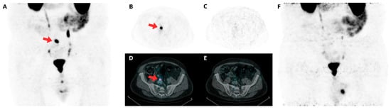
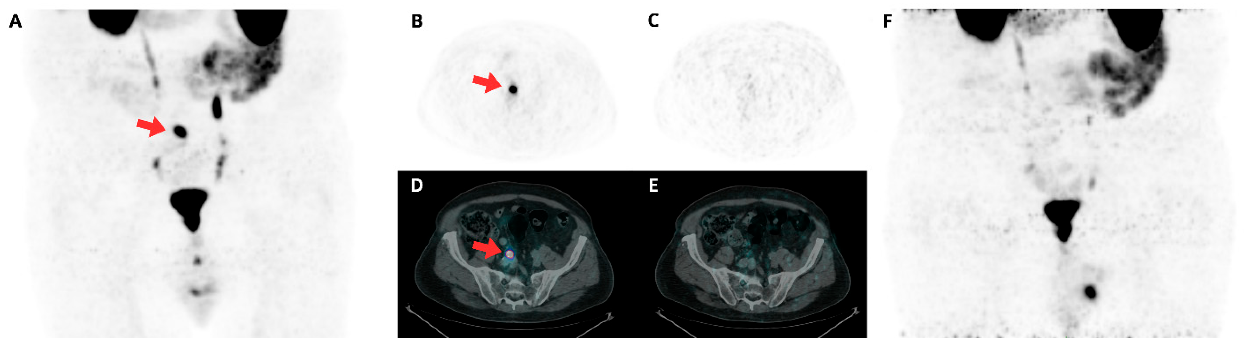
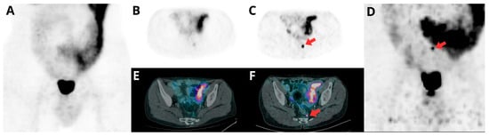
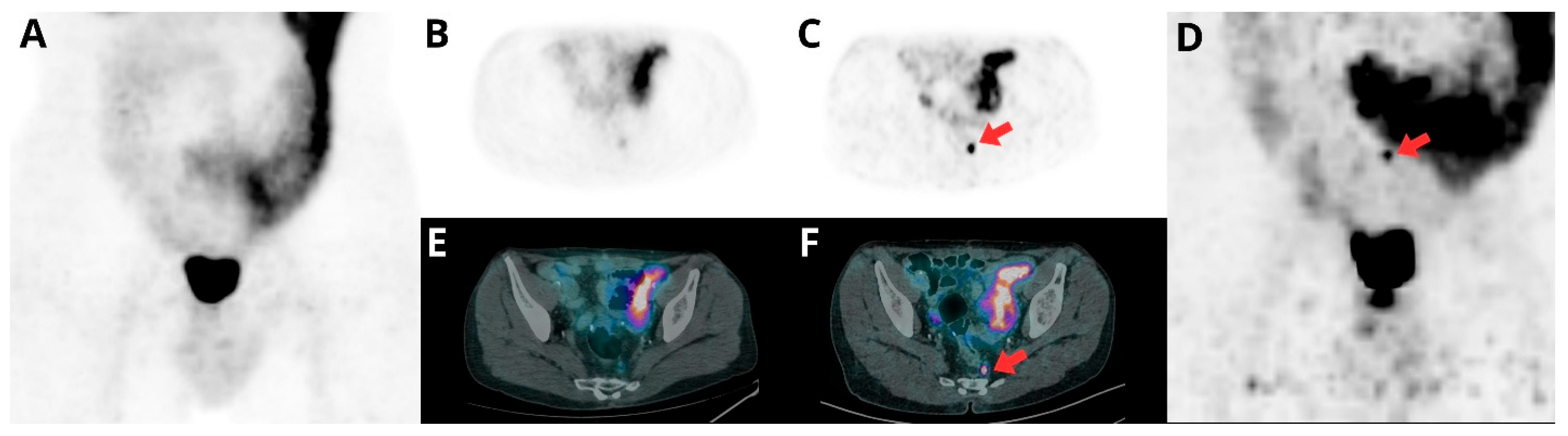
Figure 1 and Figure 2 illustrate representative clinical scenarios discussed further in the biological rationale subsections below.
Figure 1.
Patient case demonstrating that late-timepoint 68Ga-PSMA-11 PET/CT provided additional information related to physiologic ureteral activity, which supported clinical decision making. A patient with prostate carcinoma (Gleason score 3 + 4) and associated papillary urothelial carcinoma (pT1, G3) underwent radical prostatectomy in 2006 (preoperative PSA: 8 ng/mL; postoperative PSA: 0.1 ng/mL), without adjuvant radiotherapy or chemotherapy. The patient subsequently demonstrated a progressive PSA rise from August 2018 onward (0.28, 0.239, 0.35, 0.42, 0.59, and 0.6 ng/mL through January 2020), consistent with biochemical recurrence. 68Ga-PSMA-11 PET/CT was acquired at 60 min (standard timepoint) and 120 min (late timepoint) after administration; both standard- and late-timepoint images were acquired after bladder voiding. Standard-timepoint imaging (A,B,D) demonstrates a focal area of increased tracer uptake suspicious for pathologic involvement (red arrows). On late-timepoint imaging (C,E,F), the finding is no longer visualized, consistent with transient physiologic radiotracer activity within a tortuous right ureter rather than a true lesion. Fused PET/CT images show PET tracer uptake (color) overlaid on CT (grayscale).
Figure 2.
Increased conspicuity of a pathologic pararectal lymph node on late-timepoint 68Ga-PSMA-11 PET/CT. A patient with prostate adenocarcinoma (Gleason score 4 + 4, pT3aN0) underwent robot-assisted radical prostatectomy (RARP) in November 2024 (initial PSA, April 2024: 5.8 ng/mL; postoperative PSA: 0.18 ng/mL and 0.14 ng/mL in December 2024). 68Ga-PSMA-11 PET/CT was performed on 17 January 2025, with imaging acquired at 60 min (standard timepoint) and approximately 90 min (late timepoint) after administration. Standard-timepoint imaging (A,B,E) shows a small pararectal lymph node with low tracer uptake. On late-timepoint imaging (C,D,F), uptake within the same node appears more visually intense (red arrows), supporting increased diagnostic confidence and classification as a pathologic finding. The patient subsequently received stereotactic radiotherapy to the presacral lymph node, which was well visualized on late-timepoint imaging; PSA was undetectable as of September 2026. Fused PET/CT images show PET tracer uptake (color) overlaid on CT (grayscale).
4.1. Biological Rationale—Tumor-to-Background Contrast
PSMA ligands bind extracellular PSMA and may undergo receptor-mediated internalization, supporting lesion retention, while blood-pool and soft-tissue background generally decline. Early clinical and experimental observations showed increasing 68Ga-PSMA-11 uptake in prostate cancer cells or proven malignant lesions over time, accompanied by more rapid clearance from many background tissues [38,39]. In a histopathology- or imaging-referenced series, malignant lesions demonstrated significantly greater increases in standardized uptake value (SUV) and TBR than benign findings between approximately 1 and 3 h after administration [39]. Figure 2 illustrates this phenomenon in a patient with a pararectal lymph node metastasis, in which increased conspicuity on late-timepoint imaging increased diagnostic confidence (see Figure 2 legend for case details).
For some lesions, particularly small-volume or equivocal lesions, this dynamic may increase the quantitative parameters (e.g., SUV, TBR), thereby impacting lesion conspicuity or reader confidence [10,12,18,33,40,41]. In patients with BCR and PSA levels < 2 ng/mL, Hohberg et al. reported a 74% increase in mean SUVmax, a 36% increase in mean SUVpeak, and an approximately 3-fold increase in mean TBR on late compared with standard images; however, only a subset of lesions was better or exclusively visualized at either timepoint [13]. The clearest incremental visualization occurred in a subset of lesions, consistent with a selective-use strategy [20]. Similarly, prospective multiphasic imaging in patients with PSA levels < 1 ng/mL helped clarify equivocal findings, with late-timepoint imaging contributing primarily to interpreting nodal or osseous lesions and highlighting its utility for identifying specific lesions that may require further characterization [20].
Temporal behavior may also assist lesion characterization, but it is not a stand-alone discriminator of malignancy. For example, late-timepoint imaging demonstrated increasing SUVmax in most lymph node metastases, whereas most ganglia had stable or decreasing uptake. Overlap in uptake kinetics confirmed that temporal behavior should be interpreted together with CT morphology, anatomic location, and the overall disease distribution [42]. Morphologic characterization and anatomic localization therefore remain essential when interpreting temporal uptake changes. Lesion kinetics can also vary by lesion type, and not every prostate cancer lesion demonstrates increasing uptake at a later timepoint [38]; declining quantitative uptake between early and 60 min imaging was reported in 13 of 100 patients in a primary-staging cohort [43].
In primary disease, the potential benefit may extend beyond lesion conspicuity to interpretation of intraprostatic uptake. In a selected surgical cohort, late-timepoint imaging was associated with reduced background and benign-lesion activity, increased primary-tumor uptake, a higher proportion of lesions meeting the PRIMARY score’s SUVmax-based threshold, and improved intraobserver and interobserver agreement [11]. These results suggest a possible role for temporal uptake patterns in distinguishing malignant from benign intraprostatic findings, although the selected population and retrospective design limit generalizability to broader biopsy-naïve or screening populations.
Collectively, observations demonstrate that diagnostically relevant temporal information can be obtained with 68Ga-PSMA-11 PET/CT within a same-day workflow.
4.2. Biological Rationale—Physiologic Urinary Activity
Urinary excretion of 68Ga-PSMA-11 is a well-characterized pharmacokinetic feature that is effectively managed through standard patient preparation, including hydration and voiding, and by reviewing images in axial, sagittal, and coronal projections with adjusted display windows [2]. Reliable detection of prostate, nodal, and bladder-neck lesions has been consistently demonstrated [44,45,46], and urinary excretion is common across PSMA PET agents rather than unique to 68Ga-PSMA-11 [2]. This consideration is particularly relevant for patients with urinary retention (e.g., due to benign prostatic enlargement), among whom incomplete bladder emptying can prolong urinary tracer activity and limit the effectiveness of standard voiding-based mitigation strategies.
Late-timepoint imaging may provide complementary information when a small, potentially actionable finding is immediately adjacent to the bladder or ureter if the patient images demonstrate high urinary activity, though this scenario arises in only a minority of examinations. In the experience of some of the authors, this scenario most frequently arises as focal periurethral uptake in the midline just below the bladder, at the urethral origin. In this setting, the potential value of late-timepoint imaging derives from both elapsed uptake time and an intervening void, and the contribution of these factors cannot always be separated. Studies combining late pelvic imaging with bladder emptying have reported improved visualization of selected local recurrences and pelvic lesions, with incremental detection concentrated in a small number of clinically relevant findings [14,17]. Distinguishing physiologic periurethral activity from true local recurrence is most relevant in the BCR setting, where correlation with recent prostate MRI may help anatomically localize focal uptake to the urethra or, in patients after prostatectomy, the vesicourethral anastomosis. Figure 1 illustrates this scenario in a patient in whom a focal finding adjacent to the urinary tract on standard imaging was clarified on late-timepoint, postvoid imaging and attributed to transient physiologic ureteral activity (see Figure 1 legend for case details).
Late-timepoint imaging is therefore best framed as an adjunct in patients or workflows in which routine urinary-activity reduction strategies are difficult; for example, in patients with urinary retention, urgency, incontinence, frailty, or mobility limitations, where a focused late acquisition may add confidence without implying that late-timepoint imaging is required for routine care [18,19]. It may also be selectively useful when standard imaging shows an equivocal focus near the ureter or bladder, a suspected local recurrence in a region where interpretation may benefit from clarification of adjacent urinary activity, or a subtle pelvic lesion for which greater contrast could alter interpretation or treatment planning [14,15,17,20]. When standard imaging already provides a definitive, management-sufficient interpretation, a late acquisition may be unnecessary; selection should therefore be guided by whether complementary information could affect the clinical decision.
4.3. Evidence from Late-Timepoint Imaging Studies
Numerous studies have evaluated late-timepoint imaging with 68Ga-PSMA-11 PET/CT. Across heterogeneous studies, late-timepoint imaging is reported to offer potential gains in lesion contrast or diagnostic confidence, with management impact expected to be greatest when it is applied to a predefined, clinically actionable question. Table 2 summarizes representative studies, and a systematic review has cataloged the substantial heterogeneity in PSMA PET protocol variations beyond standard acquisition [21]. Most of these studies specifically examined the value of late-timepoint imaging in the BCR patient population.
Table 2.
Representative studies illustrating clinical scenarios for late-timepoint 68Ga-PSMA-11 PET/CT imaging.
Several studies in Table 2 report increases in SUVmax, SUVpeak, or TBR on late images. These parameters reflect measured uptake and image contrast; increases do not, by themselves, establish improved diagnostic accuracy or an impact on patient care. Higher uptake does not demonstrate that a lesion is malignant, that an equivocal finding has been correctly reclassified, or that a lesion would have been missed on standard imaging. Interpretation also depends on the reference standard (histopathology or adequate clinical follow-up versus imaging- or consensus-based reference standards). In addition, uptake on late images can rise in benign findings and remain stable or fall in malignant ones, so an increase alone cannot distinguish them [38,42]. Benign PSMA-avid structures, such as ganglia, may show stable or increasing uptake over time [42], and false-positive rates by timepoint were not systematically assessed in the studies summarized in Table 2. To our knowledge, only one study reported specificity at both timepoints (delayed PRIMARY scoring), with lower specificity but unchanged PPV [11]. Whether late-timepoint imaging alters false-positive rates therefore remains uncertain and warrants prospective evaluation with histopathologic reference standards. Late findings should be interpreted together with CT morphology and location rather than temporal change alone [38,42]. In several studies, increased uptake was not accompanied by changes in primary-tumor sensitivity [49], staging or treatment plans [12], or lesion positivity rates and therapeutic decisions for most patients [48]. More informative endpoints include lesion detection, changes in diagnostic classification, reader confidence, and interobserver agreement, ideally assessed against an appropriate reference standard. Where reported, these benefits were modest and concentrated in a subset of patients, such as improved diagnostic certainty in 34% of patients with BCR [14], improved reader confidence and interobserver agreement with only modest overall changes in diagnostic accuracy [19], and improved PRIMARY score sensitivity and interobserver agreement in a selected surgical cohort [11].
The evidence base has several limitations. Many studies are retrospective and single-center, with heterogeneous patient populations that vary by PSA level and prior therapy. Uptake times and validation methods also vary, and management impact is defined inconsistently; some studies emphasize lesion counts or SUV changes rather than patient-level decisions. Acquisition extent, patient preparation, diuretic use, scanner technology, reconstruction, and definitions of management impact also differ across studies. Findings from protocols using 1 scanner, uptake interval, or patient-preparation method should therefore not automatically be generalized to another clinical workflow. Available studies have primarily evaluated diagnostic and management endpoints rather than survival, progression-free survival, or quality of life (QoL), creating an opportunity for future outcome-focused research. Future studies should prospectively define management-impact endpoints (i.e., change in disease classification, radiation field modification, MDT selection, and systemic therapy escalation or de-escalation) and identify the patient subgroups most likely to benefit, while also evaluating cost-effectiveness and operational feasibility.
4.4. Practical Rationale
Radionuclide characteristics and scanner technology are two important considerations for the optimal imaging window, but neither determines whether a late acquisition is clinically necessary. Clinical necessity arises only when a late acquisition is reasonably expected to clarify a finding that could affect management. Technical feasibility should therefore be considered after, rather than before, the clinical indication has been established.
Highly PSMA-expressing lesions may continue to demonstrate relatively greater uptake compared with lower-expressing background structures, and ongoing radiotracer clearance may improve the signal-to-noise ratio. These potential gains must be balanced against radionuclide decay, which reduces available counts and can degrade image quality if not offset by more sensitive acquisition geometry.
Lower remaining activity may also reduce absolute scatter and random coincidence rates, but these changes do not necessarily compensate for the loss of useful true counts. Overall image quality reflects the balance among lesion-to-background contrast, true count statistics, random and scatter contributions, patient size, acquisition duration, detector sensitivity, acquisition geometry, and reconstruction method [35,37].
When a late acquisition is determined to be clinically appropriate, highly sensitive digital or LAFOV systems can help compensate for reduced count density. In a small 10-patient LAFOV case series, imaging at 4 h after administration increased mean TBR from 1.93 to 3.41 and increased mean signal-to-noise ratio from 24.80 to 33.02, with only a modest reduction in subjective image quality [32]. In a larger 300-patient BCR cohort imaged using a LAFOV system (Siemens Biograph Vision Quadra), positivity was reported in 67% of patients with PSA < 0.2 ng/mL, further illustrating the sensitivity gains available with this technology [34]. Because scanner sensitivity, acquisition geometry, count-rate conditions, and reconstruction can all affect apparent late-timepoint performance, these parameters should be specified whenever standard and late acquisitions are compared, and both acquisitions should use the same system and reconstruction settings when temporal change in uptake is itself the interpretive criterion. Phantom data from a 148 cm LAFOV system demonstrate that quantitative recovery is relatively stable across axial positions for larger objects but becomes less precise for small objects toward the field-of-view periphery, underscoring the interaction among lesion size, partial-volume effects, count statistics, and reconstruction [50]. Results demonstrate the technical feasibility of 68Ga-PSMA-11 imaging performed up to 4 h after administration under selected conditions, reinforcing that patient selection—not technical feasibility—remains the central consideration.
Scanner performance can also confound apparent time-dependent differences when serial acquisitions are performed on different systems. In a prospective randomized scanner-sequence study, a silicon photomultiplier-based system identified additional sites compared with a photomultiplier-tube system, whereas the short-interval later acquisition did not independently increase overall positivity [31]. This finding illustrates why effects attributable to uptake time should be separated from differences in detector sensitivity, spatial resolution, reconstruction method, acquisition duration, and scan order. Interpreters should also recognize that measured differences between standard and late images may partly reflect partial-volume effects, lesion size, axial position within the field of view, count statistics, and reconstruction settings rather than a true biologic change in uptake [38]. In a multi-radionuclide LAFOV phantom study, contrast recovery was comparatively stable across axial positions for larger objects, whereas small-object measurements showed greater dispersion toward the field-of-view periphery; radionuclide-specific point-spread-function modeling improved contrast recovery for 68Ga but did not eliminate count-statistics-related variability [50].
The clinical yield is also dependent on the indication and how late imaging is deployed. Studies enrolling patients with biochemical recurrence and low-volume or equivocal disease have generally reported greater incremental value than studies evaluating unselected staging populations or lesions already conspicuous on standard images [13,17,20,47,48,49]. Even within favorable cohorts, the benefit is concentrated in a minority of patients. For example, routine pelvic imaging at approximately 90 min improved reader-reported diagnostic certainty in 34% of patients with biochemical recurrence and identified additional lesions in 11%, and the additional findings informed focal radiation dose escalation in a clinically selected 3% of the total cohort [14]. This distribution of benefit supports an on-demand strategy based on review of the standard acquisition rather than prospective scheduling of two acquisitions for all patients. Canadian expert consensus similarly supports selective rather than routine delayed pelvic PSMA PET/CT acquisition when additional clarification could meaningfully influence management [51].
A small surgical series found increasing dominant-lesion uptake over time without a corresponding increase in primary-tumor sensitivity, suggesting that repeated acquisitions are best reserved for a defined interpretive question rather than applied routinely [49]. This finding illustrates why measurable kinetic change alone is insufficient to justify repeated imaging.
4.5. Tracer-Specific Context
Copper-64 (64Cu) has a physical half-life of approximately 12.7 h, compared with approximately 68 min for gallium-68 (68Ga), making acquisition over a substantially longer interval technically possible. However, the clinically relevant imaging window is better characterized by the radiopharmaceutical’s effective half-life, which incorporates both physical half-life and biological clearance rate. This distinction is particularly relevant to bivalent constructs such as 64Cu-SAR-bisPSMA: because the ligand exhibits prolonged tumor retention together with slow clearance from blood and background tissue, the effective half-life—and therefore the practical imaging window—is governed as much by ligand pharmacokinetics as by the physical decay of 64Cu itself. Consequently, the additional lesions identified on next-day imaging in early-phase studies of this agent may reflect a combination of prolonged radionuclide availability and ligand-driven retention/background clearance. This extended physical imaging window should be distinguished from evidence that imaging later provides greater clinical value. The performance of a radiopharmaceutical reflects the combined effects of the radionuclide and molecular construct, including ligand binding, uptake, retention, clearance, administered activity, and acquisition protocol.
In the Co-PSMA trial, 64Cu-SAR-bisPSMA was imaged at approximately 1 and 24 h and compared with standard-timepoint 68Ga-PSMA-11 PET/CT. More patients were reported positive on the 24 h than on the 1 h 64Cu-SAR-bisPSMA images. However, the within-tracer timing comparison was descriptive rather than the trial’s primary comparison, and the result reflects the complete 64Cu-SAR-bisPSMA protocol rather than the isolated effect of physical half-life [52]. The difference between the 1 h and 24 h images may reflect the combined influence of the bivalent ligand, uptake and retention characteristics, radionuclide decay, background clearance, acquisition timing, and other protocol conditions.
A longer physical half-life therefore explains why next-day imaging is possible but does not by itself demonstrate that a next-day protocol is diagnostically superior, clinically necessary, or operationally preferable. The investigators also acknowledged that the required 24 h acquisition represents a departure from standard imaging practice that may limit adoption [52]. By contrast, selected standard and late 68Ga-PSMA-11 PET/CT acquisitions can be completed in a single patient visit using established clinical systems and workflows. This same-day flexibility is operationally relevant when temporal information is needed to resolve a specific finding, but it does not imply that late-timepoint imaging should be routine.
5. Clinical Scenarios in Which Late-Timepoint Imaging May Add Value
As established in Section 3, detection and clinical utility are related but not identical: additional findings identified on a late acquisition matter clinically only if they alter interpretation in a way that changes management. Identifying an additional lesion in a patient with established disseminated metastatic disease, for example, may not change therapy. Conversely, confirming a single distant lesion in a patient initially thought to have isolated local recurrence may change treatment intent from local salvage alone to MDT, systemic therapy, or combined-modality treatment.
This distinction is particularly important in early BCR, where management often hinges on disease distribution. The highest-yield question is frequently whether recurrence is confined locally or includes regional nodal disease, particularly when localization of small-volume disease could alter salvage radiotherapy planning, radiation field design, nodal treatment, or consideration of MDT. Local-only recurrence can support prostate-bed salvage radiotherapy whereas pelvic nodal disease may lead to expanded nodal fields, a nodal boost, surgical nodal dissection, or the addition of systemic therapy; distant metastatic disease may further alter treatment intent. Late-timepoint imaging is therefore expected to be most valuable when it can move the interpretation across one of these management boundaries. For example, if late-timepoint imaging improves detection or confidence of local-only recurrence relative to standard-timepoint imaging, management typically remains unchanged, as these patients would receive salvage radiotherapy regardless of this finding. Late-timepoint imaging is unlikely to affect therapy when standard-timepoint imaging has already resolved the management question but may influence planning when it clarifies a clinically actionable finding [48]. Consistent with this framework, management generally remained unchanged when standard-timepoint imaging was already sufficient, whereas late-timepoint imaging affected treatment in selected patients when it clarified local or regional disease distribution [47,48].
Based on the literature and expert clinical experience from the authors’ centers (see Box 1), the highest-value settings for late-timepoint 68Ga-PSMA-11 imaging are expected to be those in which additional information can alter disease classification or treatment planning. Management impact is most likely in early BCR when clarification of small-volume local or regional nodal disease could change salvage radiotherapy fields, nodal treatment, or MDT consideration. For instance, late-timepoint imaging influenced management in 16% of PSMA-positive patients with BCR in 1 series [47].
Box 1. Real-world integration of selective late-timepoint imaging in nuclear medicine practice.
Based on the authors’ experience, late-timepoint imaging
may be integrated as an on-demand problem-solving examination, rather than
routinely scheduled for every patient. Standard images should be reviewed
promptly while the patient remains available. An additional late acquisition
may be considered when a discrete, clinically actionable finding remains
indeterminate and could reasonably be clarified by temporal assessment,
improved lesion-to-background contrast, or postvoid imaging.
At the Bologna center, approximately 4100 clinical
(non-trial) 68Ga-PSMA-11 PET/CT examinations have been performed
over approximately 5 years; current volume is approximately 8 to 10 per day
(volume has increased since 2021). Standard images are reviewed by a
specialist within approximately 5 min of acquisition while the patient waits
for approximately 15 min. Late imaging, which is not tracked separately, is
performed in no more than approximately 5–10% of patients, generally 1
patient per day, and is typically limited to 2 to 3 bed positions. The short
acquisition can be inserted between scheduled examinations with minimal
workflow disruption.
At the Essen center, daily volume ranges from
approximately 6 to 8 examinations on a regular day to 40 on a dedicated PSMA
day; routine review for possible late imaging has not yet been implemented.
These experiences illustrate that implementation should be adapted to local
patient volume, staffing, and scanner availability.
Across institutions, the key best-practice principle is
patient selection. Late imaging is most relevant when:
- The standard acquisition leaves a specific finding unresolved;
- Clarification could change management;
- Focused delayed or postvoid imaging could reasonably resolve the uncertainty.
When performed, the late acquisition should generally be
limited to the relevant anatomy, use consistent imaging and reconstruction
methods, and be documented in the report. Please note that the above examples
reflect individual-center practice and have not been prospectively validated.
In selected patients with high-risk prostate cancer before initial surgery, a late acquisition may also be considered when an indeterminate pelvic nodal finding could change whether pelvic lymph node dissection (PLND) is performed. If PLND would be performed or omitted regardless of the result, late imaging has no clear management rationale. Similarly, late-timepoint imaging has no established routine role in selecting patients before PSMA-directed therapy. A management-anchored framework is presented in Table 3.
Table 3.
Management-focused framework for selective late-timepoint imaging with 68Ga-PSMA-11.
5.1. Principal Problem-Solving Applications
Based on the available literature and expert clinical rationale, two principal problem-solving applications can be identified when the standard acquisition leaves a clinically actionable question unresolved: (1) assessment of the temporal behavior of an indeterminate focus, where increasing uptake or lesion-to-background contrast may support a pathologic origin when integrated with anatomic and morphologic findings [20]; and (2) characterization of prostate-bed uptake adjacent to physiologic urinary activity, particularly after voiding [17]. The first may inform local-versus-regional disease classification, nodal treatment, MDT consideration, or PLND in selected patients, whereas the second may inform local salvage planning. In either case, a late acquisition is justified only if clarification could change management.
Late-timepoint imaging may also help when standard imaging is technically or clinically limited but the result is potentially actionable—for example, when voiding or diuretic strategies are impractical, or when a small lesion near physiologic activity could change radiation planning. The key is not the presence of any equivocal finding, but the presence of an equivocal finding that matters.
5.2. Scenarios in Which Standard Imaging Is Likely Sufficient
Lower-yield settings include unequivocally positive or unequivocally negative studies in which management would not change, and patients with extensive known metastatic disease in whom one additional lesion is unlikely to alter the treatment strategy. In these cases, routine late-timepoint imaging may increase scan time and operational burden when the standard acquisition has already provided management-sufficient information.
6. Suggested Clinical Workflow
Standard whole-body 68Ga-PSMA-11 PET/CT should remain the default acquisition. The workflow below reflects best practices derived from the published literature and the authors’ clinical experience (Box 1). Note that its feasibility depends on local organizational resources.
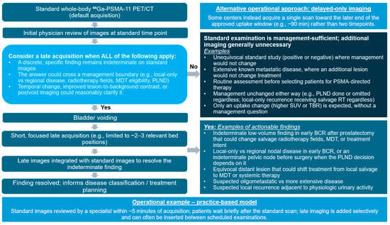
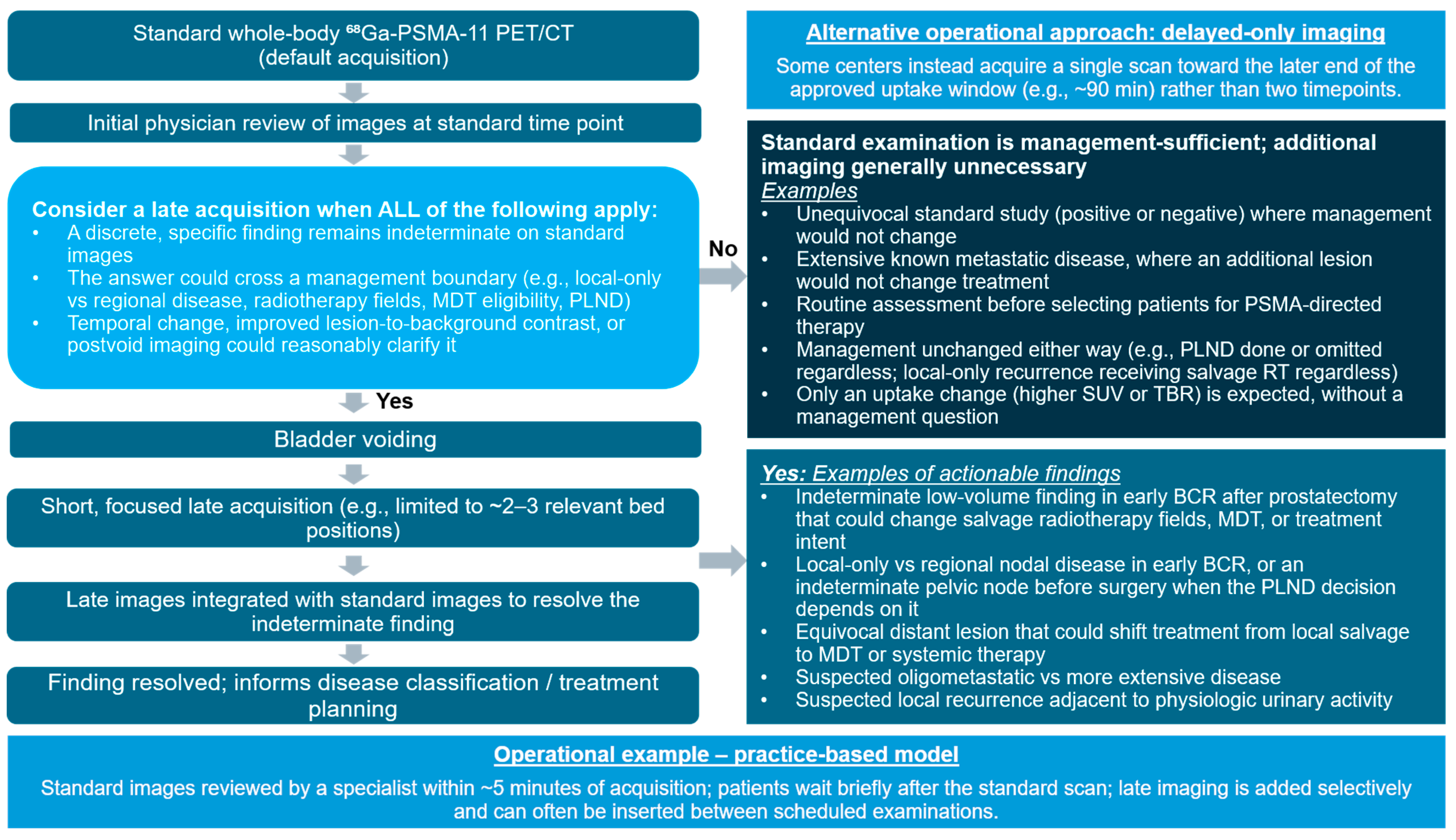
A practical late-timepoint workflow is to review the standard-timepoint images within a few minutes of acquisition while the patient remains in the imaging department. An additional late acquisition may be considered when this review identifies a discrete, clinically actionable finding that remains indeterminate and could reasonably be clarified by temporal assessment, improved lesion-to-background contrast, or postvoid imaging [51]. If the standard examination has already resolved the management question, an additional acquisition is generally unnecessary (Figure 3).
Figure 3.
Proposed clinical workflow for selective late-timepoint 68Ga-PSMA-11 PET/CT, with examples of actionable and non-actionable scenarios.
When late-timepoint imaging is warranted, a short, focused acquisition may be performed after bladder voiding and limited to relevant anatomy, such as two to three bed positions, to minimize scan duration and workflow disruption (Figure 3; see Box 1 for a practical operational model). These operational details reflect local experience and have not been validated across clinical settings; therefore, adapt the proposed workflow according to institutional patient volume, staffing, scanner availability, and validated imaging protocols.
Some centers may instead select a single acquisition toward the later end of the approved uptake window (delayed-only imaging; Table 1; Figure 3). However, this approach does not permit direct assessment of temporal change within the same examination. Selection of a single late acquisition versus an on-demand late acquisition should reflect the clinical objective. Two timepoints are required when additional temporal information regarding uptake or appearance over time is itself being used to characterize an indeterminate finding.
Operational planning for selective late-timepoint imaging should account for scanner availability, patient throughput, visit duration, and reader availability for standard timepoint review. A practical limitation is that interpretation is frequently performed remotely rather than on-site; in such settings, timely review to determine whether a late acquisition is warranted may require real-time contact with the remote reader for a preliminary interpretation while the patient remains available. Prompt same-visit review of standard-timepoint images may not be feasible in all institutions or for all patients, and implementation will depend on local resources, including reader availability, scanner capacity, and staffing. When scheduling permits limited flexibility, a short, focused late acquisition can often be accommodated between examinations without reserving routine dual-acquisition slots. The patient should be informed that an additional 20 to 30 min interval, including voiding and focused imaging, is being recommended to clarify a specific finding and improve the precision of the report. When feasible, selective late-timepoint imaging may therefore preserve access while enabling high-value, management-focused problem solving.
7. Evidence Gaps and Future Research
The case for late-timepoint imaging can be viewed as a chain of evidence: (1) late acquisition changes measured uptake or lesion conspicuity; (2) these changes improve lesion detection, diagnostic classification, reader confidence, or diagnostic accuracy against an appropriate reference standard; (3) the improved interpretation changes clinical management; and (4) the changed management improves patient outcomes. Published studies most consistently support step 1 and provide variable evidence (mostly from selected cohorts) for steps 2 and 3. To our knowledge, no current evidence provides direct support for step 4 nor does it demonstrate a survival or QoL benefit from routine late-timepoint imaging. The selective, management-anchored approach proposed in this review reflects the published imaging and management-impact literature together with the sustained clinical experience of centers that have implemented late-timepoint imaging and established workflows for its use (Box 1).
Future studies should prospectively define management-impact endpoints and evaluate diagnostic performance against appropriate reference standards. They should report lesion detection, changes in diagnostic classification, reader confidence, and interobserver agreement alongside uptake measures. They should also identify the patient subgroups most likely to benefit, determine whether management changes attributable to late imaging translate into improved patient outcomes, and assess operational feasibility and cost-effectiveness.
8. Conclusions and Future Direction
68Ga-PSMA-11 PET/CT provides excellent diagnostic performance at standard imaging timepoints, including clinically meaningful detection at low PSA levels and high specificity for pelvic nodal metastases, with reproducible performance across centers and technologies. This robust baseline supports a selective, rather than routine, approach to late-timepoint imaging. Late-timepoint imaging (standard acquisition followed by late acquisition) may be most valuable for clarifying equivocal or borderline findings that could alter disease classification, radiation treatment planning, MDT eligibility, or systemic therapy decisions, most notably in early BCR with low-volume disease. Future prospective studies should define management-impact endpoints, identify the subgroups most likely to benefit, and evaluate operational feasibility and cost-effectiveness, so that late-timepoint imaging can be applied where it delivers genuine, patient-centered clinical value.
Supplementary Materials
The following supporting information can be downloaded at: https://www.mdpi.com/article/10.3390/cancers18223703/s1, Table S1: Foundational evidence supporting standard-timepoint 68Ga-PSMA-11 PET across the prostate cancer disease continuum [53].
Author Contributions
M.G., K.H., R.Y., and S.F. contributed to the conception, direction, writing, review, and editing of the manuscript. S.F. made significant contributions, including providing the clinical images used in Figure 1 and Figure 2. M.G., K.H., R.Y., and S.F. contributed clinical expertise and best-practice guidance to inform the content of this review. All authors have read and agreed to the published version of the manuscript.
Funding
This work received no external funding. The article processing charge was funded by Telix Pharmaceuticals.
Institutional Review Board Statement
Not applicable.
Informed Consent Statement
Not applicable.
Data Availability Statement
No new data were created or analyzed in this study. Data sharing is not applicable to this article.
Acknowledgments
The authors thank Andrea Di Giorgio and Fabio Ghetti (Nuclear Medicine Unit, Alma Mater Studiorum–University of Bologna, IRCCS Azienda Ospedaliero-Universitaria di Bologna, Bologna, Italy) for providing the case details and images used in Figure 1 and Figure 2. Medical writing and editing support provided by Julia Green (Telix Pharmaceuticals).
Conflicts of Interest
M.G. reported professional relationships (including consulting, advisory, speaking, and/or research support activities, as applicable) with Siemens, Telix, Lantheus, ITM, Serac Life Sciences, Novartis, Blue Earth, Bayer, Cardinal, Genoplex, and Nusano, outside the submitted work. K.H. reported receiving consultant fees from Advanced Accelerator Applications, a Novartis company, Actithera, Amgen, AstraZeneca, Bain Capital, Bayer, Bicycle Therapeutics, Boston Scientific, Convergent, Curium, Debiopharm, EcoR1, Full Life, Fusion, GE Healthcare, Immedica, Isotopen Technologien München, Janssen, Merck, MSD, Molecular Partners, NVision, POINT Biopharma, Pentixapharm, Pfizer, Radiopharm Theranostics, Rhine Pharma, Siemens Healthineers, Sofie Biosciences, Telix, Theragnostics, and ymabs; receiving research grants from Advanced Accelerator Applications, a Novartis company, Boston Scientific, and Janssen; and having stock or other ownership interests with AdvanCell, Aktis Oncology, Bicycle Therapeutics, Convergent, NVision, Perspective Therapeutics, Radiopharm Theranostics, Rhine Pharma, Sofie Biosciences and Yellowbird Diagnostics. R.Y. reported a professional relationship with GE Healthcare outside the submitted work. S.F. reported honoraria/travel support from AAA, Amgen, Astellas, Astra Zeneca, Bayer, Blue Earth, Debio, GE, Immedica, Ipsen, Janssen, Lilly, Novartis, Telix, and United.
Abbreviations
The following abbreviations are used in this manuscript:
| 64Cu | Copper-64 |
| 68Ga | Gallium-68 |
| 68Ga-PSMA-11 | Gallium-68 prostate-specific membrane antigen-11 |
| BCR | Biochemical recurrence |
| CT | Computed tomography |
| h | Hour(s) |
| IAEA | International Atomic Energy Agency |
| ISUP | International Society of Urological Pathology |
| LAFOV | Long axial field of view |
| MBq | Megabecquerel(s) |
| MDT | Metastasis-directed therapy |
| min | Minute(s) |
| MRI | Magnetic resonance imaging |
| NPV | Negative predictive value |
| PCa | Prostate cancer |
| PET | Positron emission tomography |
| PLND | Pelvic lymph node dissection |
| PPV | Positive predictive value |
| PSA | Prostate-specific antigen |
| PSMA | Prostate-specific membrane antigen |
| pt(s) | Patient(s) |
| QoL | Quality of life |
| RARP | Robot-assisted radical prostatectomy |
| RP RT | Radical prostatectomy Radiotherapy |
| SAFOV | Short axial field of view |
| SUV | Standardized uptake value |
| SUVmax | Maximum standardized uptake value |
| SUVpeak | Peak standardized uptake value |
| TBR | Tumor-to-background ratio |
References
- Perera, M.; Papa, N.; Roberts, M.; Williams, M.; Udovicich, C.; Vela, I.; Christidis, D.; Bolton, D.; Hofman, M.S.; Lawrentschuk, N.; et al. Gallium-68 prostate-specific membrane antigen positron emission tomography in advanced prostate cancer-updated diagnostic utility, sensitivity, specificity, and distribution of prostate-specific membrane antigen-avid lesions: A systematic review and meta-analysis. Eur. Urol. 2020, 77, 403–417. [Google Scholar] [CrossRef] [Scilit] [PubMed]
- Fendler, W.P.; Eiber, M.; Beheshti, M.; Bomanji, J.; Calais, J.; Ceci, F.; Cho, S.Y.; Fanti, S.; Giesel, F.L.; Goffin, K.; et al. PSMA PET/CT: Joint EANM procedure guideline/SNMMI procedure standard for prostate cancer imaging 2.0. Eur. J. Nucl. Med. Mol. Imaging 2023, 50, 1466–1486. [Google Scholar] [CrossRef] [Scilit] [PubMed]
- Hennrich, U.; Eder, M. [(68)Ga]Ga-PSMA-11: The first FDA-approved (68)Ga-radiopharmaceutical for PET imaging of prostate cancer. Pharmaceuticals 2021, 14, 713. [Google Scholar] [CrossRef] [Scilit] [PubMed]
- Fendler, W.P.; Calais, J.; Eiber, M.; Flavell, R.R.; Mishoe, A.; Feng, F.Y.; Nguyen, H.G.; Reiter, R.E.; Rettig, M.B.; Okamoto, S.; et al. Assessment of 68Ga-PSMA-11 PET Accuracy in Localizing Recurrent Prostate Cancer: A Prospective Single-Arm Clinical Trial. JAMA Oncol. 2019, 5, 856–863. [Google Scholar] [CrossRef] [Scilit] [PubMed]
- Hope, T.A.; Eiber, M.; Armstrong, W.R.; Juarez, R.; Murthy, V.; Lawhn-Heath, C.; Behr, S.C.; Zhang, L.; Barbato, F.; Ceci, F.; et al. Diagnostic Accuracy of 68Ga-PSMA-11 PET for Pelvic Nodal Metastasis Detection Prior to Radical Prostatectomy and Pelvic Lymph Node Dissection: A Multicenter Prospective Phase 3 Imaging Trial. JAMA Oncol. 2021, 7, 1635–1642. [Google Scholar] [CrossRef] [Scilit] [PubMed]
- Hofman, M.S.; Lawrentschuk, N.; Francis, R.J.; Tang, C.; Vela, I.; Thomas, P.; Rutherford, N.; Martin, J.M.; Frydenberg, M.; Shakher, R.; et al. Prostate-specific membrane antigen PET-CT in patients with high-risk prostate cancer before curative-intent surgery or radiotherapy (proPSMA): A prospective, randomised, multicentre study. Lancet 2020, 395, 1208–1216. [Google Scholar] [CrossRef] [Scilit] [PubMed]
- Telix Pharmaceuticals. Illuccix (Kit for the Preparation of Gallium Ga 68 Gozetotide Injection) Full Prescribing Information; Telix Pharmaceuticals: Fishers, IN, USA, 2026. [Google Scholar]
- Telix Pharmaceuticals. Gozellix (Kit for the Preparation of Gallium Ga 68 Gozetotide Injection) Full Prescribing Information; Telix Pharmaceuticals: Fishers, IN, USA, 2025. [Google Scholar]
- Telix Pharmaceuticals. Illuccix 25 Micrograms Kit for Radiopharmaceutical Preparation, Package Leaflet; Telix Pharmaceuticals: Dresden, Germany, 2025. [Google Scholar]
- Afshar-Oromieh, A.; Sattler, L.P.; Mier, W.; Hadaschik, B.A.; Debus, J.; Holland-Letz, T.; Kopka, K.; Haberkorn, U. The Clinical Impact of Additional Late PET/CT Imaging with (68)Ga-PSMA-11 (HBED-CC) in the Diagnosis of Prostate Cancer. J. Nucl. Med. 2017, 58, 750–755. [Google Scholar] [CrossRef] [Scilit] [PubMed]
- Akçay, K.; Beydağı, G.; Şahin, O.E.; Akyel, R.; Akgün, E.; Ekmekçioğlu, Ö.; Selçuk, N.A.; Toklu, T.; Ekici, A.I.D.; Kapran, K.; et al. Improved Accuracy and Reliability of PRIMARY Scoring Using Delayed [68Ga] Ga-PSMA PET/CT Imaging. Mol. Imaging Radionucl. Ther. 2025, 34, 1–9. [Google Scholar] [CrossRef] [Scilit] [PubMed]
- Can, C.; Komek, H. Contribution of 5th minute and 2nd hour images to standard imaging in (68Ga)PSMA 11 PET/CT. Ann. Nucl. Med. 2020, 34, 163–173. [Google Scholar] [CrossRef] [Scilit] [PubMed]
- Hohberg, M.; Kobe, C.; Täger, P.; Hammes, J.; Schmidt, M.; Dietlein, F.; Wild, M.; Heidenreich, A.; Drzezga, A.; Dietlein, M. Combined Early and Late [(68)Ga]PSMA-HBED-CC PET Scans Improve Lesion Detectability in Biochemical Recurrence of Prostate Cancer with Low PSA Levels. Mol. Imaging Biol. 2019, 21, 558–566. [Google Scholar] [CrossRef] [Scilit] [PubMed]
- Jia, K.Y.; Kuten, J.; Jia, H.Y.; Bent, E.H.; Silveira, P.; Pasquini, L.; Nalavenkata, S.; Schoder, H.; Fox, J.J.; Yeh, R.; et al. Diagnostic Value of Routine Delayed Pelvic Imaging for Patients with Biochemical Recurrence of Prostate Cancer Undergoing 68Ga-PSMA PET/CT. J. Nucl. Med. 2026, 67, 1261–1267. [Google Scholar] [CrossRef] [Scilit] [PubMed]
- Unterrainer, L.M.; Ruchalski, K.; Allen-Auerbach, M.S.; Calais, J.; Benz, M.R. PSMA PET/CT Dual-Time-Point Imaging: Nice to Have or Need to Have? J. Nucl. Med. 2024, 65, 818–819. [Google Scholar] [CrossRef] [Scilit] [PubMed]
- Sachpekidis, C.; Pan, L.; Hadaschik, B.A.; Kopka, K.; Haberkorn, U.; Dimitrakopoulou-Strauss, A. 68Ga-PSMA-11 PET/CT in prostate cancer local recurrence: Impact of early images and parametric analysis. Am. J. Nucl. Med. Mol. Imaging 2018, 8, 351–359. [Google Scholar] [PubMed]
- Morawitz, J.; Kirchner, J.; Hertelendy, J.; Loberg, C.; Schimmöller, L.; Dabir, M.; Häberle, L.; Mamlins, E.; Antke, C.; Arsov, C.; et al. Is there a diagnostic benefit of late-phase abdomino-pelvic PET/CT after urination as part of whole-body (68) Ga-PSMA-11 PET/CT for restaging patients with biochemical recurrence of prostate cancer after radical prostatectomy? EJNMMI Res. 2022, 12, 12. [Google Scholar] [CrossRef] [Scilit] [PubMed]
- Alberts, I.; Niklas-Hünermund, J.; Sachpekidis, C.; Zacho, H.D.; Mingels, C.; Dijkstra, L.; Bohn, K.P.; Läppchen, T.; Gourni, E.; Rominger, A.; et al. Combination of Forced Diuresis with Additional Late Imaging in (68)Ga-PSMA-11 PET/CT: Effects on Lesion Visibility and Radiotracer Uptake. J. Nucl. Med. 2021, 62, 1252–1257. [Google Scholar] [CrossRef] [Scilit] [PubMed]
- Bauckneht, M.; Miceli, A.; Signori, A.; Albano, D.; Capitanio, S.; Piva, R.; Laudicella, R.; Franchini, A.; D’Amico, F.; Riondato, M.; et al. Combined forced diuresis and late acquisition on [68Ga]Ga-PSMA-11 PET/CT for biochemical recurrent prostate cancer: A clinical practice-oriented study. Eur. Radiol. 2023, 33, 3343–3353. [Google Scholar] [CrossRef] [Scilit] [PubMed]
- Beheshti, M.; Manafi-Farid, R.; Geinitz, H.; Vali, R.; Loidl, W.; Mottaghy, F.M.; Langsteger, W. Multiphasic 68Ga-PSMA PET/CT in the Detection of Early Recurrence in Prostate Cancer Patients with a PSA Level of Less Than 1 ng/mL: A Prospective Study of 135 Patients. J. Nucl. Med. 2020, 61, 1484–1490. [Google Scholar] [CrossRef] [Scilit] [PubMed]
- Sadeghpour, S.; Saidi, B.; Sahafi, P.; Maleki, F.; Ranjbar, S.; Ghorbani, M.; Hakim, H.G.; Aghaee, A.; Fanti, S.; Beheshti, M.; et al. Maximizing diagnostic yield: A systematic review and deep dive into PSMA PET scan protocol variations for prostate cancer. Semin. Nucl. Med. 2026, 56, 759–775. [Google Scholar] [CrossRef] [Scilit] [PubMed]
- Abghari-Gerst, M.; Armstrong, W.R.; Nguyen, K.; Calais, J.; Czernin, J.; Lin, D.; Jariwala, N.; Rodnick, M.; Hope, T.A.; Hearn, J.; et al. A Comprehensive Assessment of 68Ga-PSMA-11 PET in Biochemically Recurrent Prostate Cancer: Results from a Prospective Multicenter Study on 2,005 Patients. J. Nucl. Med. 2022, 63, 567–572. [Google Scholar] [CrossRef] [Scilit] [PubMed]
- Burgard, C.; Hoffmann, M.A.; Frei, M.; Buchholz, H.G.; Khreish, F.; Marlowe, R.J.; Schreckenberger, M.; Ezziddin, S.; Rosar, F. Detection Efficacy of (68)Ga-PSMA-11 PET/CT in Biochemical Recurrence of Prostate Cancer with Very Low PSA Levels: A 7-Year, Two-Center “Real-World” Experience. Cancers 2023, 15, 1376. [Google Scholar] [CrossRef] [Scilit] [PubMed]
- Fendler, W.P.; Ferdinandus, J.; Czernin, J.; Eiber, M.; Flavell, R.R.; Behr, S.C.; Wu, I.W.K.; Lawhn-Heath, C.; Pampaloni, M.H.; Reiter, R.E.; et al. Impact of (68)Ga-PSMA-11 PET on the Management of Recurrent Prostate Cancer in a Prospective Single-Arm Clinical Trial. J. Nucl. Med. 2020, 61, 1793–1799. [Google Scholar] [CrossRef] [Scilit] [PubMed]
- Giesel, F.L.; Koerber, S.A.; Hadaschik, B.; Kiefer, S.; Schwarzenboeck, S.; Rahbar, K.; Derlin, T.; Grott, C.A.; Heck, M.; Jilg, C.; et al. The German Cancer Consortium (DKTK) multi-center prospective phase 1/2 68Ga-PSMA-11 PET-imaging trial in newly-diagnosed high-risk prostate cancer: Safety and diagnostic accuracy compared to histopathology and their impact on patient management. Eur. J. Nucl. Med. Mol. Imaging 2026, 53, 2994–3006. [Google Scholar] [CrossRef] [Scilit] [PubMed]
- Cerci, J.J.; Fanti, S.; Lobato, E.E.; Kumar, R.; Kunikowska, J.; Al-Ibraheem, A.; Nasir, M.; Redondo Moneda, F.; Garcia, O.; Haidar, M.; et al. (68)Ga-PSMA-11 in Staging of Unfavorable Intermediate- and High-Risk Prostate Cancer Reduces Indication for Noncurative Prostatectomy: A Prospective, Multicenter, IAEA Study. J. Nucl. Med. 2026, 67, 67–70. [Google Scholar] [CrossRef] [Scilit] [PubMed]
- Sartor, O.; de Bono, J.; Chi, K.N.; Fizazi, K.; Herrmann, K.; Rahbar, K.; Tagawa, S.T.; Nordquist, L.T.; Vaishampayan, N.; El-Haddad, G.; et al. Lutetium-177-PSMA-617 for Metastatic Castration-Resistant Prostate Cancer. N. Engl. J. Med. 2021, 385, 1091–1103. [Google Scholar] [CrossRef] [Scilit] [PubMed]
- Szigeti, F.; Schweighofer-Zwink, G.; Meissnitzer, M.; Hauser-Kronberger, C.; Hitzl, W.; Kunit, T.; Forstner, R.; Pirich, C.; Beheshti, M. Incremental Impact of [(68) Ga]Ga-PSMA-11 PET/CT in Primary N and M Staging of Prostate Cancer Prior to Curative-Intent Surgery: A Prospective Clinical Trial in Comparison with mpMRI. Mol. Imaging Biol. 2022, 24, 50–59. [Google Scholar] [CrossRef] [Scilit] [PubMed]
- Ataya, M.N.; Bahler, C.D.; Koch, M.O.; Tann, M.; Green, M.A.; Ferguson, E.; Huelster, H.L. Can the sensitivity of prostate cancer with 68Ga-PSMA PET/CT improve with newer higher resolution PET-CT cameras? In Proceedings of the American Urological Association Annual Meeting 2025, Las Vegas, NV, USA, 26–29 April 2025; p. e1098. [Google Scholar]
- Cerci, J.J.; Fanti, S.; Lobato, E.E.; Kunikowska, J.; Alonso, O.; Medina, S.; Novruzov, F.; Lengana, T.; Granados, C.; Kumar, R.; et al. Diagnostic Performance and Clinical Impact of 68Ga-PSMA-11 PET/CT Imaging in Early Relapsed Prostate Cancer After Radical Therapy: A Prospective Multicenter Study (IAEA-PSMA Study). J. Nucl. Med. 2022, 63, 240–247. [Google Scholar] [CrossRef] [Scilit] [PubMed]
- Duan, H.; Baratto, L.; Hatami, N.; Liang, T.; Aparici, C.M.; Davidzon, G.A.; Iagaru, A. 68Ga-PSMA11 PET/CT for biochemically recurrent prostate cancer: Influence of dual-time and PMT- vs SiPM-based detectors. Transl. Oncol. 2022, 15, 101293. [Google Scholar] [CrossRef] [Scilit] [PubMed]
- Alberts, I.; Prenosil, G.; Mingels, C.; Bohn, K.P.; Viscione, M.; Sari, H.; Rominger, A.; Afshar-Oromieh, A. Feasibility of late acquisition [68Ga]Ga-PSMA-11 PET/CT using a long axial field-of-view PET/CT scanner for the diagnosis of recurrent prostate cancer-first clinical experiences. Eur. J. Nucl. Med. Mol. Imaging 2021, 48, 4456–4462. [Google Scholar] [CrossRef] [Scilit] [PubMed]
- Yu, X.; Xu, L.; Huang, G.; Liu, J.; Chen, R.; Chen, Y. The image quality and feasibility of solitary delayed [68Ga]Ga-PSMA-11 PET/CT using long field-of-view scanning in patients with prostate cancer. EJNMMI Res. 2024, 14, 14. [Google Scholar] [CrossRef] [Scilit] [PubMed]
- Thomas, S.; Callahan, J.; Conway, P.; Moon, D.; Morgan, H.; Ingbritsen, J.; Munro, M.; Ware, R.E.; Dundee, P.; Hicks, R.J. High Detection Rates for Prostate-specific Membrane Antigen-avid Prostate Cancer Recurrence at Low Prostate-specific Antigen levels on Extended Axial Field-of-view Positron Emission Tomography/Computed Tomography. Eur. Urol. Open Sci. 2025, 71, 49–56. [Google Scholar] [CrossRef] [Scilit] [PubMed]
- Brasse, D.; Kinahan, P.E.; Lartizien, C.; Comtat, C.; Casey, M.; Michel, C. Correction methods for random coincidences in fully 3D whole-body PET: Impact on data and image quality. J. Nucl. Med. 2005, 46, 859–867. [Google Scholar] [PubMed]
- Abufaraj, M.; Grubmüller, B.; Zeitlinger, M.; Kramer, G.; Seitz, C.; Haitel, A.; Baltzer, P.; Hacker, M.; Wadsak, W.; Pfaff, S.; et al. Prospective evaluation of the performance of [(68)Ga]Ga-PSMA-11 PET/CT(MRI) for lymph node staging in patients undergoing superextended salvage lymph node dissection after radical prostatectomy. Eur. J. Nucl. Med. Mol. Imaging 2019, 46, 2169–2177. [Google Scholar] [CrossRef] [Scilit] [PubMed]
- Wangerin, K.A.; Muzi, M.; Peterson, L.M.; Linden, H.M.; Novakova, A.; O’Sullivan, F.; Kurland, B.F.; Mankoff, D.A.; Kinahan, P.E. Effect of 18F-FDG uptake time on lesion detectability in PET imaging of early stage breast cancer. Tomography 2015, 1, 53–60. [Google Scholar] [CrossRef] [Scilit] [PubMed]
- Alberts, I.; Sachpekidis, C.; Gourni, E.; Boxler, S.; Gross, T.; Thalmann, G.; Rahbar, K.; Rominger, A.; Afshar-Oromieh, A. Dynamic patterns of [68Ga]Ga-PSMA-11 uptake in recurrent prostate cancer lesions. Eur. J. Nucl. Med. Mol. Imaging 2020, 47, 160–167. [Google Scholar] [CrossRef] [Scilit] [PubMed]
- Sahlmann, C.O.; Meller, B.; Bouter, C.; Ritter, C.O.; Ströbel, P.; Lotz, J.; Trojan, L.; Meller, J.; Hijazi, S. Biphasic 68Ga-PSMA-HBED-CC-PET/CT in patients with recurrent and high-risk prostate carcinoma. Eur. J. Nucl. Med. Mol. Imaging 2016, 43, 898–905. [Google Scholar] [CrossRef] [Scilit] [PubMed]
- Wang, Y.; Dong, L.; Zhao, H.; Li, L.; Huang, G.; Xue, W.; Liu, J.; Chen, R. The superior detection rate of total-body [68Ga]Ga-PSMA-11 PET/CT compared to short axial field-of-view [68Ga]Ga-PSMA-11 PET/CT for early recurrent prostate cancer patients with PSA <0.2 ng/mL after radical prostatectomy. Eur. J. Nucl. Med. Mol. Imaging 2024, 51, 2484–2494. [Google Scholar] [CrossRef] [Scilit] [PubMed]
- Wang, Y.; Zhu, L.; Xu, L.; Dong, L.; Zhao, H.; Li, L.; Huang, G.; Chen, R.; Liu, J. Superiority of long axial field-of-view 68Ga-PSMA-11 PET over short axial field-of-view PET in prostate cancer: A head-to-head comparison. Eur. J. Nucl. Med. Mol. Imaging 2026, 53, 1811–1821. [Google Scholar] [CrossRef] [Scilit] [PubMed]
- Alberts, I.; Sachpekidis, C.; Dijkstra, L.; Prenosil, G.; Gourni, E.; Boxler, S.; Gross, T.; Thalmann, G.; Rahbar, K.; Rominger, A.; et al. The role of additional late PSMA-ligand PET/CT in the differentiation between lymph node metastases and ganglia. Eur. J. Nucl. Med. Mol. Imaging 2020, 47, 642–651. [Google Scholar] [CrossRef] [Scilit] [PubMed]
- Weitzer, F.; Pernthaler, B.; Plhak, E.; Riedl, R.; Aigner, R.M. Diagnostic value of two-time point [68Ga]Ga-PSMA-11 PET/CT in the primary staging of untreated prostate cancer. Sci. Rep. 2023, 13, 8297. [Google Scholar] [CrossRef] [Scilit] [PubMed]
- Yilmaz, B.; Turkay, R.; Colakoglu, Y.; Baytekin, H.F.; Ergul, N.; Sahin, S.; Tugcu, V.; Inci, E.; Tasci, A.I.; Cermik, T.F. Comparison of preoperative locoregional Ga-68 PSMA-11 PET-CT and mp-MRI results with postoperative histopathology of prostate cancer. Prostate 2019, 79, 1007–1017. [Google Scholar] [CrossRef] [Scilit] [PubMed]
- Park, S.Y.; Zacharias, C.; Harrison, C.; Fan, R.E.; Kunder, C.; Hatami, N.; Giesel, F.; Ghanouni, P.; Daniel, B.; Loening, A.M.; et al. Gallium 68 PSMA-11 PET/MR Imaging in Patients with Intermediate- or High-Risk Prostate Cancer. Radiology 2018, 288, 495–505. [Google Scholar] [CrossRef] [Scilit] [PubMed]
- Pallavi, U.N.; Gogoi, S.; Thakral, P.; Malasani, V.; Sharma, K.; Manda, D.; Das, S.S.; Pant, V.; Sen, I. Incremental Value of Ga-68 Prostate-Specific Membrane Antigen-11 Positron-Emission Tomography/Computed Tomography Scan for Preoperative Risk Stratification of Prostate Cancer. Indian. J. Nucl. Med. 2020, 35, 93–99. [Google Scholar] [CrossRef] [Scilit] [PubMed]
- Kunikowska, J.; Pełka, K.; Tayara, O.; Krolicki, L. Ga-68-PSMA-11 PET/CT in Patients with Biochemical Recurrence of Prostate Cancer after Primary Treatment with Curative Intent-Impact of Delayed Imaging. J. Clin. Med. 2022, 11, 3311. [Google Scholar] [CrossRef] [Scilit] [PubMed]
- Hoffmann, M.A.; Buchholz, H.G.; Wieler, H.J.; Rosar, F.; Miederer, M.; Fischer, N.; Schreckenberger, M. Dual-Time Point [68Ga]Ga-PSMA-11 PET/CT Hybrid Imaging for Staging and Restaging of Prostate Cancer. Cancers 2020, 12, 2788. [Google Scholar] [CrossRef] [Scilit] [PubMed]
- Guner, L.A.; Unal, K.; Beylergil, V.; Tuna, M.B.; Saglican, Y.; Vardareli, E.; Kural, A.R. Enhancing PSMA PET/CT imaging of prostate cancer: Investigating the impact of multiple time point evaluation, diuretic administration, cribriform pattern, and intraductal carcinoma. Ann. Nucl. Med. 2023, 37, 618–628. [Google Scholar] [CrossRef] [Scilit] [PubMed]
- Zhang, H.; Yang, G.; Li, L.; Liu, M.; Wang, T.; Yang, X.; Liang, Z.; Yan, M.; Jing, H.; Xing, H.; et al. Quantification of image quality and partial volume effect along the axial field-of-view of uMI Panorama GS PET/CT system for multiple radionuclides. EJNMMI Phys. 2026, 13, 101. [Google Scholar] [CrossRef] [Scilit] [PubMed]
- Lalani, A.-K.A.; Swiha, M.; Yogasingam, S.; Arsenault, F.; Chi, K.N.; Jiang, D.M.; Emmenegger, U.; Hotte, S.J.; Juneau, D.; Laidley, D.T.; et al. Theranostics for prostate cancer: A Canadian, multidisciplinary expert report. Can. Urol. Assoc. J. 2026, 20, 309–332. [Google Scholar] [CrossRef] [Scilit] [PubMed]
- Khan, S.; Papa, N.; Kneebone, A.; Eade, T.; Ayati, N.; Nguyen, A.; Wong, K.; Chen, J.; Ho, B.; Idiare, J.; et al. Prospective Comparison of 64Copper [64Cu]SAR-bisPSMA vs 68Gallium[68Ga] PSMA-11 PET/CT for Biochemical Recurrence of Prostate Cancer Following Radical Prostatectomy (Co-PSMA Trial). Eur. Urol. 2026, 89, 512–520. [Google Scholar] [CrossRef] [Scilit] [PubMed]
- van Kalmthout, L.W.M.; van Melick, H.H.E.; Lavalaye, J.; Meijer, R.P.; Kooistra, A.; de Klerk, J.M.H.; Braat, A.J.A.T.; Kaldeway, H.P.; de Bruin, P.C.; de Keizer, B.; et al. Prospective Validation of Gallium-68 Prostate Specific Membrane Antigen-Positron Emission Tomography/Computerized Tomography for Primary Staging of Prostate Cancer. J. Urol. 2020, 203, 537–545. [Google Scholar] [CrossRef] [Scilit]

Disclaimer/Publisher’s Note: The statements, opinions and data contained in all publications are solely those of the individual author(s) and contributor(s) and not of MDPI and/or the editor(s). MDPI and/or the editor(s) disclaim responsibility for any injury to people or property resulting from any ideas, methods, instructions or products referred to in the content. |
© 2026 by the authors. Licensee MDPI, Basel, Switzerland. This article is an open access article distributed under the terms and conditions of the Creative Commons Attribution (CC BY) license.