


Simple Summary
Accurate imaging is essential for determining the extent of prostate cancer and selecting appropriate treatment strategies. This study compared the diagnostic performance of gallium-68–prostate-specific membrane antigen-11 positron emission tomography/computed tomography (68Ga-PSMA-11 PET/CT) and multiparametric magnetic resonance imaging (mpMRI) in 440 patients who underwent radical prostatectomy. Using the final surgical pathology results as the reference standard, we evaluated the ability of both imaging techniques to identify cancer in each side of the prostate and detect tumor extension beyond the prostate or into the seminal vesicles. 68Ga-PSMA-11 PET/CT showed higher sensitivity for detecting cancer in each prostate lobe, whereas mpMRI was more sensitive for identifying tumor extension beyond the prostate and seminal vesicle invasion. These findings suggest that 68Ga-PSMA-11 PET/CT and mpMRI may provide complementary information rather than serve as interchangeable imaging techniques.
Abstract
Background/Objectives: Prostate-specific membrane antigen positron emission tomography/computed tomography (PSMA PET/CT) remains insufficient for the initial local staging of prostate cancer (PC). This study aimed to compare the diagnostic accuracy of 68Ga-PSMA-11 PET/CT and multiparametric magnetic resonance imaging (mpMRI) for localised or locally advanced PC. Methods: We retrospectively reviewed patients who underwent preoperative 68Ga-PSMA-11 PET/CT and prostate mpMRI followed by radical prostatectomy (RP) at two tertiary institutions between August 2022 and April 2026. Side-specific PC, extraprostatic extension (EPE) and seminal vesicle invasion (SVI) were assessed using two imaging modalities and compared to pathologic T staging via RP. The diagnostic performance of 68Ga-PSMA-11 PET/CT and mpMRI was investigated. Results: Overall, 440 patients were included. Regarding side-specific PC detection, 68Ga-PSMA-11 PET/CT showed significantly higher sensitivity than mpMRI (72.9% vs. 61.8%, p < 0.001), whereas mpMRI had higher specificity (93.3% vs. 76.7%, p = 0.009). The two modalities showed similar overall discrimination (area under the receiver operating characteristic curve [AUC], 0.748 vs. 0.776, p = 0.323). For EPE, mpMRI had higher sensitivity than 68Ga-PSMA-11 PET/CT (42.3% vs. 7.1%, p < 0.001), whereas 68Ga-PSMA-11 PET/CT had higher specificity (96.1% vs. 84.5%, p < 0.001). Overall performance favoured mpMRI (AUCs, 0.634 vs. 0.516; p < 0.001). For SVI, mpMRI outperformed 68Ga-PSMA-11 PET/CT in sensitivity (48.7% vs. 20.5%, p = 0.002) and overall discrimination (AUC, 0.730 vs. 0.598; p = 0.001), while specificity was comparable (97.3% vs. 99.0%, p = 0.052). Conclusions: 68Ga-PSMA-11 PET/CT shows promise for the initial local staging of PC. The higher sensitivity of 68Ga-PSMA-11 PET/CT for localised PC suggests a potential complementary role in surgical planning. Conversely, in patients with EPE or SVI, mpMRI provides superior diagnostic performance overall.
1. Introduction
Prostate cancer (PC) is the most commonly diagnosed cancer in men, accounting for 30% of all new diagnoses in 2025, and the second leading cause of cancer-related death in men after lung cancer [1]. In South Korea, PC has risen from the second most common cancer among men to the most frequently diagnosed cancer as of 2024 [2]. PC varies substantially in stage and grade, reflecting its heterogeneous clinical course. Approximately 83% of patients with PC are diagnosed with localised and regional disease, a distribution largely attributed to the widespread prostate-specific antigen (PSA) screening. The 5-year cancer-specific survival rate is approximately 98% [1]. Therefore, accurate diagnosis and risk stratification are crucial for selecting treatment, particularly in patients with localised PC, where therapeutic benefits must be weighed against potential adverse effects [3,4]. In this context, multiparametric magnetic resonance imaging (mpMRI) interpreted using the standardised Prostate Imaging-Reporting and Data System (PI-RADS) version 2.1 is the most valuable tool for assessing local tumour extent before definitive treatment [3,5].
In recent years, positron emission tomography–computed tomography using prostate-specific membrane antigen (PSMA PET/CT) has gained prominence in PC diagnosis, staging and management. PSMA PET/CT enables PC detection by targeting prostate-specific membrane antigen, a transmembrane protein expressed more in PC cells than in normal prostate tissue [6,7]. Consequently, PSMA PET/CT is increasingly used for primary staging of PC because it assesses whole-body disease burden and detects primary and metastatic lesions more accurately than conventional imaging modalities including computed tomography (CT), bone scans, and magnetic resonance imaging (MRI) [6,8]. In this context, international guidelines recommend PSMA PET/CT for initial staging of unfavorable intermediate- to high-risk prostate cancer, the detection of recurrent disease after treatment, the evaluation of mCRPC, therapy response evaluation, and theranostics [3,9].
However, the diagnostic performance of PSMA PET/CT in local PC staging remains unclear, whereas the proPSMA trial demonstrated its superiority over conventional imaging (CT and bone scan) in detecting pelvic nodal and distant metastases [8]. Recent studies have evaluated PSMA PET/CT for pre-treatment local staging of PC, including detecting clinically significant prostatic lesions [10,11,12], extraprostatic extension (EPE) and seminal vesicle invasion (SVI) [13,14]. Additionally, a prospective trial shows that [18F]PSMA-1007 PET/CT outperforms MRI in identifying the final pathological tumour stage accurately [15]. However, evidence remains inconsistent regarding the comparative accuracy of PSMA PET/CT and mpMRI for detecting EPE and SVI [10,16,17]. In particular, accurate assessment of EPE and SVI is crucial for guiding nerve-sparing decisions during radical prostatectomy (RP), as nerve-sparing surgery may increase the risk of ipsilateral positive surgical margins [18].
Further research is needed to determine how local staging before definitive treatment influences treatment decisions. Therefore, this study aims to assess the diagnostic performance of 68Ga-PSMA-11 PET/CT compared to mpMRI in the initial local staging of patients with localised and/or locally advanced PC.
2. Materials and Methods
2.1. Patient Population and Study Design
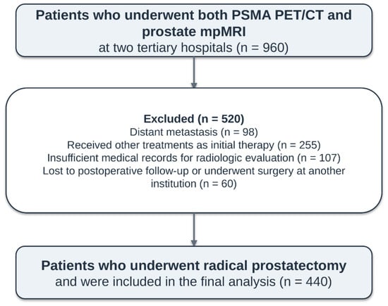
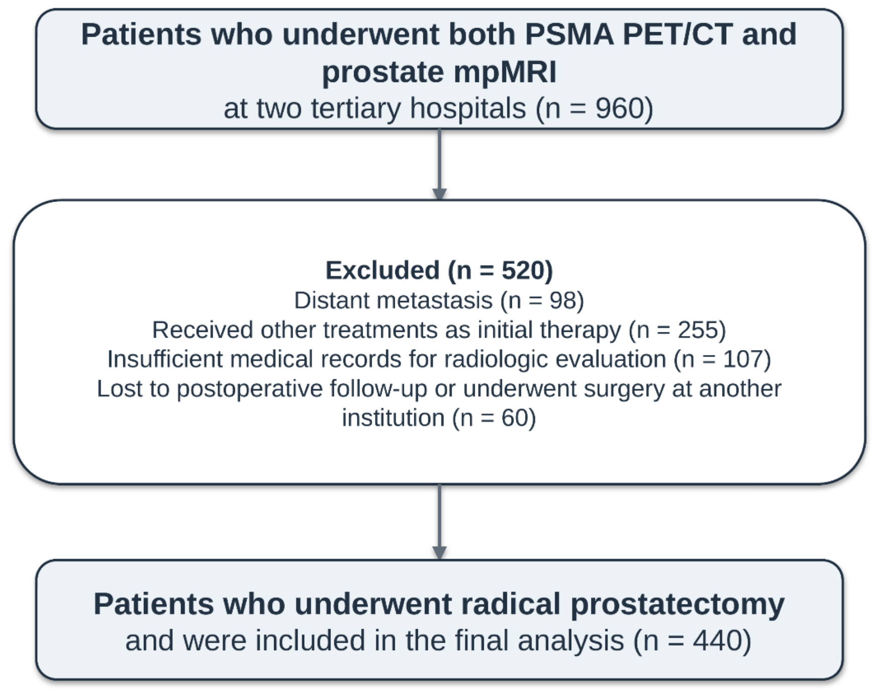
This retrospective study screened patients who visited two tertiary hospitals: Chonnam National University Hospital (Gwang-ju, Korea) and Chonnam National University Hwasun Hospital (Hwasun, Korea) for PC treatment from August 2022 to April 2026. All patients had histologically confirmed PC based on transrectal ultrasound-guided prostate (TRUS) biopsy and underwent clinical staging with radiologic evaluation through 68Ga-PSMA-11 PET/CT and mpMRI without any specific time interval between the two imaging examinations. Additionally, the study included patients who underwent RP with or without pelvic lymph node dissection as initial treatment for localised or locally advanced PC within three months of clinical staging. Patients were excluded for any of the following reasons: (1) presence of distant metastases; (2) initial treatment with radiotherapy, androgen deprivation therapy or focal therapy instead of RP; (3) unavailable medical records for radiologic evaluation; or (4) loss to follow-up. The final analysis included 440 patients with PC (Figure 1).
Figure 1.
Flowchart of patient inclusion in the study. PSMA: Prostate-specific membrane antigen; PET/CT: Positron emission tomography–computed tomography; mpMRI: Multiparametric magnetic resonance imaging.
The study variables comprised age, comorbidities (diabetes mellitus, hypertension, cardiovascular and cerebrovascular disease), preoperative Gleason grade group via TRUS biopsy, preoperative PSA and testosterone levels, radiologic stage via 68Ga-PSMA-11 PET/CT or mpMRI, EAU risk group classification [3], surgical technique (laparoscopic or robotic) and final pathologic information (Gleason grade, T stage and N stage).
2.2. Magnetic Resonance Imaging Protocols and Analysis
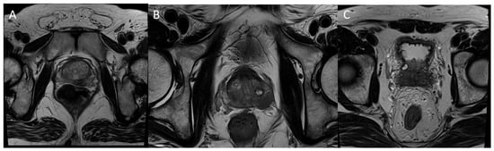
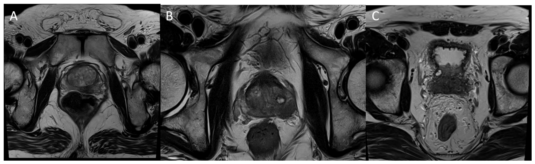
All mpMRI examinations at the two tertiary hospitals followed local protocols, which included T1-weighted and T2-weighted anatomical sequences, diffusion-weighted imaging and dynamic contrast-enhanced imaging. A dedicated uroradiologist with > 15 years of clinical experience (S.H.H) interpreted the imaging findings using PI-RADS version 2.1 [5], while blinded to the results of the other imaging modality, biopsy and final pathology. The study recorded tumour location and the presence of EPE and SVI for each patient and each side (Figure 2).
Figure 2.
Representative cases of PC staged as T2 (A), T3a (B) and T3b (C) on mpMRI. (A) Intraprostatic PI-RADS 5 lesion in peripheral zone of left lobe without suspected extraprostatic extension, classified as stage T2. (B) Visually suspected extracapsular extension of left lobe, classified as T3a. (C) Visually suspected both seminal vesicle invasion, classified as stage T3b. mpMRI: Multiparametric magnetic resonance imaging; PI-RADS; Prostate Imaging-Reporting and Data System.
2.3. Prostate-Specific Membrane Antigen Positron Emission Tomography/Computed Tomography Protocols and Analysis
All PSMA PET/CT examinations were performed at two tertiary hospitals using 68Ga-PSMA-11 radioligands. The 68Ga radioisotope was eluted from an approved 68Ge-68Ga generator (IGG100, Eckert-Ziegler, Biberstein, Switzerland). Radiolabeling was performed using a dedicated PSMA labelling kit (Illucix®, Telix Pharma, Australia), and quality control was conducted according to the instructions of the manufacturer. 68Ga-PSMA-11 was synthesized from a PSMA-11 precursor using an automated synthesis module (BIKBox®, BIK Therapeutics, Seongnam, Republic of Korea), followed by C18 cartridge purification and sterile filtration. Quality control was performed before clinical administration. Torso PET/CT imaging was performed from the vertex to the mid-thighs 60 min after intravenous administration of 68Ga-PSMA-11 at a dose of 185 MBq (5 mCi). Image acquisition was performed using dedicated PET/CT scanners (Discovery ST, Omni Legend 32, Discovery IQ and Discovery 600; GE Healthcare, Chicago, IL, USA). Supplementary Table S1 summarises the detailed imaging protocols.
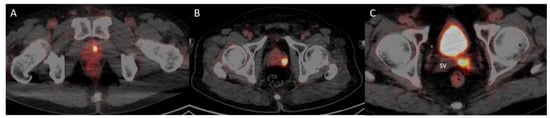
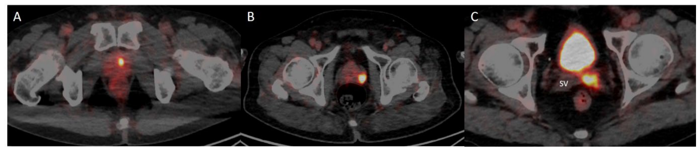
Two nuclear medicine physicians with >10 years of clinical experience (S.G.C., J.B.M.) participated in image interpretation, while blinded to the results of the other imaging modality, biopsy and final pathology. Focal PSMA uptake confined to the prostate gland was considered T2 disease. EPE (T3a) and SVI (T3b) were identified based on visually assessing definite extension of PSMA uptake beyond the prostate capsule and into the seminal vesicles, respectively, based on consensus between the two readers (Figure 3).
Figure 3.
Representative cases of PC staged as T2 (A), T3a (B) and T3b (C) on 68Ga-PSMA-11 PET/CT. (A) Intraprostatic PSMA uptake without suspected extraprostatic extension, classified as stage T2. (B) Visually suspected extracapsular extension of PSMA uptake, classified as T3a. (C) Positive PSMA uptake in the seminal vesicle, classified as stage T3b. PET/CT: positron emission tomography/computed tomography; PSMA: prostate-specific membrane antigen; SV: seminal vesicle; 68Ga-PSMA-11 PET/CT: gallium-68–prostate-specific membrane antigen-11 positron emission tomography/computed tomography; PC: prostate cancer.
2.4. Histopathologic Examination
Histopathological assessment of whole-gland and side-specific tumour stage in RP specimens served as the reference standard. All RP specimens were processed using conventional (non-whole-mount) sections according to the institutional protocol, with the entire prostate submitted for histological examination. Experienced genitourinary pathologists evaluated and graded the specimens according to the ISUP guidelines [19]. Pathology reports were prepared using the CAP protocol for RP specimens (version 4.2.0.1 until 2024 and version 4.3.0.0 thereafter) [20,21], with staging according to the AJCC 8th edition [22]. EPE (pT3a) was defined as tumour extension beyond the prostate into the periprostatic soft tissue, including periprostatic fat, posterolateral connective tissue, or the neurovascular bundle, or as microscopic invasion of the bladder neck. SVI (pT3b) was defined as tumour invasion into the muscular wall of the seminal vesicle. Synoptic reports routinely recorded EPE and SVI as left-sided, right-sided or bilateral and these data were extracted for analysis.
2.5. Statistical Analysis
Statistical analyses were performed using STATA version 16.1 (StataCorp., College Station, TX, USA). Patient demographics were summarised using descriptive statistics. Continuous variables are presented as the mean and standard deviation, whereas categorical variables are presented as frequencies (%). Diagnostic performance measures, including sensitivity, specificity, positive predictive value (PPV), negative predictive value (NPV) and accuracy, were calculated for 68Ga-PSMA-11 PET/CT and mpMRI to assess side-specific PC detection and patient-level detection of EPE and SVI, respectively. McNemar’s test was used to compare the sensitivity and specificity of 68Ga-PSMA-11 PET/CT and mpMRI. The area under the receiver operating characteristic curve (AUC) was calculated and compared using the DeLong test. A p value < 0.05 was considered statistically significant.
2.6. Ethics Statement
The study protocol was approved by the Institutional Review Board (IRB) of the Research Institute of Clinical Medicine at Chonnam National University Hospital (IRB approval No. CNUH-2026-142). This study was conducted in accordance with the Declaration of Helsinki and the Ethical Guidelines for Clinical Studies. Written informed consent was waived because of the retrospective nature of the study.
3. Results
3.1. Baseline Characteristics
Table 1 summarises the characteristics of patients who underwent preoperative 68Ga-PSMA-11 PET/CT and prostate mpMRI before RP. The mean age and preoperative PSA of the patients were 70.73 ± 5.95 years and 12.08 ± 9.88 ng/mL, respectively. Of the 440 patients included in the final analysis, 10.2% had low-risk, 52.1% had intermediate-risk and 37.7% had high-risk PC. Overall, 347 (78.9%) patients underwent robot-assisted RP.
Table 1.
Demographic characteristics of patients with PC who underwent preoperative 68Ga-PSMA-11 PET/CT and prostate mpMRI before RP.
Regarding 68Ga-PSMA-11 PET/CT T staging, 28 patients (6.4%) were classified as miT0, 198 (45.0%) as miT2 (unilateral), 181 (41.1%) as miT2 (bilateral), 19 (4.3%) as miT3a, 9 (2.0%) as miT3b and 5 (1.1%) as miT4 or N1. For mpMRI T staging, 49 patients (11.1%) were classified as T0 (no suspicious lesion), 184 (41.8%) as T2a or T2b, 67 (15.2%) as T2c, 110 (25.0%) as T3a and 30 (6.8%) as T3b. Additionally, regarding the final pathologic T stage after RP, 2 patients (0.5%) were classified as T0, 79 (18.0%) as T2 (unilateral), 153 (34.8%) as T2 (bilateral), 168 (38.2%) as T3a, 36 (8.2%) as T3b and 2 (0.5%) as T4.
3.2. Diagnostic Performance of Prostate-Specific Membrane Antigen Positron Emission Tomography/Computed Tomography Versus Multiparametric Magnetic Resonance Imaging for Detecting Side-Specific Prostate Cancer
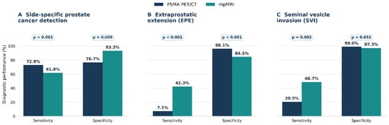
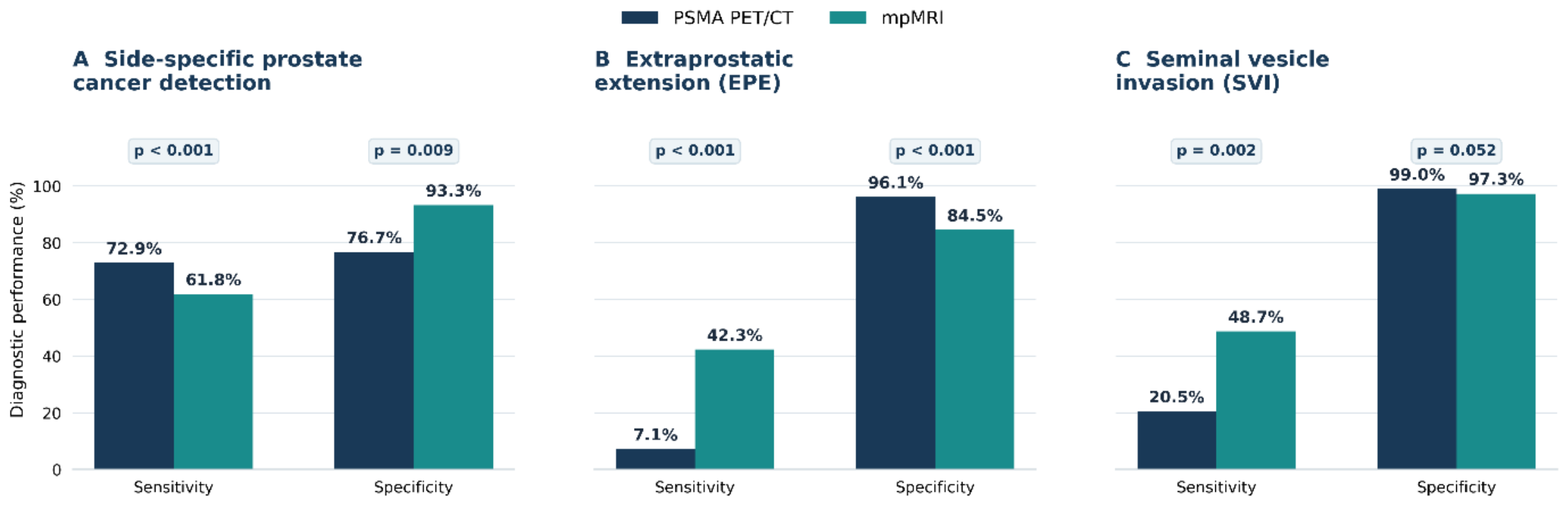
Table 2 compares the diagnostic performance of 68Ga-PSMA-11 PET/CT and mpMRI for detecting side-specific PC. 68Ga-PSMA-11 PET/CT had a sensitivity of 72.9%, significantly higher than the 61.8% sensitivity of mpMRI (95% CI, 58.5–65.1%; p < 0.001). In contrast, 68Ga-PSMA-11 PET/CT had a specificity of 76.7%, significantly lower than that of mpMRI at 93.3% (p = 0.009; Figure 4A). Overall, the accuracy of 68Ga-PSMA-11 PET/CT and mpMRI was 73.2% (95% confidence interval [CI] 70.2–76.0%) and 64.0% (95% CI, 60.8–67.1%), respectively. The PPV and NPV of 68Ga-PSMA-11 PET/CT were 97.7% and 17.2%, respectively, while those of mpMRI were 99.2% and 15.2%, respectively. The AUC did not differ significantly between 68Ga-PSMA-11 PET/CT (0.748, 95% CI: 0.692–0.804) and mpMRI (0.776, 95% CI: 0.740–0.812) (p = 0.323).
Table 2.
Diagnostic performance of 68Ga-PSMA-11 PET/CT and mpMRI for detecting side-specific PC.
Figure 4.
Comparison of sensitivity and specificity between 68Ga-PSMA-11 PET/CT and mpMRI for (A) side-specific PC detection, (B) extraprostatic extension and (C) SVI. Bar graphs comparing the sensitivity and specificity of 68Ga-PSMA-11 PET/CT and mpMRI for (A) side-specific prostate cancer detection, (B) extraprostatic extension and (C) SVI. Final histopathological findings from RP specimens served as the reference standard. Side-specific prostate cancer detection was evaluated in 880 prostate lobes from 440 patients, whereas extraprostatic extension and SVI were assessed at the patient level (n = 440). Values above the bars indicate percentages. p values were calculated using McNemar’s test. 68Ga-PSMA-11 PET/CT: gallium-68–prostate-specific membrane antigen-11 positron emission tomography/computed tomography; mpMRI, multiparametric magnetic resonance imaging; SVI, seminal vesicle invasion; PC, prostate cancer; RP, radical prostatectomy.
3.3. Diagnostic Performance of Prostate-Specific Membrane Antigen Positron Emission Tomography Versus Multiparametric Magnetic Resonance Imaging for Detecting Extraprostatic Extension and Seminal Vesicle Invasion
Table 3 presents the patient-level diagnostic performance of 68Ga-PSMA-11 PET/CT and mpMRI for detecting EPE and SVI. For EPE detection, 68Ga-PSMA-11 PET/CT showed significantly lower sensitivity (7.1% vs. 42.3%, p < 0.001) but higher specificity (96.1% vs. 84.5%, p < 0.001) than mpMRI (Figure 4B). The diagnostic accuracy of 68Ga-PSMA-11 PET/CT and mpMRI was 59.3% (95% CI: 54.6–63.9%) and 67.1% (95% CI: 62.4–71.4%), respectively. The PPVs and NPVs of 68Ga-PSMA-11 PET/CT were 56.5% and 59.5%, respectively, compared to 65.8% and 67.5% for mpMRI. The AUC was significantly lower for 68Ga-PSMA-11 PET/CT (0.516, 95% CI: 0.494–0.539) than for mpMRI (0.634, 95% CI: 0.592–0.676) (p < 0.001). For SVI detection, 68Ga-PSMA-11 PET/CT showed significantly lower sensitivity (20.5% vs. 48.7%, p = 0.002) and comparable specificity (99.0% vs. 97.3%, p = 0.052) compared to those of mpMRI (Figure 4C). The diagnostic accuracy of 68Ga-PSMA-11 PET/CT and mpMRI was 92.1% (95% CI: 89.1–94.4%) and 93.0% (95% CI: 90.2–95.2%), respectively. The PPVs and NPVs of PSMA PET/CT were 66.7% and 92.8%, respectively, compared to 63.3% and 95.1% for mpMRI. As observed for EPE, the AUC was significantly lower for 68Ga-PSMA-11 PET/CT (0.598, 95% CI: 0.533–0.662) than for mpMRI (0.730, 95% CI: 0.650–0.810; p = 0.001).
Table 3.
Diagnostic performance of 68Ga-PSMA-11 PET/CT and mpMRI for detecting EPE and SVI.
4. Discussion
PC has a heterogeneous clinical course, and treatment options vary according to disease stages and grades. Accurate diagnosis and risk stratification are crucial for guiding treatment selection in patients with localised PC [1,3,4]. Although the use of PSMA PET/CT for diagnostic assessment across different stages of PC is increasing, further studies are needed to establish its clinical utility. Therefore, this study aims to compare the diagnostic performance of 68Ga-PSMA-11 PET/CT and mpMRI for initial local staging in patients with localised and/or locally advanced PC. The results demonstrated comparable diagnostic accuracy between 68Ga-PSMA-11 PET/CT and mpMRI for detecting primary PC. However, the two modalities showed contrasting diagnostic characteristics: 68Ga-PSMA-11 PET/CT was more sensitive for detecting side-specific PC but tended to underestimate EPE and SVI, while mpMRI was more sensitive for detecting invasion into adjacent structures. These findings suggest that 68Ga-PSMA-11 PET/CT may complement mpMRI in initial local staging of localised PC and potentially inform treatment selection.
PSMA is a membrane-bound glycoprotein expressed in normal prostate tissue [23]. PSMA is overexpressed on PC cell membranes, and increased expression is associated with prostate adenocarcinoma, disease progression and castration-resistant PC [7,24]. Owing to the inherent specificity of PSMA for PC, PSMA PET/CT linked to 68Ga or 18F-labelled tracers has emerged over the past decade as an imaging tool for improving diagnostic accuracy. PSMA PET/CT has outperformed conventional imaging techniques such as CT and bone scan and may enhance MRI in local staging [25]. In clinical practice, PSMA PET/CT combines molecular oncology and imaging technology, enabling more accurate assessment of disease extent and better-informed treatment decisions for patients with PC. In the proPSMA trial, PSMA PET/CT provided more accurate staging of pelvic nodal disease and distant metastases than conventional imaging and supported appropriate treatment selection, particularly in patients eligible for definitive treatment [8,26]. Consequently, clinical practice guidelines recommend PSMA PET for PC staging, particularly in the assessment of biochemical recurrence and metastatic disease [3].
Although robust evidence supports the role of PSMA PET in initial metastatic staging, its diagnostic value in localised PC remains debated because of conflicting findings from previous studies. For detecting clinically significant PC, a previous meta-analysis reports a pooled sensitivity of 89% and specificity of 56% for PSMA PET in detecting intraprostatic lesions and cancer extent [11]. A recent systematic review and meta-analysis report good diagnostic accuracy for clinically significant PC, with pooled sensitivity and specificity of 82% and 67%, respectively, for PSMA PET [12]. These findings indicate that PSMA PET has potential diagnostic value for detecting clinically significant PC, given its high sensitivity for identifying true-positive lesions. It may be particularly effective in patients with previous negative biopsies. PSMA PET may also support clinical decision-making in patients with borderline indications for prostate biopsy, such as those with elevated PSA levels but inconclusive mpMRI findings. In this study, 68Ga-PSMA-11 PET/CT had significantly higher sensitivity for detecting side-specific PC than mpMRI (72.9% vs. 61.8%). This finding is consistent with those of previous systematic reviews and meta-analyses [11,12], suggesting that PSMA PET/CT may provide complementary information to mpMRI for side-specific PC detection during preoperative staging.
However, previous studies report conflicting findings on EPE and SVI detection. Bahler et al. studied 50 patients at risk of EPE who were scheduled for prostatectomy and report that PSMA PET/CT is more sensitive than mpMRI for detecting EPE along the neurovascular bundles. PSMA PET/CT also informed surgical decision-making regarding nerve-sparing approaches [17]. In a prospective study of 134 patients, Mookerji et al. report a sensitivity of 58% for 18F-PSMA-1007 PET/CT, significantly higher than the 33% sensitivity for mpMRI (p = 0.01). For SVI detection, PSMA PET/CT showed higher sensitivity than mpMRI (57% vs. 33%), although the difference was not statistically significant (p = 0.6) [15]. Prive et al. report that PSMA PET/CT is more accurate than mpMRI for SVI detection, whereas mpMRI performs better for detecting EPE [27]. A recent systematic review and meta-analysis also highlighted the diagnostic performance of PSMA PET-CT for SVI detection [14]. In contrast, Exterkate et al. report no additional value of PSMA PET/CT over mpMRI for detecting EPE and SVI [10]. In a prospective head-to-head trial comparing 68Ga-PSMA-11 PET/CT with mpMRI, mpMRI performs better at detecting EPE and SVI [16]. A recent large multicentre retrospective study of 550 patients reports that PSMA PET/CT had lower sensitivity for EPE detection (41% vs. 62%, p = 0.01) and comparable sensitivity for SVI detection (36% vs. 44%, p = 0.2) compared to those of MRI [12]. In the present study, 68Ga-PSMA-11 PET/CT had significantly lower sensitivity (7.1% vs. 42.3%, p < 0.001) and higher specificity (96.1% vs. 84.5%, p < 0.001) than mpMRI for EPE detection. For SVI detection, 68Ga-PSMA-11 PET/CT had significantly lower sensitivity (20.5% vs. 48.7%, p < 0.001) and comparable specificity (99.0% vs. 97.3%, p = 0.052) compared to mpMRI. Overall, mpMRI outperformed 68Ga-PSMA-11 PET/CT in EPE and SVI detection. As RP is a common treatment for localised or locally advanced PC, accurate detection of EPE and SVI is critical to surgical decision-making for nerve-sparing approaches. Further high-quality studies are needed to clarify the diagnostic value of PSMA PET for the initial staging of locally advanced PC.
In this study, for lobe- or side-specific PC detection, neither modality reliably excluded tumour, as both had an NPV below 20%. However, both showed sufficient specificity and PPValue when image abnormalities were present. These findings suggest substantial discordance between PSMA PET/CT and mpMRI, with many tumours detected based on only one modality. This is plausible because the modalities represent different aspects of PC biology. PSMA PET/CT detects cell-surface expression of a specific antigen associated with histological grade, type and cancer aggressiveness [23,28]. In contrast, mpMRI provides imaging biomarkers of tissue cellularity, vascularity and water content. These characteristics can differ between tumours and across intratumoural regions. Thus, PSMA PET/CT and mpMRI should be regarded as complementary modalities for detecting histologically confirmed intraprostatic PC. A previous study shows that combining PSMA PET with mpMRI improves diagnostic performance over mpMRI alone, particularly via improving specificity [29].
Nonetheless, the limited sensitivity of both imaging modalities, particularly PSMA PET/CT, indicates that negative imaging findings cannot reliably exclude microscopic local tumour extension. In the present study, PSMA PET/CT had significantly lower sensitivity than mpMRI for detecting EPE and SVI. This difference potentially results from several factors, including lower soft-tissue resolution, partial-volume effect, intratumoural heterogeneity in PSMA expression and PET/CT misregistration. Nevertheless, both PSMA PET/CT and mpMRI showed high specificity for EPE and SVI detection, consistent with the findings of a recent meta-analysis [30]. This meta-analysis pooled data from 10 studies involving 505 patients and reports differences in sensitivity and specificity between both modalities for detecting EPE and SVI. The present study adds to this evidence by highlighting the limitations of both imaging modalities and the need to integrate both into the diagnostic workup of PC, alongside invasive diagnostic procedures.
This study has some limitations. First, the retrospective design and restriction to patients who underwent RP may have introduced selection bias, limiting the generalizability of the findings to the broader population of patients with PC. Second, despite the two-centre design, differences in imaging equipment and acquisition protocols between centres may have influenced the diagnostic performance of both imaging modalities. In addition, interobserver agreement was not assessed between the two nuclear medicine physicians, so the results may have been influenced by interobserver bias. Third, EPE and SVI on 68Ga-PSMA-11 PET/CT were assessed visually, with positive findings defined based on definite tracer uptake extending beyond the prostatic capsule or into the seminal vesicles. This stringent criterion, alongside the limited spatial resolution of PET imaging, may have contributed to the low sensitivity for detecting local tumour extension, particularly microscopic EPE. Finally, although the findings suggest that PSMA PET/CT and mpMRI provide complementary diagnostic information, the performance of their combined interpretation was not directly evaluated. Previous studies report improved diagnostic performance when PSMA PET information is combined with MRI, particularly for EPE and SVI detection [12,13,16].
Despite these limitations, the study has several strengths. We included a relatively large cohort of 440 patients who underwent 68Ga-PSMA-11 PET/CT and mpMRI before RP, enabling a direct head-to-head comparison of the two modalities in the same patient population. Furthermore, final histopathological findings from RP specimens served as the reference standard, supporting the assessment of side-specific PC detection, EPE, and SVI. By evaluating these aspects of local staging separately, the study provides clinically relevant evidence of the differences in diagnostic performance between PSMA PET/CT and mpMRI. Further prospective studies incorporating standardised imaging protocols and interpretation criteria, and evaluating combined imaging strategies, are needed to clarify the role of PSMA PET/CT in the initial staging of localised and locally advanced PC.
5. Conclusions
68Ga-PSMA-11 PET/CT showed higher sensitivity than mpMRI for side-specific PC detection, whereas mpMRI had higher sensitivity for detecting EPE and SVI. These findings highlight the distinct diagnostic characteristics of the two imaging modalities and suggest that PSMA PET/CT may complement mpMRI rather than replace it in the initial local staging of localised and locally advanced PC.
Supplementary Materials
The following supporting information can be downloaded at: https://www.mdpi.com/article/10.3390/cancers18223697/s1, Table S1: Image acquisition and reconstruction protocols for different scanners used for 68Ga-PSMA-11 PET/CT imaging.
Author Contributions
Conceptualization, S.H.Y. and H.M.G.; Methodology, S.H.Y., S.-G.C., S.S.K. and S.H.H.; Formal analysis, S.H.Y. and H.M.G.; investigation, S.H.Y., H.M.G., H.S.C., E.C.H., S.I.J., T.W.K., D.K., J.B.M., S.-G.C., N.I.K., S.S.K. and S.H.H.; data curation, S.H.Y. and H.M.G., writing—original draft preparation, S.H.Y., S.-G.C. and S.S.K.; writing—review and editing, S.H.Y.; supervision, S.-G.C. All authors have read and agreed to the published version of the manuscript.
Funding
This research received no external funding.
Institutional Review Board Statement
The study protocol was approved by the Institutional Review Board (IRB) of Chonnam National University Hospital Research Institute of Clinical Medicine (IRB approval No. CNUH-2026-142 and approval date: 27 April 2026).
Informed Consent Statement
Written informed consent was waived because of the retrospective nature of the study.
Data Availability Statement
The data presented in this study are available in this article.
Acknowledgments
The authors used ChatGPT (OpenAI; GPT-5.6 sol) for writing graphical abstract and language refinement, including improvements in grammar, clarity, and readability. The authors reviewed and edited all output and take full responsibility for the content of the publication.
Conflicts of Interest
The authors declare no conflicts of interest.
References
- Kratzer, T.B.; Mazzitelli, N.; Star, J.; Dahut, W.L.; Jemal, A.; Siegel, R.L. Prostate cancer statistics, 2025. CA Cancer J. Clin. 2025, 75, 485–497. [Google Scholar] [CrossRef] [Scilit] [PubMed]
- Jung, K.W.; Kang, M.J.; Park, E.H.; Yun, E.H.; Kim, H.J.; Kim, J.E.; Kong, H.J.; Im, J.S.; Seo, H.G. Prediction of Cancer Incidence and Mortality in Korea, 2024. Cancer Res. Treat. 2024, 56, 372–379. [Google Scholar] [CrossRef] [Scilit] [PubMed]
- Cornford, P.; van den Bergh, R.C.N.; Briers, E.; Van den Broeck, T.; Brunckhorst, O.; Darraugh, J.; Eberli, D.; De Meerleer, G.; De Santis, M.; Farolfi, A.; et al. EAU-EANM-ESTRO-ESUR-ISUP-SIOG Guidelines on Prostate Cancer-2024 Update. Part I: Screening, Diagnosis, and Local Treatment with Curative Intent. Eur. Urol. 2024, 86, 148–163. [Google Scholar] [CrossRef] [Scilit] [PubMed]
- Al Hussein Al Awamlh, B.; Wallis, C.J.D.; Penson, D.F.; Huang, L.C.; Zhao, Z.; Conwill, R.; Talwar, R.; Morgans, A.K.; Goodman, M.; Hamilton, A.S.; et al. Functional Outcomes After Localized Prostate Cancer Treatment. JAMA 2024, 331, 302–317. [Google Scholar] [CrossRef] [Scilit] [PubMed]
- Turkbey, B.; Rosenkrantz, A.B.; Haider, M.A.; Padhani, A.R.; Villeirs, G.; Macura, K.J.; Tempany, C.M.; Choyke, P.L.; Cornud, F.; Margolis, D.J.; et al. Prostate Imaging Reporting and Data System Version 2.1: 2019 Update of Prostate Imaging Reporting and Data System Version 2. Eur. Urol. 2019, 76, 340–351. [Google Scholar] [CrossRef] [Scilit] [PubMed]
- Jochumsen, M.R.; Bouchelouche, K. PSMA PET/CT for Primary Staging of Prostate Cancer—An Updated Overview. Semin. Nucl. Med. 2024, 54, 39–45. [Google Scholar] [CrossRef] [Scilit] [PubMed]
- Evans, J.C.; Malhotra, M.; Cryan, J.F.; O’Driscoll, C.M. The therapeutic and diagnostic potential of the prostate specific membrane antigen/glutamate carboxypeptidase II (PSMA/GCPII) in cancer and neurological disease. Br. J. Pharmacol. 2016, 173, 3041–3079. [Google Scholar] [CrossRef] [Scilit] [PubMed]
- Hofman, M.S.; Lawrentschuk, N.; Francis, R.J.; Tang, C.; Vela, I.; Thomas, P.; Rutherford, N.; Martin, J.M.; Frydenberg, M.; Shakher, R.; et al. Prostate-specific membrane antigen PET-CT in patients with high-risk prostate cancer before curative-intent surgery or radiotherapy (proPSMA): A prospective, randomised, multicentre study. Lancet 2020, 395, 1208–1216. [Google Scholar] [CrossRef] [Scilit] [PubMed]
- Grozdic Milojevic, I.; Milojevic, B.; Skrijelj, D.; Bumbasirevic, U.; Janicic, A.; Kajmakovic, B.; Sobic-Saranovic, D.; Artiko, V.; Beatovic, S. The Role of Nuclear Medicine in Prostate Cancer. Diagnostics 2025, 15, 2876. [Google Scholar] [CrossRef] [Scilit] [PubMed]
- Exterkate, L.; Hermsen, R.; Kusters-Vandevelde, H.V.N.; Prette, J.F.; Baas, D.J.H.; Somford, D.M.; van Basten, J.A. Head-to-Head Comparison of (18)F-PSMA-1007 Positron Emission Tomography/Computed Tomography and Multiparametric Magnetic Resonance Imaging with Whole-mount Histopathology as Reference in Localisation and Staging of Primary Prostate Cancer. Eur. Urol. Oncol. 2023, 6, 574–581. [Google Scholar] [CrossRef] [Scilit] [PubMed]
- Kawada, T.; Yanagisawa, T.; Rajwa, P.; Sari Motlagh, R.; Mostafaei, H.; Quhal, F.; Laukhtina, E.; Aydh, A.; Konig, F.; Pallauf, M.; et al. Diagnostic Performance of Prostate-specific Membrane Antigen Positron Emission Tomography-targeted biopsy for Detection of Clinically Significant Prostate Cancer: A Systematic Review and Meta-analysis. Eur. Urol. Oncol. 2022, 5, 390–400. [Google Scholar] [CrossRef] [Scilit] [PubMed]
- Mazzone, E.; Cannoletta, D.; Quarta, L.; Chen, D.C.; Thomson, A.; Barletta, F.; Stabile, A.; Moon, D.; Eapen, R.; Lawrentschuk, N.; et al. A Comprehensive Systematic Review and Meta-analysis of the Role of Prostate-specific Membrane Antigen Positron Emission Tomography for Prostate Cancer Diagnosis and Primary Staging before Definitive Treatment. Eur. Urol. 2025, 87, 654–671. [Google Scholar] [CrossRef] [Scilit] [PubMed]
- Soeterik, T.F.W.; Heetman, J.G.; Hermsen, R.; Wever, L.; Lavalaye, J.; Vinken, M.; Bahler, C.D.; Yong, C.; Tann, M.; Kesch, C.; et al. The Added Value of Prostate-specific Membrane Antigen Positron Emission Tomography/Computed Tomography to Magnetic Resonance Imaging for Local Staging of Prostate Cancer in Patients Undergoing Radical Prostatectomy. Eur. Urol. Oncol. 2025, 8, 731–738. [Google Scholar] [CrossRef] [Scilit] [PubMed]
- Mari, A.; Cadenar, A.; Giudici, S.; Cianchi, G.; Albisinni, S.; Autorino, R.; Di Maida, F.; Gandaglia, G.; Mir, M.C.; Valerio, M.; et al. A systematic review and meta-analysis to evaluate the diagnostic accuracy of PSMA PET/CT in the initial staging of prostate cancer. Prostate Cancer Prostatic Dis. 2025, 28, 56–69. [Google Scholar] [CrossRef] [Scilit] [PubMed]
- Mookerji, N.; Pfanner, T.; Hui, A.; Huang, G.; Albers, P.; Mittal, R.; Broomfield, S.; Dean, L.; St Martin, B.; Jacobsen, N.E.; et al. Fluorine-18 Prostate-Specific Membrane Antigen-1007 PET/CT vs Multiparametric MRI for Locoregional Staging of Prostate Cancer. JAMA Oncol. 2024, 10, 1097–1103. [Google Scholar] [CrossRef] [Scilit] [PubMed]
- Sonni, I.; Felker, E.R.; Lenis, A.T.; Sisk, A.E.; Bahri, S.; Allen-Auerbach, M.; Armstrong, W.R.; Suvannarerg, V.; Tubtawee, T.; Grogan, T.; et al. Head-to-Head Comparison of (68)Ga-PSMA-11 PET/CT and mpMRI with a Histopathology Gold Standard in the Detection, Intraprostatic Localization, and Determination of Local Extension of Primary Prostate Cancer: Results from a Prospective Single-Center Imaging Trial. J. Nucl. Med. 2022, 63, 847–854. [Google Scholar] [CrossRef] [Scilit] [PubMed]
- Bahler, C.D.; Tachibana, I.; Tann, M.; Collins, K.; Swensson, J.K.; Green, M.A.; Mathias, C.J.; Tong, Y.; Yong, C.; Boris, R.S.; et al. Comparing Magnetic Resonance Imaging and Prostate-Specific Membrane Antigen-Positron Emission Tomography for Prediction of Extraprostatic Extension of Prostate Cancer and Surgical Guidance: A Prospective Nonrandomized Clinical Trial. J. Urol. 2024, 212, 290–298. [Google Scholar] [CrossRef] [Scilit] [PubMed]
- Soeterik, T.F.W.; van Melick, H.H.E.; Dijksman, L.M.; Stomps, S.; Witjes, J.A.; van Basten, J.P.A. Nerve Sparing during Robot-Assisted Radical Prostatectomy Increases the Risk of Ipsilateral Positive Surgical Margins. J. Urol. 2020, 204, 91–95. [Google Scholar] [CrossRef] [Scilit] [PubMed]
- Epstein, J.I.; Egevad, L.; Amin, M.B.; Delahunt, B.; Srigley, J.R.; Humphrey, P.A.; Grading, C. The 2014 International Society of Urological Pathology (ISUP) Consensus Conference on Gleason Grading of Prostatic Carcinoma: Definition of Grading Patterns and Proposal for a New Grading System. Am. J. Surg. Pathol. 2016, 40, 244–252. [Google Scholar] [CrossRef] [Scilit] [PubMed]
- Srigley, J.R.; Humphrey, P.A.; Amin, M.B.; Chang, S.S.; Egevad, L.; Epstein, J.I.; Grignon, D.J.; McKiernan, J.M.; Montironi, R.; Renshaw, A.A.; et al. Protocol for the examination of specimens from patients with carcinoma of the prostate gland. Arch. Pathol. Lab. Med. 2009, 133, 1568–1576. [Google Scholar] [CrossRef] [Scilit] [PubMed]
- College of American Pathologists. Cancer Protocoal Templates. Available online: https://www.cap.org/protocols-and-guidelines/cancer-protocols/current-cancer-protocols (accessed on 16 November 2026).
- Buyyounouski, M.K.; Choyke, P.L.; McKenney, J.K.; Sartor, O.; Sandler, H.M.; Amin, M.B.; Kattan, M.W.; Lin, D.W. Prostate cancer—Major changes in the American Joint Committee on Cancer eighth edition cancer staging manual. CA Cancer J. Clin. 2017, 67, 245–253. [Google Scholar] [CrossRef] [Scilit] [PubMed]
- Vetrone, L.; Mei, R.; Bianchi, L.; Giunchi, F.; Farolfi, A.; Castellucci, P.; Droghetti, M.; Presutti, M.; Degiovanni, A.; Schiavina, R.; et al. Histology and PSMA Expression on Immunohistochemistry in High-Risk Prostate Cancer Patients: Comparison with (68)Ga-PSMA PET/CT Features in Primary Staging. Cancers 2023, 15, 1716. [Google Scholar] [CrossRef] [Scilit] [PubMed]
- Mhawech-Fauceglia, P.; Zhang, S.; Terracciano, L.; Sauter, G.; Chadhuri, A.; Herrmann, F.R.; Penetrante, R. Prostate-specific membrane antigen (PSMA) protein expression in normal and neoplastic tissues and its sensitivity and specificity in prostate adenocarcinoma: An immunohistochemical study using mutiple tumour tissue microarray technique. Histopathology 2007, 50, 472–483. [Google Scholar] [CrossRef] [Scilit] [PubMed]
- Combes, A.D.; Palma, C.A.; Calopedos, R.; Wen, L.; Woo, H.; Fulham, M.; Leslie, S. PSMA PET-CT in the Diagnosis and Staging of Prostate Cancer. Diagnostics 2022, 12, 2594. [Google Scholar] [CrossRef] [Scilit] [PubMed]
- Bukavina, L.; Luckenbaugh, A.N.; Hofman, M.S.; Hope, T.; Kamran, S.C.; Murphy, D.G.; Yamoah, K.; Ost, P. Incorporating Prostate-specific Membrane Antigen Positron Emission Tomography in Management Decisions for Men with Newly Diagnosed or Biochemically Recurrent Prostate Cancer. Eur. Urol. 2023, 83, 521–533. [Google Scholar] [CrossRef] [Scilit] [PubMed]
- Prive, B.M.; Israel, B.; Schilham, M.G.M.; Muselaers, C.H.J.; Zamecnik, P.; Mulders, P.F.A.; Witjes, J.A.; Sedelaar, M.; Mehra, N.; Verzijlbergen, F.; et al. Evaluating F-18-PSMA-1007-PET in primary prostate cancer and comparing it to multi-parametric MRI and histopathology. Prostate Cancer Prostatic Dis. 2021, 24, 423–430. [Google Scholar] [CrossRef] [Scilit] [PubMed]
- Hartenbach, M.; Rasul, S.; Grubmuller, B.; Kramer, G.; Baltzer, P.; Helbich, T.; Eiber, M.; Hartenbach, S.; Grahovac, M.; Susani, M.; et al. PSMA-Directed PET/MRI Enables Noninvasive Diagnosis and Prognosis in Patients with Increased PSA Levels: Results from the Prospective Randomized RAPID Trial. J. Nucl. Med. 2026, 67, 224–231. [Google Scholar] [CrossRef] [Scilit] [PubMed]
- Margel, D.; Bernstine, H.; Groshar, D.; Ber, Y.; Nezrit, O.; Segal, N.; Yakimov, M.; Baniel, J.; Domachevsky, L. Diagnostic Performance of (68)Ga Prostate-specific Membrane Antigen PET/MRI Compared with Multiparametric MRI for Detecting Clinically Significant Prostate Cancer. Radiology 2021, 301, 379–386. [Google Scholar] [CrossRef] [Scilit] [PubMed]
- Jin, X.; Cai, Y.; Ren, X. Comparison of (68)Ga-PSMA PET and mpMRI for prostate cancer local staging: A comprehensive review and direct meta-analysis. Front. Oncol. 2024, 14, 1410229. [Google Scholar] [CrossRef] [Scilit] [PubMed]

Disclaimer/Publisher’s Note: The statements, opinions and data contained in all publications are solely those of the individual author(s) and contributor(s) and not of MDPI and/or the editor(s). MDPI and/or the editor(s) disclaim responsibility for any injury to people or property resulting from any ideas, methods, instructions or products referred to in the content. |
© 2026 by the authors. Licensee MDPI, Basel, Switzerland. This article is an open access article distributed under the terms and conditions of the Creative Commons Attribution (CC BY) license.