


Simple Summary
Three-dimensional (3D) imaging technologies, including 3D reconstruction, 3D printing, and augmented reality, are changing how surgeons plan and perform cancer operations. Instead of relying solely on standard scans, surgeons can now interact with detailed 3D models of a patient’s anatomy before entering the operating room. Despite this potential, these technologies are not yet widely used in a consistent or structured way. In this paper, we describe select examples of our experience introducing these tools across multiple cancer surgery specialties at a major cancer center. We developed a practical framework to help surgeons decide which 3D tool best matches the specific challenge they face, whether that is understanding a tumor’s relationship to nearby structures, assessing whether complete removal is possible, preparing a custom implant, or explaining a diagnosis to a patient and their family. Our goal is to provide a reproducible model that other institutions can adapt.
Abstract
Background: Three-dimensional (3D) visualization of radiological imaging has emerged as a valuable tool in surgical oncology, with applications spanning diagnostics, perioperative planning, intraoperative guidance, and patient communication. Despite its growing clinical relevance, integration into routine surgical workflows remains inconsistent and poorly standardized across specialties. Methods: We describe select cases illustrating a multispecialty feasibility and implementation experience evaluating the integration of 3D reconstruction, 3D printing, and augmented reality into surgical oncology workflows across a comprehensive cancer center. Describing cases and series of interest, we demonstrate how a structured implementation workflow was applied covering image acquisition, segmentation, volumetric rendering, and modality selection based on the specific clinical problem. A feasibility framework is proposed in terms of workflow integration, modality-to-problem fit, and clinical utility as described by the operating team. Results: Seven select case groups spanning pediatric, head and neck, urologic, gynecologic, and thoracic surgical oncology were included, with group sizes ranging from 1 to 14 patients. In 3 patients with hepatoblastoma, 3D printed anatomical models were successfully integrated into patient and family education consultations to convey tumor extent and the scope of planned resection. Segmentation-based visualization was implemented for preoperative planning in 2 patients with skull base malignancies, with the 3D workflow described as useful for operative planning by the operating team in both cases. Augmented reality (AR)-assisted 3D reconstruction was incorporated into the planning workflow for 3 patients undergoing robotic partial nephrectomy for complex renal tumors, facilitating review of vascular anatomy and assessment of the feasibility of nephron-sparing surgery. In 2 patients with suspected advanced ovarian cancer, volumetric rendering with AR was integrated into preoperative resectability planning. Among 14 patients undergoing chest wall tumor resection, patient-specific 3D printed ribcage replicas were used for preoperative implant preparation. In a separate cohort of 2 chest wall patients, AR visualization was used for spatial surgical planning. Conclusions: Across diverse oncologic contexts and surgical specialties, 3D reconstruction, printing, and augmented reality were successfully integrated into existing perioperative workflows, with each modality selected based on the specific clinical problem at hand. This experience describes a structured, modality-matched implementation framework that may serve as a practical starting point for institutions seeking to incorporate these technologies. Prospective, controlled studies are needed to establish independent clinical benefit. Broader adoption will require standardized selection criteria, prospective evaluation, and workflow adaptation for centers beyond high-volume academic institutions.
1. Introduction
Cross-sectional imaging is central to contemporary surgical oncology, guiding diagnosis, operative planning, and postoperative surveillance. Conventional imaging is inherently two-dimensional; advances in computational processing now allow these datasets to be converted into three-dimensional (3D) models that can be visualized digitally, manipulated interactively, or used to fabricate physical replicas. Across surgical disciplines, these tools have shown promise with early evidence suggesting improved spatial understanding of tumor anatomy, more confident preoperative planning, and better technical performance in the operating room, though the evidence base remains largely observational [1,2,3,4,5,6,7].
However, routine adoption of 3D models remains limited due to disease complexity, variable tumor presentations, and the absence of standardized workflows that make it difficult to define clear indications for the use of these tools. The resources required for segmentation and model generation add further practical constraints, particularly outside high-volume academic centers, leaving many surgical teams without guidance even when the technology is available [8,9,10].
This series illustrates the use of 3D models in 7 case groups spanning multiple surgical specialties and describes a practical workflow for implementing this technology within an existing institutional infrastructure. These cases represent a subset of patients treated at our institution, and they were selected for illustrative purposes.
2. Methods
2.1. 3D Imaging Technologies and Workflow
Cases were identified through retrospective review of clinical records and selected based on the use of 3D technology; this is a selected case series rather than a consecutive cohort of all eligible patients. Each case was reviewed to determine the appropriate 3D imaging modality and the purpose of its clinical application. The choice of modality was based on the intended use (patient education, preoperative planning, surgical risk stratification), presentation of the tumor, and anatomical complexity.
2.2. Data Acquisition
Medical imaging data were acquired in Digital Imaging and Communications in Medicine (DICOM) format. Cases were included from 2023 to 2026. The dataset comprised both computed tomography (CT) and magnetic resonance imaging (MRI) images that were either acquired internally or uploaded from outside institutions where image acquisition took place. Images were stored and retrieved from the institution’s Picture Archiving and Communications System (PACS). A slice thickness of 0.625 mm was acquired on CT (or 1 mm when 0.625 mm was not available), and a slice thickness of 1 mm was acquired on MRI.
2.3. Segmentation
Segmentation isolates anatomical structures from 2D imaging data and reconstructs them into 3D volumetric models, allowing different structures (such as tumor, vasculature, and bone) to be visualized separately and in spatial relation to one another. Prior to 3D segmentation, standard multiplanar CT and/or MRI reconstructions were reviewed by an attending radiologist in all cases. Segmentation was performed manually by an attending radiologist using Mimics (Materialise NV, Leuven, Belgium; version 27.0) or 3D Slicer (https://www.slicer.org/; version 5.6.2 [11]). Segmentation required approximately one hour per case. CT or MRI DICOM data were segmented to map out surgical approaches and anticipate technical challenges, including complex anatomical variations, tumors adjacent to critical structures, and vascular malformations. These segmented structures were exported as Standard Tessellation Language (STL) files, which serve as the universal file format required for both 3D printing and visualization on augmented reality (AR) platforms. Models were reviewed by a multidisciplinary team during preoperative patient care conferences.
2.4. Volumetric Rendering
Unlike traditional 2D imaging, volumetric rendering of segmented 3D models allows dynamic manipulation of the reconstructed anatomy, including rotation, sectioning, zooming, and adjustment of perspective across all three planes. This flexibility is particularly valuable for identifying subtle spatial relationships between anatomical structures that may not be appreciable on standard axial, sagittal, or coronal views. Volumetric rendering also enables visualization in unconventional planes (oblique and off-axis perspectives), which can reveal tumor involvement or anatomical relationships that would otherwise be missed on standard 2D review. Segmented volumetric models have been reported to correlate with intraoperative findings in some series; whether this translates to reductions in operative time or complications in surgical oncology has not been established by prospective comparative data [12].
2.5. 3D Printing
3D printing, also known as additive manufacturing, creates physical 3D objects from digital STL files through sequential layering of material. Following segmentation and STL file export, models were processed and optimized for printing using Materialise software. Physical models were printed using a Stratasys J5 printer with Stratasys photopolymer resins, including a clear photopolymer resin and colored photopolymer resins. Support material was removed through a combination of manual removal and ultrasonic wash bath. For models printed with the clear resin, 2 coats of epoxy resin were applied to achieve optical transparency and left to cure for 24–48 h; any remaining surface irregularities were smoothed with sandpaper prior to clinical use.
The potential role of 3D printing in healthcare has been increasingly explored over the past decade, with applications ranging from education and surgical planning to the fabrication of patient-specific implants and surgical guides [13]. Three-dimensional printing has been applied across numerous surgical specialties, including maxillofacial, cardiovascular, orthopedic, and general surgery [14]. Physical 3D anatomical models offer particular advantages over traditional 2D imaging by providing improved spatial understanding of the relationship between pathology and surrounding anatomy, and by more intuitively representing the intraoperative point of view. These strengths are especially valuable in the preparation of complex cases.
The literature characterizing 3D printing in pediatric populations is less robust than in adult cohorts; however, available studies suggest potential benefits for patient and provider education as well as surgical planning [15]. Three-dimensional printed models of neonatal and infant anatomy may enhance surgeons’ ability to visualize and interact with intricate structures, and the fabrication of custom devices can accommodate the broad range of ages and sizes encountered in pediatric patients, a particular advantage given the ongoing physiological development of pediatric anatomy. In the context of pediatric surgical oncology, which relies heavily on CT and MRI for surgical planning, 3D printing has been reported to aid complex tumor resections, surgical decision-making, preoperative planning, and surgeon confidence [16,17,18].
Three-dimensional printed models also facilitate more effective physician–patient communication and shared decision-making, helping patients and families understand tumor anatomy, surgical logistics, and procedural risks. They additionally serve as a valuable educational tool for trainees and for building consensus within support teams in multidisciplinary discussions [19,20,21,22,23,24].
2.6. Augmented Reality
The rapid advancement of extended reality technologies has led to expanding use in preoperative planning [25,26,27]. AR overlays digital images onto the real-world environment, virtual reality fully immerses the user in a computer-generated environment isolated from the real world, and mixed reality (MR) combines elements of both with the added capability of enabling direct interaction with virtual elements in real space [28].
In this study, the extended reality modality we deployed was AR, performed using the Medivis SurgicalAR platform (Medivis, New York, NY, USA). The system was implemented using a Microsoft HoloLens 2 head-mounted display (Microsoft Corporation, Redmond, WA, USA) in conjunction with a laptop workstation. PACS integration into the AR visualization software allowed direct retrieval of imaging data for use in clinical environments, including the operating theatre. Upon assessment of workflow and ease of use, the setup was streamlined to a laptop and HoloLens 2 headset for practical deployment.
Prior studies have assessed extended reality in preoperative planning [28,29] and have demonstrated improved identification of anatomic landmarks. Noted limitations include longer preoperative imaging review times and a learning curve associated with new platforms.
A key advantage of AR is the ability to visualize anatomy from unconventional oblique and off-axis planes not available in standard 2D review. This is particularly relevant in cases involving distorted anatomy, tumor encasement of critical structures, or previous surgery.
It is also valuable in preoperative planning for complex cytoreductive surgery, which is an integral component in the management of advanced ovarian cancer. Complete gross resection is associated with significant survival benefit in this patient population [30,31,32] while suboptimal cytoreduction (>1 cm of residual disease) is associated with inferior outcomes [33]. Appropriate patient selection based on preoperative imaging, facility resources, patient functional status, and the likelihood of achieving complete gross resection remains central to determining which patients are appropriate for upfront cytoreductive surgery versus neoadjuvant chemotherapy [34,35]. The decision remains challenging, and innovative approaches such as use of extended reality to enhance preoperative imaging review represent a promising avenue for improving the triage paradigm.
2.7. Ethics Statement
This study was reviewed by our Institutional Review Board and granted exempt status.
3. Results
3.1. Cases
Table 1 provides an overview of the 7 clinical case groups included in this study, with group sizes ranging from 1 to 14 patients, highlighting the diagnosis, clinical service, visualization method, and use of 3D imaging technology for the patients in each group.
Table 1.
Overview of Cases.
3.2. 3D Printing for Patient Education
Hepatoblastoma
Three cases of hepatoblastoma were identified from the Pediatric Surgery Service to create standardized 3D printed liver models for patient education. The 3D printing workflow was selected for these cases because the primary goal was tactile, patient-facing education (rather than intraoperative guidance), a context in which a physical, holdable model offers advantages over screen-based visualization alone.
These models facilitated physician–patient communication by providing a tangible reference that patients and families could engage with as the diagnosis and planned course of treatment were explained. Hepatoblastoma typically requires liver resection, and patients and families frequently underestimate the extent of the tumor and the volume of liver that must be removed. A scaled, anatomically representative 3D printed model allowed the surgeon to reference the model directly during clinical visits to communicate the logistics of the surgical procedure, clarify misconceptions about the nature of the condition, and set appropriate expectations regarding the scope of the operation.
3.3. Segmentation and 3D Reconstruction for Anatomical Clarity
3.3.1. Skull-Based Neoplasms
3D visualization on a computer monitor, generated through segmentation of CT imaging data, was used in the preoperative planning of 2 patients who presented with skull-based malignancies. This visualization method was chosen because both cases involved complex anatomical relationships between tumor and critical skull base structures that were difficult to fully appreciate on standard 2D imaging review alone.
Patient 2a:
A 46-year-old male presented with olfactory neuroblastoma after presenting with several episodes of mild epistaxis that prompted CT imaging. The tumor was characterized as Kadish B (i.e., was confined to the nasal cavity with extension to the paranasal sinuses). Segmentation revealed a tumor centered in the left nasal cavity with involvement of the left superior and middle turbinates and extension to the left maxillary sinus, with no intracranial or intraorbital extension. Intraoperative findings confirmed tumor origin at the middle and superior turbinates, and particular attention was paid to achieving complete resection at the skull base margin. All margins were negative on final surgical pathology (R0). No postoperative complications were recorded. The patient underwent adjuvant radiation therapy and remained without evidence of disease at the 2-year follow-up.
Patient 2b:
A 65-year-old male presented with intestinal-type sinonasal adenocarcinoma following nasal endoscopy and biopsy of a left nasal mass identified in the context of epistaxis. Segmentation demonstrated a mass centered in the left nasal cavity and ethmoid sinuses with involvement of the left orbital lamina papyracea, extension into the anterior cranial fossa through the cribriform plate and planum sphenoidale, and involvement of the overlying dura. Intraoperative findings confirmed invasion of the lamina papyracea and cribriform plate. The lamina was excised along with a portion of the periorbita, and a craniotomy was required to clear the tumor from the dura. The dural defect was reconstructed with a pericranial flap and a lumbar drain was inserted. The patient underwent adjuvant chemotherapy and radiation therapy; final surgical margins were negative (R0). No postoperative complications were noted. No local recurrence was recorded; however, the patient did subsequently develop bony metastases to the spine, for which further radiation therapy was administered.
3.3.2. Renal Tumor in the Setting of Crossed Fused Renal Ectopia
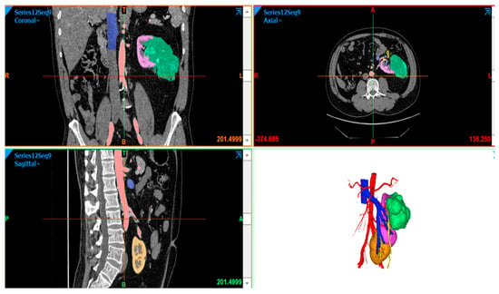
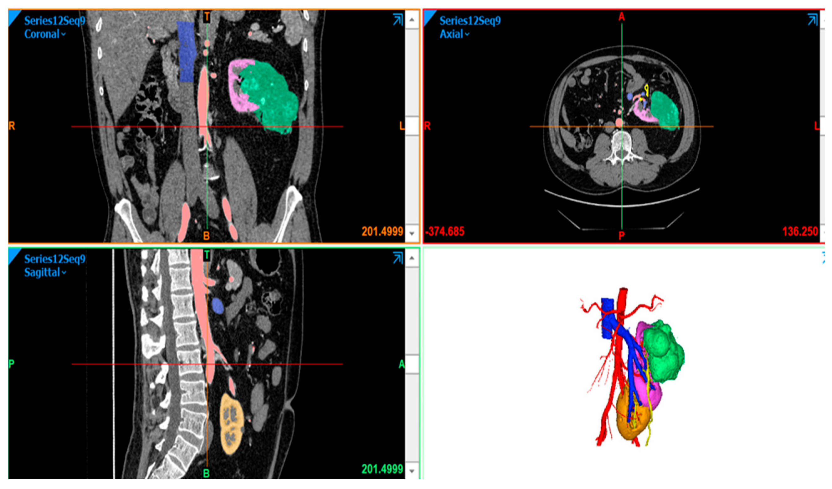
A patient presented with an 11.2 cm left renal mass in the upper pole of a kidney with crossed fused renal ectopia, a rare anatomic variant in which one kidney crosses the midline to the contralateral side, with the ureter recrossing midline and inserting into the bladder at its typical location; the two kidneys are typically fused by an isthmus of renal parenchyma [36]. This anatomic variant poses significant surgical challenges in identifying hilar structures and delineating the border between distinct renal moieties. Given this complexity, 3D visualization with AR was selected to improve spatial understanding of the tumor’s relationship to critical hilar and vascular structures prior to robotic-assisted partial nephrectomy (RAPN) (Figure 1).
Figure 1.
Three-dimensional segmentation rendering of an 11.2 cm left renal mass in the upper pole of a kidney with crossed fused renal ectopia, a rare anatomic variant in which one kidney crosses the midline to the contralateral side, with the ureter recrossing midline. The aorta (red), inferior vena cava (blue), renal parenchyma (purple, orange), and tumor (green) are individually color-coded to delineate the anomalous vascular anatomy and tumor–vessel relationships.
Preoperative planning included careful review of a 3D reconstruction of a preoperative renal protocol CT scan using the Medivis AR platform with a Microsoft HoloLens 2 head-mounted display. This allowed improved appreciation of the intimate relationship between the mass and the renal hilum. Additionally, 3D reconstruction showed that the renal veins from both moieties coalesced into a solitary renal vein and revealed the presence of four renal arteries, one of which inserted into the upper pole moiety of interest. Intraoperatively, this artery was ligated, enabling ischemic demarcation and successful removal of the moiety of interest. Final pathology demonstrated pT3a clear cell renal cell carcinoma (ccRCC) with negative margins (R0). Postoperative glomerular filtration rate (GFR) was unchanged. No postoperative complications were recorded. In this case, AR-assisted 3D reconstruction appeared to aid in the identification of a critical accessory artery that was not readily apparent on standard 2D imaging review.
3.3.3. Hilar Renal Tumors
Two patients with complex hilar renal masses were evaluated for RAPN using 3D visualization with AR (Figure 1) as an adjunct to standard preoperative imaging. In both cases, the AR-assisted 3D reconstruction workflow was selected because the endophytic location and the close proximity to hilar structures made standard 2D imaging review insufficient for confident preoperative surgical planning. The 3D reconstruction provided improved visualization of the 3D relationships between vascular structures, the collecting system, and the renal tumor.
Patient 4a:
A 6.0 cm endophytic right renal mass closely abutting hilar structures and the collecting system was evaluated with AR/3D reconstruction of a preoperative renal protocol CT. In this case, the 3D reconstruction provided a unique view of the renal hilar structures, specifically the collecting system, and its relationship to the tumor. Review of the 3D reconstruction supported the feasibility of RAPN with limited resection of the collecting system. Final pathology demonstrated ccRCC T1b with negative surgical margins (R0) and normal postoperative GFR. No postoperative complications were recorded.
Patient 4b:
A 4.2-cm entirely endophytic right renal mass with hilar abutment was similarly evaluated using AR/3D reconstruction. The reconstruction provided a view of the spatial relationship between the tumor and hilar structures, including the vessels and collecting system. Based on this assessment, RAPN was deemed feasible. Final pathology demonstrated a pT1a oncocytic tumor with negative surgical margins (R0) and normal postoperative GFR. No postoperative complications were recorded.
In both cases, AR-assisted 3D reconstruction enhanced surgeon confidence in the feasibility of nephron-sparing partial nephrectomy as opposed to radical nephrectomy, a decision that depends on the surgeons’ ability to evaluate the 3D spatial relationship between tumor and the renal hilum. The primary benefit in both cases was enhanced spatial characterization of the tumor’s relationship to the critical hilar vascular and collecting system structures, which could not be fully appreciated from sequential 2D imaging alone.
3.4. AR and Volumetric Rendering for Resectability and Spatial Planning
3.4.1. Ovarian Tumor Debulking
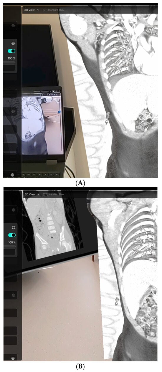
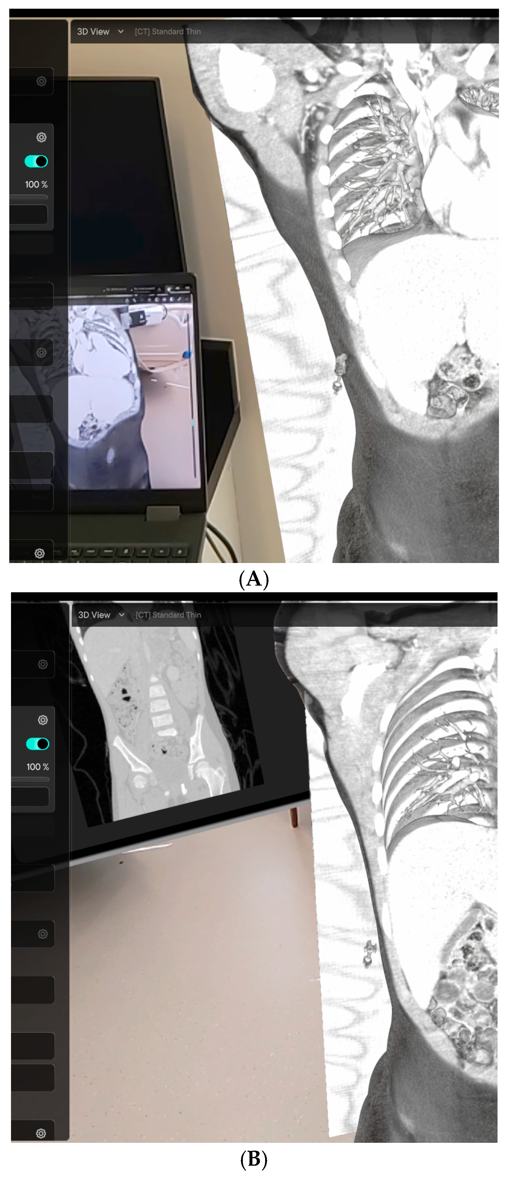
Two patients with suspected advanced ovarian cancer were evaluated using 3D volumetric rendering with an AR headset (Figure 2) as an adjunct to standard CT imaging and our institutional resectability assessment protocol (Figure 2), with the goal of improving preoperative prediction of the feasibility of complete gross resection. The AR-assisted volumetric rendering workflow was selected in these cases because accurate preoperative identification of disease distribution, particularly involvement of the small bowel, mesentery, liver, rectum, and diaphragm, is critical to determining whether upfront cytoreductive surgery or neoadjuvant chemotherapy is the appropriate initial treatment strategy. The AR method of visualizing these key anatomic spaces provided an interactive method to evaluate anatomic findings consistent with peritoneal carcinomatosis.
Figure 2.
AR volumetric rendering using the Medivis SurgicalAR platform with Microsoft HoloLens 2 headset in two patients undergoing cytoreductive surgery for advanced ovarian malignancy. (A) Case 1: coronal volumetric CT reconstruction demonstrating a large heterogeneous pelvic mass with adjacent osseous structures. (B) Case 2: coronal volumetric reconstruction showing a multilobulated pelvic mass with complex internal architecture. AR-assisted review was performed in the days prior to surgery as part of the preoperative planning workflow.
Both patients underwent our structured preoperative resectability assessment, which comprises the following steps. First, high-resolution CT and/or MRI are evaluated with volumetric segmentation and 3D reconstruction to delineate tumor boundaries, assess involvement of critical structures (including vessels, nerves, and organs), and visualize spatial relationships. Second, a multidisciplinary team comprising surgical oncology, radiology, and anesthesiology reviews the 3D reconstructions to assess the feasibility of complete resection with negative margins (R0), incorporating patient-specific factors such as pulmonary function, prior surgical history, and comorbidities. Third, tumor involvement is classified using a resectability scoring system that accounts for extent of disease distribution, vascular proximity, and peritoneal involvement; tumors are categorized as resectable, marginally resectable with 3D-assisted planning, or unresectable. Fourth, STL files are prepared for 3D printing for surgical simulation and implant preparation where applicable, and AR visualization via the Medivis platform with Microsoft HoloLens 2 allows the surgical team to rehearse resections virtually (including evaluating angles, approaches, and operative strategy). Finally, the surgical plan is finalized based on the above assessment; patient-specific guides or implants are fabricated as needed and a final preoperative review is conducted to minimize intraoperative uncertainty. This approach aligns with emerging pediatric and adult surgical oncology protocols in which 3D modeling and volumetric assessment have been shown to improve resectability decisions and reduce intraoperative complications, particularly when disease is located on anatomic structures that are fine and travel obliquely (such as the diaphragm, omentum, or bowel serosa).
All steps of the institutional resectability assessment protocol described above were applied to both patients. AR-assisted volumetric review was performed in the days prior to surgery for each patient; the individual contributions of this step are described below.
Patient 5a: Sites of disease, including small bowel serosal involvement, liver, rectum, and bilateral diaphragm, were identifiable on standard 2D CT review. However, the extent of diaphragmatic involvement was difficult to fully characterize on 2D axial imaging alone. AR-assisted volumetric rendering provided a spatially integrated view of disease distribution across all three planes simultaneously, allowing the team to better delineate the full extent of diaphragmatic disease and assess the feasibility of complete resection. This assessment supported the decision to proceed with upfront cytoreductive surgery rather than neoadjuvant chemotherapy. Intraoperative findings confirmed all predicted sites of involvement, and complete gross resection was performed with negative margins (R0). No postoperative complications were recorded.
Patient 5b: On standard 2D CT review, the absence of small bowel serosal and mesenteric involvement was not clearly established; imaging findings were equivocal and insufficient to confidently exclude peritoneal disease at these sites. AR-assisted volumetric rendering was used to enable interactive 3D assessment of the relevant anatomical regions, which the surgical team described as helpful in confirming that neither serosal nor mesenteric involvement appeared to be present. This informed the preoperative resectability discussion and supported the decision to proceed with upfront surgery. Complete gross resection was performed with negative margins (R0). No postoperative complications were recorded. This case illustrates that the 3D reconstructions can also be useful to confirm standard anatomy and the absence of malignancy.
3.4.2. Chest Wall Reconstruction: Augmented Reality
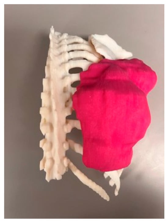
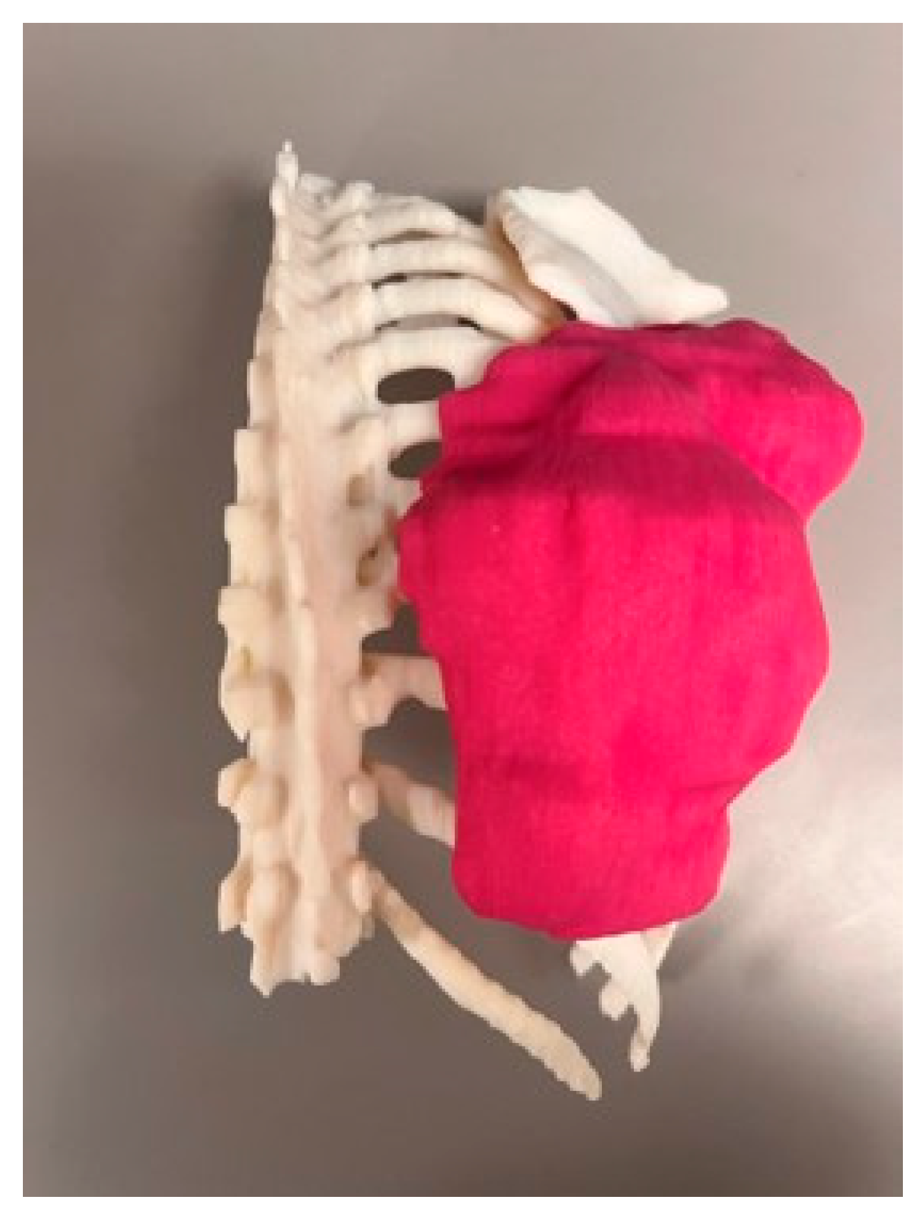
In 2 patients who underwent chest wall tumor resection and reconstruction, AR visualization was used for preoperative spatial planning in place of physical model printing (Figure 3). The AR workflow was selected to allow surgeons to interact with the virtual 3D reconstruction and appreciate tumor extent, involvement, and spatial relationship to surrounding structures prior to the procedure. AR visualization primarily enhanced the operating surgeons’ spatial understanding of tumor extent and its relationship to adjacent critical structures, supporting preoperative spatial planning for resection.
Figure 3.
Three-dimensional printed anatomical model of the thoracic spine and chest wall in a patient with a tumor involving the chest wall. The model was fabricated using segmentation data from CT imaging. The white structure represents the bony thorax and spinal column; the magenta component represents the segmented tumor. The model was used for operative planning, implant contouring, and patient and family communication prior to chest wall resection and reconstruction.
Resections were performed with negative surgical margins (R0). No postoperative complications were recorded (Table 2).
Table 2.
Case Summary: Modality Selection and Clinical Application.
3.5. 3D Printing for Implant Fabrication
Chest Wall Reconstruction: 3D Printing
In a series of 14 patients who underwent chest wall tumor resection and reconstruction, 3D reconstruction and printing were used for preoperative planning [37,38,39]. The 3D printing workflow was selected because chest wall reconstruction demands precise preoperative contouring of fixation hardware to each patient’s individual anatomy, a task that benefits from physical model fabrication.
CT and MRI were used to create detailed 3D reconstructions of the rib cage and tumor, which were then printed to produce patient-specific physical replicas. These models allowed the surgical team to pre-bend fixation plates tailored to each patient’s anatomy prior to the operative procedure, a step the team described as instrumental for implant preparation. Formal outcome metrics such as implant preparation time were not prospectively collected; the assessment of benefit was based on surgeon-reported feedback, which was consistently positive. Resections were performed with negative surgical margins (R0). No postoperative complications were recorded.
4. Discussion
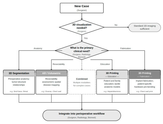
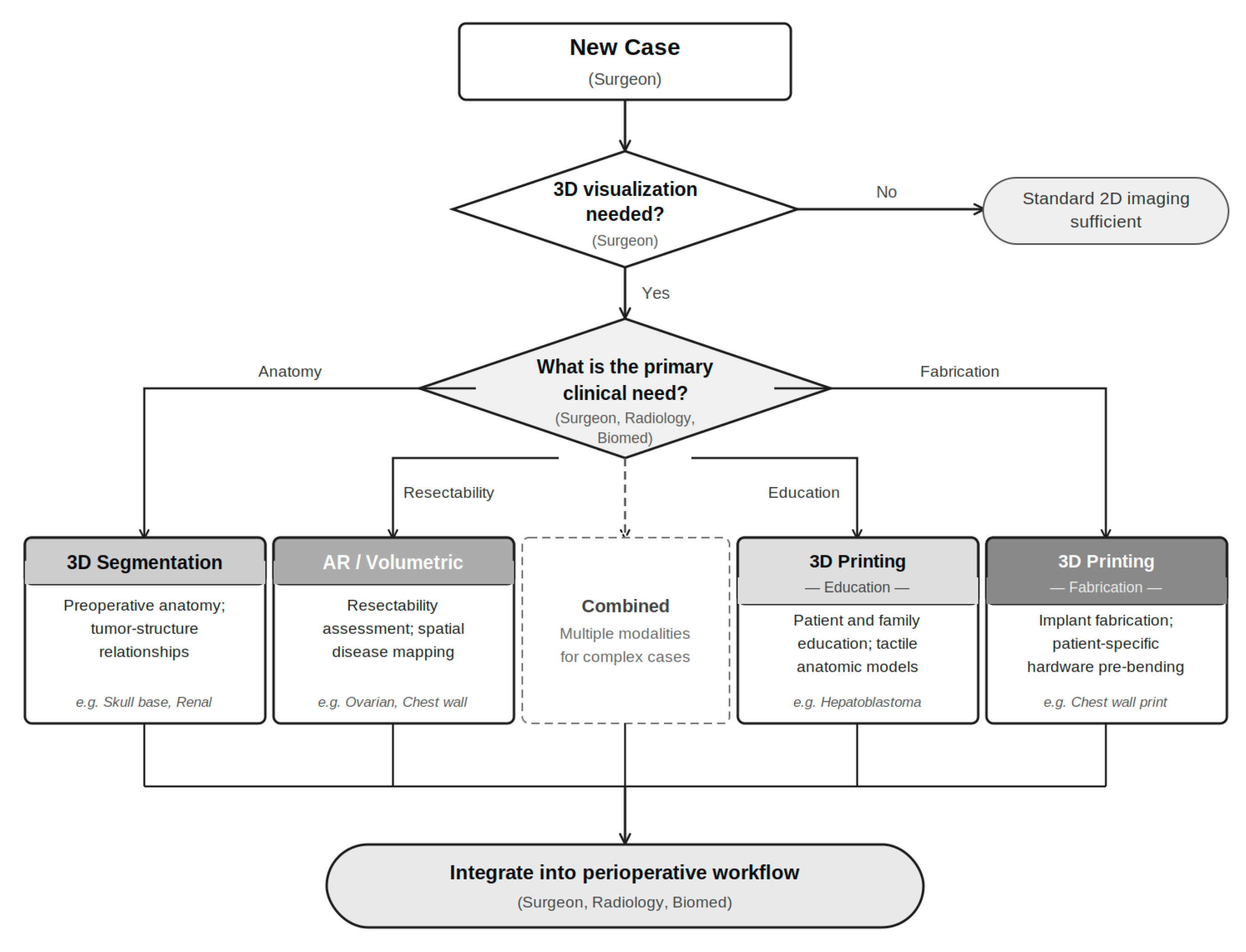
The cases presented in this series (Table 2) illustrate the feasibility of a structured, modality-matched implementation framework for 3D imaging in surgical oncology (as proposed in Figure 4). Rather than advocating for any single technology, the experience described here demonstrates that successful integration depends on matching the appropriate modality to the specific clinical problem at hand. Three distinct implementation roles emerged across the case groups. Segmentation-based 3D visualization was used when there was insufficient representation of the spatial relationships between tumor and normal anatomy on standard 2D imaging, as in the skull base and renal cases, where the relationship between tumor and critical adjacent structures could not be reliably characterized from axial, sagittal, and coronal views alone. AR and volumetric rendering were incorporated into cases requiring dynamic, multiplanar interrogation of disease distribution, as in the ovarian cases, where the clinical question was not anatomical clarity but resectability assessment across a complex peritoneal field. Physical 3D printed models were used when the clinical need was not visualization but fabrication and produced patient-specific replicas of chest wall anatomy to enable prosthetic coverage of the chest wall defect according to the principle of biomimesis [40]. This modality-to-problem framework, rather than any individual technology, is the practical contribution of this implementation experience. This framework is based on institutional experience and has not been prospectively validated; its applicability to other practice settings has not been established.
Figure 4.
Proposed modality selection decision tree for 3D imaging in surgical oncology. This decision tree provides a structured framework for selecting among segmentation-based 3D visualization, physical 3D printing, and augmented reality/volumetric rendering based on the primary clinical objective and is intended to guide surgical teams considering integration of 3D imaging technologies into perioperative workflows. It is based on institutional experience and has not been prospectively validated.
The complementary strengths of these modalities become clearest when each is considered in relation to the clinical problem it was selected to solve. Segmentation-based visualization is most valuable when the obstacle is geometric, such that a tumor’s relationship to adjacent critical structures cannot be reliably understood from sequential 2D slices alone. The skull base cases illustrate this directly: the spatial extent of disease relative to the cribriform plate, lamina papyracea, and dura was mappable only once the anatomy was rendered volumetrically and could be examined from any angle. The renal cases extend this further, as the crossed fused ectopia and endophytic hilar tumors represent anatomical configurations in which 2D review is structurally insufficient and the specific contribution of 3D reconstruction was identification of vascular anatomy that would otherwise have been discovered only intraoperatively. AR and volumetric rendering serve a different function: they are tools for assessing resectability rather than anatomical topography. In the ovarian cases, the clinical question was not tumor location, as standard CT provided that information, but rather whether the distribution was compatible with complete gross resection. AR allowed the surgical team to interrogate that question across the full 3D extent of disease, resolving ambiguity in one patient and better characterizing diaphragmatic involvement in the other. Physical 3D printed models play a different role: their value is tactile and fabricative rather than perceptual. In the chest wall cases, surgeons did not need better visualization; they needed a physical replica of each patient’s rib cage to pre-bend fixation hardware before the operation. In the patient education context, the value of a physical model is that it can be placed in a patient’s or family’s hands, examined without a screen, and pointed to during a clinical conversation, an approach that has been associated with improved patient understanding, stronger shared decision-making, and more effective physician–patient communication [41,42,43]. These are not interchangeable tools deployed for the same purpose; they are distinct instruments suited to distinct problems, and selecting among them requires a structured assessment of what the clinical obstacle actually is.
The implementation of 3D imaging technology in surgical oncology is an inherently multidisciplinary effort. The cases presented here reflect contributions from surgical oncology, radiology, medical physics, and biomedical engineering, as well as the essential involvement of anesthesia, nursing, and patient care coordination teams. Interdisciplinary collaboration was not only helpful but necessary, both for generating anatomically accurate models and for integrating them meaningfully into clinical workflows. Formal validation of accuracy was not performed; model fidelity was assessed qualitatively through comparison with intraoperative findings. Institutions seeking to develop a 3D imaging program should recognize that the technology itself is only one component; equally important is the establishment of coordinated teams, clear communication channels between subspecialties, and defined roles for each contributing discipline.
Despite the promising findings reported here, systematic integration into routine surgical oncology workflows remains inconsistent. Several practical barriers remain. The time and expertise required to generate 3D models, the institutional costs associated with hardware and software, and the absence of standardized protocols for when and how to deploy these tools across cases and institutions all represent meaningful obstacles to widespread adoption, particularly at smaller or resource-limited centers. Automated segmentation tools and streamlined AR platforms may help reduce the time burden, and continued development in these areas will be essential to making 3D imaging accessible beyond high-volume academic cancer centers.
Prospective comparative studies are needed to quantify the clinical benefits of 3D imaging technologies in surgical oncology, with rigorous outcome metrics, including operative time, margin status, complication rates, surgeon confidence, and patient understanding. The cases presented here, while demonstrating consistently favorable outcomes and positive surgeon-reported experience with these tools, represent early institutional experience rather than evidence from controlled trials. Standardized frameworks for evaluating when personalized versus standardized models are most effective, and for determining which modality is best suited to specific clinical scenarios, will be necessary to guide broader implementation. The framework developed and applied at our institution, which encompasses structured data acquisition, segmentation, multidisciplinary review, and resectability assessment, represents one model for how this integration can be operationalized, and may serve as a reproducible starting point for other institutions.
Limitations
Several limitations of this series warrant explicit acknowledgment. First, case selection was retrospective and nonrandomized, and cases in which 3D imaging was used were selected in part because of their clinical complexity, introducing the potential for favorable outcome bias. The consistently positive results reported here may therefore reflect both the utility of 3D tools and the effect of heightened preoperative preparation that accompanies their deployment, rather than an isolated contribution of the technology itself. Second, no comparator group was included; without a parallel cohort managed with standard 2D imaging alone, it is not possible to attribute observed outcomes directly to 3D-assisted planning. Third, the contribution of 3D imaging cannot be fully disentangled from surgeon experience: cases were performed by subspecialty surgeons at a high-volume academic cancer center, and the clinical judgment of experienced operators likely contributed independently to outcomes. Fourth, the workflow described here, encompassing dedicated segmentation software, AR hardware, and multidisciplinary coordination infrastructure, may not be readily replicable at lower-resource institutions, limiting generalizability beyond academic centers with established 3D imaging programs. Finally, patient numbers in most case groups were small, and the series as a whole is insufficient to support statistical inference or definitive conclusions about clinical efficacy. These limitations highlight the need for prospective, controlled studies with predefined outcome metrics to establish the independent contribution of 3D imaging technologies in surgical oncology. Formal follow-up data were not systematically collected across case groups, reflecting the retrospective and implementation-focused nature of this series rather than a prospective outcome study. Renal nephrometry scores were not available for the hilar renal cases included in this series and could not be reported.
The cases presented here demonstrate that 3D imaging technologies are implementable within existing surgical oncology workflows at a high-volume academic center. Whether these tools confer independent clinical benefit beyond the effect of structured preoperative preparation and surgeon experience cannot be determined from this descriptive series. Whether these advantages translate to improved patient outcomes, including event-free and overall survival, warrants prospective evaluation. Continued refinement of these tools, broader application across complex surgical procedures, and investment in training and institutional capacity will be necessary to determine their full clinical value and to expand access beyond high-volume academic centers.
5. Conclusions
Integration of advanced 3D imaging technologies (segmentation, volumetric rendering, 3D printing, and AR visualization) into the surgical oncology workflow is technically feasible, as demonstrated by the cases presented in this descriptive series. These observations are hypothesis-generating and cannot be attributed solely to 3D technology given the retrospective design, lack of measurable outcomes, and absence of a comparator group. Incorporation of these tools into existing perioperative workflows is a practical starting point for institutions seeking to expand access beyond high-volume specialized centers. As these technologies become more accessible and workflows more streamlined, structured selection of the appropriate modality for each clinical problem will be key to maximizing their utility, supporting patient and family education, and assisting surgical oncology teams in the management of complex oncologic cases.
Author Contributions
A.O.: Conceptualization, Data Curation, Investigation, Writing—Original Draft, Writing—Review and Editing, Project Administration. H.H.K.: Data Curation, Investigation. S.M.: Data Curation, Investigation. J.W.J.: Data Curation, Investigation. N.F.: Data Curation, Investigation. J.S.C.: Data Curation, Investigation. T.P.: Methodology, Software, Visualization. S.K.: Methodology, Software, Visualization. J.T.G.: Writing—Review and Editing, Supervision. G.B.: Methodology, Visualization. A.P.: Methodology, Visualization. J.A.C.: Investigation, Writing—Review and Editing. W.S.: Investigation, Writing—Review and Editing. G.R.: Investigation, Writing—Review and Editing. J.N.H.: Conceptualization, Supervision, Writing—Review and Editing. J.S.: Conceptualization, Supervision, Writing—Review and Editing. All authors have read and agreed to the published version of the manuscript.
Funding
This research received no external funding.
Institutional Review Board Statement
Study was reviewed and approved by our Institutional Review Board (protocol #24-243, approved on 17 July 2026) and conducted in accordance with the Declaration of Helsinki.
Informed Consent Statement
Study was granted a waiver from informed consent due to minimal risk to participants.
Data Availability Statement
The datasets used and/or analyzed during the current study are available from the corresponding authors on reasonable request.
Acknowledgments
The authors declare that no AI tools were used in the creation of this article.
Conflicts of Interest
The authors declare no conflicts of interest.
References
- Privitera, L.; Paraboschi, I.; Cross, K.; Giuliani, S. Above and Beyond Robotic Surgery and 3D Modelling in Paediatric Cancer Surgery. Front. Pediatr. 2021, 9, 777840. [Google Scholar] [CrossRef] [Scilit] [PubMed]
- Wellens, L.M.; Meulstee, J.; van de Ven, C.P.; van Scheltinga, C.E.J.T.; Littooij, A.S.; van den Heuvel-Eibrink, M.M.; Fiocco, M.; Rios, A.C.; Maal, T.; Wijnen, M.H.W.A. Comparison of 3-Dimensional and Augmented Reality Kidney Models with Conventional Imaging Data in the Preoperative Assessment of Children with Wilms Tumors. JAMA Netw. Open 2019, 2, e192633. [Google Scholar] [CrossRef] [Scilit] [PubMed]
- González-López, P.; Kuptsov, A.; Gómez-Revuelta, C.; Fernández-Villa, J.; Abarca-Olivas, J.; Daniel, R.T.; Meling, T.R.; Nieto-Navarro, J. The Integration of 3D Virtual Reality and 3D Printing Technology as Innovative Approaches to Preoperative Planning in Neuro-Oncology. J. Pers. Med. 2024, 14, 187. [Google Scholar] [CrossRef] [Scilit]
- Esperto, F.; Prata, F.; Autrán-Gómez, A.M.; Rivas, J.G.; Socarras, M.; Marchioni, M.; Albisinni, S.; Cataldo, R.; Scarpa, R.M.; Papalia, R. New Technologies for Kidney Surgery Planning 3D, Impression, Augmented Reality 3D, Reconstruction: Current Realities and Expectations. Curr. Urol. Rep. 2021, 22, 35. [Google Scholar] [CrossRef] [Scilit] [PubMed]
- Alemayehu, D.G.; Zhang, Z.; Tahir, E.; Gateau, D.; Zhang, D.-F.; Ma, X. Preoperative Planning Using 3D Printing Technology in Orthopedic Surgery. BioMed Res. Int. 2021, 2021, 7940242. [Google Scholar] [CrossRef] [Scilit] [PubMed]
- van de Woestijne, P.C.; Bakhuis, W.; Sadeghi, A.H.; Peek, J.J.; Taverne, Y.J.; Bogers, A.J. 3D Virtual Reality Imaging of Major Aortopulmonary Collateral Arteries: A Novel Diagnostic Modality. World J. Pediatr. Congenit. Heart Surg. 2021, 12, 765–772. [Google Scholar] [CrossRef] [Scilit] [PubMed]
- Tejo-Otero, A.; Buj-Corral, I.; Fenollosa-Artés, F. 3D Printing in Medicine for Preoperative Surgical Planning: A Review. Ann. Biomed. Eng. 2019, 48, 536–555. [Google Scholar] [CrossRef] [Scilit]
- Magalhães, R.; Oliveira, A.; Terroso, D.; Vilaça, A.; Veloso, R.; Marques, A.; Pereira, J.; Coelho, L. Mixed Reality in the Operating Room: A Systematic Review. J. Med. Syst. 2024, 48, 76. [Google Scholar] [CrossRef] [Scilit] [PubMed]
- Nadeem-Tariq, A.; Kazemeini, S.; Kaur, P.; Dang, G.; Davis, T.; Sraa, K.; Zitser, P.; Fang, C. Augmented Reality in Spine Surgery: A Narrative Review of Clinical Accuracy, Workflow Efficiency, and Barriers to Adoption. Cureus 2025, 17, e86803. [Google Scholar] [CrossRef] [Scilit]
- Cronin, U.M.; Cummins, N.M.; O’SUllivan, A.; O’SUllivan, L. Perceived barriers and opportunities to the use of 3D printing in a healthcare system with low adoption: A semi-structured interview study. HRB Open Res. 2025, 8, 35. [Google Scholar] [CrossRef] [Scilit]
- Fedorov, A.; Beichel, R.; Kalpathy-Cramer, J.; Finet, J.; Fillion-Robin, J.-C.; Pujol, S.; Bauer, C.; Jennings, D.; Fennessy, F.; Sonka, M.; et al. 3D Slicer as an Image Computing Platform for the Quantitative Imaging Network. Magn. Reson. Imaging 2012, 30, 1323–1341. [Google Scholar] [CrossRef] [Scilit]
- He, H.; Yu, C.; Yang, Y.; Maessen, J.G.; Nia, P.S. Three-dimensional reconstruction and virtual simulation of patient-specific anatomy for procedural planning in thoracoscopic segmentectomy: A systematic review and meta-analysis. Eur. J. Cardio-Thorac. Surg. 2025, 67, ezaf283. [Google Scholar] [CrossRef] [Scilit] [PubMed]
- Mardis, N.J. Emerging Technology and Applications of 3D Printing in the Medical Field. Mo. Med. 2018, 115, 368–373. [Google Scholar] [PubMed]
- Tack, P.; Victor, J.; Gemmel, P.; Annemans, L. 3D-printing techniques in a medical setting: A systematic literature review. Biomed. Eng. Online 2016, 15, 115. [Google Scholar] [CrossRef] [Scilit] [PubMed]
- Meyer-Szary, J.; Luis, M.S.; Mikulski, S.; Patel, A.; Schulz, F.; Tretiakow, D.; Fercho, J.; Jaguszewska, K.; Frankiewicz, M.; Pawłowska, E.; et al. The Role of 3D Printing in Planning Complex Medical Procedures and Training of Medical Professionals—Cross-Sectional Multispecialty Review. Int. J. Environ. Res. Public Health 2022, 19, 3331. [Google Scholar] [CrossRef] [Scilit] [PubMed]
- Valls-Esteve, A.; Adell-Gómez, N.; Pasten, A.; Barber, I.; Munuera, J.; Krauel, L. Exploring the Potential of Three-Dimensional Imaging, Printing, and Modeling in Pediatric Surgical Oncology: A New Era of Precision Surgery. Children 2023, 10, 832. [Google Scholar] [CrossRef] [Scilit] [PubMed]
- Youn, J.K.; Park, S.J.; Choi, Y.-H.; Han, J.-W.; Ko, D.; Byun, J.; Yang, H.-B.; Kim, H.-Y. Application of 3D printing technology for pre-operative evaluation, education and informed consent in pediatric retroperitoneal tumors. Sci. Rep. 2023, 13, 1671. [Google Scholar] [CrossRef] [Scilit] [PubMed]
- Sánchez-Sánchez, Á.; Girón-Vallejo, Ó.; Ruiz-Pruneda, R.; Fernandez-Ibieta, M.; García-Calderon, D.; Villamil, V.; Giménez-Aleixandre, M.C.; Montoya-Rangel, C.A.; Bermejo, J.P.H. Three-Dimensional Printed Model and Virtual Reconstruction: An Extra Tool for Pediatric Solid Tumors Surgery. Eur. J. Pediatr. Surg. Rep. 2018, 06, e70–e76. [Google Scholar] [CrossRef] [Scilit] [PubMed]
- Schulze, M.; Gosheger, G.; Bockholt, S.; De Vaal, M.; Budny, T.; Tönnemann, M.; Pützler, J.; Bövingloh, A.S.; Rischen, R.; Hofbauer, V.; et al. Complex Bone Tumors of the Trunk—The Role of 3D Printing and Navigation in Tumor Orthopedics: A Case Series and Review of the Literature. J. Pers. Med. 2021, 11, 517. [Google Scholar] [CrossRef] [Scilit] [PubMed]
- Parthasarathy, J.; Jonard, B.; Rees, M.; Selvaraj, B.; Scharschmidt, T. Virtual surgical planning and 3D printing in pediatric musculoskeletal oncological resections: A proof-of-concept description. Int. J. Comput. Assist. Radiol. Surg. 2022, 18, 95–104. [Google Scholar] [CrossRef] [Scilit] [PubMed]
- Shah, N.R.; Weadock, W.J.; Williams, K.M.; Moreci, R.; Stoll, T.; Joshi, A.; Petroze, R.; Newman, E.A. Use of modern three-dimensional imaging models to guide surgical planning for local control of pediatric extracranial solid tumors. Pediatr. Blood Cancer 2024, 71, e30933. [Google Scholar] [CrossRef] [Scilit] [PubMed]
- Cheng, J.; Wang, Z.F.; Yao, W.F.; Liu, J.W.; Lu, Y.; Wang, Q.; Cai, X.J. Comparison of 3D printing model to 3D virtual reconstruction and 2D imaging for the clinical education of interns in hepatocellular carcinoma: A randomized controlled study. J. Gastrointest. Oncol. 2023, 14, 325–333. [Google Scholar] [PubMed]
- Mehrzad, R.; Denour, E.; Crozier, J.; Woo, A.S. The Use of In-House Patient-Specific 3D Printed Models in Plastic-, Orthopedic and Neurosurgical Planning: A Preliminary Report. J. 3D Print. Med. 2023, 7, 3DP14. [Google Scholar] [CrossRef] [Scilit]
- Marconi, S.; Pugliese, L.; Botti, M.; Peri, A.; Cavazzi, E.; Latteri, S.; Auricchio, F.; Pietrabissa, A. Value of 3D printing for the comprehension of surgical anatomy. Surg. Endosc. 2017, 31, 4102–4110. [Google Scholar] [CrossRef] [Scilit] [PubMed]
- Fusco, J.C.; Abdelhafeez, A.H.; Krauel, L.; Honeyman, J.N.; Ehrlich, P.F.; Wijnen, M.; Lautz, T.B.; Pachl, M.; Malek, M.M. Imaging adjuvants in pediatric surgical oncology. Pediatr. Blood Cancer 2024, 72, e31241. [Google Scholar] [CrossRef] [Scilit] [PubMed]
- Shu, X.-J.; Wang, Y.; Xin, H.; Zhang, Z.-Z.; Xue, Z.; Wang, F.-Y.; Xu, B.-N. Real-time augmented reality application in presurgical planning and lesion scalp localization by a smartphone. Acta Neurochir. 2021, 164, 1069–1078. [Google Scholar] [CrossRef] [Scilit] [PubMed]
- Checcucci, E.; Amparore, D.; Pecoraro, A.; Peretti, D.; Aimar, R.; DE Cillis, S.; Piramide, F.; Volpi, G.; Piazzolla, P.; Manfrin, D.; et al. 3D mixed reality holograms for preoperative surgical planning of nephron-sparing surgery: Evaluation of surgeons’ perception. Minerva Urol. Nephrol. 2021, 73, 367–375. [Google Scholar] [CrossRef] [Scilit] [PubMed]
- Khor, W.S.; Baker, B.; Amin, K.; Chan, A.; Patel, K.; Wong, J. Augmented and virtual reality in surgery—The digital surgical environment: Applications, limitations and legal pitfalls. Ann. Transl. Med. 2016, 4, 454. [Google Scholar] [CrossRef] [Scilit] [PubMed]
- Colcuc, C.; Miersbach, M.; Cienfuegos, M.; Grüneweller, N.; Vordemvenne, T.; Wähnert, D. Comparison of virtual reality and computed tomography in the preoperative planning of complex tibial plateau fractures. Arch. Orthop. Trauma Surg. 2024, 144, 2631–2639. [Google Scholar] [CrossRef] [Scilit] [PubMed]
- Eisenhauer, E.L.; Abu-Rustum, N.R.; Sonoda, Y.; Aghajanian, C.; Barakat, R.R.; Chi, D.S. The effect of maximal surgical cytoreduction on sensitivity to platinum-taxane chemotherapy and subsequent survival in patients with advanced ovarian cancer. Gynecol. Oncol. 2008, 108, 276–281. [Google Scholar] [CrossRef] [Scilit] [PubMed]
- Chi, D.S.; Eisenhauer, E.L.; Zivanovic, O.; Sonoda, Y.; Abu-Rustum, N.R.; Levine, D.A.; Guile, M.W.; Bristow, R.E.; Aghajanian, C.; Barakat, R.R. Improved progression-free and overall survival in advanced ovarian cancer as a result of a change in surgical paradigm. Gynecol. Oncol. 2009, 114, 26–31. [Google Scholar] [CrossRef] [Scilit] [PubMed]
- Tseng, J.H.; Cowan, R.A.; Zhou, Q.; Iasonos, A.; Byrne, M.; Polcino, T.; Polen-De, C.; Gardner, G.J.; Sonoda, Y.; Zivanovic, O.; et al. Continuous improvement in primary Debulking surgery for advanced ovarian cancer: Do increased complete gross resection rates independently lead to increased progression-free and overall survival? Gynecol. Oncol. 2018, 151, 24–31. [Google Scholar] [CrossRef] [Scilit] [PubMed]
- Chi, D.S.; Liao, J.B.; Leon, L.F.; Venkatraman, E.S.; Hensley, M.L.; Bhaskaran, D.; Hoskins, W.J. Identification of Prognostic Factors in Advanced Epithelial Ovarian Carcinoma. Gynecol. Oncol. 2001, 82, 532–537. [Google Scholar] [CrossRef] [Scilit] [PubMed]
- Kahn, R.M.; Chang, S.-J.; Chi, D.S. Extra-abdominal cytoreductive techniques in ovarian cancer: How far can (should) we go? Int. J. Gynecol. Cancer 2024, 34, 379–385. [Google Scholar] [CrossRef] [Scilit] [PubMed]
- Straubhar, A.; Chi, D.S.; Roche, K.L. Update on the Role of Surgery in the Management of Advanced Epithelial Ovarian Cancer. Clin. Adv. Hematol. Oncol. 2020, 18, 723–731. [Google Scholar] [PubMed]
- Bhatt, K.; Herts, B.R. Crossed fused renal ectopia. J. Urol. 2014, 191, 475–476. [Google Scholar] [CrossRef] [Scilit] [PubMed]
- Martinez-Ferro, M.; Bellia-Munzon, G.; Toselli, L.; Sanjurjo, D. The role of 3D printing in chest wall reconstruction. J. Thorac. Dis. 2024, 16, 4053–4063. [Google Scholar] [CrossRef] [Scilit] [PubMed]
- Wang, W.; Yang, S.; Han, M.; Liu, H.; Feng, Q.; Su, Y.; Han, Y.; Wang, J. Three-dimensional printed titanium chest wall reconstruction for tumor removal in the sternal region. J. Cardiothorac. Surg. 2024, 19, 579. [Google Scholar] [CrossRef] [Scilit] [PubMed]
- Goldsmith, I. 3D bioprinting the human chest wall: Fiction or fact. npj Biomed. Innov. 2024, 1, 2. [Google Scholar] [CrossRef] [Scilit] [PubMed]
- Rocco, G. Chest Wall Resection and Reconstruction According to the Principles of Biomimesis. Semin. Thorac. Cardiovasc. Surg. 2011, 23, 307–313. [Google Scholar] [CrossRef] [Scilit] [PubMed]
- Stewart, M.A. Effective physician-patient communication and health outcomes: A review. Can. Med. Assoc. J. 1995, 152, 1423–1433. [Google Scholar]
- Zolnierek, K.B.; Dimatteo, M.R. Physician communication and patient adherence to treatment: A meta-analysis. Med. Care 2009, 47, 826–834. [Google Scholar] [CrossRef] [Scilit] [PubMed]
- Ha, J.F.; Longnecker, N. Doctor-patient communication: A review. Ochsner. J. 2010, 10, 38–43. [Google Scholar] [PubMed]

Disclaimer/Publisher’s Note: The statements, opinions and data contained in all publications are solely those of the individual author(s) and contributor(s) and not of MDPI and/or the editor(s). MDPI and/or the editor(s) disclaim responsibility for any injury to people or property resulting from any ideas, methods, instructions or products referred to in the content. |
© 2026 by the authors. Licensee MDPI, Basel, Switzerland. This article is an open access article distributed under the terms and conditions of the Creative Commons Attribution (CC BY) license.