


Simple Summary
Breast cancer patients whose disease has spread to axillary lymph nodes usually receive chemotherapy before surgery. In some patients, this treatment fully clears the cancer from the axillary lymph nodes, meaning that removing them surgically may not be necessary. However, currently it is not yet possible to reliably identify these patients prior to surgery. This study examined whether measurements from [18F]FDG PET/CT at diagnosis, combined with the amount of tumour-infiltrating lymphocytes, could help predict which patients would have their axillary lymph nodes cleared of cancer after chemotherapy. Among 135 patients, a model combining tumour features, [18F]FDG PET/CT measurements, and tumour-infiltrating lymphocytes information predicted this outcome with good accuracy. These findings suggest that such a model could eventually help identify patients who may safely avoid axillary lymph node surgery; however, this needs to be confirmed in future research.
Abstract
Background: Accurate axillary response prediction is clinically relevant in clinically node-positive (cN+) breast cancer patients treated with neoadjuvant chemo-(immuno)therapy (NAC). This study evaluated the value of [18F]FDG PET/CT-derived metabolic characteristics and tumour-infiltrating lymphocytes (TILs) to predict axillary pathologic complete response (pCR). Second, the association between [18F]FDG PET/CT-derived metabolic characteristics and TILs was evaluated. Methods: This retrospective study included cN+ breast cancer patients treated with NAC and axillary surgery from the prospective multicentre RISAS trial (NCT02800317). Baseline [18F]FDG PET/CT-derived metabolic activity of the breast tumour, axillary lymph node metastases (ALNMs), and spleen was assessed, together with TILs measured in the breast tumour and ALNMs. TILs were classified as low (<10%) versus high (≥10%). Associations were evaluated using multivariable logistic regression and receiver operating characteristic analysis. Results: Of 135 patients, 46 (34.1%) achieved axillary pCR. Higher liver mean tumour-to-background ratio (2.7 vs. 1.9, p = 0.042), trends towards higher breast and ALNM maximum standardised uptake value (SUVmax), lower splenic SUVmax and high breast tumour TILs were associated with axillary pCR. Multivariable logistic regression combining tumour grade, subtype, maximum spleen-to-liver ratio, and breast tumour TILs yielded an apparent area under the curve of 0.80 [95%CI 0.73–0.88]. Higher ALNM SUVmax and SUVmean (10.5 vs. 6.3, p = 0.024; 6.4 vs. 3.7, p = 0.015) and lower splenic SUVmean (2.6 vs. 3.1, p = 0.035) were associated with high breast tumour TILs. Conclusions: A model combining clinicopathological, metabolic, and immune-related characteristics may be informative for predicting axillary response to NAC but did not perform better than clinicopathological characteristics alone. Future studies should evaluate whether accurate prediction models can support safe axillary treatment de-escalation.
1. Introduction
In recent years, patients with clinically node-positive (cN+) breast cancer are more frequently treated with neoadjuvant chemo-(immunotherapy) (NAC) [1]. In about one third of cN+ breast cancer patients, NAC results in a pathologic complete response (pCR) of axillary lymph node metastases (ALNMs) [2,3]. Achieving axillary pCR may have important therapeutic implications, potentially de-escalating axillary surgery and adjuvant treatment.
In current clinical practice, less invasive axillary surgical staging procedures, such as sentinel lymph node biopsy (SLNB), marking axillary lymph nodes with an iodine seed (MARI) or targeted axillary dissection (TAD), are preferred over axillary lymph node dissection (ALND) after NAC [2,4,5]. The goal of axillary surgical staging is to identify axillary pCR after NAC in cN+ breast cancer patients. However, accurate response prediction before and during NAC may allow the identification of selected patients who will achieve axillary pCR, for whom axillary surgical staging could therefore be safely omitted.
Previous studies have tried to predict the response of breast tumours and ALNMs to NAC using imaging modalities and histopathological information. [18F]fluorodeoxyglucose ([18F]FDG) positron emission tomography/computed tomography (PET/CT) imaging can be considered in cN+ breast cancer for distant staging at diagnosis, as recommended by guidelines of the NCCN, EANM, and SNMMI [6,7]. [18F]FDG PET/CT provides insight into the metabolic activity of physiological and pathological cells [8]. Previous studies found that metabolic activity measured in tumour tissue may serve as an indirect marker of proliferation and oncological aggressiveness, providing predictive information regarding axillary pCR within breast cancer subtypes [9,10,11]. Alongside evaluating [18F]FDG uptake in tumour cells, immune-related organs such as the spleen are increasingly being investigated on [18F]FDG PET/CT as an indirect marker of systemic immune activity against tumour tissue. Multiple studies have demonstrated high splenic metabolic activity as a predictor of shorter survival in breast cancer and other cancer types [12,13,14,15]. In addition, tumour-infiltrating lymphocytes (TILs) are frequently used as a pathological marker that measures the immune response to tumour cells and serves as an independent predictor of response to NAC in breast cancer patients [16,17]. Previous studies have reported increased levels of breast tumour TILs to be correlated with an enhanced likelihood of pCR after NAC and a better recurrence-free survival, particularly in human epidermal growth factor receptor 2 (HER2)-positive and triple negative (TN) breast cancer subtypes [16,17,18]. However, while breast tumour TILs have been extensively studied, the assessment of pre-NAC ALNM TILs remains rarely performed in breast cancer, and their role in response prediction is still unknown.
Only a few studies have investigated the correlation between metabolic characteristics derived from [18F]FDG PET/CT and the level of TILs [19,20,21,22]. In general, these studies found weak-to-moderate correlations, with higher breast tumour metabolic activity corresponding with higher levels of breast tumour TILs, particularly in patients with HER2-positive and TN breast cancer [19,21,22]. In addition, the interplay between the systemic immune response, reflected by splenic metabolic activity, and the tumour-associated immune response, reflected by TILs, has been explored in only a few studies. Tamam et al. found a positive correlation between splenic metabolic activity and breast tumour TILs in stage 4 nonluminal breast cancer patients [20]. In contrast, Seban et al. observed an inverse association, demonstrating that a higher spleen-to-liver ratio (SLR) was associated with lower breast tumour TILs [15]. However, the combined potential of [18F]FDG PET/CT-derived metabolic characteristics and TILs to predict axillary pathological response to NAC has yet to be investigated.
Therefore, this study aimed to evaluate the value of baseline [18F]FDG PET/CT-derived metabolic activity of the breast tumour, ALNMs, and the spleen, as well as levels of pre-NAC TILs measured in the breast tumour and ALNMs, to predict axillary pathologic response in cN+ breast cancer patients treated with NAC overall and according to breast cancer subtype. Second, the association between [18F]FDG PET/CT-derived metabolic characteristics and TILs was evaluated.
2. Methods
2.1. Patients
This study presents a retrospective analysis of data collected prospectively in the multicentre RISAS trial (NCT02800317; Registration date: 15 June 2016; METC number 2016-412). Participants were women (≥18 years old) diagnosed with cN+ breast cancer and treated with NAC. NAC was administered according to national guidelines and generally consisted of anthracycline and/or taxanes. HER2-positive breast cancer patients received HER2-targeted therapy, consisting of trastuzumab with or without pertuzumab. Afterwards, surgery of the breast (except in patients with occult breast cancer) and ALNMs was performed, consisting of breast-conserving surgery or mastectomy combined with the RISAS procedure (SLNB + MARI) followed by completion ALND (cALND). Patients were excluded if they had infraclavicular or supraclavicular lymph node metastases (cN3a/cN3c), (oligo)metastatic breast cancer, SLNB performed before NAC or a history of radiotherapy to the ipsilateral axilla. Participants were recruited from 13 centres across the Netherlands between 1 March 2017 and 31 December 2019. Informed consent was obtained from all participants. Additional details regarding the RISAS trial have been previously published [5,23].
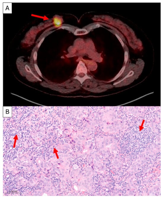
This retrospective study included all RISAS participants who underwent baseline whole-body [18F]FDG PET/CT imaging and had a core needle biopsy (CNB) of the breast tumour and/or CNB of ALNMs. Fine-needle aspiration cytology specimens of ALNMs were excluded. Breast pCR was defined as the absence of any residual invasive or in situ disease in the breast (ypT0). Axillary pCR was defined as the absence of any residual disease in the axilla, including the absence of isolated tumour cells (ypN0). [18F]FDG PET/CT imaging and TILs were centrally evaluated specifically for the purpose of this retrospective study (Figure 1).
Figure 1.
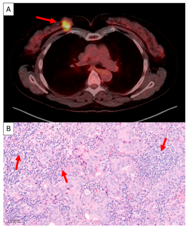
A 61-year-old patient diagnosed with HR-positive/HER2-negative grade 2 breast cancer. (A) [18F]FDG PET/CT exam showing high metabolic activity of the breast tumour (SUVmax = 14.07; red arrow). (B) Core needle biopsy of the breast tumour demonstrating high TILs (90%; red arrows). HR, hormone receptor; HER2, human epidermal growth factor receptor 2; SUVmax, maximum standardised uptake value; TILs, tumour-infiltrating lymphocytes.
2.2. [18F]FDG PET/CT Imaging
All included patients underwent whole-body [18F]FDG PET/CT imaging in supine position before the start of NAC, using a standard acquisition protocol. Blood glucose levels had to be <11 mmol/L after a fasting period of at least 4–6 h before injection of [18F]FDG. Following blood glucose assessment, [18F]FDG was administered intravenously to all patients. [18F]FDG PET/CT imaging was performed after a resting period of 45–60 min. All included [18F]FDG PET/CT exams were reconstructed according to European Association of Nuclear Medicine Research Ltd. (EARL) guidelines [6,24,25].
[18F]FDG PET/CT images were reviewed by a dedicated nuclear radiologist (T.v.N) with 6 years of clinical experience using a commercially available workstation (syngo.via 10; Siemens Healthcare, Erlangen, Germany). The maximum standardised uptake value (SUVmax) and SUVmean of the breast tumour, most hypermetabolic ALN, contralateral breast, mediastinal blood pool, liver, and spleen were measured. In addition, the metabolic tumour volume (MTV) of the breast tumour was measured. In case of multifocality of the breast tumour, the SUVmax, SUVmean, and MTV of the most hypermetabolic lesion were assessed. The number of hypermetabolic ALNs was assessed by the reader (T.v.N.) and then compared with the number of ALNs reported by the initial institution. When the number of hypermetabolic ALNs was missing from the original report or when there was a difference between the number of hypermetabolic ALNs in the original report and the first reviewer, a second reviewer (C.M.) evaluated the images to obtain consensus. The nodal-to-tumour ratio (NT-ratio) was calculated by dividing the SUVmax or SUVmean of the most hypermetabolic ALN by the SUVmax or SUVmean of the breast tumour. Liver SUVmax and SUVmean values were obtained by placing a 30 mm diameter volume of interest (VOI) in the right lobe of the liver while avoiding abnormalities such as cysts, haemangiomas, or other structural lesions. For the contralateral breast measurement, a VOI was placed in evidently normal breast tissue on the side opposite to the tumour, positioned as symmetrically as possible relative to the affected breast. In case of bilateral breast cancer, a region clearly free of tumour involvement was selected. For the mediastinal blood pool measurement, a VOI was positioned in the ascending aorta just proximal to the aortic arch, avoiding the aortic wall to prevent measurement bias. The tumour-to-background ratio (TBR) was calculated by dividing the SUVmax or SUVmean of the breast tumour by the SUVmax or SUVmean of the background tissue (liver, contralateral breast, or mediastinal blood pool). Splenic SUVmax and SUVmean values were measured by placing a 20 mm diameter spherical VOI in the central part of the spleen on the axial slice showing the largest cross-section of the spleen [26,27]. The SLR was calculated by dividing the SUVmax or SUVmean of the spleen by the SUVmax or SUVmean of the liver.
2.3. Pathologic Evaluation
All available Haematoxylin and Eosin (H&E)-stained tissue sections of the breast and ALNM CNBs were centrally collected (PALGA 2022-109-A1-6) and digitised (Pannoramic p1000 scanner, 3DHistech, Budapest, Hungary). The percentage of stromal TILs was determined on all available breast tumour and ALNM CNBs. TILs were scored according to the International TILs working group guideline by an experienced breast pathologist with 12 years of experience in breast pathology (L.K.), blinded to additional clinical information [28]. Reporting TILs in ALNMs is challenging, because leukocytes are part of the normal lymph node structure. Only the lymphocytes in altered stroma surrounding the tumour cells were registered [19,28]. TILs were later classified as low (<10%) versus high (≥10%) [29,30]. All evaluations were performed using SlideViewer (Version 2.6, 3DHistech, Budapest, Hungary).
2.4. Statistical Analysis
Statistical analysis of the data was performed using the Statistical Package for the Social Sciences (Version 28, IBM Corp., Armonk, NY, USA). Continuous variables were described as mean ± standard deviation (SD) or median (range) and compared using the unpaired t-test or Mann–Whitney U test. Categorical variables were described as absolute numbers and percentages and compared using Pearson’s Chi-square test. Univariable and multivariable logistic regression analyses were performed to determine factors that influence axillary pCR. Associations were expressed as odds ratios (ORs) with 95% confidence intervals (CI) and p-values. Splenic metabolic activity and TILs were included in the multivariable logistic regression model, given their predefined clinical relevance and the aim of the study. Additional clinicopathological variables showing a significant association in univariable regression (p < 0.05) were selected for the multivariable logistic regression model, while taking into account recommendations regarding the number of events per variable (EPV) to limit model overfitting [31,32]. Receiver operating characteristic (ROC) curves were constructed, and areas under the curve (AUC) were computed to see how accurately the combined variables could discriminate between axillary pCR and axillary residual disease. Pre-NAC SLRmax and breast tumour TIL levels were compared between patients with and without axillary pCR within each breast cancer subtype using the Mann–Whitney U test. All statistical tests were two-sided, and a p-value < 0.05 was considered statistically significant.
3. Results
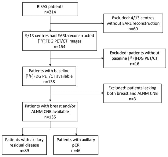
A total of 214 patients were included in the RISAS trial. After excluding patients from centres without EARL-reconstructed [18F]FDG PET/CT exams, patients without a baseline [18F]FDG PET/CT exam, and patients lacking both breast tumour and ALNM CNB for TIL assessment, 135 patients were included in this retrospective analysis (Figure 2). All patients underwent surgery of ALNMs after NAC. The overall breast and axillary pCR rates were 33.6% (45/134) and 34.1% (46/135), respectively. A total of 29.1% (39/134) achieved combined pCR of both breast and axilla. Patient and tumour characteristics, according to axillary pCR status, are presented in Table 1. Axillary pCR was significantly more often achieved in patients with oestrogen receptor (ER)-negative, progesterone receptor (PR)-negative, HER2-positive, and grade 3 tumours.
Figure 2.
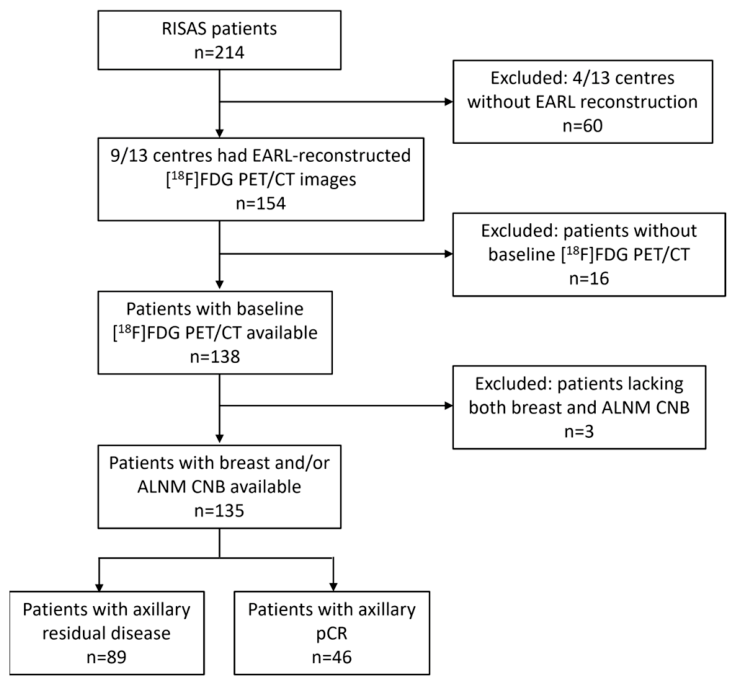
Flowchart showing patient selection process. RISAS, radioactive iodine seed placement in the axilla with sentinel lymph node biopsy; EARL, European Association of Nuclear Medicine Research Ltd.; ALNM, axillary lymph node metastases; CNB, core needle biopsy; pCR, pathological complete response.
Table 1.
Patient and tumour characteristics according to axillary pCR status.
Table 2 presents the [18F]FDG PET/CT imaging characteristics according to axillary pCR status. Among all evaluated characteristics, only TBRmean of the liver was significantly higher in patients who achieved axillary pCR (2.7 (range 0.2–6.7) vs. 1.9 (range 0.3–6.3), p = 0.042). For the remaining breast tumour and ALNM metabolic characteristics, only non-significant trends were observed, suggesting higher metabolic activity in patients who achieved axillary pCR. However, upon stratification by breast cancer subtype, this pattern shifted, with a trend towards lower breast tumour SUVmax values being associated with axillary pCR in all subtypes except TN breast cancer (Online Resource 1). Additionally, although not statistically significant, both splenic SUVmax and SLR demonstrated a trend towards lower metabolic activity in patients who achieved axillary pCR (Table 2 and Online Resource 1).
Table 2.
[18F]FDG PET/CT imaging characteristics according to axillary pCR status.
High breast tumour TILs (≥10%) were more frequently observed in patients with axillary pCR than in patients with axillary residual disease (71.1% vs. 55.3%; p = 0.079). No association was observed between ALNM TILs and axillary pCR (50% vs. 61.5%, p = 0.382) (Table 3).
Table 3.
Association between breast tumour and ALNM TIL levels and axillary pCR status.
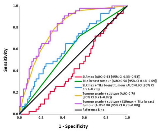
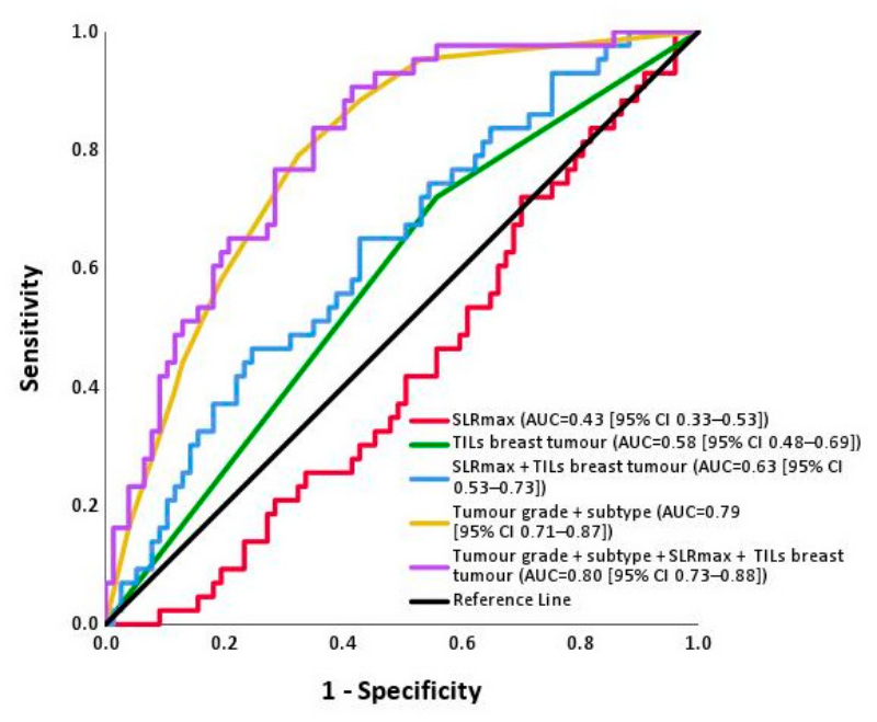
Results of univariable and multivariable logistic regression analyses of factors associated with axillary pCR are presented in Table 4. Multivariable logistic regression combining tumour grade (grade 3 vs. 1–2; OR 2.19 [95% CI 0.87–5.55], p = 0.097), breast cancer subtype (Ref HR+/HER2−-; HR+/HER2+ OR 10.38 [95% CI 2.95–36.46], p < 0.001; HR−-/HER2+ OR 13.66 [95% CI 2.84–65.75], p = 0.001; TN OR 5.13 [95% CI 1.44–18.29], p = 0.012), SLRmax (OR 0.01 [95% CI 0.00–2.74], p = 0.111), and breast tumour TILs (OR 1.25 [95% CI 0.45–3.48], p = 0.666), resulted in an apparent AUC of 0.80 [95% CI 0.73–0.88]; Figure 3). In comparison, the multivariable model including only SLRmax and breast tumour TILs resulted in an apparent AUC of 0.63 [95% CI 0.53–0.73], while the multivariable model including only breast cancer subtype and tumour grade resulted in an apparent AUC of 0.79 [95% CI 0.71–0.87].
Table 4.
Univariable and multivariable logistic regression analysis of characteristics associated with axillary pCR.
Figure 3.
Receiver operating characteristic (ROC) curve analysis illustrating the discriminative ability of SLRmax, TILs breast tumour, SLRmax + TILs breast tumour, tumour grade + breast cancer subtype, and the multivariable model of Table 4 (Tumour grade + breast cancer subtype + SLRmax + TILs breast tumour) for axillary pCR. AUC, area under the curve; TILs, tumor-infiltrating lymphocytes; SLR, spleen-to-liver ratio; pCR, pathologic complete response.
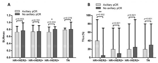
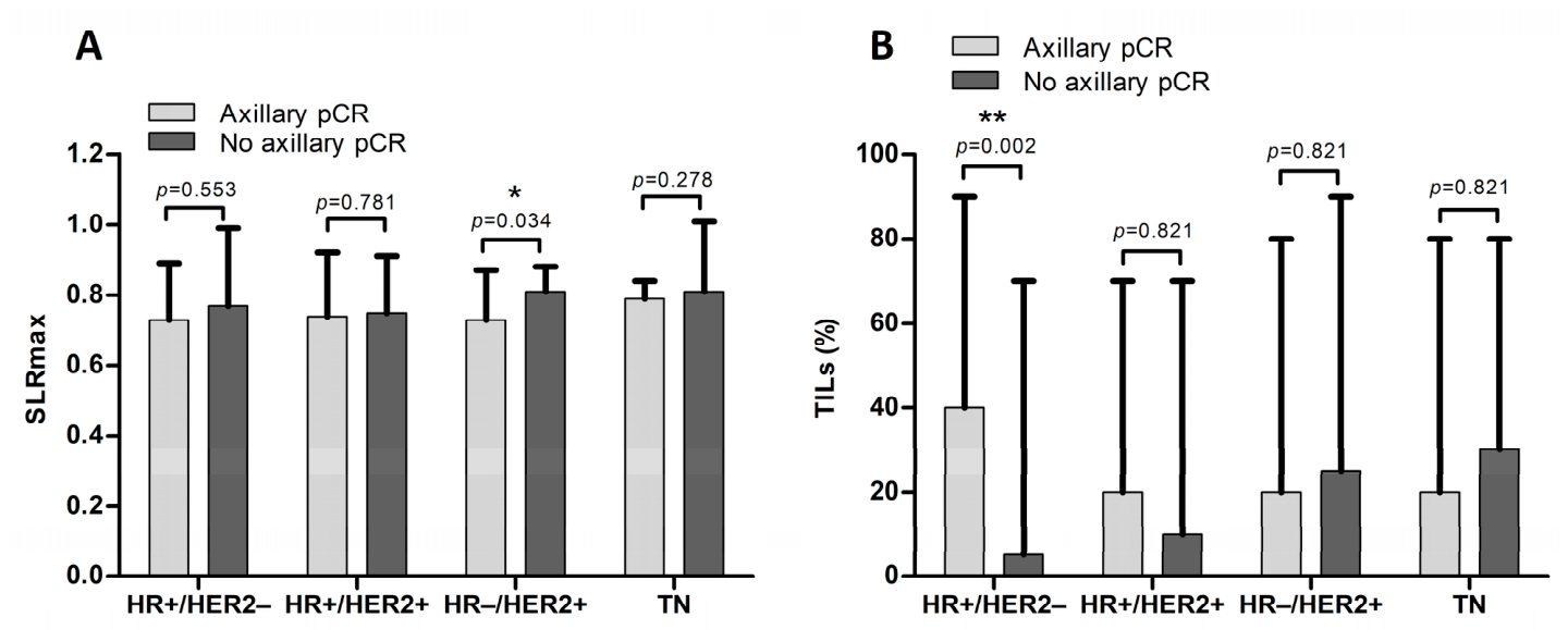
Figure 4 illustrates pre-NAC SLRmax and breast tumour TILs according to axillary pCR rates across breast cancer subtypes. Within the HR-negative/HER2-positive subtype, baseline SLRmax was significantly lower in patients achieving axillary pCR (p = 0.034; Figure 4A). The percentage of pre-NAC breast tumour TILs was significantly higher in patients achieving axillary pCR within the HR-positive/HER2-negative subtype (p = 0.002; Figure 4B). No significant differences in SLRmax and breast tumour TILs were observed between patients with and without axillary pCR in the other subtypes.
Figure 4.
(A) Baseline maximum spleen-to-liver ratio (SLRmax) across patients with axillary pCR (light grey) and without axillary pCR (dark grey) according to breast cancer subtypes. (B) Percentage of pre-NAC TILs of the breast tumour (continuous) across patients with axillary pCR (light grey) and without axillary pCR (dark grey) according to breast cancer subtypes. pCR, pathologic complete response; SLR, spleen-to-liver ratio; HR, hormone receptor; HER2, human epidermal growth factor receptor 2; TN, triple negative; NAC, neoadjuvant chemo-(immuno)therapy; TILs, tumour-infiltrating lymphocytes. * p < 0.05; ** p < 0.01.
Table 5 summarises the association between [18F]FDG PET/CT-derived metabolic characteristics and breast tumour and ALNM TILs. For breast tumour [18F]FDG PET/CT-derived characteristics, SUVmax, SUVmean, MTV, TBR liver, TBR blood pool and TBR contralateral breast were all higher in patients with high breast tumour TILs; however, none of these differences reached statistical significance. Metabolic activity in ALNMs was significantly higher in patients with high breast tumour TILs (≥10%), reflected by an increased SUVmax (10.5 vs. 6.3, p = 0.024), SUVmean (6.4 vs. 3.7, p = 0.015), and NT-ratio (0.8 (0.2–5.0) vs. 0.7 (0.2–3.6), p = 0.013). At the systemic level, lower splenic SUVmean was observed in patients with high breast tumour TILs (≥10%) compared to those with low TIL levels (2.6 vs. 3.1, p = 0.035). No significant associations were observed between [18F]FDG PET/CT-derived metabolic characteristics and pre-NAC ALNM TIL levels.
Table 5.
[18F]FDG PET/CT imaging characteristics according to breast tumour and axillary TIL levels.
4. Discussion
Patients with cN+ breast cancer who achieve axillary pCR after NAC may not benefit from any axillary surgery. Therefore, accurate non-invasive identification of patients achieving axillary pCR may allow omission of axillary surgery in the future. In this study, we explored the value of baseline [18F]FDG PET/CT-derived metabolic characteristics and TILs to predict axillary pCR in cN+ breast cancer patients treated with NAC by creating a multivariable model. The model combining [18F]FDG PET/CT-derived SLRmax and breast tumour TILs demonstrated moderate performance (apparent AUC 0.63 [95% CI 0.53–0.73]). A model including only the clinicopathological characteristics, breast cancer subtype and tumour grade, performed considerably better (apparent AUC 0.79 [95% CI 0.71–0.87], underscoring the importance of including tumour biology in response prediction. Adding SLRmax and breast tumour TILs to this clinicopathological model yielded a similar performance (apparent AUC 0.80 [95% CI 0.73–0.88]), so an incremental predictive value of these biomarkers could not be demonstrated in our cohort. Second, the association between [18F]FDG PET/CT-derived metabolic characteristics and TILs revealed higher ALNM metabolic activity and lower splenic metabolic activity in patients with high breast tumour TILs.
Regarding [18F]FDG PET/CT-derived metabolic characteristics, a trend towards higher breast tumour and ALNM [18F]FDG uptake was observed in patients achieving axillary pCR (SUVmax breast tumour 14.4 vs. 11.7, p = 0.096; SUVmax ALNM 9.6 vs. 8.3, p = 0.124). This is in contrast with previous studies, in which lower metabolic activity of the breast tumour and ALNMs was associated with achieving axillary pCR [9,10]. However, when stratified by breast cancer subtype, our data showed a shift towards lower breast tumour SUVmax values being associated with axillary pCR in all subtypes except TN breast cancer. This may reflect subtype-specific differences in tumour biology, where lower metabolic activity within one subtype may indicate a less aggressive tumour, associated with improved treatment response [9]. Regarding splenic metabolic activity on [18F]FDG PET/CT, a trend towards lower SUV values was observed in patients achieving axillary pCR. To our knowledge, no prior studies evaluated the association between splenic metabolic activity and pCR in breast cancer. However, multiple previous studies in breast cancer as well as in other cancer types have reported higher splenic metabolic values to be associated with worse prognosis [12,13,15,33,34]. Lower splenic metabolic activity may reflect a less active systemic immune response, or alternatively, a more effective tumour-directed immune response, contributing to improved treatment response and prognosis [15,20].
Regarding the tumour-associated immune response, reflected by TILs, this study showed that patients with high levels of breast tumour TILs (≥10%) more often achieved axillary pCR. This observation is in accordance with results of previous studies, which also described higher pCR rates and better disease-free and overall survival rates in patients with high levels of breast tumour TILs [18,35,36]. However, the independent predictive value of TILs may be limited in multivariable analyses of our study due to their association with breast cancer subtype and tumour grade. No association was observed between ALNM TILs and axillary pCR. To our knowledge, only a limited number of previous studies have evaluated immune cell infiltration in ALNMs, all focusing on specific immune cell subsets rather than standardised TIL assessment [37,38]. This may be due to the methodological challenges associated with measuring the level of ALNM TILs [35]. In the absence of tumour stroma, it is difficult to differentiate lymphocytes physiologically present in the ALNs from lymphocytes migrating to ALNMs as part of the immune response to attack the tumour cells [35,39]. Therefore, the reliability of ALNM TIL assessment remains questionable. Overall, these findings confirm the relevance of breast tumour TILs for the prediction of axillary pCR, whereas ALNM TILs, although not previously investigated, do not appear to have a predictive value for axillary pCR.
Concerning the relationship between [18F]FDG PET/CT-derived metabolic characteristics and TILs, our study demonstrated significantly higher metabolic activity in ALNMs among patients with high breast tumour TILs compared to those with low TILs. This aligns with Seban et al., who also reported a significant positive correlation between ALNM SUVmax and breast tumour TIL levels [21]. Most other previous studies focused on the correlation between breast tumour [18F]FDG uptake and breast tumour TIL levels, reporting a higher uptake with increasing TILs [19,22]. In our study, a trend was observed between breast tumour metabolic activity and TILs. This may be explained by differences in the usage of TIL cut-offs, nodal status and included breast cancer subtypes. Moreover, ALNMs represent key sites of immune activity, where TILs are being activated as part of the anti-tumour immune response and subsequently recruited into tumour tissue [40]. This immunological interaction may explain the stronger association between ALNM metabolic activity and breast tumour TILs; however, future studies are needed to confirm this. Regarding splenic metabolic activity, we observed a trend towards an inverse association with breast tumour TILs, which may suggest migration of systemic splenic immune cells to the local tumour microenvironment. This is in line with findings of Seban et al., who reported higher splenic glucose metabolism to be associated with a more immunosuppressive tumour microenvironment, characterised by lower breast tumour TILs [15]. In contrast, Tamam et al. reported a positive association between splenic metabolic activity and breast tumour TILs in stage 4 breast cancer, whereas in stage 2 disease, a weak inverse association was observed [20]. These differences may reflect variation in systemic immune activation across disease stages [20].
This study has several limitations. First, [18F]FDG PET/CT exams and CNBs were collected retrospectively. Although this enabled a centralised reassessment of both [18F]FDG PET and TIL measurements by a single radiologist and pathologist, interobserver variability was not assessed. Second, the absence of post-NAC [18F]FDG PET/CT imaging precluded the assessment of changes in quantitative PET parameters (e.g., SUV) in both the breast tumour and ALNMs as well as in non-target organs, such as the liver and spleen, which may provide additional information. Third, [18F]FDG PET/CT exams were collected from 13 different centres in the Netherlands. Because acquisition and reconstruction protocols differ between centres, we included only scans from EARL-accredited centres (nine centres) for this retrospective analysis. This selection may have introduced selection bias and limits the generalisability of our findings. Although EARL accreditation harmonises quantitative measurements across scanners, minor residual differences in scanner type, reconstruction protocol and acquisition parameters cannot be fully excluded. Fourth, detailed information on patients’ medical history was lacking. Therefore, patients with conditions that may have influenced immune function, such as chronic inflammation, autoimmune disorders, or those who used immunomodulatory medication, were not excluded from analysis, while these are known to potentially influence both splenic metabolic activity and levels of TILs [41,42]. Fifth, as no internal or external validation was performed and model calibration was not assessed, the reported AUCs reflect apparent performance in the development cohort. As a result, model performance may be overestimated, and generalisability to independent populations remains to be established. Sixth, variable selection was partly based on univariable significance, which may result in unstable models and overestimated effect sizes, particularly given the limited number of axillary pCR events (n = 46). Together with the absence of internal and external validation, this underlines that the model should be considered exploratory and hypothesis-generating.
Future studies should aim to further optimise model performance before omission of axillary surgery can be considered, as models would need to achieve high discriminative performance, in particular a very low false-negative rate, to avoid undertreatment and safely guide treatment de-escalation.
5. Conclusions
In conclusion, this study found that the model combining clinicopathological characteristics, splenic metabolic activity, and breast tumour TILs may be informative for the axillary response to NAC but did not perform better than clinicopathological characteristics alone. Increased metabolic activity of ALNMs was associated with high breast tumour TILs, while an inverse relationship with splenic metabolic activity was observed. Validation in independent cohorts is required before any clinical application can be considered, and future studies should evaluate whether sufficiently accurate prediction models can support safe treatment de-escalation.
Supplementary Materials
The following supporting information can be downloaded at: https://www.mdpi.com/article/10.3390/cancers18203302/s1, Table S1: [18F]FDG PET/CT imaging characteristics according to axillary pCR status across breast cancer subtypes; Collaborators REFINE study group.
Author Contributions
M.L.: Conceptualisation, Data curation, Formal analysis, Investigation, Methodology, Project administration, Resources, Validation, Visualisation, Writing—original draft, Writing—review and editing. L.F.S.K.: Conceptualisation, Data curation, Investigation, Methodology, Project administration, Resources, Validation, Writing—review and editing. C.C.P.A.: Data curation, Formal analysis, Investigation, Visualisation, Writing—original draft, Writing—review and editing. F.J.G.v.A.: Data curation, Methodology, Project administration, Resources, Writing—review and editing. S.M.J.v.K.: Writing—review and editing. P.J.v.D.: Writing—review and editing. F.M.M.: Writing—review and editing. C.M.: Writing—review and editing. E.J.T.L.: Writing—review and editing. C.C.v.d.P.: Writing—review and editing. L.B.K.: Writing—review and editing. J.M.S.: Writing—review and editing. M.L.S.: Funding acquisition, Methodology, Resources, Supervision, Writing—review and editing. T.J.A.v.N.: Conceptualisation, Data curation, Funding acquisition, Investigation, Methodology, Project administration, Supervision, Resources, Validation, Visualisation, Writing—review and editing. All authors have read and agreed to the published version of the manuscript.
Funding
M. Lenaerts received funding from Kankeronderzoeksfonds Limburg. T.J.A. van Nijnatten and F.J.G. van Amstel received funding from the Dutch Cancer Society (REFINE-trial; project 14055). None of the other authors received support from any organisation for the submitted work.
Institutional Review Board Statement
This retrospective study was conducted according to the principles of the Declaration of Helsinki (Finland, 2024) and approved by the medical ethics research committee azM/UM (METC 2023-0032).
Informed Consent Statement
For the RISAS trial, written informed consent was obtained from all participants (NCT02800317; Registration date: 15 June 2016; METC number 2016-412).
Data Availability Statement
Data and code for the final analyses are available from the corresponding author upon reasonable request.
Conflicts of Interest
Loes Kooreman received an educational fee not associated with the content of this study and reports participation in medical advisory board meetings for Sakura and Sectra, not related to the content of this study. Marjolein L. Smidt received institutional research funding not related to this study from Servier Pharma, Nutricia and Illumina for microbiota research and Roche for unrelated research. Thiemo van Nijnatten received speaker honoraria from Bayer, GE Healthcare and Hologic Inc., institutional grant support from Bayer and GE Healthcare and consultancy support for Screenpoint Medical, not related to the content of this study. The other authors have no conflicts of interest to declare that are relevant to the content of this article.
References
- Globus, O.; Greenhouse, I.; Sella, T.; Gal-Yam, E.N. The neoadjuvant systemic treatment of early breast cancer: A narrative review. Ann. Breast Surg. 2022, 7, 39. [Google Scholar] [CrossRef] [Scilit]
- Simons, J.M.; van Nijnatten, T.J.A.; van der Pol, C.C.; Luiten, E.J.T.; Koppert, L.B.; Smidt, M.L. Diagnostic Accuracy of Different Surgical Procedures for Axillary Staging After Neoadjuvant Systemic Therapy in Node-positive Breast Cancer: A Systematic Review and Meta-analysis. Ann. Surg. 2019, 269, 432–442. [Google Scholar] [CrossRef] [Scilit] [PubMed]
- Samiei, S.; Simons, J.M.; Engelen, S.M.E.; Beets-Tan, R.G.H.; Classe, J.M.; Smidt, M.L.; Group, E. Axillary Pathologic Complete Response After Neoadjuvant Systemic Therapy by Breast Cancer Subtype in Patients With Initially Clinically Node-Positive Disease: A Systematic Review and Meta-analysis. JAMA Surg. 2021, 156, e210891. [Google Scholar] [CrossRef] [Scilit] [PubMed]
- van Hemert, A.K.E.; van Loevezijn, A.A.; Baas, M.P.D.; Stokkel, M.P.M.; Groen, E.J.; van der Noort, V.; Loo, C.E.; Sonke, G.S.; Russell, N.; van Duijnhoven, F.H.; et al. Omitting axillary lymph node dissection in breast cancer patients with extensive nodal disease and excellent response to primary systemic therapy using the MARI protocol. Breast 2025, 80, 104411. [Google Scholar] [CrossRef] [Scilit] [PubMed]
- Simons, J.M.; van Nijnatten, T.J.A.; van der Pol, C.C.; van Diest, P.J.; Jager, A.; van Klaveren, D.; Kam, B.L.R.; Lobbes, M.B.I.; de Boer, M.; Verhoef, C.; et al. Diagnostic Accuracy of Radioactive Iodine Seed Placement in the Axilla With Sentinel Lymph Node Biopsy After Neoadjuvant Chemotherapy in Node-Positive Breast Cancer. JAMA Surg. 2022, 157, 991–999. [Google Scholar] [CrossRef] [Scilit] [PubMed]
- Vaz, S.C.; Woll, J.P.P.; Cardoso, F.; Groheux, D.; Cook, G.J.R.; Ulaner, G.A.; Jacene, H.; Rubio, I.T.; Schoones, J.W.; Peeters, M.V.; et al. Joint EANM-SNMMI guideline on the role of 2-[18F]FDG PET/CT in no special type breast cancer: (endorsed by the ACR, ESSO, ESTRO, EUSOBI/ESR, and EUSOMA). Eur. J. Nucl. Med. Mol. Imaging 2024, 51, 2706–2732. [Google Scholar] [CrossRef] [Scilit] [PubMed]
- Gradishar, W.J.; Moran, M.S.; Abraham, J.; Aft, R.; Agnese, D.; Allison, K.H.; Anderson, B.; Burstein, H.J.; Chew, H.; Dang, C.; et al. Breast Cancer, Version 3.2022, NCCN Clinical Practice Guidelines in Oncology. J. Natl. Compr. Cancer Netw. 2022, 20, 691–722. [Google Scholar] [CrossRef] [Scilit] [PubMed]
- Boers, J.; de Vries, E.F.J.; Glaudemans, A.; Hospers, G.A.P.; Schroder, C.P. Application of PET Tracers in Molecular Imaging for Breast Cancer. Curr. Oncol. Rep. 2020, 22, 85. [Google Scholar] [CrossRef] [Scilit] [PubMed]
- de Mooij, C.M.; Mitea, C.; Mottaghy, F.M.; Smidt, M.L.; van Nijnatten, T.J.A. Value of 18F-FDG PET/CT for predicting axillary pathologic complete response following neoadjuvant systemic therapy in breast cancer patients: Emphasis on breast cancer subtype. EJNMMI Res. 2021, 11, 116. [Google Scholar] [CrossRef] [Scilit] [PubMed]
- Lee, I.H.; Lee, S.J.; Lee, J.; Jung, J.H.; Park, H.Y.; Jeong, S.Y.; Lee, S.W.; Chae, Y.S. Utility of 18F-FDG PET/CT for predicting pathologic complete response in hormone receptor-positive, HER2-negative breast cancer patients receiving neoadjuvant chemotherapy. BMC Cancer 2020, 20, 1106. [Google Scholar] [CrossRef] [Scilit] [PubMed]
- Lenaerts, M.; van Amstel, F.J.G.; Mottaghy, F.M.; Geurts, S.M.E.; Tjan-Heijnen, V.C.G.; Smidt, M.L.; van Nijnatten, T.J.A. A review of the association between baseline [18F] fluorodeoxyglucose uptake and axillary pathological complete response in node-positive breast cancer patients: Focus on clinical subtypes. Nucl. Med. Commun. 2025, 46, 384–391. [Google Scholar] [CrossRef] [Scilit] [PubMed]
- Wang, Y.; Li, Y.; Jiang, H.; Zuo, C.; Xu, W. Elevated splenic 18F-fluorodeoxyglucose positron emission tomography/computed tomography activity is associated with 5-year risk of recurrence in non-metastatic invasive ductal carcinoma of the breast. Br. J. Radiol. 2024, 97, 237–248. [Google Scholar] [CrossRef] [Scilit] [PubMed]
- De Jaeghere, E.A.; Laloo, F.; Lippens, L.; Van Bockstal, M.; De Man, K.; Naert, E.; Van Dorpe, J.; Van de Vijver, K.; Tummers, P.; Makar, A.; et al. Splenic 18F-FDG uptake on baseline PET/CT is associated with oncological outcomes and tumor immune state in uterine cervical cancer. Gynecol. Oncol. 2020, 159, 335–343. [Google Scholar] [CrossRef] [Scilit] [PubMed]
- Seban, R.D.; Nemer, J.S.; Marabelle, A.; Yeh, R.; Deutsch, E.; Ammari, S.; Moya-Plana, A.; Mokrane, F.Z.; Gartrell, R.D.; Finkel, G.; et al. Prognostic and theranostic 18F-FDG PET biomarkers for anti-PD1 immunotherapy in metastatic melanoma: Association with outcome and transcriptomics. Eur. J. Nucl. Med. Mol. Imaging 2019, 46, 2298–2310. [Google Scholar] [CrossRef] [Scilit] [PubMed]
- Seban, R.D.; Rouzier, R.; Latouche, A.; Deleval, N.; Guinebretiere, J.M.; Buvat, I.; Bidard, F.C.; Champion, L. Total metabolic tumor volume and spleen metabolism on baseline [18F]-FDG PET/CT as independent prognostic biomarkers of recurrence in resected breast cancer. Eur. J. Nucl. Med. Mol. Imaging 2021, 48, 3560–3570. [Google Scholar] [CrossRef] [Scilit] [PubMed]
- Loi, S.; Sirtaine, N.; Piette, F.; Salgado, R.; Viale, G.; Van Eenoo, F.; Rouas, G.; Francis, P.; Crown, J.P.; Hitre, E.; et al. Prognostic and predictive value of tumor-infiltrating lymphocytes in a phase III randomized adjuvant breast cancer trial in node-positive breast cancer comparing the addition of docetaxel to doxorubicin with doxorubicin-based chemotherapy: BIG 02-98. J. Clin. Oncol. 2013, 31, 860–867. [Google Scholar] [CrossRef] [Scilit] [PubMed]
- Denkert, C.; von Minckwitz, G.; Darb-Esfahani, S.; Lederer, B.; Heppner, B.I.; Weber, K.E.; Budczies, J.; Huober, J.; Klauschen, F.; Furlanetto, J.; et al. Tumour-infiltrating lymphocytes and prognosis in different subtypes of breast cancer: A pooled analysis of 3771 patients treated with neoadjuvant therapy. Lancet Oncol. 2018, 19, 40–50. [Google Scholar] [CrossRef] [Scilit] [PubMed]
- Yokotani, T.; Ikeda, N.; Hirao, T.; Tanaka, Y.; Morita, K.; Fujii, T.; Ohbayashi, C.; Nakamura, T.; Kobayashi, T.; Sho, M. Predictive value of tumor-infiltrating lymphocytes for pathological response to neoadjuvant chemotherapy in breast cancer patients with axillary lymph node metastasis. Surg. Today 2021, 51, 595–604. [Google Scholar] [CrossRef] [Scilit] [PubMed]
- Murakami, W.; Tozaki, M.; Sasaki, M.; Hida, A.I.; Ohi, Y.; Kubota, K.; Sagara, Y. Correlation between 18F-FDG uptake on PET/MRI and the level of tumor-infiltrating lymphocytes (TILs) in triple-negative and HER2-positive breast cancer. Eur. J. Radiol. 2020, 123, 108773. [Google Scholar] [CrossRef] [Scilit] [PubMed]
- Tamam, M.; Ozcevik, H.; Kulduk, G.; Acar Tayyar, M.N.; Babacan, G.B. Evaluating the correlation between pretreatment 18F-FDG PET/CT metabolic parameters and tumor-infiltrating lymphocyte levels in nonluminal breast cancer and impact on survival. Pathol. Oncol. Res. 2024, 30, 1612014. [Google Scholar] [CrossRef] [Scilit] [PubMed]
- Seban, R.D.; Rebaud, L.; Djerroudi, L.; Vincent-Salomon, A.; Bidard, F.C.; Champion, L.; Buvat, I. Are [18F]FDG PET/CT imaging and cell blood count-derived biomarkers robust non-invasive surrogates for tumor-infiltrating lymphocytes in early-stage breast cancer? Ann. Nucl. Med. 2025, 39, 1347–1358. [Google Scholar] [CrossRef] [Scilit] [PubMed]
- Park, S.; Min, E.K.; Bae, S.J.; Cha, C.; Kim, D.; Lee, J.; Cha, Y.J.; Ahn, S.G.; Jeong, J. Relationship of the standard uptake value of 18F-FDG-PET-CT with tumor-infiltrating lymphocytes in breast tumors measuring ≥ 1 cm. Sci. Rep. 2021, 11, 12046. [Google Scholar] [CrossRef] [Scilit] [PubMed]
- van Nijnatten, T.J.A.; Simons, J.M.; Smidt, M.L.; van der Pol, C.C.; van Diest, P.J.; Jager, A.; van Klaveren, D.; Kam, B.L.R.; Lobbes, M.B.I.; de Boer, M.; et al. A Novel Less-invasive Approach for Axillary Staging After Neoadjuvant Chemotherapy in Patients With Axillary Node-positive Breast Cancer by Combining Radioactive Iodine Seed Localization in the Axilla With the Sentinel Node Procedure (RISAS): A Dutch Prospective Multicenter Validation Study. Clin. Breast Cancer 2017, 17, 399–402. [Google Scholar] [CrossRef] [Scilit] [PubMed]
- Boellaard, R.; O’Doherty, M.J.; Weber, W.A.; Mottaghy, F.M.; Lonsdale, M.N.; Stroobants, S.G.; Oyen, W.J.; Kotzerke, J.; Hoekstra, O.S.; Pruim, J.; et al. FDG PET and PET/CT: EANM procedure guidelines for tumour PET imaging: Version 1.0. Eur. J. Nucl. Med. Mol. Imaging 2010, 37, 181–200. [Google Scholar] [CrossRef] [Scilit] [PubMed]
- Boellaard, R.; Delgado-Bolton, R.; Oyen, W.J.; Giammarile, F.; Tatsch, K.; Eschner, W.; Verzijlbergen, F.J.; Barrington, S.F.; Pike, L.C.; Weber, W.A.; et al. FDG PET/CT: EANM procedure guidelines for tumour imaging: Version 2.0. Eur. J. Nucl. Med. Mol. Imaging 2015, 42, 328–354. [Google Scholar] [CrossRef] [Scilit] [PubMed]
- Prigent, K.; Lasnon, C.; Ezine, E.; Janson, M.; Coudrais, N.; Joly, E.; Cesaire, L.; Stefan, A.; Depontville, M.; Aide, N. Assessing immune organs on 18F-FDG PET/CT imaging for therapy monitoring of immune checkpoint inhibitors: Inter-observer variability, prognostic value and evolution during the treatment course of melanoma patients. Eur. J. Nucl. Med. Mol. Imaging 2021, 48, 2573–2585. [Google Scholar] [CrossRef] [Scilit] [PubMed]
- Wong, A.; Callahan, J.; Keyaerts, M.; Neyns, B.; Mangana, J.; Aberle, S.; Herschtal, A.; Fullerton, S.; Milne, D.; Iravani, A.; et al. 18F-FDG PET/CT based spleen to liver ratio associates with clinical outcome to ipilimumab in patients with metastatic melanoma. Cancer Imaging 2020, 20, 36. [Google Scholar] [CrossRef] [Scilit] [PubMed]
- Salgado, R.; Denkert, C.; Demaria, S.; Sirtaine, N.; Klauschen, F.; Pruneri, G.; Wienert, S.; Van den Eynden, G.; Baehner, F.L.; Penault-Llorca, F.; et al. The evaluation of tumor-infiltrating lymphocytes (TILs) in breast cancer: Recommendations by an International TILs Working Group 2014. Ann. Oncol. 2015, 26, 259–271. [Google Scholar] [CrossRef] [Scilit] [PubMed]
- Xia, G.; Zhang, Z.; Jiang, Q.; Wang, H.; Wang, J. Predictive value of stromal tumor-infiltrating lymphocytes in patients with breast cancer treated with neoadjuvant chemotherapy: A meta-analysis. Medicine 2024, 103, e36810. [Google Scholar] [CrossRef] [Scilit] [PubMed]
- Kooreman, L.F.S.; Dieleman, S.; van Kuijk, S.M.J.; Zur Hausen, A.; Smidt, M.L.; Grabsch, H.I. The prognostic value of the histological shape of tumor negative sentinel nodes in breast cancer. Front. Immunol. 2023, 14, 1258641. [Google Scholar] [CrossRef] [Scilit] [PubMed]
- Peduzzi, P.; Concato, J.; Kemper, E.; Holford, T.R.; Feinstein, A.R. A simulation study of the number of events per variable in logistic regression analysis. J. Clin. Epidemiol. 1996, 49, 1373–1379. [Google Scholar] [CrossRef] [Scilit] [PubMed]
- Vittinghoff, E.; McCulloch, C.E. Relaxing the rule of ten events per variable in logistic and Cox regression. Am. J. Epidemiol. 2007, 165, 710–718. [Google Scholar] [CrossRef] [Scilit] [PubMed]
- Sahin, E.; Elboga, U. Relationship between reticuloendothelial systems’ FDG uptake level and clinicopathological features in patient with invasive ductal breast cancer. Radiol. Med. 2017, 122, 785–792. [Google Scholar] [CrossRef] [Scilit] [PubMed]
- Bang, J.I.; Yoon, H.J.; Kim, B.S. Clinical utility of FDG uptake within reticuloendothelial system on F-18 FDG PET/CT for prediction of tumor recurrence in breast cancer. PLoS ONE 2018, 13, e0208861. [Google Scholar] [CrossRef] [Scilit] [PubMed]
- Chin, K.; Landen, A.H.; Kovacs, A.; Warnberg, F.; Ekholm, M.; Karlsson, P.; Olofsson Bagge, R. Tumor-infiltrating lymphocytes as a predictor of axillary and primary tumor pathological response after neoadjuvant chemotherapy in patients with breast cancer: A retrospective cohort study. Breast Cancer Res. Treat. 2024, 207, 49–63. [Google Scholar] [CrossRef] [Scilit] [PubMed]
- Janssen, L.M.; de Vries, B.; Janse, M.H.A.; van der Wall, E.; Elias, S.G.; Salgado, R.; van Diest, P.J.; Gilhuijs, K.G.A. Tumor infiltrating lymphocytes and change in tumor load on MRI to assess response and prognosis after neoadjuvant chemotherapy in breast cancer. Breast Cancer Res. Treat. 2025, 209, 167–175. [Google Scholar] [CrossRef] [Scilit] [PubMed]
- Kaewkangsadan, V.; Verma, C.; Eremin, J.M.; Cowley, G.; Ilyas, M.; Eremin, O. Tumour-draining axillary lymph nodes in patients with large and locally advanced breast cancers undergoing neoadjuvant chemotherapy (NAC): The crucial contribution of immune cells (effector, regulatory) and cytokines (Th1, Th2) to immune-mediated tumour cell death induced by NAC. BMC Cancer 2018, 18, 123. [Google Scholar] [CrossRef] [Scilit] [PubMed]
- Kohrt, H.E.; Nouri, N.; Nowels, K.; Johnson, D.; Holmes, S.; Lee, P.P. Profile of immune cells in axillary lymph nodes predicts disease-free survival in breast cancer. PLoS Med. 2005, 2, e284. [Google Scholar] [CrossRef] [Scilit] [PubMed]
- Hendry, S.; Salgado, R.; Gevaert, T.; Russell, P.A.; John, T.; Thapa, B.; Christie, M.; van de Vijver, K.; Estrada, M.V.; Gonzalez-Ericsson, P.I.; et al. Assessing Tumor-infiltrating Lymphocytes in Solid Tumors: A Practical Review for Pathologists and Proposal for a Standardized Method From the International Immunooncology Biomarkers Working Group: Part 1: Assessing the Host Immune Response, TILs in Invasive Breast Carcinoma and Ductal Carcinoma In Situ, Metastatic Tumor Deposits and Areas for Further Research. Adv. Anat. Pathol. 2017, 24, 235–251. [Google Scholar] [CrossRef] [Scilit] [PubMed]
- Liu, F.; Hardiman, T.; Wu, K.; Quist, J.; Gazinska, P.; Ng, T.; Purushotham, A.; Salgado, R.; Guo, X.; Pinder, S.E.; et al. Systemic immune reaction in axillary lymph nodes adds to tumor-infiltrating lymphocytes in triple-negative breast cancer prognostication. npj Breast Cancer 2021, 7, 86. [Google Scholar] [CrossRef] [Scilit] [PubMed]
- Maier, J.A.; Castiglioni, S.; Petrelli, A.; Cannatelli, R.; Ferretti, F.; Pellegrino, G.; Sarzi Puttini, P.; Fiorina, P.; Ardizzone, S. Immune-Mediated Inflammatory Diseases and Cancer—A dangerous liaison. Front. Immunol. 2024, 15, 1436581. [Google Scholar] [CrossRef] [Scilit] [PubMed]
- Fang, L.; Liu, K.; Liu, C.; Wang, X.; Ma, W.; Xu, W.; Wu, J.; Sun, C. Tumor accomplice: T cell exhaustion induced by chronic inflammation. Front. Immunol. 2022, 13, 979116. [Google Scholar] [CrossRef] [Scilit] [PubMed]

Disclaimer/Publisher’s Note: The statements, opinions and data contained in all publications are solely those of the individual author(s) and contributor(s) and not of MDPI and/or the editor(s). MDPI and/or the editor(s) disclaim responsibility for any injury to people or property resulting from any ideas, methods, instructions or products referred to in the content. |
© 2026 by the authors. Licensee MDPI, Basel, Switzerland. This article is an open access article distributed under the terms and conditions of the Creative Commons Attribution (CC BY) license.