


Simple Summary
PIK3CA is one of the most frequently altered cancer-related genes and has become an important focus of precision oncology. However, the biological and clinical implications of PIK3CA alterations vary considerably across tumor types, and translating this knowledge into effective treatments remains challenging. This review provides a comprehensive, updated, and comparative overview of PIK3CA biology, its molecular alterations across human cancers, their prognostic and therapeutic relevance, and the current development of treatments targeting the pathway activated by PIK3CA. We critically examine both established evidence and remaining limitations, including treatment toxicity and resistance, and discuss emerging therapeutic and molecular approaches. By bringing these different aspects together, we aim to provide oncologists and researchers with a practical framework for understanding the current state of the field, interpreting available evidence, identifying unresolved questions, and designing future clinical and translational studies.
Abstract
PIK3CA, which encodes the p110α catalytic subunit of phosphoinositide 3-kinase (PI3K), is one of the most frequently altered oncogenes in human cancer and a major driver of tumor initiation, progression, metastasis, and therapeutic resistance. Over the past two decades, advances in structural biology, cancer genomics, and translational research have substantially expanded our understanding of PIK3CA function and established the PI3K pathway as a clinically actionable therapeutic target. This review provides an overview of the structural organization and physiological functions of the PI3Kα complex, the molecular mechanisms underlying oncogenic activation, and the diverse spectrum of PIK3CA alterations across human malignancies. We also summarize the current landscape of PI3K-targeted therapies, highlighting both approved agents and emerging therapeutic strategies. Clinical evidence supports the rational integration of PI3K inhibitors with endocrine therapy, CDK4/6 inhibitors, MAPK pathway inhibitors, dual PI3K/mTOR inhibition, and immune checkpoint blockade. In addition, accumulating evidence indicates that PIK3CA plays a pivotal role in shaping the tumor immune microenvironment, providing a biological rationale for combining PI3K inhibition with immunotherapy. Finally, we discuss future directions in precision oncology, emphasizing integrated molecular profiling, liquid biopsy, single-cell and spatial technologies, functional genomics, and evolutionary approaches as complementary strategies to refine patient selection, overcome therapeutic resistance, and optimize clinical outcomes.
1. Introduction
Phosphatidylinositol-4,5-bisphosphate 3-kinase catalytic subunit alpha (PIK3CA) encodes the p110α catalytic subunit of class I phosphoinositide 3-kinases (PI3Ks), which play a central role in plasma membrane receptor-initiated signaling networks governing growth, proliferation, survival, metabolism, and angiogenesis [1]. Among the numerous genetic alterations implicated in human malignancies, mutations in PIK3CA rank among the most frequent, being detected across a wide range of tumor types including breast, colorectal, endometrial, cervical, and head and neck squamous cell carcinomas [1,2]. These mutations often confer a gain-of-function phenotype, promoting constitutive activation of the PI3K/protein kinase B (AKT)/mechanistic target of rapamycin (mTOR) pathway signaling cascade and thereby facilitating oncogenesis [2,3].
Over the past two decades, a growing body of evidence has underscored the significance of PIK3CA not only as a driver of tumorigenesis but also as a clinically actionable biomarker [4,5]. The identification of recurrent hotspot mutations, such as E542K, E545K, and H1047R, has catalyzed the development of targeted inhibitors and informed patient selection strategies in precision oncology [2,3,6]. Furthermore, comprehensive genomic profiling from consortia like The Cancer Genome Atlas (TCGA) and the International Cancer Genome Consortium (ICGC) has delineated the mutational landscape of PIK3CA and its co-alteration patterns, shedding light on both tumor-specific dependencies and resistance mechanisms [1,2].
In this review, we present a synthesis of the structural organization and biological functions of the PI3K complex, with a particular focus on the p110α subunit. We examine the prevalence and functional consequences of PIK3CA alterations in diverse cancer types, exploring how these mutations confer selective growth advantages and influence tumor behavior. We also discuss the spectrum of PIK3CA genetic aberrations, from canonical hotspots to rare variants, and their implications for therapy. Finally, we provide a critical appraisal of current and emerging therapeutic strategies targeting PIK3CA, including data from clinical trials evaluating the efficacy of isoform-specific inhibitors, and outline future directions for optimizing treatment of PIK3CA-driven malignancies.
2. Structure of PIK3CA
PI3Ks are a family of lipid kinases that phosphorylate the 3′-hydroxyl group of the inositol ring of phosphoinositides, producing second messengers essential for signal transduction [7]. Based on their structure, substrate specificity, and regulatory mechanisms, PI3Ks are divided into three classes (I, II, and III), with Class I PI3Ks being the most extensively studied due to their central role in cancer biology [7,8]. Class I PI3Ks are heterodimeric complexes composed of a catalytic subunit (p110) and a regulatory subunit (p85) (Figure 1) [7,9].
Figure 1.
Structural organization of the Class IA phosphoinositide 3-kinase (PI3K) complex and domain architecture of the PIK3CA-encoded p110α catalytic subunit. (A) Linear domain organization of the p110α catalytic subunit (PIK3CA). The protein comprises five highly conserved functional domains arranged from the N- to the C-terminus: the Adaptor-Binding Domain (ABD; aa 1–108), which mediates binding to the inter-Src homology 2 (iSH2; Inter-Src Homology 2) domain of the p85 regulatory subunit and contributes to stabilization of p110α; the RAS-Binding Domain (RBD; aa 108–186), responsible for interaction with guanosine triphosphate-bound RAS (RAS-GTP) and integration of RAS-dependent signaling; the C2 domain (Conserved Region 2; aa 186–291), which promotes phospholipid binding and membrane association; the Helical domain (aa 291–487), a key structural element involved in conformational regulation and a frequent site of activating mutations (e.g., E542K and E545K); and the Kinase domain (aa 487–1068), which contains the catalytic core responsible for phosphorylating phosphatidylinositol-4,5-bisphosphate (PIP2) into phosphatidylinositol-3,4,5-trisphosphate (PIP3) and harbors the common activating hotspot mutation H1047R. (B) The figure depicts the secondary structure-based organization of the p85α regulatory and p110α catalytic subunits in the autoinhibited state. Three major regulatory interfaces are highlighted: (1) the high-affinity interaction between the p85α iSH2 domain and the p110α adaptor-binding domain (ABD), which anchors and stabilizes the heterodimer; (2) additional contacts between iSH2 and the p110α C2 domain, contributing to catalytic regulation and membrane recruitment; and (3) the principal autoinhibitory interface between the p85α nSH2 domain and the p110α helical domain. The latter involves the helical-domain loop containing the oncogenic hotspot residues E542 and E545 and is complemented by contacts with the C2 and kinase domains. The enlarged panels illustrate the structural features of these three distinct interfaces.
The PIK3CA gene, located on chromosome 3q26.32, encodes the p110α catalytic subunit of class IA PI3Ks. This isoform is ubiquitously expressed and preferentially activated downstream of receptor tyrosine kinases (RTKs), such as epidermal growth factor receptor (EGFR), human epidermal growth factor receptor 2 (HER2), insulin-like growth factor 1 receptor (IGF1R), and fibroblast growth factor receptor (FGFR), as well as G protein-coupled receptors (GPCRs) through interactions with the p85 regulatory subunit [encoded by the phosphoinositide-3-kinase regulatory subunit 1 (PIK3R1), phosphoinositide-3-kinase regulatory subunit 2 (PIK3R2), or phosphoinositide-3-kinase regulatory subunit 3 (PIK3R3) genes] [7,9].The p110α protein is composed of five functional domains [7,9,10]:
- Adaptor-binding domain (ABD): Located at the N-terminus, this domain interacts with the p85 regulatory subunit and is crucial for stabilizing the catalytic subunit in the cytoplasm.
- Ras-binding domain (RBD): Mediates interaction with guanosine triphosphate-bound RAS (RAS-GTP), linking PI3K activity to RAS signaling.
- C2 domain: Involved in membrane binding and lipid interaction, anchoring the protein to the inner leaflet of the plasma membrane.
- Helical domain: Participates in conformational regulation and is frequently affected by oncogenic mutations (e.g., E542K, E545K).
- Kinase domain: The C-terminal catalytic core responsible for the phosphorylation of phosphatidylinositol-4,5-bisphosphate (PIP2) into phosphatidylinositol-3,4,5-trisphosphate (PIP3). The H1047R mutation in this domain is one of the most common and functionally activating alterations in cancer.
The regulatory subunit p85 also contains multiple domains, including SH2 (Src homology 2) domains that recognize phosphotyrosine motifs on activated receptors or scaffolding proteins, and an inter-SH2 domain (iSH2) that binds tightly to the ABD of p110α. In its basal state, p85 stabilizes and inhibits p110α; upon receptor activation, p85 binds to the tyrosine-phosphorylated receptor docking sites, which releases its inhibitory grip, enabling catalytic activation of PI3K [7,9]. Importantly, structural studies using X-ray crystallography and cryo-EM have revealed that oncogenic mutations in the helical or kinase domains (see legend to Figure 1 for details) disrupt either the inhibitory interface between p110α and p85, or enhance plasma membrane association and lipid kinase activity, respectively, both leading to PI3K activation independent of upstream signals. These mutations can also promote membrane association or enhance substrate affinity, further contributing to hyperactivation of downstream effectors such as AKT and mTOR [9,10].
Overall, the modular and tightly regulated architecture of the PI3K complex allows for precise control of signaling in normal physiology. However, structural perturbations introduced by oncogenic mutations in PIK3CA result in sustained activation of growth-promoting and anti-apoptotic pathways, establishing p110α as a critical node in cancer pathogenesis and a rational target for therapeutic intervention.
3. Biology and Function of PIK3CA
The biological functions of p110α are primarily mediated through activation of the PI3K/AKT/mTOR signaling axis, a key pathway frequently dysregulated in human cancers [7,11,12]. The canonical mechanism of p110α activation and downstream signaling is summarized in Figure 2.
Figure 2.
Canonical mechanism of activation of the Class IA phosphoinositide 3-kinase (PI3K)/protein kinase B (AKT)/mechanistic target of rapamycin (mTOR) signaling pathway. Following stimulation of receptor tyrosine kinases (RTKs) or G protein-coupled receptors (GPCRs), intracellular phosphotyrosine residues generated on activated receptors or adaptor proteins are recognized by the Src homology 2 (SH2) domains of the p85 regulatory subunit. This interaction relieves the inhibitory constraint exerted by p85 on the phosphatidylinositol-4,5-bisphosphate 3-kinase catalytic subunit alpha (PIK3CA)-encoded p110α catalytic subunit, promoting its recruitment to the inner leaflet of the plasma membrane and subsequent catalytic activation. Activated p110α phosphorylates phosphatidylinositol-4,5-bisphosphate (PIP2) to generate phosphatidylinositol-3,4,5-trisphosphate (PIP3), a key lipid second messenger that recruits proteins containing a pleckstrin homology (PH) domain, including AKT, to the plasma membrane. Activated AKT initiates multiple downstream signaling cascades, most notably the mTOR pathway, thereby promoting cell survival, growth, proliferation, and metabolic reprogramming. In tumors harboring activating PIK3CA mutations, this signaling cascade becomes constitutively active, resulting in persistent downstream signaling independently of upstream receptor stimulation.
The production of PIP3 at the membrane serves as a docking site for pleckstrin homology (PH) domain-containing proteins, notably AKT and PDK1 (3-phosphoinositide-dependent protein kinase-1). Activated AKT phosphorylates a wide array of substrates involved in cell cycle progression [e.g., cyclin D1, cyclin-dependent kinase inhibitor 1A (p21^CIP1/WAF1), and cyclin-dependent kinase inhibitor 1B (p27^KIP1)], inhibition of apoptosis [e.g., BCL2-associated agonist of cell death (BAD), caspase-9, and Forkhead box O (FOXO) transcription factors], and metabolic reprogramming, including activation of mechanistic target of rapamycin complex 1 (mTORC1) through phosphorylation of tuberous sclerosis complex 2 (TSC2). This signaling axis is crucial for integrating environmental cues such as growth factors, nutrient availability, and stress signals, underscoring the central role of PI3K in maintaining cellular homeostasis [11,12,13].
Emerging evidence suggests that p110α also participates in non-canonical, kinase-independent functions. These include scaffolding interactions that facilitate complex formation at the membrane and regulation of cytoskeletal dynamics and vesicular trafficking [8].
PIK3CA is expressed in virtually all tissues but is particularly abundant in metabolically active organs such as the liver, muscle, and adipose tissue. In developmental biology, mouse models with conditional knockout of PIK3CA demonstrate embryonic lethality, highlighting its essential role in cell viability and organogenesis [14]. In adult physiology, p110α regulates glucose uptake and insulin signaling, angiogenesis, and tissue regeneration [7].
In the immune system, PI3K signaling modulates the development and activation of various leukocyte populations. For instance, in T cells, PI3K activation promotes survival, differentiation, and cytokine production, while in B cells, it regulates receptor-mediated signaling crucial for antibody class switching and maturation. In endothelial cells, p110α regulates vascular permeability and neovascularization, partly through AKT-mediated phosphorylation of endothelial nitric oxide synthase (eNOS) and modulation of vascular endothelial growth factor (VEGF) signaling [15].
The PI3K pathway is embedded in a complex network of feedback loops and crosstalk with other signaling cascades. One prominent example is the negative feedback from mTORC1 to insulin receptor substrate (IRS) proteins, which modulates upstream PI3K activation [11,12,13]. Additionally, PTEN (phosphatase and tensin homolog) antagonizes PI3K function by dephosphorylating PIP3 back to PIP2, thereby acting as a critical tumor suppressor and maintaining PI3K pathway homeostasis [7,16].
PI3K also exhibits extensive crosstalk with the RAS/mitogen-activated protein kinase (MAPK) signaling pathway through direct interaction of RAS-GTP with the RBD of p110α, as well as through downstream signaling intermediates that regulate extracellular signal-regulated kinase (ERK) activity. This functional redundancy and compensation explain, in part, why dual inhibition of both pathways is often necessary for effective anti-cancer therapy in PI3K-driven tumors [9,12].
4. Cancers with PIK3CA Alterations and Their Oncogenic Role
Mutations in PIK3CA are among the most recurrent somatic alterations in human malignancies, driving oncogenesis through multifaceted mechanisms that affect nearly every hallmark of cancer. Their high prevalence is a consequence of both the functional impact of PI3K pathway activation and the adaptive advantages conferred to evolving tumor clones [17]. The oncogenic activation of the PI3K/AKT/mTOR pathway via PIK3CA mutation or amplification provides a selective advantage during tumor initiation and progression by promoting cell proliferation, metabolic rewiring, and anti-apoptotic signaling, which leads to resistance to stress and therapy. Here, we provide an overview of the cancer types most frequently harboring PIK3CA alterations, the mechanistic implications of these alterations for tumor biology, and the reasons for their prevalence and selective advantage in the cancer context.
4.1. Spectrum of PIK3CA Alterations in Cancer
PIK3CA alterations in cancer are diverse and extend beyond classical hotspot point mutations, encompassing a broad range of genomic events that collectively contribute to aberrant PI3K pathway activation. These alterations can be classified into several main categories: missense mutations at hotspot residues, copy number variations (CNVs), gene amplifications, rare non-hotspot mutations, and structural variants [18]. Understanding the spectrum of these genomic changes is critical for deciphering the mechanisms of oncogenic PI3K signaling and for optimizing targeted therapeutic strategies.
The majority of oncogenic PIK3CA mutations cluster within two key functional domains of the p110α catalytic subunit: the helical domain (exon 9) and the kinase domain (exon 20). The three most frequent hotspots include: E542K and E545K in the helical domain and H1047R in the kinase domain. These mutations increase the level of lipid kinase activity by disrupting the interaction with the regulatory subunit p85α or by directly enhancing catalytic function, resulting in persistent downstream activation of AKT and mTOR signaling. Hotspot mutations have been extensively validated in preclinical models and are the primary targets of PI3Kα-specific inhibitors currently in clinical use [6].
Beyond hotspots, PIK3CA harbors a spectrum of less frequent mutations scattered throughout the gene, including missense mutations in exons 1, 4, 7, and 13. While individually rare, collectively these variants may contribute to oncogenicity through diverse mechanisms such as altered protein stability, subcellular localization, or interaction with partner proteins. Functional characterization of these mutations remains limited but is increasingly recognized as necessary for comprehensive genomic interpretation [19]. From a clinical sequencing perspective, however, the detection of a rare PIK3CA variant should not be equated automatically with oncogenic activation or therapeutic actionability. Current somatic-variant interpretation relies on an integrated, evidence-based framework that considers population frequency, recurrence in cancer datasets, localization within established functional domains or mutational hotspots, computational and structural predictions, experimental functional data, and clinical evidence of drug response. Consensus recommendations from ClinGen, the Cancer Genomics Consortium, and the Variant Interpretation for Cancer Consortium classify somatic variants according to the strength of evidence supporting oncogenicity, while knowledge bases and interpretation platforms such as the Cancer Genome Interpreter can complement manual curation by integrating driver prediction and available therapeutic evidence [20,21]. Importantly, in silico annotation alone is insufficient for rare variants of uncertain significance (VUS), and functional validation remains particularly valuable when clinical evidence is sparse. Assays assessing PI3K/AKT pathway activation, cellular transformation, growth-factor-independent signaling, drug sensitivity, and increasingly high-throughput mutational approaches can distinguish activating from neutral variants. This distinction is clinically relevant for PIK3CA: in HNSCC, systematic functional characterization of 32 noncanonical mutations showed that 69% were activating, and a patient harboring the noncanonical Q75E variant experienced a durable response to alpelisib, illustrating that potentially actionable alterations may extend beyond classical hotspots [22,23]. Nevertheless, functional activation does not by itself establish clinical predictiveness across tumor types; lineage, allelic configuration, clonality, co-mutations, and the specific PI3K inhibitor remain important determinants of therapeutic response. Accordingly, rare PIK3CA variants identified by next-generation sequencing should be interpreted through convergent genomic, computational, structural, functional, and clinical evidence rather than by mutation position alone.
PIK3CA gene amplification is observed in a subset of tumors, often correlating with increased mRNA and protein expression levels. Amplifications tend to be more frequent in breast, ovarian, and head and neck cancers and can coexist with point mutations. While amplification alone may modestly activate the PI3K pathway, it may also potentiate the oncogenic effects of co-occurring mutations [24] and has been associated with poor prognosis in certain cancers and may modulate sensitivity to PI3K pathway inhibitors [25,26,27].
Though rare, PIK3CA structural rearrangements such as gene fusions have been described, especially in pediatric tumors and sarcomas. These fusions often result in aberrant PI3K activity via constitutive activation or altered regulation. Examples include fusions with regulatory partners that promote constitutive membrane localization or bypass normal feedback inhibition. The clinical relevance of PIK3CA fusions is an emerging area of research, with implications for novel targeted approaches [28].
The oncogenic potential of PIK3CA alterations is frequently modulated by co-mutations in other genes within the PI3K/AKT/mTOR axis or parallel signaling pathways such as RAS/RAF/MEK. Co-occurrence with PTEN loss, KRAS mutations, or TP53 alterations is common and can influence both tumor aggressiveness and therapeutic responses. For instance, PIK3CA mutations coexisting with KRAS mutations are associated with more aggressive phenotypes and resistance to EGFR-targeted therapies, as observed in CRC [6,19,29].
4.2. Prevalence and Distribution of PIK3CA Mutations Across Cancer Types
Large-scale genomic resources and clinical sequencing cohorts, including The Cancer Genome Atlas (TCGA), Memorial Sloan Kettering–Integrated Mutation Profiling of Actionable Cancer Targets (MSK-IMPACT), and the Catalogue Of Somatic Mutations In Cancer (COSMIC), consistently identify PIK3CA as one of the most frequently altered oncogenes across solid tumors [30,31,32]. However, a single pan-cancer prevalence figure is potentially misleading because estimates depend on cohort composition, tumor lineage and subtype, disease stage, specimen type, sequencing platform, assay coverage, and whether copy-number changes are included. For example, a large pan-cancer analysis of 352,392 tumor samples reported PIK3CA mutations in 12.1% of specimens, illustrating the dataset dependence of global estimates [19]. Accordingly, the frequencies reported in Table 1 are presented as approximate descriptive ranges rather than directly comparable point estimates.
Table 1.
Prevalence and spectrum of PIK3CA alterations across human malignancies.
4.2.1. Breast Cancer
PIK3CA mutations are highly prevalent in breast cancer, particularly within hormone receptor-positive (HR+)/HER2-negative subtypes, where mutation frequencies reach 30–40% [33,34]. They are also detected, albeit to a lesser extent, in HER2-positive and triple-negative breast cancers (TNBC) [35,36]. In HR+ tumors, the frequent co-activation of the estrogen receptor (ER) and PI3K/AKT/mTOR pathways underlies both ligand-independent proliferation and adaptive resistance to endocrine therapies [37]. The prognostic significance of PIK3CA mutation in breast cancer is, however, context-dependent rather than uniformly favorable. In early-stage disease, large pooled analyses have associated PIK3CA mutations with more favorable clinicopathologic features and improved invasive disease-free survival, although the independent effect is attenuated after adjustment for conventional prognostic variables [38,39]. Conversely, in advanced HR+/HER2-negative breast cancer not treated with PI3K-targeted agents, a meta-analysis of clinical-trial cohorts associated PIK3CA mutations with shorter PFS and OS [40]. Moreover, mutation-domain analyses suggest that helical- and kinase-domain alterations may not carry identical prognostic information [41]. Thus, disease stage, molecular subtype, treatment exposure, and mutation domain should be considered when interpreting PIK3CA as a prognostic biomarker. PIK3CA-mutant tumors may also show reduced sensitivity to selected systemic treatments, emphasizing the distinction between prognostic and predictive effects [42]. Clinical trials with PIK3CA-specific inhibitors, such as alpelisib, have shown improved progression-free survival (PFS) specifically in patients with PIK3CA-mutant HR+ disease, reinforcing the biomarker’s utility in precision oncology [43].
4.2.2. Endometrial Carcinoma
Among all cancers, endometrial carcinoma exhibits one of the highest frequencies of PIK3CA mutations, with estimates approaching 50%, particularly in the endometrioid subtype [44,45]. These mutations commonly co-occur with PTEN loss or inactivating mutations, resulting in synergistic hyperactivation of the PI3K cascade [46]. Importantly, in this setting, PIK3CA mutations correlate with PI3K-dependence and may identify tumors likely to respond to PI3K or dual PI3K/mTOR inhibitors [45,46]. Given their frequent occurrence in early-stage disease, PIK3CA alterations may also serve as molecular markers of precancerous transformation, opening avenues for preventive strategies [47]. From a prognostic perspective, however, the evidence is not definitive. A systematic review and meta-analysis found only a non-significant trend toward impaired survival for tumors harboring exon 9 and/or exon 20 PIK3CA mutations (RR 1.28, 95% CI 0.84–1.94), with substantial context dependence across histologic type and grade [48]. Individual studies have further suggested that adverse associations may be concentrated in specific molecular or histologic subsets, including grade 3 endometrioid carcinoma and selected exon 9 alterations, rather than representing a universal effect of PIK3CA mutation [49,50].
4.2.3. Cervical Cancer
In cervical squamous cell carcinoma (CSCC) and cervical adenocarcinoma (CAC), PIK3CA mutations are detected in approximately 25–40% of cases [51]. These alterations frequently arise early during human papillomavirus (HPV)-driven carcinogenesis and can already be identified in preinvasive lesions such as cervical intraepithelial neoplasia (CIN). Their early occurrence suggests that PIK3CA activation contributes to viral-mediated tumorigenesis, likely acting downstream of the E6 and E7 viral oncoproteins [52,53]. Functionally, these alterations promote sustained proliferative signaling and facilitate escape from growth-suppressive mechanisms. Clinically, a subset of these tumors may exhibit varying degrees of dependence on PI3K pathway signaling, providing a biological basis for the investigation of PI3K-targeted therapeutic strategies.
4.2.4. Colorectal Cancer (CRC)
In colorectal adenocarcinomas, PIK3CA mutations are observed in 15–20% of cases and show preferential localization in proximal (right-sided) tumors and those with mucinous histology [29,54,55]. These mutations frequently coexist with KRAS or BRAF (B-Raf proto-oncogene, serine/threonine kinase) mutations and have been associated with resistance to anti-EGFR monoclonal antibodies (e.g., cetuximab and panitumumab), especially in the setting of dual PI3K and RAS/RAF pathway activation. The presence of PIK3CA mutations alone does not universally predict resistance, but co-occurrence with KRAS mutations has emerged as a significant biomarker of therapeutic failure with anti-EGFR strategies [56,57]. Therefore, assessing the combinatorial mutational landscape is crucial for accurate treatment planning. The prognostic effect is more nuanced than a simple adverse association. In metastatic CRC, PIK3CA-mutant tumors have been associated with shorter overall survival (OS) in a contemporary cohort [58], but earlier large series showed that the effect may depend on mutation pattern: concomitant exon 9 and exon 20 mutations were associated with markedly worse cancer-specific and OS, whereas a mutation in either exon alone was not [59]. In stage III colon cancer, exon 20 kinase-domain mutations have likewise been linked to poorer disease-free and cancer-specific survival, an association not observed in stage I–II disease [60]. These data indicate that stage, exon/domain, co-mutations, (microsatellite instability) MSI status, and tumor sidedness can materially modify the prognostic meaning of PIK3CA alterations.
4.2.5. Head and Neck Squamous Cell Carcinoma (HNSCC)
In HNSCC, mutation frequencies range from 20–25%, with a predilection for HPV-positive tumors, where PIK3CA mutations act in concert with viral oncogene expression [61,62]. The biological relevance of PI3K pathway activation and its frequent involvement in HPV-positive HNSCC strongly support PI3K inhibition as a promising therapeutic strategy deserving further prospective validation in biomarker-driven clinical trials [22,63]. Importantly, mutation status should not be interpreted as an established adverse prognostic marker in HNSCC. A systematic review/meta-analysis reported heterogeneous results and an overall trend toward improved prognosis in PIK3CA-mutant tumors; in the accompanying cohort, mutation status was not significantly associated with OS, whereas HPV status remained the dominant prognostic factor, with evidence of an interaction between HPV and PIK3CA for PFS [62]. Accordingly, HPV status and anatomic/molecular subgroup are major potential confounders when evaluating the independent prognostic contribution of PIK3CA.
Lung Cancer. In non-small cell lung cancer (NSCLC), particularly adenocarcinomas, PIK3CA mutations occur in approximately 2–4% of cases [64]. These are often subclonal events, and their biological role is less well defined. Nonetheless, PIK3CA mutations may contribute to acquired resistance to EGFR tyrosine kinase inhibitors (TKIs) or other targeted agents, particularly in post-treatment biopsies, where selection pressure may enrich for PI3K-activated subpopulations [65]. The prognostic literature is likewise heterogeneous. In surgically treated or systemically treated NSCLC, PIK3CA mutations have not consistently shown an independent negative impact on survival [66], whereas in EGFR-mutant disease treated with EGFR-TKIs, concurrent PIK3CA mutations have been associated with shorter PFS and OS [67]. Conversely, a cohort of early-stage squamous cell lung cancers reported longer OS and PFS in PIK3CA-mutant cases [68]. These apparently discordant findings reinforce the need to distinguish histology, co-driver status, disease stage, and treatment context.
4.2.6. Ovarian Cancer
PIK3CA mutations are relatively enriched in clear cell and endometrioid ovarian carcinomas, with mutation rates of 20–30%, while they are rare in high-grade serous ovarian carcinomas [69,70]. These mutations likely participate in early tumorigenesis, and preclinical studies suggest that clear cell ovarian tumors may exhibit functional PI3K pathway dependency, making them attractive candidates for pathway-specific inhibition [70]. Prognostic data in ovarian clear cell carcinoma are also inconsistent. One clinicopathologic series associated PIK3CA mutation with favorable OS [70], whereas a later molecularly stratified cohort found PIK3CA mutation, particularly when considered together with ARID1A status, to be associated with disease progression and impaired stage-adjusted PFS/OS [71]. Therefore, PIK3CA alone cannot currently be regarded as a robust directionally consistent prognostic marker in this histotype.
4.2.7. Bladder and Urothelial Cancers
Approximately 15–20% of bladder tumors harbor PIK3CA mutations, frequently co-occurring with mutations in the telomerase reverse transcriptase (TERT) promoter and the fibroblast growth factor receptor 3 (FGFR3) gene [72,73]. Their frequent co-occurrence in non-muscle-invasive bladder cancer suggests a role in early bladder carcinogenesis, although their prognostic impact remains unclear [73]. Nonetheless, PI3K/AKT inhibitors are being explored in this setting.
4.2.8. Gliomas
Although relatively rare, PIK3CA mutations can be detected in pediatric gliomas and IDH (isocitrate dehydrogenase)-mutant low-grade gliomas, where they may contribute to treatment resistance, particularly to radiotherapy and alkylating agents [74]. The functional relevance in gliomagenesis is still under investigation.
4.2.9. Other Solid Tumors
Lower-frequency mutations in PIK3CA have also been described in gastric, esophageal, prostate, pancreatic, and hepatocellular carcinomas, though typically within genomically defined subsets. For instance, PIK3CA mutations in gastric cancer may enrich for Epstein–Barr virus–positive tumors [75], while in prostate cancer they often emerge as subclonal escape mechanisms during androgen receptor-targeted therapy [76]. Interestingly, in gastric cancer, a meta-analysis found no significant association between PIK3CA gene mutation and OS, despite an adverse prognostic association for high PIK3CA protein expression [77].
Overall, the available evidence argues against assigning a single pan-cancer prognostic direction to PIK3CA mutations. Their clinical meaning depends on tumor lineage, disease stage, hotspot/domain, clonality, co-alterations, and treatment exposure; consequently, prognostic associations should be separated conceptually from predictive sensitivity or resistance to PI3K-directed and other systemic therapies.
4.3. Oncogenic Role and Functional Consequences of PIK3CA Alterations
The oncogenic behavior of PIK3CA mutations is fundamentally rooted in their capacity to enforce a persistent activation of the PI3K signaling cascade, independent of extracellular stimuli. Unlike the tightly regulated, ligand-dependent activation observed in physiological contexts, oncogenic mutations in PIK3CA—particularly those affecting the helical and kinase domains of p110α— promote a global increase in enzymatic activity that leads to continuous production of PIP3 [9,12]. This, in turn, promotes chronic activation of downstream effectors such as AKT and mTOR, thereby enabling a broad range of cellular programs that support neoplastic transformation and progression [12].
One of the most immediate consequences of this aberrant activation is a marked enhancement of cell survival. Activated AKT phosphorylates and inactivates several pro-apoptotic proteins, including members of the BCL-2 family such as BAD, and critical components of the intrinsic apoptotic pathway like caspase-9 [78]. Additionally, AKT activity leads to the cytoplasmic retention of FOXO transcription factors, which under normal conditions translocate to the nucleus to promote the expression of genes involved in cell cycle arrest and apoptosis [79,80]. The net result is an environment in which cells are shielded from death-inducing stimuli and equipped to withstand various forms of cellular stress.
This survival advantage is paralleled by a significant deregulation of proliferative control. The sustained activation of AKT enhances the expression of cyclin D1, a key driver of G1/S transition, while simultaneously suppressing the expression and function of CDK inhibitors such as p21^CIP1 and p27^KIP1 [81]. In addition, mTORC1, a critical regulator of anabolic processes and protein synthesis, is activated downstream of AKT, further fueling the biosynthetic machinery required for continuous cell division [82]. Collectively, these alterations disrupt the normal checks and balances of cell cycle control, allowing for unchecked cellular proliferation. However, beyond survival and proliferation, PIK3CA mutations also instigate profound changes in cellular metabolism, consistent with the metabolic reprogramming that characterizes malignant cells. Mutant PIK3CA signaling upregulates glucose transporter expression [notably GLUT1 (glucose transporter 1)], increases glycolytic flux, and enhances lipid biosynthesis [83]. These metabolic adaptations, often referred to as the Warburg effect, allow cancer cells to maintain high proliferative rates even under hypoxic or nutrient-limited conditions by promoting biomass accumulation and maintaining redox balance [84]. A further hallmark of PIK3CA-driven oncogenesis is the promotion of angiogenesis and invasive potential. Sustained PI3K signaling enhances the transcription and secretion of VEGF, a potent pro-angiogenic cytokine that facilitates neovascularization and increases vascular permeability [85,86]. In parallel, the expression of matrix-degrading enzymes such as matrix metalloproteinases (MMPs) is upregulated, thereby disrupting the extracellular matrix and promoting local invasion and metastatic dissemination [87].
4.4. PIK3CA Mutations, Tumor Microenvironment, and Anti-Tumor Immunity
Accumulating evidence indicates that the oncogenic consequences of PIK3CA mutations extend well beyond cancer cell-intrinsic proliferation and survival, profoundly influencing the tumor immune microenvironment (TME). Increased activation of the PI3K/AKT/mTOR axis reshapes the cytokine and chemokine landscape, generating an immunosuppressive milieu that impairs effective anti-tumor immune surveillance [88,89]. In particular, aberrant PI3K signaling reduces the production of chemokines involved in the recruitment of cytotoxic lymphocytes while increasing the expression of soluble mediators that favor the accumulation and activation of immunosuppressive myeloid populations. Consequently, tumors harboring activating PIK3CA mutations exhibit reduced infiltration and impaired activation of CD8+ T lymphocytes together with increased recruitment of myeloid-derived suppressor cells (MDSCs), regulatory T cells (Tregs), and alternatively activated tumor-associated macrophages (TAMs), all of which cooperate to suppress adaptive immune responses and promote tumor progression [88,89]. Mechanistically, this remodeling of the cytokine and chemokine landscape can be traced to transcriptional nodes downstream of, or functionally coupled to, PI3K–AKT–mTOR signaling. AKT can potentiate nuclear factor-κB (NF-κB) transcriptional activity through phosphorylation-dependent mechanisms involving the IκB kinase complex and NF-κB subunits, thereby linking PI3K activation to the transcription of inflammatory and immune-regulatory genes [90]. NF-κB also engages in extensive crosstalk with signal transducer and activator of transcription 3 (STAT3): NF-κB-dependent cytokines, particularly interleukin-6 (IL-6), can activate STAT3 in tumor and immune cells, while cooperative NF-κB/STAT3 activity sustains feed-forward transcriptional programs that reinforce tumor-promoting inflammation and immune suppression [91]. In parallel, PI3K–AKT–mTOR signaling can enhance hypoxia-inducible factor 1α (HIF-1α) activity through oxygen-independent mechanisms, providing an additional transcriptional route linking oncogenic signaling to glycolytic, angiogenic, and microenvironmental adaptation [92]. Thus, activating PIK3CA alterations should not be viewed as merely changing cytokine abundance downstream; rather, they can rewire cytokine and chemokine expression through intersecting NF-κB-, STAT3-, and HIF-1α-centered transcriptional programs, the relative contribution of which is likely to remain tumor- and context-dependent.
In parallel, activation of PI3K signaling directly contributes to immune escape through the regulation of immune checkpoint pathways. Activation of the PI3K/AKT/mTOR cascade has been consistently associated with increased expression of programmed death-ligand 1 (PD-L1) on tumor cells and, in some settings, on infiltrating myeloid cells, thereby promoting T-cell exhaustion through the programmed cell death protein 1 (PD-1)/PD-L1 immune checkpoint axis and impairing endogenous antitumor immunity [93,94,95]. Importantly, growing preclinical evidence indicates that pharmacological inhibition of PI3K not only suppresses oncogenic signaling but also reprograms the TME by reducing immunosuppressive myeloid cells, restoring CD8+ T-cell infiltration and effector function, and enhancing the activity of immune checkpoint blockade. In cervical cancer models, for example, selective PI3K inhibition significantly potentiated the therapeutic efficacy of anti-PD-1 antibodies, providing a biological rationale for combining PI3K inhibitors with immunotherapy [94]. These observations should, however, be interpreted primarily as preclinical evidence of pharmacological synergy rather than as evidence of established clinical benefit. Although early-phase clinical investigation of PI3K inhibition combined with immune checkpoint blockade has begun, the available human data remain limited. For example, a phase I/II study of the PI3Kβ inhibitor GSK2636771 plus pembrolizumab in PTEN-loss metastatic castration-resistant prostate cancer was designed primarily to establish safety and a recommended phase II dose, with antitumor activity assessed in a small, molecularly selected cohort [96]. Thus, such studies are hypothesis-generating and cannot yet establish that PI3K inhibition improves the efficacy of immune checkpoint blockade in routine clinical practice.
Recent experimental evidence has further demonstrated that PIK3CA mutations orchestrate immune remodeling through metabolic reprogramming of the TME. Hyperactivation of PI3Kα induces a Warburg-like metabolic phenotype characterized by increased aerobic glycolysis, upregulation of glycolytic regulators, including HIF-1α, MYC proto-oncogene, and excessive lactate production. Beyond supporting tumor bioenergetics, lactate acts as an immunomodulatory metabolite that promotes macrophage recruitment and polarization toward an M2/TAM-like phenotype, facilitates the accumulation of lipid-associated triggering receptor expressed on myeloid cells 2-positive (TREM2+) and lymphatic vessel endothelial hyaluronan receptor 1-positive (Lyve1+) macrophages, promotes the expansion of MDSCs, and reduces the infiltration of both cluster of differentiation 8-positive (CD8+) T lymphocytes and natural killer (NK) cells. These changes are accompanied by reduced systemic and local levels of pro-inflammatory cytokines, including interferon-γ (IFN-γ), tumor necrosis factor-α (TNF-α), and interleukin-6 (IL-6), together with extracellular matrix remodeling, collagen deposition, and fibrosis, ultimately establishing an immune-tolerant niche that closely resembles the tumor microenvironment of established malignancies [97]. Importantly, these mechanistic observations derive from experimental systems and demonstrate biological plausibility, but they do not by themselves establish that PIK3CA-driven immune remodeling is therapeutically reversible in patients or that such reversal translates into improved response or survival with immune checkpoint inhibitors.
Collectively, these findings support PIK3CA as an important regulator of both tumor cell biology and immune ecosystem remodeling (Figure 3). Through the coordinated regulation of metabolism, cytokine production, extracellular matrix organization, immune checkpoint expression, and immune cell composition, activating PIK3CA mutations generate a tumor-permissive microenvironment that favors immune evasion and therapeutic resistance. Conversely, preclinical models indicate that PI3K inhibition may reverse several of these alterations, transforming an immunologically “cold” tumor into a more inflamed and immune-responsive microenvironment and thereby providing a compelling biological rationale for integrating PI3K-targeted agents with immune checkpoint inhibitors in biomarker-selected patients. At present, however, the evidentiary hierarchy remains clearly unbalanced: the biological rationale is well supported and the preclinical evidence is substantial, but clinical validation remains preliminary. No definitive randomized evidence currently demonstrates that adding a PI3K inhibitor to PD-1/PD-L1 blockade improves clinically meaningful outcomes specifically because of PIK3CA mutation or PI3K-pathway activation. Moreover, pathway complexity, isoform-specific effects on tumor and immune cells, treatment-related toxicity, and the absence of a validated predictive biomarker may limit translation of preclinical synergy into clinical benefit. Accordingly, PI3K–immune checkpoint combinations should currently be regarded as an investigational strategy requiring prospective biomarker-driven validation rather than as an established therapeutic approach.
Figure 3.
PIK3CA mutations and potential modulation of the antitumor tumor microenvironment. The figure provides a schematic representation of the proposed and predominantly preclinically supported relationships between activating PIK3CA mutations, tumor immune escape, and the potential immune-restorative effects of PI3Kα inhibition. The depicted mechanisms should not be interpreted as uniformly established across tumor types, as their magnitude and biological relevance may vary according to tumor lineage, molecular context, experimental model, and treatment setting. (Left) panel: PIK3CA mutations—potential mechanisms of immune suppression and tumor progression. Activating mutations, such as H1047R or E545K, result in sustained activation of the phosphoinositide 3-kinase alpha (PI3Kα)–protein kinase B (AKT)–mechanistic target of rapamycin (mTOR) signaling axis in tumor cells. Experimental studies suggest that, in selected biological contexts, this oncogenic signaling may contribute to remodeling of the tumor microenvironment through altered production of cytokines and chemokines, including reduced signals involved in the recruitment of cytotoxic cluster of differentiation 8-positive (CD8+) T lymphocytes. Preclinical models have consequently reported reduced CD8+ T-cell infiltration and/or activity, accompanied by decreased production of effector molecules, including interferon-γ (IFN-γ), granzyme B, and perforin. PI3K pathway activation has also been associated in experimental systems with the accumulation or functional polarization of immunosuppressive cell populations, including myeloid-derived suppressor cells (MDSCs), regulatory T cells (Tregs), and M2-like tumor-associated macrophages (TAMs), potentially contributing to impaired T-cell function, tumor growth, and angiogenesis. In addition, PI3K pathway activation has been linked, in a context-dependent manner, to increased expression of immune checkpoint molecules, including programmed death-ligand 1 (PD-L1) on tumor and/or myeloid cells, which may favor CD8+ T-cell dysfunction through programmed cell death protein 1 (PD-1) signaling. Collectively, these mechanisms provide a biological framework through which PIK3CA activation could contribute to immune evasion and tumor progression; however, their relative contribution in human tumors and their impact on clinical responsiveness to anti-PD-1/PD-L1 immune checkpoint blockade remain incompletely defined. (Right) panel: PI3Kα inhibition—potential restoration of antitumor immunity. Pharmacological inhibition of PI3Kα attenuates downstream AKT–mTOR signaling and, in several experimental models, has been shown to partially reverse selected immunosuppressive features of the tumor microenvironment. These effects may include reduced production of immunosuppressive cytokines and chemokines, increased expression of T-cell-attracting signals, and enhanced recruitment and activation of cytotoxic CD8+ T lymphocytes. Preclinical studies have also described restoration of T-cell effector functions, including increased production of IFN-γ, granzyme B, and perforin, together with reductions in the abundance and/or suppressive activity of MDSCs, Tregs, and M2-like TAMs. Such changes could theoretically reduce immune suppression and pro-angiogenic support and improve immune-mediated tumor control. Overall, these findings provide a biological and preclinical rationale for combining PI3Kα inhibition with immune checkpoint blockade.
4.5. Why PIK3CA Mutations Are Frequent and Favored in Cancer
The remarkable prevalence of PIK3CA mutations across a wide spectrum of human malignancies reflects not only the centrality of the PI3K pathway in cellular physiology but also the selective advantages these alterations confer during oncogenesis. One of the key reasons for their frequency lies in the early clonal advantage they provide. In many tumor types, activating mutations in PIK3CA arise at initial stages of neoplastic transformation, particularly within proliferative epithelial compartments. These mutations promote increased cell survival, proliferative autonomy, and metabolic support, enabling the mutant clone to outcompete neighboring cells and progressively dominate the developing lesion. The persistence of such clones through subsequent stages of progression indicates their fitness in evolving tumor ecosystems [98].
Another aspect that favors the positive selection of PIK3CA mutations is the biological tolerance of the p110α protein to structural alterations. Unlike more rigid oncogenes such as RAS or MYC, where hyperactivation may lead to oncogene-induced senescence or apoptosis, p110α can accommodate activating mutations—such as E545K or H1047R—without immediately disrupting cellular homeostasis. This “modularity” enables mutant PI3K signaling to enhance growth without crossing thresholds that would otherwise trigger defensive cellular programs. As a result, PIK3CA mutations can act as “safe” drivers, pushing cells toward transformation while avoiding catastrophic signaling overload [99,100].
Moreover, the cooperativity between PIK3CA mutations and other oncogenic lesions further enhances their selection during tumorigenesis. These mutations frequently co-occur with alterations in TP53, KRAS, BRAF, or loss of tumor suppressors such as PTEN, particularly in cancers like colorectal, breast, and endometrial carcinomas [101]. Such combinations result in synergistic oncogenic signaling that supports not only tumor initiation but also progression and resistance to stress. The interaction with pathways such as RAS/MAPK or WNT [family of secreted signaling proteins, derived historically from the fusion of the Wingless (Wg) gene in Drosophila and the Int-1 gene in mice] contributes to a more robust and adaptive tumor phenotype.
In specific tissue contexts—particularly hormone-responsive organs such as the breast and endometrium—PIK3CA mutations exhibit a unique advantage. In these settings, PI3K signaling interacts intimately with estrogen or progesterone receptor activity, amplifying the effects of hormone-dependent proliferation. This functional crosstalk contributes to therapeutic resistance, especially in hormone receptor-positive breast cancers, where PIK3CA-mutant clones may persist or expand under endocrine treatment pressure [102,103].
Finally, there is growing evidence that in certain tumor types PIK3CA mutations may emerge later as subclonal events, particularly in response to therapeutic interventions. In EGFR-mutant lung cancers or HER2-positive breast cancers, PIK3CA mutations appear in resistant subpopulations following targeted therapy [104,105,106]. This adaptive selection reflects the capacity of PI3K pathway activation to bypass upstream inhibition and sustain survival signaling in the face of pharmacologic stress, contributing to acquired resistance and disease recurrence.
The frequent selection of PIK3CA mutations in cancer reflects a convergence of evolutionary factors: their early oncogenic potential, biological compatibility with sustained signaling, functional synergy with co-mutations, context-dependent amplification of growth signals, and adaptability under therapeutic pressure. These features establish PIK3CA not merely as a common mutational target, but as a versatile and highly exploitable node in the malignant signaling network.
4.6. Preclinical Models of PIK3CA-Driven Cancer
The oncogenic potential of PIK3CA has been extensively investigated using a variety of genetically engineered mouse models (GEMMs) and functional in vitro systems. These models have been instrumental not only in confirming the transforming capability of specific PIK3CA mutations, but also in elucidating their cooperative behavior with other oncogenic or tumor suppressor alterations within defined tissue compartments [107,108].
One of the most extensively investigated mutations, H1047R, located within the kinase domain of p110α, has been introduced into the mouse genome under the control of tissue-specific promoters using conditional Cre (Causes recombination) recombinase–loxP (Cre-loxP) systems. In the mammary epithelium, mouse models driven by the mouse mammary tumor virus (MMTV)-Cre or whey acidic protein (WAP)-Cre promoters have demonstrated that expression of the PIK3CA H1047R allele is sufficient to initiate neoplastic transformation, leading to the development of multifocal mammary tumors that closely recapitulate the histopathological features of human luminal breast cancer [109]. These tumors often display high AKT phosphorylation and are enriched in stem/progenitor-like populations, highlighting the impact of PI3K signaling on both differentiation and self-renewal.
Similarly, in the endometrial epithelium, expression of the PIK3CA H1047R allele under the control of the lactotransferrin (Ltf)-Cre system results in glandular hyperplasia and progression to endometrial carcinoma, providing direct evidence that PIK3CA activation alone is sufficient to initiate tumorigenesis in hormone-responsive tissues [110]. Importantly, these models also recapitulate hormonal crosstalk observed in human cancers, including estrogen-induced amplification of PI3K signaling.
Beyond single-gene models, combinatorial approaches have revealed the cooperative oncogenic behavior of PIK3CA mutations. For example, when PIK3CA H1047R is co-expressed with TP53 deletion, there is a marked acceleration of tumor onset, increased histologic grade, and enhanced genomic instability [108]. In colonic epithelium, models utilizing the Villin–Cre system to drive PIK3CA mutation alone, inducing low-penetrance dysplastic changes, but in the context of concurrent APC (adenomatous polyposis coli) loss or KRAS activation, the same mutation contributes to rapid progression toward invasive carcinoma [111]. These findings underscore the role of PIK3CA as both a driver and facilitator of progression, depending on the molecular landscape.
Functionally, murine tumors harboring PIK3CA mutations exhibit hyperactivation of the PI3K/AKT/mTOR axis, resistance to apoptosis, increased vascularization, and metabolic reprogramming, closely paralleling features seen in human tumors. Tumor-derived cell lines from these models have been used to evaluate pharmacologic inhibitors of PI3K and have proven invaluable for preclinical testing and biomarker discovery. Notably, response to PI3Kα-selective inhibitors is often contingent on the presence of co-mutations, reinforcing the importance of contextual oncogenicity [10].
In addition to in vivo models, CRISPR (clustered regularly interspaced short palindromic repeats)/Cas9 (CRISPR-associated protein 9)-based knock-in systems in human and murine epithelial organoids have allowed the introduction of specific PIK3CA mutations into otherwise normal tissues, enabling precise study of early transformation events. These organoid systems, particularly those derived from the colon, lung, or mammary gland, demonstrate that PIK3CA activation leads to increased organoid size, survival under growth factor deprivation, and activation of downstream signaling in the absence of external stimuli [112,113].
Complementing GEMMs and organoid models, xenograft and patient-derived xenograft (PDX) models bearing endogenous PIK3CA mutations have been critical for evaluating therapeutic responses. Tumors with canonical H1047R or E545K mutations often show PI3K pathway dependency, but also develop resistance mechanisms, such as feedback activation of HER3 or ERK signaling, which can be explored in these models [114].
Collectively, these experimental approaches have established PIK3CA mutations as functional oncogenic drivers in vivo and in vitro. They have also provided mechanistic insight into the biology of PIK3CA-driven cancers, offering preclinical validation for the development of PI3K inhibitors and rational drug combinations. Importantly, these models continue to inform our understanding of tumor heterogeneity, drug resistance, and the broader therapeutic vulnerabilities associated with PI3K pathway dysregulation.
5. Actionability of PIK3CA Alterations: Therapeutic Approaches and Clinical Development
The therapeutic targeting of the PIK3CA-driven PI3Kα signaling pathway represents a pivotal advancement in precision oncology, capitalizing on the dependency of multiple tumor types on aberrant PI3K signaling for survival and proliferation. The concept of actionability encompasses the ability to therapeutically exploit PIK3CA alterations through selective inhibitors, stratified patient populations, and integration of molecular diagnostics into clinical decision-making. This section provides an extensive overview of current and emerging strategies to inhibit PIK3CA-driven oncogenic signaling, including the development of PI3Kα-specific inhibitors, combination regimens, and insights from phase II-III clinical trials.
5.1. PI3Kα-Selective Inhibitors and Their Mechanisms of Action
Early attempts to inhibit the PI3K pathway employed pan-PI3K inhibitors, which indiscriminately targeted all class I PI3K isoforms, resulting in dose-limiting toxicities and modest efficacy [115]. The advent of PIK3CA-selective inhibitors revolutionized the field by providing greater specificity, thereby reducing off-target effects and improving tolerability. The most clinically advanced agent in this class is Alpelisib (BYL719), an oral small molecule inhibitor that binds the ATP-binding pocket of the p110α catalytic subunit, selectively inhibiting its kinase activity. By blocking PI3Kα, Alpelisib abrogates downstream AKT/mTOR signaling, inducing cell cycle arrest and apoptosis in PIK3CA-mutant tumor cells [116].
5.2. Clinical Trials and Regulatory Approvals
The clinical development of PIK3CA-targeted inhibitors has been driven by evaluation across multiple tumor types, with the most compelling evidence emerging in HR+/HER2-negative advanced breast cancer, where PIK3CA mutations are highly prevalent.
However, the translation of the preclinical rationale into clinically meaningful benefit has been uneven. Across tumor types, many early pan-PI3K and isoform-selective strategies produced modest response rates, short PFS, or negative randomized results, often accompanied by dose-limiting toxicities [117,118,119,120,121,122,123]. Thus, pathway activation or the presence of a PIK3CA alteration should not be assumed to confer uniform PI3K dependence in patients.
A summary of the major phase II–III clinical trials evaluating PIK3CA-targeted inhibitors across different malignancies is provided in Table 2 [43,117,118,119,120,121,122,123,124,125,126,127,128,129,130,131,132,133,134,135,136,137]. Among selective PI3Kα inhibitors, alpelisib, serabelisib, and inavolisib represent successive stages in the clinical development of this therapeutic class. Alpelisib (BYL719) entered first-in-human clinical investigation in 2010 and subsequently became the first PI3Kα-selective inhibitor to achieve regulatory approval, receiving FDA approval in 2019 for patients with PIK3CA-mutated, HR-positive/HER2-negative advanced breast cancer. Serabelisib (TAK-117; MLN1117/INK1117) was developed during the same early generation of isoform-selective PI3Kα inhibitors; its first-in-human study established clinical feasibility but also highlighted the limited activity of monotherapy [124]. Importantly, serabelisib subsequently entered randomized phase II evaluation in combination with the dual mTORC1/2 inhibitor sapanisertib. In advanced clear-cell renal cell carcinoma, sapanisertib plus serabelisib did not improve PFS versus everolimus and was less tolerable [125]; similarly, the serabelisib-containing arm of a randomized phase II study in advanced or recurrent endometrial cancer was closed after a prespecified futility analysis [126]. Inavolisib (GDC-0077) represents a more recent generation of PI3Kα-targeted agents and is distinguished mechanistically by its ability not only to inhibit PI3Kα activity but also to promote degradation of mutant p110α. Its development culminated in the phase III INAVO120 trial, in which inavolisib plus palbociclib and fulvestrant significantly prolonged PFS in endocrine-resistant PIK3CA-mutated, HR-positive/HER2-negative advanced breast cancer, supporting regulatory approval in 2024 [127].
Table 2.
Pivotal clinical studies of PIK3CA-targeted PI3K inhibitors across different solid malignancies.
The pivotal Phase III SOLAR-1 trial evaluated Alpelisib combined with fulvestrant versus placebo plus fulvestrant in patients with HR+, HER2-negative advanced breast cancer harboring PIK3CA mutations. This study demonstrated a statistically significant improvement in PFS, leading to regulatory approval by the FDA and EMA in 2019 [43]. Nevertheless, this success should not be generalized to PIK3CA-mutant cancers as a whole. The overall clinical performance of PI3K inhibition has been considerably less impressive than the extensive preclinical evidence of pathway dependence might have suggested. In SOLAR-1, the absolute median PFS gain was clinically meaningful but still limited (11.0 vs. 5.7 months), and treatment was associated with substantial on-target toxicity: grade 3/4 hyperglycemia occurred in 36.6% of patients receiving alpelisib versus 0.7% with placebo, grade 3 rash in 9.9% versus 0.3%, and grade 3 diarrhea in 6.7% versus 0.3%; adverse events led to treatment discontinuation in 25.0% versus 4.2%, respectively [43]. These toxicities are clinically relevant because hyperglycemia and rash often emerge early, require active monitoring and supportive treatment, and may necessitate treatment interruption or dose reduction; consequently, maintaining adequate dose intensity can be challenging in routine practice. The experience with taselisib further illustrates this translational gap: in SANDPIPER, PIK3CA selection enriched for benefit, but the median PFS improvement was only 2 months (7.4 vs. 5.4 months), whereas serious adverse events occurred in 32.0% of taselisib-treated patients versus 8.9% with placebo; dose reductions were required in 36.5% versus 2.3%, and treatment discontinuation because of adverse events that occurred in 16.8% versus 2.3%, respectively [128]. Thus, even when pathway inhibition produces statistically significant antitumor activity, the magnitude of benefit may be partly offset by a narrow therapeutic window and the need for dose modification. More importantly, PIK3CA mutation has not behaved as a tumor-agnostic predictive biomarker. In the biomarker-selected NCI-MATCH cohort, taselisib produced no objective responses across 61 patients with PIK3CA-mutant non-breast/non-squamous-lung solid tumors, with a median PFS of only 3.1 months [129]. Similarly, mutation-selected squamous NSCLC showed minimal activity and early trial closure for futility [130], while a recent biomarker-defined HNSCC study found comparable disease control in PIK3CA-mutant and wild-type cohorts, arguing against clear enrichment by mutation status [131]. Conversely, the activity of broad PI3K/mTOR blockade in PIK3CA-wild-type HR+/HER2-negative breast cancer in VIKTORIA-1 further indicates that therapeutic dependence on this pathway cannot be inferred solely from PIK3CA genotype [132]. These discrepancies likely reflect several layers of biological complexity that are incompletely captured by preclinical models, including tumor-lineage-specific signaling, functional differences among PIK3CA variants, clonality, feedback reactivation of receptor tyrosine kinase and MAPK signaling, and co-alterations in PTEN, KRAS, BRAF, ESR1, or other pathway components [115,129]. Intrinsic resistance may therefore arise when PIK3CA mutation is not the dominant oncogenic dependency or when parallel signaling pathways maintain downstream survival despite PI3K blockade. Acquired resistance can subsequently emerge through adaptive relief of negative-feedback loops, reactivation of receptor tyrosine kinase signaling, restoration of AKT/mTOR output, activation of the RAS/MAPK axis, or selection of resistant subclones [115]. These adaptive responses help explain why deep and durable pathway suppression is difficult to achieve with PI3K inhibition alone and provide a biological rationale for combination strategies, although such combinations may further compound toxicity. In addition, drug selectivity, achievable target inhibition, metabolic toxicity, prior treatment exposure, and intratumoral heterogeneity can restrict the therapeutic window in patients. Taken together, these limitations are particularly evident outside HR+/HER2-negative breast cancer, where objective responses have generally been uncommon and randomized studies have frequently failed to demonstrate clinically meaningful benefit despite biological or molecular selection [117,118,119,120,121,122,123,128,129,130,131]. Accordingly, PIK3CA mutation is best regarded as a validated predictive biomarker only in specific drug–disease-treatment contexts, most clearly for PI3K-directed therapy combined with endocrine treatment in HR+/HER2-negative advanced breast cancer, rather than as a universal predictor of sensitivity to PI3K inhibitors. Dynamic biomarkers may further refine patient selection: in alpelisib-treated advanced breast cancer, early decreases in mutant ctDNA (circulating tumor DNA) were associated with response, clinical benefit, and improved PFS [133]. These observations support integrating PIK3CA genotype with tumor lineage, mutation subtype, co-genomic context, and longitudinal molecular assessment rather than using mutation status as an isolated binary biomarker.
5.3. Combination Therapies and Overcoming Resistance
A major challenge in the clinical application of PIK3CA inhibitors is the frequent emergence of resistance, which can be either intrinsic or acquired during treatment. These resistance mechanisms often involve reactivation of the PI3K/AKT/mTOR axis or activation of compensatory signaling networks that bypass PI3K inhibition. To overcome this limitation, several rational combination strategies have been developed with the aim of enhancing antitumor efficacy and delaying or preventing resistance. One of the most successful therapeutic strategies has been the combination of PIK3CA-targeted PI3K inhibitors with endocrine therapy in HR+ breast cancer, as previously demonstrated in the pivotal SOLAR-1 trial [43]. In this setting, dual targeting of the estrogen receptor (ER) and the PI3K pathway—exemplified by the clinically approved combination of alpelisib and fulvestrant—has produced significant clinical benefit, capitalizing on the well-established bidirectional crosstalk between ER and PI3K signaling.
Another promising therapeutic strategy involves the co-inhibition of PI3K and mTOR, a key downstream effector of the PI3K signaling pathway. By simultaneously targeting both nodes of the pathway, these combinations are designed to suppress compensatory feedback loops, prevent pathway reactivation, and achieve more profound and durable pathway inhibition. Gedatolisib, a reversible, ATP-competitive dual PI3K/mTOR inhibitor targeting all class I PI3K isoforms, exemplifies this therapeutic approach and has emerged as one of the most clinically advanced agents in this class [132]. Similarly, the concurrent use of PIK3CA inhibitors with CDK4/6 (cyclin-dependent kinase 4/6) inhibitors is being actively explored, particularly in HR+ breast cancers, based on the rationale that both pathways converge on cell cycle progression [132]. Emerging data also point to the potential of combining PIK3CA inhibitors with immunotherapy. As previously discussed, inhibition of PI3K can modulate the TME—such as by reducing the suppressive activity of regulatory T cells or altering myeloid cell function—thereby possibly enhancing the efficacy of immune checkpoint inhibitors. The combination of PI3K inhibition with immune checkpoint blockade represents one of the most promising and rapidly emerging frontiers in precision oncology. This therapeutic strategy is supported by compelling preclinical evidence demonstrating that inhibition of the PI3K pathway can reshape the tumor immune microenvironment, enhance antitumor immunity, and potentially overcome both primary and acquired resistance to immune checkpoint inhibitors. Translating these findings into the clinical setting, the ongoing phase Ib SELENA trial (ClinicalTrials.gov identifier: NCT06545682) is evaluating the combination of the PI3Kα inhibitor alpelisib with the anti-PD-1 antibody pembrolizumab in patients with metastatic breast cancer or melanoma. The primary objective is to establish the maximum tolerated dose (MTD) and recommended phase II dose (RP2D), while secondary and exploratory endpoints include preliminary antitumor activity, pharmacokinetics, and comprehensive immune biomarker analyses.
Importantly, resistance to PI3Kα inhibitors is not only driven by adaptive signaling but also by genomic alterations. Secondary mutations within PIK3CA itself, activation of parallel oncogenic pathways such as RAS/MAPK, or loss of tumor suppressors like PTEN have all been implicated in mediating drug escape. To address these complex dynamics, modern clinical trials are increasingly adopting adaptive designs that incorporate molecular re-biopsies and serial analysis of ctDNA [139,140,141]. These approaches enable real-time tracking of tumor evolution and may guide the timely adaptation of therapeutic strategies, ushering in a more personalized and responsive model of cancer treatment.
Mechanistically, resistance to PI3Kα inhibition is highly heterogeneous and can arise through both adaptive, non-genetic signaling responses and selection of resistant genomic clones. A recurrent early adaptive event is relief of negative feedback on receptor tyrosine kinases (RTKs): suppression of PI3K–AKT signaling can increase the expression and/or activity of insulin receptor (IR), insulin-like growth factor 1 receptor (IGF-1R), HER2 and HER3, thereby restoring upstream signaling and promoting reactivation of AKT and parallel MAPK/ERK pathways [142,143]. HER3 is particularly relevant because its multiple p85-binding motifs provide a powerful route for re-engaging PI3K signaling; accordingly, enhanced HER-family signaling after PI3K inhibition has been demonstrated in experimental breast cancer models, supporting combined PI3K–HER2/HER3 or PI3K–MEK strategies in selected molecular contexts [142,143]. These feedback responses illustrate why apparently complete pharmacologic inhibition of PI3Kα does not necessarily translate into durable pathway suppression at the cellular level. Genomic evolution can reinforce or bypass these adaptive responses. Acquired PTEN loss is among the best documented mechanisms of clinical resistance to PI3Kα-selective therapy. In a seminal analysis of a patient with PIK3CA-mutant metastatic breast cancer treated with alpelisib (BYL719), multiple progressing metastatic lesions independently acquired genetic events converging on PTEN loss; experimentally, PTEN loss shifted signaling toward PI3Kβ dependence and reduced sensitivity to PI3Kα inhibition [144]. Longitudinal tumor and plasma analyses have subsequently confirmed PTEN alterations as recurrent resistance events and have also identified expansion of activating ESR1 mutations during alpelisib plus endocrine therapy, emphasizing that resistance may arise either from reactivation of the PI3K pathway or from escape through the estrogen–receptor axis [145]. Additional on-target mechanisms have now been described: serial liquid-biopsy and autopsy studies identified secondary PIK3CA mutations affecting the orthosteric inhibitor-binding pocket, together with activating AKT1 alterations and other PI3K-pathway changes, in tumors progressing on PI3Kα inhibitors [146]. These observations support repeated molecular profiling at progression rather than assuming that the pretreatment PIK3CA genotype remains the dominant therapeutic dependency.
Downstream pathway reactivation and metabolic plasticity provide further layers of resistance. Persistent or reacquired AKT–mTORC1 signaling can maintain protein synthesis, survival and anabolic metabolism despite proximal PI3Kα blockade, while alternative effectors such as PIM (Proviral Integration site for Moloney murine leukemia virus) kinases can sustain downstream signaling in an AKT-independent manner [147]. More recently, experimental models of alpelisib-resistant PIK3CA-mutant breast cancer have linked constitutive mTORC1 activation to suppression of autophagy and extensive metabolic rewiring; although this phenotype confers drug resistance, it may simultaneously create vulnerabilities to agents that perturb glycolysis or mitochondrial metabolism [148]. Such metabolic approaches remain predominantly preclinical and require prospective validation, but they broaden the concept of resistance beyond purely genomic escape and suggest that metabolic state may become a therapeutically exploitable component of treatment adaptation.
Current combination strategies therefore attempt to suppress several resistance routes simultaneously. Clinically validated examples include sustained cotargeting of ER and PI3Kα, while newer approaches combine PI3K-pathway inhibition with CDK4/6 blockade or target downstream AKT/mTOR signaling. The phase III INAVO120 trial provides an important proof of principle: in patients with PIK3CA-mutated, HR-positive/HER2-negative advanced breast cancer recurring during or shortly after adjuvant endocrine therapy, the addition of the PI3Kα inhibitor inavolisib to palbociclib plus fulvestrant significantly prolonged progression-free survival compared with palbociclib–fulvestrant alone [127].
5.4. Biomarker Development and Patient Selection
Accurate molecular diagnostics are essential for the successful clinical deployment of PI3K inhibitors. Next-generation sequencing (NGS) panels enable sensitive detection of PIK3CA mutations in tumor tissue and liquid biopsies. Moreover, quantitative assessment of mutant allele frequency and clonality may inform prognosis and response prediction. Clinical guidelines now recommend PIK3CA mutation testing as a standard biomarker for therapy selection in advanced HR+ breast cancer [149]. Efforts are underway to standardize testing methodologies and to expand biomarker-driven approaches to other malignancies with PIK3CA alterations.
6. Conclusions and Future Perspectives
Over the past two decades, PIK3CA has emerged as one of the most frequently mutated oncogenes across solid tumors, playing a pivotal role in tumor initiation, progression, and therapeutic resistance. The clinical success of selective PI3Kα inhibitors, particularly alpelisib in hormone receptor-positive breast cancer, has established PI3Kα as a clinically actionable target. Nevertheless, the overall benefit of PI3K inhibition remains constrained by intrinsic and acquired resistance mechanisms, as well as treatment-related toxicities. In particular, hyperglycemia, an on-target adverse event resulting from the central role of PI3Kα in insulin receptor signaling and glucose homeostasis, represents a major cause of dose modification and treatment discontinuation. These limitations highlight the need for improved therapeutic strategies with greater selectivity and a more favorable therapeutic index.
Increasing evidence indicates that the biological and clinical impact of PIK3CA mutations is highly context-dependent. Their oncogenic potential is influenced by tissue lineage, co-occurring genomic alterations, epigenetic regulation, and metabolic state, suggesting that mutation status alone is insufficient to predict therapeutic response. Future patient stratification will therefore require integrated molecular profiling, incorporating co-mutation patterns, transcriptomic signatures, and longitudinal assessment of tumor evolution through liquid biopsy and other dynamic biomarkers. In this framework, liquid biopsy is particularly attractive because plasma ctDNA can provide a minimally invasive and repeatable source of tumor-derived genomic information. In advanced HR+/HER2-negative breast cancer, ctDNA-based PIK3CA testing can identify patients eligible for PI3Kα-directed therapy and may complement tissue genotyping when archival tissue is unavailable, inadequate, or potentially unrepresentative of contemporary disease [149,150]. Importantly, serial rather than single-time-point analysis may add a dynamic dimension to treatment monitoring: longitudinal tumor/plasma profiling during alpelisib-based therapy has detected the emergence or expansion of resistance-associated PTEN and ESR1 alterations [145], while decreases in PI3K-pathway-mutant ctDNA during alpelisib treatment have been associated with clinical benefit and longer progression-free survival [133]. Thus, ctDNA kinetics could potentially provide an early molecular readout of response or clonal escape and support molecularly informed treatment adaptation before unequivocal radiological progression. However, this application remains incompletely validated for routine PI3K-directed treatment adaptation, and negative plasma results require cautious interpretation because ctDNA shedding, tumor burden, metastatic site, assay sensitivity, and pre-analytical factors can substantially influence detectability [149,151]. Single-cell and spatially resolved approaches address a complementary limitation of bulk sequencing by resolving intratumoral heterogeneity and the anatomical organization of resistant clones and microenvironmental states. Single-cell analyses have demonstrated that PIK3CA mutations can be subclonal and spatially segregated from other actionable alterations, and that treatment can modify the frequency and topology of PIK3CA-mutant populations [152]. More broadly, single-cell transcriptomic profiling can identify rare drug-tolerant cell states and adaptive signaling programs that would be obscured in bulk measurements, including PI3K/AKT/mTOR pathway rewiring associated with therapeutic persistence [153]. Spatial transcriptomics can add positional information to these cellular states, potentially linking PI3K-pathway activation to immune, stromal, and metabolic niches and thereby helping to distinguish tumor-cell-intrinsic resistance from microenvironment-mediated escape. At present, however, the direct clinical evidence supporting spatial transcriptomics for selecting PI3K inhibitors or adapting treatment remains limited; its principal value in this field is currently translational and hypothesis-generating. Integration of serial ctDNA with single-cell and spatial profiling may ultimately enable a more complete, temporally and spatially resolved representation of PIK3CA-driven tumors, supporting biomarker refinement, mechanism-based combination therapies, and increasingly individualized therapeutic strategies.
Combination therapies are likely to represent the next major step in PI3K-targeted treatment. Clinical and preclinical studies support combining PI3K inhibitors with endocrine therapy, CDK4/6 inhibitors, MAPK pathway inhibitors, or immunotherapy to enhance efficacy and delay resistance. At the same time, the development of next-generation PI3K inhibitors—including mutant-selective, isoform-specific, covalent, and allosteric compounds—may improve both therapeutic efficacy and tolerability. A particularly relevant example is RLY-2608 (zovegalisib), a first-in-class, pan-mutant-selective allosteric PI3Kα inhibitor designed to preferentially inhibit mutant PI3Kα while sparing wild-type PI3Kα, thereby reducing the on-target metabolic liabilities associated with conventional orthosteric PI3Kα inhibition. Preclinical and early clinical proof-of-concept data showed antitumor activity across kinase- and helical-domain PIK3CA mutations with limited perturbation of glucose homeostasis, and also indicated activity against secondary PIK3CA mutations that confer resistance to orthosteric PI3Kα inhibitors [146,154]. Although these findings are promising, the clinical benefit and comparative therapeutic index of this strategy require confirmation in later-phase trials. Targeting downstream pathway nodes provides a complementary strategy. Capivasertib, an oral pan-AKT inhibitor, has demonstrated clinically meaningful activity in combination with fulvestrant in hormone receptor-positive/HER2-negative advanced breast cancer, particularly in tumors harboring PIK3CA, AKT1, or PTEN alterations; in the phase III CAPItello-291 trial, this approach significantly prolonged progression-free survival and subsequently led to regulatory approval in this biomarker-defined population [155]. These data support broader investigation of rational combinations and sequential strategies across the PI3K-AKT-mTOR axis, while emphasizing that pathway intensification must be balanced against overlapping toxicities and should ideally be guided by the dominant molecular mechanism of pathway activation or resistance.
Advances in functional genomics, CRISPR-based screening, patient-derived organoids, genetically engineered mouse models, and single-cell and spatial technologies will further refine our understanding of PI3K biology and accelerate the identification of predictive biomarkers and rational combination strategies.
In conclusion, PIK3CA exemplifies both the opportunities and challenges of precision oncology. Maximizing the clinical impact of PI3K-targeted therapies will require moving beyond static mutation-based treatment decisions toward dynamic, context-aware therapeutic approaches that integrate tumor genomics, evolutionary dynamics, and microenvironmental interactions. Such strategies have the potential to achieve more durable clinical responses and expand the benefit of PI3K-targeted therapies across multiple tumor types.
Author Contributions
Conceptualization, A.O., C.P. and M.S.; methodology, M.C. (Marco Correra), M.C. (Maurizio Capuozzo), C.C., F.I. and N.D.C.; software, A.A.; validation, A.O., F.I. and G.S.; investigation, M.S. and M.B.; resources, G.S.; data curation, A.O., L.T., C.C., A.B. and S.S.; writing—original draft preparation, A.O.; writing—review and editing, M.C. (Maurizio Capuozzo), A.C.C., G.N. and G.S.; supervision, M.C. (Michele Caraglia), A.O. and G.S. All authors have read and agreed to the published version of the manuscript.
Funding
This research received no external funding.
Institutional Review Board Statement
Not applicable.
Informed Consent Statement
Not applicable.
Data Availability Statement
No new data were created.
Acknowledgments
We thank Daniela Capobianco for technical editing and writing assistance.
Conflicts of Interest
Giovanni Savarese is employed by AMES, Via Padre Carmine Fico 24, 80013 Casalnuovo di Napoli, Italy. The remaining authors declare that they have no commercial or financial relationships that could be construed as a potential conflict of interest.
Abbreviations
| ABD | Adaptor-binding domain |
| AE | Adverse event |
| AI | Aromatase inhibitor |
| AKT | Protein kinase B |
| APC | Adenomatous polyposis coli |
| ARID1A | AT-rich interaction domain 1A |
| BAD | BCL2-associated agonist of cell death |
| BH | Breakpoint Cluster Region Homology |
| BID | Twice daily |
| BRAF | B-Raf proto-oncogene, serine/threonine kinase |
| C2 | Conserved Region 2 |
| CAC | Cervical adenocarcinoma |
| Cas9 | CRISPR-associated protein 9 |
| CB | Clinical benefit |
| CBR | Clinical benefit rate |
| CD8 | Cluster of differentiation 8 |
| CDK4/6 | Cyclin-dependent kinase 4 and 6 |
| CGC | Cancer Genomics Consortium |
| CI | Confidence interval |
| CIN | Cervical intraepithelial neoplasia |
| CNVs | Copy number variations |
| COSMIC | Catalogue Of Somatic Mutations In Cancer |
| CR | Complete response |
| CRC | Colorectal cancer |
| CRISPR | Clustered regularly interspaced short palindromic repeats |
| CRPC | Castration-resistant prostate cancer |
| CSCC | Cervical squamous cell carcinoma |
| ctDNA | Circulating tumor DNA |
| DCR | Disease control rate |
| EGFR | Epidermal growth factor receptor |
| EMA | European Medicines Agency |
| ER | Estrogen receptor |
| ERK | Extracellular signal-regulated kinase |
| ESR1 | Estrogen receptor 1 |
| FDA | Food and Drug Administration |
| FGFR | Fibroblast growth factor receptor |
| FGFR3 | Fibroblast growth factor receptor 3 |
| FOXO | Forkhead box O |
| FTC | Follicular thyroid carcinoma |
| GBM | Glioblastoma |
| GEMMs | Genetically engineered mouse models |
| GI | Gastrointestinal |
| GLUT1 | Glucose transporter 1 |
| GPCRs | G protein-coupled receptors |
| HCC | Hepatocellular carcinoma |
| HER2 | Human epidermal growth factor receptor 2 |
| HER3 | Human epidermal growth factor receptor 3 |
| HIF-1α | Hypoxia-inducible factor 1α |
| HNSCC | Head and neck squamous cell carcinoma |
| HPV | Human papillomavirus |
| HR | Hormone receptor |
| ICGC | International Cancer Genome Consortium |
| IDH | Isocitrate dehydrogenase |
| IFN-γ | Interferon-γ |
| IGF-1R | Insulin-like growth factor 1 receptor |
| IGF1R | Insulin-like growth factor 1 receptor |
| IL-6 | Interleukin-6 |
| IR | Insulin receptor |
| IRS | Insulin receptor substrate |
| iSH2 | Inter-Src homology 2 |
| ITT | Intention-to-treat |
| KRAS | KRAS proto-oncogene, GTPase |
| Ltf | Lactotransferrin |
| LYVE-1 | Lymphatic vessel endothelial hyaluronan receptor 1 |
| MAPK | Mitogen-activated protein kinase |
| MDSCs | Myeloid-derived suppressor cells |
| MEK | Mitogen-activated protein kinase kinase |
| MMPs | Matrix metalloproteinases |
| MMTV | Mouse mammary tumor virus |
| MRI | Magnetic resonance imaging |
| MSI | Microsatellite instability |
| MSK-IMPACT | Memorial Sloan Kettering–Integrated Mutation Profiling of Actionable Cancer Targets |
| MTD | Maximum tolerated dose |
| mTOR | Mechanistic target of rapamycin |
| mTORC1 | Mechanistic target of rapamycin complex 1 |
| MYC | MYC proto-oncogene, basic helix-loop-helix |
| NF-κB | Nuclear factor-κB |
| NGS | Next-generation sequencing |
| NK | Natural killer |
| NSCLC | Non-small cell lung cancer |
| ORR | Objective response rate |
| OS | Overall survival |
| p21^CIP1 | Cyclin-dependent kinase inhibitor 1A |
| p27^KIP1 | Cyclin-dependent kinase inhibitor 1B |
| pCR | Pathologic complete response |
| PD-1 | Programmed cell death protein 1 |
| PD-L1 | Programmed death-ligand 1 |
| PDK1 | 3-Phosphoinositide-dependent protein kinase-1 |
| PDX | Patient-derived xenograft |
| PFS | Progression-free survival |
| PH | Pleckstrin homology |
| PI3K | Phosphoinositide 3-kinase |
| PI3Kα | Phosphoinositide 3-kinase alpha |
| PI3Kβ | Phosphoinositide 3-kinase beta |
| PIK3CA | Phosphatidylinositol-4,5-bisphosphate 3-kinase catalytic subunit alpha |
| PIK3R1 | Phosphoinositide-3-kinase regulatory subunit 1 |
| PIK3R2 | Phosphoinositide-3-kinase regulatory subunit 2 |
| PIK3R3 | Phosphoinositide-3-kinase regulatory subunit 3 |
| PIP2 | Phosphatidylinositol-4,5-bisphosphate |
| PIP3 | Phosphatidylinositol-3,4,5-trisphosphate |
| PTC | Papillary thyroid carcinoma |
| PTEN | Phosphatase and tensin homolog |
| RAF | Rapidly accelerated fibrosarcoma kinase |
| RAS | Rat sarcoma (small GTPase family) |
| RAS-GTP | Guanosine triphosphate-bound RAS |
| RBD | RAS-binding domain |
| RP2D | Recommended phase II dose |
| RTKs | Receptor tyrosine kinases |
| SH2 | Src homology 2 |
| SH3 | Src homology 3 |
| STAT3 | Signal transducer and activator of transcription 3 |
| TAMs | Tumor-associated macrophages |
| TCGA | The Cancer Genome Atlas |
| TEAEs | Treatment-emergent adverse events |
| TERT | Telomerase reverse transcriptase |
| TKIs | Tyrosine kinase inhibitors |
| TME | Tumor microenvironment |
| TNBC | Triple-negative breast cancer |
| TNF-α | Tumor necrosis factor-α |
| TP53 | Tumor protein p53 |
| Tregs | Regulatory T cells |
| TREM2 | Triggering receptor expressed on myeloid cells 2 |
| TSC2 | Tuberous sclerosis complex 2 |
| VEGF | Vascular endothelial growth factor |
| VUS | Variant of uncertain significance |
| WAP | Whey acidic protein |
| WNT | Family of secreted signaling proteins, historically named from the fusion of the Wingless (Wg) gene in Drosophila and the Int-1 gene in mice |
References
- Samuels, Y.; Ericson, K. Oncogenic mutations of PIK3CA in human cancers. Proc. Natl. Acad. Sci. USA 2004, 101, 18292–18297. [Google Scholar] [CrossRef] [Scilit]
- Janku, F.; Tsimberidou, A.M.; Garrido-Laguna, I.; Wang, X.; Luthra, R.; Hong, D.S.; Naing, A.; Falchook, G.S.; Moroney, J.W.; Piha-Paul, S.A.; et al. PIK3CA mutations in patients with advanced cancers treated with PI3K/AKT/mTOR axis inhibitors. Mol. Cancer Ther. 2011, 10, 558–565. [Google Scholar] [CrossRef] [Scilit]
- Janku, F.; Wheler, J.J.; Naing, A.; Falchook, G.S.; Hong, D.S.; Stepanek, V.M.; Fu, S.; Piha-Paul, S.A.; Lee, J.J.; Luthra, R.; et al. PIK3CA mutation H1047R is associated with response to PI3K/AKT/mTOR signaling pathway inhibitors in early-phase clinical trials. Cancer Res. 2013, 73, 276–284. [Google Scholar] [CrossRef] [Scilit]
- Van Landingham, N.K.; Nazarenko, A.; Grandis, J.R.; Johnson, D.E. The mutational profiles and corresponding therapeutic implications of PI3K mutations in cancer. Adv. Biol. Regul. 2023, 87, 100934. [Google Scholar] [CrossRef] [Scilit]
- Li, H.; Wen, X.; Ren, Y.; Fan, Z.; Zhang, J.; He, G.; Fu, L. Targeting PI3K family with small-molecule inhibitors in cancer therapy: Current clinical status and future directions. Mol. Cancer 2024, 23, 164. [Google Scholar] [CrossRef] [Scilit]
- Wang, Y.; Rozen, V.; Zhao, Y.; Wang, Z. Oncogenic activation of PIK3CA in cancers: Emerging targeted therapies in precision oncology. Genes Dis. 2025, 12, 101430. [Google Scholar] [CrossRef] [Scilit]
- Fruman, D.A.; Chiu, H.; Hopkins, B.D.; Bagrodia, S.; Cantley, L.C.; Abraham, R.T. The PI3K pathway in human disease. Cell 2017, 170, 605–635. [Google Scholar] [CrossRef] [Scilit]
- Burke, J.E.; Triscott, J.; Emerling, B.M.; Hammond, G.R.V. Beyond PI3Ks: Targeting phosphoinositide kinases in disease. Nat. Rev. Drug Discov. 2023, 22, 357–386. [Google Scholar] [CrossRef] [Scilit]
- Sheng, Z.; Beck, P.; Gabby, M.; Habte-Mariam, S.; Mitkos, K. Molecular Basis of Oncogenic PI3K Proteins. Cancers 2024, 17, 77. [Google Scholar] [CrossRef] [Scilit]
- Liu, X.; Chen, Y.; Han, W.; Chen, Y.; Feng, W.; Zhou, Q.; Wang, M.W. Structural Insights into the Development of Inhibitors Against Cancer-Specific Mutations of PI3Kα. Annu. Rev. Pharmacol. Toxicol. 2026, 66, 465–485. [Google Scholar] [CrossRef] [Scilit]
- Manning, B.D.; Cantley, L.C. AKT/PKB signaling: Navigating downstream. Cell 2007, 129, 1261–1274. [Google Scholar] [CrossRef] [Scilit]
- Glaviano, A.; Foo, A.S.C.; Lam, H.Y.; Yap, K.C.H.; Jacot, W.; Jones, R.H.; Eng, H.; Nair, M.G.; Makvandi, P.; Geoerger, B.; et al. PI3K/AKT/mTOR signaling transduction pathway and targeted therapies in cancer. Mol. Cancer 2023, 22, 138. [Google Scholar] [CrossRef] [Scilit]
- Paplomata, E.; O’Regan, R. The PI3K/AKT/mTOR pathway in breast cancer: Targets, trials and biomarkers. Ther. Adv. Med. Oncol. 2014, 6, 154–166. [Google Scholar] [CrossRef] [Scilit]
- Morin, G.M.; Zerbib, L.; Kaltenbach, S.; Fraissenon, A.; Balducci, E.; Asnafi, V.; Canaud, G. PIK3CA-Related Disorders: From Disease Mechanism to Evidence-Based Treatments. Annu. Rev. Genom. Hum. Genet. 2024, 25, 211–237. [Google Scholar] [CrossRef] [Scilit]
- Okkenhaug, K.; Vanhaesebroeck, B. PI3K in lymphocyte development, differentiation and activation. Nat. Rev. Immunol. 2003, 3, 317–330. [Google Scholar] [CrossRef] [Scilit]
- Li, J.; Yen, C.; Liaw, D.; Podsypanina, K.; Bose, S.; Wang, S.I.; Puc, J.; Miliaresis, C.; Rodgers, L.; McCombie, R.; et al. PTEN, a putative protein tyrosine phosphatase gene mutated in human brain, breast, and prostate cancer. Science 1997, 275, 1943–1947. [Google Scholar] [CrossRef] [Scilit]
- Ubaid, S.; Kushwaha, R.; Kashif, M.; Singh, V. Comprehensive analysis of oncogenic determinants across tumor types via multi-omics integration. Cancer Genet 2025, 298-299, 44–62. [Google Scholar] [CrossRef] [Scilit]
- Tharin, Z.; Richard, C.; Derangère, V.; Ilie, A.; Arnould, L.; Ghiringhelli, F.; Boidot, R.; Ladoire, S. PIK3CA and PIK3R1 tumor mutational landscape in a pan-cancer patient cohort and its association with pathway activation and treatment efficacy. Sci. Rep. 2023, 13, 4467. [Google Scholar] [CrossRef] [Scilit]
- Sivakumar, S.; Jin, D.X.; Rathod, R.; Ross, J.; Cantley, L.C.; Scaltriti, M.; Chen, J.W.; Hutchinson, K.E.; Wilson, T.R.; Sokol, E.S.; et al. Genetic Heterogeneity and Tissue-specific Patterns of Tumors with Multiple PIK3CA Mutations. Clin. Cancer Res. 2023, 29, 1125–1136. [Google Scholar] [CrossRef] [Scilit]
- Horak, P.; Griffith, M.; Danos, A.M.; Pitel, B.A.; Madhavan, S.; Liu, X.; Chow, C.; Williams, H.; Carmody, L.; Barrow-Laing, L.; et al. Standards for the classification of pathogenicity of somatic variants in cancer (oncogenicity): Joint recommendations of Clinical Genome Resource (ClinGen), Cancer Genomics Consortium (CGC), and Variant Interpretation for Cancer Consortium (VICC). Genet Med. 2022, 24, 986–998. [Google Scholar] [CrossRef] [Scilit]
- Tamborero, D.; Rubio-Perez, C.; Deu-Pons, J.; Schroeder, M.P.; Vivancos, A.; Rovira, A.; Tusquets, I.; Albanell, J.; Rodon, J.; Tabernero, J.; et al. Cancer Genome Interpreter annotates the biological and clinical relevance of tumor alterations. Genome Med. 2018, 10, 25. [Google Scholar] [CrossRef] [Scilit]
- Jin, N.; Keam, B.; Cho, J.; Lee, M.J.; Kim, H.R.; Torosyan, H.; Jura, N.; Ng, P.K.; Mills, G.B.; Li, H.; et al. Therapeutic implications of activating noncanonical PIK3CA mutations in head and neck squamous cell carcinoma. J. Clin. Investig. 2021, 131, e150335. [Google Scholar] [CrossRef] [Scilit]
- Rugo, H.S.; Raskina, K.; Schrock, A.B.; Madison, R.W.; Graf, R.P.; Sokol, E.S.; Sivakumar, S.S.; Lee, J.K.; Fisher, V.; Oxnard, G.R.; et al. Biology and Targetability of the Extended Spectrum of PIK3CA Mutations Detected in Breast Carcinoma. Clin. Cancer Res. 2023, 29, 1056–1067. [Google Scholar] [CrossRef] [Scilit]
- Lee, J.K.; Quintanilha, J.C.F.; Chen, K.T.; Fendler, B.; Tambaoan, C.F.B.; Graf, R.; Odzer, N.; Pusztai, L.; Lustberg, M.; Singh, H.; et al. The Pan-Tumor Landscape of Gene Amplifications and Copy Number Amplification Ratio for Established and Emerging Clinical Targets. Clin. Cancer Res. 2026, 32, 2653–2666. [Google Scholar] [CrossRef] [Scilit]
- Dugo, E.; Piva, F.; Giulietti, M.; Giannella, L.; Ciavattini, A. Copy number variations in endometrial cancer: From biological significance to clinical utility. Int. J. Gynecol. Cancer 2024, 34, 1089–1097. [Google Scholar] [CrossRef] [Scilit]
- Wei, Y.; Wei, C.; Chen, L.; Liu, N.; Ou, Q.; Yin, J.C.; Pang, J.; Fang, Z.; Wu, X.; Wang, X.; et al. Genomic Correlates of Unfavorable Outcome in Locally Advanced Cervical Cancer Treated with Neoadjuvant Chemoradiation. Cancer Res. Treat. 2022, 54, 1209–1218. [Google Scholar] [CrossRef] [Scilit]
- Foffano, L.; Davis, A.A.; Reduzzi, C.; Podany, E.L.; Medford, A.J.; Velimirovic, M.; Clifton, K.; Pastò, B.; Pontolillo, L.; Gianni, C.; et al. Prognostic impact of PIK3CA amplifications and co-occurring mutations detected through circulating tumor DNA (ctDNA) in metastatic breast cancer (MBC). J. Clin. Oncol. 2026, 44, 1036. [Google Scholar] [CrossRef] [Scilit]
- Chen, N.; Zhang, Q.; Sun, L.; You, X.; Chen, S.; Chen, D.; Yang, F. Comprehensive study of gene fusions in sarcomas. Investig. New Drugs 2025, 43, 3–17. [Google Scholar] [CrossRef] [Scilit]
- Wang, H.; Tang, R.; Jiang, L.; Jia, Y. The role of PIK3CA gene mutations in colorectal cancer and the selection of treatment strategies. Front. Pharmacol. 2024, 15, 1494802. [Google Scholar] [CrossRef] [Scilit]
- Hoadley, K.A.; Yau, C.; Hinoue, T.; Wolf, D.M.; Lazar, A.J.; Drill, E.; Shen, R.; Taylor, A.M.; Cherniack, A.D.; Thorsson, V.; et al. Cell-of-Origin Patterns Dominate the Molecular Classification of 10,000 Tumors from 33 Types of Cancer. Cell 2018, 173, 291–304.e6. [Google Scholar] [CrossRef] [Scilit]
- Zehir, A.; Benayed, R.; Shah, R.H.; Syed, A.; Middha, S.; Kim, H.R.; Srinivasan, P.; Gao, J.; Chakravarty, D.; Devlin, S.M.; et al. Mutational Landscape of Metastatic Cancer Revealed from Prospective Clinical Sequencing of 10,000 Patients. Nat. Med. 2017, 23, 703–713. [Google Scholar] [CrossRef] [Scilit]
- Tate, J.G.; Bamford, S.; Jubb, H.C.; Sondka, Z.; Beare, D.M.; Bindal, N.; Boutselakis, H.; Cole, C.G.; Creatore, C.; Dawson, E.; et al. COSMIC: The Catalogue of Somatic Mutations in Cancer. Nucleic Acids Res. 2019, 47, D941–D947. [Google Scholar] [CrossRef] [Scilit]
- Martínez-Sáez, O.; Chic, N.; Pascual, T.; Adamo, B.; Vidal, M.; González-Farré, B.; Sanfeliu, E.; Schettini, F.; Conte, B.; Brasó-Maristany, F.; et al. Frequency and spectrum of PIK3CA somatic mutations in breast cancer. Breast Cancer Res. 2020, 22, 45. [Google Scholar] [CrossRef] [Scilit]
- Chao, T.C.; Tsai, Y.F.; Liu, C.Y.; Lien, P.J.; Lin, Y.S.; Feng, C.J.; Chen, Y.J.; Lai, J.I.; Hsu, C.Y.; Lynn, J.J.; et al. Prevalence of PIK3CA mutations in Taiwanese patients with breast cancer: A retrospective next-generation sequencing database analysis. Front. Oncol. 2023, 13, 1192946. [Google Scholar] [CrossRef] [Scilit]
- Lee, S.; Kim, H.Y.; Jung, Y.J.; Jung, C.S.; Im, D.; Kim, J.Y.; Lee, S.M.; Oh, S.H. Comparison of mutational profiles between triple-negative and hormone receptor-positive/human epidermal growth factor receptor 2-negative breast cancers in T2N0-1M0 stage: Implications of TP53 and PIK3CA mutations in Korean early-stage breast cancers. Curr. Probl. Cancer 2022, 46, 100843. [Google Scholar] [CrossRef] [Scilit]
- Mallick, S.; Duttaroy, A.K.; Dutta, S. The PIK3CA gene and its pivotal role in tumor tropism of triple-negative breast cancer. Transl. Oncol. 2024, 50, 102140. [Google Scholar] [CrossRef] [Scilit]
- Vasan, N.; Toska, E.; Scaltriti, M. Overview of the relevance of PI3K pathway in HR-positive breast cancer. Ann. Oncol. 2019, 30, x3–x11. [Google Scholar] [CrossRef] [Scilit]
- Shi, Q.; Xuhong, J.; Tian, H.; Qu, M.; Zhang, Y.; Jiang, J.; Qi, X. Predictive and prognostic value of PIK3CA mutations in HER2-positive breast cancer treated with tyrosine kinase inhibitors: A systematic review and meta-analysis. Bio-Chim. Biophys. Acta Rev. Cancer 2023, 1878, 188847. [Google Scholar] [CrossRef] [Scilit]
- Zardavas, D.; Te Marvelde, L.; Milne, R.L.; Fumagalli, D.; Fountzilas, G.; Kotoula, V.; Razis, E.; Papaxoinis, G.; Joensuu, H.; Moynahan, M.E.; et al. Tumor PIK3CA Genotype and Prognosis in Early-Stage Breast Cancer: A Pooled Analysis of Individual Patient Data. J. Clin. Oncol. 2018, 36, 981–990. [Google Scholar] [CrossRef] [Scilit]
- Fillbrunn, M.; Signorovitch, J.; André, F.; Wang, I.; Lorenzo, I.; Ridolfi, A.; Park, J.; Dua, A.; Rugo, H.S. PIK3CA mutation status, progression and survival in advanced HR+/HER2- breast cancer: A meta-analysis of published clinical trials. BMC Cancer 2022, 22, 1002. [Google Scholar] [CrossRef] [Scilit]
- Barbareschi, M.; Buttitta, F.; Felicioni, L.; Cotrupi, S.; Barassi, F.; Del Grammastro, M.; Ferro, A.; Palma, P.D.; Galligioni, E.; Marchetti, A. Different prognostic roles of mutations in the helical and kinase domains of the PIK3CA gene in breast carcinomas. Clin. Cancer Res. 2007, 13, 6064–6069. [Google Scholar] [CrossRef] [Scilit]
- Zhu, K.; Wu, Y.; He, P.; Fan, Y.; Zhong, X.; Zheng, H.; Luo, T. PI3K/AKT/mTOR-Targeted Therapy for Breast Cancer. Cells 2022, 11, 2508. [Google Scholar] [CrossRef] [Scilit]
- André, F.; Ciruelos, E.; Rubovszky, G.; Campone, M.; Loibl, S.; Rugo, H.S.; Iwata, H.; Conte, P.; Mayer, I.A.; Kaufman, B.; et al. Alpelisib for PIK3CA-Mutated, Hormone Receptor–Positive Advanced Breast Cancer. N. Engl. J. Med. 2019, 380, 1929–1940. [Google Scholar] [CrossRef] [Scilit]
- Le Gallo, M.; Bell, D.W. The emerging genomic landscape of endometrial cancer. Clin. Chem. 2014, 60, 98–110. [Google Scholar] [CrossRef] [Scilit]
- The Cancer Genome Atlas Research Network; Levine, D.A. Integrated genomic characterization of endometrial carcinoma. Nature 2013, 497, 67–73. [Google Scholar] [CrossRef] [Scilit]
- Oda, K.; Stokoe, D.; Taketani, Y.; McCormick, F. High frequency of coexistent PIK3CA mutations and PTEN loss in endometrial carcinoma. Cancer Res. 2005, 65, 1066–1072. [Google Scholar] [CrossRef] [Scilit]
- Niu, S.; Molberg, K.; Castrillon, D.H.; Lucas, E.; Chen, H. Biomarkers in the Diagnosis of Endometrial Precancers. Mo-lecular Characteristics, Candidate Immunohistochemical Markers, and Promising Results of Three-Marker Panel: Current Status and Future Directions. Cancers 2024, 16, 1159. [Google Scholar] [CrossRef] [Scilit]
- Bredin, H.K.; Krakstad, C.; Hoivik, E.A. PIK3CA mutations and their impact on survival outcomes of patients with endometrial cancer: A systematic review and meta-analysis. PLoS ONE 2023, 18, e0283203. [Google Scholar] [CrossRef] [Scilit]
- McIntyre, J.B.; Nelson, G.S.; Ghatage, P.; Morris, D.; Duggan, M.A.; Lee, C.-H.; Doll, C.M.; Köbel, M. PIK3CA missense mutation is associated with unfavorable outcome in grade 3 endometrioid carcinoma but not in serous endometrial carcinoma. Gynecol. Oncol. 2014, 132, 188–193. [Google Scholar] [CrossRef] [Scilit]
- Mjos, S.; Werner, H.M.J.; Birkeland, E.; Holst, F.; Berg, A.; Halle, M.K.; Tangen, I.L.; Kusonmano, K.; Mauland, K.K.; Oyan, A.M.; et al. PIK3CA exon9 mutations associate with reduced survival, and are highly concordant between matching primary tumors and metastases in endometrial cancer. Sci. Rep. 2017, 7, 10240. [Google Scholar] [CrossRef] [Scilit]
- Voutsadakis, I.A. PI3KCA Mutations in Uterine Cervix Carcinoma. J. Clin. Med. 2021, 10, 220. [Google Scholar] [CrossRef] [Scilit]
- Medda, A.; Duca, D.; Chiocca, S. Human Papillomavirus and Cellular Pathways: Hits and Targets. Pathogens 2021, 10, 262. [Google Scholar] [CrossRef] [Scilit]
- Jiang, W.; He, T.; Liu, S.; Zheng, Y.; Xiang, L.; Pei, X.; Wang, Z.; Yang, H. The PIK3CA E542K and E545K mutations promote glycolysis and proliferation via induction of the β-catenin/SIRT3 signaling pathway in cervical cancer. J. Hematol. Oncol. 2018, 11, 139. [Google Scholar] [CrossRef] [Scilit]
- Taniguchi, H.; Uehara, K.; Nakayama, G.; Nakayama, H.; Aiba, T.; Hattori, N.; Kataoka, M.; Nakano, Y.; Kawase, Y.; Okochi, O.; et al. Tumor Location Is Associated with the Prevalence of Braf and Pik3ca Mutations in Patients with Wild-Type Ras Colorectal Cancer: A Prospective Multi-Center Cohort Study in Japan. Transl. Oncol. 2020, 13, 100786. [Google Scholar] [CrossRef] [Scilit]
- Ciepiela, I.; Szczepaniak, M.; Ciepiela, P.; Hińcza-Nowak, K.; Kopczyński, J.; Macek, P.; Kubicka, K.; Chrapek, M.; Tyka, M.; Góźdź, S.; et al. Tumor location matters, next generation sequencing mutation profiling of left-sided, rectal, and right-sided colorectal tumors in 552 patients. Sci. Rep. 2024, 14, 4619. [Google Scholar] [CrossRef] [Scilit]
- Leite, L.F.; Noronha, M.M.; de Menezes, J.S.A.; da Conceição, L.D.; Almeida, L.F.C.; Cappellaro, A.P.; Belotto, M.; Biachi de Castria, T.; Peixoto, R.D.; Megid, T.B.C. Anti-EGFR Therapy in Metastatic Colorectal Cancer: Identifying, Tracking, and Overcoming Resistance. Cancers 2025, 17, 2804. [Google Scholar] [CrossRef] [Scilit]
- Voutsadakis, I.A. From PIK3CA mutations to rational PI3K inhibition for the treatment of colorectal cancer. Mol. Diagn. Ther. 2025; online ahead of print. [CrossRef] [Scilit]
- Tan, E.S.; Fan, W.; Knepper, T.C.; Schell, M.J.; Sahin, I.H.; Fleming, J.B.; Xie, H. Prognostic and Predictive Value of PIK3CA Mutations in Metastatic Colorectal Cancer. Target Oncol. 2022, 17, 483–492. [Google Scholar] [CrossRef] [Scilit]
- Liao, X.; Morikawa, T.; Lochhead, P.; Imamura, Y.; Kuchiba, A.; Yamauchi, M.; Nosho, K.; Qian, Z.R.; Nishihara, R.; Meyerhardt, J.A.; et al. Prognostic role of PIK3CA mutation in colorectal cancer: Cohort study and literature review. Clin. Cancer Res. 2012, 18, 2257–2268. [Google Scholar] [CrossRef] [Scilit]
- Sarasqueta, A.F.; Zeestraten, E.C.M.; van Wezel, T.; van Lijnschoten, G.; van Eijk, R.; Dekker, J.W.T.; Kuppen, P.J.K.; Goossens-Beumer, I.J.; Lemmens, V.E.P.P.; van de Velde, C.J.H.; et al. PIK3CA kinase domain mutation identifies a subgroup of stage III colon cancer patients with poor prognosis. Cell. Oncol. 2011, 34, 523–531. [Google Scholar] [CrossRef] [Scilit]
- Nichols, A.C.; Palma, D.A.; Chow, W.; Tan, S.; Rajakumar, C.; Rizzo, G.; Fung, K.; Kwan, K.; Wehrli, B.; Winquist, E.; et al. High frequency of activating PIK3CA mutations in human papillomavirus-positive oropharyngeal cancer. JAMA Otolaryngol. Head. Neck Surg. 2013, 139, 617–622. [Google Scholar] [CrossRef] [Scilit]
- Cochicho, D.; Esteves, S.; Rito, M.; Silva, F.; Martins, L.; Montalvão, P.; Cunha, M.; Magalhães, M.; Gil da Costa, R.M.; Felix, A. PIK3CA Gene Mutations in HNSCC: Systematic Review and Correlations with HPV Status and Patient Survival. Cancers 2022, 14, 1286. [Google Scholar] [CrossRef] [Scilit]
- Jung, K.; Kang, H.; Mehra, R. Targeting phosphoinositide 3-kinase (PI3K) in head and neck squamous cell carcinoma (HNSCC). Cancers Head Neck 2018, 3, 3. [Google Scholar] [CrossRef] [Scilit]
- Rossi, S.; Pagliaro, A.; Masini, S.; Finocchiaro, G.; Toschi, L.; Bria, E.; Antonio, V.; Alessio, S.; Inno, A.; Gori, S.; et al. PIK3CA Alterations in NSCLC: Clinical Characteristics of a “Neglected” Population of Oncogene-Addicted Patients. Biomedicines 2026, 14, 362. [Google Scholar] [CrossRef] [Scilit]
- Zhao, J.; Xu, W.; Zhou, F.; Zhang, X.; Zhou, M.; Miao, D.; Yu, L.; Zhang, Y.; Fan, J.; Zhou, C.; et al. Navigating the landscape of EGFR TKI resistance in EGFR-mutant NSCLC—Mechanisms and evolving treatment approaches. Nat. Rev. Clin. Oncol. 2026, 23, 63–83. [Google Scholar] [CrossRef] [Scilit]
- Scheffler, M.; Bos, M.; Gardizi, M.; König, K.; Michels, S.; Fassunke, J.; Heydt, C.; Künstlinger, H.; Ihle, M.; Ueckeroth, F.; et al. PIK3CA mutations in non-small cell lung cancer (NSCLC): Genetic heterogeneity, prognostic impact and incidence of prior malignancies. Oncotarget 2015, 6, 1315–1326. [Google Scholar] [CrossRef] [Scilit]
- Qiu, X.; Wang, Y.; Liu, F.; Peng, L.; Fang, C.; Qian, X.; Zhang, X.; Wang, Q.; Xiao, Z.; Chen, R.; et al. Survival and prognosis analyses of concurrent PIK3CA mutations in EGFR mutant non-small cell lung cancer treated with EGFR tyrosine kinase inhibitors. Am. J. Cancer Res. 2021, 11, 3189–3200. [Google Scholar]
- McGowan, M.; Hoven, A.S.; Lund-Iversen, M.; Solberg, S.; Helland, Å.; Hirsch, F.R.; Brustugun, O.T. PIK3CA mutations as prognostic factor in squamous cell lung carcinoma. Lung Cancer 2017, 103, 52–57. [Google Scholar] [CrossRef] [Scilit]
- Shaik, B.; Thokala, M.; Shaik, F.; Nakka, S.; Pinjari, O.B.; Marabathula, R.B.; Al-Farga, A.; Anday, M.K. Emerging molecular targets in gynaecologic cancers: Clinical implications for personalized therapy. Mol. Biol. Rep. 2026, 53, 1038. [Google Scholar] [CrossRef] [Scilit]
- Rahman, M.; Nakayama, K.; Rahman, M.T.; Nakayama, N.; Ishikawa, M.; Katagiri, A.; Iida, K.; Nakayama, S.; Otsuki, Y.; Shih, I.-M.; et al. Clinicopathologic and biological analysis of PIK3CA mutation in ovarian clear cell carcinoma. Hum. Pathol. 2012, 43, 2197–2206. [Google Scholar] [CrossRef] [Scilit]
- Hein, A.; Oliveira, D.-V.N.P.; Christiansen, A.P.; Høgdall, C.; Høgdall, E. Prognostic impact of molecular profiles and molecular signatures in clear cell ovarian cancer. Gynecol. Oncol. 2023, 278, 9–16. [Google Scholar] [CrossRef] [Scilit]
- Lopez-Beltran, A.; Blanca, A.; Downes, M.R.; Cimadamore, A.; Montironi, R.; Cheng, L. Molecular pathology of bladder cancer. Histopathology 2026, 88, 65–85. [Google Scholar] [CrossRef] [Scilit]
- Hurst, C.D.; Knowles, M.A. Mutational landscape of non-muscle-invasive bladder cancer. Urol. Oncol. 2022, 40, 295–303. [Google Scholar] [CrossRef] [Scilit]
- Tomás, A.; Pojo, M. PIK3CA Mutations: Are They a Relevant Target in Adult Diffuse Gliomas? Int. J. Mol. Sci. 2025, 26, 5276. [Google Scholar] [CrossRef] [Scilit]
- Miaozhen, Q.; Qiu, M.; Yang, X.; Zhou, D.; Ma, J.; Long, Y.; Ye, Z.; Xu, B.; Zhao, Q.; Jin, Y.; et al. Classification of gastric cancer by EBV status combined with molecular profiling predicts patient prognosis. Clin. Transl. Med. 2020, 10, 353–362. [Google Scholar] [CrossRef] [Scilit]
- Raith, F.; O’Donovan, D.H.; Lemos, C.; Politz, O.; Haendler, B. Addressing the Reciprocal Crosstalk between the AR and the PI3K/AKT/mTOR Signaling Pathways for Prostate Cancer Treatment. Int. J. Mol. Sci. 2023, 24, 2289. [Google Scholar] [CrossRef] [Scilit]
- Li, H.; Chen, S.; Li, H.; Cui, J.; Gao, Y.; Wu, D.; Luan, S.; Qin, Y.; Zhai, T.; Liu, D.; et al. Association between PIK3CA alteration and prognosis of gastric cancer patients: A meta-analysis. Oncotarget 2018, 9, 7651–7659. [Google Scholar] [CrossRef] [Scilit]
- Mustafa, M.; Ahmad, R.; Tantry, I.Q.; Ahmad, W.; Siddiqui, S.; Alam, M.; Abbas, K.; Moinuddin; Hassan, M.I.; Habib, S.; et al. Apoptosis: A Comprehensive Overview of Signaling Pathways, Morphological Changes, and Physiological Significance and Therapeutic Implications. Cells 2024, 13, 1838. [Google Scholar] [CrossRef] [Scilit]
- Guo, X.; Peng, K.; He, Y.; Xue, L. Mechanistic regulation of FOXO transcription factors in the nucleus. Biochim. Biophys. Acta Rev. Cancer 2024, 1879, 189083. [Google Scholar] [CrossRef] [Scilit]
- Orea-Soufi, A.; Paik, J.; Bragança, J.; Donlon, T.A.; Willcox, B.J.; Link, W. FOXO transcription factors as therapeutic targets in human diseases. Trends Pharmacol. Sci. 2022, 43, 1070–1084. [Google Scholar] [CrossRef] [Scilit]
- Wan, Q.; Yang, Z.; Huang, L.; Xia, Y.; Long, L.; Xu, Z.; Li, J. Tumor cell cycle regulation: Integrated perspective of stage characteristics, regulatory networks, and signaling pathway intervention strategies. Mol. Biomed. 2026, 7, 14. [Google Scholar] [CrossRef] [Scilit]
- Zhu, J.; Wang, H.; Jiang, X. MTORC1 beyond anabolic metabolism: Regulation of cell death. J. Cell Biol. 2022, 221, e202208103. [Google Scholar] [CrossRef] [Scilit]
- Fontana, F.; Giannitti, G.; Marchesi, S.; Limonta, P. The PI3K/Akt Pathway and Glucose Metabolism: A Dangerous Liaison in Cancer. Int. J. Biol. Sci. 2024, 20, 3113–3125. [Google Scholar] [CrossRef] [Scilit]
- Pavlova, N.N.; Zhu, J.; Thompson, C.B. The hallmarks of cancer metabolism: Still emerging. Cell Metab. 2022, 34, 355–377. [Google Scholar] [CrossRef] [Scilit]
- Karar, J.; Maity, A. PI3K/AKT/mTOR Pathway in Angiogenesis. Front. Mol. Neurosci. 2011, 4, 51. [Google Scholar] [CrossRef] [Scilit]
- Kim, S.J.; Jung, K.H.; Son, M.K.; Park, J.H.; Yan, H.H.; Fang, Z.; Kang, Y.W.; Han, B.; Lim, J.H.; Hong, S.S. Tumor vessel normalization by the PI3K inhibitor HS-173 enhances drug delivery. Cancer Lett. 2017, 403, 339–353. [Google Scholar] [CrossRef] [Scilit]
- Wolosowicz, M.; Prokopiuk, S.; Kaminski, T.W. The Complex Role of Matrix Metalloproteinase-2 (MMP-2) in Health and Disease. Int. J. Mol. Sci. 2024, 25, 13691. [Google Scholar] [CrossRef] [Scilit]
- Collins, N.B.; Al Abosy, R.; Miller, B.C.; Bi, K.; Zhao, Q.; Quigley, M.; Ishizuka, J.J.; Yates, K.B.; Pope, H.W.; Manguso, R.T.; et al. PI3K activation allows immune evasion by promoting an inhibitory myeloid tumor microenvironment. J. Immunother. Cancer 2022, 10, e003402. [Google Scholar] [CrossRef] [Scilit]
- Conduit, S.E.; Vasan, N.; Brown, J.R.; Perry, M.W.D.; Vanhaesebroeck, B. A renaissance in targeting the PI3K/AKT/mTOR pathway. Nat. Rev. Drug Discov. 2026, 25, 469–493. [Google Scholar] [CrossRef] [Scilit]
- Sun, E.J.; Wankell, M.; Palamuthusingam, P.; McFarlane, C.; Hebbard, L. Targeting the PI3K/Akt/mTOR Pathway in Hepatocellular Carcinoma. Biomedicines 2021, 9, 1639. [Google Scholar] [CrossRef] [Scilit]
- Wang, H.Q.; Man, Q.W.; Huo, F.Y.; Gao, X.; Lin, H.; Li, S.R.; Wang, J.; Su, F.C.; Cai, L.; Shi, Y.; et al. STAT3 pathway in cancers: Past, present, and future. MedComm 2022, 3, e124. [Google Scholar] [CrossRef] [Scilit] [PubMed]
- Mafi, S.; Mansoori, B.; Taeb, S.; Sadeghi, H.; Abbasi, R.; Cho, W.C.; Rostamzadeh, D. MTOR-Mediated Regulation of Immune Responses in Cancer and Tumor Microenvironment. Front. Immunol. 2022, 12, 774103. [Google Scholar] [CrossRef] [Scilit]
- Quan, Z.; Yang, Y.; Zheng, H.; Zhan, Y.; Luo, J.; Ning, Y.; Fan, S. Clinical implications of the interaction between PD-1/PD-L1 and PI3K/AKT/mTOR pathway in progression and treatment of non-small cell lung cancer. J. Cancer 2022, 13, 3434–3443. [Google Scholar] [CrossRef] [Scilit]
- Jiang, W.; Ouyang, X.; Li, C.; Long, Y.; Chen, W.; Ji, Z.; Shen, X.; Xiang, L.; Yang, H. Targeting PI3Kα increases the efficacy of anti-PD-1 antibody in cervical cancer. Immunology 2023, 170, 419–438. [Google Scholar] [CrossRef] [Scilit]
- Wang, S.; Liu, C.; Yang, C.; Jin, Y.; Cui, Q.; Wang, D.; Ge, T.; He, G.; Li, W.; Zhang, G.; et al. PI3K/AKT/mTOR and PD 1/CTLA 4/CD28 pathways as key targets of cancer immunotherapy. Oncol. Lett. 2024, 28, 567. [Google Scholar] [CrossRef] [Scilit]
- Dumbrava, E.E.; Burton, E.M.; Subudhi, S.K.; Milton, D.R.; Aparicio, A.; Yap, T.A.; Naing, A.; Corn, P.G.; Pilié, P.G.; Zurita, A.J.; et al. Phase I/II study of the selective PI3Kβ inhibitor GSK2636771 in combination with pembrolizumab in patients with metastatic castration-resistant prostate cancer and PTEN loss. J. Clin. Oncol. 2022, 40, 5052. [Google Scholar] [CrossRef] [Scilit]
- Galasso, I.; Bayard, C.; Blériot, C.; Ladraa, S.; Kong, W.T.; Cassaca, R.; Hoguin, C.; Protic, S.; Megret, J.; Goudin, N.; et al. Oncogenic PIK3CA mutations shape an immunoregulatory microenvironment in mosaic overgrowth disorders. PNAS Nexus 2026, 5, gag163. [Google Scholar] [CrossRef] [Scilit]
- Nicoś, M.; Krawczyk, P. Genetic Clonality as the Hallmark Driving Evolution of Non-Small Cell Lung Cancer. Cancers 2022, 14, 1813. [Google Scholar] [CrossRef] [Scilit]
- Kato, S.; Lippman, S.M.; Flaherty, K.T.; Kurzrock, R. The Conundrum of Genetic “Drivers” in Benign Conditions. J. Natl. Cancer Inst. 2016, 108, djw036. [Google Scholar] [CrossRef] [Scilit]
- Vasan, N.; Cantley, L.C. At a crossroads: How to translate the roles of PI3K in oncogenic and metabolic signalling into improvements in cancer therapy. Nat. Rev. Clin. Oncol. 2022, 19, 471–485. [Google Scholar] [CrossRef] [Scilit]
- Lee, J.K.; Sivakumar, S.; Schrock, A.B.; Madison, R.; Fabrizio, D.; Gjoerup, O.; Ross, J.S.; Frampton, G.M.; Napalkov, P.; Montesion, M.; et al. Comprehensive pan-cancer genomic landscape of KRAS altered cancers and real-world outcomes in solid tumors. npj Precis. Oncol. 2022, 6, 91. [Google Scholar] [CrossRef] [Scilit]
- Khatpe, A.S.; Adebayo, A.K.; Herodotou, C.A.; Kumar, B.; Nakshatri, H. Nexus between PI3K/AKT and Estrogen Receptor Signaling in Breast Cancer. Cancers 2021, 13, 369. [Google Scholar] [CrossRef] [Scilit]
- Guha, P.; Sen, K.; Chowdhury, P.; Mukherjee, D. Estrogen receptors as potential therapeutic target in endometrial cancer. J. Recept. Signal Transduct. Res. 2023, 43, 19–26. [Google Scholar] [CrossRef] [Scilit]
- Koulouris, A.; Tsagkaris, C.; Corriero, A.C.; Metro, G.; Mountzios, G. Resistance to TKIs in EGFR-Mutated Non-Small Cell Lung Cancer: From Mechanisms to New Therapeutic Strategies. Cancers 2022, 14, 3337. [Google Scholar] [CrossRef] [Scilit]
- Johnson, M.; Garassino, M.C.; Mok, T.; Mitsudomi, T. Treatment strategies and outcomes for patients with EGFR-mutant non-small cell lung cancer resistant to EGFR tyrosine kinase inhibitors: Focus on novel therapies. Lung Cancer 2022, 170, 41–51. [Google Scholar] [CrossRef] [Scilit]
- Zhong, H.; Zhou, Z.; Wang, H.; Wang, R.; Shen, K.; Huang, R.; Wang, Z. The Biological Roles and Clinical Applications of the PI3K/AKT Pathway in Targeted Therapy Resistance in HER2-Positive Breast Cancer: A Comprehensive Review. Int. J. Mol. Sci. 2024, 25, 13376. [Google Scholar] [CrossRef] [Scilit]
- Yuan, W.; Stawiski, E.; Janakiraman, V.; Chan, E.; Durinck, S.; Edgar, K.A.; Kljavin, N.M.; Rivers, C.S.; Gnad, F.; Roose-Girma, M.; et al. Conditional activation of Pik3ca(H1047R) in a knock-in mouse model promotes mammary tumorigenesis and emergence of mutations. Oncogene 2013, 32, 318–326. [Google Scholar] [CrossRef] [Scilit]
- Adams, J.R.; Xu, K.; Liu, J.C.; Agamez, N.M.; Loch, A.J.; Wong, R.G.; Wang, W.; Wright, K.L.; Lane, T.F.; Zacksenhaus, E.; et al. Cooperation between Pik3ca and p53 mutations in mouse mammary tumor formation. Cancer Res. 2011, 71, 2706–2717. [Google Scholar] [CrossRef] [Scilit]
- Hare, L.M.; Schwarz, Q.; Wiszniak, S.; Gurung, R.; Montgomery, K.G.; Mitchell, C.A.; Phillips, W.A. Heterozygous expression of the oncogenic Pik3ca(H1047R) mutation during murine development results in fatal embryonic and extraembryonic defects. Dev. Biol. 2015, 404, 14–26. [Google Scholar] [CrossRef] [Scilit]
- Reske, J.J.; Wilson, M.R.; Holladay, J.; Siwicki, R.A.; Skalski, H.; Harkins, S.; Adams, M.; Risinger, J.I.; Hostetter, G.; Lin, K.; et al. Co-existing TP53 and ARID1A mutations promote aggressive endometrial tumorigenesis. PLoS Genet. 2021, 17, e1009986. [Google Scholar] [CrossRef] [Scilit]
- Jackstadt, R.; Sansom, O.J. Mouse models of intestinal cancer. J. Pathol. 2016, 238, 141–151. [Google Scholar] [CrossRef] [Scilit]
- Ramakrishna, G.; Babu, P.E.; Singh, R.; Trehanpati, N. Application of CRISPR-Cas9 based gene editing to study the pathogenesis of colon and liver cancer using organoids. Hepatol. Int. 2021, 15, 1309–1317. [Google Scholar] [CrossRef] [Scilit]
- Hai, J.; Zhang, H.; Zhou, J.; Wu, Z.; Chen, T.; Papadopoulos, E.; Dowling, C.M.; Pyon, V.; Pan, Y.; Liu, J.B.; et al. Generation of Genetically Engineered Mouse Lung Organoid Models for Squamous Cell Lung Cancers Allows for the Study of Combinatorial Immunotherapy. Clin. Cancer Res. 2020, 26, 3431–3442. [Google Scholar] [CrossRef] [Scilit]
- Liu, Y.; Wu, W.; Cai, C.; Zhang, H.; Shen, H.; Han, Y. Patient-derived xenograft models in cancer therapy: Technologies and applications. Signal Transduct. Target Ther. 2023, 8, 160. [Google Scholar] [CrossRef] [Scilit]
- Castel, P.; Toska, E.; Engelman, J.A.; Scaltriti, M. The present and future of PI3K inhibitors for cancer therapy. Nat. Cancer 2021, 2, 587–597. [Google Scholar] [CrossRef] [Scilit]
- Li, H.; Prever, L.; Hirsch, E.; Gulluni, F. Targeting PI3K/AKT/mTOR Signaling Pathway in Breast Cancer. Cancers 2021, 13, 3517. [Google Scholar] [CrossRef] [Scilit]
- Levy, B.; Spira, A.; Becker, D.; Evans, T.; Schnadig, I.; Camidge, D.R.; Bauman, J.E.; Hausman, D.; Walker, L.; Nemunaitis, J.; et al. A randomized, phase 2 trial of docetaxel with or without PX-866, an irreversible oral phosphatidylinositol 3-kinase inhibitor, in patients with relapsed or metastatic non-small-cell lung cancer. J. Thorac. Oncol. 2014, 9, 1031–1035. [Google Scholar] [CrossRef] [Scilit]
- Jimeno, A.; Bauman, J.E.; Weissman, C.; Adkins, D.; Schnadig, I.; Beauregard, P.; Bowles, D.W.; Spira, A.; Levy, B.; Seetharamu, N.; et al. A randomized, phase 2 trial of docetaxel with or without PX-866, an irreversible oral phosphatidylinositol 3-kinase inhibitor, in patients with relapsed or metastatic head and neck squamous cell cancer. Oral Oncol. 2015, 51, 383–388. [Google Scholar] [CrossRef] [Scilit]
- Jimeno, A.; Shirai, K.; Choi, M.; Laskin, J.; Kochenderfer, M.; Spira, A.; Cline-Burkhardt, V.; Winquist, E.; Hausman, D.; Walker, L.; et al. A randomized, phase II trial of cetuximab with or without PX-866, an irreversible oral phosphatidylinositol 3-kinase inhibitor, in patients with relapsed or metastatic head and neck squamous cell cancer. Ann. Oncol. 2015, 26, 556–561. [Google Scholar] [CrossRef] [Scilit]
- Vansteenkiste, J.F.; Canon, J.L.; De Braud, F.; Grossi, F.; De Pas, T.; Gray, J.E.; Su, W.C.; Felip, E.; Yoshioka, H.; Gridelli, C.; et al. Safety and efficacy of buparlisib (BKM120) in patients with PI3K pathway-activated non-small cell lung cancer: Results from the phase II BASALT-1 study. J. Thorac. Oncol. 2015, 10, 1319–1327. [Google Scholar] [CrossRef] [Scilit]
- Pitz, M.W.; Eisenhauer, E.A.; MacNeil, M.V.; Thiessen, B.; Easaw, J.C.; Macdonald, D.R.; Eisenstat, D.D.; Kakumanu, A.S.; Salim, M.; Chalchal, H.; et al. Phase II study of PX-866 in recurrent glioblastoma. Neuro Oncol. 2015, 17, 1270–1274. [Google Scholar] [CrossRef] [Scilit]
- Bowles, D.W.; Kochenderfer, M.; Cohn, A.; Sideris, L.; Nguyen, N.; Cline-Burkhardt, V.; Schnadig, I.; Choi, M.; Nabell, L.; Chaudhry, A.; et al. A randomized, phase II trial of cetuximab with or without PX-866, an irreversible oral phosphatidylinositol 3-kinase inhibitor, in patients with metastatic colorectal carcinoma. Clin. Colorectal Cancer 2016, 15, 337–344.e2. [Google Scholar] [CrossRef] [Scilit]
- Dickler, M.N.; Saura, C.; Richards, D.A.; Krop, I.E.; Cervantes, A.; Bedard, P.L.; Patel, M.R.; Pusztai, L.; Oliveira, M.; Cardenas, A.K.; et al. Phase II study of taselisib (GDC-0032) in combination with fulvestrant in patients with HER2-negative, hormone receptor-positive advanced breast cancer. Clin. Cancer Res. 2018, 24, 4380–4387. [Google Scholar] [CrossRef] [Scilit]
- Juric, D.; de Bono, J.S.; LoRusso, P.M.; Nemunaitis, J.; Heath, E.I.; Kwak, E.L.; Mercadé, T.M.; Geuna, E.; de Miguel-Luken, M.J.; Patel, C.; et al. A first-in-human, phase I, dose-escalation study of TAK-117, a selective PI3Kα isoform inhibitor, in patients with advanced solid malignancies. Clin. Cancer Res. 2017, 23, 5015–5023. [Google Scholar] [CrossRef] [Scilit]
- Choueiri, T.K.; Porta, C.; Suárez, C.; Hainsworth, J.; Voog, E.; Duran, I.; Reeves, J.; Czaykowski, P.; Castellano, D.; Chen, J.; et al. Randomized Phase II Trial of Sapanisertib ± TAK-117 vs. Everolimus in Patients with Advanced Renal Cell Carcinoma After VEGF-Targeted Therapy. Oncologist 2022, 27, 1048–1057. [Google Scholar] [CrossRef] [Scilit]
- Han, S.N.; Oza, A.; Colombo, N.; Oaknin, A.; Raspagliesi, F.; Wenham, R.M.; Braicu, E.I.; Jewell, A.; Makker, V.; Krell, J.; et al. A randomized phase 2 study of sapanisertib in combination with paclitaxel versus paclitaxel alone in women with advanced, recurrent, or persistent endometrial cancer. Gynecol. Oncol. 2023, 178, 110–118. [Google Scholar] [CrossRef] [Scilit]
- Turner, N.C.; Im, S.A.; Saura, C.; Juric, D.; Loibl, S.; Kalinsky, K.; Schmid, P.; Loi, S.; Sunpaweravong, P.; Musolino, A.; et al. Inavolisib-Based Therapy in PIK3CA-Mutated Advanced Breast Cancer. N Engl. J. Med. 2024, 391, 1584–1596. [Google Scholar] [CrossRef] [Scilit]
- Dent, S.; Cortés, J.; Im, Y.H.; Diéras, V.; Harbeck, N.; Krop, I.E.; Wilson, T.R.; Cui, N.; Schimmoller, F.; Hsu, J.Y.; et al. Phase III randomized study of taselisib or placebo with fulvestrant in estrogen receptor-positive, PIK3CA-mutant, HER2-negative, advanced breast cancer: The SANDPIPER trial. Ann. Oncol. 2021, 32, 197–207. [Google Scholar] [CrossRef] [Scilit]
- Krop, I.E.; Jegede, O.A.; Grilley-Olson, J.E.; Lauring, J.D.; Mitchell, E.P.; Zwiebel, J.A.; Gray, R.J.; Wang, V.; McShane, L.M.; Rubinstein, L.V.; et al. Phase II study of taselisib in PIK3CA-mutated solid tumors other than breast and squamous lung cancer: Results from the NCI-MATCH ECOG-ACRIN trial (EAY131) subprotocol I. JCO Precis. Oncol. 2022, 6, e2100424. [Google Scholar] [CrossRef] [Scilit]
- Langer, C.J.; Redman, M.W.; Wade, J.L., 3rd; Aggarwal, C.; Bradley, J.D.; Crawford, J.; Stella, P.J.; Knapp, M.H.; Miao, J.; Minichiello, K.; et al. SWOG S1400B (NCT02785913), a phase II study of GDC-0032 (taselisib) for previously treated PI3K-positive patients with stage IV squamous cell lung cancer (Lung-MAP sub-study). J. Thorac. Oncol. 2019, 14, 1839–1846. [Google Scholar] [CrossRef] [Scilit]
- Gauduchon, T.; Digue, L.; Ferlay, C.; Treilleux, I.; Wang, Q.; Attignon, V.; Lefebvre, G.; Kotecki, N.; Daste, A.; Even, C.; et al. Efficacy of buparlisib according to PIK3CA mutation status in recurrent or metastatic head and neck squamous cell carcinoma: A multicenter phase II trial. Oral Oncol. 2026, 178, 108004. [Google Scholar] [CrossRef] [Scilit]
- Hurvitz, S.A.; Layman, R.M.; Curigliano, G.; André, F.; Cristofanilli, M.; Kim, S.B.; Martínez Rodríguez, J.L.; Nadal, J.C.; Kim, G.M.; Lo, L.; et al. VIKTORIA-1 Study Group. VIKTORIA-1 trial of gedatolisib plus fulvestrant with or without palbociclib in hormone receptor-positive/HER2-negative/PIK3CA wild-type advanced breast cancer. J. Clin. Oncol. 2026, 44, 1108–1119. [Google Scholar] [CrossRef] [Scilit]
- Savas, P.; Lo, L.L.; Luen, S.J.; Blackley, E.F.; Callahan, J.; Moodie, K.; van Geelen, C.T.; Ko, Y.A.; Weng, C.F.; Wein, L.; et al. Alpelisib monotherapy for PI3K-altered, pretreated advanced breast cancer: A phase II study. Cancer Discov. 2022, 12, 2058–2073. [Google Scholar] [CrossRef] [Scilit]
- Saura, C.; Hlauschek, D.; Oliveira, M.; Zardavas, D.; Jallitsch-Halper, A.; de la Peña, L.; Nuciforo, P.; Ballestrero, A.; Dubsky, P.; Lombard, J.M.; et al. Neoadjuvant letrozole plus taselisib versus letrozole plus placebo in postmenopausal women with oestrogen receptor-positive, HER2-negative, early-stage breast cancer (LORELEI): A multicentre, randomised, double-blind, placebo-controlled, phase 2 trial. Lancet Oncol. 2019, 20, 1226–1238. [Google Scholar] [CrossRef] [Scilit]
- Rugo, H.S.; Lerebours, F.; Ciruelos, E.; Drullinsky, P.; Ruiz-Borrego, M.; Neven, P.; Park, Y.H.; Prat, A.; Bachelot, T.; Juric, D.; et al. Alpelisib plus fulvestrant in PIK3CA-mutated, hormone receptor-positive advanced breast cancer after a CDK4/6 inhibitor (BYLieve): One cohort of a phase 2, multicentre, open-label, non-comparative study. Lancet Oncol. 2024, 25, e629–e638. [Google Scholar] [CrossRef] [Scilit]
- Konstantinopoulos, P.A.; Kim, J.W.; Freyer, G.; Lee, J.Y.; Gaba, L.; Grisham, R.N.; Colombo, N.; Wu, X.; Sehouli, J.; Cruz, F.; et al. Primary analysis of EPIK-O/ENGOT-ov61: Alpelisib plus olaparib versus chemotherapy in platinum-resistant or platinum-refractory high-grade serous ovarian cancer without BRCA mutation. J. Clin. Oncol. 2025, 43, 2908–2917. [Google Scholar] [CrossRef] [Scilit]
- Voorthuis, R.A.B.; Oliveira, M.; van Rossum, A.G.J.; de Boo, L.W.; Mandjes, I.A.M.; Saura, C.; Muñoz, S.; López García, D.; Schrier, M.; Sikorska, K.; et al. PI3K inhibition in combination with tamoxifen in patients with metastatic HR-positive/HER2-negative breast cancer: Clinical and circulating tumor DNA results. Clin. Cancer Res. 2026, 32, 1983–1994. [Google Scholar] [CrossRef] [Scilit]
- Hotte, S.J.; Chi, K.N.; Joshua, A.M.; Tu, D.; Macfarlane, R.J.; Gregg, R.W.; Ruether, J.D.; Basappa, N.S.; Finch, D.; Salim, M.; et al. A Phase II Study of PX-866 in Patients With Recurrent or Metastatic Castration-resistant Prostate Cancer: Canadian Cancer Trials Group Study IND205. Clin. Genitourin. Cancer 2019, 17, 201–208.e1. [Google Scholar] [CrossRef] [Scilit]
- Malapelle, U.; Tiseo, M.; Vivancos, A.; Kapp, J.; Serrano, M.J.; Tiemann, M. Liquid Biopsy for Biomarker Testing in Non-Small Cell Lung Cancer: A European Perspective. J. Mol. Pathol. 2021, 2, 255–273. [Google Scholar] [CrossRef] [Scilit]
- Di Mauro, A.; Berretta, M.; Santorsola, M.; Ferrara, G.; Picone, C.; Savarese, G.; Ottaiano, A. Towards Post-Genomic Oncology: Embracing Cancer Complexity via Artificial Intelligence, Multi-Targeted Therapeutics, Drug Repurposing, and Innovative Study Designs. Int. J. Mol. Sci. 2025, 26, 7723. [Google Scholar] [CrossRef] [Scilit]
- Ottaiano, A.; Ianniello, M.; Santorsola, M.; Ruggiero, R.; Sirica, R.; Sabbatino, F.; Perri, F.; Cascella, M.; Di Marzo, M.; Berretta, M.; et al. From Chaos to Opportunity: Decoding Cancer Heterogeneity for Enhanced Treatment Strategies. Biology 2023, 12, 1183. [Google Scholar] [CrossRef] [Scilit]
- Chandarlapaty, S.; Sawai, A.; Scaltriti, M.; Rodrik-Outmezguine, V.; Grbovic-Huezo, O.; Serra, V.; Majumder, P.K.; Baselga, J.; Rosen, N. AKT inhibition relieves feedback suppression of receptor tyrosine kinase expression and activity. Cancer Cell 2011, 19, 58–71. [Google Scholar] [CrossRef] [Scilit]
- Serra, V.; Scaltriti, M.; Prudkin, L.; Eichhorn, P.J.A.; Ibrahim, Y.H.; Chandarlapaty, S.; Markman, B.; Rodriguez, O.; Guzman, M.; Rodriguez, S.; et al. PI3K inhibition results in enhanced HER signaling and acquired ERK dependency in HER2-overexpressing breast cancer. Oncogene 2011, 30, 2547–2557. [Google Scholar] [CrossRef] [Scilit]
- Juric, D.; Castel, P.; Griffith, M.; Griffith, O.L.; Won, H.H.; Ellis, H.; Ebbesen, S.H.; Ainscough, B.J.; Ramu, A.; Iyer, G.; et al. Convergent loss of PTEN leads to clinical resistance to a PI(3)Kα inhibitor. Nature 2015, 518, 240–244. [Google Scholar] [CrossRef] [Scilit]
- Razavi, P.; Dickler, M.N.; Shah, P.D.; Toy, W.; Brown, D.N.; Won, H.H.; Li, B.T.; Shen, R.; Vasan, N.; Modi, S.; et al. Alterations in PTEN and ESR1 promote clinical resistance to alpelisib plus aromatase inhibitors. Nat. Cancer 2020, 1, 382–393. [Google Scholar] [CrossRef] [Scilit]
- Varkaris, A.; Fece de la Cruz, F.; Martin, E.E.; Norden, B.L.; Chevalier, N.; Kehlmann, A.M.; Leshchiner, I.; Barnes, H.; Ehnstrom, S.; Stavridi, A.-M.; et al. Allosteric PI3Kα Inhibition Overcomes On-target Resistance to Orthosteric Inhibitors Mediated by Secondary PIK3CA Mutations. Cancer Discov. 2024, 14, 227–239. [Google Scholar] [CrossRef] [Scilit]
- Le, X.; Antony, R.; Razavi, P.; Treacy, D.J.; Luo, F.; Ghandi, M.; Castel, P.; Scaltriti, M.; Baselga, J.; Garraway, L.A. Systematic Functional Characterization of Resistance to PI3K Inhibition in Breast Cancer. Cancer Discov. 2016, 6, 1134–1147. [Google Scholar] [CrossRef] [Scilit]
- Gremke, N.; Besong, I.; Stroh, A.; von Wichert, L.; Witt, M.; Elmshäuser, S.; Wanzel, M.; Fromm, M.F.; Taudte, R.V.; Schmatloch, S.; et al. Targeting PI3K inhibitor resistance in breast cancer with metabolic drugs. Signal Transduct. Target Ther. 2025, 10, 92. [Google Scholar] [CrossRef] [Scilit]
- Marchiò, C.; Rojo, F.; Copson, E.R.; Schmitt, F.; Bellosillo, B.; Fremd, C. Expert recommendations for PIK3CA testing in HR+/HER2- locally advanced and metastatic breast cancer. npj Breast Cancer 2026, 12, 100. [Google Scholar] [CrossRef] [Scilit]
- Keraite, I.; Alvarez-Garcia, V.; Garcia-Murillas, I.; Beaney, M.; Turner, N.C.; Bartos, C.; Oikonomidou, O.; Kersaudy-Kerhoas, M.; Leslie, N.R. PIK3CA mutation enrichment and quantitation from blood and tissue. Sci. Rep. 2020, 10, 17082. [Google Scholar] [CrossRef] [Scilit]
- Pascual, J.; Attard, G.; Bidard, F.C.; Curigliano, G.; De Mattos-Arruda, L.; Diehn, M.; Italiano, A.; Lindberg, J.; Merker, J.; Montagut, C.; et al. ESMO recommendations on the use of circulating tumour DNA assays for patients with cancer: A report from the ESMO Precision Medicine Working Group. Ann. Oncol. 2022, 33, 750–768. [Google Scholar] [CrossRef] [Scilit]
- Janiszewska, M.; Liu, L.; Almendro, V.; Kuang, Y.; Paweletz, C.; Sakr, R.A.; Weigelt, B.; Hanker, A.B.; Chandarlapaty, S.; King, T.A.; et al. In situ single-cell analysis identifies heterogeneity for PIK3CA mutation and HER2 amplification in HER2-positive breast cancer. Nat. Genet. 2015, 47, 1212–1219. [Google Scholar] [CrossRef] [Scilit]
- Chang, C.A.; Jen, J.; Jiang, S.; Sayad, A.; Mer, A.S.; Brown, K.R.; Nixon, A.M.L.; Dhabaria, A.; Tang, K.H.; Venet, D.; et al. Ontogeny and Vulnerabilities of Drug-Tolerant Persisters in HER2+ Breast Cancer. Cancer Discov. 2022, 12, 1022–1045. [Google Scholar] [CrossRef] [Scilit]
- Varkaris, A.; Pazolli, E.; Gunaydin, H.; Wang, Q.; Pierce, L.; Boezio, A.A.; Bulku, A.; DiPietro, L.; Fridrich, C.; Frost, A.; et al. Discovery and Clinical Proof-of-Concept of RLY-2608, a First-in-Class Mutant-Selective Allosteric PI3Kα Inhibitor That Decouples Antitumor Activity from Hyperinsulinemia. Cancer Discov. 2024, 14, 240–257. [Google Scholar] [CrossRef] [Scilit]
- Turner, N.C.; Oliveira, M.; Howell, S.J.; Dalenc, F.; Cortes, J.; Moreno, H.L.G.; Hu, X.; Jhaveri, K.; Krivorotko, P.; Loibl, S.; et al. CAPItello-291 Study Group. Capivasertib in Hormone Receptor-Positive Advanced Breast Cancer. N Engl. J. Med. 2023, 388, 2058–2070. [Google Scholar] [CrossRef] [Scilit]
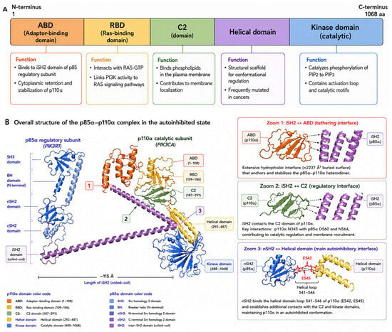
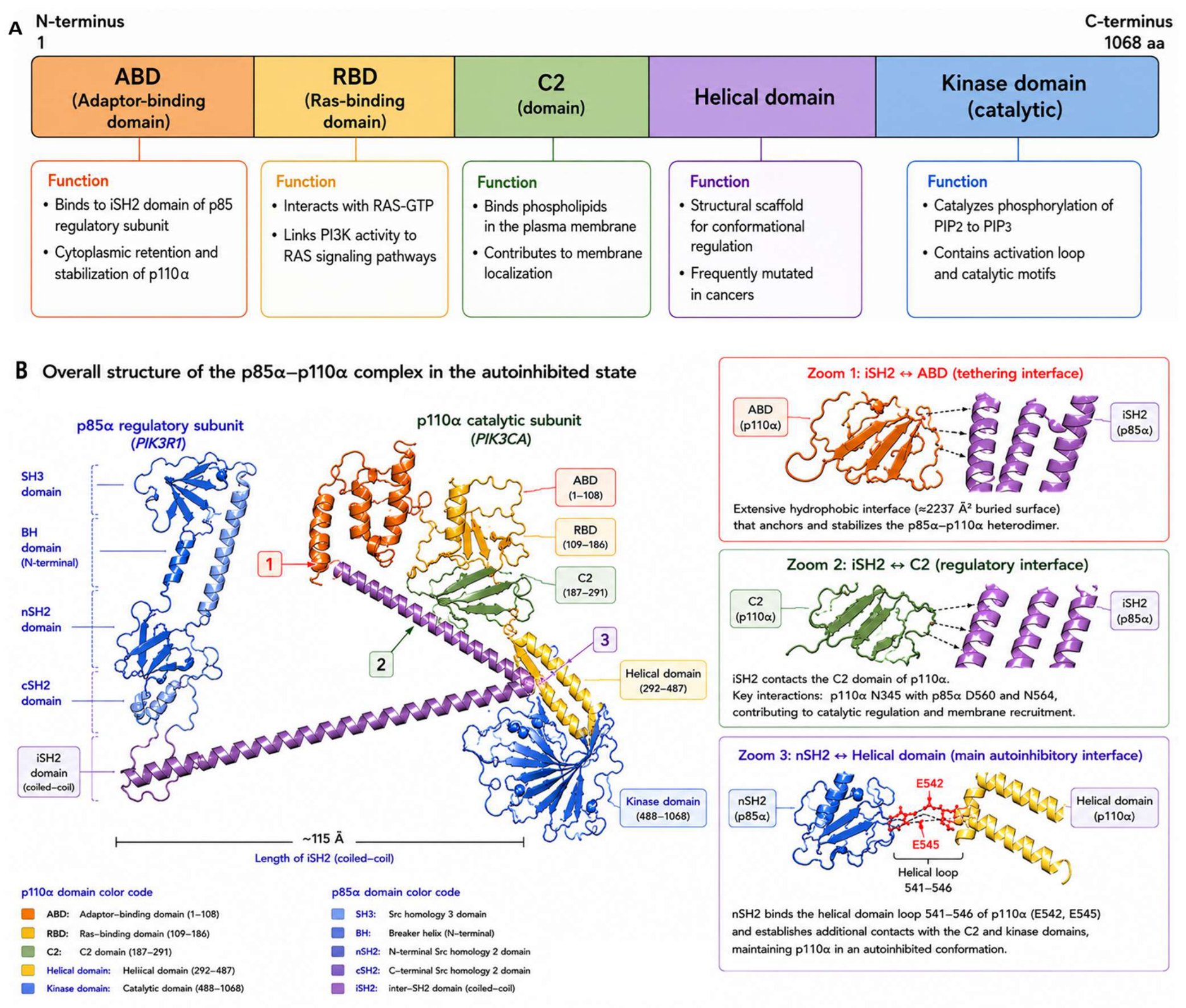
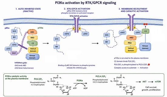
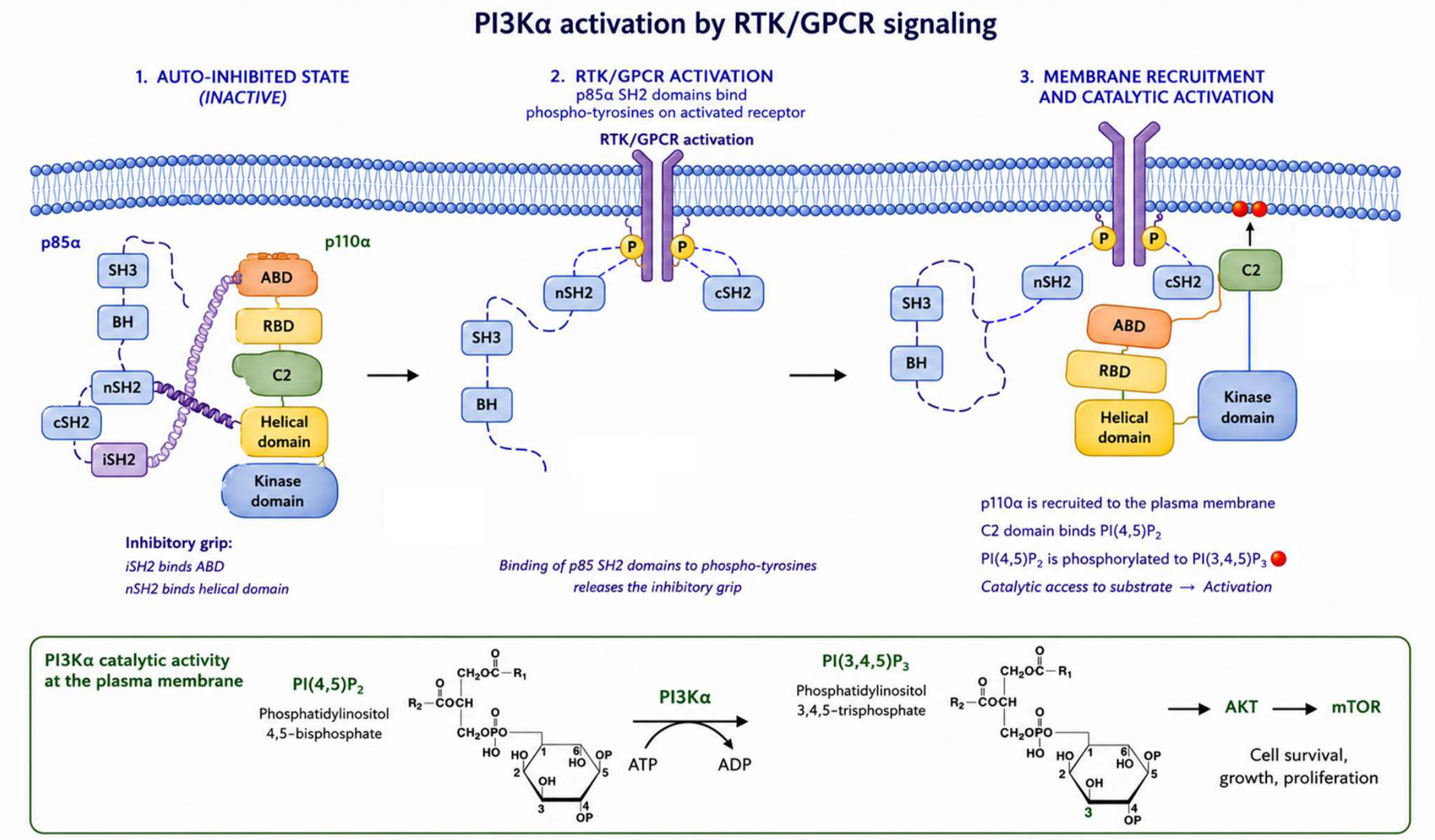
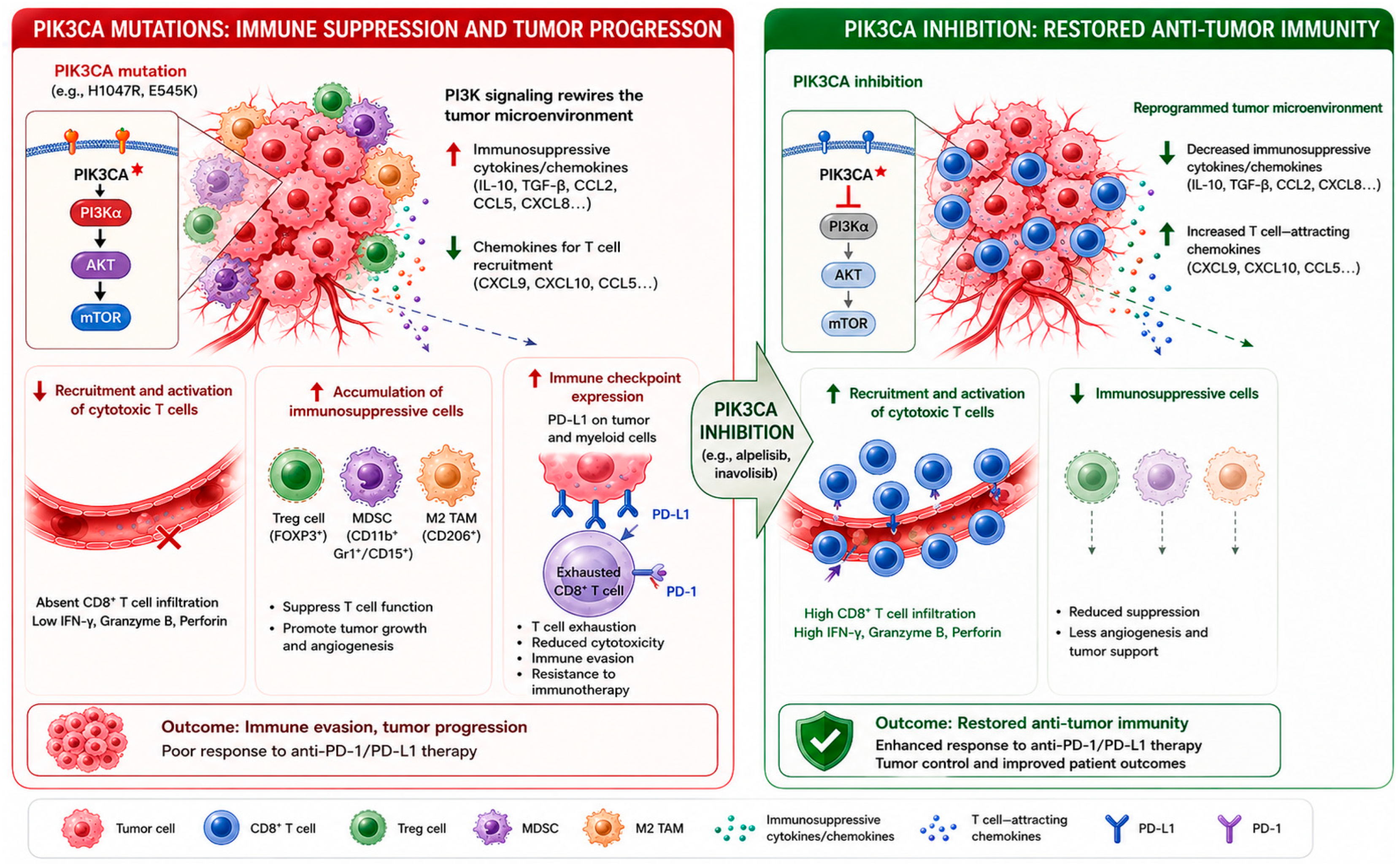

Disclaimer/Publisher’s Note: The statements, opinions and data contained in all publications are solely those of the individual author(s) and contributor(s) and not of MDPI and/or the editor(s). MDPI and/or the editor(s) disclaim responsibility for any injury to people or property resulting from any ideas, methods, instructions or products referred to in the content. |
© 2026 by the authors. Licensee MDPI, Basel, Switzerland. This article is an open access article distributed under the terms and conditions of the Creative Commons Attribution (CC BY) license.