


Simple Summary
Whole-pelvic radiotherapy may improve outcomes for some patients with localized prostate cancer, but identifying who is most likely to benefit remains challenging. In this study, we evaluated whether a multimodal artificial intelligence model that combines information from biopsy tissue images and clinical characteristics could identify patients with different outcome patterns following whole-pelvic versus prostate-only radiotherapy. Using data from a randomized clinical trial, we found that patients classified as high risk by the model showed larger numerical differences in biochemical failure rates with whole-pelvic radiotherapy, although the formal statistical interaction test was not significant. These findings should be considered exploratory and hypothesis-generating. Nevertheless, these findings support further investigation of artificial intelligence-based risk assessment as a tool for risk stratification and hypothesis generation in future prospective clinical trials.
Abstract
Background/Objectives: This study explored whether a multimodal artificial intelligence (MMAI) model integrating digitized histopathology and clinical features can identify prostate cancer patients who may benefit from neoadjuvant hormonal therapy (NHT) and whole-pelvic radiotherapy (WPRT). Methods: This secondary analysis of NRG/RTOG 9413 included NHT-treated patients with digitized biopsy slides and clinical data who were not part of the MMAI model optimization. A previously validated MMAI model estimated long-term risk, and Fine-Gray models evaluated interactions between MMAI-derived scores and the radiation field (WPRT vs. prostate-only RT [PORT]) for biochemical failure (BF), chosen over progression-free survival because of extended follow-up and distant metastasis (DM), with subgroup analyses by predefined MMAI strata. Results: Among 81 eligible patients, the MMAI-by-treatment interaction for BF did not confirm a differential effect (p = 0.30). Therefore, subgroup findings should be interpreted as descriptive and hypothesis-generating. Nevertheless, the magnitude effect of WPRT was numerically greater in the MMAI high-risk subgroup (5-yr: 41% vs. 79%; 10-yr: 47% vs. 79%; aHR 0.35 [0.14–0.86]) than in the low–intermediate group (5-yr: 18% vs. 33%; 10-yr: 44% vs. 57%; aHR 0.66 [0.29–1.48]). Conclusions: Although no statistically significant treatment-by-MMAI interaction was demonstrated, these findings are hypothesis-generating and support further investigation of MMAI approaches for guiding WPRT in NRG/RTOG 0534, 0924, GETUG-01, and POP-RT trials.
1. Introduction
External-beam radiotherapy (EBRT) with hormonal therapy (HT) is a standard of care for intermediate- and high-risk prostate cancer [1,2,3,4,5,6,7,8]. Still, the role of whole-pelvic RT (WPRT) remains debated, especially regarding which patients are likely to benefit. RTOG 9413 demonstrated a progression-free survival (PFS) benefit with WPRT versus prostate-only RT (PORT) [9], with long-term follow-up confirming durable benefit among patients receiving neoadjuvant-HT (NHT) [10]. However, GETUG-01 did not show a PFS improvement with WPRT [11,12]. Recent data highlight the importance of patient selection, including high-risk disease [13] and younger age [14]. Current risk stratification tools lack precision in identifying who benefits from WPRT [15,16]. Multimodal artificial intelligence (MMAI) algorithms may help by integrating clinical variables and digitized histopathology [17]. While these models have been validated in predicting clinical outcomes [18,19,20,21,22], their potential role in informing treatment selection—particularly for WPRT—remains unexplored.
In this study, leveraging patient-level data from RTOG 9413, we applied a previously validated MMAI prognostic model to explore whether MMAI-derived risk groups exhibit distinct patterns of biochemical or metastatic outcomes following NHT with WPRT versus PORT. This secondary analysis is intended to provide insights that may motivate validation in trials such as POP-RT, GETUG-01, NRG/RTOG 0534, and 0924.
2. Materials and Methods
2.1. Study Cohort
This retrospective analysis included consenting patients enrolled in the NRG/RTOG 9413 trial, which compared WPRT versus PORT and NHT versus adjuvant-HT in localized prostate cancer. Details of the trial design have been previously described [9,10,23,24].
The current analytic cohort excluded patients used in the MMAI model development (v1.2) [25]. This cohort corresponds to the internal validation group used in MMAI model development and was further reduced to patients receiving NHT; the subgroup guided the design of NRG/RTOG 0924 [26,27].
2.2. Data Acquisition
Digitized H&E-stained biopsy slides and clinical data were obtained for a subset of NRG/RTOG 9413 patients with pretreatment biopsy images, complete clinical data, and no prior use in model optimization. Slides were scanned at 20× magnification and underwent centralized quality assurance.
2.3. Application of the MMAI Model
We applied a validated MMAI prognostic model integrating biopsy histopathology with clinical data (PSA, age, and clinical T-stage) to generate a prognostic risk score [17,20,25]. This model was trained and validated across multiple randomized trials and demonstrated superior prediction performance for distant metastasis (DM) and biochemical failure (BF) compared to conventional tools. The model combined convolutional neural network-extracted image features with clinical variables through a fully connected neural network. The model combined image-derived features from biopsy histopathology with clinical variables using a neural network.
2.4. Statistical Analysis
The endpoints of interest were BF and DM, analyzed using time-to-event methods. BF was selected over the trial’s original PFS endpoint due to the extended duration of follow-up, during which PFS events increasingly reflected non-cancer-related deaths [10], limiting its utility in assessing prostate cancer-specific outcomes. BF was chosen for its clearer signal of disease control and treatment effect, while DM was included because it is the endpoint toward which the MMAI model was initially developed.
To explore whether BF or DM differed by MMAI between WPRT and PORT, Fine-Gray models with interaction terms between treatment and MMAI were used. Correspondingly, estimated treatment effects were adjusted for biomarker interaction (adjusted hazard ratio, aHR), and 95% confidence intervals (CIs) were reported. Subgroup summaries were generated within MMAI risk strata (low–intermediate vs. high) using previously validated thresholds (low risk: <0.273; intermediate risk: 0.273–0.499; and high risk: >0.499). Cumulative incidence functions accounting for competing risks (defined as death without an event) were used to provide BF and DM risk estimates for the MMAI subgroups and comparisons between treatment groups (WPRT vs. PORT) based on Gray’s test were treated as descriptive and not intended for inference.
All tests were two-sided at a 0.05 significance level, without multiplicity adjustments. Given the exploratory design, findings should be considered hypothesis-generating.
3. Results
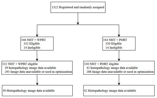
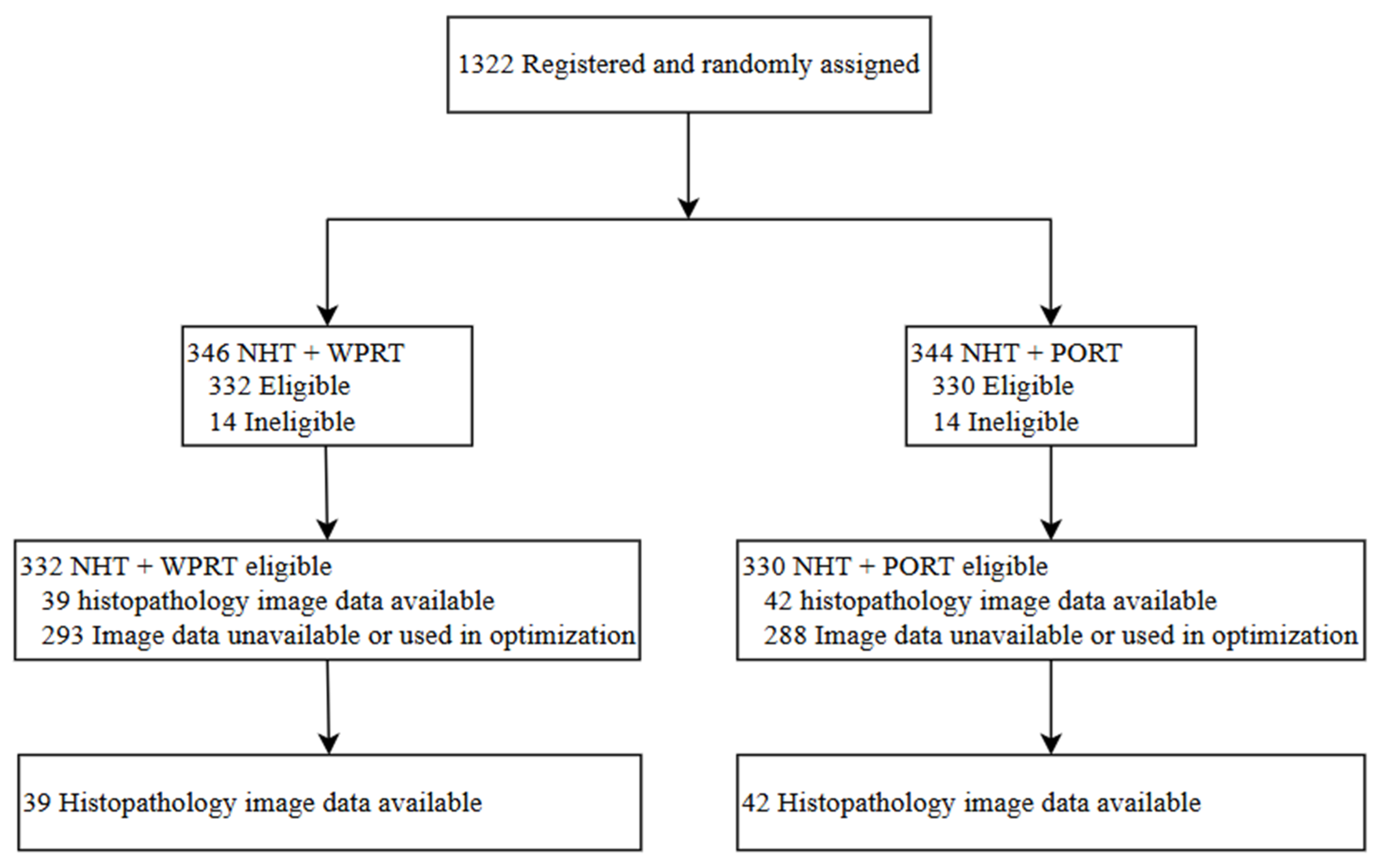
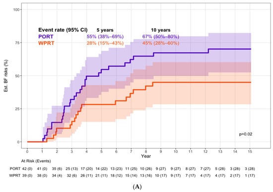
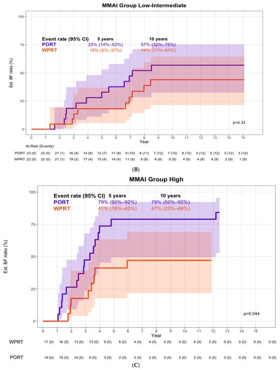
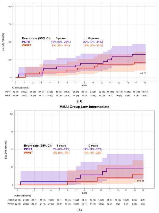
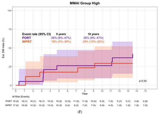
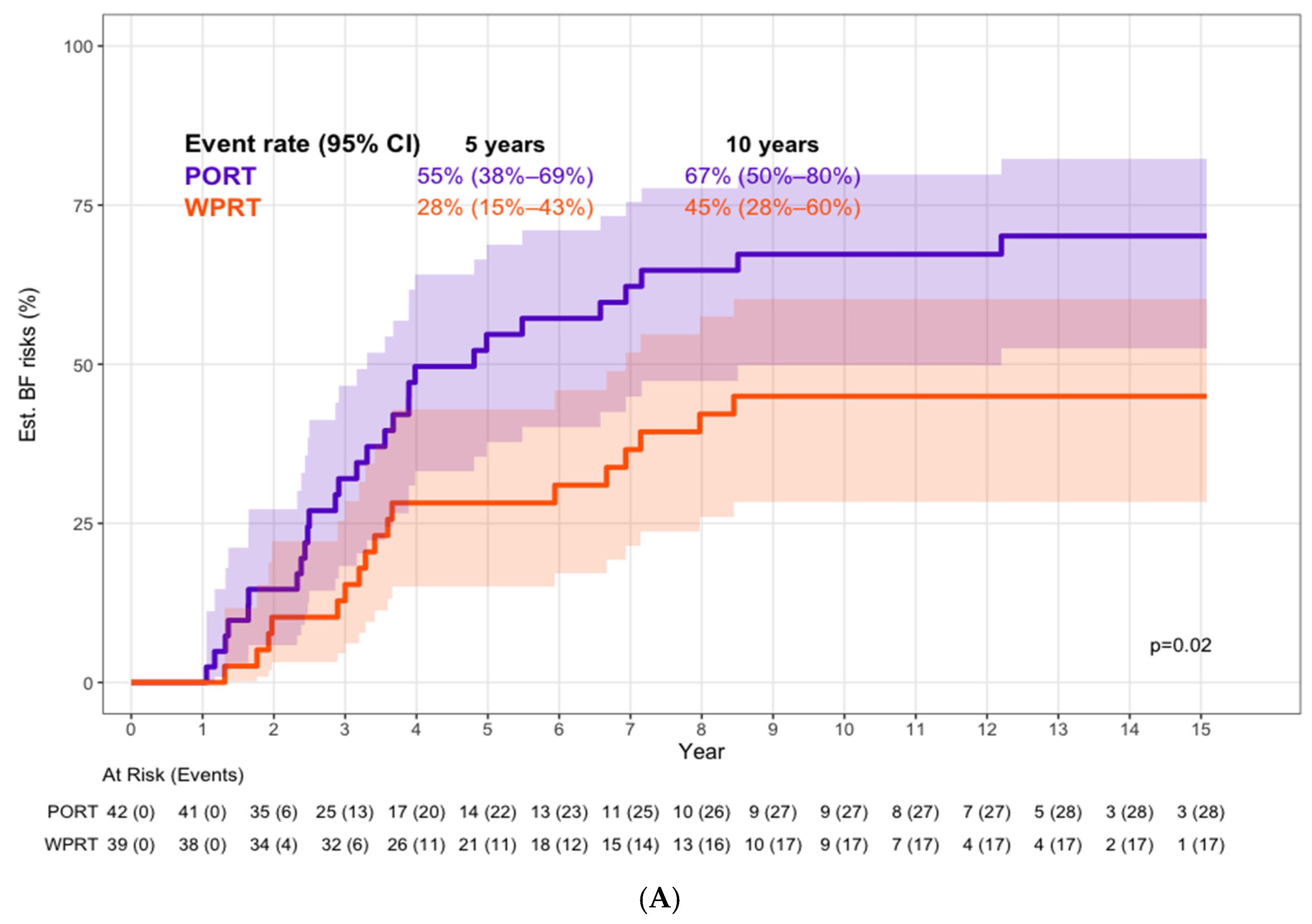
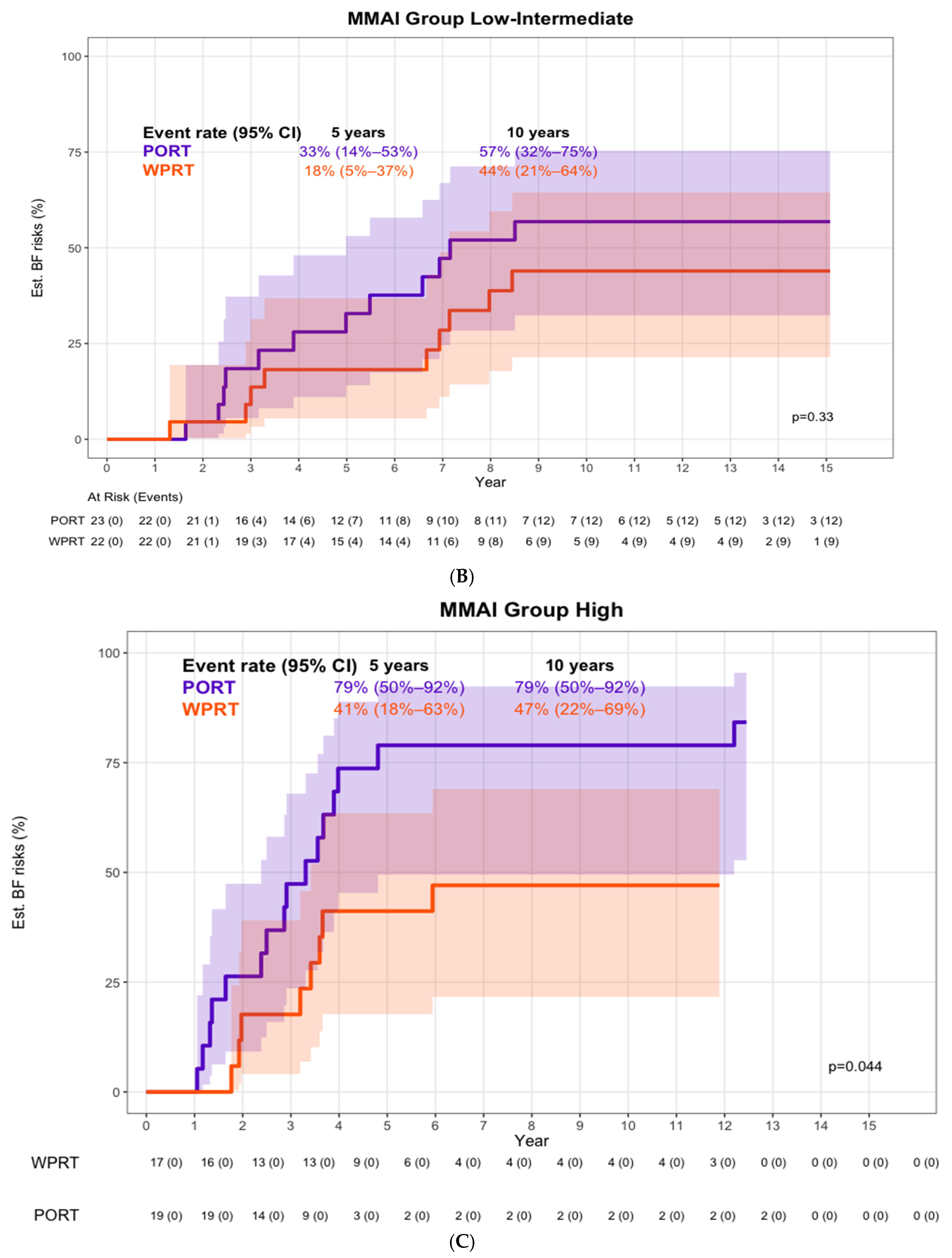
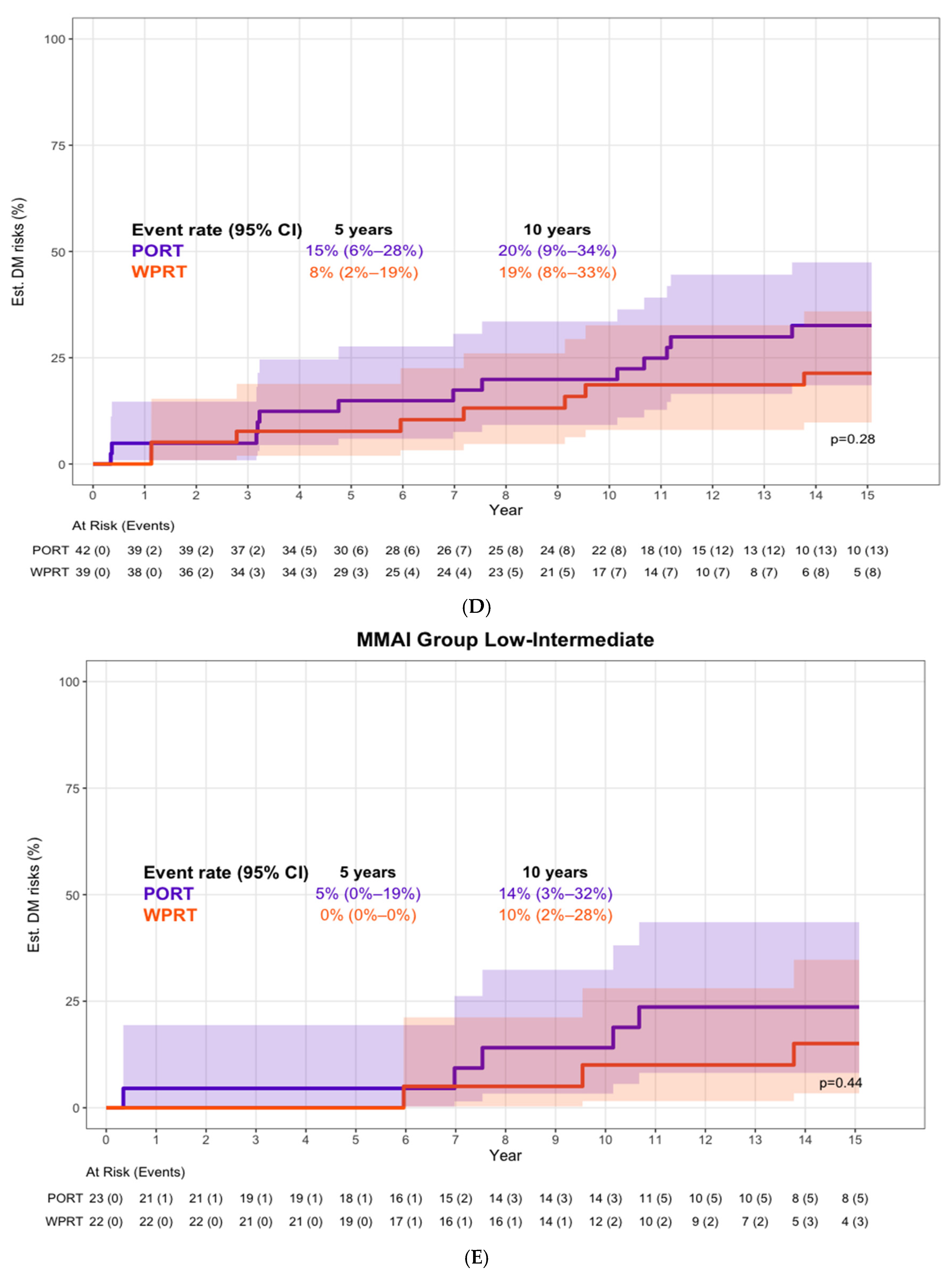
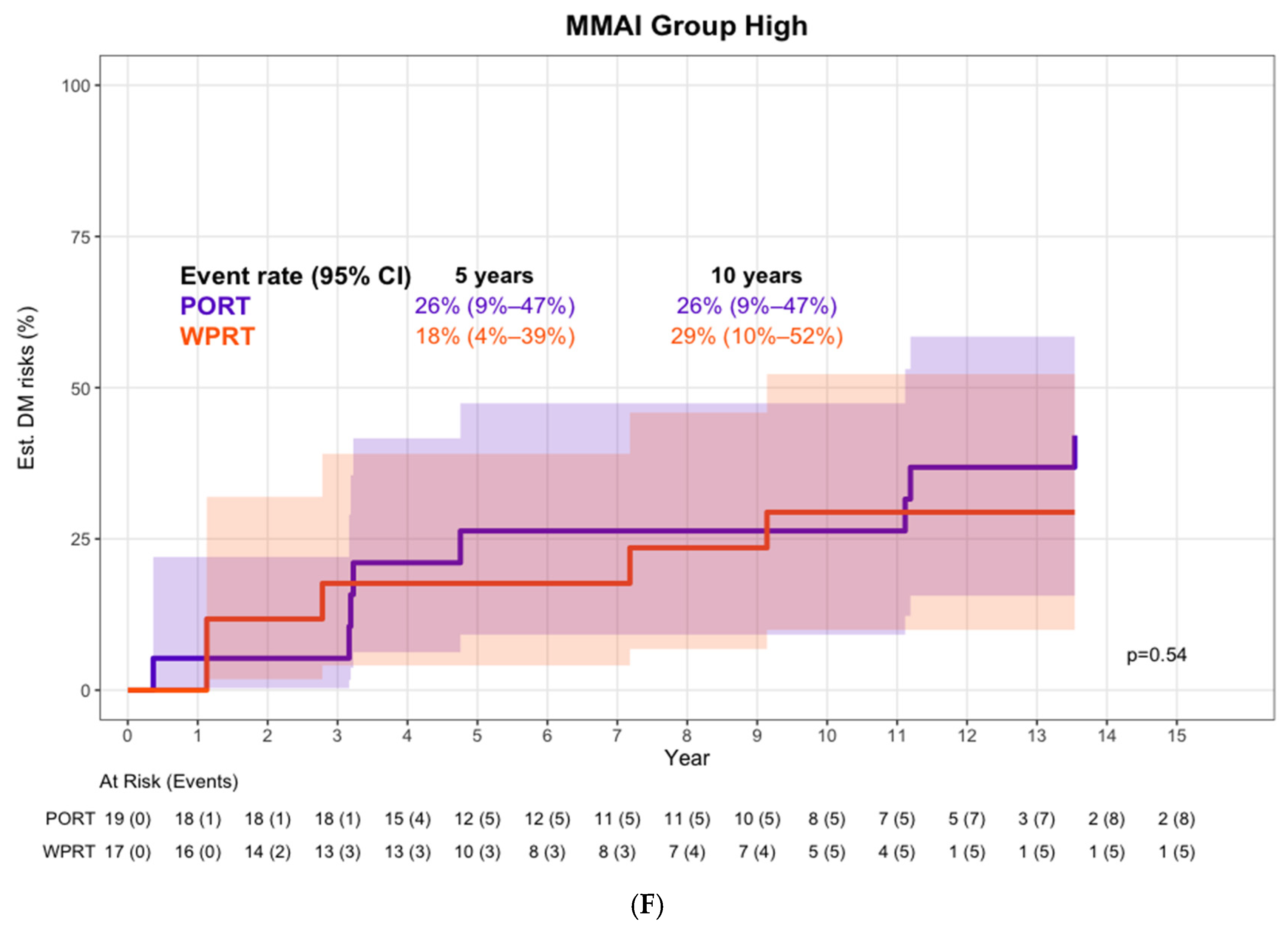
Eighty-one patients were included in this analysis (Figure 1; Table 1). BF rates were lower with WPRT (5-year: 28% vs. 55%; 10-year: 45% vs. 67%; aHR 0.49 [0.27–0.89]). The treatment-by-MMAI interaction was not statistically significant (interaction p = 0.30), indicating no evidence of treatment effect heterogeneity in this cohort. Accordingly, all subgroup findings should be interpreted as descriptive and hypothesis-generating. Nevertheless, exploratory subgroup estimates showed numerically larger risk differences between WPRT and PORT among patients classified as MMAI high-risk (5-year: 41% vs. 79%; 10-year: 47% vs. 79%; exploratory-aHR 0.35 [0.14–0.86]) than among those in the low–intermediate group (5-year: 18% vs. 33%; 10-year: 44% vs. 57%; exploratory-aHR 0.66 [0.29–1.48]) (Figure 2). For DM, no apparent differences were observed between WPRT and PORT (5-year: 8% vs. 15%; 10-year: 19% vs. 20%; exploratory-aHR 0.62 [0.26–1.48]). The interaction between the MMAI category and the radiation field on DM was assessed (interaction p = 0.82). WPRT vs. PORT treatment effects were aHR 0.56 [0.14–2.25] for MMAI low–intermediate subgroup vs. aHR 0.69 [0.22–2.16] for MMAI high subgroup (Figure 2). Because baseline PSA differed numerically between treatment groups, a sensitivity analysis was performed by adding PSA as a continuous covariate to the interaction models. For BF, the treatment-by-MMAI interaction estimate remained similar and was modestly strengthened after PSA adjustment (primary model: sHR 0.52, p = 0.30; PSA-adjusted model: sHR 0.45, p = 0.20). For DM, results were essentially unchanged (primary model: sHR 1.22, p = 0.82; PSA-adjusted model: sHR 1.20, p = 0.85). Exploratory NCCN risk group analyses are presented in Supplementary Table S1. These analyses yielded unstable estimates because only 13 patients were classified as NCCN intermediate-risk and events were sparse.
Figure 1.
CONSORT diagram. Abbreviations: NHT, neoadjuvant hormonal therapy; PORT, prostate only radiotherapy; WPRT, whole-pelvic radiotherapy.
Table 1.
Baseline characteristics of patients who received neoadjuvant hormonal therapy, stratified by radiation treatment volume.
Figure 2.
Cumulative incidence estimates and 95% confidence bands of biochemical failure (A–C) and distant metastasis (D–F) among patients who received neoadjuvant hormonal therapy by MMAI risk classification and radiation treatment volume. Shaded areas represent the 95% confidence bands around each cumulative incidence curve.
4. Discussion
This exploratory analysis of NRG/RTOG 9413 examined whether MMAI-derived risk groups showed differing treatment effect patterns for WPRT versus PORT. Numerically larger BF differences were observed among MMAI high-risk patients than in the low–intermediate group, whereas DM estimates were similar across strata. However, the treatment-by-MMAI interaction was not statistically significant; therefore, these findings should be interpreted as descriptive and hypothesis-generating. Further retrospective evaluation in larger randomized datasets will be needed to determine whether these exploratory patterns are reproducible.
Despite validation of prior nodal risk models [28], more sensitive, biologically driven methods are needed to guide WPRT. The clinical relevance of these findings lies in the potential of MMAI risk classification to meet this unmet need, offering an efficient tool to personalize treatment decisions and inform future validation efforts.
These findings arrive at a time when the optimal use of WPRT remains uncertain, with randomized trials yielding inconsistent results. RTOG 9413 demonstrated a PFS benefit with WPRT [9,10], while GETUG-01 did not [11,12], and POP-RT showed BF benefits but lacks long-term data [13]. Recent observational work also suggests benefit in biologically selected subgroups [14]. Our analysis extends these insights by applying a validated MMAI framework.
Importantly, these findings should be considered within the context of contemporary PSMA-PET staging. Although PSMA-PET has substantially improved detection of nodal and distant metastases compared with conventional imaging, its sensitivity for microscopic pelvic nodal disease remains imperfect. Surgical validation studies have demonstrated that a meaningful proportion of patients with negative PSMA-PET findings harbor occult pathologic nodal metastases [29,30,31]. In this setting, tissue-based MMAI approaches may provide information complementary to imaging by capturing biologic features associated with aggressive disease and patterns of disease progression that are not constrained by imaging resolution. Rather than replacing PSMA-PET, MMAI may ultimately provide complementary information, with PSMA-PET identifying radiographically detectable disease and MMAI providing additional biologic risk stratification. Future studies should evaluate whether integrating MMAI with PSMA-PET improves risk stratification and further refines the study of WPRT in randomized clinical trial datasets.
NRG/RTOG 0534 and 0924 trials provide opportunities to assess whether MMAI can refine patient selection for WPRT and serve as a biomarker to guide treatment intensification. Additional validation opportunities may emerge from ongoing pelvic nodal radiotherapy trials, including PIVOTALboost and PEARLS. Future efforts should include prospective validation in larger randomized datasets adequately powered to test interactions. Using the observed interaction effect as a planning assumption, approximately 500 patients (corresponding to roughly 275 BF events) may be required to achieve 80% power for treatment-by-MMAI interaction testing.
Several limitations warrant consideration. This was a retrospective analysis, and treatment was not stratified by MMAI risk. Only a small subset (n = 81) of the original NHT cohort was available because many patients were used for original model optimization or lacked biopsy slides. Additionally, staging was based on pre-PSMA PET imaging, which may limit its applicability to modern practice. Moreover, while BF is a clinically relevant endpoint, its long-term association with prostate cancer-specific and overall mortality remains variable, particularly in the contemporary era of salvage therapies [32,33]. Nonetheless, BF occurred frequently enough in this cohort to permit more stable descriptive estimation of outcome patterns. In contrast, events for DM, PCSM, or OS were too sparse to support reliable subgroup summaries. Similarly, exploratory interaction analyses using NCCN risk groups yielded unstable estimates because only 13 patients were classified as NCCN intermediate-risk and events were sparse, resulting in clinically implausible treatment effect estimates that should be interpreted with caution (Supplementary Table S1). Given the limited sample size, all treatment effect estimates, particularly within MMAI risk strata, should be interpreted cautiously, and no confirmatory conclusions regarding treatment effect heterogeneity can be drawn.
BF was selected over the trial’s original primary endpoint of PFS because the extended follow-up of RTOG 9413—now exceeding two decades—led many progression events to be driven by non-cancer-related deaths rather than PSA progression, and neither the original trial nor the subset analyzed here was powered to assess DM or OS. As a result, PFS has become increasingly difficult to interpret as a cancer-specific measure of treatment efficacy. BF, by contrast, offers a more direct signal of disease control and treatment effect, consistent with its use in recent trials such as POP-RT and NRG/RTOG 0924.
5. Conclusions
This study observed a stronger association between WPRT and reduced BF among MMAI high-risk patients than among low- and intermediate-risk patients. Because no significant treatment-by-MMAI interaction was observed, these findings should be considered exploratory and hypothesis-generating. MMAI may offer a potential avenue for exploring personalized treatment selection beyond conventional risk stratification. These results motivate further adequately powered validation to support the broader integration of digital pathology-based AI tools in future clinical trial design.
Supplementary Materials
The following supporting information can be downloaded at https://www.mdpi.com/article/10.3390/cancers18121982/s1: Table S1: Exploratory NCCN risk group analyses, neoadjuvant ADT cohort (n = 81). (A) NCCN by radiation field interaction effect; (B) Subgroup-specific treatment effects (WPRT vs. PORT) within each NCCN risk group to aid clinical interpretation of the interaction effect.
Author Contributions
Conceptualization: M.R.III, E.L.S., M.S., P.T.T. Methodology: H.-C.H., M.S. Validation: H.-C.H. Formal analysis: H.-C.H. Investigation: I.J.B., E.M.G., M.R.III, E.L.S. Resources: A.P.D., J.D.P., S.L.P., T.N.S. Data Curation: E.M.G., H.-C.H., I.-C.H., S.L.P. Writing—Original Draft: M.S. Writing—Review & Editing: I.J.B., A.P.D., E.M.G., G.D.G., H.-C.H., M.A.H., A.M.M., P.L.N., J.D.P., M.R.III, E.L.S., M.S., P.T.T., S.L.P., T.N.S. and M.R. Visualization: H.-C.H. Supervision: M.R.III, E.L.S., P.T.T., T.N.S. Project Administration: E.L.S., P.T.T. Funding Acquisition: P.T.T. All authors have read and agreed to the published version of the manuscript.
Funding
Research reported in this publication was supported by the National Cancer Institute of the National Institutes of Health under Award Number UG1CA189867 and UG1CA189858 (NCORP), U10CA180822 (NRG Oncology SDMC), U24CA196067 (NRG Specimen Bank), U10CA180868 (NRG Oncology Operations), and CTEP. The content is solely the responsibility of the authors and does not necessarily represent the official views of the National Institutes of Health. This project was also supported by Artera.
Institutional Review Board Statement
The NRG/RTOG 9413 trial was registered at ClinicalTrials.gov (NCT00769548) and approved by the Ethics Committee of NRG (protocol code: 20027; date of approval: 29 September 2021).
Informed Consent Statement
Written informed consent was obtained from all patients enrolled in the original RTOG 9413 clinical trial. This secondary analysis was conducted using de-identified data.
Data Availability Statement
All data will be made available in accordance with the National Clinical Trials Network Data Archive policies and procedures.
Acknowledgments
Mutlay Sayan, and NRG Oncology would like to thank the following accrual institutions for their contributions to our mission to improve the lives of patients with cancer through practice-changing, multi-institutional clinical and translational research: Akron City Hospital; Albert Einstein Medical Center; Baptist Cancer Institute; Brooklyn MB-CCOP/SUNY Downstate; Centre Hospitalier de l`Université de Montréal-Notre Dame; Christiana Care Health Services, Inc. CCOP; Columbia-Saint Mary’s Milwaukee; Community Memorial Hospital; Cross Cancer Institute; Dartmouth Hitchcock Medical Center; Delaware County Memorial Hospital; East Carolina Medical School; Emory University; Froedtert and the Medical College of Wisconsin; Greater Baltimore Medical Center; Gunderson Clinic; H. Lee Moffitt Cancer Center & Research Institute; Henry Ford Hospital; John Muir Medical Center-Concord Campus; Kaiser Permanente Los Angeles Medical Center; Laura and Isaac Perlmutter Cancer Center at NYU Langone; London Regional Cancer Program; McGill University; Montefiore Medical Center-Moses Campus; Northern Indiana Cancer Research Consortium CCOP; Nova Scotia Cancer Centre; Radiation Oncology Associates of W. Alabama PC; Regional Hospital of Scranton; Regional Medical Center; Saint Agnes Medical Center; Santa Cruz Radiation Oncology; Saskatoon Cancer Center; South Jersey Hospital Systems; Southeast Cancer Control Consortium, Inc., CCOP; St. Lukes Hospital; St. Mary’s Medical Center; Stanford University Medical Center; The New York Hospital Medical Center/Queens; Thomas Jefferson University Hospital; Toledo Community Hospital Oncology Program CCOP; Toledo Hospital; Tom Baker Cancer Centre; University of Alabama at Birmingham Medical Center; University of Chicago Comprehensive Cancer Center; University of Pennsylvania Medical Center; Veterans Affairs Medical Center; Virginia Baptist Hospital; Virginia Commonwealth University; Virginia Mason CCOP; Virtua Memorial; Wake Forest University Health Sciences; Walter Reed National Military Medical Center; Washington University; WellSpan Health-York Hospital.
Conflicts of Interest
Authors with nothing to disclose: Barani, Gore, Hallman, Hsu, McDonald, Pennington, Roach, Rooney, Sayan. The following authors declare support for the work reported in this manuscript without time limit: Hallman-To Institution: RTOG/NRG Oncology; Huang-To You: Employee of Artera Inc.; Pennington-To Institution: NCI grant 5UG1CA189858; Pugh-To Institution: Grant from NCI; Stewart-To You: Artera Inc. The following authors declare relationships that were present during the 36 months prior to publication: Dicker-Grants To Institution: Artera; Grass-Grants To Institution: Artera AI; Huang-Stock or stock options To You: Employee of Artera Inc.; Other To You: Employee of Artera Inc.; Nguyen-Consultancy To You: J&J Oncology, Bayer, Novartis, Astellas, MDxHealth, Boston Scientific, Nanocan, AIQ; Grants To Institution: J&J Oncology, Bayer, Astellas; Stock or stock options To You: Stratagen Bio, Nanocan, Reversal Therapeutics; Showalter-Leadership or Fiduciary Role To You: I previously served as Chief Medical Officer at Artera Inc.; Stock or stock options To You: Stock in Artera Inc.; Stewart-Stock or stock options To You: Artera Inc.; Other To You: Employee of Artera Inc.; Tran-Consultancy To You: J&J, Bayer, Natsar Pharm, Pfizer, Novartis, Lantheus, Regeneron, Reflexion Medical and AZ; Grants To You: Bayer; Items or Services Received: To You Veracyte & Artera; Leadership or Fiduciary Role To You: ASTRO, ASCO and AACR; Patents To You: Licensed and royalties from Natsar Pharm; Payment or Honoraria To You: J&J, Bayer, Natsar Pharm, Pfizer, Novartis, Lantheus, Regeneron, Reflexion Medical and AZ; Royalties or Licenses To You: Licensed and royalties from Natsar Pharm; Travel Support To You: J&J, Bayer, Natsar Pharm, Pfizer, Novartis, Lantheus, Regeneron, Reflexion Medical and AZ. Author Huei-Chung Huang, Erin L. Stewart, and Timothy N. Showalter were employed by the company Artera. The remaining authors declare that the research was conducted in the absence of any commercial or financial relationships that could be construed as a potential conflict of interest.
Abbreviations
The following abbreviations are used in this manuscript:
| ADT | Androgen deprivation therapy |
| aHR | Adjusted hazard ratio |
| BF | Biochemical failure |
| CI | Confidence interval |
| DM | Distant metastasis |
| EBRT | External-beam radiotherapy |
| H&E | Hematoxylin and eosin |
| HT | Hormonal therapy |
| IQR | Interquartile range |
| MMAI | Multimodal artificial intelligence |
| NCCN | National Comprehensive Cancer Network |
| NHT | Neoadjuvant hormonal therapy |
| OS | Overall survival |
| PCSM | Prostate cancer-specific mortality |
| PFS | Progression-free survival |
| POP-RT | Prostate-Only Versus Whole-Pelvic Radiation Therapy trial |
| PORT | Prostate-only radiotherapy |
| PSA | Prostate-specific antigen |
| PSMA PET | Prostate-specific membrane antigen positron emission tomography |
| RT | Radiotherapy |
| WPRT | Whole-pelvic radiotherapy |
References
- Jones, C.U.; Hunt, D.; McGowan, D.G.; Amin, M.B.; Chetner, M.P.; Bruner, D.W.; Leibenhaut, M.H.; Husain, S.M.; Rotman, M.; Souhami, L.; et al. Radiotherapy and short-term androgen deprivation for localized prostate cancer. N. Engl. J. Med. 2011, 365, 107–118. [Google Scholar] [CrossRef] [Scilit]
- Pisansky, T.M.; Hunt, D.; Gomella, L.G.; Amin, M.B.; Balogh, A.G.; Chinn, D.M.; Seider, M.J.; Duclos, M.; Rosenthal, S.A.; Bauman, G.S.; et al. Duration of androgen suppression before radiotherapy for localized prostate cancer: Radiation therapy oncology group randomized clinical trial 9910. J. Clin. Oncol. 2015, 33, 332–339. [Google Scholar] [CrossRef] [Scilit] [PubMed]
- Eastham, J.A.; Auffenberg, G.B.; Barocas, D.A.; Chou, R.; Crispino, T.; Davis, J.W.; Eggener, S.; Horwitz, E.M.; Kane, C.J.; Kirkby, E.; et al. Clinically Localized Prostate Cancer: AUA/ASTRO Guideline, Part I: Introduction, Risk Assessment, Staging, and Risk-Based Management. J. Urol. 2022, 208, 10–18. [Google Scholar] [CrossRef] [Scilit] [PubMed]
- Eastham, J.A.; Auffenberg, G.B.; Barocas, D.A.; Chou, R.; Crispino, T.; Davis, J.W.; Eggener, S.; Horwitz, E.M.; Kane, C.J.; Kirkby, E.; et al. Clinically Localized Prostate Cancer: AUA/ASTRO Guideline, Part II: Principles of Active Surveillance, Principles of Surgery, and Follow-Up. J. Urol. 2022, 208, 19–25. [Google Scholar] [CrossRef] [Scilit] [PubMed]
- Bekelman, J.E.; Rumble, R.B.; Freedland, S.J. Clinically Localized Prostate Cancer: ASCO Clinical Practice Guideline Endorsement of an AUA/ASTRO/SUO Guideline Summary. J. Oncol. Pract. 2018, 14, 618–624. [Google Scholar] [CrossRef] [Scilit] [PubMed]
- Spratt, D.E.; Srinivas, S.; Adra, N.; Ahmed, B.; An, Y.; Bitting, R.; Chapin, B.; Cheng, H.H.; Cho, S.Y.; D’Amico, A.V.; et al. Prostate Cancer, Version 3.2026, NCCN Clinical Practice Guidelines In Oncology. J. Natl. Compr. Cancer Netw. 2025, 23, 469–493. [Google Scholar] [CrossRef] [Scilit] [PubMed]
- Parker, C.; Castro, E.; Fizazi, K.; Heidenreich, A.; Ost, P.; Procopio, G.; Tombal, B.; Gillessen, S. Prostate cancer: ESMO Clinical Practice Guidelines for diagnosis, treatment and follow-up. Ann. Oncol. 2020, 31, 1119–1134. [Google Scholar] [CrossRef] [Scilit] [PubMed]
- Cornford, P.; van den Bergh, R.C.N.; Briers, E.; Van den Broeck, T.; Brunckhorst, O.; Darraugh, J.; Eberli, D.; De Meerleer, G.; De Santis, M.; Farolfi, A.; et al. EAU-EANM-ESTRO-ESUR-ISUP-SIOG Guidelines on Prostate Cancer-2024 Update. Part I: Screening, Diagnosis, and Local Treatment with Curative Intent. Eur. Urol. 2024, 86, 148–163. [Google Scholar] [CrossRef] [Scilit] [PubMed]
- Roach, M., 3rd; DeSilvio, M.; Lawton, C.; Uhl, V.; Machtay, M.; Seider, M.J.; Rotman, M.; Jones, C.; Asbell, S.O.; Valicenti, R.K.; et al. Phase III trial comparing whole-pelvic versus prostate-only radiotherapy and neoadjuvant versus adjuvant combined androgen suppression: Radiation Therapy Oncology Group 9413. J. Clin. Oncol. 2003, 21, 1904–1911. [Google Scholar] [CrossRef] [Scilit] [PubMed]
- Roach, M.; Moughan, J.; Lawton, C.A.F.; Dicker, A.P.; Zeitzer, K.L.; Gore, E.M.; Kwok, Y.; Seider, M.J.; Hsu, I.C.; Hartford, A.C.; et al. Sequence of hormonal therapy and radiotherapy field size in unfavourable, localised prostate cancer (NRG/RTOG 9413): Long-term results of a randomised, phase 3 trial. Lancet Oncol. 2018, 19, 1504–1515. [Google Scholar] [CrossRef] [Scilit] [PubMed]
- Pommier, P.; Chabaud, S.; Lagrange, J.L.; Richaud, P.; Lesaunier, F.; Le Prise, E.; Wagner, J.P.; Hay, M.H.; Beckendorf, V.; Suchaud, J.P.; et al. Is There a Role for Pelvic Irradiation in Localized Prostate Adenocarcinoma? Preliminary Results of GETUG-01. J. Clin. Oncol. 2007, 25, 5366–5373. [Google Scholar] [CrossRef] [Scilit] [PubMed]
- Pommier, P.; Chabaud, S.; Lagrange, J.L.; Richaud, P.; Le Prise, E.; Wagner, J.P.; Azria, D.; Beckendorf, V.; Suchaud, J.P.; Bernier, V.; et al. Is There a Role for Pelvic Irradiation in Localized Prostate Adenocarcinoma? Update of the Long-Term Survival Results of the GETUG-01 Randomized Study. Int. J. Radiat. Oncol. Biol. Phys. 2016, 96, 759–769. [Google Scholar] [CrossRef] [Scilit] [PubMed]
- Murthy, V.; Maitre, P.; Kannan, S.; Panigrahi, G.; Krishnatry, R.; Bakshi, G.; Prakash, G.; Pal, M.; Menon, S.; Phurailatpam, R.; et al. Prostate-Only Versus Whole-Pelvic Radiation Therapy in High-Risk and Very High-Risk Prostate Cancer (POP-RT): Outcomes From Phase III Randomized Controlled Trial. J. Clin. Oncol. 2021, 39, 1234–1242. [Google Scholar] [CrossRef] [Scilit] [PubMed]
- Sayan, M.; Chen, M.H.; Loffredo, M.; McMahon, E.; Moningi, S.; Orio, P.F.; Nguyen, P.L.; D’Amico, A.V. Elective Pelvic Lymph Node Radiation Therapy and the Risk of Death in Patients With Unfavorable-Risk Prostate Cancer: A Postrandomization Analysis. J. Clin. Oncol. 2024, 42, 2558–2564. [Google Scholar] [CrossRef] [Scilit] [PubMed]
- Partin, A.W.; Yoo, J.; Carter, H.B.; Pearson, J.D.; Chan, D.W.; Epstein, J.I.; Walsh, P.C. The Use of Prostate Specific Antigen, Clinical Stage and Gleason Score to Predict Pathological Stage in Men with Localized Prostate Cancer. J. Urol. 1993, 150, 110–114. [Google Scholar] [CrossRef] [Scilit] [PubMed]
- Chilukuri, S.; Sayan, M.; Pra, A.D.; Murthy, V. Elective Regional Nodal Irradiation in Prostate Cancer. Semin Radiat. Oncol. 2025, 35, 374–384. [Google Scholar] [CrossRef] [Scilit] [PubMed]
- Esteva, A.; Feng, J.; van der Wal, D.; Huang, S.C.; Simko, J.P.; DeVries, S.; Chen, E.; Schaeffer, E.M.; Morgan, T.M.; Sun, Y.; et al. Prostate cancer therapy personalization via multi-modal deep learning on randomized phase III clinical trials. npj Digit. Med. 2022, 5, 71. [Google Scholar] [CrossRef] [Scilit] [PubMed]
- Spratt, D.E.; Tang, S.; Sun, Y.; Huang, H.C.; Chen, E.; Mohamad, O.; Armstrong, A.J.; Tward, J.D.; Nguyen, P.L.; Lang, J.M.; et al. Artificial Intelligence Predictive Model for Hormone Therapy Use in Prostate Cancer. NEJM Evid. 2023, 2, EVIDoa2300023. [Google Scholar] [CrossRef] [Scilit] [PubMed]
- Roach, M.; Zhang, J.; Mohamad, O.; Wal, D.v.d.; Simko, J.P.; DeVries, S.; Huang, H.-C.; Joun, S.; Schaeffer, E.M.; Morgan, T.M.; et al. Assessing Algorithmic Fairness With a Multimodal Artificial Intelligence Model in Men of African and Non-African Origin on NRG Oncology Prostate Cancer Phase III Trials. JCO Clin. Cancer Inform. 2025, 9, e2400284. [Google Scholar] [CrossRef] [Scilit] [PubMed]
- Parker, C.T.A.; Mendes, L.; Liu, V.Y.T.; Grist, E.; Joun, S.; Yamashita, R.; Mitani, A.; Chen, E.; Parry, M.A.; Sachdeva, A.; et al. External validation of a digital pathology-based multimodal artificial intelligence-derived prognostic model in patients with advanced prostate cancer starting long-term androgen deprivation therapy: A post-hoc ancillary biomarker study of four phase 3 randomised controlled trials of the STAMPEDE platform protocol. Lancet Digit. Health 2025, 7, 100885. [Google Scholar] [CrossRef] [Scilit] [PubMed]
- Armstrong, A.J.; Liu, V.Y.T.; Selvaraju, R.R.; Chen, E.; Simko, J.P.; DeVries, S.; Sartor, O.; Sandler, H.M.; Mohamad, O.; Huang, H.C.; et al. Development and Validation of an Artificial Intelligence Digital Pathology Biomarker to Predict Benefit of Long-Term Hormonal Therapy and Radiotherapy in Men With High-Risk Prostate Cancer Across Multiple Phase III Trials. J. Clin. Oncol. 2025, 43, 3494–3504. [Google Scholar] [CrossRef] [Scilit] [PubMed]
- Morgan, T.M.; Ren, Y.; Tang, S.; Zwerink, W.; Chen, E.; Mitani, A.; Huang, H.C.; Simko, J.P.; DeVries, S.; Pollack, A.; et al. Development and Validation of a Multimodal Artificial Intelligence-derived Digital Pathology-based Biomarker Predicting Metastasis Among Patients with Biochemical Recurrence After Radical Prostatectomy in NRG/RTOG Trials. Eur. Urol. 2025; in press. [CrossRef] [Scilit] [PubMed]
- Lawton, C.A.F.; Lin, X.; Hanks, G.E.; Lepor, H.; Grignon, D.J.; Brereton, H.D.; Bedi, M.; Rosenthal, S.A.; Zeitzer, K.L.; Venkatesan, V.M.; et al. Duration of Androgen Deprivation in Locally Advanced Prostate Cancer: Long-Term Update of NRG Oncology RTOG 9202. Int. J. Radiat. Oncol. Biol. Phys. 2017, 98, 296–303. [Google Scholar] [CrossRef] [Scilit] [PubMed]
- Lawton, C.A.; DeSilvio, M.; Roach, M., 3rd; Uhl, V.; Kirsch, R.; Seider, M.; Rotman, M.; Jones, C.; Asbell, S.; Valicenti, R.; et al. An update of the phase III trial comparing whole pelvic to prostate only radiotherapy and neoadjuvant to adjuvant total androgen suppression: Updated analysis of RTOG 94-13, with emphasis on unexpected hormone/radiation interactions. Int. J. Radiat. Oncol. Biol. Phys. 2007, 69, 646–655. [Google Scholar] [CrossRef] [Scilit] [PubMed]
- Gerrard, P.; Zhang, J.; Yamashita, R.; Huang, H.-C.; Nag, S.; Nhek, S.; Kish, J.M.; Cole, A.; Silberman, N.; Royce, T.J.; et al. Analytical Validation of a Clinical Grade Prognostic and Classification Artificial Intelligence Laboratory Test for Men with Prostate Cancer. AI Precis. Oncol. 2024, 1, 119–126. [Google Scholar] [CrossRef] [Scilit]
- National Cancer Institute; NRG Oncology. Androgen Deprivation Therapy and High Dose Radiotherapy with or Without Whole-Pelvic Radiotherapy in Unfavorable Intermediate or Favorable High Risk Prostate Cancer: A Phase III Randomized Trial; National Cancer: Philadelphia, PA, USA; NRG Oncology: Philadelphia, PA, USA, 2011.
- Roach, M.I.; Karrison, T.; Chung, H.; Chang, A.J.; Gomella, L.G.; Hsu, I.-C.; Morton, G.; Wallace, R.E.; Movsas, B.; Bruner, D.W.; et al. Androgen Deprivation Therapy (ADT) and High Dose Definitive Radiotherapy (RT) +/− Whole Pelvic RT in Patients with Unfavorable Intermediate or Favorable High-Risk Prostate Cancer: Early Results of a Phase III Randomized Trial, NRG/RTOG 0924. In Proceedings of the ASTRO Annual Meeting, San Francisco, CA, USA, 28–30 September 2025. [Google Scholar]
- Abdollah, F.; Cozzarini, C.; Suardi, N.; Gallina, A.; Capitanio, U.; Bianchi, M.; Tutolo, M.; Salonia, A.; La Macchia, M.; Di Muzio, N.; et al. Indications for pelvic nodal treatment in prostate cancer should change. Validation of the Roach formula in a large extended nodal dissection series. Int. J. Radiat. Oncol. Biol. Phys. 2012, 83, 624–629. [Google Scholar] [CrossRef] [Scilit] [PubMed]
- Yaxley, J.W.; Raveenthiran, S.; Nouhaud, F.X.; Samartunga, H.; Yaxley, A.J.; Coughlin, G.; Delahunt, B.; Egevad, L.; McEwan, L.; Wong, D. Outcomes of Primary Lymph Node Staging of Intermediate and High Risk Prostate Cancer with (68)Ga-PSMA Positron Emission Tomography/Computerized Tomography Compared to Histological Correlation of Pelvic Lymph Node Pathology. J. Urol. 2019, 201, 815–820. [Google Scholar] [CrossRef] [Scilit] [PubMed]
- Ozen, A.; Sarikaya, Z.; Gur, C.; Ozboru, D.; Canat, H.L. Pelvic Lymph Node Staging With PSMA PET in Prostate Cancer: Surgical Validation With Implications for Radiotherapy Planning. Prostate 2026, 86, 648–654. [Google Scholar] [CrossRef] [Scilit] [PubMed]
- Pickersgill, N.A.; Yu, R.; Vertosick, E.A.; Fletcher, S.A.; Liso, N.; Kluckert, J.; Akin, O.; Ehdaie, B.; Laudone, V.P.; Eastham, J.A.; et al. Diagnostic and prognostic utility of PSMA-PET/CT in node-metastatic prostate cancer. BJU Int. 2026. Online ahead of print. [Google Scholar] [CrossRef] [Scilit]
- Dignam, J.J.; Hamstra, D.A.; Lepor, H.; Grignon, D.; Brereton, H.; Currey, A.; Rosenthal, S.; Zeitzer, K.L.; Venkatesan, V.M.; Horwitz, E.M.; et al. Time Interval to Biochemical Failure as a Surrogate End Point in Locally Advanced Prostate Cancer: Analysis of Randomized Trial NRG/RTOG 9202. J. Clin. Oncol. 2019, 37, 213–221. [Google Scholar] [CrossRef] [Scilit] [PubMed]
- Valle, L.F.; Lehrer, E.J.; Markovic, D.; Elashoff, D.; Levin-Epstein, R.; Karnes, R.J.; Reiter, R.E.; Rettig, M.; Calais, J.; Nickols, N.G.; et al. A Systematic Review and Meta-analysis of Local Salvage Therapies After Radiotherapy for Prostate Cancer (MASTER). Eur. Urol. 2021, 80, 280–292. [Google Scholar] [CrossRef] [Scilit] [PubMed]

Disclaimer/Publisher’s Note: The statements, opinions and data contained in all publications are solely those of the individual author(s) and contributor(s) and not of MDPI and/or the editor(s). MDPI and/or the editor(s) disclaim responsibility for any injury to people or property resulting from any ideas, methods, instructions or products referred to in the content. |
© 2026 by the authors. Licensee MDPI, Basel, Switzerland. This article is an open access article distributed under the terms and conditions of the Creative Commons Attribution (CC BY) license.