


Simple Summary
Extracellular vesicles (EVs) secreted by cancer cells were previously believed to contribute to metastasis promotion. However, recent studies have shown that EVs secreted by stemness-high cells, such as stem cells and stemness-intensified cancer cells, may exert significant suppressive effects on cancer metastasis. These suppressive effects are speculated to be mediated by immune cells, and therefore, elucidating the functional roles of various contents of EVs in regulating immune cells is recognized as an urgent challenge. A current key aim is to identify high-performance miRNAs from EVs and clarify their functional roles and target genes. This review aims to contribute to achieving this objective by providing up-to-date, relevant information.
Abstract
Preventing metastasis and recurrence after cancer treatment remains a challenge. Extracellular vesicles (EVs) have long garnered attention as tools for vaccination. To develop high-performance vaccines, there has been an ongoing search for high-performance miRNAs and high-performance EVs as resources. In recent years, stemness-high cells have been reported to represent valuable resources for EVs, offering a level of performance not previously achieved. A differential miRNA analysis was performed between metastasis-suppressive EVs and metastasis-promoting EVs to predict miRNAs specific to metastasis regulation. These newly identified miRNAs were expected to show good performance in metastasis suppression. It is noticeable that these miRNAs seem to be categorized differently from the cancer-associated miRNAs that have been extensively studied to date. In order to further develop vaccine therapy, it is widely recognized that continuing to explore methods for further enhancing the metastasis-suppressive potential of EVs and mRNAs is a fundamental and urgent task. Significant progress has been made through the discovery of stemness-high cells as new EV resources and the results of miRNA exploration focused on metastasis regulation. This review aims to address current challenges by presenting relevant, up-to-date information and providing insights that may lead to new discoveries.
1. Introduction
Cancer immunotherapy research using extracellular vesicles (EVs) is currently undergoing rapid development. EVs include exosomes (30–150 nm), microvesicles (150–1000 nm), and apoptotic bodies (1–5 μm). Many landmark reports have been published regarding exosomes, but in recent years, there have been a number of reports suggesting that EVs more widely, not limited to exosomes, represent an extremely promising resource for use in developing substances for cancer therapy [1,2,3,4,5]. In addition, although limited, vaccine therapy using EVs is also being carried out [6]. The main reason why EVs are attracting attention from the perspective of cancer treatment is that EVs are expected to offer a trump card for use against the problems of metastasis and recurrence caused by small numbers of residual cancer cells, which are difficult to solve using standard treatments such as surgery, anticancer drugs, and radiotherapy. In other words, EVs can be used as vaccines and delivered systemically in a cell-free manner and may be effective in attacking minute amounts of cancer cells that are difficult to locate.
However, EVs contain a complex and diverse group of molecules, and it is extremely difficult to select only those components that are effective in suppressing metastasis. In addition, the mechanism behind metastasis suppression is not the same for each molecule, and it is difficult to establish optimal conditions for the simultaneous application of these molecules. Even if an optimal set of molecules can be prepared, it is also important to improve the technologies used to deliver them stably to the relevant sites in the body [7,8].
Many recent reviews have concluded that the properties and roles of individual components in EVs need to be studied in more detail. For the development of vaccine therapy, it is widely recognized that continuing to explore methods of further enhancing the metastasis-suppressive potential of EVs and mRNAs is a fundamental and urgent task. Recently, it has been shown that EVs obtained from stemness-high cells may exert significant effects in metastasis suppression [3,9,10]. This strongly suggests that EVs from stemness-high cells could provide novel resources that offer better performance than any previously used.
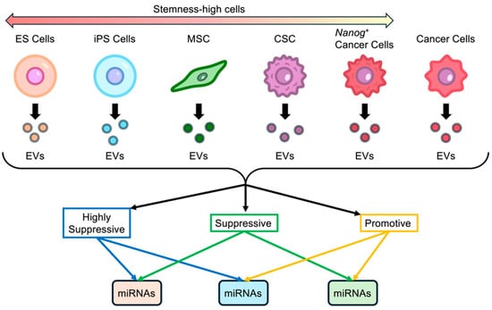
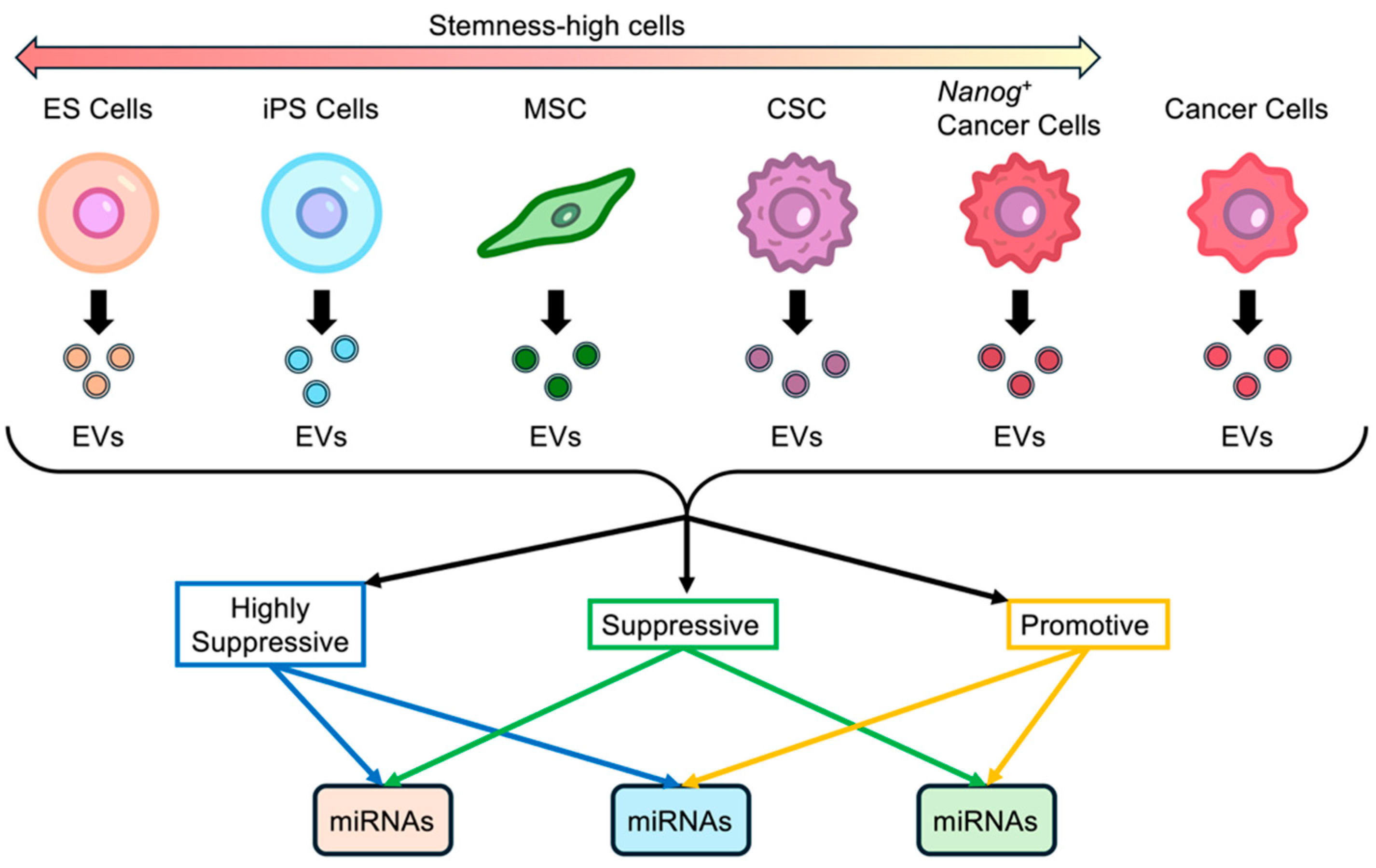
There are no quantitative indicators for stemness, and therefore, there is no fixed definition of stemness-high cells. The term is used by the authors for convenience in contrast with cells that have differentiated into specific functions (Figure 1). Embryonic stem (ES) cells, which are undifferentiated cells, can be considered the most stemness-high cells. This is followed by induced pluripotent stem (iPS) cells, mesenchymal stem (MS) cells, and others. Cancer cells may be considered cells that have differentiated into specific functional cells. Cancer cells obtained by introducing a stemness-intensifying factor such as Nanog are considered to have more stemness-high than the cancer cells of the original wild cell line. Additionally, cancer stem (CS) cells are considered to have more stemness-high than cancer cells [11,12,13].
Figure 1.
Stemness-high cells are EV resources with significant metastasis suppression effects. ES cells, embryonic stem cells; iPS cells, induced pluripotent cells; MS cells, mesenchymal stem cells; CS cells, cancer stem cells; Nanog+ cancer cells, Nanog overexpressing cancer cells.
Differential miRNA analysis between metastasis-suppressive EVs and metastasis-promoting EVs may predict miRNAs specific to metastasis regulation. This analytical method is not new but is especially effective in identifying metastasis-regulating miRNAs that are differently categorized from the cancer-associated miRNAs that have been extensively studied to date.
The effects of EVs secreted by stemness-high cells on cancer metastasis, whether they are promotive or suppressive, might not necessarily be the same as was previously understood. These EVs may play more multifaceted roles than anticipated. There is a growing hope that we will be able to discover novel functional molecules such as miRNAs and their target genes that can be used to develop cancer metastasis therapies.
2. Activation of Immune Cells with EVs Derived from Highly Metastatic Cancer Cells
EVs secreted from cancer cells play a dual role in vivo. One role is the promotion of growth in cancer cells. EVs secreted by cancer cells are taken up by tissue cells, thereby introducing cancer cell information into those tissue cells. This creates a metastasis-inducing niche, an environment that promotes the engraftment and proliferation of cancer cells in the vicinity [14,15].
The other role is the inhibition of the growth of cancer cells. When EVs secreted by cancer cells are taken up by dendritic cells, the dendritic cells are activated to secrete EVs that can activate T cells and macrophages to transform them into anticancer cells. Elements of the acquired immunity system, such as T cells, selectively exhibit anticancer properties against the original cancer cells secreting the EVs that initially activated the dendritic cells. There is a memory effect, meaning that the anticancer activity is retained for a certain period of time [16,17]. On the other hand, macrophages of the innate immune system have also been reported to exhibit this memory effect [18,19,20]. Although nonspecific, this suggests the possibility that responses to what is recognized as foreign to the organism may be sustained and remembered for a certain period of time.
Cancer cells encountered by immune cells during the evolution of the disease are those that have survived conventional treatment and are therefore likely to be highly malignant. In order for immune cells to exhibit sufficient anticancer activity against such cells, it is necessary to activate them using EVs derived from malignant cancer cells. Experimentally, metastatic potency represents a practical indicator of the malignancy of cancer cells.
3. Is Achieving Zero Risk of Cancer Metastasis Possible?
Multiple genes have been proposed as stemness-intensifying factors. Among them, the homeodomain transcription factor Nanog is the key factor for maintaining self-renewal and pluripotency characteristics. In humans, the Nanog protein, coded by the Nanog1 gene, consists of 305 amino acids and contains a conserved homeodomain that binds to DNA. There are ten pseudogenes (NanogP2-P11), with NanogP8 being highly expressed in cancer cells [21]. In mice, however, no pseudogene has been reported. Nanog proteins are central regulators controlling both embryonic stem cells and cancer stem cells and are considered a marker of poor prognosis in many types of cancer.
Therefore, it was predicted that the overexpression of Nanog in cancer cells would increase the metastatic potential of the cancer and lead to the progression of malignancy. To demonstrate this effect of Nanog, mouse melanoma was selected as the test material, because there are several cell lines of the same lineage with different intensities of metastatic potential (B16F0, B16F10, B16BL6, etc.). These cell lines are considered to be useful as controls for the quantitative evaluation of changes in the intensity of metastatic potential. Another reason why mouse melanoma was selected was that it forms solid black metastatic colonies, which are relatively easy to analyze (in terms of number and volume) via microscopic measurements. As expected, the Nanog-overexpressing strain showed higher metastatic potential than the wild-type strain [22].
However, when the properties of EVs from the Nanog-overexpressing and wild-type strains were examined, unexpected results were obtained. The initial expectation was that EVs from any cell would be metastasis-promoting, with the stronger metastasis-promoting EVs being from Nanog-overexpressing cells (Nanog+F10-EVs). In fact, however, EVs from a wild strain F10 (F10-EVs) were metastasis-promoting, while the Nanog+F10-EVs were, contrary to expectations, metastasis-suppressive [9]. Colon cancer showed similar results. Nanog-overexpressing colon 26 (Nanog+colon26) had a higher metastatic potential than wild-type colon 26, while the EVs obtained from Nanog+colon26-EVs showed metastasis suppression [23].
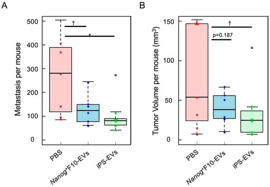
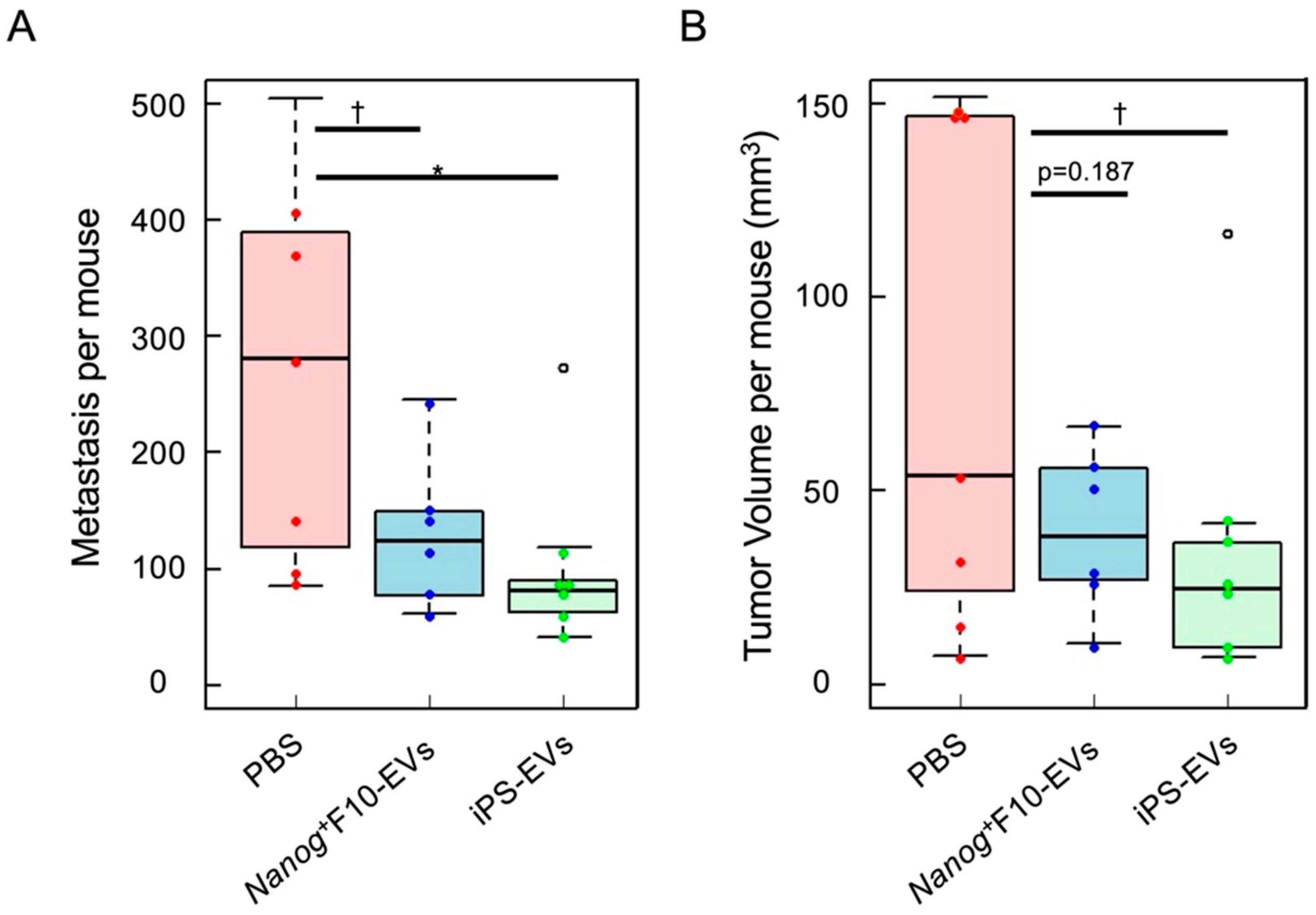
It was considered that the metastasis-suppressive effect of EVs might be closely related to the fact that the cells that secreted EVs were stemness-high cells. Therefore, EVs secreted from cells with higher stemness may exhibit a more intense suppression effect than Nanog+F10-EVs. However, when embryonic stem cells and iPS cells were focused on, and EVs derived from mouse iPS cells (iPS-EVs) were applied to metastasis experiments using mouse melanoma (Nanog+F10), they showed even stronger metastasis suppression than Nanog+F10-EVs (Figure 2).
Figure 2.
Metastasis-suppressive effects of Nanog+F10-EVs or iPS-EVs in mice [3]. Total number (A) and volume (B) of colonies in the liver and lungs. PBS n = 7, Nanog+F10-EVs n = 6, and iPS-EVs n = 7. (°) outlier, * p < 0.05, † p < 0.1.
It was a mystery why iPS-EVs showed a suppressive effect on melanoma metastasis when there was no information about melanoma in the iPS-EVs. It was speculated that Nanog+F10-EVs and iPS-EVs exerted their suppressive effects through different molecular mechanisms. Therefore, it was expected that a combination of the different types of EVs may show additive effects and reduce metastasis to almost zero.
4. Candidates for Metastasis-Regulating miRNAs and Their Target Genes
miRNA sequencing analysis was performed on five types of EVs (Nanog+F10-EVs [S], F10-EVs [P], Nanog+colon26-EVs [S], colon26-EVs [P], and iPS-EVs [S]) ([S], metastasis-suppressive; [P], metastasis-promotive). miRNAs that changed significantly were determined using the differential analysis of [S]/[P] and are shown in Figure 2. Since iPS-EVs showed higher metastasis suppression than Nanog+F10-EVs[S], iPS-EVs[S]/Nanog+F10-EVs[S] differential analysis was performed. Based on the results, the miRNAs with the top 4–6 highest fold changes were predicted. After predicting the target genes of each miRNA using TargetScanMouse 8.0 at CWCS ≤ −0.4, the strength of functional association was analyzed using STRING for the predicted gene clusters, and the top genes with the strongest functional association were analyzed using Cytoscan/cytoHubba. The predicted top genes are candidates for hub genes, which represent the core of metastasis suppression signaling pathways.
STRING gene function information is compiled based on publicly available information on a gene. It provides a relatively comprehensive description of the functions of cells and tissues. Since particular emphasis was placed on information related to the immune system, keywords related to immune function were established, and we ranked the function-related intensities based on these keywords. Consequently, candidate metastasis-regulating miRNAs and target hub genes were predicted (Table 1).
Table 1.
Candidates for melanoma metastasis-regulating miRNAs and their target genes with high functional association [3,23].
5. Metastasis-Regulating miRNAs Are Different from Cancer-Associated miRNAs
Cancer-associated miRNAs have been extensively studied [24]. In this review, the applications of miRNA-based diagnostic and therapeutic methods in cancer medicine are listed in a seven-page table citing 73 references. In total, 177 types of miRNA were identified in 17 types of cancer. In the cited literature, breast cancer and lung cancer were the most common, with 14 cases each.
In the case of breast cancer-associated miRNAs [25], for example, five miRNAs are identified as “oncogenic miRNAs” that are involved in tumorigenesis, metastasis, and drug resistance, and four types of miRNAs are identified as “tumor-suppressive miRNAs” that are involved in the suppression of tumor-related properties (Table 2).
Table 2.
Tumor-related miRNAs in breast cancer and their relationship with tumorigenesis, metastasis, and drug resistance [25].
Of the miRNAs in Table 1, however, only miR-19a-3p and miR-19b-3p were mentioned in the review [24], and no type of miRNA was common between those in Table 1 and Table 2, suggesting that there were very few previous studies concerning the miRNAs in Table 1. To further confirm the lack of studies on the miRNAs in Table 1, relevant studies in the literature were searched for through PubMed-NCBI (https://pmc.ncbi.nlm.nih.gov/). A search for “miR-21&cancer”, for instance, selected 43,979 articles. The average number of articles selected for five oncogenic miRNAs and four tumor-suppressing miRNAs in Table 2 was 22,709 and 20,390, respectively. In contrast, the average number of selected articles for five up-miRNAs and seven down-miRNAs in Table 1 was only 1434 and 1224, respectively.
This indicates that metastasis-regulating miRNAs seem to form a small group, different from the large group of cancer-associated miRNAs. Furthermore, miRNAs assigned as metastasis-regulating miRNAs have only recently begun to attract interest as a research topic.
6. Recent Work on miRNAs Associated with Stemness-High Cells
Recent work on miRNAs’ roles in ES, iPS, and MS cells (including some miRNAs in EVs) is summarized in Table 3. An emphasis is placed on whether the miRNAs were tumor-promotive or tumor-suppressive and whether they were able to serve as indicators of the properties of tumor cells or immune cells.
Table 3.
Potential roles of miRNAs in stemness-high cells.
6.1. ES Cells
Human embryonic stem (hES) cells are pluripotent cell lines established from internal cell masses harvested from human blastocysts. To investigate ES cell-specific miRNAs, miRNAs expressed in ES cells and embryonic cancer cells (EC cells) were comparatively analyzed. Of the 36 miRNAs identified via cDNA cloning, 8 were expressed in both ES and EC cells, while 7 were expressed only in ES cells [26]. The temporal changes in the expression of miRNAs during early differentiation from ES cells were investigated. The results showed that they could be classified into five clusters. Once a cluster with a high correlation with safety (free from cancer risk) was determined, the miRNAs expressed in that cluster would represent the next safety marker [27]. However, there have been no reports involving safety markers since then.
6.2. iPS Cells
For practical use in tissue regeneration, iPS cells are easier to use than ES cells. In such cases, the quality of the cells, including safety, is important. miRNAs are candidates for such indicators; one is miR-27b, which is highly conserved in vertebrates and involved in the regulation of various target genes in signaling pathways [49]. miR-27b also plays a role in the suppression of embryonic development in human iPS cells by acting antagonistically to the BMP signaling pathway [30]. Therefore, miR-27b must be downregulated to promote embryogenesis. Before differentiation, the maintenance of pluripotency is essential, but the selectivity and efficiency are important when differentiating in a specific direction. miRNAs involved in these cases are described in [31,33] and [32,34,35,37], respectively.
If EVs are to be used for practical purposes, then their mass preparation is required. An index consisting of ten miRNAs has been proposed for such a purpose [38].
A striking suggestion is that cancer cells could be rendered stemness-high via a novel method using miRNAs [36]. Four types of miRNAs were introduced into colon cancer cells, and ES cell-like cells were obtained, expressing undifferentiation marker genes of Nanog, Oct3/4, SOX2, and Klf4, as well as tumor-associated antigen 1–60. Previously, ES cell-like cells could also be produced via induction with four transcription factors (Oct3/4, Sox2, c-Myc, and Klf4) according to the conventional remodeling method. However, the expression of the oncogene c-Myc remained at a high level, which was a serious problem. The novel miRNA method solved this problem.
6.3. MS Cells
Mesenchymal stem cells (MS cells) can also be differentiated into various cell types. miRNAs are involved in promotion, regulation, and suppression in differentiation processes.
A suppressive role has been observed in osteogenesis. When MS cells were co-cultured with human periodontal ligament cells (HPL cells) for periodontal tissue regeneration therapy, bone formation was suppressed under the influence of fluid secretions from HPL cells. miR-299-5p was involved in this suppressive effect [39]. When MS cells were co-cultured with human gingival fibroblasts (HGFs), bone formation was also suppressed under the influence of humoral secretions from HGFs. In this case, miR-101-3p was involved in the suppressive effect [40].
miR-503 promoted vascular smooth muscle cell (VSMC) differentiation by inhibiting SMAD7 via the TGF-β pathway from upstream. In addition, miR-143 and miR-145 were upregulated by activated TGF-β1. On the other hand, miR-222-5p conversely repressed VSMC differentiation by downregulating ROCK2 and α-SMA [41]. This is an example of a situation in which promotion and suppression occur simultaneously. Numerous similar cases have been observed.
miR-200b-3p promoted the differentiation of MS cells into insulin-producing cells through interaction with the zinc finger E-box binding homeobox 2 (ZEB2) 3’ untranslated region (UTR) [42]. miR-145 promoted the differentiation of bone marrow MS cells into smooth muscle cells [43]. Osteogenic differentiation was promoted by miR-29b via the PTEN/AKT/β-catenin signaling pathway [44] and also regulated by miR-125b by targeting Cdfβ [45]. miR-34a activated alveolar differentiation by inhibiting TGF-βsignaling and activating the BPM pathway [46].
The idea of applying miRNAs as indicators of cell quality is exemplified by iPS cells. However, a similar method was applied to MS cells to determine the differentiation potential in a specific direction using the expression status of multiple miRNAs [47]. The ability of MS cells to differentiate into neurons was investigated to explore potential therapeutic tools for neurological diseases. After the induction of differentiation and conversion, miRNA expression was analyzed over time and classified into five clusters based on expression trajectories. It was found that each cluster contained unique miRNAs [48].
7. miRNAs in Innate Immune Cells
In future, when facing attacks from invading cancer cells in the absence of cancer cell information, it is considered that the main players will be those of the innate immune system, that is, macrophages and natural killer (NK) cells, and the involvement of dendritic cells (DCs) must also be considered. Therefore, we have summarized examples of the roles of miRNAs acting within macrophages, NK cells, and dendritic cells or from outside of each cell.
7.1. miRNAs Ccontributing to Macrophage Activation
There are many reviews on the involvement of miRNAs in macrophage function. With respect to data supporting the interaction between miRNAs and metabolism in macrophage inflammatory responses, the focus has been on key miRNAs and miRNA families, and furthermore, the impact of these networks on human disease has been discussed and summarized [50]. Another review comprehensively describes the molecular mechanisms of miRNAs in macrophage polarization, to analyze the potential value of regulatory pathways in the context of tumor and inflammation intervention therapies [51]. Some commentaries focus on specific diseases. For example, in asthma, there is an imbalance between classically activated macrophages (M1 cells) and selectively activated macrophages (M2 cells), with the latter predominating. Therefore, data on miRNAs known to correlate with specific human macrophage phenotypes and polarization, as well as their association with adult asthma, have been compiled [52]. Macrophages also play an important role in the progression of acute lung injury (ALI)/acute respiratory distress syndrome (ARDS) and exhibit different polar morphologies depending on the stage of disease progression. Therefore, an overview of miRNA expression in ALI/ARDS was presented, and the mechanisms and pathways by which miRNAs respond to macrophage polarization, inflammation, and apoptosis were introduced [53].
On the other hand, there is an interesting commentary on the homeostasis of the M1/M2 state of macrophages [54], which will be discussed below in detail. The polarization of macrophage M1/M2, which detects bacterial and viral motifs (pathogen-associated molecular patterns, PAMPs) in bacteria- and virus-infected tissues, increases tissue inflammation through cytokine production and kills bacteria and viruses. This inflammation-induced activation is referred to as the classically activated state or M1-like macrophages. In addition to PAMPs, the detection of inflammatory cytokines such as interferon gamma (INFγ) also results in M1-like macrophages. In contrast, the detection of proinflammatory cytokines such as interleukin 4 (IL-4) and IL-13 leads to the suppression of the inflammatory response, removal of cellular debris, and reconstruction of the extracellular matrix. This state is referred to as an alternative activation state or M2-like macrophages. Furthermore, if the M1 state persists, which may result in abnormal inflammatory activity, a feedback inhibition loop is activated to maintain homeostasis. Similarly, in the case of the M2 state, a feedback inhibition loop is activated to prevent excessive activity and maintain homeostasis. The function of macrophages is not simply polarized based on whether they are in the M1 or M2 state; rather, they have a fine-tuning function to maintain homeostasis in both states. miRNAs are mainly responsible for this fine-tuning function (Table 4).
Table 4.
miRNAs activating and regulating macrophages [54].
The action of miRNAs is a challenging issue from a therapeutic point of view, because the target cell types differ in terms of their metastasis-inhibitory effects. miR-146 is an example of complicated regulation. miR-146 is a novel miRNA that is known to suppress the metastasis of cancer cells. The activation of the Jak-Stat signaling pathway (mainly Stat3) in cancer cells increases cancer cell survival and proliferation. In contrast, suppressor of cytokine signaling 1 (SOCS1) suppresses the Jak-Stat signaling pathway, so the activation of SOCS1 in cancer cells suppresses cancer cell growth. Since miR-146 can suppress SOCS1, miR-146 can potentially promote cancer cell growth. This process is complicated: when miR-146 is upregulated in cancer cells, SOCS1 (Down)/Jak-Stat (Up)/cancer cell proliferation (Up) is observed, which is counterproductive for metastasis suppression. In macrophages, however, SOCS1 suppresses the Cytokine/Toll-like receptor (TLR) signaling pathway. As a result, macrophage survival and proliferation are suppressed. Thus, increased expression of miR-146 in macrophages has a favorable effect on metastasis suppression: SOCS1 (Down)/cytokine, TLR (Up)/macrophage proliferation (Up). Therefore, when applying miR-146 in vivo, it must be delivered selectively to macrophages only. The high quality of miRNAs means that even if there are multiple target genes, none of them are guaranteed to have a negative effect on metastasis inhibition.
7.2. miRNAs Contributing to NK Cell Activation
In the human immune system, potent miRNAs and miRNA clusters have been discovered that play a pivotal role in the regulation of gene expression, termed immune miRNAs. Immune miRNAs exert a regulatory influence on both innate and acquired immune cells. They do not necessarily activate anti-tumor effects but may conversely blunt anti-tumor responses and create an environment inducive to tumor survival [55]. B7-H3 (a member of the B7 family of immune checkpoint molecules) is involved in T cell immune responses but has been newly shown to be involved in NK cell function, as well [56]. In mouse experiments, the inhibition of B7-H3 in ovarian cancer prolonged mouse survival. In vitro, miR-29c directly targeted and inhibited the expression of B7-H3, enhancing the anti-tumor effect of NK cells. However, in vivo, miR-29c/B7-H3 suppression did not improve the anti-tumor effect. There was a decrease in miR-29 family members (miR-29a, miR-29b, miR-29c) in deceased patients, which correlated inversely with B7-H3 expression in neuroblastoma patients. miR-29 family members promoted NK cell infiltration and activation and induced tumor cell apoptosis [57].
Natural Killer Group 2 Member D (NKG2D) is an important activation receptor for NK cells. UL16-binding protein 2 (ULBP2) is constantly expressed or elevated in cancer cells and serves as an important NKG2D ligand. The binding of ULBP2 and NKG2D initiates NK cell activation and the subsequent targeted elimination of cancer cells. Thus, enhanced expression of ULBP2 leads to a more efficient elimination of these cells by NK cells. The administration of resveratrol (RES) reduced intracellular miR-17-5p levels and increased ULBP2 expression, since ULBP2 was a target of miR-17-5p. As a result, the cytotoxic activity of NK cells was increased using RES [58].
The effects of miRNAs are not always as expected. In breast cancer (BC) tissue, miR-338-3p levels were significantly reduced. This trend was more pronounced in patients with advanced stages. To inhibit breast cancer cell survival, miR-338-3p was overexpressed. However, the axis of miR-338-3p (Up) in BC/ADAM17 secretion (Down)/granzyme B, CD16, and NKG2D (Down) inNK/NK cell function (Down)/BC survival (Up) ultimately resulted in an effect that increased BC cell survival [59].
An integrated miRNA and mRNA immune cell signature was constructed to predict survival in breast and ovarian cancer patients. miRNA expression-based immune cell signatures were constructed from the breast and ovarian cancer data sets of The Cancer Genome Atlas and combined with existing mRNA immune cell signatures. The constructed signatures enabled a more accurate depiction of prognostic and predictive immune cell subsets in both cancers [60].
7.3. miRNAs Involved in Dendritic Cell Function
Using miRNA array analysis, a representative miR-5119 was identified in breast cancer as a potential regulator of PD-L1 in dendritic cells (DCs). miR-5119 mimic modified DCs effectively restored the function of exhausted CD8+ T cells in vitro and in vivo and resulted in potent anti-tumor cellular immune responses, increased cytokine production, and decreased T cell apoptosis [61].
Cancer-associated fibroblasts (CAFs), a major component of the tumor microenvironment (TME), are known to modulate immune responses through the secretion of exosomes containing various growth hormones, miRNAs, and cytokines, but their effects on DCs were unknown. To confirm the effect of CAF-derived exosomes on DC maturation, DCs were cultured in the presence of a CAF-derived acclimation medium (CAFs-CM). The role of epigenetic regulators in maturation was assessed via the expression of miR-221, miR-222, miR-155, miR-142-3p, and miR-146a. Among them, an increased expression of miR-146a was observed, which was positively correlated with an increased expression of anti-inflammatory cytokines such as IL-6, IL-10, and TGF-β and a decreased expression of TNF-α (inflammation-inducing) [62].
DCs primed with exosomes rich in miR-155 inhibited tumor growth. Primed DCs significantly increased IL-12p70 and IFN-γ in serum and accelerated the differentiation, proliferation, and cytotoxic effects of helper T cells and cytotoxic T cells [63]. miR-155-overexpressing dendritic cells (DCs) were used to generate a DC vaccine. In mice transfected with this vaccine, an increase in effector T cells, the suppression of tumor growth, and a dramatic decrease in lung metastases were observed [64].
The miRNAs cited in this section are listed in Table 5.
Table 5.
miRNAs associated with innate immune system.
8. Properties and Roles of Metastasis-Regulating miRNAs Demonstrated in Other Studies
According to a PubMed search, the number of papers dealing with metastasis-regulating miRNAs was extremely small. There was very little commonality between metastasis-regulating miRNAs (Table 1) and miRNAs of other categories (cancer-associated miRNAs and those cited in Section 6 and Section 7). Although there are few papers on these metastasis-regulating miRNAs, we have summarized their properties and functional roles as described in those papers (Table 6). In the case of research on cancer and disease, it seems that many miRNAs exacerbate the condition. In light of this situation, the remarkable effect of miRNAs in suppressing cancer metastasis seems to be an extremely rare phenomenon. This strongly suggests that the search for miRNAs contributing to the suppression of cancer metastasis is still insufficient.
9. Conclusions
A systematic study focusing on metastasis-regulating miRNAs is important in facilitating the development of novel miRNAs with enhanced antimetastatic properties and in the analyses of cellular and molecular mechanisms underlying these effects. Regarding novel miRNAs, the strategy is to design appropriate pairs in which one is metastasis-suppressive and the other is metastasis-promotive. Based on the results of numerous experimental studies and reviews, it appears that it is more difficult to obtain miRNAs that contribute to metastasis suppression than those that contribute to metastasis promotion. From this perspective, Nanog-activated cancer cells and iPS cells could be innovatively employed to prepare metastasis-suppressive EVs. EVs from Nanog-activated cancer X cells are expected to represent a resource that can suppress the metastasis of cancer X cells. On the other hand, EVs from iPS cells are expected to provide a resource that can suppress metastasis in any type of cancer. This simple idea may lead to a useful approach across cancer types.
Given the complexity of the components within EVs, it is extremely difficult to fully elucidate their functional roles and use in stemness-high cells. However, based on the research trends observed in this review, it is expected that various miRNA conditions (types, combinations, concentrations, etc.) will lead to high-performance treatments to reduce metastasis risk to almost zero.
Author Contributions
Conceptualization, M.S. and H.M.; investigation, M.S. and H.M.; resources, M.S. and H.M.; writing—original draft preparation, M.S. and H.M.; writing—review and editing, M.S. and H.M.; visualization, M.S. and H.M.; supervision, M.S.; project administration, M.S.; funding acquisition, M.S. All authors have read and agreed to the published version of the manuscript.
Funding
This study was supported by a special grant to TUAT and JSPS KAKENHI Grant Number JP24K09432.
Conflicts of Interest
The authors declare no conflicts of interest.
References
- Kitai, Y.; Kawasaki, T.; Sueyoshi, T.; Kobiyama, K.; Ishii, K.J.; Zou, J.; Akira, S.; Matsuda, T.; Kawai, T. DNA-Containing Exosomes Derived from Cancer Cells Treated with Topotecan Activate a STING-Dependent Pathway and Reinforce Antitumor Immunity. J. Immunol. 2017, 198, 1649–1659. [Google Scholar] [CrossRef] [Scilit]
- Rezaei, R.; Baghaei, K.; Hashemi, S.M.; Zali, M.R.; Ghanbarian, H.; Amani, D. Tumor-Derived Exosomes Enriched by miRNA-124 Promote Anti-tumor Immune Response in CT-26 Tumor-Bearing Mice. Front. Med. 2021, 8, 619939. [Google Scholar] [CrossRef] [Scilit]
- Khoo, C.S.M.; Henmi, T.; Saito, M. Comparative Study of Metastasis Suppression Effects of Extracellular Vesicles Derived from Anaplastic Cell Lines, Nanog-Overexpressing Melanoma, and Induced Pluripotent Stem Cells. Int. J. Mol. Sci. 2023, 24, 17206. [Google Scholar] [CrossRef] [Scilit]
- Loric, S.; Denis, J.A.; Desbene, C.; Sabbah, M.; Conti, M. Extracellular Vesicles in Breast Cancer: From Biology and Function to Clinical Diagnosis and Therapeutic Management. Int. J. Mol. Sci. 2023, 24, 7208. [Google Scholar] [CrossRef] [Scilit] [PubMed]
- Xia, W.; Tan, Y.; Liu, Y.; Xie, N.; Zhu, H. Prospect of extracellular vesicles in tumor immunotherapy. Front. Immunol. 2025, 16, 1525052. [Google Scholar] [CrossRef] [Scilit] [PubMed]
- Claridge, B.; Lozano, J.; Poh, Q.H.; Greening, D.W. Development of Extracellular Vesicle Therapeutics: Challenges, Considerations, and Opportunities. Front. Cell Dev. Biol. 2021, 9, 734720. [Google Scholar] [CrossRef] [Scilit] [PubMed]
- Al-Jipouri, A.; Eritja, À.; Bozic, M. Unraveling the Multifaceted Roles of Extracellular Vesicles: Insights into Biology, Pharmacology, and Pharmaceutical Applications for Drug Delivery. Int. J. Mol. Sci. 2023, 25, 485. [Google Scholar] [CrossRef] [Scilit]
- Wang, J.; Yin, B.; Lian, J.; Wang, X. Extracellular Vesicles as Drug Delivery System for Cancer Therapy. Pharmaceutics 2024, 16, 1029. [Google Scholar] [CrossRef] [Scilit]
- Hatakenaka, T.; Matsuki, N.; Minagawa, S.; Khoo, C.S.M.; Saito, M. Anti-Metastatic Function of Extracellular Vesicles Derived from Nanog-Overexpressing Melanoma. Curr. Oncol. 2022, 29, 1029–1046. [Google Scholar] [CrossRef] [Scilit]
- Wang, R.; Zhu, T.; Hou, B.; Huang, X. An iPSC-derived exosome-pulsed dendritic cell vaccine boosts antitumor immunity in melanoma. Mol. Ther. 2023, 31, 2376–2390. [Google Scholar] [CrossRef] [Scilit]
- Ma, Y.; Yu, W.; Shrivastava, A.; Srivastava, R.K.; Shankar, S. Inhibition of pancreatic cancer stem cell characteristics by α-Mangostin: Molecular mechanisms involving Sonic hedgehog and Nanog. J. Cell. Mol. Med. 2019, 23, 2719–2730. [Google Scholar] [CrossRef] [Scilit]
- Gawlik-Rzemieniewska, N.; Bednarek, I. The role of NANOG transcriptional factor in the development of malignant phenotype of cancer cells. Cancer Biol. Ther. 2015, 17, 1–10. [Google Scholar] [CrossRef] [Scilit] [PubMed]
- Haddadin, L.; Sun, X. Stem Cells in Cancer: From Mechanisms to Therapeutic Strategies. Cells 2025, 14, 538. [Google Scholar] [CrossRef] [Scilit]
- Lucotti, S.; Kenific, C.M.; Zhang, H.; Lyden, D. Extracellular vesicles and particles impact the systemic landscape of cancer. EMBO J. 2022, 41, e109288. [Google Scholar] [CrossRef] [Scilit]
- Naito, Y.; Yoshioka, Y.; Ochiya, T. Intercellular crosstalk between cancer cells and cancer-associated fibroblasts via extracellular vesicles. Cancer Cell Int. 2022, 22, 367. [Google Scholar] [CrossRef] [Scilit]
- Radbruch, A.; McGrath, M.A.; Siracusa, F.; Hoffmann, U.; Sercan-Alp, Ö.; Hutloff, A.; Tokoyoda, K.; Chang, H.-D.; Dong, J. Homeostasis and Durability of T-Cell Memory—The Resting and the Restless T-Cell Memory. Cold Spring Harb. Perspect. Biol. 2021, 13, a038083. [Google Scholar] [CrossRef] [Scilit] [PubMed]
- Kawabe, T.; Ciucci, T.; Kim, K.S.; Tayama, S.; Kawajiri, A.; Suzuki, T.; Tanaka, R.; Ishii, N.; Jankovic, D.; Zhu, J.; et al. Redefining the Foreign Antigen and Self-Driven Memory CD4+ T-Cell Compartments via Transcriptomic, Phenotypic, and Functional Analyses. Front. Immunol. 2022, 13, 870542. [Google Scholar] [CrossRef] [Scilit] [PubMed]
- Seeley, J.J.; Baker, R.G.; Mohamed, G.; Bruns, T.; Hayden, M.S.; Deshmukh, S.D.; Freedberg, D.E.; Ghosh, S. Induction of innate immune memory via microRNA targeting of chromatin remodelling factors. Nature 2018, 559, 114–119. [Google Scholar] [CrossRef] [Scilit]
- Netea, M.G.; Domínguez-Andrés, J.; Barreiro, L.B.; Chavakis, T.; Divangahi, M.; Fuchs, E.; Joosten, L.A.B.; van der Meer, J.W.M.; Mhlanga, M.M.; Mulder, W.J.M.; et al. Defining trained immunity and its role in health and disease. Nat. Rev. Immunol. 2020, 20, 375–388. [Google Scholar] [CrossRef] [Scilit]
- Maheshwari, A. Innate Immune Memory in Macrophages. Newborn 2023, 2, 60–79. [Google Scholar] [CrossRef] [Scilit]
- Zhang, W.; Sui, Y.; Ni, J.; Yang, T. Insights into the Nanog gene: A propeller for stemness in primitive stem cells. Int. J. Biol. Sci. 2016, 12, 1372–1381. [Google Scholar] [CrossRef] [Scilit]
- Saito, M.; Kishi, R.; Sasai, T.; Hatakenaka, T.; Matsuki, N.; Minagawa, S. Effect of Nanog overexpression on the metastatic potential of a mouse melanoma cell line B16-BL6. Mol. Cell. Biochem. 2021, 476, 2651–2661. [Google Scholar] [CrossRef] [Scilit]
- Henmi, T.; Matsuoka, H.; Katayama, N.; Saito, M. Suppression of Metastasis of Colon Cancer to Liver in Mouse Models by Pretreatment with Extracellular Vesicles Derived from Nanog-Overexpressing Colon-26 Cancer Cells. Int. J. Mol. Sci. 2024, 25, 12794. [Google Scholar] [CrossRef] [Scilit]
- Sempere, L.F.; Azmi, A.S.; Moore, A. microRNA-based diagnostic and therapeutic applications in cancer medicine. Wiley Interdiscip. Rev. RNA 2021, 12, e1662. [Google Scholar] [CrossRef] [Scilit]
- Manap, A.S.A.; Wisham, A.A.; Wong, F.W.; Najmi, H.R.A.; Ng, Z.F.; Diba, R.S. Mapping the function of MicroRNAs as a critical regulator of tumor-immune cell communication in breast cancer and potential treatment strategies. Front. Cell Dev. Biol. 2024, 12, 1390704. [Google Scholar] [CrossRef] [Scilit]
- Suh, M.-R.; Lee, Y.; Kim, J.Y.; Kim, S.-K.; Moon, S.-H.; Lee, J.Y.; Cha, K.-Y.; Chung, H.M.; Yoon, H.S.; Moon, S.Y.; et al. Human embryonic stem cells express a unique set of microRNAs. Dev. Biol. 2004, 270, 488–498. [Google Scholar] [CrossRef] [Scilit]
- Ciaudo, C.; Servant, N.; Cognat, V.; Sarazin, A.; Kieffer, E.; Viville, S.; Colot, V.; Barillot, E.; Heard, E.; Voinnet, O.; et al. Highly Dynamic and Sex-Specific Expression of microRNAs During Early ES Cell Differentiation. PLoS Genet. 2009, 5, e1000620. [Google Scholar] [CrossRef] [Scilit] [PubMed]
- Yamaguchi, S.; Yamahara, K.; Homma, K.; Suzuki, S.; Fujii, S.; Morizane, R.; Monkawa, T.; Matsuzaki, Y.; Kangawa, K.; Itoh, H. The role of microRNA-145 in human embryonic stem cell differentiation into vascular cells. Atherosclerosis 2011, 219, 468–474. [Google Scholar] [CrossRef] [Scilit]
- Yoo, J.K.; Kim, J.; Choi, S.; Noh, H.M.; Kwon, Y.D.; Yoo, H.; Yi, H.S.; Chung, H.M.; Kim, J.K. Discovery and Characterization of Novel MicroRNAs During Endothelial Differentiation of Human Embryonic Stem Cells. Stem Cells Dev. 2012, 21, 2049–2057. [Google Scholar] [CrossRef] [Scilit] [PubMed]
- Lim, J.; Sakai, E.; Sakurai, F.; Mizuguchi, H. miR-27b antagonizes BMP signaling in early differentiation of human induced pluripotent stem cells. Sci. Rep. 2021, 11, 19820. [Google Scholar] [CrossRef] [Scilit] [PubMed]
- Liu, T.; Chen, Q.; Huang, Y.; Huang, Q.; Jiang, L.; Guo, L. Low microRNA-199a expression in human amniotic epithelial cell feeder layers maintains human-induced pluripotent stem cell pluripotency via increased leukemia inhibitory factor expression. Acta Biochim. Biophys. Sin. 2012, 44, 197–206. [Google Scholar] [CrossRef] [Scilit]
- Sugawara, T.; Miura, T.; Kawasaki, T.; Umezawa, A.; Akutsu, H. The hsa-miR-302 cluster controls ectodermal differentiation of human pluripotent stem cell via repression of DAZAP2. Regen. Ther. 2020, 15, 1–9. [Google Scholar] [CrossRef] [Scilit]
- Sugawara, T.; Kawamoto, Y.; Kawasaki, T.; Umezawa, A.; Akutsu, H. A single allele of the hsa-miR-302/367 cluster maintains human pluripotent stem cells. Regen. Ther. 2022, 21, 37–45. [Google Scholar] [CrossRef] [Scilit]
- Di Bernardini, E.; Campagnolo, P.; Margariti, A.; Zampetaki, A.; Karamariti, E.; Hu, Y.; Xu, Q. Endothelial Lineage Differentiation from Induced Pluripotent Stem Cells Is Regulated by MicroRNA-21 and Transforming Growth Factor β2 (TGF-β2) Pathways. J. Biol. Chem. 2014, 289, 3383–3393. [Google Scholar] [CrossRef] [Scilit]
- Jaafarpour, Z.; Soleimani, M.; Hosseinkhani, S.; Geramizadeh, B.; Yaghmaei, P.; Mobarra, N.; Karimi, M.H. Overexpression of microRNA-375 and microRNA-122 promotes the differentiation of human induced pluripotent stem cells into hepatocyte-like cells. Biologicals 2020, 63, 24–32. [Google Scholar] [CrossRef] [Scilit]
- Miyazaki, S.; Yamamoto, H.; Miyoshi, N.; Wu, X.; Ogawa, H.; Uemura, M.; Nishimura, J.; Hata, T.; Takemasa, I.; Mizushima, T.; et al. A Cancer Reprogramming Method Using MicroRNAs as a Novel Therapeutic Approach against Colon Cancer. Ann. Surg. Oncol. 2014, 22, 1394–1401. [Google Scholar] [CrossRef] [Scilit]
- Marzano, M.; Bejoy, J.; Cheerathodi, M.R.; Sun, L.; York, S.B.; Zhao, J.; Kanekiyo, T.; Bu, G.; Meckes, D.G.; Li, Y. Differential Effects of Extracellular Vesicles of Lineage-Specific Human Pluripotent Stem Cells on the Cellular Behaviors of Isogenic Cortical Spheroids. Cells 2019, 8, 993. [Google Scholar] [CrossRef] [Scilit] [PubMed]
- Muok, L.; Sun, L.; Esmonde, C.; Worden, H.; Vied, C.; Duke, L.; Ma, S.; Zeng, O.; Driscoll, T.; Jung, S.; et al. Extracellular vesicle biogenesis of three-dimensional human pluripotent stem cells in a novel Vertical-Wheel bioreactor. J. Extracell. Biol. 2024, 3, e133. [Google Scholar] [CrossRef] [Scilit] [PubMed]
- Kaneda-Ikeda, E.; Iwata, T.; Mizuno, N.; Nagahara, T.; Kajiya, M.; Takeda, K.; Hirata, R.; Ishida, S.; Yoshioka, M.; Fujita, T.; et al. Periodontal ligament cells regulate osteogenesis via miR-299-5p in mesenchymal stem cells. Differentiation 2020, 112, 47–57. [Google Scholar] [CrossRef] [Scilit] [PubMed]
- Kaneda-Ikeda, E.; Iwata, T.; Mizuno, N.; Nagahara, T.; Kajiya, M.; Ouhara, K.; Yoshioka, M.; Ishida, S.; Kawaguchi, H.; Kurihara, H. Regulation of osteogenesis via miR-101-3p in mesenchymal stem cells by human gingival fibroblasts. J. Bone Miner. Metab. 2020, 38, 442–455. [Google Scholar] [CrossRef] [Scilit]
- Abolhasani, S.; Ahmadi, Y.; Rostami, Y.; Fattahi, D. The role of MicroRNAs in mesenchymal stem cell differentiation into vascular smooth muscle cells. Cell Div. 2025, 20, 6. [Google Scholar] [CrossRef] [Scilit] [PubMed]
- Chen, W.; Jiang, W.; Dong, J.; Wang, J.; Wang, B. miR-200b-3p Induces the Formation of Insulin-Producing Cells from Umbilical Cord Mesenchymal Stem Cells by Targeting ZEB2. Crit. Rev. Eukaryot. Gene Expr. 2022, 32, 33–46. [Google Scholar] [CrossRef] [Scilit]
- Pajoohesh, M.; Naderi-Manesh, H.; Soleimani, M. MicroRNA-145-based differentiation of human mesenchymal stem cells to smooth muscle cells. Biotechnol. Lett. 2016, 38, 1975–1981. [Google Scholar] [CrossRef] [Scilit]
- Xia, T.; Dong, S.; Tian, J. miR-29b promotes the osteogenic differentiation of mesenchymal stem cells derived from human adipose tissue via the PTEN/AKT/β-catenin signaling pathway. Int. J. Mol. Med. 2020, 46, 709–717. [Google Scholar] [CrossRef] [Scilit]
- Huang, K.; Fu, J.; Zhou, W.; Li, W.; Dong, S.; Yu, S.; Hu, Z.; Wang, H.; Xie, Z. MicroRNA-125b regulates osteogenic differentiation of mesenchymal stem cells by targeting Cbfβ in vitro. Biochimie 2014, 102, 47–55. [Google Scholar] [CrossRef] [Scilit]
- Acosta-Plasencia, M.; Castellano, J.J.; Díaz, T.; He, Y.; Marrades, R.M.; Navarro, A. Discovering genes and microRNAs involved in human lung development unveils IGFBP3/miR-34a dynamics and their relevance for alveolar differentiation. Stem Cell Res. Ther. 2024, 15, 263. [Google Scholar] [CrossRef] [Scilit]
- Alizadeh, A.H.; Lively, S.; Lepage, S.I.M.; Potla, P.; Russell, S.; Ali, S.A.; Kapoor, M.; Koch, T.G. MicroRNAs as Prognostic Markers for Chondrogenic Differentiation Potential of Equine Mesenchymal Stromal Cells. Stem Cells Dev. 2023, 32, 693–702. [Google Scholar] [CrossRef] [Scilit]
- Diener, C.; Thüre, K.; Engel, A.; Hart, M.; Keller, A.; Meese, E.; Fischer, U.; Alles, J.; Fehlmann, T.; Backes, C.; et al. Paving the way to a neural fate–RNA signatures in naive and trans-differentiating mesenchymal stem cells. Eur. J. Cell Biol. 2024, 103, 151458. [Google Scholar] [CrossRef] [Scilit]
- Rogler, C.E.; LeVoci, L.; Ader, T.; Massimi, A.; Tchaikovskaya, T.; Norel, R.; Rogler, L.E. MicroRNA-23b cluster microRNAs regulate transforming growth factor-beta/bone morphogenetic protein signaling and liver stem cell differentiation by targeting Smads†. Hepatology 2009, 50, 575–584. [Google Scholar] [CrossRef] [Scilit] [PubMed]
- Nelson, M.C.; O’COnnell, R.M. MicroRNAs: At the Interface of Metabolic Pathways and Inflammatory Responses by Macrophages. Front. Immunol. 2020, 11, 1797. [Google Scholar] [CrossRef] [Scilit] [PubMed]
- Wang, C.; Wang, X.; Zhang, D.; Sun, X.; Wu, Y.; Wang, J.; Li, Q.; Jiang, G. The macrophage polarization by miRNAs and its potential role in the treatment of tumor and inflammation (Review). Oncol. Rep. 2023, 50, 190. [Google Scholar] [CrossRef] [Scilit]
- Feketea, G.; I Bocsan, C.; Popescu, C.; Gaman, M.; A Stanciu, L.; Zdrenghea, M.T. A Review of Macrophage MicroRNAs’ Role in Human Asthma. Cells 2019, 8, 420. [Google Scholar] [CrossRef] [Scilit]
- Zhang, J.; Tian, W.; Wang, F.; Liu, J.; Huang, J.; Duangmano, S.; Liu, H.; Liu, M.; Zhang, Z.; Jiang, X. Advancements in understanding the role of microRnas in regulating macrophage polarization during acute lung injury. Cell Cycle 2023, 22, 1694–1712. [Google Scholar] [CrossRef] [Scilit]
- Sprenkle, N.T.; Serezani, C.H.; Pua, H.H. MicroRNAs in Macrophages: Regulators of Activation and Function. J. Immunol. 2023, 210, 359–368. [Google Scholar] [CrossRef] [Scilit]
- Hirschberger, S.; Hinske, L.C.; Kreth, S. MiRNAs: Dynamic regulators of immune cell functions in inflammation and cancer. Cancer Lett. 2018, 431, 11–21. [Google Scholar] [CrossRef] [Scilit]
- Deng, M.; Wu, D.; Zhang, Y.; Jin, Z.; Miao, J. MiR-29c downregulates tumor-expressed B7-H3 to mediate the antitumor NK-cell functions in ovarian cancer. Gynecol. Oncol. 2021, 162, 190–199. [Google Scholar] [CrossRef] [Scilit] [PubMed]
- Pathania, A.S.; Chava, H.; Chaturvedi, N.K.; Chava, S.; Byrareddy, S.N.; Coulter, D.W.; Challagundla, K.B. The miR-29 family facilitates the activation of NK-cell immune responses by targeting the B7-H3 immune checkpoint in neuroblastoma. Cell Death Dis. 2024, 15, 428. [Google Scholar] [CrossRef] [Scilit] [PubMed]
- Ding, B.; Li, J.; Yan, J.-L.; Jiang, C.-Y.; Qian, L.-B.; Pan, J. Resveratrol contributes to NK cell-mediated breast cancer cytotoxicity by upregulating ULBP2 through miR-17-5p downmodulation and activation of MINK1/JNK/c-Jun signaling. Front. Immunol. 2025, 16, 1515605. [Google Scholar] [CrossRef] [Scilit] [PubMed]
- Shi, Y.; Pan, J.; Hang, C.; Tan, L.; Hu, L.; Yan, Z.; Zhu, J. The estrogen/miR-338-3p/ADAM17 axis enhances the viability of breast cancer cells via suppressing NK cell’s function. Environ. Toxicol. 2023, 38, 1618–1627. [Google Scholar] [CrossRef] [Scilit]
- Ray, M.; Ruffalo, M.M.; Bar-Joseph, Z. Construction of integrated microRNA and mRNA immune cell signatures to predict survival of patients with breast and ovarian cancer. Genes Chromosom. Cancer 2018, 58, 34–42. [Google Scholar] [CrossRef] [Scilit]
- Zhang, M.; Shi, Y.; Zhang, Y.; Wang, Y.; Alotaibi, F.; Qiu, L.; Wang, H.; Peng, S.; Liu, Y.; Li, Q.; et al. miRNA-5119 regulates immune checkpoints in dendritic cells to enhance breast cancer immunotherapy. Cancer Immunol. Immunother. 2020, 69, 951–967. [Google Scholar] [CrossRef] [Scilit]
- Mirza, S.; Penny, C.; Jain, N.K.; Rawal, R.M. Curcumin mediated dendritic cell maturation by modulating cancer associated fibroblasts-derived exosomal miRNA-146a. J. Cancer Res. Ther. 2023, 19, S649–S657. [Google Scholar] [CrossRef] [Scilit]
- Asadirad, A.; Baghaei, K.; Hashemi, S.M.; Dehnavi, S.; Ghanbarian, H.; Mortaz, E.; Anissian, A.; Aghdaei, H.A.; Amani, D. Dendritic cell immunotherapy with miR-155 enriched tumor-derived exosome suppressed cancer growth and induced antitumor immune responses in murine model of colorectal cancer induced by CT26 cell line. Int. Immunopharmacol. 2022, 104, 108493. [Google Scholar] [CrossRef] [Scilit] [PubMed]
- Hodge, J.; Wang, F.; Wang, J.; Liu, Q.; Saaoud, F.; Wang, Y.; Singh, U.P.; Chen, H.; Luo, M.; Ai, W.; et al. Overexpression of microRNA-155 enhances the efficacy of dendritic cell vaccine against breast cancer. OncoImmunology 2020, 9, 1724761. [Google Scholar] [CrossRef] [Scilit]
- Zhong, Q.; Nie, Q.; Wu, R.; Huang, Y. Exosomal miR-18a-5p promotes EMT and metastasis of NPC cells via targeting BTG3 and activating the Wnt/β-catenin signaling pathway. Cell Cycle 2023, 22, 1544–1562. [Google Scholar] [CrossRef] [Scilit]
- Santos, S.A.A.; Portela, L.M.F.; Camargo, A.C.L.; Constantino, F.B.; Colombelli, K.T.; Fioretto, M.N.; Mattos, R.; Fantinatti, B.E.d.A.; Denti, M.A.; Piazza, S.; et al. miR-18a-5p Is Involved in the Developmental Origin of Prostate Cancer in Maternally Malnourished Offspring Rats: A DOHaD Approach. Int. J. Mol. Sci. 2022, 23, 14855. [Google Scholar] [CrossRef] [Scilit] [PubMed]
- Soldevilla, B.; Lens-Pardo, A.; Espinosa-Olarte, P.; Carretero-Puche, C.; Molina-Pinelo, S.; Robles, C.; Benavent, M.; Gomez-Izquierdo, L.; Fierro-Fernández, M.; Morales-Burgo, P.; et al. MicroRNA signature and integrative omics analyses define prognostic clusters and key pathways driving prognosis in patients with neuroendocrine neoplasms. Mol. Oncol. 2023, 17, 582–597. [Google Scholar] [CrossRef] [Scilit]
- Honkala, A.T.; Tailor, D.; Malhotra, S.V. Guanylate-Binding Protein 1: An Emerging Target in Inflammation and Cancer. Front. Immunol. 2020, 10, 3139. [Google Scholar] [CrossRef] [Scilit]
- Du, C.; Duan, X.; Yao, X.; Wan, J.; Cheng, Y.; Wang, Y.; Yan, Y.; Zhang, L.; Zhu, L.; Ni, C.; et al. Tumour-derived exosomal miR-3473b promotes lung tumour cell intrapulmonary colonization by activating the nuclear factor-κB of local fibroblasts. J. Cell. Mol. Med. 2020, 24, 7802–7813. [Google Scholar] [CrossRef] [Scilit] [PubMed]
- Li, Y.; Shen, Z.; Jiang, X.; Wang, Y.; Yang, Z.; Mao, Y.; Wu, Z.; Li, G.; Chen, H. Mouse mesenchymal stem cell-derived exosomal miR-466f-3p reverses EMT process through inhibiting AKT/GSK3β pathway via c-MET in radiation-induced lung injury. J. Exp. Clin. Cancer Res. 2022, 41, 128. [Google Scholar] [CrossRef] [Scilit]
- Besharat, Z.M.; Sabato, C.; Po, A.; Gianno, F.; Abballe, L.; Napolitano, M.; Miele, E.; Giangaspero, F.; Vacca, A.; Catanzaro, G.; et al. Low Expression of miR-466f-3p Sustains Epithelial to Mesenchymal Transition in Sonic Hedgehog Medulloblastoma Stem Cells Through Vegfa-Nrp2 Signaling Pathway. Front. Pharmacol. 2018, 9, 1281. [Google Scholar] [CrossRef] [Scilit]
- Ji, J.; He, Q.; Xia, Y.; Sha, X.; Liang, Q.; Xu, Y.; Chen, P.; Dong, C.; Zhao, R.; Yang, J.; et al. Circulating plasma derived exosomes from systemic lupus erythematosus aggravate lupus nephritis through miR-122-5p/FOXO3-mediated macrophage activation. J. Nanobiotechnol. 2024, 22, 779. [Google Scholar] [CrossRef] [Scilit]
- Han, Z.; Li, Y.; Zhang, J.; Guo, C.; Li, Q.; Zhang, X.; Lan, Y.; Gu, W.; Xing, Z.; Liang, L.; et al. Tumor-derived circulating exosomal miR-342-5p and miR-574-5p as promising diagnostic biomarkers for early-stage Lung Adenocarcinoma. Int. J. Med. Sci. 2020, 17, 1428–1438. [Google Scholar] [CrossRef] [Scilit]
- Conrad, O.; Burgy, M.; Foppolo, S.; Jehl, A.; Thiéry, A.; Guihard, S.; Vauchelles, R.; Jung, A.C.; Mourtada, J.; Macabre, C.; et al. Tumor-Suppressive and Immunomodulating Activity of miR-30a-3p and miR-30e-3p in HNSCC Cells and Tumoroids. Int. J. Mol. Sci. 2023, 24, 11178. [Google Scholar] [CrossRef] [Scilit] [PubMed]
- Wang, S.; Yang, Y.; Suen, A.; Zhu, J.; Williams, B.; Hu, J.; Chen, F.; Kozar, R.; Shen, S.; Li, Z.; et al. Role of extracellular microRNA-146a-5p in host innate immunity and bacterial sepsis. iScience 2021, 24, 103441. [Google Scholar] [CrossRef] [Scilit] [PubMed]
- Tang, Z.; Xue, Z.; Liu, X.; Zhang, Y.; Zhao, J.; Liu, J.; Zhang, L.; Guo, Q.; Feng, B.; Wang, J.; et al. Inhibition of hypoxic exosomal miR-423-3p decreases glioma progression by restricting autophagy in astrocytes. Cell Death Dis. 2025, 16, 265. [Google Scholar] [CrossRef] [Scilit] [PubMed]
- Chen, J.; Zheng, J.; Zheng, N.; Zhou, R.; Huang, L.; He, J.; Zhu, W.; Zhang, R. miR-19b-3p/PKNOX1 Regulates Viral My-ocarditis by Regulating Macrophage Polarization. Front. Genet. 2022, 13, 902453. [Google Scholar] [CrossRef] [Scilit]

Disclaimer/Publisher’s Note: The statements, opinions and data contained in all publications are solely those of the individual author(s) and contributor(s) and not of MDPI and/or the editor(s). MDPI and/or the editor(s) disclaim responsibility for any injury to people or property resulting from any ideas, methods, instructions or products referred to in the content. |
© 2025 by the authors. Licensee MDPI, Basel, Switzerland. This article is an open access article distributed under the terms and conditions of the Creative Commons Attribution (CC BY) license (https://creativecommons.org/licenses/by/4.0/).