


Abstract
Gliomas are the most common primary malignant brain tumors and are frequently associated with severe neurological comorbidities, most notably tumor-associated epilepsy. Seizures affect 40% of all patients and up to 80% of patients with IDH-mutant low-grade gliomas, while more than half of these cases are refractory to standard antiseizure medications. Despite significant advances in molecular profiling, the mechanistic interplay between infiltrating glioma cells and peritumoral neuronal circuits remains poorly understood, largely due to the lack of adequate in vitro models that recapitulate both the cytoarchitecture and the electrophysiological complexity of the tumor microenvironment. In this Perspective, we discuss the use of microfluidic Glioma-on-a-Chip platforms as a transformative tool for studying glioma-neuron interactions. These compartmentalized devices, when integrated with multi-electrode arrays and patient-derived cells (including human iPSC-derived neurons), enable real-time monitoring of the interaction of neurons and cancer cells, tumor invasion, network hyperexcitability, and pharmacological responses. The integration of these advanced platforms offers the potential to generate more physiologically relevant disease models. Recent studies have demonstrated that glioma-on-a-chip platforms are valuable for unraveling fundamental mechanisms of network hyperexcitability and may facilitate personalized drug screening, improving clinical outcomes.
1. Introduction
Primary cancers of the central nervous system (CNS) represent a significant public health challenge. Globally, the age-standardized incidence rate of primary brain and CNS cancers (ICD-10 codes C71, C72) has been reported as 4.72 and 3.88 cases per 100,000 male and female populations, respectively, corresponding to approximately 100,000 newly diagnosed patients annually [1].
Gliomas are primary tumors of the central nervous system that are thought to originate predominantly from neural and glial progenitor cells or through dedifferentiation of mature glial cells [2]. According to the WHO CNS5 classification, gliomas are graded 1–4 and classified into astrocytomas and oligodendrogliomas, based on histological features and integrated molecular markers (e.g., IDH mutation, 1p/19q codeletion), which are essential for prognosis and therapy decisions [3]. A classification of glioma into molecular subtypes characterized by abnormalities in the PDGFRA, IDH1, EGFR, and NF1 genes has been proposed [4]; however, it should be noted that cells representing all four subtypes may be found within a single patient’s tumor [5].
A particularly challenging subgroup comprises gliomas harboring mutations in the metabolic enzymes isocitrate dehydrogenase 1 or 2 (IDH1/2). These mutations are detected in approximately 65–85% of lower-grade gliomas (WHO grade 2–3) and now represent a defining molecular feature in the WHO CNS5 classification [6,7,8].
A wide range of methods are used to treat gliomas, including surgery, radiation therapy and drug combinations; however, the percentage of grade 2 tumors progressing to grades 3 and 4 remains high (70% for IDH-mutant astrocytomas, 9–75% for IDH-mutant oligodendrogliomas, depending on the treatment strategy) [9,10,11]. Glioma cells are highly mobile and invasive, leading to rapid tumor growth and poor outcomes. Seizures develop in approximately 40% of patients with brain tumors during the course of the disease [12]. Patients with IDH-mutant gliomas exhibit a striking predisposition to epileptic seizures. The incidence of seizures in this population approaches 80%, and notably, over 50% of affected individuals experience reappearance or exacerbation of seizure activity [13,14,15,16]. This refractory epilepsy profoundly impairs quality of life, accelerates cognitive decline, and complicates oncological treatment.
Gliomas induce functional reorganization of neural circuits such that task-related neuronal responses engage tumor-infiltrated cortical regions beyond those normally recruited in the healthy brain, reflecting pathological integration of glioma cells into neural networks [17]. Glioma cells form a unified electrically coupled network through gap junctions located on extended membranous protrusions known as tumor microtubes. This architecture enables the propagation of depolarizing currents generated in response to neuronal excitation, thereby establishing a route for direct neuron–glioma interaction. The impact of gliomas on the biophysical properties of neurons is further evidenced by the differential excitability of neural tissue adjacent to gliomas of varying malignancy grades [18].
Multiple additional pathophysiological mechanisms contribute to the epileptogenic process. Glioma cells actively remodel the peritumoral microenvironment through excessive secretion of glutamate and simultaneous impairment of GABAergic inhibition, thereby shifting the excitation–inhibition balance toward hyperexcitability [19,20]. The tumor microenvironment promotes a reduction in the number of GABAergic interneurons and alters the expression of the neuronal chloride transporter, thereby shifting the effect of GABA from inhibitory to excitatory, which leads to network hyperexcitability [21]. An additional mechanism by which neuronal excitability is modified involves the secretion of synaptogenic factors, such as glypican-3 and thrombospondin-1, which stimulate synaptogenesis and lead to enhanced functional connectivity between the glioma and the brain [22].
The specific mechanism underlying the development of epileptogenesis induced by IDH-mutant gliomas has been partially elucidated. The neomorphic enzymatic activity of mutant IDH converts α-ketoglutarate (α-KG) into the oncometabolite D-2-hydroxyglutarate (D-2-HG) [23]. Recent evidence demonstrates that D-2-HG released by IDH-mutant glioma cells diffuses into the peritumoral cortex, where it induces hyperactivation of the mechanistic target of rapamycin (mTOR) signaling pathway in surrounding neurons [14,24]. This mTOR-dependent hyperactivation is associated with increased network excitability and the emergence of synchronized epileptiform discharges [25]. In some models, mTOR hyperactivation reduces the threshold for action potential generation via repression of Kv1.1 channels [26]. In other studies, network hyperexcitability arises predominantly from enhanced excitatory synaptic inputs or weakened inhibition, despite reduced intrinsic excitability of individual neurons [27,28]. Furthermore, glioma infiltration disrupts the structural integrity of perineuronal nets and alters the composition of the extracellular matrix, which further compromises the stability of local neuronal circuits [29,30]. Transcriptomic analysis of human peritumoral tissue indicates a link between seizures associated with gliomas and alterations in signaling pathways mediated by inflammatory processes and glial cell activity [31].
Importantly, the relationship between glioma cells and neurons is bidirectional. Neurons and glioma cells form unidirectional connections via glutamatergic and GABAergic synapses, leading to depolarization of the tumor cells, which stimulates their proliferation and enhances brain invasion [32,33,34]. AMPA receptor (AMPAR) inhibitors, such as perampanel, have been shown to reduce the proliferation and invasion of glioma cells [34]. Up to this point, several paracrine mechanisms by which neuronal activity enhances glioma proliferation have been identified. Neuroligin-3 (NLGN3) signaling induces a synaptogenic gene expression profile in glioma cells [35]; the use of ADAM10 inhibitors, which mediate the activity-dependent cleavage of NLGN3, significantly reduced glioma growth in mouse models [36]. Brain-derived neurotrophic factor (BDNF) mediates an increase in the number of AMPA receptors on glioma cells, leading to enhanced excitatory postsynaptic potentials (EPSPs) and increased intracellular calcium concentration [32]. Inhibition of the BDNF receptor TrkB reduced the sensitivity of glioma cells to glutamate, thereby weakening neuron–glioma synaptic connections and decreasing neuronal activity-driven glioma proliferation [37]. It has been demonstrated in mice that mitral and tufted (M/T) cells, which are activated in response to olfactory stimuli, can mediate the development of highly malignant olfactory bulb glioma through the release of insulin-like growth factor 1 (IGF1) [38]. It has been shown that GABAergic synapses between neurons and diffuse midline glioma cells depolarize tumor cells via GABA-A receptors, and GABAergic currents exert a stimulatory effect on tumor growth. Studies in mice have demonstrated that administration of positive allosteric modulators of GABA-A receptors accelerates tumor growth and reduces survival time [39]. Cholinergic synapses and signaling via muscarinic acetylcholine receptors promote glioma cell proliferation and increase tumor cell migration and invasion [40,41].
Consequently, the tumor and the neuronal network become locked in a positive feedback loop: glioma-induced hyperexcitability promotes further tumor growth, which in turn exacerbates network dysfunction and epileptogenesis.
Understanding the mechanisms of mutual interaction between glioma and adjacent nervous tissue will make it possible to identify targets for suppressing the spread of tumor cells and the onset of epileptic activity. Despite these fundamental insights, the precise molecular and cellular mechanisms underlying glioma-associated epilepsy remain incompletely understood. Several critical questions persist: How do IDH-mutant cells specifically alter synaptic connectivity? What is the role of tumor location relative to functional white matter tracts? Why do only a subset of patients develop refractory seizures, and what predicts responsiveness to specific antiseizure medications (ASMs)?
Promising avenues for research into the pathophysiological mechanisms underlying glioma-associated epilepsy include the development of new models to assess neural network activity during interactions with tumor cells, as well as personalized approaches using patient-derived cells and comparing results observed in vitro and in vivo. Despite growing interest in this topic, the ability to study patient tumor tissue in the laboratory to assess interactions between various brain cells and the tumor and to analyze responses to various types and combinations of drugs, with subsequent adjustment of therapy based on specific results, remains uncommon.
A major impediment to addressing these fundamental and biomedical tasks is the lack of experimental models that faithfully recapitulate the complex, spatiotemporally dynamic interactions between an invading glioma and an electrically active neuronal network.
In vivo animal models, including genetically modified mouse models, provide a physiological context for studying the mechanisms of glioma proliferation and invasion and for testing therapeutic agents, but they are limited by ethical constraints, high costs, and low throughput [42,43]. More accessible in vitro models are used as an alternative to in vivo methods [44]. To develop physiologically relevant models, it is necessary to reproduce the tumor microenvironment, which influences the tumor’s proliferative and invasive properties [45]. At the same time, the model must be easily scalable to enable standardized testing of pharmaceutical drugs.
Traditional in vitro models rely primarily on two-dimensional (2D) monocultures of immortalized glioma cell lines. While useful for studying cell-autonomous processes and selected aspects of neuron–glioma communication, these systems cannot fully recapitulate the three-dimensional conditions of the tumor microenvironment and the full spectrum of neuron–glioma signaling mechanisms [46,47].
Transwell co-culture systems, which separate glioma and neuronal populations by a permeable membrane, enable the study of soluble factors (e.g., D-2-HG, NLGN3, BDNF) on neuronal excitability and, when used with large-pore membranes, allow quantification of chemotaxis and migration of glioma cells across the membrane [24,48]. They are simple and compatible with independent analysis of each cell population. However, these systems preclude direct cell–cell contact and cannot reproduce the complex spatial organization or microenvironmental features of brain tumors, including the physical guidance of glioma invasion along axonal or white matter tracts, which is a hallmark of diffuse gliomas in vivo [49,50].
Microfluidic approaches using chip-based and biomaterial-based constructs have been widely used to develop glioma-on-a-chip (GOC) models to simulate multiple phenomena such as tumor angiogenesis [51], tumor-vasculature interactions [52,53], invasion assays [54], and tumor cell extravasation [55,56,57]. A number of microfluidic constructs have been developed to study growth factor gradients in cancer cell migration [58,59], tumor-stromal cell interactions [60], and tumor-endothelial cell interactions [61]. A three-dimensional (3D) in vitro model of the tumor-vascular interface was previously presented, designed to precisely control microenvironmental factors and measure endothelial barrier characteristics with real-time visualization [58]. In recent years, microfluidic devices for culturing glioma tissue obtained from patients following tumor resection have been actively developed to enable evaluation of the efficacy of anticancer drugs [47,62].
Consequently, there is an urgent need for advanced in vitro platforms that bridge the gap between 2D cultures and whole-animal models, where microfluidic approaches represent a promising direction.
In this review, we focus on in vitro models reported in the literature that address research questions related to the experimental model of neuro-glioma in vitro in the context of epilepsy induced seizures. We then propose further applications of microfluidic technologies and improvements in the design of experimental models of glioma induced epileptogenesis that may uncover fundamental mechanisms of network hyperexcitability and facilitate personalized drug screening, ultimately improving clinical outcomes.
2. Engineering Glioma–Neuron Interface on a Chip
Over the last decade, microfluidic “Organ-on-a-Chip” technologies have undergone active development, aiming to recreate the structural and functional complexity of living tissues within precisely controlled microfabricated environments [63]. Glioma-on-a-chip (GoC) interfaces have been developed to recreate a tumor microenvironment, enabling the study of glioma spread and the preclinical assessment of drug effects on tumor cells under conditions mimicking the brain. Typically, these platforms feature several connected compartments: one for culturing glioma cells derived from established cell lines, hiPSCs, hESCs, or patient explants, and additional compartments for drug delivery, neurons cultivating, or the modeling of the tumoral microenvironment. GoC platforms serve as convenient, effective tools for investigating tumor cell development, interactions with other cell types and blood vessels, and responses to therapeutic agents.
The microfluidic chips underpinning these platforms contain one or more cell-culture compartments with dimensions in the tens to hundreds of microns; they may also incorporate channels that mimic blood vessels to facilitate nutrient medium flow and can be integrated with sensing elements [64].
2.1. Microfluidic Chip Production Technology
Microfluidic chips can be fabricated using various methods: soft lithography, laser ablation, 3D printing or bioprinting [64]. Most commonly microfluidic chips are produced by soft lithography from polydimethylsiloxane (PDMS). They can be designed as bilayer structures that facilitate the localized application of pharmacological agents (e.g., ASMs or chemotherapeutics) to either the glioma or the neuronal compartment, thereby enabling the dissection of drug effects with spatial resolution [65]. Master molds for soft lithography are fabricated with micrometer precision using photolithography, micromilling, or 3D printing, each offering distinct advantages in resolution and rapid prototyping [66]. PDMS-based chips can be integrated with MEAs in a reversible manner, which significantly helps to reduce the cost of the experiments.
In addition to PDMS, hydrogels such as PEGDA can be used to create microfluidic structures. In this case, the photopolymerization process takes place at low UV intensity and does not require an expensive chrome photomask [62]. Achieving stable bonding between a hydrogel and an MEA is nontrivial because hydrogels undergo volume changes that can lead to its delamination from the electrodes or the substrate, thereby compromising electrical contact and the mechanical integrity of the device [67].
However, it should be noted that PDMS and hydrogels could absorb small hydrophobic molecules and leach uncured oligomers, which can distort effective drug concentrations during screening. Alternative materials for chip fabrication, such as glass and thermoplastics (e.g., PMMA, COC, or COP), require high initial manufacturing costs [68].
Thus, PDMS is the optimal material for creating a GoC platform under laboratory conditions and offers the additional advantage of compatibility with MEAs. However, in drug studies, the partial adsorption of test compounds onto the chip surface must be taken into account [69].
The technology of combining soft lithography with nanomaterials such as poly(lactic-co-glycolic acid) nanofiber membranes is undergoing rapid development [70], or nanoimprint lithography, both utilize materials that cure under specific thermal, chemical, or optical stimuli [71].
Laser ablation is a method based on laser processing that enables the creation of complex structures from glass, hydrogels, and other materials without the need for a preliminary mask, thereby reducing the typical manufacturing process by 60% compared to traditional lithography-based approaches [72].
3D printing enables the layer-by-layer fabrication of a chip that can incorporate various complex 3D structures. Advances in the resolution and speed of 3D printing technology over the past decade have simplified the fabrication of microfluidic devices [73].
In addition to cell culture structures, bioprinting is used to develop glioma-on-a-chip models; this technique enables the simultaneous printing of various cell types and materials that mimic the extracellular matrix [74].
Thus, glioma-on-a-chip platforms are fabricated using a range of techniques, each offering distinct advantages in resolution and in the capacity to reproduce the complex three-dimensional architecture and heterogeneity of the tumor microenvironment.
2.2. Fluid Flow Modeling
For applications requiring the simulation of drug delivery through BBB or the modeling of the perivascular niche in GoC platforms, microfluidic channels are used to generate a flow of culture medium that supplies oxygen and nutrients to the tumor tissue.
Endothelial cells are used to create a vascular channel via forming a dense layer along the wall of a microfluidic channel, through which substances diffuse toward neurons and glioma cells. Shear stress plays a critical role in the formation of tight junctions between endothelial cells [75]. Establishing a physiological microarchitecture of the endothelial layer which induces specific cellular phenotypes and genotypes requires uniform fluid flow with a shear stress in the range of native blood vessels (0.1–6 dyn/cm2) [76].
In in vitro models, the cross-section of the vascular channel is most frequently rectangular. Although this is less physiological than a circular shape, which is simpler to fabricate. The dimensions of microfluidic channels should allow the seeding of endothelial cells and formation of the required shear stress levels. Typically, channel cross-sectional dimensions range from 50 µm to 6.2 mm and most commonly fall within the range of several hundred micrometers [77]. The volumetric flow rate of the fluid ranges from 0.01 to 400 µL/min and selected based on the required level of shear stress in existing blood–brain barrier (BBB) models.
The type of the flow through a vascular channel can be implemented with the different technological approaches. Perfusion systems are used to generate unidirectional flow, whereas bidirectional flow is achieved using rocker platforms. In the latter setup, the chip is mounted on the platform, and the culture medium flows through the channel under the influence of gravity [47]. A physiologically relevant approach involves pulsatile flow, which mimics the cardiac cycle. However, generating such flow is technically challenging, and pulsatile shear stress can compromise the integrity of the blood–brain barrier by reducing the expression of tight junction markers and altering cell morphology [78].
2.3. Sources of Glioma Cells
Selecting an appropriate cell model is crucial for developing “glioma-on-a-chip” (GOC) systems adapted to a specific research objective. The following types of glioma cell sources can be distinguished: (1) immortalized cell lines, (2) glioma-like cells transformed with oncogenic mutations and (3) patient-derived tumor cells.
Immortalized glioma cell lines remain widely used due to their reproducibility and ease of experimental procedures [74,79]. This model is considered suitable for fundamental research and the early stages of preclinical screening studies. Adapted to serum-containing media (typically 10% FBS), these lines lose EGFR amplification and associated mutations present in patient tumors [80,81]. Classical lines (e.g., U87MG, U251, LN229, T98G, A172, U373) lack the original EGFR amplification. Long-term culture alters gene expression profiles compared with primary tumors [82]. These models fail to recapitulate the infiltrative growth and patient-specific heterogeneity, limiting their preclinical utility [82].
Glioma-like cells transformed with oncogenic mutations can be obtained by CRISPR/Cas9-mediated genome editing of neuronal stem cells [83]. This approach enables modeling of the early stages of gliomagenesis. However, such cells do not reproduce the full genetic complexity of gliomas and require complex genome-editing protocols.
Cells isolated from surgical material contain stem and differentiated tumor cells, as well as microenvironment cells. Such cell sources may improve the physiological relevance of glioma-on-a-chip models. Within this approach, the following glioma cell models can be distinguished: (1) Patient-Derived Cells (PDCs) in 2D and Patient-Derived Organoids (PDOs) in 3D, (2) cells derived from Patient-Derived Xenografts (PDX), (3) Glioma Stem Cells (GSCs), and (4) long-term serum-cultured PDCs.
PDCs in 2D and 3D represent a heterogeneous cell population closely resembling the original tumor. To preserve the cellular composition of the tumor, the number of passages is limited to 5–8, as prolonged culture under certain conditions in vitro leads to natural selection and enrichment of certain cell types. PDCs are generally maintained under serum-free conditions with growth factors (EGF/FGF2, PDGF, HBEGF) to preserve stem-like properties [82,84,85]. Three-dimensional formats preserve the initial tumor heterogeneity significantly better, whereas 2D cultures are prone to selection and genetic drift [86,87].
To preserve the native properties of tumor cells during culture more accurately, it is important to identify the cell of origin of the tumor and to use a medium with appropriate factors. For example, if the cell of origin is established to be the oligodendrocyte progenitor type, the original properties are better preserved in medium with PDGF than in medium for neural progenitors with EGF/FGF2 [88]. Definitive identification of the cell of origin for a given patient’s tumor is only possible on an individual basis. At the same time, recent research has shown that different molecular subtypes of gliomas may be associated with origination from particular types of progenitor cells [89].
PDCs with a low passage number preserve the genetics and heterogeneity of the tumor and can be used for testing therapy sensitivity [90] and electrophysiological responses. A limitation of the method is the inability to guarantee obtaining a culture from the tumor tissue. Only about 50% of samples successfully established in vitro in serum-free medium with EGF/FGF2 [91,92]. Furthermore, the limited number of passages requires strict synchronization of experiments with the receipt of surgical material.
PDX-derived cells represent cells isolated from tumors grown in immunodeficient mice after implantation of patient material. After isolation from the animal, cells can be cultured using protocols similar to those for PDCs. Xenografts derived from patients and implanted into orthotopic brain sites allow, to a certain extent, the retention of genetic and morphological characteristics and are considered preferred models for studying the resistance of glioma cells to therapy [93]. Such a model is preferred for investigation of the invasive potential of glioma [94]. Obvious limitations of this model include the influence of the animal microenvironment on human glioma cells and the long time required to obtain cells from PDX, approximately 2–11 months [95].
Glioma Stem Cells (GSCs) represent a subpopulation of tumor cells capable of self-renewal and differentiation, which play a key role in tumor growth, heterogeneity, invasion, therapy resistance, and potential recurrence [96,97,98]. Therefore, these cells are of interest for both fundamental research and personalized screening. GSCs-enriched cultures are obtained through natural selection during prolonged culture of primary tumor cells under serum-free conditions in the presence of growth factors and nutritional supplements [82,99,100]. GSCs are characterized by the expression of the markers Nestin, SOX2, and CD133. The limitations of this cell model stem from the fact that it has not possible to establish cultures from all tumor samples, moreover gradual phenotypic changes occur during prolonged in vitro expansion [101,102].
Long-term culture of primary PDCs under adherent serum conditions resulted in loss of tumorigenicity in vivo and downregulation of neural stem cell markers. Although such cells regained tumorigenic potential at later passages, they did not reproduce the phenotype of the original tumor and underwent significant de novo genomic changes [82]. During prolonged culture in serum-containing monolayer, rapid selection of cells lacking EGFR amplification occurred, leading to the loss of key genetic drivers of the original tumor [80]. However, when non-adherent cells were not removed during the establishment of primary cultures in serum-containing medium, clusters of predominantly bipolar cells formed on top of adherent cells. These mixed cultures exhibited a hybrid expression profile between sphere serum-free and adherent serum cultures and, upon intracerebral transplantation, generated invasive lethal tumors, unlike traditional adherent cultures [103]. Thus, under certain culture conditions, tumor cells with tumorigenic potential can be maintained in serum-containing medium. In another study, cells derived from GSC-spheroid xenografts retained EGFR amplification when cultured as spheroids in serum-containing medium, but not in adherent cultures, which also confirmed the importance of the culture format [104]. GSCs obtained under serum-free conditions retained the ability to form neurospheres and initiate tumors after >60 days in serum-containing medium, however this led to aberrant differentiation and activation of alternative oncogenic pathways [105]. Today, it is accepted that serum-free cultures represent a more reliable and standardized model for studying glioma biology [99].
The specific mechanisms underlying the development of glioma-associated epileptiform activity may depend on the molecular subtype and phenotype of glioma cells. In this regard, the precise selection of the glioma cell source depending on the research objectives is important in the glioma-induced epilepsy models. IDH1-mutant gliomas are commonly used to study tumor-induced epileptogenesis since their oncometabolite D-2-hydroxyglutarate (D2HG) promotes neuronal excitatory activity. Glial progenitor cells (GPCs), including oligodendrocyte progenitor cells (OPCs) harboring IDH mutations, may represent the cellular origin of IDH-mutant glioma cells [89]. Consequently, cells of this molecular glioma subtype require culture conditions that differ from those used for gliomas originating from neuronal progenitors. Under adherent serum-free conditions with EGF/FGF2, IDH1-mutant cells demonstrated loss of the mutant phenotype within the first several passages [101,106,107]. Recently, a method for long-term culturing of non-dissociated explants of IDH1-mutant diffuse lower-grade gliomas under serum-free conditions with PDGF supplementation was described, allowing the maintenance of a heterogeneous cell composition for months [108]. To date, several stable cell lines harboring IDH mutations have been reported [109,110]. As a rule, IDH-mutant cell lines are characterized by a low proliferation rate and require specific culture protocols. For example, the IDH-mutant BT142 line was established and maintained as neurospheres in serum-free medium with a high concentration of PDGF-AA (100 ng/mL) and standard EGF/FGF2 (10 ng/mL) [109]. Thus, cultivation of IDH1-mutant glioma cells requires specific protocols.
Comparison of IDH-wild-type and IDH-mutant glioma cells has limitations related to differences in optimal medium composition and culture protocols for these two cell types, both in the case of patient-derived glioma stem cells and stable cell lines [101,109]. To study the direct effect of the IDH mutation, isogenic cells carrying an IDH mutation can be used. For example, cells with a heterozygous IDH1R132H/WT mutation generated by CRISPR-mediated genome editing in human astrocytes [111].
Results of fundamental research and drug screening require stratification by molecular subtype and validation across multiple cultures. This is especially critical for chip models using patient-derived cells. It should be taken into account that, as the number of passages increases, cell selection occurs depending on the growth and morphogen factors present in the culture medium. Therefore, when comparing studies involving cells with different passage numbers, an assessment of the comparability of cellular phenotype composition should be included.
In developing “glioma-on-a-chip” (GOC) systems for personalized research, priority should be addressed to patient-derived cell models, culture protocols and medium composition must be selected to preserve the pool of original tumor cells. Developing protocols for the derivation and maintenance of patient tumor cell cultures remains a key challenge. Future efforts must strike a balance between standardizing culture protocols and tailoring culture conditions to cells with heterogeneous and unique genetic profiles. Non-malignant components of the tumor microenvironment, such as the extracellular matrix, astrocytes and oligodendrocytes, microglia, endothelial cells, pericytes, neurons, and immune cells, can be incorporated into modern “glioma-on-a-chip” systems to enhance biological fidelity [9,112].
2.4. Microelectrode Array Electrophysiology
To study tumor-induced epilepsy in vitro, GoC platforms are integrated with microelectrode arrays (MEAs). MEAs enable non-invasive, long-term extracellular recording of the spontaneous and evoked bioelectrical activity of the neuronal compartment co-cultured with glioma cells [113,114]. Spontaneous activity of neuronal networks in vitro contain individual spikes and bursts events lasting tens of milliseconds that involve the entire network. Application of chemoconvulsants for modeling epileptiform activity induces network hyperexcitability, which is reflected in increased spiking frequency, prolonged burst duration, and a higher level of network synchrony.
The mechanisms of epilepsy are currently being actively investigated using MEAs and human stem cells in vitro [14,115]. A study was conducted to search for integrated network activity parameters for detecting epileptiform activity in homogeneous neuronal cultures derived from human pluripotent stem cells exposed to various epileptogenic substances using MEAs [116]. A periodicity was identified as a new analytical parameter that facilitated the detection of responses to epileptogenic drugs, which had previously been difficult to detect using conventional analytical parameters. Changes in burst frequency have been used as an indicator of drug toxicity (picrotoxin, isoniazid, chlorpromazine, etc.) [117,118,119,120,121]. Thus, in a homogeneous neuronal network, picrotoxin induced epileptiform electrical activity in neurons when a critical concentration was exceeded [118].
Combining microfluidics and MEA methods makes it possible to model and study the formation and development of focal epilepsy in neuronal networks, including patient-derived iPSCs [121].
Culturing glioma cells and neurons in connected chambers of a microfluidic chip integrated with a microelectrode array (MEA) enables long-term monitoring of network dynamics. This approach allows for the assessment of the glioma’s impact on neuronal network activity including the emergence of early epileptiform signatures, such as increased spiking frequency and enhanced synchrony, relative to a control group [122,123]. Such a model holds promise for evaluating both the toxicity and the efficacy of anti-tumor and anti-seizure medications.
The epileptiform activity recorded in neuronal networks in vitro only partially reflects the seizure activity observed in the brain, largely due to the absence of a complex, three-dimensional network architecture. Although approaches to increasing the architectural complexity of in vitro networks are being actively developed ranging from the use of microfluidic systems and the creation of two-dimensional modular networks to the development of three-dimensional organoids, spheroids, and models incorporating gels or 3D printing, such networks remain significantly simplified compared to the living brain.
A technical challenge lies in recording electrical activity within three-dimensional CoG models, as standard microelectrode arrays (MEAs) only capture action potentials from neurons located in close proximity to the MEA surface. Solutions include applying thin hydrogel layers over planar high-density MEAs (HD-MEAs) [124] which ensures close contact between the 3D neuronal network and the surface or the use of 3D or flexible electrode arrays [125,126]. Conformal/shell MEAs feature a mesh-like, “kirigami,” or foldable-capsule structure that fits tightly around the exterior of a spherical organoid, enabling the recording of signals from cells located on its surface [127,128,129]. Another type of flexible electrode array—stretchable mesh nanoelectronics, which feature a macroporous, mesh-like structure, is integrated into an organoid during the early stage of its formation. The organoid develops directly around the mesh, which enables the recording of activity within the deep layers of the mature tissue (up to 1–2 mm) [130]. MEAs featuring electrodes in the form of flexible 3D microneedles and pillars, with an insertion depth of up to 1 mm, allow for the recording of activity within three-dimensional neuronal cultures, although this results in localized tissue damage [131,132]. Multilayered flexible MEAs consist of several parallel layers of flexible meshes separated by biocompatible spacers. They infiltrate the organoid as it develops, recording activity layer by layer to a depth of several hundred micrometers [133].
Thus, recent developments in MEA technology allow for the investigation of activity in three-dimensional GoC networks. However, even two-dimensional models are suitable for simulating seizure-like neuronal activity and evaluating the efficacy of ASMs. MEA-based in vitro epilepsy models are not intended to replace clinical data, but can be used as a simplified scalable tool for early ASM screening, seizure-liability assessment, and personalized drug testing. However, obtained results should be validated against in vivo and clinical outcomes.
2.5. Neuron-Glioma Interaction in the GoC Platform
Microfluidic platforms are widely used for neuronal modeling and functional assessment. Neuronal networks cultured in microfluidic chips were used for modelling epileptiform activity [121] and other functional impairments [134]. Microchannels that separate neuronal processes from cell bodies can be used to study axonal injury and regeneration [135]. Axonal transport of organelles and vesicles can be assessed in a model of neuron subpopulations connected by axons within microchannels in vitro [134,136]. Furthermore, neuronal cultures in microfluidic chips can be used to assess the presence of various chemical substances in the perfused medium by monitoring changes in calcium activity [137]. Microfluidic chips made it possible to model connections between cells from different regions of the brain by culturing the relevant cells in chambers linked by microchannels [138,139].
Primary rodent neurons or human iPSC/ESC-derived neuronal cells are typically used in microfluidic platforms. Primary animal cells are obtained using relatively standard protocols and are characterized by largely stable and well-studied activity dynamics. The generation of neurons and glial cells from stem cells can be performed using various protocols that differ in cell maturation efficiency, differentiation specification, required time for networkwide activity, and other parameters [139,140,141].
GoC platforms are used to investigate the bidirectional interaction between glioma cells and neuronal networks as well as the efficacy of anti-tumor drugs. For these purposes tumor cells are seeded into neuronal cultures or cultivated in connected chip compartments. In the latter case, neuron-glioma cultures form an interface that is convenient for studying the migration and proliferative properties of glioma cells.
Tumor cell migration along neurites was demonstrated using a two-chamber chip. Hippocampal and cerebral cortical neurons from embryonic rats or dorsal root ganglion neurons from postnatal rats were cultured in one compartment, while PC-3 (prostate cancer), Panc-1 (pancreatic cancer), or MCF-7 (breast cancer) cells were cultured in the adjacent compartment. The neurites provided biophysical support for cancer cell attachment and guided their migration. Furthermore, the authors showed that blocking NE-adrenergic and ACh-muscarinic receptors on the cancer cells inhibited the migration of PC-3 cells along the neurites [142].
Subsequently, a study by Liu et al. demonstrated the migration of glioma cells along neurites. The chamber with neurons and chamber with tumor cells were connected by channels into which axons from rat hippocampal or dorsal root ganglion (DRG) neurons grew. The migration of glioma cells was more directional and covered greater distances in the presence of neurites. Furthermore, the platform proved effective for evaluating the effects of anti-tumor drugs, neurotransmitter blockers, and NGF inhibitors on tumor cell proliferation and migration [143]. Similar two-chamber platforms can be used for long-term studies of the migration of patient-derived glioma cells.
GoC platforms integrated with MEAs enable the investigation of deeper aspects of glioma-neuron interactions, including the effect of glioma cells on the functional properties of neuronal networks and the mechanisms underlying the generation of epileptiform seizures.
A group of tumors that usually have mutations in isocitrate dehydrogenase 1 (IDH1mut) are more likely to induce epilepsy than other gliomas. The IDH1mut product D-2-hydroxyglutarate (D2HG) bears a strong structural resemblance to glutamate, leading to the hypothesis that it might increase neuronal activity by mimicking glutamate’s action at the NMDA receptor. Studies using MEAs demonstrated that the firing rate of rat cortical neurons increased four- to sixfold following treatment with exogenous D2HG, an effect that was abolished in the presence of the selective NMDA receptor antagonist AP5 [6]. Thus, the ability of exogenous D2HG to excite neurons at certain concentrations was demonstrated.
However, recent studies using MEA technology have identified an alternative mechanism of action for D2HG. In models involving IDH-mutant mouse glioma cells co-cultured in transwell with primary rat neuronal-glial cultures on MEAs, it was demonstrated that D2HG released by IDH-mutant gliomas as well as exogenous D2HG increased neuronal network activity that eliminated when the mTOR pathway was inactivated by rapamycin. In the neuron-glial cortical cultures exogenous D2HG increases maximal mitochondrial respiration and exhibits upregulation of P-S6 and LDHA, associated with activation of the mTOR signaling pathway, even when bioelectrical activity was completely suppressed by tetrodotoxin [14]. Changes in excitability mediated by mTOR may be combined with direct effects of D2HG on NMDA receptors. The relative contribution of different mechanisms of D2HG action on neuronal activity at tissue-relevant concentrations remains incompletely understood and may be of interest for future studies in the glioma-on-a-chip model.
An in vitro functional model was used to evaluate the interaction between human induced pluripotent stem cell (hiPSC)-derived cortical glutamatergic neurons and pediatric high-grade glioma cells co-cultured in a single microfluidic chamber. Neuronal network firing rates increased in cultures containing tumor cells 48 h after their seeding onto mature neurons [144].
A more detailed analysis of neuronal spikes revealed specific categories of activity waveforms and temporal characteristics associated with enhanced invasion by immortalized glioma cell lines (U251 and U87 MG, IDH-wildtype). Increased gamma-wave activity suggests enhanced synchrony of spontaneous network activity, which increased intracellular calcium influx in glioma cells and facilitated their further invasion in vitro through signaling across neuron-glioma synapses [145]. These data confirm that glioma cells change the activity of neuronal networks, which may lead to the occurrence of epileptic seizures in patients. At the same time, tumor cell invasion is enhanced by neuronal activity.
There are numerous gaps in our understanding of the mechanisms underlying glioma-associated epilepsy. In particular, the precise molecular pathway linking the pTERT mutation in glioma cells, MGMT promoter methylation, neurotransmitter imbalance in the peritumoral space, and seizure development remains unclear. Debate continues regarding the expression of vimentin, Rho, p53, and LGI1 in relation to epilepsy in glioma patients. It also remains unclear whether tumor location plays a significant role in seizure onset [24].
Compared with conventional adherent cultures, GoC models provide cell–cell and cell–matrix interactions as well as a three-dimensional architecture that enables a more physiologically relevant model of in vivo conditions. Precise control over cell positioning and migration direction via chip microstructures allows quantitative assessment of glioma cell migration capacity and determination of cell-to-cell interaction distance. Integrating GoC with MEAs enables a detailed study of bioelectrical activity patterns in neuronal networks in the presence of glioma cells and the identification of mechanisms underlying glioma-associated neuronal hyperexcitability [14].
Although the number of studies investigating direct neuron-glioma interactions in on-chip models remains limited, the significant potential of this technology for exploring the underlying fundamental mechanisms has been confirmed (Figure 1). Network activity of dissociated neural cultures on MEAs differs substantially from in vivo activity. Nevertheless, dissociated cultures can be used to assess overall network excitability over extended timeframes and to analyze spiking and bursting activity characteristics valid for studying epileptiform phenomena. Network activity dynamics in dissociated cultures vary depending on cell density and culture surface area. On-chip models allow relatively precise control over these parameters, thereby reducing inter-culture variability.
Figure 1.
Investigating neuron-glioma interaction in in vitro platforms. (A) Schematic illustration of the microfluidic µCHC platform for investigating the interaction between T98G and hippocampal neurons. (B) Time-lapse images for the migratory assays of T98G along neurites, the yellow curves indicate the edge of hippocampal neurons or T98G migrating from the cell chambers. (A,B) Reprinted from [143] with permission from American Chemical Society. (C) Schematic demonstrating interactions through media of glioma cell line cultured on Transwell and cortical rat neurons and astrocytes cultured on a MEA. (D) Thirty second raster plots (bottom) and spike histograms (top) of spiking activity in eight electrode channels in a single well. Right: IDHR132H induces a greater number of bursts (blue bars) compared to IDHWT. (C,D) reprinted from [14] with permission from Oxford University Press. (E) A light microscope image (×40) of the neuronal culture on MEA. (F) A comparison of the amplitude and the duration of neuronal activity in healthy neuronal cultures and glioma cell line (CRL-2303) diseased culture. CRL-2303 diseased culture activity shows longer duration events with higher amplitudes lasting several seconds followed by periods of quiescence. (E,F) Adapted from [146] with permission from Springer Nature. (G) Schematic design of the microfluidic MEA component of the platform for real-time analysis and modulation of tumor–neural interactions. (H) Illustration of electrophysiological recordings, including the neuron-tumor cell co-culture on a high-density microelectrode array and developmental staging over days. (G,H) reprinted from [145] under the terms of the Creative Commons CC-BY license.
2.6. 3D Models and Organoids
2D GoC models are scalable and reproducible; however, the lack of spatial structure makes them less physiologically relevant compared with 3D models. In 3D models, tumor cells exhibit greater heterogeneity, nutrient and oxygen gradients form within the tissue structure, and a natural extracellular matrix develops. The following methods for creating three-dimensional neuron-glioma models can be identified: co-culturing tumor cells and healthy-cell organoids [147], bioprinting, and culturing in hydrogels [148].
Co-culturing patient-derived glioma stem cells (GSCs) and cerebral organoids derived from human embryonic stem cells (ESCs) enables the investigation of heterogeneous tumor development pathways [147,149,150]. Using the cerebral organoid glioma (GLICO) model, Linkous et al. demonstrated that GSCs infiltrate and proliferate within the organoid tissue, forming tumors with a phenotype similar to patient-derived GSCs. The resulting tumors were supported by a network of tumor microtubes that facilitate invasion into normal tissues [147]. It was shown that GSCs integrated into organoids formed hemisynapses with neurons, which may influence neuronal network functions [151].
Culturing in hydrogels facilitates the scaling up of experiments and involves embedding a glioma spheroid or individual glioma cells within a three-dimensional (3D) hydrogel, such as Matrigel [152]. To increase the physiological relevance of the model, glioma cells are co-cultured with other cell types, such as endothelial cells [153] and microglia [48].
However, models involving the co-culture of neurons and glioma cells in hydrogels have been rarely presented in the literature. A 3D model of glioma invasion in Matrigel demonstrated that the invasiveness of patient-derived glioblastoma cells is strongly dependent on the microenvironment. In cortical spheroids, invasiveness was characterized by relatively short protrusions from the glioblastoma cells, whereas in Matrigel, the cells formed long protrusions and migrated away from the core. Furthermore, the addition of cortical cells to the Matrigel led to increased MMP2 expression in the glioblastoma core cells [154].
Förster et al. presented a 3D model of a neuron-glioblastoma construct derived from human induced pluripotent stem cells (hiPSCs) and spheroids of an immortalized human glioblastoma cell line, cultivated in a type I collagen hydrogel. The model demonstrated increased secretion of factors supporting tumor proliferation and/or progression, as well as the formation of neuron-glioma synapses [155].
A bioprinting-based approach enables the rapid development of complex cellular structures. An in vitro hemispherical 3D glioma model, featuring glioma cells in the inner layer and neurons in the outer layer allowed the study of changes in cell morphology at the neuron-glioma boundary [156].
Three-dimensional models have significant limitations despite its advantages. For instance, organoids have be cultured for several months to approach postnatal transcriptional and functional states [157]. A significant issue is the immaturity of neurons, even in long-term organoid cultures, which is reflected in a fetal-like transcriptional profile of the neurons [158], reduced synaptic density, and less developed synaptic ultrastructure [159]. During long-term cultivation, the electrical activity of cortical organoids can evolve from sporadic spiking into rhythmic activity. However, its characteristics remain similar to the activity observed in the immature neural networks of premature newborns [160].
Thus, brain organoids are typically studied in the context of brain development and, at early stages, consist primarily of immature cell types [161]. To rapidly generate specific types of mature cells from iPSCs, the method of overexpressing the corresponding transcription factors can be used [162,163]. To model glioma, which often arises in the developed brain, organoids must include mature cell types: astrocytes, oligodendrocytes, myelinated neurons, and immune cells [164]. Although some progress has been made in generating astrocytes, microglia [165,166] and endothelial cells [167] within organoids, further efforts are needed to produce three-dimensional structures that more closely mimic the living brain.
Another limitation of using organoids for glioma-on-a-chip models is their structural variability, which leads to inconsistent results [168]. Variability arises at the level of cell lines [169], culture protocols [170], and batch effects [171].
The oxygen and nutrient gradients that develop within organoids resembling those found in tumors can lead to the formation of necrotic cores and uneven cell viability. This increases sample variability and complicates the interpretation of results when testing therapeutic drugs. Approaches such as vascularization, sectioning of the samples, or culturing at an air–liquid interface can help to mitigate this issue partially [172].
Furthermore, in the context of studying glioma-induced epilepsy, 3D glioma-on-a-chip (GoC) systems require specialized microelectrode arrays (MEAs) capable of recording neuronal activity within volumetric structures.
Thus, although 3D models still suffer from immaturity, structural variability, and insufficient standardization, they offer a physiologically more relevant environment for investigating neuron–glioma interactions. Further improvements in maturation protocols, vascularization, reproducibility, and volumetric MEA recording will be required before these models can be fully exploited. Nevertheless, they represent a promising and rapidly developing platform for glioma-associated epilepsy research.
2.7. Glioma Microenvironment and Blood–Brain Barrier (BBB) Model
The glioblastoma tumor microenvironment consists of non-transformed stromal cells, including the vascular network, various infiltrating and resident immune cells, and other types of glial cells, endothelial cells, neurons, and the extracellular matrix, as well as heterogeneous cancer cells. The glioblastoma tumor microenvironment can be considered compartmentalized into perivascular, perinecrotic, and peritumoral niches [173], in which cancer cells and host cell populations interact directly and via paracrine signaling [174].
Tumor-associated microglia and macrophages, or TAMs, account for approximately 30–50% of the cells in glioma tumors [175], with the highest percentage of these cells observed in the most malignant grade IV gliomas [176]. In the tumor microenvironment, macrophages and microglia alter their characteristics, beginning to produce factors that enhance the growth and invasion of glioma cells [177,178]. Various factors produced by resident microglia that contribute to glioma development have been identified [175,179,180]. Macrophages and microglia in gliomas show distinct phenotypic and functional heterogeneity and are characterized by unique gene expression profiles. However, the functional roles of specific TAM subsets remain poorly understood [181].
The role of microglia in glioma as a pro- or anti-epileptic factor remains insufficiently studied and probably depends on its phenotype. Under physiological conditions, microglia participate in the conversion of ATP, released upon activation of neurons and astrocytes, into ADP, AMP, and then adenosine, which suppresses neuronal activity. In addition, ADP can activate the P2RY12 receptor on the microglial membrane surface [182]. P2RY12 expression is high in normal physiological state but decreases sharply after microglial activation [183] and gradually declines with decreasing distance to the glioma [184]. In a recent study, pharmacological blockade of microglial P2RY12 led to enhanced spontaneous neuronal activity in areas located distally from the glioma border, suggesting its possible significance in the development of glioma-associated epilepsy [184]. The effect of microglia on neuronal activity may be associated with various mechanisms, including its contribution to neuroinflammation [185], altered ion homeostasis, and blood–brain barrier disruption [186,187].
In glioma-on-a-chip platforms, a three-dimensional tumor microenvironment can be recapitulated by integrating glioma cells, macrophages or microglia, and endothelial cells to investigate tumor cell development under the influence of macrophages and to develop anti-tumor approaches [188,189]. In a microfluidic model of glioma–microglia interaction, miR-124 delivered by extracellular vesicles suppressed the growth of human glioblastoma cells and inhibited the polarization of microglia toward a tumor-supporting phenotype [188].
The glioblastoma microenvironment is characterized by hypoxia that varies in degree between the tumor core and the peritumoral regions [190], as well as by epithelial–mesenchymal transition, migratory and invasive potential, functional impairment of the blood–brain barrier, greater molecular permeability, and a tendency toward brain edema [74]. Tumor microenvironment cells, such as astrocytes and endothelial cells, support the stem-like nature of glioma cells, thereby promoting tumor progression [191].
An excitation/inhibition imbalance emerges primarily within the peritumoral niche and may be mediated by a reduction in the number of parvalbumin-positive GABAergic neurons, impaired astrocyte-mediated uptake of synaptic glutamate [21,29], increased synaptogenesis [192], and other mechanisms.
In glioblastoma it was found that epilepsy-associated genes exhibit peak activity at the tumor leading edge with lower enrichment in the microvascular proliferation zone associated with the blood–brain barrier. These findings may indicate that seizure propensity is linked to the specific profile of activated genes and their spatial localization within the tumor microenvironment [193]. Studying the pathophysiology of epileptiform activity in the peritumoral niche and identifying microenvironmental changes that disrupt the excitation/inhibition balance could lead to the development of treatments for seizures following tumor resection.
Replicating the tumor microenvironment in “glioma-on-a-chip” platforms can involve specific microenvironmental components, such as the extracellular matrix and blood vessels, to mimic in vivo conditions (Figure 2). Blood vessels are modeled using microfluidic channels for endothelial cell culture, while tumor cells embedded in hydrogel biomaterials are seeded into an adjacent channel (Figure 2A) [194]. This type of GoC platform revealed increased expression of genes associated with neovascularization in endothelial cells co-cultured with glioma stem-like cells and the migration of these cells toward the blood vessel [194]. Another group found that stem-like tumor cells exhibit migration toward blood vessels and attachment to their walls as opposed to proliferative tumor cells. Furthermore, the ability to co-localize in the perivascular region was associated with the genetic and pathological subtype of the tumor [195].
Figure 2.
In vitro models of the tumoral microenvironment including the blood–brain barrier (BBB) (A) Design scheme of the on-chip perivascular niche and glioma interaction model. The quasi-cylindrical microvessel formed with various endothelial cells coupled with ECM-integrated PDMS channel with glioma cells [194]. (B) Co-cultivating spheroids of GFP transfected rat glioma cells (central well) and rat astrocytes (side wells) in the microfluidic chip. (C,D) Images on days 7 and 14 showing (top) GFP channel (green—glioma cells only), and (bottom) mixed channels including DAPI (dark blue—nucleus) and phalloidin (red—F-actin) and demonstrated the difference in outgrowth distance between co-culture (C) and glioma monoculture (D) samples [196]. Scale bars represent 500 µm. (E) Perivascular niche within glioblastoma tumor microenvironment model. The microfluidic chip consists of three components: tumor region, stroma region, vascular region. (F) Phase-contrast images demonstrate vasculature formation between day 0 and day 3 and (G) fluorescent images showing formed vascular networks expressing Actin and CD31. Scale bar = 20 µm [197]. All the sub-figures in Figure 2 are under the terms of the Creative Commons CC-BY license.
It has been shown that elevated CD44 expression on glioma stem cells (GSCs) is observed under conditions of severe hypoxia in the peritumoral zone, and subsequent CD44-mediated signaling promotes the invasive phenotype of these cells [190]. Co-culturing glioma spheroids and astrocytes in a microfluidic chip revealed that astrocytes acquire a reactive phenotype, characterized by increased expression of glial fibrillary acidic protein (GFAP), which is associated with enhanced glioma cell growth (Figure 2B–D) [196].
More complex glioma-on-a-chip models enable the simultaneous investigation of the effects of both astrocytes and endothelial cells on glioma infiltrativeness (Figure 2E–G). Using a three-component platform that modeled tumor tissue contact with blood vessels via an astrocyte-containing matrix, it was demonstrated that endothelial cells and astrocytes enhanced tumor cell infiltrativeness. Receptors on glioma cells that interact with secreted ligands expressed by endothelial cells and astrocytes, thereby mediating tumor cell chemotaxis were identified [197].
Glioblastoma often causes blood–brain barrier (BBB) damage due to endothelial cell dysfunction, leading to the continuous infiltration of macrophages across the compromised BBB [198]. Glioma-induced BBB disruption can also cause epileptic seizures [24]. For example, an increase in albumin concentration as result of leakage across a compromised BBB leads to its TGF-β-mediated uptake by astrocytes and impaired K+ buffering, which triggers hypersynchronized epileptiform spiking activity [199,200]. BBB-glioma-on-a-chip models enable the search for new therapeutic solutions for glioblastoma treatment. For instance, a targeted approach using nanoparticles was demonstrated in a chip consisting of a tumor compartment and two channels: a stromal channel filled with Matrigel and a distal channel containing endothelial cells and pericytes that mimic the blood–brain barrier (BBB). The nanoparticles delivered therapeutic agents to the tumor cells and showed greater efficacy in suppressing the growth and invasiveness of glioblastoma stem cells compared to free-drug counterparts [201].
The issue of epileptic seizures arising from neuron-glioma interactions involves numerous aspects, including alterations in the tumor microenvironment and blood–brain barrier permeability. Investigating this matter requires the development of sophisticated new “glioma-on-a-chip” models capable of faithfully replicating the characteristics of the peritumoral region. Previous studies have combined glioma cells with neurons in microfluidic devices [142,143,144] or on MEAs [14,145,146], and modular microfluidic MEA devices have been developed for neuronal networks without glioma cells [121,202]. Furthermore, the capability of monitoring of neurite-guided glioma migration on chip [143] and formation of perfused perivascular niche with glioma cells has been demonstrated [194,197]. The following prospective areas for investigating neuro-glioma interactions with the potential for future clinical application should be developed: BBB module with neurons and glioma, platform for studying IDH-mutant vs. wildtype comparison in a compartmentalized neuron–glioma chip and protocols for patient-derived screening that predicts treatment response. None of existing platforms combines axon-guided invasion, a white-matter compartment, a perivascular/BBB module, human iPSC-derived neurons and molecularly defined (IDH-mutant vs. IDH-wildtype) glioma cells in a single complex system. Here we compare existing models (Table 1) and propose a staged modular design that integrates these elements.
Table 1.
Comparison table summarizing the experimental models of neuron-glioma interaction and epileptiform activity on a chip related to the proposed platform.
3. Future Perspectives of the Glioma-on-a-Chip Approach
The glioma-on-a-chip models discussed here offer various options for studying the interaction between glioma and its microenvironment, ranging from simple two-dimensional models to more physiologically relevant three-dimensional ones.
There is a growing consensus that reproducing the tumor microenvironment is necessary to study glioma development in vitro, and integrating the blood–brain barrier with glioma cells on a single platform is important to evaluate drug delivery efficiency and efficacy. Co-culture of glioma cells with mature neural networks within microfluidic chips could address three main gaps: glioma invasion and development, long-term neuronal activity monitoring, and human-relevant microenvironmental control. We propose several key features for advanced Glioma-on-a-Chip technology:
Multicellular interaction: The microfluidic chip must contain multiple compartments to study glioma interaction with various brain structures (white matter, gray matter, BBB), tumor development and occurrence of epileptic seizures.
Complexity of the microenvironment: the microfluidic chips should include additional cellular components of the tumor microenvironment (astrocytes, microglia, endothelial cells) and 3D extracellular matrix hydrogels to recapitulate the in vivo tumor microenvironment.
Human iPSC integration: using iPSC-derived cells (glial cells, excitatory and inhibitory human neurons, etc.) are essential for assessing human-relevant pharmacology and genetic background effects.
3D organization. Development of physiologically relevant models of neuron-glioma interaction.
Neuronal network activity monitoring: Integration with multielectrode arrays allows monitoring network level synchronous activity initiation and seizure localisation during glioma development.
Previous studies have focused on addressing specific tasks using platforms that replicate the interaction of glioma cells with individual components of the microenvironment [144,145,203]. Integrating all or several components of the tumor microenvironment into a single platform may prove necessary in the future for both research purposes and personalized screening and therapy.
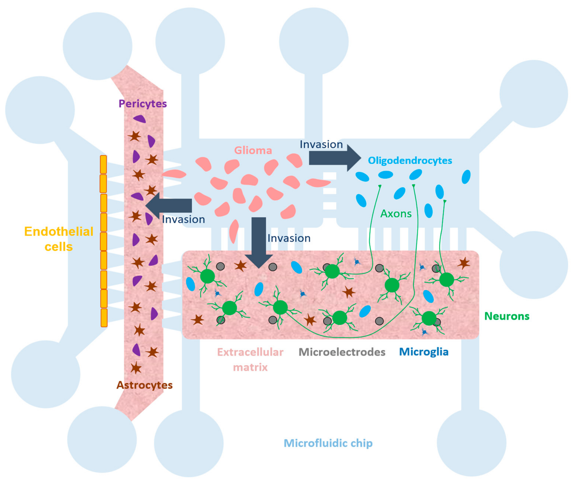
The fundamental architecture of prospective Glioma-on-a-Chip devices consists of two or more culture compartments connected by an array of microstructures (Figure 3), including two compartments designed to model the blood–brain barrier (BBB): a vascular compartment containing endothelial cells, and a matrix-containing compartment housing astrocytes and pericytes. Each compartment features two openings for cell loading and the delivery of culture medium. To generate fluid flow through the vascular compartment, a tubing is connected to the medium-supply openings—either directly or via fittings. This configuration also allows for the use of distinct culture media for the cells in the neuronal-glioma chambers and for the endothelial cells.
The cell culture chambers are about several millimeters in size, with the height of all chambers (compartments) ranging between 100 and 200 µm.
The width of the compartment for astrocytes and pericytes is determined by the technical feasibility of loading a gel into the channel, as well as the time required substances to diffuse from the vascular compartment toward the neurons and glioma; it is typically around 0.5–1.5 mm [204,205,206].
The width of a vascularization channel is typically about hundreds of micrometers [206,207,208].
The platform incorporates two types of connections between the chambers. The first consists of microchannels with a rectangular cross-section and a small height (3–5 µm), which allow axonal outgrowth from the neuronal side while physically confining neuronal cell bodies within the chamber [143]. Glial cells, characterized by their ability to change shape, are capable of migrating through microchannels that are only a few micrometers high. The second type of connection consists of a series of pillars designed to retain gel within one of the chip’s chambers; this configuration is intended for recreating the blood–brain barrier (BBB). The pillars are trapezoidal in shape, with base dimensions ranging from 70 to 450 µm [206,207,208].
The pillars prevent the gel—loaded into the channel containing astrocytes and pericytes—from entering adjacent chambers, while the gaps between them are sufficient to allow the diffusion of therapeutic agents across the blood–brain barrier (BBB) model toward glioma cells and neurons.
Electrophysiological signals from neurons can be recorded using planar microelectrode arrays (MEAs) for two-dimensional neuronal cultures, or by employing needle-type or “shell” MEAs. The setup is integrated with the MEA in such a way that the microelectrode region is positioned within the neuronal culture chamber.
Human iPSC-derived neurons are seeded into one chamber and allowed to mature over several weeks until robust spontaneous network activity occurs. The glioma and neuronal chambers are connected by microchannels. The axons extend through the microchannels forming a network that occupies most of the area of the glioma chamber. Glioma cells seeded to the glioma chamber can migrate along these axons toward the neuronal population, thereby modeling axon-guided glioma invasion.
A separate axonal chamber connected by microchannels to both the neuronal chamber and the glioma chamber is designated within the chip for modeling glioma interaction with white matter. Oligodendrocytes are added to this chamber to model myelination and its glioma-associated disruption, as well as glioma invasion along white matter observed in patients.
The mature neuronal network generates burst activity. Microelectrode arrays (MEAs) provide sufficiently high spatial and temporal resolution for reliable detection of epileptiform activity associated with an imbalance between excitation and inhibition [121]. Such a Glioma-on-a-Chip platform with MEA allows the study of mechanisms and effects of glioma-induced network hyperexcitability and its correlation with glioma invasion.
Integration of the BBB model using two compartments connected to the neuronal and glial chambers allows the study of drug efficacy within the platform. Astrocytes and pericytes embedded in Matrigel are seeded into the compartment adjacent to the cell-containing chambers (Figure 3), while endothelial cells are seeded into the distal compartment to model a blood vessel into which nutrient medium containing a drug is delivered via a peristaltic pump or a rocking platform. The integrity of a modeled barrier within a chip, as well as its permeability to drug compounds, can be assessed by measuring the permeability of tracers and characterizing tight junctions [204,205,206,207,208]. Transendothelial electrical resistance (TEER) measurement can also be used [153]. However, due to a difficulty in controlling measurement variability, the TEER is not always measured in 3D BBB-on-chip culture models [207].
The channel adjacent to the cell-containing chambers is filled with astrocytes and pericytes embedded in Matrigel. Endothelial cells are seeded into the distal channel to model a blood vessel, into which culture medium containing the drug is delivered via a peristaltic pump or a rocking platform. Furthermore, recent innovations in microfluidic chip fabrication allow for the creation of fluidic reservoirs that maintain a hydrostatic pressure differential, enabling localized drug perfusion without inducing convective flow through the channels [121,202].
Blood-derived macrophages and microglia can be added to the vascular and neuroglial compartments, respectively. Co-culturing macrophages and microglia with glioma and neuronal cells in separate compartments of microfluidic chips allows control of spatial organization and the recreation of conditions mimicking the glioma tumor core and the peritumoral niche.
The technical implementation of such a multicomponent model with a complex cellular composition requires stepwise chip preparation and sequential cell seeding. Glioma cells (e.g., PDCs or GSCs), astrocytes, oligodendrocytes, microglia, and endothelial cells must first be obtained and maintained separately under appropriate conditions with the addition of factors required for each specific cell type. First, an extracellular matrix 3D compartment containing astrocytes and pericytes should be formed. These cells secrete factors necessary for tight junction formation and transport regulation in the BBB model. Then neuronal cells should be seeded into the chip and cultured for several weeks, during which a network forms and synapses mature. Next, brain endothelial cells should be seeded, forming a monolayer and tight junctions over several days under perfusion of culturing medium. Glioma cells can be added at the final stage simultaneously with seeding of endothelial cells and BBB formation. During co-culture, neuronal, glial, and glioma cells can be maintained in a common culture medium [142,147]. Although recent studies have demonstrated the possibility of culturing individual vascular models for more than a month because of controlled physical and biological factors, the median culture duration in vascular chips across studies from 2010 to 2025 was 4 days [209]. Thus, maintaining a stable vascular cell phenotype and barrier function is a bottleneck for long-term maintenance of a fully heterogeneous model with a mature neuro-glial network and reconstituted BBB, with evaluation periods ranging from several days to weeks.
The presented generalized “Glioma-on-a-Chip” device scheme opens up the following future research strategies:
Epileptogenesis induced by gliomas of various molecular subtypes. For example, the new microfluidic approach enables the direct comparison of IDH-mutant versus IDH-wildtype gliomas in terms of their epileptogenic potential and their sensitivity to specific ASMs (e.g., valproic acid, perampanel) or chemotherapeutic agents (e.g., temozolomide). Such comparisons are critical for understanding why seizures are more prevalent in IDH-mutant tumors and for identifying the most effective therapeutic strategies for individual molecular subtypes.
Note, that comparisons between IDH-mutant and IDH-wildtype cells require glioma-free neuronal controls. Differences in culturing medium requirements can be mitigated by using a common co-culture medium after preconditioning, by limiting the co-culture duration, and by including isogenic IDH-mutant and wildtype cells as an additional control. Furthermore, experiments comparing the effects of various molecular subtypes should be conducted by assessing neuronal network activity during co-culture with glioma cells as well as growth and invasion of glioma cells.
Comparison of results obtained on the GoC platform with the cultivation of neuronal and glioma cells in isolated compartments but in the same culture medium, will allow to study how direct cell contact could be distinguished from effects mediated by soluble tumor-derived factors.
Mechanisms of glioma interaction with both neurons and the tumor microenvironment. Introducing oligodendrocytes into the axonal chamber enables the study of myelination disruption caused by the glioma.
Glioma cells modify the extracellular matrix, a process that can also be investigated using the presented platform.
The organ-on-a-chip model allows for the study of glioma-induced changes in blood–brain barrier (BBB) function, the activity of specific transporters, and permeability to various substances and immune cells.
Co-culturing macrophages and microglia with glioma and neuronal cells in microfluidic chips allows for the study of the interactions of these glioma-associated immune cells with glioma and other cells within the tumor microenvironment and the investigation of their role in glioma-associated epilepsy. Such models also offer the opportunity to study the response of these cells to various therapeutic strategies, including testing immuno-oncology drugs designed to reprogram microglia.
Mechanisms of glioma cell invasion and migration into white and gray matter, as well as along blood vessels, can be investigated. Separate compartments for neurons and axons enable the difference in tumor cell invasion characteristics in gray versus white matter. An alternative chip design featuring a vessel-mimicking channel will facilitate the study of glioma cell migration toward blood vessels.
Biomedical research. A promising practical application of the platform is the assessment of anti-tumor drugs, including their neurotoxicity and delivery across the BBB to the tumor microenvironment. A more advanced version incorporates a liver compartment, enabling the study of the drug pathway: blood vessel → hepatic metabolism → blood vessel → BBB → brain.
Figure 3.
Generalized diagram of proposed “Glioma-on-a-Chip” device for investigation of glioma interactions with various brain cell populations.
A critical step is the transition to models that combine patient-derived cells with microenvironmental components to better recapitulate the complex interactions driving glioma progression [112]. Studies utilizing “glioma-on-a-chip” systems require adjustment of parameters and chip design based on specific objectives and the chosen sources of glioma and neuronal cells.
The standardization of co-culture protocols remains a promising area of development, essential for ensuring reproducibility of results and clinical validation. Some strategies could improve the comparability of results across different “glioma-on-a-chip” systems used in various laboratories. It is necessary to specify not only donor characteristics, molecular subtype, and tumor grade, but also the phenotypic composition of the cultured cells used in the model at the time of the experiment, for example, the ratio of glioma stem cells (GSCs) to differentiated cells. Passage number and functional characteristics of these cells, such as D2HG production in IDH-mutant gliomas, proliferation rates, and respiratory metabolic profile, should be reported. Differentiation efficiency must be reported for the generation of tumor microenvironment cells, including neurons, glia, and vascular cells, from stem cells (iPSCs/ESCs) or via transdifferentiation, and should be based on both the expression of specific markers and the results of functional assessments. Standardized quantitative metrics, such as neuronal spike frequency or the transendothelial electrical resistance (TEER) of a barrier, should be established for the functional evaluation of these models. Finally, the parameters of the chip must be clearly described, including the composition of the materials and their preparation methods, the dimensions of the microchannels and chambers, the type of adhesive coating, and the medium flow conditions.
4. Toward Personalized Therapy: Functional Drug Screening
Despite advances in genomic precision oncology, only a small fraction of patients benefit from targeted therapy selected on the basis of genomic profiling. In the pan-oncology NCI-MATCH trial, which enrolled patients with multiple cancer types, the overall objective response rate across treatment arms was 10.3% [210]; glioblastoma-specific series show a similar picture—fewer than 10% of screened patients actually initiate matched targeted therapy, and objective responses are confined to individual molecular subgroups, such as tumors harboring the BRAF V600E mutation [211]. This approach is limited by the lack of actionable mutations, complex genetic interactions, and unpredictable drug responses [212]. To overcome these limitations, evaluating drug efficacy directly on patient tumor tissues ex vivo can be used. Personalized functional testing using patient-derived models offers a promising complementary approach to guide clinical care, and early feasibility studies and clinical trials are currently underway to evaluate its clinical utility [213].
GoC platforms hold considerable promise as tools for patient-specific functional drug screening because they can capture both tumor-intrinsic behavior and the effects of glioma on neuronal network function. In current practice, the selection of antiseizure medications and anticancer therapies remains largely empirical, even though glioma-associated epilepsy, invasive growth, and therapeutic resistance are strongly influenced by tumor subtype and by the surrounding microenvironment [213]. A microfluidic system assembled from patient-derived glioma cells and human iPSC-derived neurons could therefore enable parallel testing of multiple interventions across matched chip units, with concurrent assessment of tumor viability, invasion, and network electrophysiology. Therefore, GoC offers the opportunity to study tumor aggressiveness and the likelihood of recurrence.
Glioblastomas exhibit a high level of heterogeneity due to the presence of cell populations of varying levels of differentiation within the tumor. After therapy, glioma stem cells undergo a phenotypic transition from the proneuronal to the mesenchymal subtype, becoming more aggressive and leading to disease recurrence [96,97,105,214].
Confirming these data, in the microfluidic chip study, recurrent glioblastomas demonstrated greater resistance and viability than grade 1–3 tumors. Furthermore, tumors with wild-type IDH showed greater viability than tumors with IDH mutations, which correlates with the worse survival rate associated with wild-type IDH [47]. Drug screening in personalized therapy requires the preservation of the original tumor’s properties and, consequently, the selection of culture conditions based on the tumor type.
4.1. Prioritizing Antitumor Efficacy While Preserving Neural Function
For a clinically meaningful screening platform, suppression of tumor growth and recurrence must remain the primary objective, but the assay should also capture effects on adjacent neural tissue [215]. This dual readout is essential because agents that reduce glioma viability may also injure neurons or exacerbate network hyperexcitability, whereas other compounds may improve seizure control without producing a substantial antitumor effect [216]. Real-time microelectrode array recordings make it possible to quantify bursting activity, synchrony, and epileptiform discharges while the tumor compartment is simultaneously evaluated for invasion, growth, and survival.
Lomeli et al. demonstrated that temozolomide directly induces apoptosis in hippocampal neurons and impairs neural transmission by reducing dendritic branching and synaptic spine density [217]. Additionally, it causes severe mitochondrial dysfunction and oxidative stress by suppressing mitochondrial gene transcription. GoC platforms may also be useful for distinguishing antitumor efficacy from neurotoxicity. A compound that reduces glioma growth but severely suppresses healthy neuronal activity would be less desirable in a patient with limited cognitive and functional reserve. Conversely, a therapy that stabilizes network excitability without affecting tumor progression would be inadequate as a stand-alone oncologic intervention. The GoC platform therefore provides a framework for assessing efficacy and safety within a single experimental system.
4.2. Criteria for Interpreting Drug Responses
We note first that validated criteria for interpreting drug responses on glioma-on-a-chip platforms do not yet exist, and their development remains one of the tasks facing this technology. As a starting point, approaches from classical pharmacology, which distinguish efficacy from toxicity on the basis of dose–response curves, can be adapted. Within such a framework, two key distinctions are necessary.
First, genuine antitumor activity must be distinguished from general cytotoxicity. Each drug should be tested over a concentration range that includes clinically achievable concentrations of the free (unbound to plasma proteins) fraction of the drug (Cmax), on a tumor–neural network chip, on a control chip containing only the neuronal network, and on a chip with an isolated glioma culture. Following the classical approach, the selectivity index should be calculated on the basis of cytotoxicity: as the ratio of the concentration reducing neuronal population viability by 50% (IC50) to the concentration reducing tumor viability by 50%. However, because a reduction in network electrical activity can occur without neuronal death (e.g., through ion channel blockade), and because slowly developing neurotoxicity may primarily manifest as network dysfunction, this measure should be complemented by a functional assessment—the ratio of the concentration reducing network activity by 50% to the concentration reducing tumor viability by 50%. A chip with an isolated glioma culture further allows direct tumor toxicity to be separated from effects mediated by neuronal network activity.
Second, normalization of pathological neuronal network activity must be distinguished from nonspecific neuronal suppression. MEA parameters should be interpreted relative to two reference points: the tumor-exposed network without drug and the healthy network treated with the drug. A response should be considered disease-modifying only if spike and burst rates, synchrony, periodicity, and the frequency of epileptiform discharges shift toward the values observed in healthy controls, at concentrations that essentially do not alter the baseline activity of the healthy network; a comparable reduction in activity in tumor-bearing and healthy networks indicates nonspecific suppression.
Exposure duration is another critical parameter: acute (hours), sub-chronic (days), and chronic (weeks) recordings reflect different phenomena—acute channel blockade, delayed synaptic remodeling, and long-term compensatory plasticity—and only time frames compatible with the clinical decision window are informative for patient-specific testing.
Taken together, drug concentration, exposure time, tumor viability and migration, neuronal survival, and MEA-based network activity constitute a minimal interpretive framework that should be reported jointly for each tested compound.
4.3. Repurposing Antiseizure Medications
Although antiseizure medications are primarily used for symptom control, several may also exert direct effects on glioma biology. Among the most promising candidates is perampanel, a non-competitive AMPA receptor antagonist. In glioblastoma models, perampanel inhibited cell growth in vitro, and its rationale is supported by evidence that neuron-to-glioma synaptic signaling through AMPA receptors promotes tumor progression [32]. This translational concept is currently undergoing clinical evaluation in a window-of-opportunity trial in progressive glioblastoma, making perampanel a rational candidate for on-chip screening. Clinical data stimulating interest in this direction remain limited: in a small series of glioma patients receiving perampanel for seizure control, shrinkage of the hyperintense lesion on MRI was reported in the majority of observed patients [218]; however, these patients were receiving concomitant antitumor therapy, the study design was retrospective, and the observed effects cannot be reliably attributed to perampanel. Thus, the antitumor efficacy of perampanel in glioma patients has not been established: the available evidence is limited to preclinical studies, small clinical series, and an ongoing trial designed primarily to assess pharmacodynamic and clinical endpoints rather than to demonstrate efficacy [219].
Valproic acid and levetiracetam are traditionally used in neuro-oncology primarily as antiseizure medications. In glioblastoma models, valproic acid has been reported to reduce cell growth and invasiveness, and clinical meta-analyses have suggested an association between valproic acid use and improved overall survival, although the magnitude of benefit remains heterogeneous across studies [220,221]. In turn, levetiracetam enhances the efficacy of temozolomide in vitro and exhibits direct antitumor activity in cell cultures, while retrospective and population-based studies associate its use with improved overall and progression-free survival, although this clinical benefit remains a subject of debate and may depend on the duration of therapy [222]. Thus, both drugs are now considered not only as agents for seizure control, but also as potential modifiers of glioma biology [223].
In a functional screening platform, these agents should therefore be assessed not only for seizure control, but also for their effects on tumor viability, invasion, and treatment sensitivity. A useful screening platform should measure not only neuronal suppression or seizure reduction, but also whether a drug alters glioma viability, migration, and metabolic state [215].
4.4. Targeting Neuron–Tumor Crosstalk
Such a microfluidic model is also well suited for evaluating therapies that disrupt bidirectional signaling between glioma and neurons. Neuronal activity promotes cleavage and secretion of neuroligin-3, which functions as a glioma mitogen through the PI3K–mTOR pathway [224]. Another mechanism of tumor-neuron interaction may be mediated by brain-derived neurotrophic factor (BDNF) secreted by neurons. It binds to TrkB receptors on the surface of glioma cells and triggers an intracellular signaling cascade. This leads to the active insertion of additional AMPA receptors into the membrane of tumor cells, thereby strengthening neuron-to-glioma communication [37].
In this setting, the GoC platform can be used to test whether TrkB-pathway modulators attenuate malignant synaptic plasticity while preserving normal neuronal function. This should be presented as a preclinical screening strategy rather than an established therapeutic approach, because direct clinical validation of these agents in glioma remains limited.
Several practical barriers should be considered before GoC-based screening can inform decision-making in real clinical practice. First, cultures derived from patients cannot be established from every specimen: only about 50% of glioma samples grow successfully under standard serum-free conditions [91,92], and culture conditions must be carefully tailored to preserve the original tumor phenotype (Section 2.3). Second, the amount of tissue available for culture is limited, as the material must be shared among histopathological examination, genomic profiling, and culturing. Third, the turnaround time is constrained by the clinical decision window: chips must be fabricated, coated, and quality-controlled in advance, and results must be obtained within days to weeks in order to influence treatment decisions. Fourth, inter-chip reproducibility—cell seeding density, network maturation stage, hydrogel properties, operator handling—requires standardized co-culture protocols, on-chip reference standards, and automated analysis pipelines. Finally, and most importantly, no glioma-on-a-chip assay has yet undergone prospective clinical validation for its ability to predict individual treatment response or improve outcomes; analytical validity, clinical validity, and clinical utility must be demonstrated in purpose-designed studies. At present, functional screening on GoC platforms should therefore be regarded as a tool for preclinical research rather than an established clinical test. Its application in routine clinical practice remains a prospect that requires further investigation.
5. Conclusions
Glioma-associated epilepsy remains a drug-resistant disease affecting up to 80% of IDH-mutated glioma patients. Replicating this complex pathology has been hindered by the limitations of traditional models: 2D cultures lack 3D spatial organization, while animal models suffer from high costs and poor real-time control. The modern organ-on-a-chip approach, based on microfluidic technologies and cellular engineering, bridges this translational gap. In vitro microfluidic Glioma-on-a-Chip systems enable co-culture mature neural networks with glioma populations while allowing communication through microchannels that reconstruct the bidirectional glioma-neuron interface. Such GoC systems integrated with microelectrode arrays (MEAs) provide real-time monitoring of network-level hyperexcitability and synchronized seizures development during epileptogenesis. This microengineered approach will allow for the study of complex bidirectional neuron-glioma crosstalk with feedback, where glutamatergic neural transmission promotes glioma proliferation, and the invading glioma subsequently remodels peritumoral circuits, which trigger synchronized epileptiform activity.
If the technical and validation challenges outlined above are addressed, such in vitro approaches have the potential to complement existing personalized medicine strategies. Because only a small fraction of patients benefit from genome-matched targeted therapy in both cross-tumor and glioma-specific cohorts, testing antitumor drugs directly on patient-derived glioma stem cells (GSCs) under co-culture conditions may provide clinically valuable functional information. GoC platforms capture patient-specific tumor heterogeneity and establish a vital framework to prioritize antitumor efficacy while preserving healthy neural network function. This dual readout distinguishes effective antineoplastic drugs from those inducing neurotoxicity, such as temozolomide-induced mitochondrial dysfunction.
Furthermore, this approach may facilitate the repurposing of antiseizure medications with reported—though not yet clinically proven—antitumor activity in preclinical models and retrospective studies (e.g., perampanel, valproic acid, levetiracetam), as well as the screening of synaptic signaling blockers (e.g., TrkB-pathway inhibitors) to uncover bidirectional mechanisms influencing tumor progression. Looking forward, integrating fully humanized iPSC-derived cellular niches with multi-well scalability could substantially advance preclinical screening and—following prospective clinical validation—contribute to the development of personalized therapeutic strategies for patients.
Author Contributions
Conceptualization, A.P., A.G. and Y.P.; writing—original draft preparation, A.P., A.G., Y.P. and K.Y.; writing—review and editing, V.K. (Vladimir Kolpakov), V.K. (Victor Kazantsev), I.M., K.Y., D.Y., E.I., I.T. and M.Z.; supervision, A.P. and I.M. All authors have read and agreed to the published version of the manuscript.
Funding
This research was funded by the Russian Science Foundation (RSF), grant No. 26-15-00550.
Institutional Review Board Statement
Not applicable.
Informed Consent Statement
Not applicable.
Data Availability Statement
No new data were created or analyzed in this study. Data sharing is not applicable to this article.
Conflicts of Interest
The authors declare no conflicts of interest. The funders had no role in the design of the study; in the collection, analyses, or interpretation of data; in the writing of the manuscript; or in the decision to publish the results.
Abbreviations
The following abbreviations are used in this manuscript:
| ADAM10 | A Disintegrin And Metalloproteinase 10 |
| ADP | Adenosine Diphosphate |
| AMP | Adenosine Monophosphate |
| AMPA | α-amino-3-hydroxy-5-methyl-4-isoxazolepropionic acid |
| ASM | antiseizure medications |
| ATP | Adenosine triphosphate |
| BBB | blood–brain barrier |
| BDNF | brain-derived neurotrophic factor |
| CNS | central nervous system |
| D2HG | D-2-hydroxyglutarate |
| ECM | extracellular matrix |
| EGF | Epidermal growth factor |
| EGFR | Epidermal Growth Factor Receptor |
| EPSP | excitatory postsynaptic potential |
| FBS | Fetal bovine serum |
| FGF2 | Fibroblast Growth Factor 2 |
| GABA | gamma-aminobutyric acid |
| GFAP | glial fibrillary acidic protein |
| GOC | glioma-on-a-chip |
| GPC | Glial progenitor cell |
| GSC | Glioma Stem Cell |
| HBEGF | Heparin-binding EGF-like growth factor |
| hESC | human embryonic stem cell |
| hiPSC | human induced pluripotent stem cells |
| IDH | Isocitrate dehydrogenase |
| IGF1 | insulin-like growth factor 1 |
| MCF-7 | breast cancer cell line |
| MEA | multi-electrode array |
| NLGN3 | Neuroligin-3 |
| OPC | oligodendrocyte progenitor cell |
| Panc-1 | pancreatic cancer cell line |
| PC-3 | prostate cancer cell line |
| PDC | Patient-Derived Cell |
| PDGF | Platelet-Derived Growth Factor |
| PDMS | polydimethylsiloxane |
| PDO | Patient-Derived Organoid |
| PDX | Patient-Derived Xenograft |
| PEGDA | Poly(ethylene glycol) diacrylate |
| TAM | Tumor-associated microglia and macrophage |
| WHO | World Health Organization |
References
- Zhou, J.; Gu, L.; Du, F.; Li, C.; Zhang, F.; Zhang, X.; Pang, J.; Xie, B.; Wang, X.; Peng, J.; et al. The global, regional, and national brain and CNS cancers burden and trends from 1990 to 2021. Sci. Rep. 2025, 15, 19228. [Google Scholar] [CrossRef] [Scilit] [PubMed]
- Altmann, C.; Keller, S.; Schmidt, M.H.H. The role of SVZ stem cells in glioblastoma. Cancers 2019, 11, 448. [Google Scholar] [CrossRef] [Scilit] [PubMed]
- Prajapati, H.P.; Kannaujia, S.K. A simplified overview of the World Health Organization classification of central nervous system tumors 2021. Surg. Neurol. Int. 2022, 13, 252. [Google Scholar] [CrossRef] [Scilit] [PubMed]
- Verhaak, R.G.; Hoadley, K.A.; Purdom, E.; Wang, V.; Qi, Y.; Wilkerson, M.D.; Miller, C.R.; Ding, L.; Golub, T.; Mesirov, J.P.; et al. Integrated genomic analysis identifies clinically relevant subtypes of glioblastoma characterized by abnormalities in PDGFRA, IDH1, EGFR, and NF1. Cancer Cell 2010, 17, 98–110. [Google Scholar] [CrossRef] [Scilit] [PubMed]
- Patel, A.P.; Tirosh, I.; Trombetta, J.J.; Shalek, A.K.; Gillespie, S.M.; Wakimoto, H.; Cahill, D.P.; Nahed, B.V.; Curry, W.T.; Martuza, R.L.; et al. Single-cell RNA-seq highlights intratumoral heterogeneity in primary glioblastoma. Science 2014, 344, 1396–1401. [Google Scholar] [CrossRef] [Scilit] [PubMed]
- Chen, H.; Judkins, J.; Thomas, C.; Wu, M.; Khoury, L.; Benjamin, C.G.; Pacione, D.; Golfinos, J.G.; Kumthekar, P.; Ghamsari, F.; et al. Mutant IDH1 and seizures in patients with glioma. Neurology 2017, 88, 1805–1813. [Google Scholar] [CrossRef] [Scilit] [PubMed]
- Duan, W.-C.; Wang, L.; Li, K.; Wang, W.-W.; Zhan, Y.-B.; Zhang, F.-J.; Yu, B.; Bai, Y.-H.; Wang, Y.-M.; Ji, Y.-C.; et al. IDH mutations but not TERTp mutations are associated with seizures in lower-grade gliomas. Medicine 2018, 97, e13675. [Google Scholar] [CrossRef] [Scilit] [PubMed]
- Louis, D.N.; Perry, A.; Wesseling, P.; Brat, D.J.; Cree, I.A.; Figarella-Branger, D.; Hawkins, C.; Ng, H.K.; Pfister, S.M.; Reifenberger, G.; et al. The 2021 WHO classification of tumors of the central nervous system: A summary. Neuro-Oncology 2021, 23, 1231–1251. [Google Scholar] [CrossRef] [Scilit] [PubMed]
- Ustun, M.; Rahmani Dabbagh, S.; Ilci, I.S.; Bagci-Onder, T.; Tasoglu, S. Glioma-on-a-chip models. Micromachines 2021, 12, 490. [Google Scholar] [CrossRef] [Scilit] [PubMed]
- Weller, J.; Katzendobler, S.; Karschnia, P.; Lietke, S.; Egensperger, R.; Thon, N.; Weller, M.; Suchorska, B.; Tonn, J.C. PCV chemotherapy alone for WHO grade 2 oligodendroglioma: Prolonged disease control with low risk of malignant progression. J. Neurooncol. 2021, 153, 283–291. [Google Scholar] [CrossRef] [Scilit] [PubMed]
- Jansen, E.; Hamisch, C.; Ruess, D.; Heiland, D.H.; Goldbrunner, R.; Ruge, M.I.; Schnell, O.; Grau, S.J. Observation after surgery for low grade glioma: Long-term outcome in the light of the 2016 WHO classification. J. Neurooncol. 2019, 145, 501–507. [Google Scholar] [CrossRef] [Scilit] [PubMed]
- Strowd, R.E. Treatment considerations in tumor-related epilepsy. Curr. Treat. Options Neurol. 2025, 27, 47. [Google Scholar] [CrossRef] [Scilit]
- Huberfeld, G.; Vecht, C.J. Seizures and gliomas—Towards a single therapeutic approach. Nat. Rev. Neurol. 2016, 12, 204–216. [Google Scholar] [CrossRef] [Scilit] [PubMed]
- Mortazavi, A.; Fayed, I.; Bachani, M.; Dowdy, T.; Jahanipour, J.; Khan, A.; Owotade, J.; Walbridge, S.; Inati, S.K.; Steiner, J.; et al. IDH-mutated gliomas promote epileptogenesis through d-2-hydroxyglutarate-dependent mTOR hyperactivation. Neuro-Oncology 2022, 24, 1423–1435. [Google Scholar] [CrossRef] [Scilit] [PubMed]
- Chang, E.F.; Potts, M.B.; Keles, G.E.; Lamborn, K.R.; Chang, S.M.; Barbaro, N.M.; Berger, M.S. Seizure characteristics and control following resection in 332 patients with low-grade gliomas. J. Neurosurg. 2008, 108, 227–235. [Google Scholar] [CrossRef] [Scilit] [PubMed]
- Chaichana, K.L.; Parker, S.L.; Olivi, A.; Quinones-Hinojosa, A. Long-term seizure outcomes in adult patients undergoing primary resection of malignant brain astrocytomas. J. Neurosurg. 2009, 111, 282–292. [Google Scholar] [CrossRef] [Scilit] [PubMed]
- Krishna, S.; Choudhury, A.; Keough, M.B.; Seo, K.; Ni, L.; Kakaizada, S.; Lee, A.; Aabedi, A.; Popova, G.; Lipkin, B.; et al. Glioblastoma remodelling of human neural circuits decreases survival. Nature 2023, 617, 599–607. [Google Scholar] [CrossRef] [Scilit] [PubMed]
- McAlpine, H.; Rosier, M.; Rozario, J.; Wang, X.; Wimmer, V.C.; Guzulaitis, R.; Guan, H.; Hu, Y.; Chirlov, L.; Davey, C.; et al. Increased neural excitability and glioma synaptic activity drives glioma proliferation in human cortex. Nat. Neurosci. 2026, 29, 350–357. [Google Scholar] [CrossRef] [Scilit] [PubMed]
- Buckingham, S.C.; Campbell, S.L.; Haas, B.R.; Montana, V.; Robel, S.; Ogunrinu, T.; Sontheimer, H. Glutamate release by primary brain tumors induces epileptic activity. Nat. Med. 2011, 17, 1269–1274. [Google Scholar] [CrossRef] [Scilit] [PubMed]
- Campbell, S.L.; Buckingham, S.C.; Sontheimer, H. Human glioma cells induce hyperexcitability in cortical networks. Epilepsia 2012, 53, 1360–1370. [Google Scholar] [CrossRef] [Scilit] [PubMed]
- Campbell, S.L.; Robel, S.; Cuddapah, V.A.; Robert, S.; Buckingham, S.C.; Kahle, K.T.; Sontheimer, H. GABAergic disinhibition and impaired KCC2 cotransporter activity underlie tumor-associated epilepsy. Glia 2015, 63, 23–36. [Google Scholar] [CrossRef] [Scilit] [PubMed]
- Cueto-Urena, C.; Ramirez-Exposito, M.J.; Martinez-Martos, J.M. Neuron–glioma synapses in tumor progression. Biomedicines 2025, 14, 72. [Google Scholar] [CrossRef] [Scilit] [PubMed]
- Dang, L.; White, D.W.; Gross, S.; Bennett, B.D.; Bittinger, M.A.; Driggers, E.M.; Fantin, V.R.; Jang, H.G.; Jin, S.; Keenan, M.C.; et al. Cancer-associated IDH1 mutations produce 2-hydroxyglutarate. Nature 2009, 462, 739–744. [Google Scholar] [CrossRef] [Scilit] [PubMed]
- Saviuk, M.; Sleptsova, E.; Redkin, T.; Turubanova, V. Unexplained causes of glioma-associated epilepsies: A review of theories and an area for research. Cancers 2023, 15, 5539. [Google Scholar] [CrossRef] [Scilit] [PubMed]
- LaSarge, C.L.; Danzer, S.C. Mechanisms regulating neuronal excitability and seizure development following mTOR pathway hyperactivation. Front. Mol. Neurosci. 2014, 7, 18. [Google Scholar] [CrossRef] [Scilit] [PubMed]
- Sosanya, N.M.; Brager, D.H.; Wolfe, S.; Niere, F.; Raab-Graham, K.F. Rapamycin reveals an mTOR-independent repression of Kv1.1 expression during epileptogenesis. Neurobiol. Dis. 2015, 73, 96–105. [Google Scholar] [CrossRef] [Scilit] [PubMed]
- Wang, S.; Li, K.; He, Q.; Yang, H.; Yang, Y.; Wang, X.; Shu, Y.; Ding, J. Dysmorphic neurons with ultralow intrinsic excitability are paradoxically hyperexcitable in neural network in a mouse model of focal cortical dysplasia type II. Epilepsia 2025, 66, 4006–4022. [Google Scholar] [CrossRef] [Scilit] [PubMed]
- Zhong, S.; Zhao, Z.; Xie, W.; Cai, Y.; Zhang, Y.; Ding, J.; Wang, X. GABAergic interneuron and neurotransmission are mTOR-dependently disturbed in experimental focal cortical dysplasia. Mol. Neurobiol. 2021, 58, 156–169. [Google Scholar] [CrossRef] [Scilit] [PubMed]
- Tewari, B.P.; Chaunsali, L.; Campbell, S.L.; Patel, D.C.; Goode, A.E.; Sontheimer, H. Perineuronal nets decrease membrane capacitance of peritumoral fast spiking interneurons in a model of epilepsy. Nat. Commun. 2018, 9, 4724. [Google Scholar] [CrossRef] [Scilit] [PubMed]
- Vedunova, M.; Sakharnova, T.; Mitroshina, E.; Perminova, M.; Pimashkin, A.; Zakharov, Y.; Dityatev, A.; Mukhina, I. Seizure-like activity in hyaluronidase-treated dissociated hippocampal cultures. Front. Cell. Neurosci. 2013, 7, 149. [Google Scholar] [CrossRef] [Scilit] [PubMed]
- Feyissa, A.M.; Carrano, A.; Wang, X.; Allen, M.; Ertekin-Taner, N.; Dickson, D.W.; Jentoft, M.E.; Quinones-Hinojosa, A. Analysis of intraoperative human brain tissue transcriptome reveals putative risk genes and altered molecular pathways in glioma-related seizures. Epilepsy Res. 2021, 173, 106618. [Google Scholar] [CrossRef] [Scilit] [PubMed]
- Venkatesh, H.S.; Morishita, W.; Geraghty, A.C.; Silverbush, D.; Gillespie, S.M.; Arzt, M.; Tam, L.T.; Espenel, C.; Ponnuswami, A.; Ni, L.; et al. Electrical and synaptic integration of glioma into neural circuits. Nature 2019, 573, 539–545. [Google Scholar] [CrossRef] [Scilit] [PubMed]
- Venkataramani, V.; Tanev, D.I.; Strahle, C.; Studier-Fischer, A.; Fankhauser, L.; Kessler, T.; Korber, C.; Kardorff, M.; Ratliff, M.; Xie, R.; et al. Glutamatergic synaptic input to glioma cells drives brain tumour progression. Nature 2019, 573, 532–538. [Google Scholar] [CrossRef] [Scilit] [PubMed]
- Venkataramani, V.; Yang, Y.; Schubert, M.C.; Reyhan, E.; Tetzlaff, S.K.; Wißmann, N.; Botz, M.; Soyka, S.J.; Beretta, C.A.; Pramatarov, R.L.; et al. Glioblastoma hijacks neuronal mechanisms for brain invasion. Cell 2022, 185, 2899–2917. [Google Scholar] [CrossRef] [Scilit] [PubMed]
- Venkatesh, H.S.; Tam, L.T.; Woo, P.J.; Lennon, J.; Nagaraja, S.; Gillespie, S.M.; Ni, J.; Duveau, D.Y.; Morris, P.J.; Zhao, J.J.; et al. Targeting neuronal activity-regulated neuroligin-3 dependency in high-grade glioma. Nature 2017, 549, 533–537. [Google Scholar] [CrossRef] [Scilit] [PubMed]
- Pan, Y.; Hysinger, J.D.; Barron, T.; Schindler, N.F.; Cobb, O.; Guo, X.; Yalçn, B.; Anastasaki, C.; Mulinyawe, S.B.; Ponnuswami, A.; et al. NF1 mutation drives neuronal activity-dependent initiation of optic glioma. Nature 2021, 594, 277–282. [Google Scholar] [CrossRef] [Scilit] [PubMed]
- Taylor, K.R.; Barron, T.; Hui, A.; Spitzer, A.; Yalçn, B.; Ivec, A.E.; Geraghty, A.C.; Hartmann, G.G.; Arzt, M.; Gillespie, S.M.; et al. Glioma synapses recruit mechanisms of adaptive plasticity. Nature 2023, 623, 366–374. [Google Scholar] [CrossRef] [Scilit] [PubMed]
- Chen, P.; Wang, W.; Liu, R.; Lyu, J.; Zhang, L.; Li, B.; Qiu, B.; Tian, A.; Jiang, W.; Ying, H.; et al. Olfactory sensory experience regulates gliomagenesis via neuronal IGF1. Nature 2022, 606, 550–556. [Google Scholar] [CrossRef] [Scilit] [PubMed]
- Barron, T.; Yalçn, B.; Su, M.; Byun, Y.G.; Gavish, A.; Shamardani, K.; Xu, H.; Ni, L.; Soni, N.; Mehta, V.; et al. GABAergic neuron-to-glioma synapses in diffuse midline gliomas. Nature 2025, 639, 1060–1068. [Google Scholar] [CrossRef] [Scilit] [PubMed]
- Drexler, R.; Drinnenberg, A.; Gavish, A.; Yalcin, B.; Shamardani, K.; Rogers, A.E.; Mancusi, R.; Trivedi, V.; Taylor, K.R.; Kim, Y.S.; et al. Cholinergic neuronal activity promotes diffuse midline glioma growth through muscarinic signaling. Cell 2025, 188, 4640–4657. [Google Scholar] [CrossRef] [Scilit] [PubMed]
- Monje, M. The neuroscience of brain cancers. Neuron 2025, 113, 2734–2739. [Google Scholar] [CrossRef] [Scilit] [PubMed]
- El Meskini, R.; Iacovelli, A.J.; Kulaga, A.; Gumprecht, M.; Martin, P.L.; Baran, M.; Householder, D.B.; Van Dyke, T.; Weaver Ohler, Z. A preclinical orthotopic model for glioblastoma recapitulates key features of human tumors and demonstrates sensitivity to a combination of MEK and PI3K pathway inhibitors. Dis. Model. Mech. 2015, 8, 45–56. [Google Scholar] [CrossRef] [Scilit] [PubMed]
- William, D.; Mullins, C.S.; Schneider, B.; Orthmann, A.; Lamp, N.; Krohn, M.; Hoffmann, A.; Classen, C.F.; Linnebacher, M. Optimized creation of glioblastoma patient derived xenografts for use in preclinical studies. J. Transl. Med. 2017, 15, 27. [Google Scholar] [CrossRef] [Scilit] [PubMed]
- Vollmann-Zwerenz, A.; Leidgens, V.; Feliciello, G.; Klein, C.A.; Hau, P. Tumor cell invasion in glioblastoma. Int. J. Mol. Sci. 2020, 21, 1932. [Google Scholar] [CrossRef] [Scilit] [PubMed]
- Cuddapah, V.A.; Robel, S.; Watkins, S.; Sontheimer, H. A neurocentric perspective on glioma invasion. Nat. Rev. Neurosci. 2014, 15, 455–465. [Google Scholar] [CrossRef] [Scilit] [PubMed]
- Ding, H.; Nagy, A.; Gutmann, D.H.; Guha, A. A review of astrocytoma models. Neurosurg. Focus 2000, 8, 1–8. [Google Scholar] [CrossRef] [Scilit]
- Olubajo, F.; Achawal, S.; Greenman, J. Development of a microfluidic culture paradigm for ex vivo maintenance of human glioblastoma tissue: A new glioblastoma model? Transl. Oncol. 2020, 13, 1–10. [Google Scholar] [CrossRef] [Scilit] [PubMed]
- Leite, D.M.; Zvar Baskovic, B.; Civita, P.; Neto, C.; Gumbleton, M.; Pilkington, G.J. A human co-culture cell model incorporating microglia supports glioblastoma growth and migration, and confers resistance to cytotoxics. FASEB J. 2020, 34, 1710–1727. [Google Scholar] [CrossRef] [Scilit] [PubMed]
- Li, Y.; Wang, J.; Song, S.-R.; Lv, S.-Q.; Qin, J.-H.; Yu, S.-C. Models for evaluating glioblastoma invasion along white matter tracts. Trends Biotechnol. 2024, 42, 293–309. [Google Scholar] [CrossRef] [Scilit] [PubMed]
- Liebig, C.; Ayala, G.; Wilks, J.A.; Berger, D.H.; Albo, D. Perineural invasion in cancer: A review of the literature. Cancer 2009, 115, 3379–3391. [Google Scholar] [CrossRef] [Scilit] [PubMed]
- Bischel, L.L.; Young, E.W.K.; Mader, B.R.; Beebe, D.J. Tubeless microfluidic angiogenesis assay with three-dimensional endothelial-lined microvessels. Biomaterials 2013, 34, 1471–1477. [Google Scholar] [CrossRef] [Scilit] [PubMed]
- Truong, D.; Fiorelli, R.; Barrientos, E.S.; Melendez, E.L.; Sanai, N.; Mehta, S.; Nikkhah, M. A three-dimensional (3D) organotypic microfluidic model for glioma stem cells—Vascular interactions. Biomaterials 2019, 198, 63–77. [Google Scholar] [CrossRef] [Scilit] [PubMed]
- Holter, J.C.; Chang, C.W.; Avendano, A.; Garg, A.A.; Verma, A.K.; Charan, M.; Ahirwar, D.K.; Ganju, R.K.; Song, J.W. Fibroblast-derived CXCL12 increases vascular permeability in a 3-D microfluidic model independent of extracellular matrix contractility. Front. Bioeng. Biotechnol. 2022, 10, 888431. [Google Scholar] [CrossRef] [Scilit] [PubMed]
- Truong, D.; Puleo, J.; Llave, A.; Mouneimne, G.; Kamm, R.D.; Nikkhah, M. Breast cancer cell invasion into a three dimensional tumor-stroma microenvironment. Sci. Rep. 2016, 6, 1–18. [Google Scholar] [CrossRef] [Scilit] [PubMed]
- Zervantonakis, I.K.; Hughes-Alford, S.K.; Charest, J.L.; Condeelis, J.S.; Gertler, F.B.; Kamm, R.D. Three-dimensional microfluidic model for tumor cell intravasation and endothelial barrier function. Proc. Natl. Acad. Sci. USA 2012, 109, 13515–13520. [Google Scholar] [CrossRef] [Scilit] [PubMed]
- Samiei, E.; Seyfoori, A.; Toyota, B.; Ghavami, S.; Akbari, M. Investigating programmed cell death and tumor invasion in a three-dimensional (3D) microfluidic model of glioblastoma. Int. J. Mol. Sci. 2020, 21, 3162. [Google Scholar] [CrossRef] [Scilit] [PubMed]
- Kim, S.; Wan, Z.; Jeon, J.S.; Kamm, R.D. Microfluidic vascular models of tumor cell extravasation. Front. Oncol. 2022, 12, 1052192. [Google Scholar] [CrossRef] [Scilit] [PubMed]
- Zervantonakis, I.K.; Chung, S.; Sudo, R.; Zhang, M.; Charest, J.L.; Kamm, R.D. Concentration gradients in microfluidic 3D matrix cell culture systems. Int. J. Micro-Nano Scale Transp. 2010, 1, 27–36. [Google Scholar] [CrossRef] [Scilit]
- Abhyankar, V.V.; Toepke, M.W.; Cortesio, C.L.; Lokuta, M.A.; Huttenlocher, A.; Beebe, D.J. A platform for assessing chemotactic migration within a spatiotemporally defined 3D microenvironment. Lab Chip 2008, 8, 1507–1515. [Google Scholar] [CrossRef] [Scilit] [PubMed]
- Huang, C.P.; Lu, J.; Seon, H.; Lee, A.P.; Flanagan, L.A.; Kim, H.-Y.; Putnam, A.J.; Jeon, N.L. Engineering microscale cellular niches for three-dimensional multicellular co-cultures. Lab Chip 2009, 9, 1740–1748. [Google Scholar] [CrossRef] [Scilit] [PubMed]
- Song, J.W.; Cavnar, S.P.; Walker, A.C.; Luker, K.E.; Gupta, M.; Tung, Y.-C.; Luker, G.D.; Takayama, S. Microfluidic endothelium for studying the intravascular adhesion of metastatic breast cancer cells. PLoS ONE 2009, 4, e5756. [Google Scholar] [CrossRef] [Scilit] [PubMed]
- Fan, Y.; Nguyen, D.T.; Akay, Y.; Xu, F.; Akay, M. Engineering a brain cancer chip for high-throughput drug screening. Sci. Rep. 2016, 6, 25062. [Google Scholar] [CrossRef] [Scilit] [PubMed]
- Bhatia, S.N.; Ingber, D.E. Microfluidic organs-on-chips. Nat. Biotechnol. 2014, 32, 760–772. [Google Scholar] [CrossRef] [Scilit] [PubMed]
- Zimina, T.M.; Sitkov, N.O.; Gareev, K.G.; Mikhailova, N.V.; Combs, S.E.; Shevtsov, M.A. Hybrid-integrated devices for mimicking malignant brain tumors (“tumor-on-a-chip”) for in vitro development of targeted drug delivery and personalized therapy approaches. Front. Med. 2024, 11, 1452298. [Google Scholar] [CrossRef] [Scilit] [PubMed]
- Lee, H.-J.; Yoon, T.-H.; Park, J.-H.; Perumal, J.; Kim, D.-P. Characterization and fabrication of polyvinylsilazane glass microfluidic channels via soft lithographic technique. J. Ind. Eng. Chem. 2008, 14, 45–51. [Google Scholar] [CrossRef] [Scilit]
- Chavarria, D.; Ojetola, I.; Russotti, M.L.; Yates, A.K.; Lippmann, E.S. Tolerances in microfluidic master molds: A comparison of 3D printing and micromilling. RSC Adv. 2026, 16, 19437–19448. [Google Scholar] [CrossRef] [Scilit] [PubMed]
- Huang, W.-C.; Ong, X.C.; Kwon, I.S.; Gopinath, C.; Fisher, L.E.; Wu, H.; Fedder, G.K.; Gaunt, R.A.; Bettinger, C.J. Ultracompliant Hydrogel-Based Neural Interfaces Fabricated by Aqueous-Phase Microtransfer Printing. Adv. Funct. Mater. 2018, 28, 1801059. [Google Scholar] [CrossRef] [Scilit]
- Xu, Y.; Yu, W.; Li, Y.; Zhang, L. Biomedical Materials and Fabrication Methods for Construction of In Vitro Neurovascular Unit Models. Materials 2026, 19, 3590. [Google Scholar] [CrossRef] [Scilit] [PubMed]
- van Meer, B.J.; de Vries, H.; Firth, K.S.A.; van Weerd, J.; Tertoolen, L.G.J.; Karperien, H.B.J.; Jonkheijm, P.; Denning, C.; IJzerman, A.P.; Mummery, C.L. Small molecule absorption by PDMS in the context of drug response bioassays. Biochem. Biophys. Res. Commun. 2017, 482, 323–328. [Google Scholar] [CrossRef] [Scilit] [PubMed]
- Yang, X.; Li, K.; Zhang, X.; Liu, C.; Guo, B.; Wen, W.; Gao, X. Nanofiber membrane supported lung-on-a-chip microdevice for anti-cancer drug testing. Lab Chip 2018, 18, 486–495. [Google Scholar] [CrossRef] [Scilit] [PubMed]
- Xia, Q.; Pease, R.F. Nanoimprint lithography 20 years on. Nanotechnology 2015, 26, 182501. [Google Scholar] [CrossRef] [Scilit] [PubMed]
- Nawroth, J.C.; Scudder, L.L.; Halvorson, R.T.; Tresback, J.; Ferrier, J.P., Jr.; Sheehy, S.P.; Cho, A.; Kannan, S.; Sunyovszki, I.; Goss, J.A.; et al. Automated fabrication of photopatterned gelatin hydrogels for organ-on-chips applications. Biofabrication 2018, 10, 025004. [Google Scholar] [CrossRef] [Scilit] [PubMed]
- Puryear, J.R., III; Yoon, J.K.; Kim, Y. Advanced fabrication techniques of microengineered physiological systems. Micromachines 2020, 11, 730. [Google Scholar] [CrossRef] [Scilit] [PubMed]
- Xie, Z.; Chen, M.; Lian, J.; Wang, H.; Ma, J. Glioblastoma-on-a-chip construction and therapeutic applications. Front. Oncol. 2023, 13, 1183059. [Google Scholar] [CrossRef] [Scilit] [PubMed]
- Booth, R.; Kim, H. Characterization of a microfluidic in vitro model of the blood-brain barrier (μBBB). Lab Chip 2012, 12, 1784–1792. [Google Scholar] [CrossRef] [Scilit] [PubMed]
- Kim, S.; Lee, H.; Chung, M.; Jeon, N.L. Engineering of Functional, Perfusable 3D Microvascular Networks on a Chip. Lab Chip 2013, 13, 1489–1500. [Google Scholar] [CrossRef] [Scilit] [PubMed]
- Salmina, A.B.; Saridis, M.R.; Ryzhkov, V.V.; Korsakova, S.A.; Averchuk, A.S.; Bystrov, D.A.; Barbasheva, A.A.; Libet, P.A.; Kuguk, A.K.; Polynkin, L.Y.; et al. Development of Microvascular Network in Microfluidic Brain-on-a-Chip Models In Vitro: A Multidisciplinary Review. Biotechnol. J. 2025, 20, e70126. [Google Scholar] [CrossRef] [Scilit] [PubMed]
- Bouhrira, N.; DeOre, B.J.; Galie, P.A. Implementation and characterization of a physiologically relevant flow waveform in a 3D microfluidic model of the blood–brain barrier. Biotechnol. Bioeng. 2021, 118, 2411–2421. [Google Scholar] [CrossRef] [Scilit] [PubMed]
- Alves, A.H.; Nucci, M.P.; Mamani, J.B.; Valle, N.M.E.; Ribeiro, E.F.; Rego, G.N.A.; Oliveira, F.A.; Theinel, M.H.; Santos, R.S.; Gamarra, L.F. The advances in glioblastoma on-a-chip for therapy approaches. Cancers 2022, 14, 869. [Google Scholar] [CrossRef] [Scilit] [PubMed]
- Schulte, A.; Gunther, H.S.; Martens, T.; Zapf, S.; Riethdorf, S.; Wulfing, C.; Stoupiec, M.; Westphal, M.; Lamszus, K. Glioblastoma stem-like cell lines with either maintenance or loss of high-level EGFR amplification, generated via modulation of ligand concentration. Clin. Cancer Res. 2012, 18, 1901–1913. [Google Scholar] [CrossRef] [Scilit] [PubMed]
- William, D.; Mokri, P.; Lamp, N.; Linnebacher, M.; Classen, C.F.; Erbersdobler, A.; Schneider, B. Amplification of the EGFR gene can be maintained and modulated by variation of EGF concentrations in in vitro models of glioblastoma multiforme. PLoS ONE 2017, 12, e0185208. [Google Scholar] [CrossRef] [Scilit] [PubMed]
- Lee, J.; Kotliarova, S.; Kotliarov, Y.; Li, A.; Su, Q.; Donin, N.M.; Pastorino, S.; Purow, B.W.; Christopher, N.; Zhang, W.; et al. Tumor stem cells derived from glioblastomas cultured in bFGF and EGF more closely mirror the phenotype and genotype of primary tumors than do serum-cultured cell lines. Cancer Cell 2006, 9, 391–403. [Google Scholar] [CrossRef] [Scilit] [PubMed]
- DeSouza, P.A.; Ishahak, M.; Qu, X.; McCornack, C.; Annamalai, D.; Mao, D.D.; Vessoni, A.T.; Tang, Y.; Millman, J.R.; Kim, A.H. INSM1 governs a neuronal progenitor state that drives glioblastoma in a human stem cell model. Nat. Commun. 2026, 17, 31. [Google Scholar] [CrossRef] [Scilit] [PubMed]
- Ledur, P.F.; Onzi, G.R.; Zong, H.; Lenz, G. Culture conditions defining glioblastoma cells behavior: What is the impact for novel discoveries? Oncotarget 2017, 8, 69185. [Google Scholar] [CrossRef] [Scilit] [PubMed]
- Vasileva, N.S.; Kuligina, E.V.; Dymova, M.A.; Savinovskaya, Y.I.; Zinchenko, N.D.; Ageenko, A.B.; Mishinov, S.V.; Dome, A.S.; Stepanov, G.A.; Richter, V.A.; et al. Transcriptome changes in glioma cells cultivated under conditions of neurosphere formation. Cells 2022, 11, 3106. [Google Scholar] [CrossRef] [Scilit] [PubMed]
- Sivakumar, H.; Forsythe, S.D.; Laxton, A.W.; Tatter, S.B.; Miller, L.D.; Strowd, R.E.; Skardal, A. Hyaluronan-based glioblastoma tumor constructs maintain patient tumor drug responses and genomic parity. Micromachines 2026, 17, 276. [Google Scholar] [CrossRef] [Scilit] [PubMed]
- Blandin, A.F.; Durand, A.; Litzler, M.; Tripp, A.; Guerin, E.; Ruhland, E.; Fontanilles, M.; Van der Ent, W.; Entz-Werle, N.; Martin, S.; et al. Hypoxic environment and paired hierarchical 3D and 2D models of pediatric H3.3-mutated gliomas recreate the patient tumor complexity. Cancers 2019, 11, 1875. [Google Scholar] [CrossRef] [Scilit] [PubMed]
- Ledur, P.F.; Liu, C.; He, H.; Harris, A.R.; Minussi, D.C.; Zhou, H.Y.; Mandel, J.J.; Shaffrey, M.E.; Lopes, M.B.; Zong, H.; et al. Culture conditions tailored to the cell of origin are critical for maintaining native properties and tumorigenicity of glioma cells. Neuro-Oncology 2016, 18, 1413–1424. [Google Scholar] [CrossRef] [Scilit] [PubMed]
- Park, J.W.; Kwak, J.; Kim, K.W.; Jung, S.; Nam, C.H.; Kim, H.J.; Lee, S.M.; Choi, C.; Ahn, Y.; Park, J.H.; et al. IDH-mutant gliomas arise from glial progenitor cells harboring the initial driver mutation. Science 2026, 391, eadt0559. [Google Scholar] [CrossRef] [Scilit] [PubMed]
- Wu, M.; Wang, T.; Ji, N.; Lu, T.; Yuan, R.; Wu, L.; Zhang, J.; Li, M.; Cao, P.; Zhao, J.; et al. Multi-omics and pharmacological characterization of patient-derived glioma cell lines. Nat. Commun. 2024, 15, 6740. [Google Scholar] [CrossRef] [Scilit] [PubMed]
- Gunther, H.S.; Schmidt, N.O.; Phillips, H.S.; Kemming, D.; Kharbanda, S.; Soriano, R.; Modrusan, Z.; Meissner, H.; Westphal, M.; Lamszus, K. Glioblastoma-derived stem cell-enriched cultures form distinct subgroups according to molecular and phenotypic criteria. Oncogene 2008, 27, 2897–2909. [Google Scholar] [CrossRef] [Scilit] [PubMed]
- Galli, R.; Binda, E.; Orfanelli, U.; Cipelletti, B.; Gritti, A.; De Vitis, S.; Fiocco, R.; Foroni, C.; Dimeco, F.; Vescovi, A. Isolation and characterization of tumorigenic, stem-like neural precursors from human glioblastoma. Cancer Res. 2004, 64, 7011–7021. [Google Scholar] [CrossRef] [Scilit] [PubMed]
- Patrizii, M.; Bartucci, M.; Pine, S.R.; Sabaawy, H.E. Utility of glioblastoma patient-derived orthotopic xenografts in drug discovery and personalized therapy. Front. Oncol. 2018, 8, 23. [Google Scholar] [CrossRef] [Scilit] [PubMed]
- Hettiarachchi, P.; Park, T. Choice of animal models to investigate cell migration and invasion in glioblastoma. Cancers 2025, 17, 2776. [Google Scholar] [CrossRef] [Scilit] [PubMed]
- Oliveira-Nunes, M.C.; Ma, W.; Mao, C.; Huo, B.S.; Rios Angulo, A.; Landa Bianchi, G.; Ray, P.; Chen, Q. Brain metastasis patient-derived xenograft models to characterize the contribution of brain microenvironment to metastatic outgrowth. Neuro-Oncol. Adv. 2025, 7, vdaf239. [Google Scholar] [CrossRef] [Scilit] [PubMed]
- He, J.; Yan, X.; Hu, S. Glioma stem cells: Drivers of tumor progression and recurrence. Stem Cell Res. Ther. 2025, 16, 293. [Google Scholar] [CrossRef] [Scilit] [PubMed]
- Singh, S.; Dey, D.; Barik, D.; Mohapatra, I.; Kim, S.; Sharma, M.; Prasad, S.; Wang, P.; Singh, A.; Singh, G. Glioblastoma at the crossroads: Current understanding and future therapeutic horizons. Signal Transduct. Target. Ther. 2025, 10, 213. [Google Scholar] [CrossRef] [Scilit] [PubMed]
- Liu, S.; Gong, Z.; Zhou, D.; Ayala-Nunez, V.; Xu, T.; Wick, P. Bridging the gap: How organ-on-a-chip technology facilitates the battle against glioma. Small Sci. 2026, 6, e202500154. [Google Scholar] [CrossRef] [Scilit] [PubMed]
- Kim, S.S.; Pirollo, K.F.; Chang, E.H. Isolation and culturing of glioma cancer stem cells. Curr. Protoc. Cell Biol. 2015, 67, 23.10. [Google Scholar] [CrossRef] [Scilit] [PubMed]
- Ignatova, T.N.; Kukekov, V.G.; Laywell, E.D.; Suslov, O.N.; Vrionis, F.D.; Steindler, D.A. Human cortical glial tumors contain neural stem-like cells expressing astroglial and neuronal markers in vitro. Glia 2002, 39, 193–206. [Google Scholar] [CrossRef] [Scilit] [PubMed]
- Balvers, R.K.; Kleijn, A.; Kloezeman, J.J.; French, P.J.; Kremer, A.; van den Bent, M.J.; Dirven, C.M.F.; Leenstra, S.; Lamfers, M.L.M. Serum-free culture success of glial tumors is related to specific molecular profiles and expression of extracellular matrix-associated gene modules. Neuro-Oncology 2013, 15, 1684–1695. [Google Scholar] [CrossRef] [Scilit] [PubMed]
- Stoczynska-Fidelus, E.; Piaskowski, S.; Bienkowski, M.; Banaszczyk, M.; Hulas-Bigoszewska, K.; Winiecka-Klimek, M.; Radomiak-Zaluska, A.; Och, W.; Borowiec, M.; Zieba, J.; et al. The failure in the stabilization of glioblastoma-derived cell lines: Spontaneous in vitro senescence as the main culprit. PLoS ONE 2014, 9, e87136. [Google Scholar] [CrossRef] [Scilit] [PubMed][Green Version]
- Behnan, J.; Stangeland, B.; Hosainey, S.A.M.; Joel, M.; Olsen, T.K.; Micci, F.; Glover, J.C.; Isakson, P.; Brinchmann, J.E. Differential propagation of stroma and cancer stem cells dictates tumorigenesis and multipotency. Oncogene 2017, 36, 570–584. [Google Scholar] [CrossRef] [Scilit] [PubMed]
- Talasila, K.M.; Brekka, N.; Mangseth, K.; Stieber, D.; Evensen, L.; Rosland, G.V.; Torsvik, A.; Niclou, S.P.; Mahesparan, R.; Miletic, H.; et al. Tumor versus stromal cells in culture—Survival of the fittest? PLoS ONE 2013, 8, e81183. [Google Scholar] [CrossRef] [Scilit] [PubMed]
- Balasubramaniyan, V.; Vaillant, B.; Wang, S.; Gumin, J.; Butalid, M.E.; Sai, K.; Mukheef, F.; Kim, S.H.; Boddeke, H.W.G.M.; Lang, F.; et al. Aberrant mesenchymal differentiation of glioma stem-like cells: Implications for therapeutic targeting. Oncotarget 2015, 6, 31007–31017. [Google Scholar] [CrossRef] [Scilit] [PubMed][Green Version]
- Shin, K.; Shin, H.; Cho, H.J.; Kang, H.; Lee, J.-K.; Seo, Y.J.; Shin, Y.J.; Kim, D.; Koo, H.; Kong, D.S.; et al. Sphere-forming culture for expanding genetically distinct patient-derived glioma stem cells by cellular growth rate screening. Cancers 2020, 12, 549. [Google Scholar] [CrossRef] [Scilit] [PubMed]
- Piaskowski, S.; Bienkowski, M.; Stoczynska-Fidelus, E.; Stawski, R.; Sieruta, M.; Szybka, M.; Papierz, W.; Wolanczyk, M.; Jaskolski, D.J.; Liberski, P.P.; et al. Glioma cells showing IDH1 mutation cannot be propagated in standard cell culture conditions. Br. J. Cancer 2011, 104, 968–970. [Google Scholar] [CrossRef] [Scilit] [PubMed]
- Aguilar-Cazares, K.; Pineau, D.; Garcia, L.; Huang, G.H.; Lv, S.Q.; Duffau, H.; Hugnot, J.P. A protocol for explant cultures of IDH1-mutant diffuse low-grade gliomas. J. Vis. Exp. 2025, 219, e67260. [Google Scholar] [CrossRef] [Scilit] [PubMed]
- Luchman, H.A.; Stechishin, O.D.; Dang, N.H.; Blough, M.D.; Chesnelong, C.; Kelly, J.J.; Nguyen, S.A.; Chan, J.A.; Weljie, A.M.; Cairncross, J.G.; et al. An in vivo patient-derived model of endogenous IDH1-mutant glioma. Neuro-Oncology 2012, 14, 184–191. [Google Scholar] [CrossRef] [Scilit] [PubMed]
- Pianka, S.T.; Li, T.; Prins, T.J.; Eldred, B.S.C.; Kevan, B.M.; Liang, H.; Zapanta Rinonos, S.; Kornblum, H.I.; Nathanson, D.A.; Pellegrini, M.; et al. D-2-HG inhibits IDH1mut glioma growth via FTO inhibition and resultant m6A hypermethylation. Cancer Res. Commun. 2023, 3, 192–201. [Google Scholar] [CrossRef] [Scilit] [PubMed]
- Wei, S.; Wang, J.; Oyinlade, O.; Ma, D.; Wang, S.; Kratz, L.; Lal, B.; Xu, Q.; Liu, S.; Shah, S.R.; et al. Heterozygous IDH1R132H/WT created by “single base editing” inhibits human astroglial cell growth by downregulating YAP. Oncogene 2018, 37, 5160–5174. [Google Scholar] [CrossRef] [Scilit] [PubMed]
- Maity, S.; Bhuyan, T.; Jewell, C.; Kawakita, S.; Sharma, S.; Nguyen, H.T.; Hassani Najafabadi, A.; Ermis, M.; Falcone, N.; Chen, J.; et al. Recent developments in glioblastoma-on-a-chip for advanced drug screening applications. Small 2025, 21, e2405511. [Google Scholar] [CrossRef] [Scilit] [PubMed]
- Brofiga, M.; Losacco, S.; Poggio, F.; Zerbo, R.A.; Milanese, M.; Massobrio, P.; Burlando, B. Multiple neuron clusters on micro-electrode arrays as an in vitro model of brain network. Sci. Rep. 2023, 13, 15604. [Google Scholar] [CrossRef] [Scilit] [PubMed]
- le Feber, J.; Rutten, W.L.C.; Stegenga, J.; Wolters, P.S.; Ramakers, G.J.A.; Van Pelt, J. Conditional firing probabilities in cultured neuronal networks: A stable underlying structure in widely varying spontaneous activity patterns. J. Neural Eng. 2007, 4, 5. [Google Scholar] [CrossRef] [Scilit] [PubMed]
- Javaid, M.S.; Tan, T.; Dvir, N.; Anderson, A.; O’Brien, T.J.; Kwan, P.; Antonic-Baker, A. Human in vitro models of epilepsy using embryonic and induced pluripotent stem cells. Cells 2022, 11, 3957. [Google Scholar] [CrossRef] [Scilit] [PubMed]
- Ishibashi, Y.; Odawara, A.; Kinoshita, K.; Okamura, A.; Shirakawa, T.; Suzuki, I. Principal component analysis to distinguish seizure liability of drugs in human iPS cell-derived neurons. Toxicol. Sci. 2021, 184, 265–275. [Google Scholar] [CrossRef] [Scilit] [PubMed]
- Kreir, M.; Van Deuren, B.; Versweyveld, S.; De Bondt, A.; Van den Wyngaert, I.; Van der Linde, H.; Lu, H.R.; Teuns, G.; Gallacher, D.J. Do in vitro assays in rat primary neurons predict drug-induced seizure liability in humans? Toxicol. Appl. Pharmacol. 2018, 346, 45–57. [Google Scholar] [CrossRef] [Scilit] [PubMed]
- Tukker, A.M.; Wijnolts, F.M.; de Groot, A.; Wubbolts, R.W.; Westerink, R.H. In vitro techniques for assessing neurotoxicity using human iPSC-derived neuronal models. Cell Cult. Tech. 2019, 17–35. [Google Scholar] [CrossRef] [Scilit]
- Bradley, J.A.; Luithardt, H.H.; Metea, M.R.; Strock, C.J. In vitro screening for seizure liability using microelectrode array technology. Toxicol. Sci. 2018, 163, 240–253. [Google Scholar] [CrossRef] [Scilit] [PubMed]
- Fan, J.; Thalody, G.; Kwagh, J.; Burnett, E.; Shi, H.; Lewen, G.; Chen, S.-J.; Levesque, P. Assessing seizure liability using multi-electrode arrays (MEA). Toxicol. Vitr. 2019, 55, 93–100. [Google Scholar] [CrossRef] [Scilit] [PubMed]
- Pelkonen, A.; Mzezewa, R.; Sukki, L.; Ryynanen, T.; Kreutzer, J.; Hyvarinen, T.; Vinogradov, A.; Aarnos, L.; Lekkala, J.; Kallio, P.; et al. A modular brain-on-a-chip for modelling epileptic seizures with functionally connected human neuronal networks. Biosens. Bioelectron. 2020, 168, 112553. [Google Scholar] [CrossRef] [Scilit] [PubMed]
- Pimashkin, A.; Kastalskiy, I.; Simonov, A.; Koryagina, E.; Mukhina, I.; Kazantsev, V. Spiking signatures of spontaneous activity bursts in hippocampal cultures. Front. Comput. Neurosci. 2011, 5, 46. [Google Scholar] [CrossRef] [Scilit] [PubMed]
- Gladkov, A.; Grinchuk, O.; Pigareva, Y.; Mukhina, I.; Kazantsev, V.; Pimashkin, A. Theta rhythm-like bidirectional cycling dynamics of living neuronal networks in vitro. PLoS ONE 2018, 13, e0192468. [Google Scholar] [CrossRef] [Scilit] [PubMed]
- Schroter, M.; Wang, C.; Terrigno, M.; Hornauer, P.; Huang, Z.; Jagasia, R.; Hierlemann, A. Functional imaging of brain organoids using high-density microelectrode arrays. MRS Bull. 2022, 47, 530–544. [Google Scholar] [CrossRef] [Scilit] [PubMed]
- Pesantez Torres, F.; Manning, A.; Paredes-Espinosa, M.B.; Tokranova, N.; Bertucci, T.; Kiehl, T.R.; Paluh, J.L.; Cady, N.C.; Xie, Y.; Sharfstein, S.T. 3D flexible self-folding microelectrode array for organoid electrophysiology analysis. Cell Biomater. 2025, 1, 100179. [Google Scholar] [CrossRef] [Scilit]
- Paek, J.; Park, W.; Song, H.; Jeong, I.; Oh, M.; Kim, E.; Kim, D.; An, S.H.; Kim, Y.; Chung, W.G.; et al. Emerging diverse 3D neural electrode architectures for bioelectronics. Nanoscale Horiz. 2026, 11, 1536–1560. [Google Scholar] [CrossRef] [Scilit] [PubMed]
- Park, Y.; Franz, C.K.; Ryu, H.; Luan, H.; Cotton, K.Y.; Kim, J.U.; Chung, T.S.; Zhao, S.; Vazquez-Guardado, A.; Yang, D.S.; et al. Three-dimensional, multifunctional neural interfaces for cortical spheroids and engineered assembloids. Sci. Adv. 2021, 7, eabf9153. [Google Scholar] [CrossRef] [Scilit] [PubMed]
- Kim, D.; Ryu, H. Recent Advances in Microelectrode Array Interfaces for Organoids. Biomimetics 2026, 11, 142. [Google Scholar] [CrossRef] [Scilit] [PubMed]
- Tasnim, K.; Liu, J. Emerging bioelectronics for brain organoid electrophysiology. J. Mol. Biol. 2022, 434, 167165. [Google Scholar] [CrossRef] [Scilit] [PubMed]
- Le Floch, P.; Li, Q.; Lin, Z.; Zhao, S.; Liu, R.; Tasnim, K.; Jiang, H.; Liu, J. Stretchable Mesh Nanoelectronics for 3D Single-Cell Chronic Electrophysiology from Developing Brain Organoids. Adv. Mater. 2022, 34, e2106829. [Google Scholar] [CrossRef] [Scilit] [PubMed]
- Lee, J.Y.; Park, S.H.; Kim, Y.; Cho, Y.U.; Park, J.; Hong, J.-H.; Kim, K.; Shin, J.; Ju, J.E.; Min, I.S.; et al. Foldable three dimensional neural electrode arrays for simultaneous brain interfacing of cortical surface and intracortical multilayers. npj Flex. Electron. 2022, 6, 86. [Google Scholar] [CrossRef] [Scilit]
- Kopic, I.; Peng, H.; Schmidt, S.; Berezin, O.; Wang, S.; Westmeyer, G.G.; Wolfrum, B. Inkjet-printed 3D sensor arrays with FIB-induced electrode refinement for low-noise amperometric recordings in hiPSC-derived brain organoids. ACS Sens. 2025, 10, 6426. [Google Scholar] [CrossRef] [Scilit] [PubMed]
- Kim, N.; Kang, M.; Ji, J.; Kim, H.; Kim, H.N.; Choi, N.; Lee, Y.J. Multilayered microelectrode array for monitoring electrophysiological signals of 3D neural networks in cerebral organoid. Microsyst. Nanoeng. 2026, 12, 201. [Google Scholar] [CrossRef] [Scilit] [PubMed]
- Virlogeux, A.; Moutaux, E.; Christaller, W.; Genoux, A.; Bruyere, J.; Fino, E.; Charlot, B.; Cazorla, M.; Saudou, F. Reconstituting Corticostriatal Network on-a-Chip Reveals the Contribution of the Presynaptic Compartment to Huntington’s Disease. Cell Rep. 2018, 22, 110–122. [Google Scholar] [CrossRef] [Scilit] [PubMed]
- Taylor, A.M.; Blurton-Jones, M.; Rhee, S.W.; Cribbs, D.H.; Cotman, C.W.; Jeon, N.L. A microfluidic culture platform for CNS axonal injury, regeneration and transport. Nat. Methods 2005, 2, 599–605. [Google Scholar] [CrossRef] [Scilit] [PubMed]
- Altman, T.; Maimon, R.; Ionescu, A.; Pery, T.G.; Perlson, E. Axonal Transport of Organelles in Motor Neuron Cultures using Microfluidic Chambers System. J. Vis. Exp. 2020, 159, e60993. [Google Scholar] [CrossRef] [Scilit] [PubMed]
- Zhu, D.; Nilghaz, A.; Tong, Z.; Poole, D.P.; O’Sullivan, K.; Imlach, W.L.; Haberberger, R.V.; Veldhuis, N.A.; Matusica, D.; Voelcker, N.H. Pain-on-a-Chip: A microfluidic device for neuron differentiation and functional discrimination in animal models of chronic pain. Biosens. Bioelectron. 2025, 279, 117401. [Google Scholar] [CrossRef] [Scilit] [PubMed]
- Moutaux, E.; Christaller, W.; Scaramuzzino, C.; Genoux, A.; Charlot, B.; Cazorla, M.; Saudou, F. Neuronal network maturation differently affects secretory vesicles and mitochondria transport in axons. Sci. Rep. 2018, 8, 13429. [Google Scholar] [CrossRef] [Scilit] [PubMed]
- Charalampopoulou, A.; Taga, A.; Rust, K.; Luciani, E.; Marshall, K.; Montgomery, E.; Mansinghka, A.; Singh, R.; Zhao, Y.; O’Keefe, C.; et al. Development of a human iPSC-derived corticospinal tract-on-a-chip. Cell Rep. Methods 2026, 6, 101457. [Google Scholar] [CrossRef] [Scilit] [PubMed]
- Parodi, G.; Brofiga, M.; Pastore, V.P.; Chiappalone, M.; Martinoia, S. Deepening the role of excitation/inhibition balance in human iPSCs-derived neuronal networks coupled to MEAs during long-term development. J. Neural Eng. 2023, 20, 056011. [Google Scholar] [CrossRef] [Scilit] [PubMed]
- Izsak, J.; Seth, H.; Theiss, S.; Hanse, E.; Illes, S. Human Cerebrospinal Fluid Promotes Neuronal Circuit Maturation of Human Induced Pluripotent Stem Cell-Derived 3D Neural Aggregates. Stem Cell Rep. 2020, 14, 1044–1059. [Google Scholar] [CrossRef] [Scilit] [PubMed]
- Lei, Y.; Li, J.; Wang, N.; Yang, X.; Hamada, Y.; Li, Q.; Zheng, W.; Jiang, X. An on-chip model for investigating the interaction between neurons and cancer cells. Integr. Biol. 2016, 8, 359–367. [Google Scholar] [CrossRef] [Scilit] [PubMed]
- Liu, X.; Zhang, W.; Zheng, W.; Jiang, X. Micropatterned coculture platform for screening nerve-related anticancer drugs. ACS Nano 2021, 15, 637–649. [Google Scholar] [CrossRef] [Scilit] [PubMed]
- Fuchs, Q.; Batut, A.; Gleyzes, M.; Rontard, J.; Miny, L.; Libralato, M.; Vieira, J.; Debis, D.; Larramendy, F.; Honegger, T.; et al. Co-culture of glutamatergic neurons and pediatric high-grade glioma cells into microfluidic devices to assess electrical interactions. J. Vis. Exp. 2021, 177, e62748. [Google Scholar] [CrossRef] [Scilit] [PubMed]
- Xu, T.; Zhang, X.; Jiang, Y.; Sheng, K.; Li, J.; Ren, J.; He, J.; Liang, C.; Yu, Z.; Jin, H.; et al. A machine learning-driven electrophysiological platform for real-time tumor-neural interaction analysis and modulation. Nat. Commun. 2026, 17, 49. [Google Scholar] [CrossRef] [Scilit] [PubMed]
- Savarraj, J.P.; Kelly, K.C.; DeCoster, M.A. Early glioma is associated with abnormal electrical events in cortical cultures. Med. Biol. Eng. Comput. 2019, 57, 1645–1656. [Google Scholar] [CrossRef] [Scilit] [PubMed]
- Linkous, A.; Balamatsias, D.; Snuderl, M.; Edwards, L.; Miyaguchi, K.; Milner, T.; Reich, B.; Cohen-Gould, L.; Storaska, A.; Nakayama, Y.; et al. Modeling patient-derived glioblastoma with cerebral organoids. Cell Rep. 2019, 26, 3203–3211. [Google Scholar] [CrossRef] [Scilit] [PubMed]
- Yi, H.-G.; Jeong, Y.H.; Kim, Y.; Choi, Y.-J.; Moon, H.E.; Park, S.H.; Kang, K.S.; Bae, M.; Jang, J.; Youn, H.; et al. A bioprinted human-glioblastoma-on-a-chip for the identification of patient-specific responses to chemoradiotherapy. Nat. Biomed. Eng. 2019, 3, 509–519. [Google Scholar] [CrossRef] [Scilit] [PubMed]
- Azzarelli, R.; Ori, M.; Philpott, A.; Simons, B.D. Three-dimensional model of glioblastoma by co-culturing tumor stem cells with human brain organoids. Biol. Open 2021, 10, bio056416. [Google Scholar] [CrossRef] [Scilit] [PubMed]
- Ogawa, J.; Pao, G.M.; Shokhirev, M.N.; Verma, I.M. Glioblastoma model using human cerebral organoids. Cell Rep. 2018, 23, 1220–1229. [Google Scholar] [CrossRef] [Scilit] [PubMed]
- Goranci-Buzhala, G.; Mariappan, A.; Gabriel, E.; Ramani, A.; Ricci-Vitiani, L.; Buccarelli, M.; D’Alessandris, Q.G.; Pallini, R.; Gopalakrishnan, J. Rapid and efficient invasion assay of glioblastoma in human brain organoids. Cell Rep. 2020, 31, 107738. [Google Scholar] [CrossRef] [Scilit] [PubMed]
- Vinci, M.; Box, C.; Eccles, S.A. Three-dimensional (3D) tumor spheroid invasion assay. J. Vis. Exp. 2015, 99, e52686. [Google Scholar] [CrossRef] [Scilit] [PubMed]
- Marino, A.; Tricinci, O.; Battaglini, M.; Filippeschi, C.; Mattoli, V.; Sinibaldi, E.; Ciofani, G. A 3D real-scale, biomimetic, and biohybrid model of the blood-brain barrier fabricated through two-photon lithography. Small 2018, 14, 1702959. [Google Scholar] [CrossRef] [Scilit] [PubMed]
- Abedin, M.J.; Michelhaugh, S.K.; Mittal, S.; Berdichevsky, Y. 3D models of glioblastoma interaction with cortical cells. Front. Bioeng. Biotechnol. 2023, 11, 1150772. [Google Scholar] [CrossRef] [Scilit] [PubMed]
- Forster, N.; Isosaari, L.; Kulta, O.; Junnila, O.; Vuolanto, V.; Pollari, M.; Rautajoki, K.J.; Narkilahti, S. Functional 3D human neuron–glioblastoma model reveals cellular interactions enabling drug safety assessments. FASEB J. 2025, 39, e70567. [Google Scholar] [CrossRef] [Scilit] [PubMed]
- Bai, L.; Hao, Z.; Wang, S.; Zhou, J.; Yao, S.; Pei, N.; Zhu, H.; Zhang, K.; Reis, R.L.; Oliveira, J.M.; et al. Biomimetic three-dimensional glioma model printed in vitro for the studies of glioma cells and neurons interactions. Int. J. Bioprinting 2023, 9, 715. [Google Scholar] [CrossRef] [Scilit] [PubMed]
- Gordon, A.; Yoon, S.J.; Tran, S.S.; Makinson, C.D.; Park, J.Y.; Andersen, J.; Valencia, A.M.; Horvath, S.; Xiao, X.; Huguenard, J.R.; et al. Long-term maturation of human cortical organoids matches key early postnatal transitions. Nat. Neurosci. 2021, 24, 331–342. [Google Scholar] [CrossRef] [Scilit] [PubMed]
- Herring, C.A.; Simmons, R.K.; Freytag, S.; Poppe, D.; Moffet, J.J.D.; Pflueger, J.; Buckberry, S.; Vargas-Landin, D.B.; Clement, O.; Echeverria, E.G.; et al. Human prefrontal cortex gene regulatory dynamics from gestation to adulthood at single-cell resolution. Cell 2022, 185, 4428–4447.e28. [Google Scholar] [CrossRef] [Scilit] [PubMed]
- Ohlenschläger, M.S.; Criscuolo, L.; Jensen, P.; Lloyd-Davies Sanchez, D.J.; Sutcliffe, M.; Bhosale, S.; Bogetofte, H.; Tahir, M.; Jakobsen, L.A.; Pihl, M.; et al. Modeling Synaptic Maturation from Growth Cone to Synapse in Human Organoids. J. Neurochem. 2026, 170, e70458. [Google Scholar] [CrossRef] [Scilit] [PubMed]
- Mancinelli, S.; Bariselli, S.; Lodato, S. The emergence of electrical activity in human brain organoids. Stem Cell Rep. 2025, 20, 102632. [Google Scholar] [CrossRef] [Scilit] [PubMed]
- Lancaster, M.A.; Renner, M.; Martin, C.A.; Wenzel, D.; Bicknell, L.S.; Hurles, M.E.; Homfray, T.; Penninger, J.M.; Jackson, A.P.; Knoblich, J.A. Cerebral organoids model human brain development and microcephaly. Nature 2013, 501, 373–379. [Google Scholar] [CrossRef] [Scilit] [PubMed]
- Zhang, Y.; Pak, C.; Han, Y.; Ahlenius, H.; Zhang, Z.; Chanda, S.; Marro, S.; Patzke, C.; Acuna, C.; Covy, J.; et al. Rapid single-step induction of functional neurons from human pluripotent stem cells. Neuron 2013, 78, 785–798. [Google Scholar] [CrossRef] [Scilit] [PubMed]
- Joung, J.; Ma, S.; Tay, T.; Geiger-Schuller, K.R.; Kirchgatterer, P.C.; Verdine, V.K.; Guo, B.; Arias-Garcia, M.A.; Allen, W.E.; Singh, A.; et al. A transcription factor atlas of directed differentiation. Cell 2023, 186, 209–229. [Google Scholar] [CrossRef] [Scilit] [PubMed]
- Mariappan, A.; Goranci-Buzhala, G.; Ricci-Vitiani, L.; Pallini, R.; Gopalakrishnan, J. Trends and challenges in modeling glioma using 3D human brain organoids. Cell Death Differ. 2021, 28, 15–23. [Google Scholar] [CrossRef] [Scilit] [PubMed]
- Sloan, S.A.; Andersen, J.; Pasca, A.M.; Birey, F.; Pasca, S.P. Generation and assembly of human brain region-specific three-dimensional cultures. Nat. Protoc. 2018, 13, 2062–2085. [Google Scholar] [CrossRef] [Scilit] [PubMed]
- Pasca, A.M.; Sloan, S.A.; Clarke, L.E.; Tian, Y.; Makinson, C.D.; Huber, N.; Kim, C.H.; Park, J.-Y.; O’Rourke, N.A.; Nguyen, K.D.; et al. Functional cortical neurons and astrocytes from human pluripotent stem cells in 3D culture. Nat. Methods 2015, 12, 671–678. [Google Scholar] [CrossRef] [Scilit] [PubMed]
- Cakir, B.; Xiang, Y.; Tanaka, Y.; Kural, M.H.; Parent, M.; Kang, Y.-J.; Chapeton, K.; Patterson, B.; Yuan, Y.; He, C.-S.; et al. Engineering of human brain organoids with a functional vascular-like system. Nat. Methods 2019, 16, 1169–1175. [Google Scholar] [CrossRef] [Scilit] [PubMed]
- Faravelli, I.; Anton-Bolanos, N.; Brown, J.R.; Arlotta, P. Brain organoids: Tools for understanding the uniqueness and individual variability of the human brain. Annu. Rev. Genom. Hum. Genet. 2025, 26, 299–320. [Google Scholar] [CrossRef] [Scilit] [PubMed]
- Sandoval, S.O.; Cappuccio, G.; Kruth, K.; Osenberg, S.; Khalil, S.M.; Mendez-Albelo, N.M.; Padmanabhan, K.; Wang, D.; Niciu, M.J.; Bhattacharyya, A.; et al. Rigor and reproducibility in human brain organoid research: Where we are and where we need to go. Stem Cell Rep. 2024, 19, 796–816. [Google Scholar] [CrossRef] [Scilit] [PubMed]
- Aiello, G.; Nemir, M.; Vidimova, B.; Ramel, C.; Viguie, J.; Ravera, A.; Wrzesinski, K.; Bagni, C. Increased reproducibility of brain organoids through controlled fluid dynamics. EMBO Rep. 2025, 26, 6209–6239. [Google Scholar] [CrossRef] [Scilit] [PubMed]
- Glass, M.R.; Waxman, E.A.; Yamashita, S.; Lafferty, M.; Beltran, A.A.; Farah, T.; Patel, N.K.; Singla, R.; Matoba, N.; Ahmed, S.; et al. Cross-site reproducibility of human cortical organoids reveals consistent cell type composition and architecture. Stem Cell Rep. 2024, 19, 1351–1367. [Google Scholar] [CrossRef] [Scilit] [PubMed]
- Giandomenico, S.L.; Mierau, S.B.; Gibbons, G.M.; Wenger, L.M.D.; Masullo, L.; Sit, T.; Sutcliffe, M.; Boulanger, J.; Tripodi, M.; Derivery, E.; et al. Cerebral organoids at the air-liquid interface generate diverse nerve tracts with functional output. Nat. Neurosci. 2019, 22, 669–679. [Google Scholar] [CrossRef] [Scilit] [PubMed]
- Ah-Pine, F.; Khettab, M.; Bedoui, Y.; Slama, Y.; Daniel, M.; Doray, B.; Gasque, P. On the origin and development of glioblastoma: Multifaceted role of perivascular mesenchymal stromal cells. Acta Neuropathol. Commun. 2023, 11, 104. [Google Scholar] [CrossRef] [Scilit] [PubMed]
- Hambardzumyan, D.; Bergers, G. Glioblastoma: Defining tumor niches. Trends Cancer 2015, 1, 252–265. [Google Scholar] [CrossRef] [Scilit] [PubMed]
- Solga, A.C.; Pong, W.W.; Kim, K.-Y.; Cimino, P.J.; Toonen, J.A.; Walker, J.; Wylie, T.; Magrini, V.; Griffith, M.; Griffith, O.L.; et al. RNA sequencing of tumor-associated microglia reveals Ccl5 as a stromal chemokine critical for neurofibromatosis-1 glioma growth. Neoplasia 2015, 17, 776–788. [Google Scholar] [CrossRef] [Scilit] [PubMed]
- Pinton, L.; Masetto, E.; Vettore, M.; Solito, S.; Magri, S.; D’Andolfi, M.; Del Bianco, P.; Lollo, G.; Benoit, J.-P.; Okada, H.; et al. The immune suppressive microenvironment of human gliomas depends on the accumulation of bone marrow-derived macrophages in the center of the lesion. J. Immunother. Cancer 2019, 7, 58. [Google Scholar] [CrossRef] [Scilit] [PubMed]
- Roesch, S.; Rapp, C.; Dettling, S.; Herold-Mende, C. When immune cells turn bad—Tumor-associated microglia/macrophages in glioma. Int. J. Mol. Sci. 2018, 19, 436. [Google Scholar] [CrossRef] [Scilit] [PubMed]
- Pan, Y.; Yu, Y.; Wang, X.; Zhang, T. Tumor-associated macrophages in tumor immunity. Front. Immunol. 2020, 11, 583084. [Google Scholar] [CrossRef] [Scilit] [PubMed]
- da Fonseca, A.C.C.; Romao, L.; Amaral, R.F.; Assad Kahn, S.; Lobo, D.; Martins, S.; Marcondes de Souza, J.; Moura-Neto, V.; Lima, F.R. Microglial stress inducible protein 1 promotes proliferation and migration in human glioblastoma cells. Neuroscience 2012, 200, 130–141. [Google Scholar] [CrossRef] [Scilit] [PubMed]
- Yi, Y.; Huang, S.; Chen, L.; Chen, X.; Yang, Z.; Ke, Y. Atorvastatin suppresses glioma invasion and migration by reducing microglial MT1-MMP expression. J. Neuroimmunol. 2013, 260, 1–8. [Google Scholar] [CrossRef] [Scilit] [PubMed]
- Zheng, Y.; Alzoubi, I.; Graeber, M.B.; Anand, A.; Barazzuol, L.; Dorostkar, M.; Frontzek, K.J.; Golębiewska, A.; Hadjipanayis, C.G.; Jansen, G.S.M.; et al. Consensus statement on microglial and macrophage functions in gliomas. Acta Neuropathol. 2026, 151, 39. [Google Scholar] [CrossRef] [Scilit] [PubMed]
- Badimon, A.; Strasburger, H.J.; Ayata, P.; Chen, X.; Nair, A.; Ikegami, A.; Hwang, P.; Wang, Y.-C.; Graves, S.M.; Kenny, P.J.; et al. Negative feedback control of neuronal activity by microglia. Nature 2020, 586, 417–423. [Google Scholar] [CrossRef] [Scilit] [PubMed]
- Haynes, S.E.; Hollopeter, G.; Yang, G.; Kurpius, D.; Dailey, M.E.; Gan, W.-B.; Julius, D. The P2Y12 receptor regulates microglial activation by extracellular nucleotides. Nat. Neurosci. 2006, 9, 1512–1519. [Google Scholar] [CrossRef] [Scilit] [PubMed]
- Yei, J.; Lee, N.K.; Ryu, S.; Ryu, S.-E.; Lee, J.; Park, T.; Jeong, Y.; Kang, R.; Kwon, H.-K.; Kim, S.-G.; et al. Glioma-associated microglia potentiate neuronal hyper-excitability in the glioma environment. Neuro-Oncology 2025, 27, 3058–3071. [Google Scholar] [CrossRef] [Scilit] [PubMed]
- Rodriguez-Gomez, J.A.; Kavanagh, E.; Engskog-Vlachos, P.; Engskog, M.K.R.; Herrera, A.J.; Espinosa-Oliva, A.M.; Joseph, B.; Hajji, N.; Venero, J.L.; Burguillos, M.A. Microglia: Agents of the CNS pro-inflammatory response. Cells 2020, 9, 1717. [Google Scholar] [CrossRef] [Scilit] [PubMed]
- Golab, F.; Hajimirzaei, P.; Zarbakhsh, S.; Zolfaghari, S.; Hayat, P.; Joghataei, M.T.; Bakhtiarzadeh, F.; Ahmadirad, N. Interplay of neuroinflammation and epilepsy in glioblastoma multiforme: Mechanisms and therapeutic implications. J. Mol. Neurosci. 2025, 75, 68. [Google Scholar] [CrossRef] [Scilit] [PubMed]
- Tripathi, S.; Nathan, C.L.; Tate, M.C.; Horbinski, C.M.; Templer, J.W.; Rosenow, J.M.; Sita, T.L.; James, C.D.; Deneen, B.; Miller, S.D.; et al. The immune system and metabolic products in epilepsy and glioma-associated epilepsy: Emerging therapeutic directions. JCI Insight 2024, 9, e174753. [Google Scholar] [CrossRef] [Scilit] [PubMed]
- Hong, S.; You, J.Y.; Paek, K.; Park, J.; Kang, S.J.; Han, E.H.; Choi, N.; Chung, S.; Rhee, W.J.; Kim, J.A. Inhibition of tumor progression and M2 microglial polarization by extracellular vesicle-mediated microRNA-124 in a 3D microfluidic glioblastoma microenvironment. Theranostics 2021, 11, 9687–9704. [Google Scholar] [CrossRef] [Scilit] [PubMed]
- Lin, L.; He, Z.; Jie, M.; Lin, J.-M.; Zhang, J. 3D microfluidic tumor models for biomimetic engineering of glioma niche and detection of cell morphology, migration and phenotype change. Talanta 2021, 234, 122702. [Google Scholar] [CrossRef] [Scilit] [PubMed]
- Inoue, A.; Ohnishi, T.; Nishikawa, M.; Ohtsuka, Y.; Kusakabe, K.; Yano, H.; Tanaka, J.; Kunieda, T. A narrative review on CD44′s role in glioblastoma invasion, proliferation, and tumor recurrence. Cancers 2023, 15, 4898. [Google Scholar] [CrossRef] [Scilit] [PubMed]
- Nakod, P.S.; Kim, Y.; Rao, S.S. The impact of astrocytes and endothelial cells on glioblastoma stemness marker expression in multicellular spheroids. Cell. Mol. Bioeng. 2021, 14, 639–651. [Google Scholar] [CrossRef] [Scilit] [PubMed]
- Wang, Y.-H.; Huang, T.-L.; Chen, X.; Yu, S.-X.; Li, W.; Chen, T.; Li, Y.; Kuang, Y.-Q.; Shu, H.-F. Glioma-derived TSP2 promotes excitatory synapse formation and results in hyperexcitability in the peritumoral cortex of glioma. J. Neuropathol. Exp. Neurol. 2021, 80, 137–149. [Google Scholar] [CrossRef] [Scilit] [PubMed]
- Soeung, V.; Puchalski, R.B.; Noebels, J.L. The complex molecular epileptogenesis landscape of glioblastoma. Cell Rep. Med. 2024, 5, 101691. [Google Scholar] [CrossRef] [Scilit] [PubMed]
- Gerigk, M.; Bulstrode, H.; Shi, H.H.; Tonisen, F.; Cerutti, C.; Morrison, G.; Rowitch, D.; Huang, Y.Y.S. On-chip perivascular niche supporting stemness of patient-derived glioma cells in a serum-free, flowable culture. Lab Chip 2021, 21, 2343–2358. [Google Scholar] [PubMed]
- Xiao, Y.; Kim, D.; Dura, B.; Zhang, K.; Yan, R.; Li, H.; Han, E.; Ip, J.; Zou, P.; Liu, J.; et al. Ex vivo dynamics of human glioblastoma cells in a microvasculature-on-a-chip system correlates with tumor heterogeneity and subtypes. Adv. Sci. 2019, 6, 1801531. [Google Scholar] [CrossRef] [Scilit] [PubMed]
- Daoud, J.P.A.; Collisson, A.E.; Asaid, D.; do Couto Lopes, E.C.; Paraskevopoulos, D.; Chapman, C.A.R. Development and characterization of an on-chip glioma outgrowth model. Vitr. Models 2025, 1–13. [Google Scholar] [CrossRef] [Scilit]
- Adjei-Sowah, E.A.; O’Connor, S.A.; Veldhuizen, J.; Lo Cascio, C.; Plaisier, C.; Mehta, S.; Nikkhah, M. Investigating the interactions of glioma stem cells in the perivascular niche at single-cell resolution using a microfluidic tumor microenvironment model. Adv. Sci. 2022, 9, 2201436. [Google Scholar] [CrossRef] [Scilit] [PubMed]
- Li, X.; Gao, W.; Long, X.; Wu, M. New insights into monocyte-derived macrophages in glioblastoma. Research 2025, 8, 0836. [Google Scholar] [CrossRef] [Scilit] [PubMed]
- van Vliet, E.A.; da Costa Araujo, S.; Redeker, S.; van Schaik, R.; Aronica, E.; Gorter, J.A. Blood-brain barrier leakage may lead to progression of temporal lobe epilepsy. Brain 2007, 130, 521–534. [Google Scholar] [CrossRef] [Scilit] [PubMed]
- Ivens, S.; Kaufer, D.; Flores, L.P.; Bechmann, I.; Zumsteg, D.; Tomkins, O.; Seifert, E.; Heinemann, U.; Friedman, A. TGF-β receptor-mediated albumin uptake into astrocytes is involved in neocortical epileptogenesis. Brain 2007, 130, 535–547. [Google Scholar] [CrossRef] [Scilit] [PubMed]
- Manoharan, T.J.M.; Wang, T.-Y.; Mantri, S.; Alarnous, H.; Mehta, S.; Wang, K.-C.; Nikkhah, M. Advancing targeted drug delivery in glioblastoma multiforme through biomimetic nanomedicine using 3D tumor-on-a-chip model. Adv. Healthc. Mater. 2026, 15, e02454. [Google Scholar] [CrossRef] [Scilit] [PubMed]
- Pigareva, Y.; Gladkov, A.; Kolpakov, V.; Mukhina, I.; Kazantsev, V.; Pimashkin, A. Microfluidic bi-layer platform to study functional interaction between co-cultured neural networks with unidirectional synaptic connectivity. Micromachines 2023, 14, 835. [Google Scholar] [CrossRef] [Scilit] [PubMed]
- Feng, J.; Yang, J. Glioma-neuron interactions: Insights from neural plasticity. Front. Oncol. 2025, 15, 1661897. [Google Scholar] [CrossRef] [Scilit] [PubMed]
- Campisi, M.; Shin, Y.; Osaki, T.; Hajal, C.; Chiono, V.; Kamm, R.D. 3D self-organized microvascular model of the human blood-brain barrier with endothelial cells, pericytes and astrocytes. Biomaterials 2018, 180, 117–129. [Google Scholar] [CrossRef] [Scilit] [PubMed]
- Qiu, B.; Pompe, S.; Xenaki, K.T.; Di Maggio, A.; en Henegouwen, P.M.; Oliveira, S.; Mastrobattista, E.; Caiazzo, M. Generation of a perfusable three-dimensional human neurovascular chip to model brain drug delivery and immune extravasation. J. Control. Release 2025, 387, 114257. [Google Scholar] [CrossRef] [Scilit] [PubMed]
- Uwamori, H.; Higuchi, T.; Arai, K.; Sudo, R. Integration of neurogenesis and angiogenesis models for constructing a neurovascular tissue. Sci. Rep. 2017, 7, 17349. [Google Scholar] [CrossRef] [Scilit] [PubMed]
- Adriani, G.; Ma, D.; Pavesi, A.; Kamm, R.D.; Goh, E.L. A 3D neurovascular microfluidic model consisting of neurons, astrocytes and cerebral endothelial cells as a blood–brain barrier. Lab Chip 2017, 17, 448–459. [Google Scholar] [CrossRef] [Scilit] [PubMed]
- Wevers, N.R.; Kasi, D.G.; Gray, T.; Wilschut, K.J.; Smith, B.; van Vught, R.; Shimizu, F.; Sano, Y.; Kanda, T.; Marsh, G.; et al. A perfused human blood-brain barrier on-a-chip for high-throughput assessment of barrier function and antibody transport. Fluids Barriers CNS 2018, 15, 23. [Google Scholar] [CrossRef] [Scilit] [PubMed]
- Pandey, R.; Lee, J.; Mojiri, A.; Cooke, J.P.; Jain, A. Time and architecture: The next two dimensions of microphysiological systems. npj Biomed. Innov. 2026, 3, 22. [Google Scholar] [CrossRef] [Scilit] [PubMed]
- O’Dwyer, P.J.; Gray, R.J.; Flaherty, K.T.; Chen, A.P.; Li, S.; Wang, V.; McShane, L.M.; Patton, D.R.; Tricoli, J.V.; Williams, P.M.; et al. The NCI-MATCH trial: Lessons for precision oncology. Nat. Med. 2023, 29, 1349–1357. [Google Scholar] [CrossRef] [Scilit] [PubMed]
- Weller, J.; Potthoff, A.L.; Zeyen, T.; Schaub, C.; Duffy, C.; Schneider, M.; Herrlinger, U. Current status of precision oncology in adult glioblastoma. Mol. Oncol. 2024, 18, 2927–2950. [Google Scholar] [CrossRef] [Scilit] [PubMed]
- Mann, B.; Artz, N.; Darawsheh, R.; Kram, D.E.; Hingtgen, S.; Satterlee, A.B. Opportunities and challenges for patient-derived models of brain tumors in functional precision medicine. npj Precis. Oncol. 2025, 9, 47. [Google Scholar] [CrossRef] [Scilit] [PubMed]
- Yashin, K.S.; Yuzhakova, D.V.; Sachkova, D.A.; Kukhnina, L.S.; Kharitonova, T.M.; Zolotova, A.S.; Medyanik, I.A.; Shirmanova, M.V. Personalized medicine in brain gliomas: Targeted therapy, patient-derived tumor models (review). Sovrem. Tekhnol. Med. 2023, 15, 61. [Google Scholar] [CrossRef] [Scilit] [PubMed]
- Mondal, A.; Kumari Singh, D.; Panda, S.; Shiras, A. Extracellular vesicles as modulators of tumor microenvironment and disease progression in glioma. Front. Oncol. 2017, 7, 144. [Google Scholar] [CrossRef] [Scilit] [PubMed]
- Mann, B.; Zhang, X.; Bell, N.; Adefolaju, A.; Thang, M.; Dasari, R.; Kanchi, K.; Valdivia, A.; Yang, Y.; Buckley, A.A.; et al. A living ex vivo platform for functional, personalized brain cancer diagnosis. Cell Rep. Med. 2023, 4, 101042. [Google Scholar] [CrossRef] [Scilit] [PubMed]
- Khalili, B.F.; Dixit, K.; Kamson, D.O.; Horbinski, C.; Przybyla, D.J.; Tate, M.C.; Heimberger, A.B.; Lukas, R.V.; Templer, J.W. The excitatory milieu in glioma: Mechanisms and therapeutic avenues. Neuro-Oncology 2025, 27, 1932–1945. [Google Scholar] [CrossRef] [Scilit] [PubMed]
- Lomeli, N.; Di, K.; Pearre, D.C.; Chung, T.F.; Bota, D.A. Mitochondrial-associated impairments of temozolomide on neural stem/progenitor cells and hippocampal neurons. Mitochondrion 2020, 52, 56–66. [Google Scholar] [CrossRef] [Scilit] [PubMed]
- Izumoto, S.; Miyauchi, M.; Tasaki, T.; Okuda, T.; Nakagawa, N.; Nakano, N.; Kato, A.; Fujita, M. Seizures and Tumor Progression in Glioma Patients with Uncontrollable Epilepsy Treated with Perampanel. Anticancer Res. 2018, 38, 4361–4366. [Google Scholar] [CrossRef] [Scilit] [PubMed]
- Heuer, S.; Burghaus, I.; Gose, M.; Kessler, T.; Sahm, F.; Vollmuth, P.; Venkataramani, V.; Herrlinger, U.; Wick, W.; Platten, M.; et al. PerSurge (NOA-30) phase II trial of perampanel treatment around surgery in patients with progressive glioblastoma. BMC Cancer 2024, 24, 135. [Google Scholar] [CrossRef] [Scilit] [PubMed]
- Han, W.; Guan, W. Valproic acid: A promising therapeutic agent in glioma treatment. Front. Oncol. 2021, 11, 687362. [Google Scholar] [CrossRef] [Scilit] [PubMed]
- Sullivan, J.K.; Fahey, P.P.; Agho, K.E.; Hurley, S.P.; Feng, Z.; Day, R.O.; Lim, D. Valproic acid as a radio-sensitizer in glioma: A systematic review and meta-analysis. Neurooncol. Pract. 2022, 10, 13–23. [Google Scholar] [CrossRef] [Scilit] [PubMed]
- Lee, Y.; Lee, E.; Roh, T.H.; Kim, S.H. Association between levetiracetam use and survival in patients with glioblastoma: A nationwide population-based study. Cancer Res. Treat. 2025, 57, 369–377. [Google Scholar] [CrossRef] [Scilit] [PubMed]
- Khalili, B.F.; Walbert, T.; Horbinski, C.; Dixit, K.; Gururangan, K.; Thio, H.; Tate, M.C.; Stupp, R.; Lukas, R.V.; Templer, J.W. Levetiracetam and valproic acid in glioma: Antiseizure and potential antineoplastic effects. Future Oncol. 2025, 21, 483–491. [Google Scholar] [CrossRef] [Scilit] [PubMed]
- Venkatesh, H.S.; Johung, T.B.; Caretti, V.; Noll, A.; Tang, Y.; Nagaraja, S.; Gibson, E.M.; Mount, C.W.; Polepalli, J.; Mitra, S.S.; et al. Neuronal activity promotes glioma growth through neuroligin-3 secretion. Cell 2015, 161, 803–816. [Google Scholar] [CrossRef] [Scilit] [PubMed]
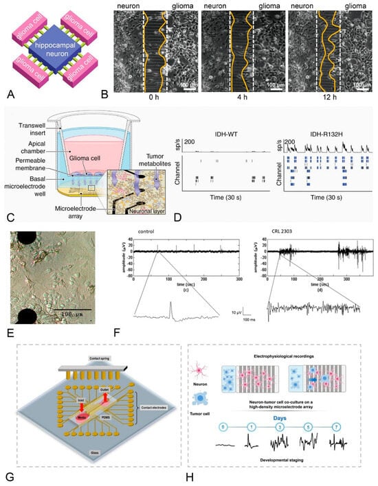
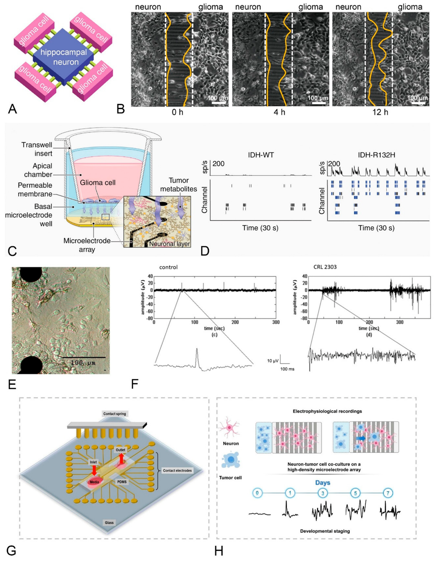
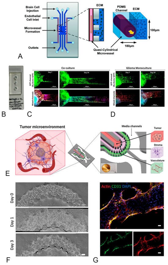
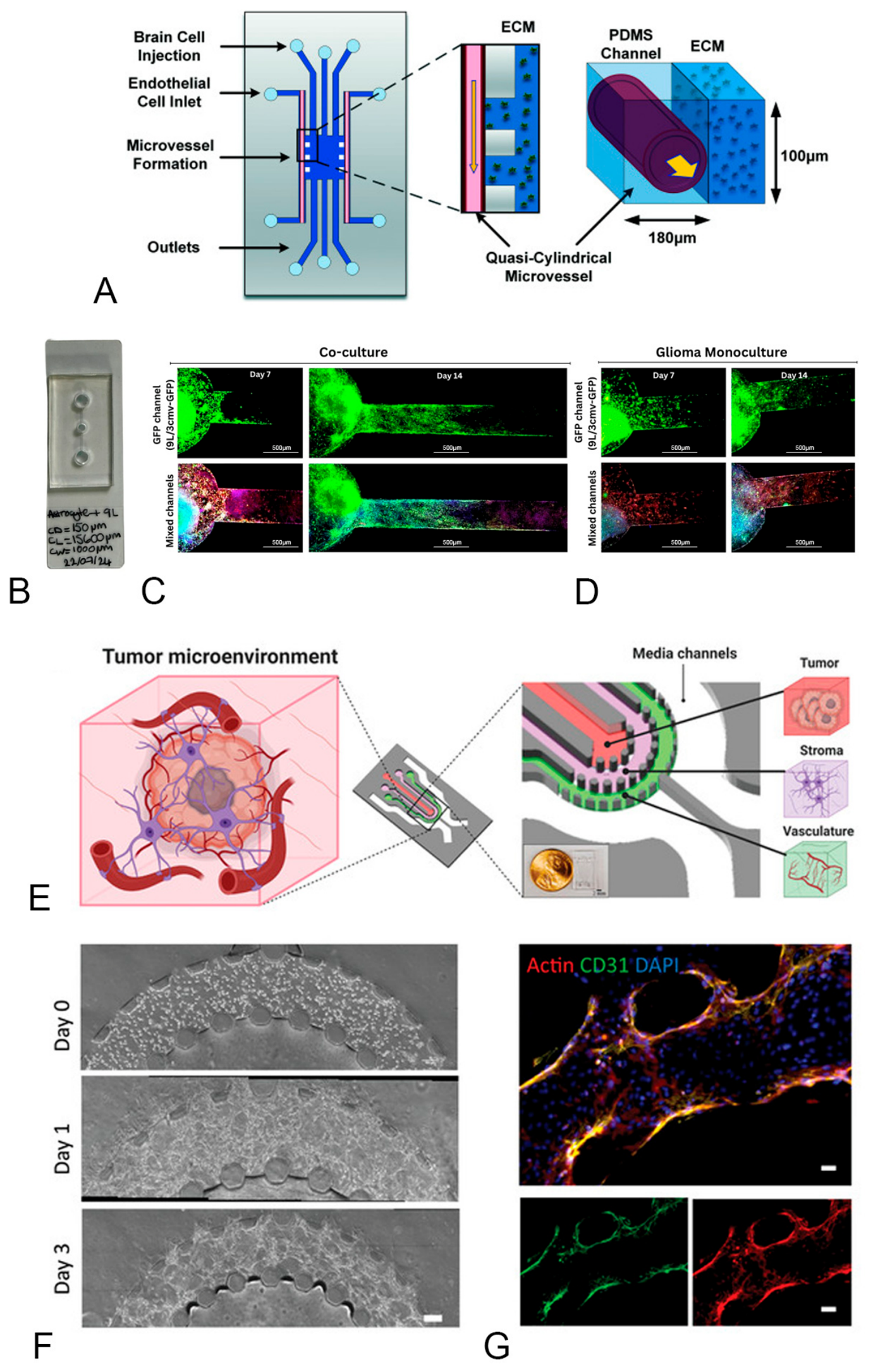

Disclaimer/Publisher’s Note: The statements, opinions and data contained in all publications are solely those of the individual author(s) and contributor(s) and not of MDPI and/or the editor(s). MDPI and/or the editor(s) disclaim responsibility for any injury to people or property resulting from any ideas, methods, instructions or products referred to in the content. |
© 2026 by the authors. Licensee MDPI, Basel, Switzerland. This article is an open access article distributed under the terms and conditions of the Creative Commons Attribution (CC BY) license.