Article

Biomechanical Investigation of Red Cell Sedimentation Using Blood Shear Stress and Blood Flow Image in a Capillary Chip


Department of Mechanical Engineering, Chosun University, 309 Pilmun-daero, Dong-gu, Gwangju 61452, Republic of Korea
Micromachines 2023, 14(8), 1594; https://doi.org/10.3390/mi14081594
Submission received: 1 July 2023 / Revised: 1 August 2023 / Accepted: 11 August 2023 / Published: 13 August 2023
(This article belongs to the Special Issue Biosensors for Biomedical and Environmental Applications)

Abstract

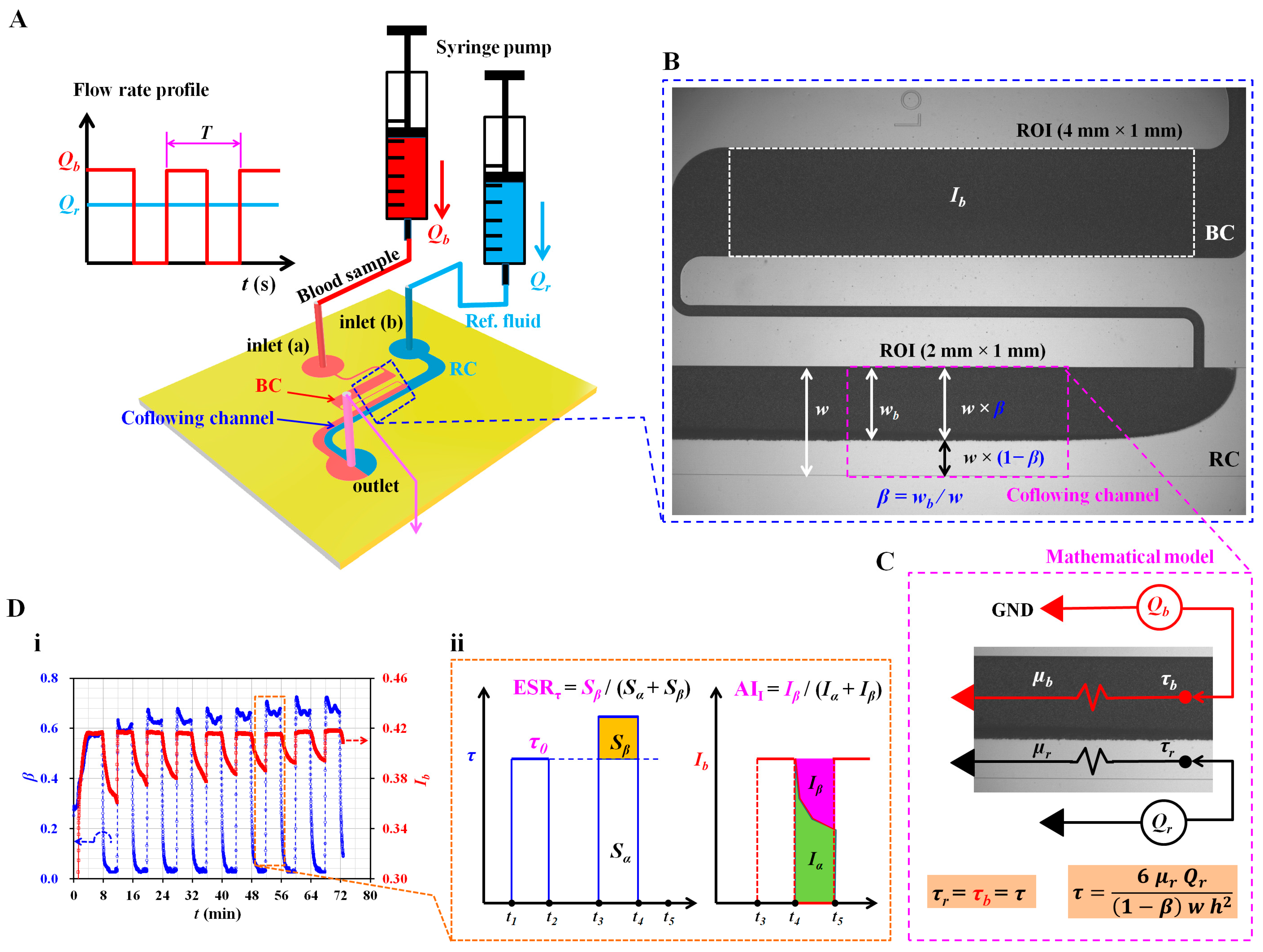
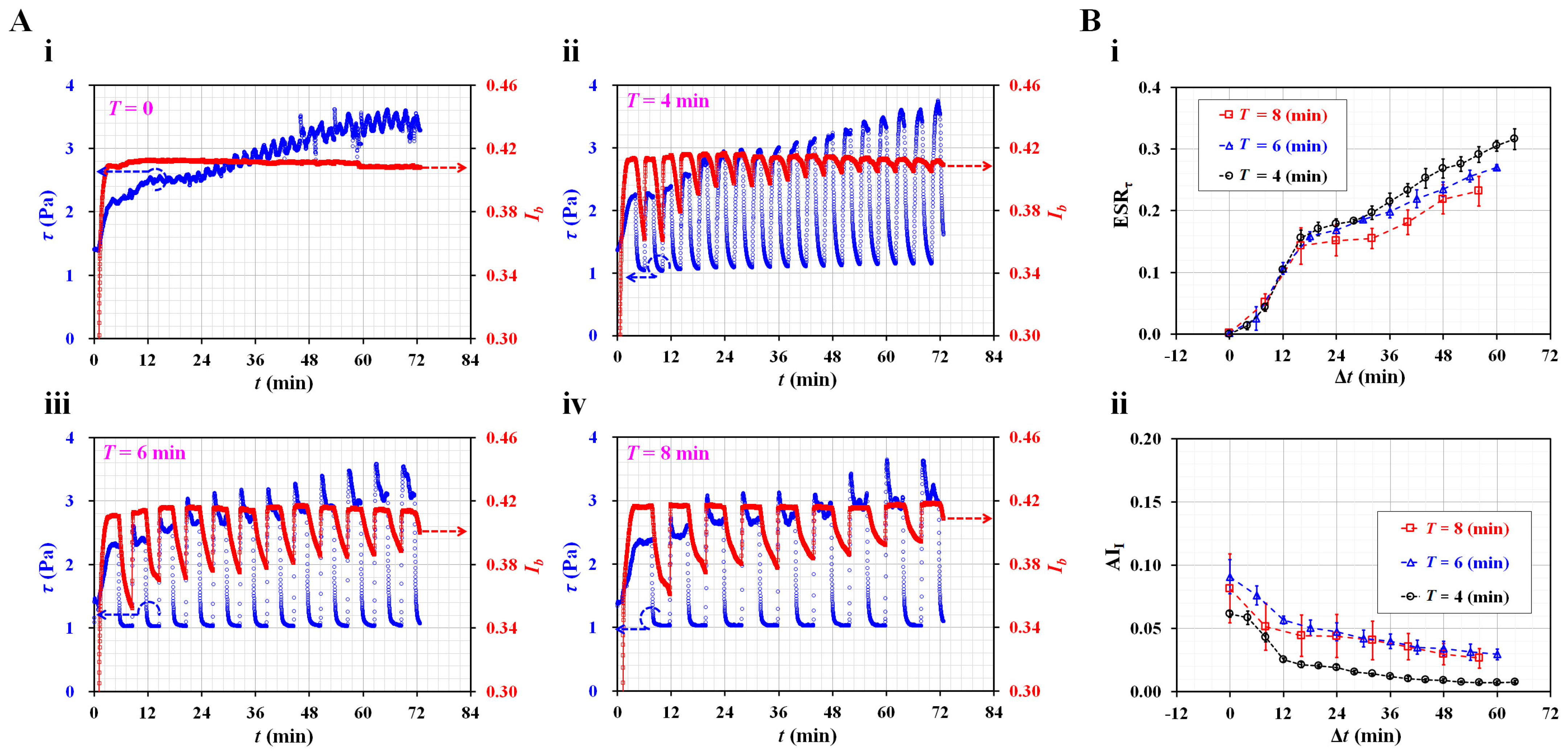
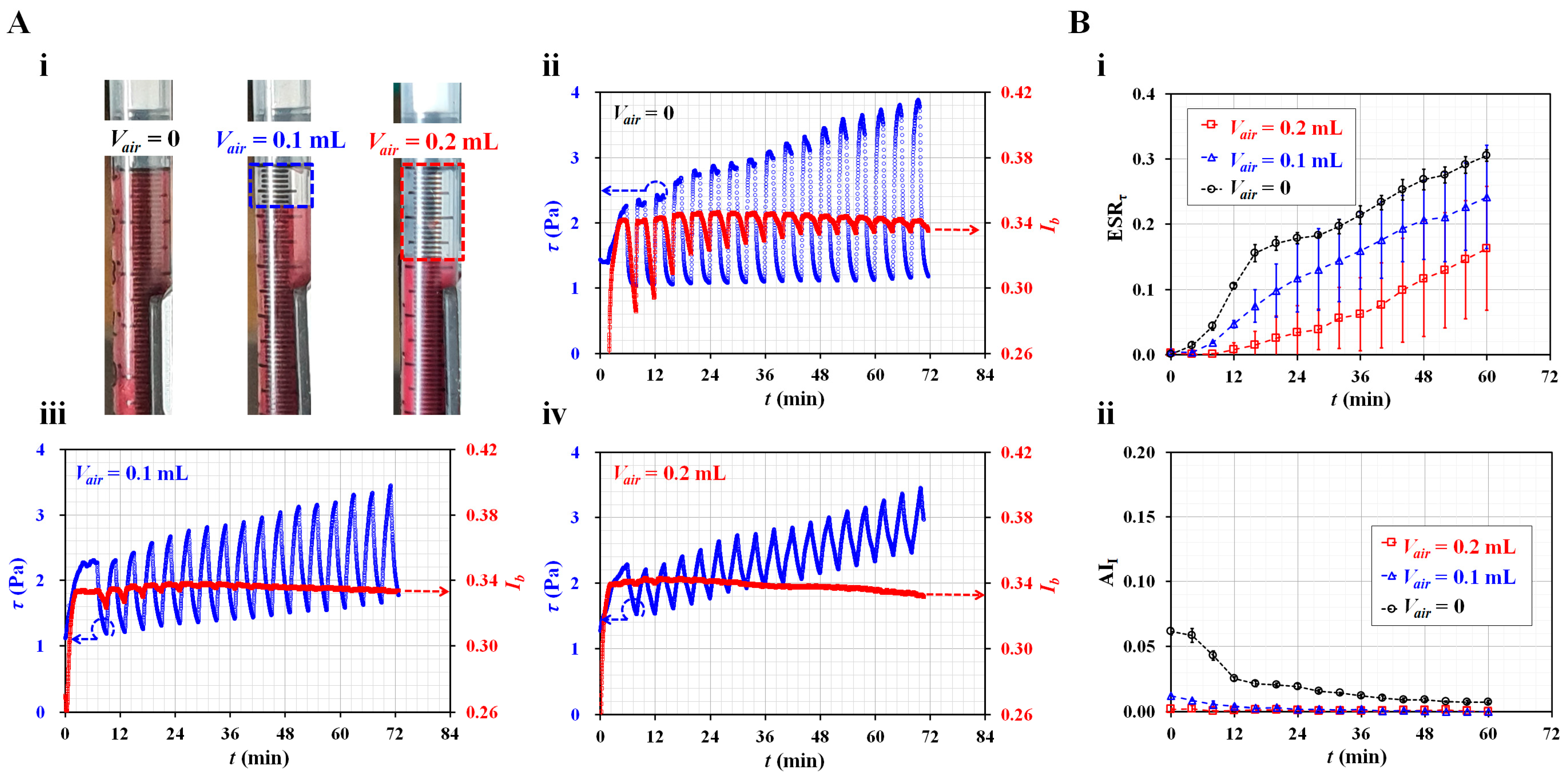
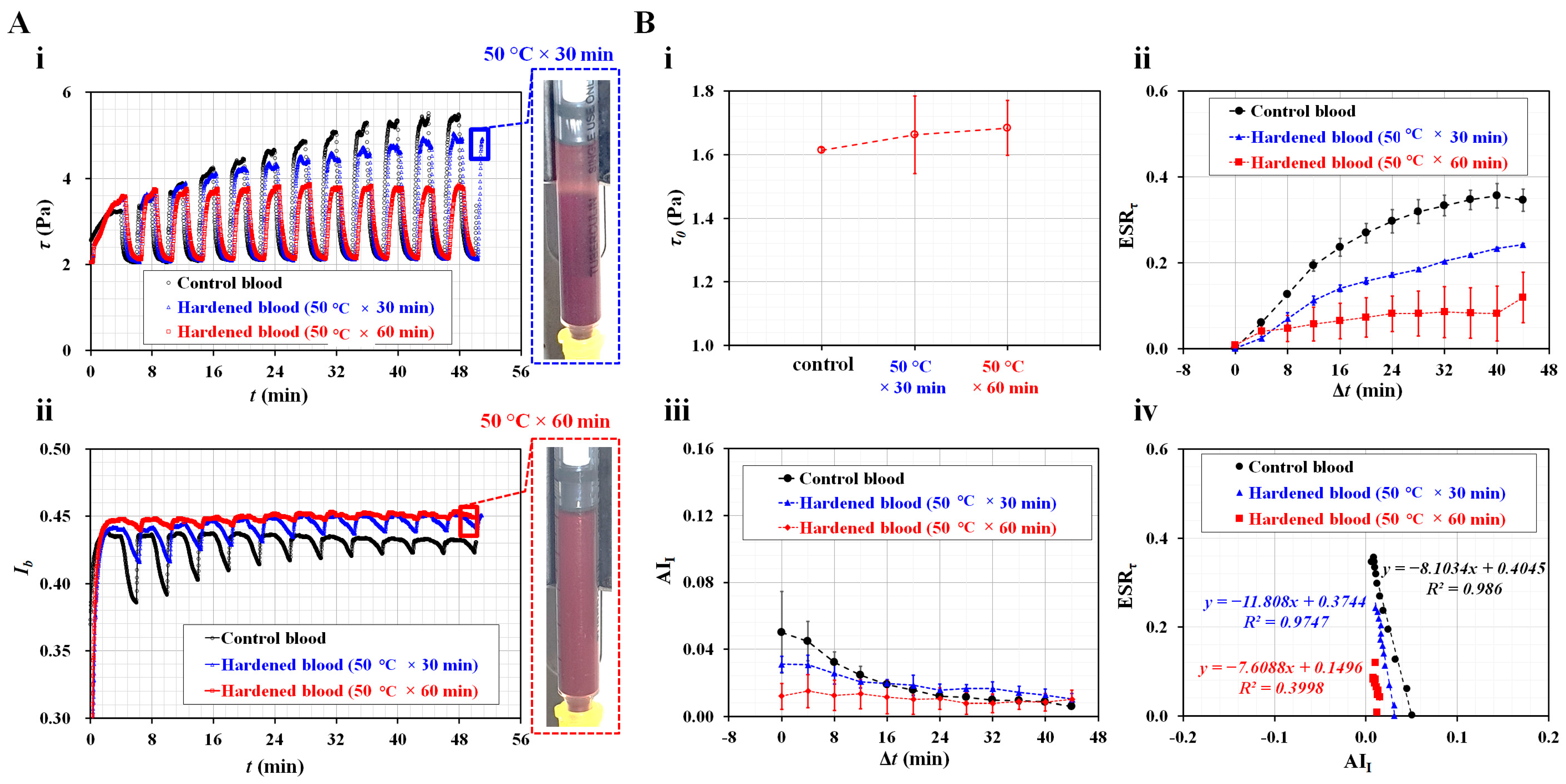
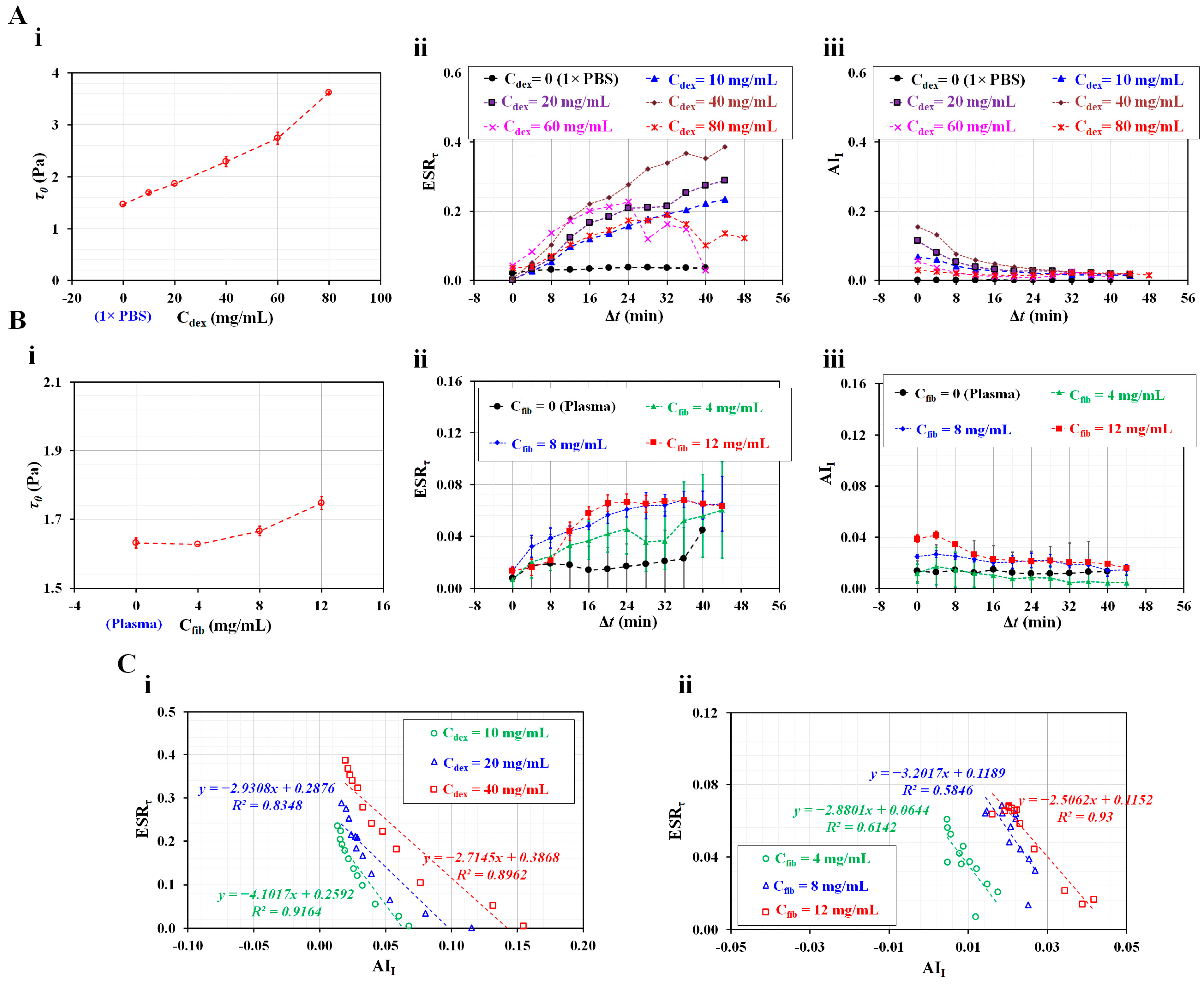
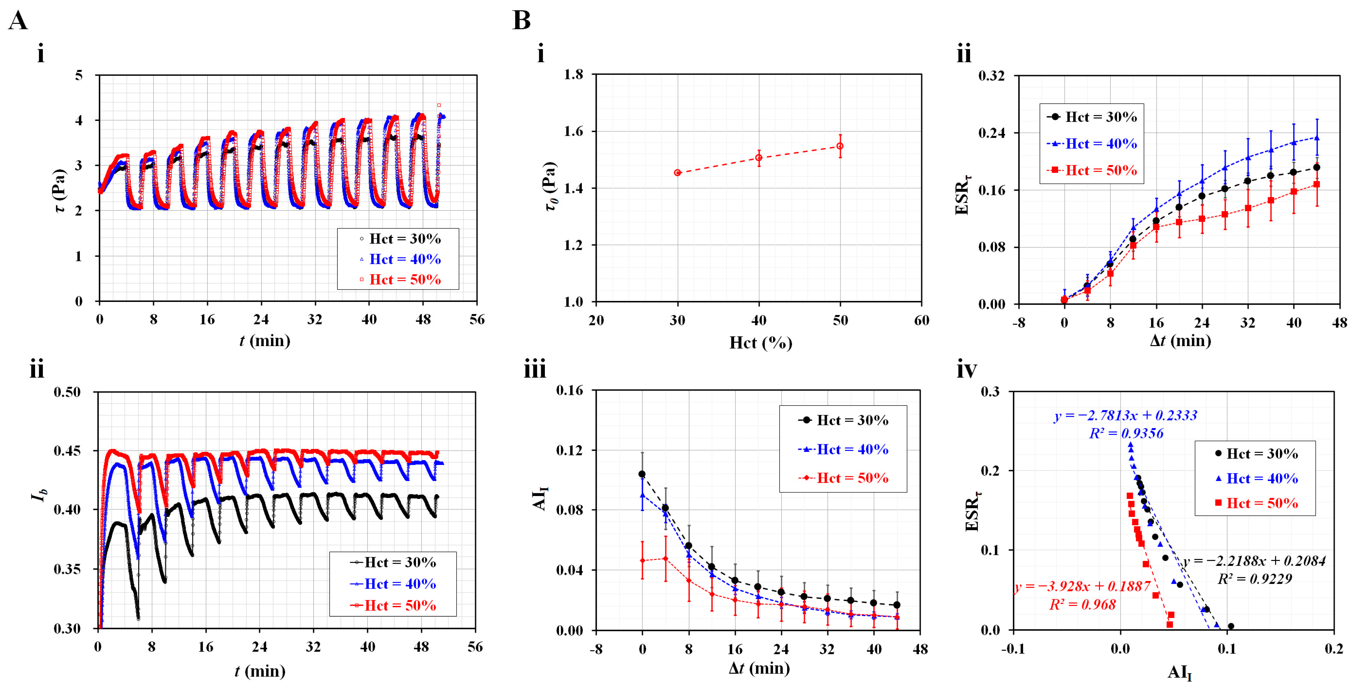
Blood image intensity has been used to detect erythrocyte sedimentation rate (ESR). However, it does not give information on the biophysical properties of blood samples under continuous ESR. In this study, to quantify mechanical variations of blood under continuous ESR, blood shear stress and blood image intensity were obtained by analyzing blood flows in the capillary channel. A blood sample is loaded into a driving syringe to demonstrate the proposed method. The blood flow rate is set in a periodic on–off pattern. A blood sample is then supplied into a capillary chip, and microscopic blood images are captured at specific intervals. Blood shear stress is quantified from the interface of the bloodstream in the coflowing channel. τ0 is defined as the maximum shear stress obtained at the first period. Simultaneously, ESRτ is then obtained by analyzing temporal variations of blood shear stress for every on period. AII is evaluated by analyzing the temporal variation of blood image intensity for every off period. According to the experimental results, a shorter period of T = 4 min and no air cavity contributes to the high sensitivity of the two indices (ESRτ and AII). The τ0 exhibits substantial differences with respect to hematocrits (i.e., 30–50%) as well as diluents. The ESRτ and AII showed a reciprocal relationship with each other. Three suggested properties represented substantial differences for suspended blood samples (i.e., hardened red blood cells, different concentrations of dextran solution, and fibrinogen). In conclusion, the present method can detect variations in blood samples under continuous ESR effectively.
Keywords: erythrocyte sedimentation rate (ESR); ESR index; aggregation index; blood shear stress; blood flow intensity; capillary chip erythrocyte sedimentation rate (ESR); ESR index; aggregation index; blood shear stress; blood flow intensity; capillary chip