


Abstract
Large-scale cultivation of transgenic Bt crops has led to target pest resistance to Bt proteins, especially Cry1Ac. Under persistent selection pressure, Helicoverpa armigera has evolved resistance to Bt proteins in both laboratory and field populations, highlighting an urgent need to comprehensively elucidate the molecular mechanisms underlying pest Bt resistance. Previous studies have confirmed that the eukaryotic translation initiation factor eIF2 plays a critical role in regulating Bt resistance. Our prior RNA-seq data showed significant differences in the expression level of eIF3L between susceptible and Cry1Ac-resistant strains of H. armigera. In this study, we further observed that the mRNA level of eIF3L was significantly reduced in larvae surviving Cry1Ac exposure. More importantly, heterologous expression of eIF3L increased the susceptibility of Sf9 cells to Cry1Ac. Consistently, knockdown of eIF3L was associated with reduced mortality in H. armigera larvae exposed to Cry1Ac. Together, our in vivo and in vitro results suggest that eIF3L is involved in Cry1Ac toxicity. Furthermore, eIF3L affects the expression of the Cry1Ac receptor genes CAD, ABCC2, and ABCC3. Collectively, these results indicate that down-regulation of eIF3L is associated with decreased susceptibility of H. armigera to Cry1Ac, providing novel insights into the molecular mechanisms underlying insect resistance to Bt proteins.
Key Contribution:
The extensive cultivation of Bt crops has intensified selection pressure, driving the evolution of resistance in target pests and threatening the long-term efficacy of Bt proteins. Understanding the molecular mechanisms underlying Bt resistance is essential for effective monitoring and management. While eIF2 has been implicated in Bt resistance, the function of eIF3L remains unclear. This study found that the down-regulation of eIF3L is associated with decreased susceptibility of Helicoverpa armigera to Cry1Ac, providing new insights into the mechanism of Bt resistance.
1. Introduction
Bacillus thuringiensis (Bt), a Gram-positive bacterium, produces insecticidal proteins that are used to control global agricultural pests [1,2]. Insecticide developed based on the Bt protein is not only specific and effective, but also possesses ecological safety, accounting for 75~95% of the global microbial insecticide market [3]. More importantly, transgenic Bt crops bring about huge economic and environmental benefits [4,5]. However, with the widespread application of transgenic Bt crops, the occurrence of target pest resistance to Bt proteins has been gradually increasing, involving at least eleven pest species and nine insecticidal proteins, which seriously threatens the long-term efficacy of Bt proteins [6,7]. Comprehensively understanding the resistance mechanisms of target pests to Bt proteins is crucial for developing resistance monitoring technologies and formulating resistance management strategies.
Crystalline (Cry) toxins are primary Bt proteins used for pest control [8]. Current studies indicate that upon ingestion by insects, Cry proteins are proteolytically activated and then bind with specific midgut receptors, damaging epithelial cells and ultimately causing insect death [9,10,11]. Furthermore, previous studies have shown that mutations or expression changes in genes involved in toxin activation and toxin-binding can lead to resistance [12]. Among them, mutation or down-regulation of the midgut receptors is considered to be the main cause of resistance [2,10,13]. The identified midgut receptors mainly include alkaline phosphatase (ALP), cadherin (CAD), aminopeptidase (APN), and ATP-binding cassette (ABC) transporter [13]. Recently, studies have also shown that transcription factors FOXA and GATA are involved in regulating the expression of Cry1Ac receptor genes, affecting the susceptibility of Helicoverpa armigera to Cry1Ac [14,15]. Furthermore, some studies have found that the MAPK signaling pathway regulates the expression of Cry midgut receptor genes through transcription factors [16,17,18]. Recent studies have highlighted the importance of transcriptional and translational regulation in modulating receptor expression.
In eukaryotic cells, the regulation of protein translation is essential for gene expression, and primarily occurs at the initiation phase. The eukaryotic translation initiation factor (eIF) is the major regulatory factor in the initiation stage of protein synthesis [19,20]. Recent studies have shown that eIFs have additional regulatory functions, possibly exerting their effects beyond translation initiation, such as regulation of cellular and organism physiology [21,22,23,24]. In Tribolium castaneum, eIF2γ can regulate the 20E biosynthesis pathway, thereby influencing its metamorphic development [25]. Notably, the transcription level of eIF2 is significantly lower in the Cry1Ac-resistant strain of the cotton bollworm H. armigera, and eIF2 regulates the expression of receptor genes CAD, ABCC2, and ABCC3, which consequently confers Cry1Ac resistance to H. armigera [26]. eIF3 is composed of 13 conserved subunits (subunits a ~ m) and plays a central role in initial processes [27,28,29]. Studies have demonstrated that eIF3 subunits are essential for organismal survival under various stress conditions [23,27,30]. In Caenorhabditis elegans, mutation of eIF3k or eIF3l increases general stress resistance [23]. Previous studies have implicated eIF3 subunits in Cry1Ac toxicity. Specifically, eIF3L was identified as a potential interacting protein of Cry1Ac in yeast two-hybrid screens [31]. However, it remains unclear whether eIF3L is involved in Cry1Ac toxicity.
H. armigera, a primary target pest of Bt crops, has developed resistance to Cry1Ac proteins under long-term selection pressure from Bt toxins [32,33]. Given the vital functional of eIF2 in Bt resistance, we sought to explore the roles of other eIFs in the toxicity of Cry1Ac. Our previous RNA-seq data identified eIF3L as differentially expressed between susceptible and resistant strains of H. armigera [34]. In this study, we investigated the role of eIF3L in Cry1Ac toxicity and Cry1Ac resistance, through qRT-PCR, cell cytotoxicity assays and RNAi, to offer insights into the Cry1Ac resistance mechanism of target pests.
2. Results
2.1. Bioinformatic Characterization and Phylogenetic Analysis of eIF3L
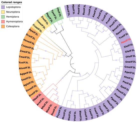
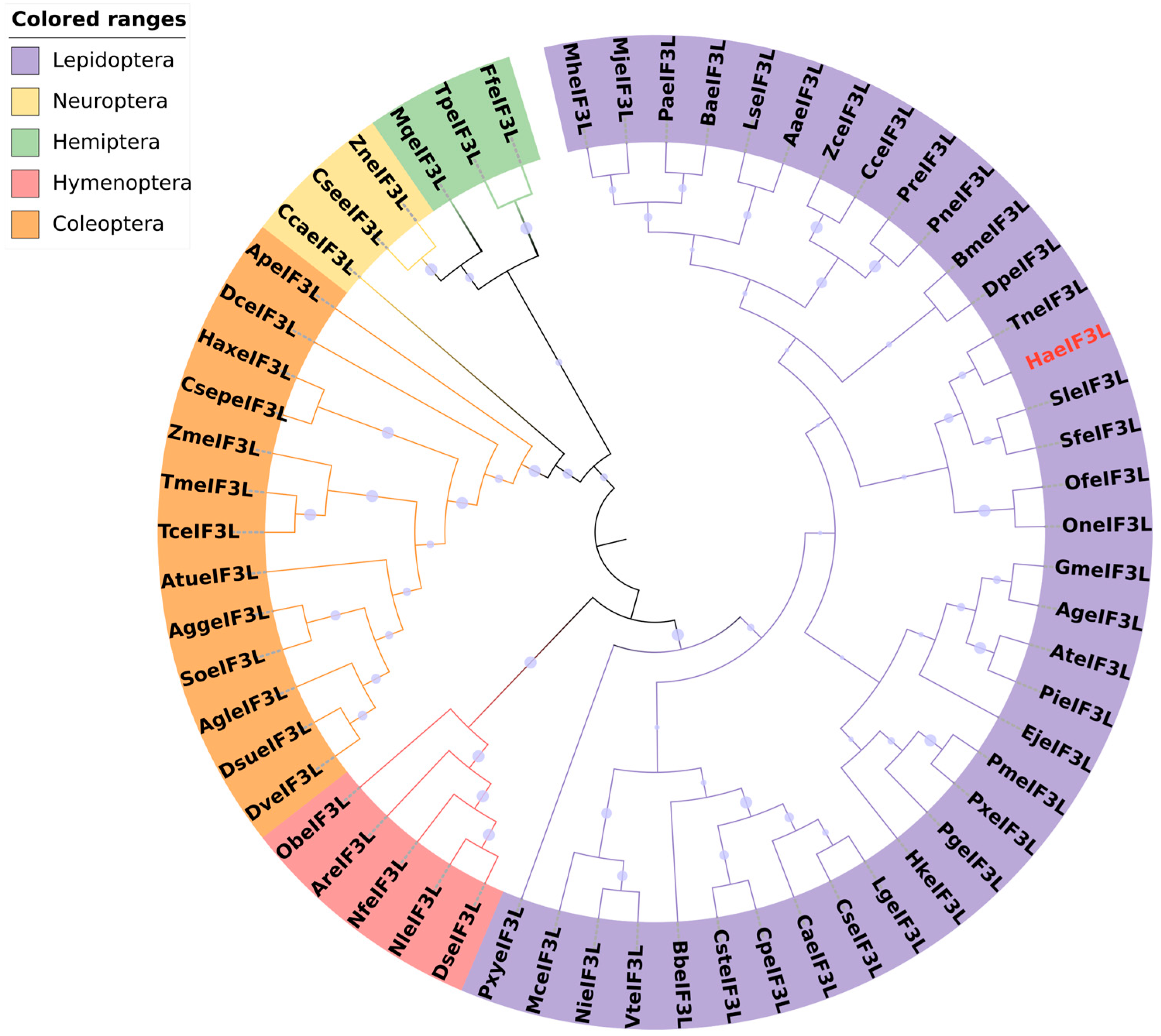
The full-length open reading frame (ORF) of eIF3L was cloned from H. armigera. The sequence has been uploaded to the NCBI database (GenBank accession: PQ002175). The ORF of eIF3L was 1632 bp in length, encoding 543 amino acid residues. eIF3L was predicted to have a molecular weight of 63.43 kDa and a pI of 6.11. We obtained 61 sequences from different insect species in the NCBI database (Table S2). Phylogenetic analysis was performed with the NJ method. The results revealed that eIF3L protein was most homologous with T. ni and evolutionarily conserved (Figure 1).
Figure 1.
Bioinformatic analysis of eIF3L. The amino acid sequence of eIF3L was downloaded from the NCBI database (accessions are shown in Table S2). Phylogenetic analysis was performed with the neighbor-joining method. The sequence of H. armigera is marked in red font. Species differences are shown through the shaded areas of different colors.
2.2. Spatio-Temporal Expression of eIF3L in H. armigera
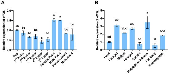
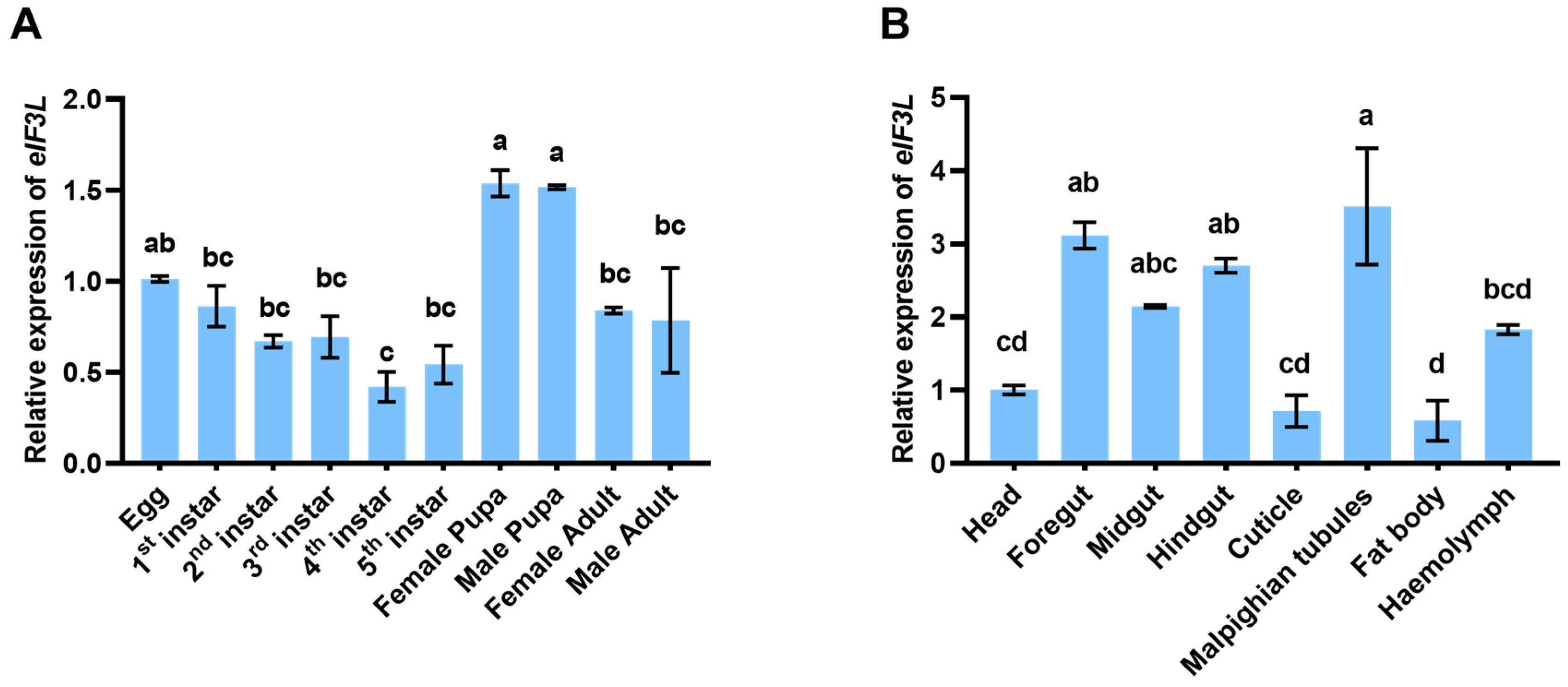
We examined eIF3L expression by qRT-PCR at different life stages and in different tissues. The results showed that eIF3L was expressed throughout life stages at varying levels. The transcript level of eIF3L peaked in the pupal stage, followed by the egg, larval and adult stages. Among them, the transcriptional level is the lowest in the fourth-instar larvae (Figure 2A). In different tissues of fifth-instar larvae, eIF3L transcript abundance was the highest in the Malpighian tubules, followed by the foregut, hindgut, midgut, hemolymph, head, epidermis and fat body (Figure 2B).
Figure 2.
Spatio-temporal expression of eIF3L in H. armigera. (A) Relative expression of eIF3L at different life stages. (B) Relative expression of eIF3L in each tissue of the 5th-instar larvae. The data in the figure are presented as the mean value ± SEM. Different letters denote statistical significance (one-way ANOVA with Tukey’s test, p < 0.05).
2.3. Reduced eIF3L Expression in Cry1Ac-Treated Larvae and the Resistant Strain
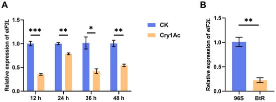
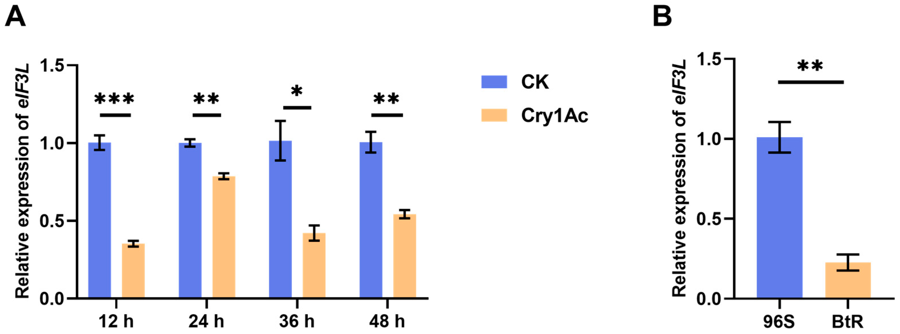
To determine whether eIF3L is related to the toxicity process of Cry1Ac, the transcriptional levels of eIF3L in the midgut tissues of the larvae were detected at four time points after Cry1Ac treatment in the susceptible strain 96S. The results showed that compared to the control group, the relative expression of eIF3L was significantly decreased after Cry1Ac treatment (Figure 3A). In addition, we compared its transcript levels between the susceptible strain 96S and Cry1Ac-resistant strain BtR and found that eIF3L expression was significantly down-regulated in the BtR strain (Figure 3B).
Figure 3.
Effect of Cry1Ac treatment on eIF3L transcription levels. (A) Relative expression of eIF3L in 5th-instar larvae of the 96S strain after exposure to the Cry1Ac artificial diet (100 μg/g). At different treatment points (12 h, 24 h, 36 h and 48 h), relative expression of the eIF3L gene was detected. (B) The expression level of eIF3L in the 96S strain and the BtR strain. Values in the figure represent the mean ± SEM. Data was analyzed using Student’s t-test (“*” p < 0.05, “**” p < 0.01, “***” p < 0.001).
2.4. Transient Expression of eIF3L Enhanced the Susceptibility of Sf9 Cells to Cry1Ac
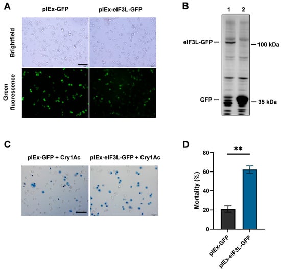
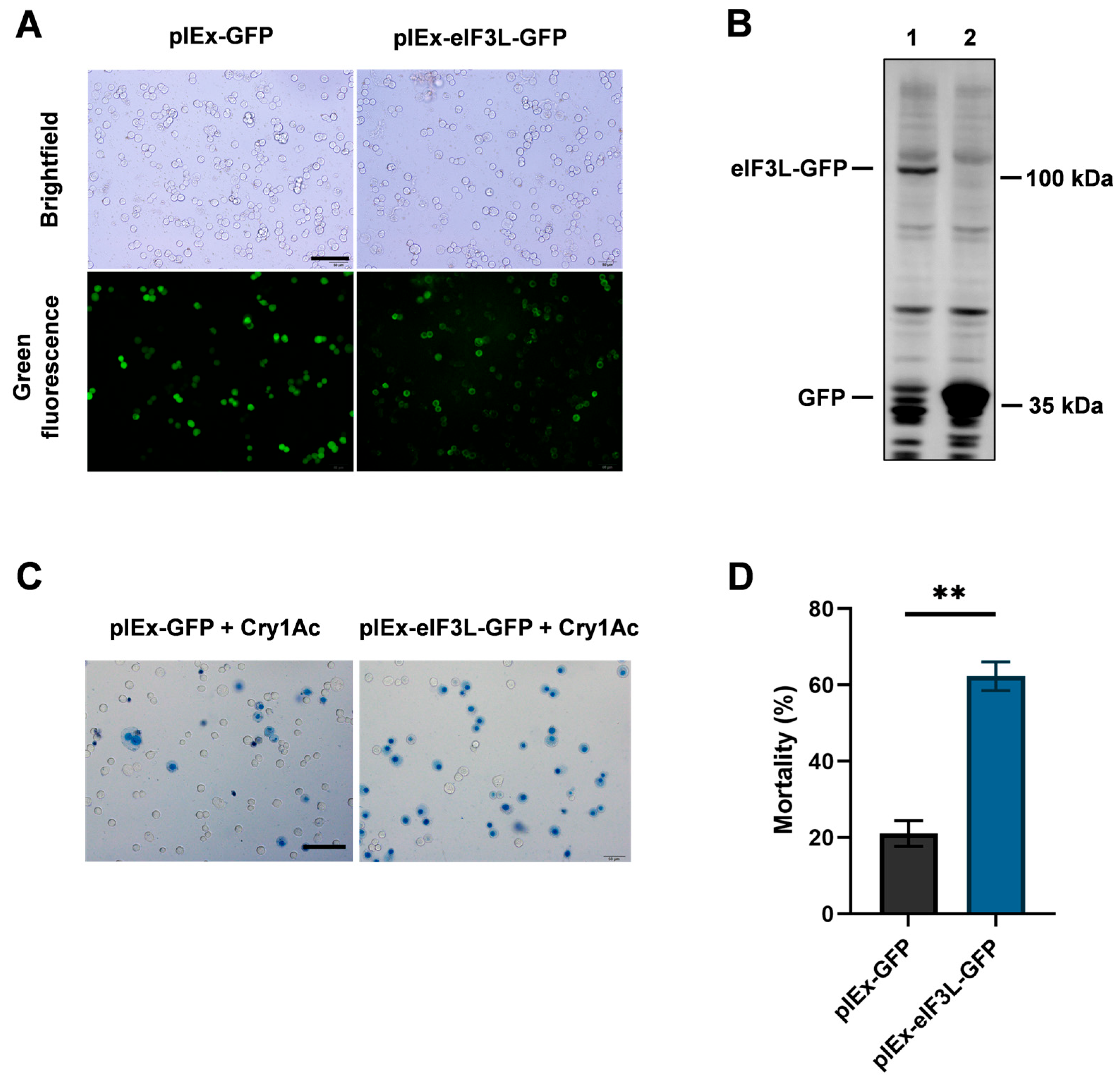
To investigate the functional role of eIF3L in Cry1Ac toxicity, eIF3L-GFP fusion protein was constructed and transient-expressed in Sf9 cells. Transfected Sf9 cells (pIEx-eIF3L-GFP/pIEx-GFP) exhibited GFP fluorescence, confirming recombinant protein expression (Figure 4A). Furthermore, the results of WB analysis confirmed the successful overexpression of eIF3L at the protein level in transfected cells (Figure 4B). Subsequently, after transfection for 48 h, activated Cry1Ac protein was added. Unlike the growth status in cells that only express GFP, the mortality of cells expressing eIF3L-GFP significantly increased by approximately 41 percentage points (Figure 4C,D). Importantly, heterologous expression of eIF3L significantly increased the cell’s sensitivity to Cry1Ac.
Figure 4.
Effect of eIF3L expression on the susceptibility of Sf9 cells to Cry1Ac. (A) Fluorescence microscopic observation after transfection. (B) Western blot analysis: Lane 1: pIEx-eIF3L-GFP, Lane 2: pIEx-GFP. (C) Representative images of cells after Cry1Ac treatment. Bars, 100 μm. After transfection for 48 h, activated Cry1Ac protein was added. Thirty minutes later, trypan blue solution was used for observation (blue indicates dead cells). (D) Cell mortality. Values in the figure represent the mean ± SEM. Data was analyzed using Student’s t-test (“**” p < 0.01).
2.5. Knockdown of eIF3L Is Associated with Reduced Larval Mortality Under Cry1Ac Treatment
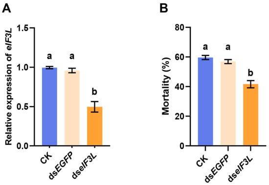
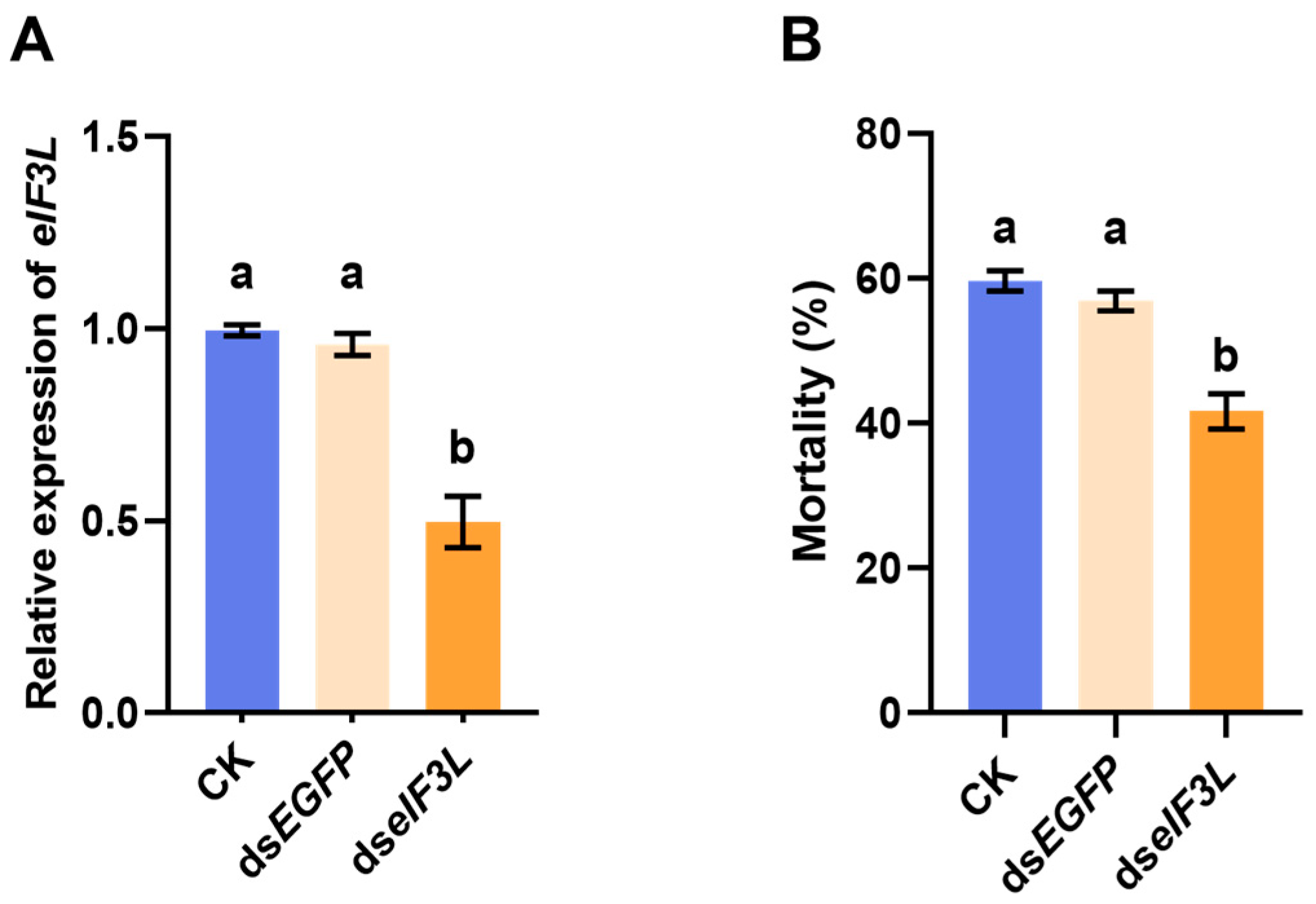
Based on the above results, we further verified the effect of eIF3L on Cry1Ac toxicity in the 96S strain using RNAi. dsRNA was injected into third-instar larvae. At 48 h post injection, qRT-PCR was performed to assess gene silencing efficiency. The results showed that the transcript level of eIF3L was significantly decreased compared to the control group (CK and dsEGFP). The efficiency of gene silencing was approximately 49% (Figure 5A). Subsequently, a bioassay was conducted on the larvae. Under the treatment of 300 μg/g of Cry1Ac insecticidal protein, larvae mortality was significantly reduced compared to the control, by approximately 17 percentage points (Figure 5B). These findings suggest that knockdown of eIF3L is associated with reduced larval mortality under Cry1Ac treatment.
Figure 5.
Effect of eIF3L knockdown on larval mortality under Cry1Ac treatment. (A) Silencing efficiency of eIF3L. Midgut tissues were extracted from larvae 48 h after dsRNA injection to measure the relative expression levels of eIF3L. (B) Larval mortality. Larvae were transitioned to an artificial diet containing Cry1Ac toxin at 48 h post injection, and their growth status was examined 5 d later. Values in the figure represent the mean ± SEM. Different letters denote statistical significance (one-way ANOVA with Tukey’s test, p < 0.05).
2.6. eIF3L Affects the Expression of Receptor Genes CAD, ABCC2, and ABCC3
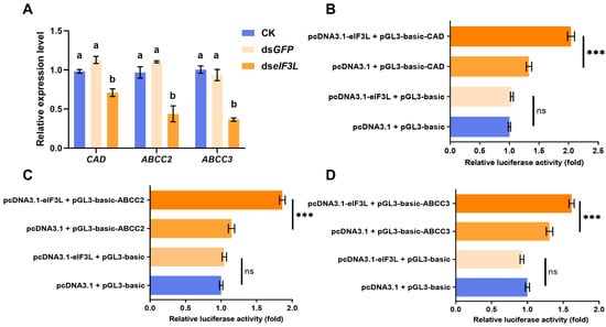
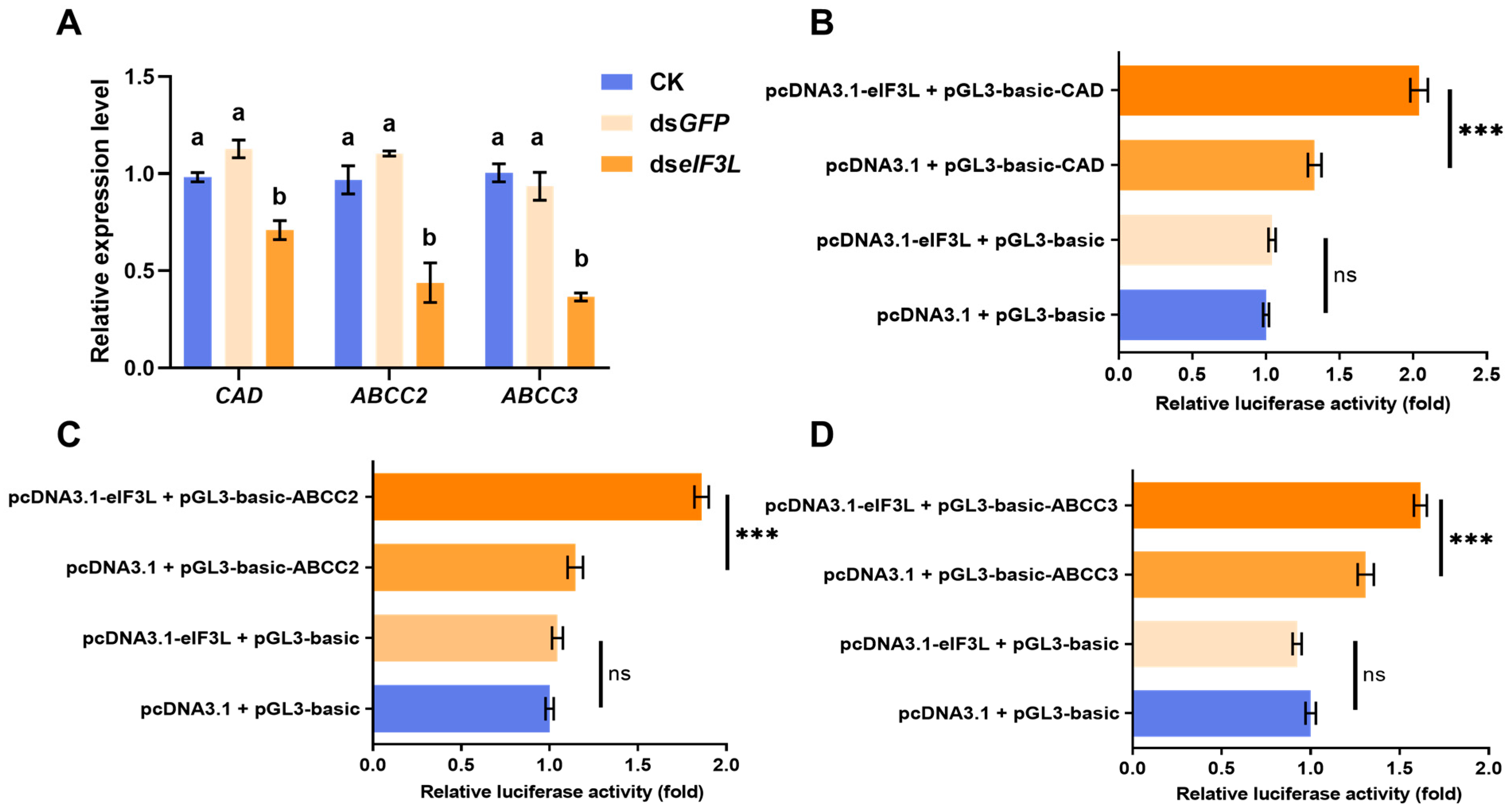
To investigate whether eIF3L contributes to the regulation of Cry1Ac receptor genes, we measured the mRNA levels of receptor genes after eIF3L silencing using qRT-PCR. Knockdown of eIF3L significantly decreased the mRNA abundance of CAD, ABCC2, and ABCC3 (Figure 6A). Furthermore, to further assess the effect of eIF3L on receptor gene promoter activity, a dual-luciferase reporter assay was conducted. The results showed that co-transfection with eIF3L significantly increased luciferase activity compared with the control group (Figure 6B–D). These results indicate that eIF3L affects the expression of CAD, ABCC2, and ABCC3.
Figure 6.
Effects of eIF3L on the expression of CAD, ABCC2, and ABCC3. (A) Transcript levels of CAD, ABCC2, and ABCC3 upon eIF3L knockdown. Different letters denote statistical significance (one-way ANOVA with Tukey’s test, p < 0.05). (B–D) eIF3L co-transfection on promoter activity of Cry1Ac receptor genes (CAD, ABCC2, and ABCC3). The receptor gene promoter construct was co-transfected with the pcDNA3.1-eIF3L vector to determine luciferase activity. Four groups were established: (1) the experimental group (pcDNA3.1-eIF3L + pGL3-basic-specific promoter + pRL-TK); (2) the corresponding control group (empty pcDNA3.1 + pGL3-basic-specific promoter + pRL-TK); (3) negative control (pcDNA3.1-eIF3L + empty pGL3-basic + pRL-TK); and (4) blank control (empty pcDNA3.1 + empty pGL3-basic + pRL-TK). Values in the figure represent the mean ± SEM (ns, not significant; “***” p< 0.001, t-test).
3. Discussion
The evolution of Cry protein resistance in target pests severely threatens the sustainable application of Bt insecticidal proteins [7]. Nevertheless, the complete regulatory network governing resistance mechanisms remain poorly understood. Elucidating these mechanisms is crucial for formulating resistance management strategies. Eukaryotic translation initiation factors exert critical functions in regulating gene expression, cell differentiation and apoptosis [21]. As the largest and most intricate subfamily among these factors, eIF3 has been reported to mediate host–pathogen interactions [30,35,36]. Our recent work revealed that eIF3L participates in the toxic action of Cry1Ac [31,34]. In addition, research on the nematode Caenorhabditis elegans demonstrated that eIF3L modulates cellular and organismal physiological processes [23].
In the study, we cloned the eIF3L gene from H. armigera. Its deduced amino acid sequence exhibits high similarity to homologous proteins of other Lepidoptera, reflecting evolutionary conservation across this insect (Figure 1). Our results showed that eIF3L was ubiquitously expressed in stages and tissues (Figure 2). This is consistent with the expression patterns of other eIF genes in lepidopteran insects Antheraea pernyi and Bombyx mori [37,38,39]. Notably, the expression level of eIF3L was higher in the Malpighian tubules and the gut. Previous studies have shown that genes involved in detoxification, such as cytochrome P450s and glutathione S-transferases, are often highly expressed in Malpighian tubules [40,41]. While we did not directly test the role of eIF3L in detoxification, its high expression in these tissues suggests a potential involvement in stress responses. Just as in C. elegans, the mutation of eIF3k/eIF3l can enhance the tolerance to endoplasmic reticulum stress without affecting overall translation, suggesting that these subunits may play a regulatory role in the stress response [23]. Similarly, eIF3m was specifically expressed in the hemipteran insect Rhodnius prolixus gut, and it was found to be important for digestion, reproduction, and growth [36]. Therefore, the expression pattern of eIF3L in H. armigera may reflect a role in modulating cellular stress responses to Bt toxin exposure.
eIF3 is essential for proper stress responses [27]. Here, we observed that the eIF3L transcript level was reduced in larvae surviving Cry1Ac treatment, implying its potential role in the toxicity process of Cry1Ac (Figure 3). This is consistent with previous studies on the eIF gene, supporting its significant role in stress response [23,36]. However, gut samples in this study were obtained solely from larvae that survived Cry1Ac exposure. Consequently, we cannot conclusively determine whether the decline in eIF3L expression was caused by Cry1Ac-mediated suppression or by the selective survival of larvae that naturally had low eIF3L levels. To resolve this, future experiments should monitor larval growth at each time point and collect gut tissues from all individuals. In eukaryotes, the eIF3 complex is mainly studied in mammals. Mutations or dysregulation of their expression levels can lead to developmental disorders [42]. Moreover, a decrease in the expression of eIF3 subunits causes developmental disorders in zebrafish and mice (h and m subunits) [43], and in Caenorhabditis elegans, eIF3 subunits can also modulate stress tolerance and extend its lifespan (k and l subunits) [23]. Here, we observed that transient expression of eIF3L enhanced the susceptibility of Sf9 cells to Cry1Ac (Figure 4). Consistently, RNAi-mediated knockdown of eIF3L was associated with reduced larval mortality under Cry1Ac treatment (Figure 5). And this is similar to the previous research results, confirming the significant role that eIF3L plays in the body’s response to pathogen infection [23,30]. However, because eIF3L is a translation initiation factor, its knockdown may alter general larval physiology, which could indirectly contribute to the reduced mortality observed. Further studies should include no-toxin controls and measure larval feeding and physiological parameters to fully exclude these nonspecific effects. Taken together, just like the research on eIF2, our results indicated that eIF3L also affected Cry1Ac toxicity [26].
Furthermore, our data suggest that eIF3L affects the expression of CAD, ABCC2, and ABCC3 (Figure 6). However, our results were limited to transcriptional level and promoter activity analysis, leaving the underlying mechanism unclear. Therefore, these results should be regarded as preliminary evidence suggesting that eIF3L may affect the expression of receptor genes, rather than definitive proof of direct transcriptional regulation. Future studies should use insect cell lines and include controls to distinguish between transcriptional and translational effects. Nevertheless, this study is the first to report an association between eIF3L and the expression of Cry1Ac receptor genes, providing a theoretical basis for future investigation of its molecular mechanisms. Notably, eIF3L exhibits dynamic subcellular distribution and relocalizes in response to specific stimuli [44,45,46]. That is, in addition to its important role in translation initiation, eIF3L may also have other physiological functions. Furthermore, in human disease research, androgen-induced palmitoylation has been demonstrated to drive eIF3L membrane localization in mammalian cells [45]. A study in H. armigera suggested that eIF3L may interact with Cry1Ac protein [31]. Based on studies in C. elegans [23], we speculate that eIF3L may interact with Cry1Ac directly or indirectly. First, eIF3L regulates receptor gene expression, thereby affecting Cry1Ac binding capacity and toxicity. Second, eIF3L may interact with Cry1Ac or modulate cellular stress responses, influencing toxicity independently of receptor expression. Future studies should further investigate to distinguish between these two hypotheses. In this study, although eIF3L knockdown reduced the transcript levels of receptor genes, receptor protein levels and Cry1Ac binding capacity were not measured. Therefore, we cannot definitively link changes in receptor mRNA to changes in susceptibility. Future studies should measure receptor protein levels by Western blot and perform Cry1Ac binding assays to determine whether eIF3L-mediated changes in receptor expression affect toxin binding. Our research indicated that down-regulation of eIF3L is associated with decreased susceptibility of H. armigera to Cry1Ac; however, the underlying mechanism still requires further investigation. Previously, MAPK signaling pathway plays a crucial role in the regulation of eIF functions [22,47]. In addition, studies have reported that eIF was also closely associated with insect reproduction and metamorphic development [36,39,43]. This raises the following question: does eIF interact with immunity or hormone pathways, thereby influencing insect resistance to the Cry1Ac insecticidal protein? This hypothesis warrants further investigation. eIF regulates gene expression through multi-layered mechanisms and plays a crucial role in insect development and immune responses. Further analysis of this functional network using molecular and genetic approaches will help clarify the molecular basis of Cry1Ac susceptibility in H. armigera and may point to candidate targets for resistance management.
In conclusion, we found that knockdown of eIF3L was associated with a decrease in the susceptibility of H. armigera larvae to Cry1Ac. These findings provide new insights into the resistance mechanism of cotton bollworm to Cry1Ac, and have a positive impact on the development of pest resistance management strategies.
4. Materials and Methods
4.1. Insects and Bt Toxins
The susceptible strain 96S was obtained from a field in Xinxiang County, Henan Province, China, in 1996. The Cry1Ac-resistant strain BtR was generated by laboratory selection. The larvae showed an approximately 3000-fold resistance ratio to Cry1Ac [48]. In the laboratory, an artificial diet was given to the larvae [49]. Culture conditions consisted of 26 ± 1 °C, 75 ± 5% relative humidity, and a 14 L:10 D photoperiod.
Cry1Ac protoxin and activated Cry1Ac toxin were purchased from Beijing Meiyan Agricultural Technology Co., Ltd. (Beijing, China).
4.2. Sample Collection
Developmental samples of the 96S strain included eggs (400); first- to fifth-instar larvae at 100, 50, 20, 10, and 10 individuals, respectively; female and male pupae (10 each); and female and male adults (10 each). Tissue samples were collected from fifth -instar larvae (30), including the head, foregut, midgut, hindgut, cuticle, Malpighian tubules, fat body and hemolymph. Three replications were prepared for each treatment. The collected samples were immediately frozen with liquid nitrogen and then transferred to −80 °C.
4.3. Gene Cloning
Total RNA was extracted with TRIzol reagent (Thermo Fisher, Waltham, MA, USA) following the manufacturer’s instructions. Subsequently, purity and concentration were measured using NanoDrop 1000 (Thermo Fisher, Waltham, MA, USA). First-strand cDNA was reverse-transcribed from 1 μg RNA using the FastKing gDNA Dispelling RT SuperMix (Tiangen, Beijing, China). The coding sequence (CDS) of H. armigera eIF3L was obtained from the NCBI database (GenBank: XM_021325791.2) and further corrected against published transcriptome data [33]. Gene-cloning primers (Table S1) were designed using Primer3Plus (https://www.primer3plus.com/index.html (accessed on 1 September 2023)). PCR was performed with 2× Phanta Max Master Mix (Dye Plus) (Vazyme, Nanjing, China). The PCR procedure was as follows: 95 °C for 3 min, 1 cycle; 95 °C for 30 s, 58 °C for 30 s, 72 °C for 2 min, 35 cycles; and 72 °C for 5 min. After purification, the amplicon was subcloned into the pEASY-Blunt Simple Cloning Vector (TransGen, Beijing, China).
4.4. Sequence Analysis
The molecular weight and isoelectric point of protein were predicted using the Expasy MW/pI tool (https://web.expasy.org/compute_pi/ (accessed on 20 October 2023)). Homologous protein sequence alignment was performed with DNAMAN 9.0 (Lynnon, San Ramon, CA, USA). The phylogenetic tree was constructed using the neighbor-joining method in MEGA 7.0.
4.5. qRT-PCR
cDNA was generated as described earlier. Primers for qRT-PCR (Table S1) were designed via NCBI Primer-BLAST (https://www.ncbi.nlm.nih.gov/tools/primer-blast/ (accessed on 7 November 2023)). qRT-PCR assays were conducted using SuperReal PreMix Plus (SYBR Green) (TransGen, Beijing, China) on an ABI 7500 Real-Time PCR System (Thermo Fisher, Waltham, MA, USA). RPS15, ribosomal protein S15 (GenBank: AY818611.1), was used as a reference gene [50]. Gene relative expression was calculated via the 2−ΔΔCt method [51], using qRT-PCR data obtained from three biologically independent samples, each assayed in triplicate.
4.6. The Transcriptional Level of eIF3L Under Cry1Ac Stress
We added Cry1Ac protoxin to the artificial diet to make a mixed toxic diet with a final concentration of 100 μg/g. Fifth-instar larvae were starved for 6 h and then fed an artificial diet containing Cry1Ac (100 μg/g). A solvent (50 mmol/L Na2CO3, pH 10.0) was used as the control. At different treatment points (12 h, 24 h, 36 h and 48 h), the midgut tissues of survival individuals were sampled respectively, and the midgut of 20 larvae was collected from each sample. Three replications were prepared for each treatment. Finally, the transcriptional level of the eIF3L was detected using qRT-PCR.
4.7. Transient Expression in Sf9 Cells and Cytotoxicity Assay
pIEx-4-GFP vector was provided by Dr. Jizhen Wei (Henan Agricultural University). Green fluorescent protein (GFP) (GenBank: U50963.1) was inserted between pIEx-4 vector KpnI and NotI restriction sites. The eIF3L CDS was inserted into the SacI and BglII restriction sites of this vector via Gibson assembly (NEB, Ipswich, MA, USA). Primers used were listed in Table S1.
Sf9 cells were maintained at 27 ± 1 °C in Sf-900 II Serum-free medium (Gibco, New York, NY, USA) supplemented with penicillin (50 U/mL) and streptomycin (50 μg/mL) (Gibco, New York, NY, USA). For transfection, Sf9 cells were first seeded into a 24-well plate (5 × 10⁵ cells per well). After 24 h, the cells were washed with fresh medium. Transient expression was performed using FuGENE HD transfection reagent (Promega, Madison, WI, USA). Further, 500 ng/well pIEx-eIF3L-GFP and pIEx-GFP were respectively mixed with transfection reagent at a ratio of 1:3 (w/w). At 48 h post transfection, activated Cry1Ac toxin (40 μg/mL, diluted in the culture medium) was added. After 30 min of treatment, cell growth was observed by 0.4% trypan blue staining (Solarbio, Beijing, China), and mortality was calculated. Cell death was quantified by counting the number of dead cells (stained with trypan blue) relative to the total number of cells in each field of view. Three independent biological replicates were performed for each treatment.
4.8. Western Blot
At 48 h post transfection, cell proteins were extracted using RIPA lysis buffer (Beyotime, Shanghai, China) with 10 μL phenylmethylsulfonyl fluoride and protease inhibitor cocktail (Coolaber). Proteins were resolved by SDS-PAGE and then transferred onto a PVDF membrane (Merck, Darmstadt, Germany). The membrane was blocked with 5% skim milk (1 h, room temperature), and then incubated overnight at 4 °C with rabbit anti-GFP IgG primary antibody (Invitrogen) diluted at a ratio of 1:10,000 in 3% BSA. The next day, the PVDF membrane was washed with 1× TBST and then incubated for 1 h at room temperature with an alpaca anti-rabbit IgG-HRP secondary antibody (HUABIO) diluted at a ratio of 1:10,000 in 3% BSA. Finally, immunoblots were imaged using Touch Imager (e-Blot, Shanghai, China).
4.9. dsRNA Synthesis and Microinjection
dsRNA-specific primers were designed with E-RNAi [52]. Primers for dsRNA synthesis are given in Table S1. dsRNA product sizes were 492 bp for eIF3L and 571 bp for EGFP. dsRNA was synthesized with the T7 RiboMAXTM Express RNAi System (Promega, Madison, WI, USA) according to the product’s protocol. For microinjection, third-instar larvae were chilled on ice for 30 min and then injected with 2 μg dsRNA using a NANOJECT III microinjection system (Drummond, Broomall, PA, USA). The control group included CK (not injected, blank control) and dsEGFP (dsEGFP was injected, negative control). At 48 h post injection, larvae midguts were collected to assess gene silencing efficiency. Each sample consisted of 10 midgut, and three replicates were set up.
4.10. Larvae Bioassays
At 48 h post injection, an artificial diet toxicity bioassay was used to assess the sensitivity of H. armigera to Cry1Ac insecticidal protein. We added Cry1Ac protoxin to the artificial diet to make a mixed toxic diet with a final concentration of 300 μg/g. The mixed artificial diet was evenly distributed into 24-well plates. Larvae were then started on the diet. Each treatment group comprised 24 larvae, with three replicates. After 5 days, larval growth and development were assessed, and mortality was recorded.
4.11. Dual-Luciferase Assay
Promoter fragments of receptors CAD (GenBank: MN097698.1), ABCC2 (GenBank: MF186781.1) and ABCC3 (GenBank: MF197473.1) were respectively cloned into the pGL3-basic vector. Primers used are listed in Table S1. To assess the effects of eIF3L on the regulatory activity of receptors, the CDS of eIF3L was ligated into the pcDNA3.1. Then, the pGL3-basic-CAD (pGL3-basic-ABCC2 and pGL3-basic-ABCC3) and pcDNA3.1-eIF3L plasmids were co-transfected into HEK-293T cells. Four groups were established: (1) the experimental group (pcDNA3.1-eIF3L + pGL3-basic-specific promoter + pRL-TK); (2) the corresponding control group (empty pcDNA3.1 + pGL3-basic-specific promoter + pRL-TK); (3) the negative control (pcDNA3.1-eIF3L + empty pGL3-basic + pRL-TK); and (4) the blank control (empty pcDNA3.1 + empty pGL3-basic + pRL-TK). After 72 h, luciferase activity was assessed using a dual-luciferase reporter gene assay kit (Yeasen Biotechnology, Shanghai). Each experiment was performed with 4 independent replicates. The pRL-TK vector, which carries the Renilla luciferase gene driven by a constitutive thymidine kinase (TK) promoter, served as an internal reference for correcting transfection efficiency and well-to-well variation. The firefly signal was divided by the Renilla signal for normalization, and relative luciferase activity was determined by comparing the normalized firefly signal of each experimental group with that of the control group.
4.12. Statistical Analyses
All data are presented as mean ± SEM. Statistical analyses were performed in R software (version 4.3.1, https://www.r-project.org (accessed on 3 December 2024)). Differences between two groups were evaluated using Student’s t-test, with significance indicated as * p < 0.05, ** p < 0.01, and *** p < 0.001. When comparing the data of more than two groups, we used one-way ANOVA followed by Tukey’s honest significant difference (HSD) for multiple comparisons.
Supplementary Materials
The following supporting information can be downloaded at: https://www.mdpi.com/article/10.3390/toxins18110471/s1, Table S1: Primers used in the study; Table S2: The gene names and accession numbers of amino acid sequences used in the neighbor-joining tree.
Author Contributions
Conceptualization, N.L., C.Z., G.L. and L.C.; methodology, N.L., C.Z. and Z.L.N.; software, N.L.; validation, E.T.S., H.Y. and X.L.; formal analysis, N.L.; investigation, N.L. and C.Z.; resources, G.L. and L.C.; data curation, N.L.; writing—original draft preparation, N.L.; writing—review and editing, C.Z., Z.L.N., E.T.S., G.L. and L.C.; visualization, N.L. and C.Z.; supervision, G.L. and L.C.; project administration, G.L. and L.C.; funding acquisition, G.L. All authors have read and agreed to the published version of the manuscript.
Funding
This work was supported by the Biological Breeding National Science and Technology Major Project (2022ZD04021); the National Natural Science Foundation of China (32372543); and the Agricultural Science and Technology Innovation Program (CAAS-CSCB-202303).
Institutional Review Board Statement
Not applicable.
Informed Consent Statement
Not applicable.
Data Availability Statement
The original contributions presented in this study are included in the article. Further inquiries can be directed to the corresponding authors.
Acknowledgments
We thank Jizhen Wei (Henan Agricultural University, Zhengzhou, Henan, China) for providing the pIEx-4-GFP vector and for technical advice on cell line experiments.
Conflicts of Interest
The authors declare no conflicts of interest.
References
- Palma, L.; Muñoz, D.; Berry, C.; Murillo, J.; Caballero, P. Bacillus thuringiensis toxins: An overview of their biocidal activity. Toxins 2014, 6, 3296–3325. [Google Scholar] [CrossRef] [Scilit] [PubMed]
- Bel, Y.; Ferré, J.; Hernández-Martínez, P. Bacillus thuringiensis Toxins: Functional Characterization and Mechanism of Action. Toxins 2020, 12, 785. [Google Scholar] [CrossRef] [Scilit] [PubMed]
- Bravo, A.; Likitvivatanavong, S.; Gill, S.S.; Soberón, M. Bacillus thuringiensis: A story of a successful bioinsecticide. Insect Biochem. Mol. Biol. 2011, 41, 423–431. [Google Scholar] [CrossRef] [Scilit] [PubMed]
- Tabashnik, B.E.; Brévault, T.; Carrière, Y. Insect resistance to Bt crops: Lessons from the first billion acres. Nat. Biotechnol. 2013, 31, 510–521. [Google Scholar] [CrossRef] [Scilit] [PubMed]
- Lu, Y.; Wu, K.; Jiang, Y.; Guo, Y.; Desneux, N. Widespread adoption of Bt cotton and insecticide decrease promotes biocontrol services. Nature 2012, 487, 362–365. [Google Scholar] [CrossRef] [Scilit] [PubMed]
- Tabashnik, B.E.; Carrière, Y. Global Patterns of Resistance to Bt Crops Highlighting Pink Bollworm in the United States, China, and India. J. Econ. Entomol. 2019, 112, 2513–2523. [Google Scholar] [CrossRef] [Scilit] [PubMed]
- Tabashnik, B.E.; Fabrick, J.A.; Carrière, Y. Global Patterns of Insect Resistance to Transgenic Bt Crops: The First 25 Years. J. Econ. Entomol. 2023, 116, 297–309. [Google Scholar] [CrossRef] [Scilit] [PubMed]
- Sanahuja, G.; Banakar, R.; Twyman, R.M.; Capell, T.; Christou, P. Bacillus thuringiensis: A century of research, development and commercial applications. Plant Biotechnol. J. 2011, 9, 283–300. [Google Scholar] [CrossRef] [Scilit] [PubMed]
- Heckel, D.G. A return to the pore—Dissecting Bacillus thuringiensis toxin mode of action via voltage clamp experiments. FEBS J. 2016, 283, 4458–4461. [Google Scholar] [CrossRef] [Scilit] [PubMed]
- Pardo-López, L.; Soberón, M.; Bravo, A. Bacillus thuringiensis insecticidal three-domain Cry toxins: Mode of action, insect resistance and consequences for crop protection. FEMS Microbiol. Rev. 2013, 37, 3–22. [Google Scholar] [CrossRef] [Scilit] [PubMed]
- Ma, Y.; Zhang, J.; Xiao, Y.; Yang, Y.; Liu, C.; Peng, R.; Yang, Y.; Bravo, A.; Soberón, M.; Liu, K. The Cadherin Cry1Ac Binding-Region is Necessary for the Cooperative Effect with ABCC2 Transporter Enhancing Insecticidal Activity of Bacillus thuringiensis Cry1Ac Toxin. Toxins 2019, 11, 538. [Google Scholar] [CrossRef] [Scilit] [PubMed]
- Xiao, Y.; Wu, K. Recent progress on the interaction between insects and Bacillus thuringiensis crops. Phil. Trans. R. Soc. Lond. B Biol. Sci. 2019, 374, 20180316. [Google Scholar] [CrossRef] [Scilit] [PubMed]
- Jurat-Fuentes, J.L.; Heckel, D.G.; Ferré, J. Mechanisms of Resistance to Insecticidal Proteins from Bacillus thuringiensis. Annu. Rev. Entomol. 2021, 66, 121–140. [Google Scholar] [CrossRef] [Scilit] [PubMed]
- Li, J.; Ma, Y.; Yuan, W.; Xiao, Y.; Liu, C.; Wang, J.; Peng, J.; Peng, R.; Soberón, M.; Bravo, A.; et al. FOXA transcriptional factor modulates insect susceptibility to Bacillus thuringiensis Cry1Ac toxin by regulating the expression of toxin-receptor ABCC2 and ABCC3 genes. Insect Biochem. Mol. Biol. 2017, 88, 1–11. [Google Scholar] [CrossRef] [Scilit] [PubMed]
- Wei, W.; Pan, S.; Ma, Y.; Xiao, Y.; Yang, Y.; He, S.; Bravo, A.; Soberón, M.; Liu, K. GATAe transcription factor is involved in Bacillus thuringiensis Cry1Ac toxin receptor gene expression inducing toxin susceptibility. Insect Biochem. Mol. Biol. 2020, 118, 103306. [Google Scholar] [CrossRef] [Scilit] [PubMed]
- Qin, J.; Guo, L.; Ye, F.; Kang, S.; Sun, D.; Zhu, L.; Bai, Y.; Cheng, Z.; Xu, L.; Ouyang, C.; et al. MAPK-Activated Transcription Factor PxJun Suppresses PxABCB1 Expression and Confers Resistance to Bacillus thuringiensis Cry1Ac Toxin in Plutella xylostella (L.). Appl. Environ. Microbiol. 2021, 87, e0046621. [Google Scholar] [CrossRef] [Scilit] [PubMed]
- Guo, Z.; Kang, S.; Sun, D.; Gong, L.; Zhou, J.; Qin, J.; Guo, L.; Zhu, L.; Bai, Y.; Ye, F.; et al. MAPK-dependent hormonal signaling plasticity contributes to overcoming Bacillus thuringiensis toxin action in an insect host. Nat. Commun. 2020, 11, 3003. [Google Scholar] [CrossRef] [Scilit] [PubMed]
- Guo, Z.; Guo, L.; Qin, J.; Ye, F.; Sun, D.; Wu, Q.; Wang, S.; Crickmore, N.; Zhou, X.; Bravo, A.; et al. A single transcription factor facilitates an insect host combating Bacillus thuringiensis infection while maintaining fitness. Nat. Commun. 2022, 13, 6024. [Google Scholar] [CrossRef] [Scilit] [PubMed]
- Haimov, O.; Sinvani, H.; Dikstein, R. Cap-dependent, scanning-free translation initiation mechanisms. Biochim. Biophys. Acta 2015, 1849, 1313–1318. [Google Scholar] [CrossRef] [Scilit] [PubMed]
- Andreev, D.E.; Dmitriev, S.E.; Loughran, G.; Terenin, I.M.; Baranov, P.V.; Shatsky, I.N. Translation control of mRNAs encoding mammalian translation initiation factors. Gene 2018, 651, 174–182. [Google Scholar] [CrossRef] [Scilit] [PubMed]
- Brito Querido, J.; Díaz-López, I.; Ramakrishnan, V. The molecular basis of translation initiation and its regulation in eukaryotes. Nat. Rev. Mol. Cell Biol. 2024, 25, 168–186. [Google Scholar] [CrossRef] [Scilit] [PubMed]
- Hao, P.; Yu, J.; Ward, R.; Liu, Y.; Hao, Q.; An, S.; Xu, T. Eukaryotic translation initiation factors as promising targets in cancer therapy. Cell Commun. Signal. 2020, 18, 175. [Google Scholar] [CrossRef] [Scilit] [PubMed]
- Cattie, D.J.; Richardson, C.E.; Reddy, K.C.; Ness-Cohn, E.M.; Droste, R.; Thompson, M.K.; Gilbert, W.V.; Kim, D.H. Mutations in Nonessential eIF3k and eIF3l Genes Confer Lifespan Extension and Enhanced Resistance to ER Stress in Caenorhabditis elegans. PLoS Genet. 2016, 12, e1006326. [Google Scholar] [CrossRef] [Scilit] [PubMed]
- Rollwage, L.; Van Houtte, H.; Hossain, R.; Wynant, N.; Willems, G.; Varrelmann, M. Recessive resistance against beet chlorosis virus is conferred by the eukaryotic translation initiation factor (iso)4E in Beta vulgaris. Plant Biotechnol. J. 2024, 22, 2129–2141. [Google Scholar] [CrossRef] [Scilit] [PubMed]
- Song, X.; Zhong, Q.; Peng, G.; Ji, Y.; Zhang, Y.; Tang, J.; Xie, J.; Bi, J.; Feng, F.; Li, B. Functional characterization of a special dicistronic transcription unit encoding histone methyltransferase su(var)3-9 and translation regulator eIF2γ in Tribolium castaneum. Biochem. J. 2020, 477, 3059–3074. [Google Scholar] [CrossRef] [Scilit] [PubMed]
- Zhang, C.; Wei, J.; Li, W.; Li, N.; Soe, E.T.; Naing, Z.L.; Tang, J.; Yu, H.; Fang, F.; Li, X.; et al. Eukaryotic Translation Initiation Factor 2 Modulates the Expression of Midgut Receptors to Confer Resistance to Bacillus thuringiensis Cry1Ac Toxin in Helicoverpa armigera. J. Agric. Food Chem. 2025, 73, 7179–7186. [Google Scholar] [CrossRef] [Scilit] [PubMed]
- Raabe, K.; Honys, D.; Michailidis, C. The role of eukaryotic initiation factor 3 in plant translation regulation. Plant Physiol. Biochem. 2019, 145, 75–83. [Google Scholar] [CrossRef] [Scilit] [PubMed]
- Farache, D.; Antine, S.P.; Lee, A.S.Y. Moonlighting translation factors: Multifunctionality drives diverse gene regulation. Trends Cell Biol. 2022, 32, 762–772. [Google Scholar] [CrossRef] [Scilit] [PubMed]
- Simonetti, A.; Brito Querido, J.; Myasnikov, A.G.; Mancera-Martinez, E.; Renaud, A.; Kuhn, L.; Hashem, Y. eIF3 Peripheral Subunits Rearrangement after mRNA Binding and Start-Codon Recognition. Mol. Cell 2016, 63, 206–217. [Google Scholar] [CrossRef] [Scilit] [PubMed]
- Lin, L.; Cao, J.; Du, A.; An, Q.; Chen, X.; Yuan, S.; Batool, W.; Shabbir, A.; Zhang, D.; Wang, Z.; et al. eIF3k Domain-Containing Protein Regulates Conidiogenesis, Appressorium Turgor, Virulence, Stress Tolerance, and Physiological and Pathogenic Development of Magnaporthe oryzae Oryzae. Front. Plant Sci. 2021, 12, 748120. [Google Scholar] [CrossRef] [Scilit] [PubMed]
- Duan, Y.; Yao, X.; Li, P.; Zhao, Y.; Zhang, B.; An, S.; Wei, J.; Li, X. Death-Associated LIM-Only Protein Reduces Cry1Ac Toxicity by Sequestration of Cry1Ac Protoxin and Activated Toxin in Helicoverpa armigera. J. Agric. Food Chem. 2024, 72, 18708–18719. [Google Scholar] [CrossRef] [Scilit] [PubMed]
- Wei, J.; Guo, Y.; Liang, G.; Wu, K.; Zhang, J.; Tabashnik, B.E.; Li, X. Cross-resistance and interactions between Bt toxins Cry1Ac and Cry2Ab against the cotton bollworm. Sci. Rep. 2015, 5, 7714. [Google Scholar] [CrossRef] [Scilit] [PubMed]
- Tabashnik, B.E.; Wu, K.; Wu, Y. Early detection of field-evolved resistance to Bt cotton in China: Cotton bollworm and pink bollworm. J. Invertebr. Pathol. 2012, 110, 301–306. [Google Scholar] [CrossRef] [Scilit] [PubMed]
- Wei, J.; Yang, S.; Chen, L.; Liu, X.; Du, M.; An, S.; Liang, G. Transcriptomic Responses to Different Cry1Ac Selection Stresses in Helicoverpa armigera. Front. Physiol. 2018, 9, 1653. [Google Scholar] [CrossRef] [Scilit] [PubMed]
- Duan, H.; Zhang, S.; Zarai, Y.; Öllinger, R.; Wu, Y.; Sun, L.; Hu, C.; He, Y.; Tian, G.; Rad, R.; et al. eIF3 mRNA selectivity profiling reveals eIF3k as a cancer-relevant regulator of ribosome content. EMBO J. 2023, 42, e112362. [Google Scholar] [CrossRef] [Scilit] [PubMed]
- Ameijeiras, P.; Capriotti, N.; Ons, S.; Oliveira, P.L.; Sterkel, M. eIF3 subunit M regulates blood meal digestion in Rhodnius prolixus affecting ecdysis, reproduction, and survival. Insect Sci. 2023, 30, 1282–1292. [Google Scholar] [CrossRef] [Scilit] [PubMed]
- Duan, J.; Li, R.; Cheng, D.; Fan, W.; Zha, X.; Cheng, T.; Wu, Y.; Wang, J.; Mita, K.; Xiang, Z.; et al. SilkDB v2.0: A platform for silkworm (Bombyx mori) genome biology. Nucleic Acids Res. 2010, 38, D453–D456. [Google Scholar] [CrossRef] [Scilit] [PubMed]
- Chen, M.M.; Liu, Y.Q.; Li, Y.; Yao, R.; Chen, M.; Xia, R.X.; Li, Q.; Qin, L. Molecular characterization and phylogenetic analysis of the eukaryotic translation initiation factor 4A gene in Antheraea pernyi (Lepdoptera: Saturniidae). J. Insect Sci. 2014, 14, 168. [Google Scholar] [CrossRef] [Scilit] [PubMed]
- Li, C.; Wang, Y.; Ge, R.; Zhang, L.; Du, H.; Zhang, J.; Li, B.; Chen, K. Eukaryotic initiation factor 6 modulates the metamorphosis and reproduction of Tribolium castaneum. Insect Mol. Biol. 2023, 32, 106–117. [Google Scholar] [CrossRef] [Scilit] [PubMed]
- Xu, Y.J.; Zhang, Y.N.; Xue, Y.; Hao, S.P.; Wang, Y.J.; Yang, X.X.; Shen, Y.Q.; Su, Q.; Xiao, Y.D.; Liu, J.Q.; et al. Proteotranscriptomic analyses of the midgut and Malpighian tubules after a sublethal concentration of Cry1Ab exposure on Spodoptera litura. Pest Manag. Sci. 2024, 80, 2587–2595. [Google Scholar] [CrossRef] [Scilit] [PubMed]
- Zhao, Z.; Li, Y.; Xiao, Y.; Ali, A.; Dhiloo, K.H.; Chen, W.; Wu, K. Distribution and Metabolism of Bt-Cry1Ac Toxin in Tissues and Organs of the Cotton Bollworm, Helicoverpa armigera. Toxins 2016, 8, 212. [Google Scholar] [CrossRef] [Scilit] [PubMed]
- Gomes-Duarte, A.; Lacerda, R.; Menezes, J.; Romão, L. eIF3: A factor for human health and disease. RNA Biol. 2018, 15, 26–34. [Google Scholar] [CrossRef] [Scilit] [PubMed]
- Choudhuri, A.; Maitra, U.; Evans, T. Translation initiation factor eIF3h targets specific transcripts to polysomes during embryogenesis. Proc. Natl. Acad. Sci. USA 2013, 110, 9818–9823. [Google Scholar] [CrossRef] [Scilit] [PubMed]
- Morris-Desbois, C.; Réty, S.; Ferro, M.; Garin, J.; Jalinot, P. The human protein HSPC021 interacts with Int-6 and is associated with eukaryotic translation initiation factor 3. J. Biol. Chem. 2001, 276, 45988–45995. [Google Scholar] [CrossRef] [Scilit] [PubMed]
- Cui, L.; Liu, M.; Lai, S.; Hou, H.; Diao, T.; Zhang, D.; Wang, M.; Zhang, Y.; Wang, J. Androgen upregulates the palmitoylation of eIF3L in human prostate LNCaP cells. Onco Targets Ther. 2019, 12, 4451–4459. [Google Scholar] [CrossRef] [Scilit] [PubMed]
- Kachaev, Z.M.; Ivashchenko, S.D.; Kozlov, E.N.; Lebedeva, L.A.; Shidlovskii, Y.V. Localization and Functional Roles of Components of the Translation Apparatus in the Eukaryotic Cell Nucleus. Cells 2021, 10, 3239. [Google Scholar] [CrossRef] [Scilit] [PubMed]
- Roux, P.P.; Topisirovic, I. Signaling Pathways Involved in the Regulation of mRNA Translation. Mol. Cell. Biol. 2018, 38, e00070-18. [Google Scholar] [CrossRef] [Scilit] [PubMed]
- Cao, G.; Feng, H.; Guo, F.; Wu, K.; Li, X.; Liang, G.; Desneux, N. Quantitative analysis of fitness costs associated with the development of resistance to the Bt toxin Cry1Ac in Helicoverpa armigera. Sci. Rep. 2014, 4, 5629. [Google Scholar] [CrossRef] [Scilit] [PubMed]
- Liang, G.M.; Wu, K.M.; Yu, H.K.; Li, K.K.; Feng, X.; Guo, Y.Y. Changes of inheritance mode and fitness in Helicoverpa armigera (Hübner) (Lepidoptera: Noctuidae) along with its resistance evolution to Cry1Ac toxin. J. Invertebr. Pathol. 2008, 97, 142–149. [Google Scholar] [CrossRef] [Scilit] [PubMed]
- Zhang, S.; An, S.; Li, Z.; Wu, F.; Yang, Q.; Liu, Y.; Cao, J.; Zhang, H.; Zhang, Q.; Liu, X. Identification and validation of reference genes for normalization of gene expression analysis using qRT-PCR in Helicoverpa armigera (Lepidoptera: Noctuidae). Gene 2015, 555, 393–402. [Google Scholar] [CrossRef] [Scilit] [PubMed]
- Livak, K.J.; Schmittgen, T.D. Analysis of relative gene expression data using real-time quantitative PCR and the 2(-Delta Delta C(T)) Method. Methods 2001, 25, 402–408. [Google Scholar] [PubMed]
- Horn, T.; Boutros, M. E-RNAi: A web application for the multi-species design of RNAi reagents--2010 update. Nucleic Acids Res. 2010, 38, W332–W339. [Google Scholar] [CrossRef] [Scilit] [PubMed]
Disclaimer/Publisher’s Note: The statements, opinions and data contained in all publications are solely those of the individual author(s) and contributor(s) and not of MDPI and/or the editor(s). MDPI and/or the editor(s) disclaim responsibility for any injury to people or property resulting from any ideas, methods, instructions or products referred to in the content. |
© 2026 by the authors. Licensee MDPI, Basel, Switzerland. This article is an open access article distributed under the terms and conditions of the Creative Commons Attribution (CC BY) license.