

Doses of Botulinum Toxin in Cervical Dystonia: Does Ultrasound Guidance Change Injection Practices?

 ,
,  ,
,
Abstract
1. Introduction
2. Results
3. Discussion
4. Materials and Methods
4.1. Ethics
4.2. Inclusion and Non-Inclusion Criteria
4.3. Data
4.4. Statistical Analysis
Supplementary Materials
Author Contributions
Funding
Institutional Review Board Statement
Informed Consent Statement
Data Availability Statement
Conflicts of Interest
References
- Jaeger, G. Caput Obstipum Affectum Rariorem in Libris et Praxi; Litteris Roebelianis: Tübingen, Germany, 1937. [Google Scholar]
- Defazio, G.; Jankovic, J.; Giel, J.L.; Papapetropoulos, S. Descriptive Epidemiology of Cervical Dystonia. Tremor Other Hyperkinetic Mov. 2013, 3, tre-03-193-4374-2. [Google Scholar] [CrossRef]
- Rodrigues, F.B.; Duarte, G.S.; Marques, R.E.; Castelão, M.; Ferreira, J.; Sampaio, C.; Moore, A.P.; Costa, J. Botulinum Toxin Type A Therapy for Cervical Dystonia. Cochrane Database Syst. Rev. 2020, 11, CD003633. [Google Scholar] [CrossRef] [PubMed]
- Jost, W.H.; Tatu, L.; Pandey, S.; Sławek, J.; Drużdż, A.; Biering-Sørensen, B.; Altmann, C.F.; Kreisler, A. Frequency of Different Subtypes of Cervical Dystonia: A Prospective Multicenter Study According to Col-Cap Concept. J. Neural Transm. 2020, 127, 45–50. [Google Scholar] [CrossRef]
- Charles, P.D.; Adler, C.H.; Stacy, M.; Comella, C.; Jankovic, J.; Manack Adams, A.; Schwartz, M.; Brin, M.F. Cervical Dystonia and Pain: Characteristics and Treatment Patterns from CD PROBE (Cervical Dystonia Patient Registry for Observation of OnabotulinumtoxinA Efficacy). J. Neurol. 2014, 261, 1309–1319. [Google Scholar] [CrossRef] [PubMed]
- Nijmeijer, S.W.R.; Koelman, J.H.T.M.; Kamphuis, D.J.; Tijssen, M.A.J. Muscle Selection for Treatment of Cervical Dystonia with Botulinum Toxin—A Systematic Review. Park. Relat. Disord. 2012, 18, 731–736. [Google Scholar] [CrossRef]
- Schramm, A.; Bäumer, T.; Fietzek, U.; Heitmann, S.; Walter, U.; Jost, W.H. Relevance of Sonography for Botulinum Toxin Treatment of Cervical Dystonia: An Expert Statement. J. Neural Transm. 2015, 122, 1457–1463. [Google Scholar] [CrossRef]
- Kreisler, A.; Djelad, S.; Simonin, C.; Baille, G.; Mutez, E.; Degardin, A.; Defebvre, L.; Labreuche, J.; Cailliau, E.; Duhamel, A. Does Ultrasound-Guidance Improve the Outcome of Botulinum Toxin Injections in Cervical Dystonia? Rev. Neurol. 2022, 178, 591–602. [Google Scholar] [CrossRef]
- Tyślerowicz, M.; Dulski, J.; Gawryluk, J.; Sławek, J. Does Ultrasound Guidance Improve the Effectiveness of Neurotoxin Injections in Patients with Cervical Dystonia? (A Prospective, Partially-Blinded, Clinical Study). Toxins 2022, 14, 674. [Google Scholar] [CrossRef]
- Kreisler, A.; Gerrebout, C.; Defebvre, L.; Demondion, X. Accuracy of Non-Guided versus Ultrasound-Guided Injections in Cervical Muscles: A Cadaver Study. J. Neurol. 2021, 268, 1894–1902. [Google Scholar] [CrossRef]
- Garcia Ruiz, P.J.; Martínez Castrillo, J.C.; Burguera, J.A.; Campos, V.; Castro, A.; Cancho, E.; Chacón, J.; Hernández Vara, J.; Val, J.L.; Lopez Garcia, E.; et al. Evolution of Dose and Response to Botulinum Toxin A in Cervical Dystonia: A Multicenter Study. J. Neurol. 2011, 258, 1055–1057. [Google Scholar] [CrossRef]
- Hsiung, G.-Y.R.; Das, S.K.; Ranawaya, R.; Lafontaine, A.-L.; Suchowersky, O. Long-Term Efficacy of Botulinum Toxin A in Treatment of Various Movement Disorders over a 10-Year Period. Mov. Disord. 2002, 17, 1288–1293. [Google Scholar] [CrossRef] [PubMed]
- Brin, M.F.; Comella, C.L.; Jankovic, J.; Lai, F.; Naumann, M.; for the CD-017 BoNTA Study Group. Long-Term Treatment with Botulinum Toxin Type A in Cervical Dystonia Has Low Immunogenicity by Mouse Protection Assay. Mov. Disord. 2008, 23, 1353–1360. [Google Scholar] [CrossRef] [PubMed]
- Gill, C.; Manus, N.; Pelster, M.; Cook, J.; Title, W.; Molinari, A.; Charles, D. Continuation of Long-Term Care for Cervical Dystonia at an Academic Movement Disorders Clinic. Toxins 2013, 5, 776–783. [Google Scholar] [CrossRef] [PubMed]
- Bentivoglio, A.R.; Di Stasio, E.; Mulas, D.; Cerbarano, M.L.; Ialongo, T.; Laurienzo, A.; Petracca, M. Long-Term Abobotulinumtoxin A Treatment of Cervical Dystonia. Neurotox. Res. 2017, 32, 291–300. [Google Scholar] [CrossRef]
- Mejia, N.I.; Vuong, K.D.; Jankovic, J. Long-Term Botulinum Toxin Efficacy, Safety, and Immunogenicity. Mov. Disord. 2005, 20, 592–597. [Google Scholar] [CrossRef]
- Ramirez-Castaneda, J.; Jankovic, J. Long-Term Efficacy, Safety, and Side Effect Profile of Botulinum Toxin in Dystonia: A 20-Year Follow-Up. Toxicon 2014, 90, 344–348. [Google Scholar] [CrossRef]
- Hefter, H.; Brauns, R.; Ürer, B.; Rosenthal, D.; Albrecht, P. Effective Long-Term Treatment with Incobotulinumtoxin (Xeomin®) without Neutralizing Antibody Induction: A Monocentric, Cross-Sectional Study. J. Neurol. 2020, 267, 1340–1347. [Google Scholar] [CrossRef] [PubMed]
- Kessler, K.R.; Skutta, M.; Benecke, R. Long-Term Treatment of Cervical Dystonia with Botulinum Toxin A: Efficacy, Safety, and Antibody Frequency. J. Neurol. 1999, 246, 265–274. [Google Scholar] [CrossRef]
- Jochim, A.; Meindl, T.; Mantel, T.; Zwirner, S.; Zech, M.; Castrop, F.; Haslinger, B. Treatment of Cervical Dystonia with Abo- and OnabotulinumtoxinA: Long-Term Safety and Efficacy in Daily Clinical Practice. J. Neurol. 2019, 266, 1879–1886. [Google Scholar] [CrossRef]
- Sen, A.; Soysal, A.; Arpaci, B. Clinical Characteristics and Response to Long-Term Botulinum Toxin Type A Therapy in Patients with Cervical Dystonia at a Neurology Clinic. Arch. Neuropsychiatr. 2014, 51, 383–388. [Google Scholar] [CrossRef]
- Haussermann, P.; Marczoch, S.; Klinger, C.; Landgrebe, M.; Conrad, B.; Ceballos-Baumann, A. Long-term Follow-up of Cervical Dystonia Patients Treated with Botulinum Toxin A. Movement Disorders 2004, 19, 303–308. [Google Scholar] [CrossRef] [PubMed]
- Skogseid, I.M.; Kerty, E. The Course of Cervical Dystonia and Patient Satisfaction with Long-term Botulinum Toxin A Treatment. Euro J. Neurol. 2005, 12, 163–170. [Google Scholar] [CrossRef]
- Mohammadi, B.; Buhr, N.; Bigalke, H.; Krampfl, K.; Dengler, R.; Kollewe, K. A Long-Term Follow-up of Botulinum Toxin A in Cervical Dystonia. Neurol. Res. 2009, 31, 463–466. [Google Scholar] [CrossRef] [PubMed]
- Maia, F.M.; Kanashiro, A.K.; Chien, H.F.; Gonçalves, L.R.; Barbosa, E.R. Clinical Changes of Cervical Dystonia Pattern in Long-Term Botulinum Toxin Treated Patients. Park. Relat. Disord. 2010, 16, 8–11. [Google Scholar] [CrossRef]
- Truong, D.; Brodsky, M.; Lew, M.; Brashear, A.; Jankovic, J.; Molho, E.; Orlova, O.; Timerbaeva, S. Global Dysport Cervical Dystonia Study Group Long-Term Efficacy and Safety of Botulinum Toxin Type A (Dysport) in Cervical Dystonia. Park. Relat. Disord. 2010, 16, 316–323. [Google Scholar] [CrossRef]
- Camargo, C.H.F.; Teive, H.A.G.; Becker, N.; Munhoz, R.P.; Werneck, L.C. Botulinum Toxin Type A and Cervical Dystonia: A Seven-Year Follow-Up. Arq. Neuro-Psiquiatr. 2011, 69, 745–750. [Google Scholar] [CrossRef] [PubMed]
- Vivancos-Matellano, F.; Ybot-Gorrin, I.; Diez-Tejedor, E. A 17-Year Experience of AbobotulinumtoxinA in Cervical Dystonia. Int. J. Neurosci. 2012, 122, 354–357. [Google Scholar] [CrossRef]
- Dressler, D.; Paus, S.; Seitzinger, A.; Gebhardt, B.; Kupsch, A. Long-Term Efficacy and Safety of IncobotulinumtoxinA Injections in Patients with Cervical Dystonia. J. Neurol. Neurosurg. Psychiatry 2013, 84, 1014–1019. [Google Scholar] [CrossRef]
- Jog, M.; Wein, T.; Bhogal, M.; Dhani, S.; Miller, R.; Ismail, F.; Beauchamp, R.; Trentin, G. Real-World, Long-Term Quality of Life Following Therapeutic OnabotulinumtoxinA Treatment. Can. J. Neurol. Sci. 2016, 43, 687–696. [Google Scholar] [CrossRef]
- Moll, M.; Rosenthal, D.; Hefter, H. Quality of Life in Long-Term Botulinum Toxin Treatment of Cervical Dystonia: Results of a Cross Sectional Study. Park. Relat. Disord. 2018, 57, 63–67. [Google Scholar] [CrossRef]
- the INTEREST IN CD2 study group; Colosimo, C.; Charles, D.; Misra, V.P.; Maisonobe, P.; Om, S. How Satisfied Are Cervical Dystonia Patients after 3 Years of Botulinum Toxin Type A Treatment? Results from a Prospective, Long-Term Observational Study. J. Neurol. 2019, 266, 3038–3046. [Google Scholar] [CrossRef] [PubMed]
- Tatu, L.; Jost, W.H. Anatomy and Cervical Dystonia: “Dysfunction Follows Form”. J. Neural Transm. 2017, 124, 237–243. [Google Scholar] [CrossRef] [PubMed]
- Scaglione, F. Conversion Ratio between Botox®, Dysport®, and Xeomin® in Clinical Practice. Toxins 2016, 8, 65. [Google Scholar] [CrossRef] [PubMed]
- Hong, J.S.; Sathe, G.G.; Niyonkuru, C.; Munin, M.C. Elimination of Dysphagia Using Ultrasound Guidance for Botulinum Toxin Injections in Cervical Dystonia. Muscle Nerve 2012, 46, 535–539. [Google Scholar] [CrossRef]
- Reichel, G.; Stenner, A.; Jahn, A. The phenomenology of cervical dystonia. Fortschr. Neurol. Psychiatr. 2009, 77, 272–277. [Google Scholar] [CrossRef]

| Study | Main Objective | Number of Patients with CD | Follow-Up | BoNT | Mean Dose | Course of Doses |
|---|---|---|---|---|---|---|
| Kessler et al. 1999 [19] | Efficacy and safety in CD | 303 | From 2 to 6 years (6 to 21 injections) | Abo | 778 ± 253 U | Starting dose: 1072 ± 373 U According to Figure 2B in [19], the dose reduced after a booster dose, then increased again after 10 cycles No statistical evaluation of dose increase was performed |
| Hsiung et al., 2002 [12] | Outcome in various dystonic conditions and hemifacial spasm | 106 | More than 5 years | Ona | 222 U [70–400] (mean [range]) | Dose at year 5 was superior to at year 1 and years 2 to 4 (Figure 1 in [12]), but the change was not significant |
| Hausserman et al., 2004 [22] | Adherence to treatment, perception of improvement and reasons for discontinuation in CD | 100 | 61.02 ± 54.53 months (13.82 ± 14.33 cycles) | Abo | 800.79 ± 241 U | No data |
| Skogseid et al., 2005 [23] | Efficacy in idiopathic CD | 78 | More than 1.5 years | Ona | 111 U [82–190] (median [range]) | No data |
| Mejia et al., 2005 [16] | Efficacy and safety in CD | 19 | From 12 to 18.9 years (32.4 ± 17.0 cycles) | Ona | No data | No data |
| Brin et al., 2008 [13] | Rate of clinical non-responsiveness and BoNT-A neutralizing Ab formation in CD | 326 | From 0.26 to 4.2 years (1 to 15 cycles) | Ona | 187.0 ± 76.5 U | Treatment 1: 148 U (326 subjects) Treatment 12: 213 U (44 subjects) Dose increase was attributed to initial titration No statistical evaluation of dose increase was performed |
| Mohammadi et al., 2009 [24] | Efficacy and safety in CD | 207 | Ona: 5.2 ± 2.2 years Abo: 7.3 ± 3.1 years | Abo (163 patients) Ona (44 patients) | Abo: 389 ± 144 U Ona: 145 ± 44 U | No data |
| Maia et al., 2010 [25] | To determine if a change in the clinical pattern of CD over time is associated with a worse response to BoNT therapy | 67 | Stable pattern: 80 months (15 cycles) Pattern change: 62 months (11 cycles) | Ona | Stable pattern: 219.17 U Pattern change: 204.78 U | No data |
| Truong et al., 2010 [26] | Efficacy and safety in CD | 55 | From 3.9 to 94.0 weeks (1 to 4 cycles) | Abo | No data | Cycle 1: 502.3 U (108 subjects) Cycle 2: 642.5 U (100 subjects) Cycle 3: 716.1 U (96 subjects) Cycle 4: 775.6 U (88 subjects) No statistical evaluation of dose increase was performed |
| Camargo et al., 2011 [27] | Efficacy and safety in CD | 28 | No data | Abo Ona Prosigne® | No data | No data |
| Garcia Ruiz et al. 2011 [11] | Mean dose of BoNT-A per session and appearance of resistance during the first 5 years of treatment in CD | 275 | 5 years | Ona | No data | First injected before 2000 (old batch): significant increase (from 180 ± 65 to 203 ± 63 U) First injected after 2000 (new, current batch): no increase (181+/−75 U) |
| Vivancos et al., 2012 [28] | Efficacy and safety in CD | 37 | From 1 to 17 years | Abo | 487 ± 55 U | No data |
| Dressler et al., 2013 [29] | Efficacy and safety in CD | 64 (including a maximum of 19 BoNT-naïve patients) | From 49.3 to 114.1 weeks (5 cycles) | Inco | No data | First injection: 151.4 ± 57.8 U Fifth injection: 192.2 ± 69.1 U No statistical evaluation of dose increase was performed |
| Gill et al., 2013 [14] | Clinical characteristics of CD patients who continued care with BoNT, and reasons for stopping care | 70 (including 65 patients treated at least once with BoNT-A) | From 0 to 39 cycles (median: 14) | Ona (97% of the patients at treatment initiation) Rima Abo | Ona: 267.2 ± 81.4 U Rima: 12,770 ± 5228 U | In patients who continued care, the starting dose was 230.8 ± 74.3 U of Ona Successive doses appear in Figures 1 and 2 in [14], but no statistical evaluation of dose increase was performed |
| Ramirez-Castaneda and Jankovic, 2014 [17] (continuation of Mejia et al., 2005) | Efficacy and safety in various dystonic conditions | 51 | From 10 to 26 years | Ona (95% of the patients) Rima (3.1% of the patients) Abo (0.44% of the patients) Inco (0.12% of the patients) Clinical trial product (1.02% of the patients) | Ona: 266.18 ± 103.71 U | No specific data for CD |
| Sen et al., 2014 [21] | Clinical characteristics and efficacy in CD | 45 | From 12 to 120 months (mean: 36.13 ± 29.17) | Abo (44 patients) Ona (1 patient) | Abo: 643.23 ± 154.01 U | First injection: 599.00 ± 147.60 Last injection: 681.66 ± 188.09 U Significant increase |
| Jog et al., 2016 [30] | Benefit of Ona on quality of life of patients with CD | 234 | 5 cycles | Ona | No data | No data |
| Bentivoglio et al., 2017 [15] | Efficacy and safety in primary CD | 39 | From 2 to 6 years (6 to 40 cycles) | Abo | 701.5 ± 280.6 | First injection: 492.6 ± 275.9 U 20th injection: 806.3 ± 197.5 (18 patients) Successive doses appear in Figure 3 in [15], but no statistical evaluation of dose increase was performed |
| Moll et al., 2018 [31] | Quality of life in CD | 211 | From 2 to 21.5 years (mean: 11.7 ± 5.3 years) | Abo only (128 patients) Ona only (36 patients) Rima (1 patient) Several toxins (Abo, Ona, Inco) (46 patients) | Abo only: 702 ± 133 U Ona only: 187 ± 32 U Rima only: 7500 U Inco: 232 ± 45 U | No data |
| Colossimo et al., 2019 [32] | Satisfaction of patients with CD | 995 patients at baseline; 583 patients completed the survey | 34.2 ± 9.9 months (8.65 ± 3.25 cycles) | Abo (689 patients) Ona (247 patients) Inco (59 patients) | Abo: 500.0 [50.0-1833.3] Ona: 150.0 [13.3–500.0] Inco: 198.6 [45.6–514.3] (Median [range]) | No data |
| Current real-life treatment of CD | 334 | At least 3 injections Abo: 11.0 ± 7.7 years Ona: 5.9 ± 5.0 years | Abo (209 patients) Ona (135 patients) | Abo: 663 ± 249 U Ona: 138 ± 51 U | Abo: First year: 585 ± 254 Fifth year: 648 ± 240 Ona: First year: 124 ± 54 Fifth year: 144 ± 55 Significant increase with both toxins | |
| Jochim et al., 2019 [20] | Incidence and prevalence of neutralizing antibodies’ formation under Inco in various dystonic and spastic conditions, and hemifacial spasm | 73 | All the patients: 2149 ± 1225 days Unknown in the CD subgroup | Inco only Ona then Inco Abo then Inco | Unknown | Initial dose about 200 U (Figure 1a in [20]) Further doses unknown |
| Hefter et al., 2020 [18] | Incidence and prevalence of neutralizing antibodies formation under Inco in various dystonic and spastic conditions, and hemifacial spasm. | 73 | All the patients: 2149+/-1225 days. Unknown in the CD subgroup. | Inco only Ona then Inco Abo then Inco | Unknown | Inital dose about 200 U (Figure 1a in [18]) Further doses unknown. |
| US Group (n = 82) | No US Group (n = 48) | ||
|---|---|---|---|
| Gender | Women | 58 (70.3%) | 31 (64.6%) |
| Men | 24 (29.7%) | 17 (35.4%) | |
| Time between onset of symptoms and first injection (median (Q1; Q3)) | 1.89 years (0.7; 7.6) | 2.96 years (1.2; 8.6) | |
| Age at first injection (median (Q1; Q3)) | 51.00 years (45.0; 62.0) | 48.23 years (42.5; 57.0) | |
| Etiology of the dystonia | Idiopathic | 74 (90.2%) | 46 (95.8%) |
| Iatrogenic | 2 (2.4%) | 2 (4.2%) | |
| Degenerative neurologic disorder | 4 (4.9%) | 0 | |
| Post-traumatic | 1 (1.2%) | 0 | |
| Mitochondrial cytopathy | 1 (1.2%) | 0 | |
| Main dystonic subtype | Torticaput | 52 (63.4%) | 24 (50.0%) |
| Torticollis | 5 (6.1%) | 4 (8.3%) | |
| Laterocaput | 18 (22.0%) | 13 (27.1%) | |
| Laterocollis | 2 (2.4) | 2 (4.2%) | |
| Antecaput | 0 | 0 | |
| Antecollis | 0 | 0 | |
| Retrocaput | 5 (6.1%) | 5 (10.4%) | |
| Retrocollis | 0 | 1 (2.1%) | |
| Number of injection cycles (median (Q1;Q3)) | All | 28.0 (19.0;43.0) | 31.5 (19.5;45.0) |
| Non-guided | 16.5 (8.0;30.0) | 31.5 (19.5;45.0) | |
| US-guided | 12.0 (7.0;15.0) | 0.00 (0.00;0.00) | |
| Follow-up (years) (median (Q1;Q3)) | All | 10.0 (6.5;14.6) | 10.8 (7.6;14.0) |
| Non-guided | 5.1 (2.7;10.6) | 10.8 (7.6;14.0) | |
| US-guided | 3.8 (2.3;4.7) | 0.00 (0.00;0.00) | |
| Reason for switching from non-guided to US-guided injections | Low efficacy on posture | 59 (72.0%) | - |
| Low efficacy on tremor | 20 (24.4%) | - | |
| Difficulty to find anatomical landmarks (for example, obesity) | 3 (3.7%) | - | |
| Muscle | US Group | No US Group | |
|---|---|---|---|
| Non-Guided Injections | US-Guided Injections | ||
| Sternocleidomastoid | 12.0 (6.0; 25.0) 17.70 ± 15.48 | 10.0 (6.0; 14.0) 9.93 ± 5.62 | 31.00 (17.00; 45.00) 31.48 ± 20.09 |
| Splenius capitis | 15.0 (7.0; 28.0) 19.02 ± 15.32 | 8.0 (5.0; 13.0) 8.79 ± 5.98 | 31.50 (19.50; 43.50) 33.88 ± 18.88 |
| Trapezius | 5.0 (1.0; 9.0) 7.23 ± 8.02 | 7.0 (2.0; 13.0) 7.85 ± 6.61 | 5.50 (0.00; 18.50) 10.83 ± 11.75 |
| Levator scapulae | 6.0 (2.0; 13.0) 9.40 ± 11.20 | 7.0 (4.0; 13.0) 8.55 ± 6.20 | 19.50 (10.00; 26.50) 19.88 ± 14.62 |
| Semispinalis capitis | 0.0 (0.0; 2.0) 1.87 ± 3.67 | 4.0 (0.0; 10.0) 5.35 ± 5.54 | 0.00 (0.00; 0.00) 2.96 ± 6.28 |
| Longissimus capitis | 0.0 (0.0; 0.0) 0.00 ± 0.00 | 0.0 (0.0; 0.0) 1.12 ± 2.80 | 0.00 (0.00; 0.00) 0.00 ± 0.00 |
| Scalenus anterior | 0.0 (0.0; 0.0) 0.11 ± 0.89 | 0.0 (0.0; 0.0) 0.88 ± 2.70 | 0.00 (0.00; 0.00) 0.00 ± 0.00 |
| Scalenus medius | 0.0 (0.0; 0.0) 0.62 ± 2.00 | 0.0 (0.0; 0.0) 0.85 ± 2.66 | 0.00 (0.00; 0.00) 1.92 ± 5.55 |
| Obliquus capitis anterior | 0.0 (0.0; 0.0) 0.02 ± 0.16 | 4.5 (0.0; 14.0) 7.01 ± 7.40 | 0.00 (0.00; 0.00) 0.10 ± 0.59 |
| Rectus capitis major | 0.0 (0.0; 0.0) 0.00 ± 0.00 | 0.0 (0.0; 0.0) 0.12 ± 0.84 | 0.00 (0.00; 0.00) 0.00 ± 0.00 |
| Semispinalis cervicis | 0.0 (0.0; 0.0) 0.09 ± 0.32 | 0.0 (0.0; 1.0) 2.07 ± 4.58 | 0.00 (0.00; 0.00) 0.10 ± 0.37 |
| Muscle | US Group (n = 82) | No US Group (n = 48) | p ** (Wilcoxon Test) | ||
|---|---|---|---|---|---|
| Non-Guided Injections | US-Guided Injections | p * (Wilcoxon Test) | |||
| Sternocleidomastoid | 1.00 (0.87; 1.00) 0.86 ± 0.27 | 1.00 (0.91; 1.00) 0.87 ± 0.30 | 0.17 | 1.0 (0.98; 1.00) 0.90 ± 0.25 | 0.027 |
| Splenius capitis | 1.00 (0.95; 1.00) 0.93 ± 0.19 | 1.00 (0.76; 1.00) 0.80 ± 0.35 | 0.0237 | 1.0 (1.00; 1.00) 0.99 ± 0.02 | 0.026 |
| Trapezius | 0.36 (0.06; 0.75) 0.42 ± 0.36 | 0.90 (0.28; 1.00) 0.67 ± 0.41 | <0.001 | 0.35 (0.00; 0.78) 0.37 ± 0.37 | 0.29 |
| Levator scapulae | 0.49 (0.16; 0.81) 0.47 ± 0.34 | 1.00 (0.67; 1.00) 0.77 ± 0.38 | <0.001 | 0.73 (0.41; 0.91) 0.61 ± 0.34 | 0.026 |
| Semispinalis capitis | 0.00 (0.00; 0.13) 0.12 ± 0.23 | 0.57 (0.0; 1.00) 0.51 ± 0.44 | <0.001 | 0.0 (0.0; 0.0) 0.10 ± 0.26 | 0.12 |
| Longissimus capitis | 0.00 (0.00; 0.00) 0.00 ± 0.00 | 0.0 (0.00; 0.02) 0.10 ± 0.23 | <0.001 | 0.0 (0.0; 0.0) 0.00 ± 0.00 | 1 |
| Scalenus anterior | 0.00 (0.00; 0.00) 0.00 ± 0.03 | 0.0 (0.00; 0.00) 0.07 ± 0.20 | 0.002 | 0.0 (0.0; 0.0) 0.00 ± 0.00 | 0.28 |
| Scalenus medius | 0.00 (0.00; 0.00) 0.03 ± 0.11 | 0.0 (0.00; 0.00) 0.06 ± 0.18 | 0.39 | 0.0 (0.0; 0.0) 0.04 ± 0.16 | 0.42 |
| Obliquus capitis anterior | 0.00 (0.00; 0.00) 0.00 ± 0.01 | 0.64 (0.00; 1.00) 0.56 ± 0.46 | <0.001 | 0.0 (0.0; 0.0) 0.00 ± 0.01 | 0.60 |
| Rectus capitis major | 0.00 (0.00; 0.00) 0.00 ± 0.00 | 0.00 (0.00; 0.00) 0.01 ± 0.08 | 0.50 | 0.0 (0.0; 0.0) 0.00 ± 0.00 | 1 |
| Semispinalis cervicis | 0.0 (0.0; 0.0) 0.01 ± 0.06 | 0.00 (0.00; 0.14) 0.15 ± 0.30 | <0.001 | 0.0 (0.0; 0.0) 0.00 ± 0.03 | 0.85 |
| Muscle | US Group | No US Group | |||
|---|---|---|---|---|---|
| Non-Guided Injections | US-Guided Injections | p ** | p *** | ||
| Sternocleidomastoid | 50.0 (30.0; 72.0) 53.53 ± 34.46 | 40.0 (30.0; 60.0) 43.30 ± 24.55 | 0.004 | 50.0 (40.0; 70.0) 56.54 ± 34.45 | 0.31 |
| Splenius capitis | 71.0 (50.0; 100.0) 81.20 ± 44.56 | 40.0 (8.0; 80.0) 45.93 ± 36.16 | <10−3 | 80.0 (60.0; 100.0) 90.50 ± 47.53 | 0.037 |
| Trapezius | 0.0 (0.0; 30.0) 16.05 ± 24.96 | 32.0 (0.0; 40.0) 29.91 ± 24.00 | 0.002 | 0.0 (0.0; 30.0) 14.72 ± 24.80 | 0.59 |
| Levator scapulae | 0.0 (0.0; 40.0) 21.54 ± 28.97 | 40.0 (20.0; 56.0) 36.69 ± 25.94 | <10−3 | 30.0 (0.0; 40.0) 26.45 ± 26.89 | 0.016 |
| Total dose | 200.0 (150.0; 240.0) 206.72 ± 85.75 | 226.07 (160.0; 293.3) 261.68 ± 107.42 | 0.03 | 180.0 (140.0; 220.0) 220.75 ± 87.40 | 0.06 |
| Mean dose per muscle * | 60.0 (46.0; 76.7) 63.22 ± 26.96 | 45.2 (36.7; 58.0) 47.86 ± 16.6 | <10−3 | 60.0 (48.0; 72.0) 63.48 ± 26.35 | 0.79 |
| Number of target muscles per cycle | 3.0 (3.0; 4.0) 3.55 ± 1.5 | 5.0 (4.0; 7.0) 5.62 ± 1.7 | <10−3 | 4.0 (3.0; 4.0) 3.68 ± 1.3 | 0.16 |
| Muscle | US Group | No US Group | ||||||
|---|---|---|---|---|---|---|---|---|
| First Dose (Non-Guided) | Last Dose (Non-Guided) | Last Dose (Guided) | p * | p ** | First Dose | Last Dose | p *** | |
| Sternocleidomastoid | 42.5 (30.0; 50.0) 39.64 ± 16.30 | 50.0 (40.0; 80.0) 57.51 ± 31.46 | 40.0 (31.0; 50.0) 42.41 ± 20.15 | <0.001 | <0.001 | 40.0 (30.0; 50.0) 40.91 ± 14.25 | 50.0 (40.0; 60.0) 55.95 ± 30.57 | <0.001 |
| Splenius capitis | 50.0 (40.0; 60.0) 50.20 ± 17.28 | 70.0 (50.0; 100.0) 83.09 ± 46.03 | 48.0 (32.0; 70.0) 53.01 ± 27.24 | <0.001 | <0.001 | 50.0 (50.0; 60.0) 58.43 ± 21.15 | 80.0 (60.0; 100.0) 84.36 ± 39.10 | <0.001 |
| Trapezius | 40.0 (30.0; 50.0) 37.65 ± 13.40 | 40.0 (30.0; 50.0) 45.49 ± 22.71 | 40.0 (30.0; 40.0) 38.38 ± 16.71 | 0.030 | 0.004 | 40. (40.0; 50.0) 48.00 ± 19.24 | 40.0 (38.0; 50.0) 47.67 ± 25.78 | 0.75 |
| Levator scapulae | 30.0 (30.0; 48.0) 35.96 ± 16.23 | 40.0 (30.0; 50.0) 44.79 ± 16.86 | 40.0 (32.0; 58.0) 43.36 ± 16.21 | 0.001 | 0.23 | 30.0 (25.0; 30.0) 26.67+/- 5.77 | 40.0 (30.0; 53.0) 45.31 ± 18.30 | 0.50 |
| Total dose | 100.0 (76.25; 120.0) 103.20 ± 47.95 | 230.0 (182.5; 287.0) 241.59 ± 91.77 | 222.0 (162.0; 300.0) 245.33 ± 107.6 | <0.001 | 0.54 | 100.0 (80.0; 132.5) 113.13 ± 52.06 | 235.0 (170.0; 297.0) 239.27 ± 86.81 | 0.001 |
| Number of target muscles | 2.0 (2.0; 3.0) 2.44 ± 1.12 | 4.0 (3.0; 5.75) 4.56 ± 1.79 | 6.0 (5.0; 7.0) 5.88 ± 1.86 | <0.001 | <0.001 | 2.0 (2.0; 3.0) 2.44 ± 0.87 | 4.0 (3.75; 5.0) 4.44 ± 1.53 | 0.001 |
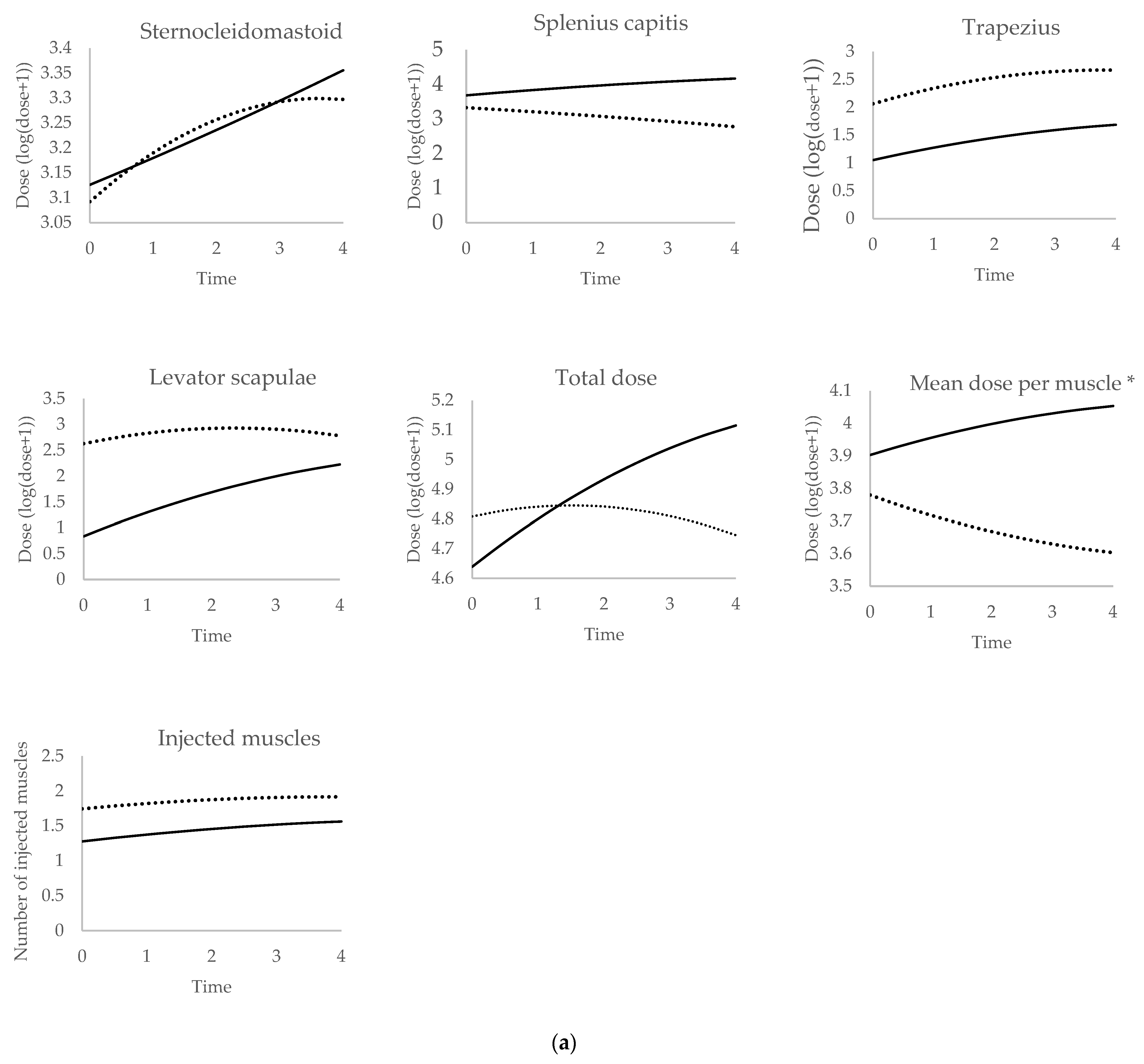
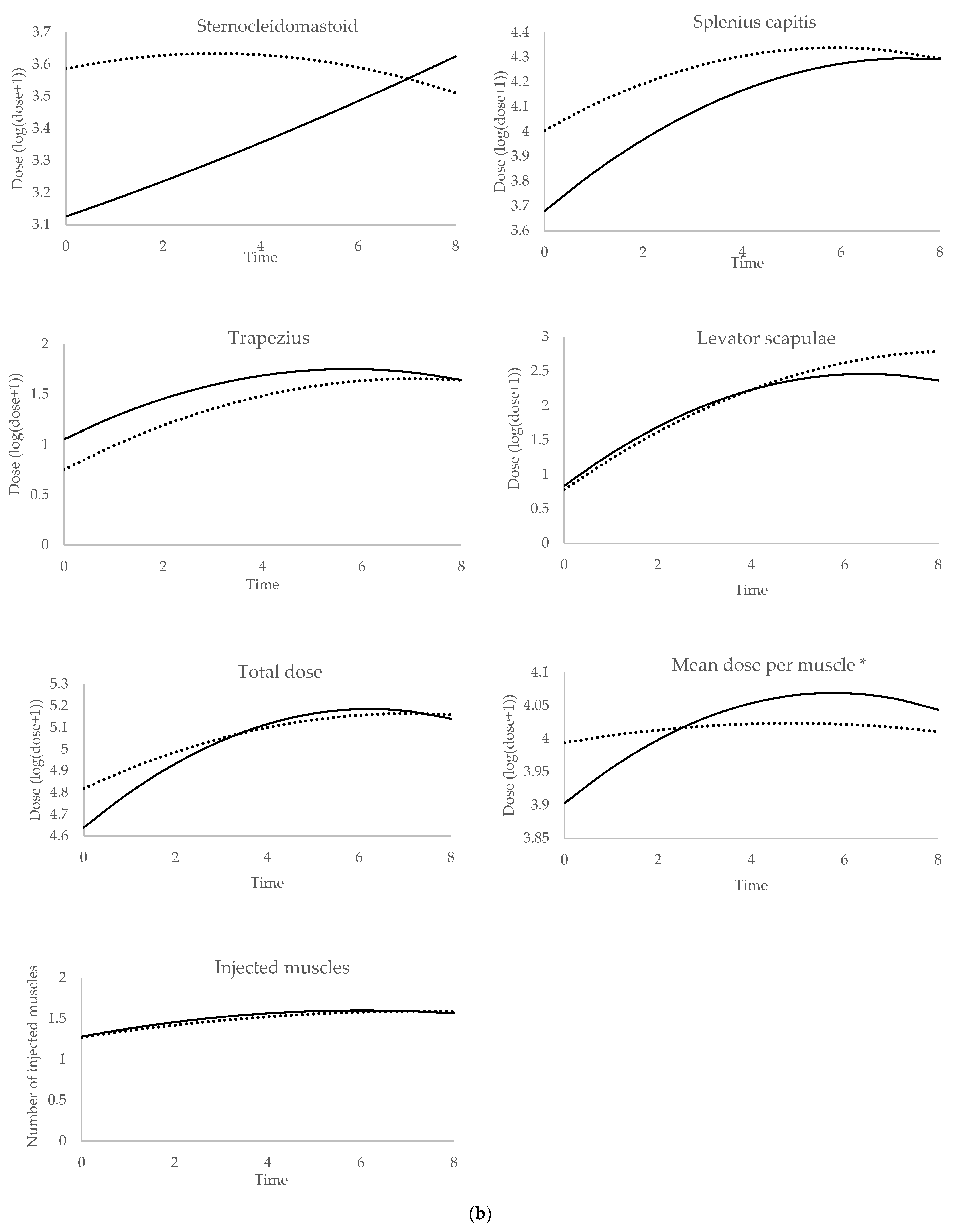
| Muscle | US Group: Non-Guided vs. US-Guided Injections * | US Group Non-Guided Injections vs. No US Group * |
|---|---|---|
| Sternocleidomastoid | NA | NA |
| Splenius capitis | <10−3 | 0.47 |
| Trapezius | 0.67 | 0.94 |
| Levator scapulae | 0.0018 | 0.57 |
| Total dose | <10−3 | 0.068 |
| Mean dose per muscle ** | <10−3 | 0.064 |
| Number of injected muscles | 0.15 | 0.82 |
Disclaimer/Publisher’s Note: The statements, opinions and data contained in all publications are solely those of the individual author(s) and contributor(s) and not of MDPI and/or the editor(s). MDPI and/or the editor(s) disclaim responsibility for any injury to people or property resulting from any ideas, methods, instructions or products referred to in the content. |
© 2024 by the authors. Licensee MDPI, Basel, Switzerland. This article is an open access article distributed under the terms and conditions of the Creative Commons Attribution (CC BY) license (https://creativecommons.org/licenses/by/4.0/).
Share and Cite
Kreisler, A.; Mortain, L.; Watel, K.; Mutez, E.; Defebvre, L.; Duhamel, A. Doses of Botulinum Toxin in Cervical Dystonia: Does Ultrasound Guidance Change Injection Practices? Toxins 2024, 16, 439. https://doi.org/10.3390/toxins16100439
Kreisler A, Mortain L, Watel K, Mutez E, Defebvre L, Duhamel A. Doses of Botulinum Toxin in Cervical Dystonia: Does Ultrasound Guidance Change Injection Practices? Toxins. 2024; 16(10):439. https://doi.org/10.3390/toxins16100439
Chicago/Turabian StyleKreisler, Alexandre, Léa Mortain, Kaëlig Watel, Eugénie Mutez, Luc Defebvre, and Alain Duhamel. 2024. "Doses of Botulinum Toxin in Cervical Dystonia: Does Ultrasound Guidance Change Injection Practices?" Toxins 16, no. 10: 439. https://doi.org/10.3390/toxins16100439
APA StyleKreisler, A., Mortain, L., Watel, K., Mutez, E., Defebvre, L., & Duhamel, A. (2024). Doses of Botulinum Toxin in Cervical Dystonia: Does Ultrasound Guidance Change Injection Practices? Toxins, 16(10), 439. https://doi.org/10.3390/toxins16100439