


Abstract
Gut microbiota play crucial roles in host health, including immune regulation, metabolism, and nutrient absorption. Its dysregulation has been linked to various diseases. When administered in sufficient amounts, probiotics can contribute to restoring the gut microbial balance and maintain homeostasis. Species of the genus Bacillus and related genera (Alkalihalobacillus and Heyndrickxia) have been extensively studied and commercialized as probiotics due to their ability to form endospores, the dormant cell forms that provide remarkable resistance to adverse conditions. Understanding the mechanisms of the action of spore-forming probiotics is essential for harnessing their therapeutic potential. This review explores the mechanisms underlying the action of spore-forming probiotics, with a special focus on Alkalihalobacillus clausii. Many beneficial properties such as resilience in extreme conditions, multiplication in the gut, gut adhesion, immunomodulatory effects, the production of bioactive and antimicrobial compounds, as well as efficacy in human health and diseases are extensively dissected. In parallel, this review underscores the limitations of spore-forming probiotics, focusing on safety concerns, issues related to dose standardization and quality control, as well as potential off-target effects and risks in immunocompromised individuals.
1. Introduction
The gastrointestinal (GI) tract hosts a complex ecosystem composed of a diverse array of microorganisms, collectively referred to as the gut microbiota. The gut microbiota closely interacts with its host, regulating and supporting different functions, including the immune, hormonal, and nervous system through complex signaling pathways [1,2,3]. It also helps in maintaining gut homeostasis, influencing various physiological processes, such as digestion, nutrient absorption, and metabolism [4,5]. In contrast, the dysregulation of host–microbe interactions has been implicated in the pathogenesis of various diseases, including inflammatory bowel diseases (IBD), obesity, and metabolic syndrome [2]. Probiotics have garnered significant attention from the scientific and medical communities as they offer an effective and non-invasive restoration of this dysregulation, resulting in a myriad of potential health benefits [6].
Of the different species of probiotics consumed around the world for their health benefits, Gram-positive bacteria belonging to the genus Bacillus or related genera, such as Alkalihalobacillus and Heyndrickxia, are among the most extensively studied and are characterized by the unique ability to form endospores [7,8,9].
Endospores could be defined as dormant, resistant, and non-reproductive structures produced by certain bacteria belonging to the phylum Bacillota (formerly Firmicutes) in adverse conditions. When conditions become favorable for germination, spores can revert to the vegetative state and resume their metabolic activities.
Spores are highly resistant to harsh environmental conditions, including extreme temperatures, pH, and desiccation. This remarkable resistance can be attributed to their unique properties, such as a thick proteinaceous coat, high levels of calcium-dipicolinic acid (Ca2+-DPA) in the spore core, a low water content, and minimal metabolic activity [10,11]. Additionally, spore DNA is protected by its saturation with α/β-type small acid-soluble proteins (SASPs), which bind to DNA, altering its conformation and making it resistant to damage [10]. Such characteristics enable spores to survive in extreme environments, but also to persist in the host’s GI tract and exert their beneficial effects [12]. It was indeed observed that commercial probiotics containing members of the genus Bacillus and related genera, such as Bacillus cereus, Alkalihalobacillus clausii (formerly Bacillus clausii), and Bacillus pumilus, could persist in the mouse GI tract for up to 16 days [13]. Among these spore-forming species, A. clausii and Heyndrickxia coagulans (formerly Bacillus coagulans) represent the most largely commercialized probiotics worldwide. Various studies highlighted the distinguishing physiological properties of these microorganisms, including their tolerance to harsh GI environments, the enhancement of the gut barrier function, broad-spectrum antibiotic resistance, and their ability to synthesize vitamins [14,15,16,17,18].
Although spores are often included in commercial probiotic formulations, the importance of these forms of resistance has been under-appreciated from a clinical and microbiological perspective. In order to elucidate the therapeutic potential of spore-forming probiotics and optimize their clinical applicability, it is essential to comprehend their mechanisms of action. In view of the above, this review aims to shed more light on the distinctive attributes of spore-forming bacteria as probiotics, with a particular emphasis on the advantages conferred by their ability to form spores. Since an extensive review on H. coagulans has been recently published [19], herein we will particularly focus on the use of A. clausii as a probiotic, discussing its potential applications in health and disease.
2. Key Attributes of Spore-Forming Probiotics
2.1. Resistance to Extreme Conditions
The GI tract provides a challenging environment for bacterial survival due to partial or complete anoxia, low pH, bile salts, and an extremely high concentration of commensal microbes that compete for nutrients and the ecological niche [20].
The efficacy of orally administered probiotics depends on their ability to resist these conditions, colonize the gut, and exert functional benefits [15,21,22,23]. In this context, bacterial spores are known to remain inert and transit unrestrained through the gut (Figure 1) despite unfavorable conditions [20]. Spores of A. clausii were reported to survive in acidic environments, in the presence of bile and anoxic conditions [15,24].
Figure 1.
Mechanism of action of spore-forming probiotics.
Spore germination involves tightly regulated transcriptional changes followed by morphological cell changes [25]. The events typically include rehydration, the release of Ca2+-DPA and other core components, followed by the formation of metabolically active vegetative cells [26]. Once formed, the vegetative cells become less resistant to environmental stress. Despite this, they can adapt to specific environments at different stages of their life cycle. This adaptation is made possible through responses that adjust to the environment, which are controlled by complex molecular mechanisms.
The H+ homeostasis in A. clausii was shown to be maintained by the F0F1 ATP synthase complex that works by hydrolyzing ATP to pump protons (H+) from the cytoplasm [16,24]. Various proteins involved in the general stress adaptation by A. clausii ENTPro were identified by their annotation and Pfam domain search [16]. Notably, the universal stress protein UspA (PF00582) encoded by a gene present in the A. clausii ENTPro genome was found crucial for survival during cellular growth arrest and reprogramming the cell’s defence mechanisms during stress in other bacteria [27,28]. Molecular chaperones, such as GroES (PF00166) and GroEL, can contribute to the resistance of A. clausii against environmental stressors. In addition, the heat shock protein 33 (PF01430), the cold shock proteins CspA (PF00313), Clp proteases (PF00574), and HtpX and HrcA-like heat shock proteins could help in maintaining cellular growth and nucleic acid stability, while preventing the formation of inclusion bodies. The chaperone protein DnaJ (PF00226) and the nucleotide exchange factor GrpE (PF01025) can provide resistance to hyperosmotic stress and heat. Moreover, two copies of the methionine sulfoxide reductase A-encoding gene can confer resistance to oxidative stress [16].
2.2. Germination and Multiplication
Spore-forming probiotics demonstrate extreme stability and resistance to unfavorable conditions, attributes that facilitate their transit in the gut and survival at the low pH of the gastric barrier [12]. Furthermore, their stability and survival could be improved using various strategies and technologies, such as inducing sub-lethal stress, drying, encapsulation, and immobilization [26,27,28,29,30,31,32,33,34]. After spore germination, vegetative bacteria can rapidly multiply (Figure 1), allowing the quick and temporary colonization of the gut [35].
In vitro studies demonstrated that specific germinant receptors on the spore surface recognize signals such as amino acids, sugars, and bile salts, triggering spore germination [36]. In the case of Bacillus subtilis spores, micromolar concentrations of peptidoglycan-derived muropeptides act as potent germinants [37].
In vivo studies on Bacillus spores indicated that they may have similar life cycles in mammals and avian species, but with species- and strain-specific differences in the survival and persistence of the spores in the gut [20]. However, studies elucidating the mechanisms of spore germination in the human gut are scarce. Moreover, the mechanisms are considered different from that of other mammals, as the majority of studies have been conducted in vitro using artificial GI tract models [38,39,40,41].
In a study conducted by Shinde and co-authors [42], the spores of H. coagulans MTCC 5856 were shown to tolerate simulated conditions mimicking the digestion process. In addition, the H. coagulans strain contained in a mono-strain commercial probiotic product was found to persist in artificial gastrointestinal fluids for prolonged time [43]. Vecchione et al. [15] compared the in vitro behavior of various probiotic formulations commercialized in Italy and reported the remarkable survival of a probiotic mixture containing four A. clausii strains, as simulated gastric fluids. In the same study, an about 3-Log increase in the number of A. clausii cells was observed after 240 min of incubation in an artificial intestinal juice, thus highlighting the ability of A. clausii to multiply in intestinal-like conditions [15]. Furthermore, in a randomized, open-label, cross-over trial, Ghelardi et al. [44] investigated the GI fate of orally administered A. clausii using two commercial probiotic formulations containing spores of strains O/C, N/R, SIN, and T administered as a single dose to human volunteers. It was reported that the spores survived transit through the human gut and maintained considerable intestinal titer for up to 12 days after administration, and then underwent germination, outgrowth, and multiplication as vegetative forms. The strains also exhibited specific adaptation to the gut environment. In addition, an in vitro study by Mazzantini et al. [18] underscored the ability of A. clausii strains N/R and T and H. coagulans ATCC 7050 to survive in simulated intestinal fluids for as long as 8 h and the multiplication of vegetative cells of A. clausii O/C and SIN in these conditions. A systematic review of the literature by Morelli and Pellegrino [45] on the ability of viable probiotic strains to survive and reproduce in the gastrointestinal tract of healthy adults also highlighted a better survival of spore-forming probiotics in the GI tract. Lately, Ghelardi et al. [44] reported the survival in simulated gastrointestinal fluids of A. clausii spores contained in formulations, namely Enterogermina 4B, Enterogermina 6B, and Enterogermina Sporattiva. Also, the A. clausii SIN spores in Enterogermina Sporattiva were found to multiply in the simulated intestinal environment. In a recent in vitro study [46], A. clausii spores contained in Enterogermina 2B were shown to germinate in intestinal-like conditions following activation in a gastric juice. Additionally, A. clausii cells were shown to multiply in the intestinal fluid, increasing their number of about 3-Log [46].
2.3. Adhesion to the Gut
The adhesion of probiotic bacteria to the host gut is required for the competitive exclusion of gut pathogens, the release of antimicrobial substances, short-chain fatty acids (SCFAs), and organic acids, and the modulation of the host immune system [13,16,47,48]. The good adherence capacity of spore-forming probiotics (Figure 1) promotes the gut residence time, pathogen elimination, and host immune modulation [16]. The spore coat and exosporium were found to have a crucial role in spore adhesion [4]. Among the key proteins identified on the surface of the spores are mannose-specific lectins and spore-coat-associated proteins [49]. A Pfam analysis of the surface proteins of Gram-positive bacteria revealed the involvement of a mucus-binding protein with the ‘Gram_pos_anchor’ Pfam domain (PF00746) at the C-terminus, a collagen-binding protein with an LPXTG motif at the C-terminus, and a fibronectin-binding protein in facilitating the adhesion of a probiotic bacterium to the intestinal mucosal layer and further interactions [16,50]. In addition, adhesion is determined by many factors such as cell wall hydrophobicity, the adhesion potential, and auto-aggregation [51,52,53]. With few exceptions, spores exhibit higher adhesion compared to vegetative cells [49,54]. This could be attributed to their higher hydrophobicity and lower energy barrier, compared to vegetative cells [55]. In fact, the spores of H. coagulans MTCC 5856 were shown to strongly adhere to human colonic cell lines [42]. Vegetative cells of H. coagulans ATCC 7050 and A. clausii O/C, N/R, SIN, and T and spores of A. clausii were found to adhere to gastrointestinal mucins, thus highlighting their potential ability to colonize the mucus layer of the gut [18,46]. This behavior was supposed to be attributed to the presence of mucus-binding proteins and type-IV filamentous adhesions on the cell surface [16,18,56].
2.4. Production of Bioactive Compounds
Spore-forming probiotics produce various active metabolites (Figure 1), including SCFAs, such as acetate, propionate, and butyrate, as a result of dietary carbohydrate and protein digestion [57,58]. SCFAs are volatile fatty acids produced by the gut microbiota in the large bowel as fermentation products from food components that are unabsorbed/undigested in the small intestine [58]. SCFAs have distinct physiological effects. They contribute to shaping the gut environment, influence the physiology of the colon, and can be used as energy sources by host cells and by the intestinal microbiota [58]. They also participate in different host-signaling mechanisms [58]. In addition, SCFAs promote gut microbiota-mediated alterations in the gut motility through the maintenance of colonic serotonin homeostasis and participate in different host-signaling mechanisms [58,59]. SCFAs can serve as energy sources for host cells and for the gut microbiota to conduct varied functions in the host intestine; thus, their amount and relative abundance may be considered as biomarkers of a healthy status. Reduced levels of acetic, propionic, and butyric acids in the gut often coincide with health issues, suggesting that supplementing with SCFA-producing microbes could alleviate related symptoms. Calvigioni et al. [60] demonstrated for the first time that A. clausii strains N/R, O/C, SIN, and T can produce acetic, propionic, and butyric acids in vitro, thus suggesting that they could produce these molecules also in vivo. Interestingly, the strain T resulted to be the highest producer of acetic acid (602.00 ± 54.15 ng/mL) and was found to secrete about two-fold more propionic acid than A. clausii NR and SIN. In contrast, no differences in the amount of secreted butyric acid were evidenced between the four A. clausii strains. The production/secretion of these SCFAs in vitro, particularly acetic acid, was recently demonstrated for a formulation containing a spore mixture of A. clausii N/R, O/C, SIN, and T [46]. In the study from Calvigioni and colleagues, the production of acetic acid (~300 ng/mL), but not of propionic and butyric acids, was evidenced also for H. coagulans ATCC 7050 [60]. Interesting, the effect of the A. clausii strains on SCFA production was also highlighted in a model of the GI tract of patients treated with proton pump inhibitors, with the increased production of butyrate and decreased production of acetate, thus suggesting an effect on the overall production of these molecules [5,61].
The administration of probiotics producing B-group vitamins is advantageous for human health, since these vitamins play a pivotal role in the metabolism of carbohydrates, proteins, and fatty acids, in the prevention of oxidative stress, and in the maintenance of neuronal functionality [62]. Mazzantini et al. [18] demonstrated in vitro the synthesis of vitamins, particularly vitamin B2 (riboflavin), by the vegetative A. clausii strains O/C, N/R, SIN, and T, as well as by H. coagulans ATCC 7050. In particular, the amount of vitamin B2 produced by H. coagulans ATCC 7050 was found to be more than 10-fold higher than that produced by the A. clausii strains [18]. Riboflavin produced by the A. clausii strains was previously found to support the growth of a riboflavin-deficient mutant of B. cereus on riboflavin-depleted media [14]. In addition, the secretion of vitamins B2 (22.86 ± 1.61 ng/mL), B8 (3.93 ± 0.01 ng/mL), B9 (2.01 ± 0.12 ng/mL), and B12 (1.17 ± 0.64 ng/mL) was recently demonstrated in vitro for a formulation containing spores of the same A. clausii strains [46].
Over recent decades, numerous studies have suggested that probiotics play a crucial role in preventing oxidative stress and in various diseases associated with reactive oxygen species (ROS) by producing antioxidants. Specifically, Bacillus and related genera are known to produce enzymes like catalase (CAT) and superoxide dismutase (SOD), which help the host in counteracting oxidative stress [63,64]. Lippolis and coauthors identified CAT in the secretomes of A. clausii O/C, N/R, SIN, and T, with strains O/C and N/R secreting higher levels of the proteins than the other strains [65]. In the same study, all the A. clausii strains were found to secrete SOD and the strain O/C resulted to be the highest producer. Thereafter, CAT and SOD activities were detected in both cell lysates and culture supernatants obtained from H. coagulans ATCC 7050, A. clausii O/C, N/R, SIN, and T, and from the formulation containing a spore mixture of these A. clausii strains [18,46]. An in vitro study hypothesized that the administration of these probiotic strains could potentially reduce or alleviate ROS accumulation in vivo, thus preventing oxidative stress [18].
Spore-forming bacteria are known to produce a variety of bioactive enzymes that promote food digestion and nutrients adsorption. These include proteases, lipases, and amylases, which digest proteins, lipids, and carbohydrates, respectively. Other relevant enzymes are α- and β-galactosidases, which hydrolyze complex galactooligosaccharides and lactose, respectively, and pectinases responsible for pectin digestion. A recent review of Nicolas and Ma [66] collected evidence on the ability of some probiotic strains of H. coagulans (e.g., KM-1, MA-13, NL01, RCS3, and GBI−30 6086) to produce α- and β-galactosidases, amylases, alkaline proteases, lipases, and/or pectinases in vitro. Furthermore, the production of β-galactosidase was demonstrated in vitro for H. coagulans ATCC 7050, A. clausii (i.e., O/C, N/R, SIN, and T), and for a formulation containing spores of these A. clausii strains [18,46]. Interestingly, H. coagulans ATCC 7050 was found to produce two-fold more β-galactosidase than A. clausii N/R, O/C, and T, and three-fold more of the enzyme than A. clausii SIN [18]. Since this enzyme is responsible for the degradation of lactose, the ability of these microbes to produce β-galactosidase have been hypothesized to contribute in ameliorating the symptomatology associated with lactose intolerance [18].
The production of alkaline proteases was demonstrated in vitro for A. clausii I-52 and UBBC07 [67,68,69]. In addition, a comparative proteomic analysis of A. clausii O/C, N/R, SIN, and T identified an alkaline protease (AprE) and an aminopeptidase (AmpS) in the extracellular proteomes of all strains. Interestingly, stains O/C and T resulted in being able to secrete higher amounts of AprE compared to strains N/R and SIN [65]. Interestingly, Lin and coauthors [70] evidenced the ability of B. subtilis ubsp. natto NTU-18 to produce a nattokinase able to interfere with the transfer of vancomycin resistance in Enterococcus faecalis.
2.5. Production of Antimicrobial Compounds
Bacillus spp. produce various antimicrobial compounds that aid in the competitive exclusion of pathogenic microbes in the gut [71,72,73]. These compounds include bacteriocins and bacteriocin-like inhibitory substances such as subtilin and coagulin, or lipopeptides, such as surfactin, iturins, bacillomycins, and mycosubtilins. Moreover, Bacillus spp. are well known producers of lantibiotics, post-translationally modified antimicrobial peptides that are active against Gram-positive bacteria and a few Gram-negative bacteria including pathogens and antibiotic-resistant bacteria [71,74,75,76,77,78].
H. coagulans strains can secrete a bacteriocin, a well-known antibacterial substance, as well as other antimicrobial substances, such as lactic acid and its derivatives, which may be produced and act in the human gut [79,80,81,82].
In a preclinical study by Paparo et al. [83], the protective mechanisms of A. clausii against the rotavirus infection have been elucidated, providing insights into the mechanistic basis underlying the clinical efficacy of A. clausii in pediatric viral acute gastroenteritis. Using the human pediatric enterocyte model, it was demonstrated that vegetative cells of A. clausii strains O/C, N/R, SIN, and T induce the synthesis of antimicrobial peptides, human beta defensin 2, and cathelicidin, that aid in fighting infections and rescuing cell proliferation, which is impaired by rotavirus. Additionally, A. clausii strains were reported to inhibit ROS production by rotavirus and suppress the release of pro-inflammatory cytokines, such as interleukin (IL)-8 and interferon (IFN)-β, and genes associated with the Toll-like receptor (TLR)-3 pathway [83].
The vegetative cells of A. clausii produce antimicrobial peptides with inhibitory effects against a broad spectrum of bacteria, including Salmonella typhimurium, Escherichia coli, Shigella flexneri, Staphylococcus aureus, Listeria monocytogenes, and Enterococcus faecalis. Urdaci et al. [84] reported that the release of these antimicrobial substances occurs during a stationary growth phase and coincides with sporulation. Specifically, two strains of A. clausii, UBBC07 and O/C, were found to produce in vitro a peptide called clausin that demonstrates antimicrobial activity primarily against Gram-positive bacteria. In addition, clausin also mitigates the cytotoxic effects on Caco2 cells induced by Clostridioides difficile [77,85]. It also targets lipid intermediates involved in bacterial peptidoglycan synthesis, providing further insights into its mechanism of action against bacterial pathogens [86].
2.6. Antibiotic Resistance
The presence of antibiotic resistance genes (ARGs) on mobile genetic elements (i.e., plasmids, transposons, and phages) has been highlighted as a critical safety concern [87,88]. In fact, these genes can be transmitted to other microbes by a horizontal gene transfer, thus contributing to the spreading of antibiotic resistance. In contrast, the existence of non-transmissible AGRs stably residing in bacterial chromosomes could be considered an added value since some probiotics are administered in concomitance with antibiotics [16,89]. Several ARGs that enable probiotics’ survival and persistence during antibiotic treatment have been identified within the genomes of Bacillus spp. For example, cfr-like genes that encode ribosome methyltransferases have been identified in several Bacillus spp. These genes determine the ability of Bacillus spp. to develop resistance against multiple antibiotic classes including phenicols, oxazolidinones, lincosamides, pleuromutilins, and streptogramin A [90]. Probiotic A. clausii strains harbor specific antibiotic defence genes such as an aminoglycoside resistance gene (aadD2), a chloramphenicol acetyltransferase gene (cat(Bcl)), and a β-lactamase gene (blaBCL-1) that contribute to their ability to withstand antibiotic pressure and maintain their probiotic function in the gut environment [91,92,93].
Multidrug resistance proteins, universal stress proteins, the EmrB multidrug efflux system, tetracycline resistance proteins, and different classes of beta-lactamases are present in various Bacillus spp., conferring resistance against common antibiotics (Figure 1). However, vancomycin resistance was only identified in Bacillus toyonensis and chloramphenicol resistance in all included bacteria except for H. coagulans, highlighting the diversity of antibiotic resistance mechanisms within the genus Bacillus and related genera [16]. In addition, it was reported that the antibiotic sensitivity patterns of four A. clausii strains, O/C, SIN, N/R, and T, differ from that of the wild A. clausii strain DSM8716. Although the strains show similarities, such as resistance to macrolides, beta-lactams, and aminoglycosides, A. clausii DSM8716 is sensitive to chloramphenicol, rifampicin, streptomycin, and tetracycline, indicating that the probiotic strains O/C, SIN, N/R, and T have acquired resistance to these compounds through selective mutagenesis [94]. Furthermore, the analysis of mutations involved in the resistance to streptomycin and rifampicin revealed the K101R mutation in the rpsL gene, associated with streptomycin resistance in the SIN strain, and the S488F mutation in the rpoB gene, associated with rifampicin resistance in the N/R strain [85].
The analysis of the composite genome of A. clausii ENTPro confirmed the presence of chromosomic genes encoding chloramphenicol acetyltransferase, tetracycline resistance domains, two proteins with the streptomycin adenylyltransferase domain (PF04439), six proteins with the aminoglycoside phosphotransferase (PF01909) domain, and one protein with the kanamycin nucleotidyltransferase (PF07827) domain imparting resistance to streptomycin [16]. The identification of specific protein domains associated with antibiotic resistance in A. clausii highlights the diverse repertoire of molecular mechanisms underlying its ability to resist certain antibiotics as well as the differences between bacteria of the same species [16].
2.7. Regulation of the Immune System
The intricate interplay between the gut microbiota and the mucosal immune system profoundly influences the maintenance of the gut immune system and metabolic homeostasis [95]. Probiotics exert their immunomodulatory effects through direct interactions with the intestinal epithelium, particularly in the small intestine, where the commensal microbiota are less abundant [96]. Additionally, through various effector molecules, including bacterial cell wall components, such as peptidoglycan and lipoteichoic acid, as well as specific proteins, probiotics moderate host immune responses by modulating receptor signaling cascades involved in the immune system regulation [97,98]. Beyond the conventional immune functions, emerging evidence highlights the role of the gut microbiota and their components in regulating energy, glucose, and lipid metabolism, demonstrating a link between them and host metabolism [99,100,101]. Thus, manipulating the gut microbiota using probiotics offers promising avenues for modulating host metabolism and potentially managing metabolic disorders [102,103].
The immunomodulatory effect of spore-forming probiotics can be attributed to cell wall components, surface-associated proteins, glycoproteins, and secreted factors. An in vitro evaluation of the immunomodulatory properties of whole vegetative cells of A. clausii on Swiss and C57 Bl/6j murine cells revealed the induction of nitric oxide synthetase activity, IFN-γ production, and CD4+ T-cell proliferation [84]. The cell wall fraction of H. coagulans JBI-YZ6.3 was shown to increase the level of expression of CD25 and CD69 activator markers on NK, T, and dendritic cells, as well as on monocytes [104]. Interestingly, CD69 expression on monocytes has been linked to the activation of the 5-lipoxygenase pathway, which results in the production of leukotrienes responsible for immune cells’ recruitment [105]. In addition, the same fraction was found to increase the production of IFN-γ and some pro-inflammatory cytokines, as well as of the stem cell-mobilizing growth factor (G-CSF), thus enhancing post-injury and post-inflammation repair processes [104]. The immunomodulatory activity exerted by the cell wall fraction reasonably involves different bacterial glycans and host pattern recognition receptors. In fact, as described for other Gram-positive probiotics, peptidoglycan and its components muramyl dipeptide and N-acetylglucosamine can be recognized by the Toll-Like Receptor (TLR) 2, leading to the activation of the Nuclear Factor kappa-light-chain-enhancer of activated B cells (NF-κB) and the Mitogen-Activated Protein Kinase (MAPK) pathways [106,107,108]. Furthermore, the D-alanine in the cell wall can amplify TLR2 signaling, directly impacting cytokine production [109]. Additionally, muramyl dipeptides in the cell wall can activate the nucleotide-binding oligomerization domain 2 (NOD2) receptors, which stimulates the activation of NF-κB and MAPK pathways through the recruitment of the receptor-interacting protein kinase 2 (RIP2) [107,108]. An elegant in vitro study from Zhang and coworkers [110] clarified the possible mechanism of action of the cell wall lipoteichoic acid of H. coagulans HOM5301 in the activation of RAW 264.7 macrophages. In particular, lipoteichoic acid of this strain appeared able to stimulate TLR2 and induce the activation of MAPK and NF-κB pathways by recruiting the Myeloid Differentiation Primary-Response Protein 88 (MyD88) and the Interleukin-1 Receptor-Associated Kinases (IRAK), which in turn induce the expression of pro-inflammatory cytokines and chemokines [110].
To the best of our knowledge, no reports evaluating the specific immunomodulatory properties of isolated surface-associated proteins of spore-forming probiotics are available. Nevertheless, these molecules can also play a key role in immunomodulation exerted by these microbes. For instance, the interaction between bacterial s-layer proteins and the dendritic cell-specific ICAM-3-grabbing non-integrin (DC-SIGN), SIGN3, Decitin-1, and macrophage-inducible C-type lectin can help in the immune response modulation and protection of the gut barrier integrity, as described for other Gram-positive probiotics [107]. Furthermore, as observed in Limosilactobacillus reuteri, mucus-binding proteins can enhance immunomodulation by the binding of C-type lectin receptors, thus inducing a Th1-polarized immune response [111]. Glycoproteins constituting surface appendages (pili and flagella) can also be involved in the stimulation of the immune response induced by spore-forming bacteria, being recognized by TLR2 receptors [107].
Regarding secreted factors, A. clausii supernatants were observed to protect intestinal cells from cytotoxicity induced by C. difficile and B. cereus through a serine protease, which rescues cell viability and mitochondrial activity, proving its clinical relevance in the prophylaxis of C. difficile-associated diarrhea [78].
Zhu et al. [103] highlighted that Bacillus-derived metabolites, such as organic acids and exoenzymes, play a crucial role in modulating the host immune response (Figure 1). These metabolites enhance the host’s immune regulation by promoting the production of anti-inflammatory cytokines and inhibiting the growth of pathogenic bacteria. For instance, SCFAs can activate G-protein-coupled receptors (GPCRs), improving the production of anti-inflammatory factors by T-cells and reducing pro-inflammatory molecules [94]. Furthermore, pyruvate produced by bacilli can induce the GPCR-mediated dendrite protrusion of C-X3-C motif chemokine receptor 1+ cells, triggering an immune response. In particular, activated GPCRs modulate the host immune response through different signaling cascades, including the MAPK, the signal transducer and activator of transcription 3 (STAT3), and the mammalian target of rapamycin (mTOR) pathways [112]. These interactions underscore the potential of spore-forming probiotics in maintaining immune homeostasis and protecting against enteric infections. Cell wall components and metabolites from H. coagulans PTA-6086 have been shown to increase the activity of different immune cells [113,114].
A. clausii strains or their supernatants also improved the gut barrier integrity by reducing necrotic or apoptotic enterocytes, increasing mucin production, and enhancing the synthesis of tight junction proteins in an in vitro model of retrovirus infection [76]. A. clausii CSI08 cells and their supernatants were shown to attenuate the expression of pro-inflammatory cytokines by down-regulating the NF-κB transcription factor [115].
An in vitro study from Vicente-Gil and coauthors [116] investigated the immunomodulatory effects of extracellular vesicles produced by probiotic B. subtilis ABP1 on the intestinal epithelial cell line RTgutGC and splenic leukocytes. In both cell lines, vesicles were found to increase the expression of genes encoding some pro-inflammatory cytokines (i.e., IL-1β and IL-8) and antimicrobial peptides (i.e., hepcidin and cathelicidin 2). Furthermore, they stimulated the expression of genes correlated with B-cell differentiation and the production of MHC-II and IgM in splenic leukocytes [116]. Interestingly, the authors evidenced markedly different effects when vesicles obtained from Bacillus megaterium were used, thus suggesting a species- or even strain-dependent immunostimulatory activity [116].
Some in vivo studies also highlighted the immunomodulatory properties of spore-forming probiotics. Bomko and co-workers revealed that H. coagulans is able to normalize the number of splenic lymphocytes, macrophages, and T-lymphocytes and their functional activity to promote the host immune system when administered to mice with colitis [117]. Similarly, the administration of A. clausii (O/C, SIN, N/R, T) to mice alleviates the symptoms of colitis by modulating the gut microbiota [118]. Zhao and coworkers [119] investigated the in vivo immunomodulatory properties of H. coagulans MZY531 whole cells administered to cyclophosphamide-induced immunosuppressed mice. The authors evidenced the downregulation of TLR4/MyD88/NF-κB pathways and the upregulation of anti-inflammatory cytokines (INF-γ, IL-2, IL-4, and IL-10) [119].
A. clausii demonstrated therapeutic potential, indicating its clinical applicability in various inflammatory conditions, as evidenced by the following studies. In a mouse model of ovalbumin-induced asthma, A. clausii administration reduced eosinophils, neutrophils, and lymphocytes, while also attenuating airway epithelium thickening and decreasing IL-4 and IL-5 levels [120]. Additionally, A. clausii MTCC-8326 induced a controlled inflammatory response in murine macrophages, balancing pro-inflammatory and anti-inflammatory cytokine expression [121]. In mice with schistosomiasis, A. clausii increased the levels of the anti-inflammatory cytokine IL-10, Treg, and Th17 cells and reduced the levels of the pro-inflammatory cytokines IFN-γ, tumor necrosis factor (TNF)-α, and IL-6, resulting in a reduction in inflammation [122].
3. A. clausii in the Treatment Landscape of Various Diseases
Many probiotic formulations containing A. clausii spores are commercialized around the world [18,43,123,124,125,126]. Most of these formulations contain only one A. clausii strain (i.e., UBBC-07, 088AE, SIN), while others contain more than one strain or a combination of A. clausii and other bacteria. In many studies, a formulation consisting of spores of the A. clausii strains O/C, N/R, SIN, and T (Enterogermina) has been used. This product is licensed and marketed as a prophylactic medicine in more than 50 countries around the world [9,73] for its demonstrated tolerability and efficacy in humans over several decades [44,94,127,128].
Despite their extensive use and proven efficacy, A. clausii probiotics have been relatively less reported and disseminated in the literature compared to probiotics belonging to other genera such as Lactobacillus and Bifidobacterium [7]. Several clinical studies proved the efficacy of A. clausii spores in the management of various health disorders (Table 1).
Table 1.
Clinical studies reporting the efficacy of A. clausii in various disorders.
4. Safety, Regulatory-Related Considerations, and Limitation of Spore-Forming Probiotics
Spore-forming bacteria are commonly included in a plethora of commercial formulations that are sold as foods, food supplements, or drugs/live biotherapeutic products (LBPs), depending on the intended use [149,150].
Regarding safety, bacteria used as probiotics in foods and food supplements should have a long history of safe use in humans and should possess the Generally Recognized As Safe (GRAS) or the Qualified Presumption of Safety (QPS) status [87,151,152]. In contrast, microbes contained in drugs/LBPs undergo meticulous safety assessments by performing in vitro and in vivo studies, as well as clinical trials in humans [88].
A limited number of cases of bacteremia and sepsis in immunocompromised patients or individuals with underlying comorbidities, due to the consumption of A. clausii probiotics, have been published [153,154]. The development of bacteremia and sepsis following the administration of products containing A. clausii has also been reported in two patients hospitalized in the intensive care unit [155], in an infant with short bowel syndrome [156], in an 87-year-old man [157], and in a pediatric patient affected by a rare autoinflammatory disease [158]. Furthermore, cases of fatal septic shock in a 4-month-old infant with congenital heart disease [159] and in an infant with malnutrition, acute diarrhea, and moderate-to-severe dehydration [160] have been reported. In light of this evidence, physicians should exercise caution while considering the prolonged administration of spore-forming preparations to immunocompromised individuals and subjects with underlying comorbidities.
Spore-forming bacteria, particularly those constituting drugs/LBPs, should adhere to rigorous standards related to potency, identity, and purity [161]. Regarding potency, commercial products should contain the number of living microbes labeled by manufacturers, since this amount is crucial for providing the desired beneficial effect to consumers [162]. Compared to other probiotics, spore-forming microbes have the advantage that spores can survive unaffected during the industrial production process and for the entire product shelf-life. Nevertheless, the use of appropriate methods for enumerating spores at the time of manufacturing is important to guarantee potency [161]. In fact, a different number of spores than that labeled was evidenced in different studies investigating the quality of some Bacillus-containing products [43,123,124,125,126].
The United States Food and Drug Administration (FDA) and the European Medicines Agency (EMA) require that microbes included in drugs/LBPs are identified at the strain level and genotypically and phenotypically characterized [88]. The identification of microbes used in LBPs should be accurately performed, possibly using two different methods, as recommended by the FDA and EMA, to guarantee the strain identity and product purity [88]. Regarding probiotics included in foods and food supplements, the Food and Agriculture Organization of The United Nations (FAO) and the World Health Organization (WHO) defined the methods to be applied for their correct identification [151]. The use of inappropriate identification methods combined with flaws in manufacturer quality controls could result in the inclusion of different Bacillus strains, contaminant microorganisms, and even potential pathogens in commercial products, as evidenced in some studies [123,124,125,126].
The characterization of spore-forming strains is aimed at excluding the presence of virulence factors (e.g., toxins) that can constitute a potential risk for consumers. Although the presence of chromosomal non-transmissible ARGs is tolerated, probiotic spore-formers must not possess ARGs localized on mobile genetic elements, since these genes can be transmitted to gut commensals or pathogenic microbes through a horizontal gene transfer [87,88,151]. Although the strains analyzed in this review only possess chromosomal and non-transmissible ARGs, spore-forming microbes should always be screened to exclude the presence of transmissible ARGs before their inclusion in foods, food supplements, or drugs/LBPs.
As highlighted in this review, some of the beneficial properties exerted by spore-forming bacteria (e.g., multiplication in simulated intestinal fluid, the production of SCFAs) appeared to be species- or even strain-dependent. For this reason, the choice of the right product to administer should take into account its microbial composition and should be personalized based on the specific requirements of each individual. In parallel, it is also important to mention that some attributes of spore-forming bacteria (e.g., mucin adhesion, spore germination, and vegetative cell multiplication in a gut-like environment) could be unwanted in immunocompromised people. In fact, these properties can enhance the persistence of bacilli in the human gut, thus increasing the risk of off-target effects ranging from digestive discomfort to blood translocation and systemic infections [88]. Compared to other probiotics (e.g., lactobacilli, bifidobacteria), a limited number of papers investigating the beneficial properties of spore-forming bacteria are available, thus leading the knowledge on the potential of these microbes still being inadequate. We believe that more in vitro and in vivo studies should be performed to dissect almost all the attributes of these microbes, thus expanding the range of beneficial effects they could provide for human health. It is also important to remark that most of the properties evidenced in this review come from in vitro studies or in vivo investigations conducted on animal models, thus limiting their translational value. Therefore, new clinical studies should be performed for validating the beneficial attributes of spore-forming bacteria also in humans.
5. Conclusions
Spore-forming probiotics, such as H. coagulans and A. clausii, exhibit unique properties and mechanisms of action that distinguish them from non-spore-forming probiotics, providing alternative viable solutions for enhancing digestive health and overall well-being. The ability to form spores enables these microbes to withstand harsh environmental conditions, such as high temperatures and an acidic pH, thereby enhancing the stability and shelf life. Spore-forming bacteria that adhere to gastrointestinal mucins and A. clausii can multiply in gut-like conditions, thus emphasizing the ability to persist in the human intestine for prolonged times. In addition, spore-forming probiotics display promising immunomodulatory properties and appear able to produce antimicrobial molecules and bioactive compounds (i.e., SCFAs, B-group vitamins, and enzymes) that can improve human health. Despite their promise, it is essential to approach their use with a balanced perspective. As research in this field continues to advance, harnessing their potential holds prospects for improving human health and combating a range of human disorders. In addition to their beneficial effects, some limitations to the use of spore-forming probiotics have been evidenced (paragraph 4) and should be taken into account when these microbes are considered for human consumption.
Author Contributions
E.G. and D.M. contributed to the literature search and analysis, substantially reviewed and edited the manuscript, and approved its final submitted version. All authors have read and agreed to the published version of the manuscript.
Funding
The APC was funded by Opella.
Institutional Review Board Statement
Not applicable.
Informed Consent Statement
Not applicable.
Data Availability Statement
No new data were created or analyzed in this study. Data Sharing is not applicable to this article.
Acknowledgments
Medical writing support was provided by Meena Ravuri and Leander Corrie, employees of Opella. The authors would like to thank Paolo Pellegrino, a former employee of Opella, for his insightful feedback and collaboration.
Conflicts of Interest
E.G. and D.M. have been lecturers for Opella. E.G. and D.M. declare that this research was conducted in the absence of any commercial or financial relationships that could be construed as a potential conflicts of interest.
References
- Round, J.L.; Mazmanian, S.K. The gut microbiota shapes intestinal immune responses during health and disease. Nat. Rev. Immunol. 2009, 9, 313–323. [Google Scholar] [CrossRef] [Scilit]
- Belkaid, Y.; Hand, T.W. Role of the microbiota in immunity and inflammation. Cell 2014, 157, 121–141. [Google Scholar] [CrossRef] [Scilit] [PubMed]
- Cryan, J.F.; Dinan, T.G. Mind-altering microorganisms: The impact of the gut microbiota on brain and behaviour. Nat. Rev. Neurosci. 2012, 13, 701–712. [Google Scholar] [CrossRef] [Scilit]
- Koopman, N.; Remijas, L.; Seppen, J.; Setlow, P.; Brul, S. Mechanisms and applications of bacterial sporulation and germination in the intestine. Int. J. Mol. Sci. 2022, 23, 3405. [Google Scholar] [CrossRef] [Scilit]
- den Besten, G.; van Eunen, K.; Groen, A.K.; Venema, K.; Reijngoud, D.J.; Bakker, B.M. The role of short-chain fatty acids in the interplay between diet, gut microbiota, and host energy metabolism. J. Lipid Res. 2013, 54, 2325–2340. [Google Scholar] [CrossRef] [Scilit]
- Nithya, V.; Halami, P.M. Evaluation of the probiotic characteristics of Bacillus species isolated from different food sources. Ann. Microbiol. 2013, 63, 129–137. [Google Scholar] [CrossRef] [Scilit]
- Elshaghabee, F.M.F.; Rokana, N.; Gulhane, R.D.; Sharma, C.; Panwar, H. Bacillus as potential probiotics: Status, concerns, and future perspectives. Front. Microbiol. 2017, 8, 1490. [Google Scholar] [CrossRef] [Scilit]
- Sanders, M.E.; Morelli, L.; Tompkins, T.A. Sporeformers as human probiotics: Bacillus, Sporolactobacillus, and Brevibacillus. Compr. Rev. Food Sci. Food Saf. 2003, 2, 101–110. [Google Scholar] [CrossRef] [Scilit] [PubMed]
- Hong, H.A.; Huang, J.M.; Khaneja, R.; Hiep, L.V.; Urdaci, M.C.; Cutting, S.M. The safety of Bacillus subtilis and Bacillus indicus as food probiotics. J. Appl. Microbiol. 2008, 105, 510–520. [Google Scholar] [CrossRef] [Scilit] [PubMed]
- Setlow, P. Spores of Bacillus subtilis: Their resistance to and killing by radiation, heat and chemicals. J. Appl. Microbiol. 2006, 101, 514–525. [Google Scholar] [CrossRef] [Scilit]
- McKenney, P.T.; Driks, A.; Eichenberger, P. The Bacillus subtilis endospore: Assembly and functions of the multilayered coat. Nat. Rev. Microbiol. 2013, 11, 33–44. [Google Scholar] [CrossRef] [Scilit] [PubMed]
- Cutting, S.M. Bacillus probiotics. Food Microbiol. 2011, 28, 214–220. [Google Scholar] [CrossRef] [Scilit]
- Duc, L.H.; Hong, H.A.; Barbosa, T.M.; Henriques, A.O.; Cutting, S.M. Characterization of Bacillus probiotics available for human use. Appl. Environ. Microbiol. 2004, 70, 2161–2171. [Google Scholar] [CrossRef] [Scilit]
- Salvetti, S.; Celandroni, F.; Ghelardi, E.; Baggiani, A.; Senesi, S. Rapid determination of vitamin B2 secretion by bacteria growing on solid media. J. Appl. Microbiol. 2003, 95, 1255–1260. [Google Scholar] [CrossRef] [Scilit]
- Vecchione, A.; Celandroni, F.; Mazzantini, D.; Senesi, S.; Lupetti, A.; Ghelardi, E. Compositional quality and potential gastrointestinal behavior of Probiotic Products Commercialized in Italy. Front. Med. 2018, 5, 59. [Google Scholar] [CrossRef] [Scilit]
- Khatri, I.; Sharma, G.; Subramanian, S. Composite genome sequence of Bacillus clausii, a probiotic commercially available as Enterogermina(®), and insights into its probiotic properties. BMC Microbiol. 2019, 19, 307. [Google Scholar] [CrossRef] [Scilit]
- Cao, J.; Yu, Z.; Liu, W.; Zhao, J.; Zhang, H.; Zhai, Q.; Chen, W. Probiotic characteristics of Bacillus coagulans and associated implications for human health and diseases. J. Funct. Foods 2020, 64, 103643. [Google Scholar] [CrossRef] [Scilit]
- Mazzantini, D.; Calvigioni, M.; Celandroni, F.; Lupetti, A.; Ghelardi, E. In vitro assessment of probiotic attributes for strains contained in commercial formulations. Sci. Rep. 2022, 12, 21640. [Google Scholar] [CrossRef] [Scilit]
- Liang, J.; Li, C.; Chen, Z.; Guo, F.; Dou, J.; Wang, T.; Xu, Z.S. Progress of research and application of Heyndrickxia coagulans (Bacillus coagulans) as probiotic bacteria. Front. Cell Infect. Microbiol. 2024, 14, 1415790. [Google Scholar] [CrossRef] [Scilit] [PubMed]
- Bernardeau, M.; Lehtinen, M.J.; Forssten, S.D.; Nurminen, P. Importance of the gastrointestinal life cycle of Bacillus for probiotic functionality. J. Food Sci. Technol. 2017, 54, 2570–2584. [Google Scholar] [CrossRef] [Scilit] [PubMed]
- Huys, G.; Botteldoorn, N.; Delvigne, F.; De Vuyst, L.; Heyndrickx, M.; Pot, B.; Dubois, J.-J.; Daube, G. Microbial characterization of probiotics–advisory report of the working group “8651 Probiotics” of the Belgian Superior Health Council (SHC). Mol. Nutr. Food Res. 2013, 57, 1479–1504. [Google Scholar] [CrossRef] [Scilit]
- Shobharani, P.; Halami, P.M. Cellular fatty acid profile and H(+)-ATPase activity to assess acid tolerance of Bacillus sp. for potential probiotic functional attributes. Appl. Microbiol. Biotechnol. 2014, 98, 9045–9058. [Google Scholar] [CrossRef] [Scilit] [PubMed]
- Kolaček, S.; Hojsak, I.; Berni Canani, R.; Guarino, A.; Indrio, F.; Orel, R.; Pot, B.; Shamir, R.; Szajewska, H.; Vandenplas, Y.; et al. Commercial probiotic products: A call for improved quality control. A position paper by the ESPGHAN working group for probiotics and prebiotics. J. Pediatr. Gastroenterol. Nutr. 2017, 65, 117–124. [Google Scholar] [CrossRef] [Scilit] [PubMed]
- Cenci, G.; Trotta, F.; Caldini, G. Tolerance to challenges miming gastrointestinal transit by spores and vegetative cells of Bacillus clausii. J. Appl. Microbiol. 2006, 101, 1208–1215. [Google Scholar] [CrossRef] [Scilit]
- Dworkin, J.; Shah, I.M. Exit from dormancy in microbial organisms. Nat. Rev. Microbiol. 2010, 8, 890–896. [Google Scholar] [CrossRef] [Scilit]
- Setlow, P. Spore germination. Curr. Opin. Microbiol. 2003, 6, 550–556. [Google Scholar] [CrossRef] [Scilit]
- Nachin, L.; Nannmark, U.; Nyström, T. Differential roles of the universal stress proteins of Escherichia coli in oxidative stress resistance, adhesion, and motility. J. Bacteriol. 2005, 187, 6265–6272. [Google Scholar] [CrossRef] [Scilit]
- Seifart Gomes, C.; Izar, B.; Pazan, F.; Mohamed, W.; Mraheil, M.A.; Mukherjee, K.; Billion, A.; Aharonowitz, Y.; Chakraborty, T.; Hain, T. Universal stress proteins are important for oxidative and acid stress resistance and growth of Listeria monocytogenes EGD-e in vitro and in vivo. PLoS ONE 2011, 6, e24965. [Google Scholar] [CrossRef] [Scilit]
- Huq, T.; Khan, A.; Khan, R.A.; Riedl, B.; Lacroix, M. Encapsulation of probiotic bacteria in biopolymeric system. Crit. Rev. Food Sci. Nutr. 2013, 53, 909–916. [Google Scholar] [CrossRef] [Scilit] [PubMed]
- Mitropoulou, G.; Nedovic, V.; Goyal, A.; Kourkoutas, Y. Immobilization technologies in probiotic food production. J. Nutr. Metab. 2013, 2013, 716861. [Google Scholar] [CrossRef] [Scilit] [PubMed]
- Riaz, Q.U.A.; Masud, T. Recent trends and applications of encapsulating materials for probiotic stability. Crit. Rev. Food Sci. Nutr. 2013, 53, 231–244. [Google Scholar] [CrossRef] [Scilit]
- Nguyen, H.T.; Razafindralambo, H.; Blecker, C.; N’Yapo, C.; Thonart, P.; Delvigne, F. Stochastic exposure to sub-lethal high temperature enhances exopolysaccharides (EPS) excretion and improves Bifidobacterium bifidum cell survival to freeze–drying. Biochem. Eng. J. 2014, 88, 85–94. [Google Scholar] [CrossRef] [Scilit]
- Broeckx, G.; Vandenheuvel, D.; Claes, I.J.; Lebeer, S.; Kiekens, F. Drying techniques of probiotic bacteria as an important step towards the development of novel pharmabiotics. Int. J. Pharm. 2016, 505, 303–318. [Google Scholar] [CrossRef] [Scilit] [PubMed]
- Opara, E.C. Applications of cell microencapsulation. Methods Mol. Biol. 2017, 1479, 23–39. [Google Scholar] [CrossRef] [Scilit]
- Davis, C. Enumeration of probiotic strains: Review of culture-dependent and alternative techniques to quantify viable bacteria. J. Microbiol. Methods 2014, 103, 9–17. [Google Scholar] [CrossRef] [Scilit]
- Moir, A. How do spores germinate? J. Appl. Microbiol. 2006, 101, 526–530. [Google Scholar] [CrossRef] [Scilit]
- Shah, I.M.; Laaberki, M.H.; Popham, D.L.; Dworkin, J. A eukaryotic-like Ser/Thr kinase signals bacteria to exit dormancy in response to peptidoglycan fragments. Cell 2008, 135, 486–496. [Google Scholar] [CrossRef] [Scilit] [PubMed]
- Maathuis, A.J.; Keller, D.; Farmer, S. Survival and metabolic activity of the GanedenBC30 strain of Bacillus coagulans in a dynamic in vitro model of the stomach and small intestine. Benef. Microbes 2010, 1, 31–36. [Google Scholar] [CrossRef] [Scilit]
- Ceuppens, S.; Uyttendaele, M.; Drieskens, K.; Heyndrickx, M.; Rajkovic, A.; Boon, N.; Van de Wiele, T. Survival and germination of Bacillus cereus spores without outgrowth or enterotoxin production during in vitro simulation of gastrointestinal transit. Appl. Environ. Microbiol. 2012, 78, 7698–7705. [Google Scholar] [CrossRef] [Scilit] [PubMed]
- Ceuppens, S.; Uyttendaele, M.; Drieskens, K.; Rajkovic, A.; Boon, N.; Wiele, T.V. Survival of Bacillus cereus vegetative cells and spores during in vitro simulation of gastric passage. J. Food Prot. 2012, 75, 690–694. [Google Scholar] [CrossRef] [Scilit] [PubMed]
- Hatanaka, M.; Nakamura, Y.; Maathuis, A.J.; Venema, K.; Murota, I.; Yamamoto, N. Influence of Bacillus subtilis C-3102 on microbiota in a dynamic in vitro model of the gastrointestinal tract simulating human conditions. Benef. Microbes 2012, 3, 229–236. [Google Scholar] [CrossRef] [Scilit]
- Shinde, T.; Vemuri, R.; Shastri, M.D.; Perera, A.P.; Tristram, S.; Stanley, R.; Eri, R. Probiotic Bacillus coagulans MTCC 5856 spores exhibit excellent in-vitro functional efficacy in simulated gastric survival, mucosal adhesion and immunomodulation. J. Funct. Foods 2019, 52, 100–108. [Google Scholar] [CrossRef] [Scilit]
- Ghelardi, E.; Mazzantini, D.; Celandroni, F.; Calvigioni, M.; Panattoni, A.; Lupetti, A.; Bois De Fer, B.; Perez, M. Analysis of the microbial content of probiotic products commercialized worldwide and survivability in conditions mimicking the human gut environment. Front. Microbiol. 2023, 14, 1127321. [Google Scholar] [CrossRef] [Scilit]
- Ghelardi, E.; Celandroni, F.; Salvetti, S.; Gueye, S.A.; Lupetti, A.; Senesi, S. Survival and persistence of Bacillus clausii in the human gastrointestinal tract following oral administration as spore-based probiotic formulation. J. Appl. Microbiol. 2015, 119, 552–559. [Google Scholar] [CrossRef] [Scilit] [PubMed]
- Morelli, L.; Pellegrino, P. A critical evaluation of the factors affecting the survival and persistence of beneficial bacteria in healthy adults. Benef. Microbes 2021, 12, 15–25. [Google Scholar] [CrossRef] [Scilit]
- Mazzantini, D.; Calvigioni, M.; Celandroni, F.; Saba, A.; Ghelardi, E. In vitro analysis of an Alkalihalobacillus clausii spore-based probiotic formulation clarifies the mechanisms underlying its beneficial properties. Biomolecules 2025, 15, 1294. [Google Scholar] [CrossRef] [Scilit]
- Tuomola, E.M.; Ouwehand, A.C.; Salminen, S.J. Chemical, physical and enzymatic pre-treatments of probiotic Lactobacilli alter their adhesion to human intestinal mucus glycoproteins. Int. J. Food Microbiol. 2000, 60, 75–81. [Google Scholar] [CrossRef] [Scilit] [PubMed]
- Kurian, S.J.; Baral, T.; Sekhar, M.S.; Rao, M. Chapter 28—Role of probiotics and prebiotics in digestion, metabolism, and immunity. In Nutrition and Functional Foods in Boosting Digestion, Metabolism and Immune Health; Bagchi, D., Ohia, S.E., Eds.; Academic Press: New York, NY, USA, 2022; pp. 501–522. [Google Scholar] [CrossRef] [Scilit]
- Tallon, R.; Arias, S.; Bressollier, P.; Urdaci, M.C. Strain- and matrix-dependent adhesion of Lactobacillus plantarum is mediated by proteinaceous bacterial compounds. J. Appl. Microbiol. 2007, 102, 442–451. [Google Scholar] [CrossRef] [Scilit] [PubMed]
- Sánchez, B.; Arias, S.; Chaignepain, S.; Denayrolles, M.; Schmitter, J.M.; Bressollier, P.; Urdaci, M.C. Identification of surface proteins involved in the adhesion of a probiotic Bacillus cereus strain to mucin and fibronectin. Microbiology 2009, 155, 1708–1716. [Google Scholar] [CrossRef] [Scilit]
- Del Re, B.; Sgorbati, B.; Miglioli, M.; Palenzona, D. Adhesion, autoaggregation and hydrophobicity of 13 strains of Bifidobacterium longum. Lett. Appl. Microbiol. 2000, 31, 438–442. [Google Scholar] [CrossRef] [Scilit] [PubMed]
- Pan, W.H.; Li, P.L.; Liu, Z. The correlation between surface hydrophobicity and adherence of Bifidobacterium strains from centenarians’ faeces. Anaerobe 2006, 12, 148–152. [Google Scholar] [CrossRef] [Scilit]
- Thakur, N.; Rokana, N.; Panwar, H. Probiotics: Selection criteria, safety and role in health and disease. J. Innov. Biol. 2016, 3, 259–270. [Google Scholar]
- Sebastian, A.P.; Keerthi, T.R. Adhesion and cell surface properties of wild species of spore formers against enteric pathogens. Asian Pac. J. Trop. Med. 2013, 6, 110–114. [Google Scholar] [CrossRef] [Scilit]
- Harimawan, A.; Zhong, S.; Lim, C.T.; Ting, Y.P. Adhesion of B. subtilis spores and vegetative cells onto stainless steel--DLVO theories and AFM spectroscopy. J. Colloid Interface Sci. 2013, 405, 233–241. [Google Scholar] [CrossRef] [Scilit] [PubMed]
- Khatri, I.; Sharma, S.; Ramya, T.N.; Subramanian, S. Complete genomes of Bacillus coagulans S-lac and Bacillus subtilis TO-A JPC, two phylogenetically distinct probiotics. PLoS ONE 2016, 11, e0156745. [Google Scholar] [CrossRef] [Scilit] [PubMed]
- Macfarlane, G.T.; Macfarlane, S. Bacteria, colonic fermentation, and gastrointestinal health. J. AOAC Int. 2012, 95, 50–60. [Google Scholar] [CrossRef] [Scilit]
- Ríos-Covián, D.; Ruas-Madiedo, P.; Margolles, A.; Gueimonde, M.; de los Reyes-Gavilán, C.G.; Salazar, N. Intestinal short chain fatty acids and their link with diet and human health. Front. Microbiol. 2016, 7, 185. [Google Scholar] [CrossRef] [Scilit] [PubMed]
- Reigstad, C.S.; Salmonson, C.E.; Rainey, J.F.; Szurszewski, J.H., 3rd; Linden, D.R.; Sonnenburg, J.L.; Farrugia, G.; Kashyap, P.C. Gut microbes promote colonic serotonin production through an effect of short-chain fatty acids on enterochromaffin cells. FASEB J. 2015, 29, 1395–1403. [Google Scholar] [CrossRef] [Scilit]
- Calvigioni, M.; Bertolini, A.; Codini, S.; Mazzantini, D.; Panattoni, A.; Massimino, M.; Celandroni, F.; Zucchi, R.; Saba, A.; Ghelardi, E. HPLC-MS-MS quantification of short-chain fatty acids actively secreted by probiotic strains. Front. Microbiol. 2023, 14, 1124144. [Google Scholar] [CrossRef] [Scilit]
- Duysburgh, C.; Verstrepen, L.; Broeck, M.V.D.; Righetto, Z.; Perez, M., 3rd. Investigation of Enterogermina’s protective and restorative mechanisms on the gut microbiota with PPI, using SHIME technology. Nutrients 2023, 15, 653. [Google Scholar] [CrossRef] [Scilit] [PubMed]
- Tardy, A.-L.; Pouteau, E.; Marquez, D.; Yilmaz, C.; Scholey, A. Vitamins and Minerals for Energy, Fatigue and Cognition: A Narrative Review of the Biochemical and Clinical Evidence. Nutrients 2020, 12, 228. [Google Scholar] [CrossRef] [Scilit] [PubMed]
- Wang, Y.; Wu, Y.; Wang, Y.; Xu, H.; Mei, X.; Yu, D.; Wang, Y.; Li, W. Antioxidant properties of probiotic bacteria. Nutrients 2017, 9, 521. [Google Scholar] [CrossRef] [Scilit]
- Ali, S.S.; Ahsan, H.; Zia, M.K.; Siddiqui, T.; Khan, F.H. Understanding oxidants and antioxidants: Classical team with new players. J. Food Biochem. 2020, 44, e13145. [Google Scholar] [CrossRef] [Scilit] [PubMed]
- Lippolis, R.; Siciliano, R.A.; Mazzeo, M.F.; Abbrescia, A.; Gnoni, A.; Sardanelli, A.M.; Papa, S. Comparative secretome analysis of four isogenic Bacillus clausii probiotic strains. Proteome Sci. 2013, 11, 28. [Google Scholar] [CrossRef] [Scilit] [PubMed]
- Nicolas, G.M.; Ma, S. Effects of Heyndrickxia coagulans probiotics in the human gut microbiota and their health implications: A review. Food Wellness 2025, 1, 100003. [Google Scholar] [CrossRef] [Scilit]
- Joo, H.S.; Kumar, C.G.; Park, G.C.; Paik, S.R.; Chang, C.S. Oxidant and SDS-stable alkaline protease from Bacillus clausii I-52: Production and some properties. J. Appl. Microbiol. 2003, 95, 267–272. [Google Scholar] [CrossRef] [Scilit]
- Kazan, D.; Bal, H.; Denizci, A.A.; Ozturk, N.C.; Ozturk, H.U.; Dilgimen, A.S.; Ozturk, D.C.; Erarslan, A. Studies on alkaline serine protease produced by Bacillus clausii GMBE 22. Prep. Biochem. Biotechnol. 2009, 39, 289–307. [Google Scholar] [CrossRef] [Scilit]
- Guleria, J.; Khan, M.R.; Khan, M.A. A novel protease from Bacillus clausii UBBC07, a healthful solution against diarrhea-causing pathogens. J. App. Biol. Biotech. 2025, 13, 112–118. [Google Scholar] [CrossRef] [Scilit]
- Lin, Y.C.; Chen, E.H.; Chen, R.P.; Dunny, G.M.; Hu, W.S.; Lee, K.T. Probiotic Bacillus Affects Enterococcus faecalis Antibiotic Resistance Transfer by Interfering with Pheromone Signaling Cascades. Appl. Environ. Microbiol. 2021, 87, e0044221. [Google Scholar] [CrossRef] [Scilit]
- Hong, H.A.; Duc, L.H.; Cutting, S.M. The use of bacterial spore formers as probiotics. FEMS Microbiol. Rev. 2005, 29, 813–835. [Google Scholar] [CrossRef] [Scilit]
- Banat, I.M.; Franzetti, A.; Gandolfi, I.; Bestetti, G.; Martinotti, M.G.; Fracchia, L.; Smyth, T.J.; Marchant, R. Microbial biosurfactants production, applications and future potential. Appl. Microbiol. Biotechnol. 2010, 87, 427–444. [Google Scholar] [CrossRef] [Scilit]
- Acosta-Rodríguez-Bueno, C.P.; Abreu, Y.A.A.T.; Guarner, F.; Guno, M.J.V.; Pehlivanoğlu, E.; Perez, M., 3rd. Bacillus clausii for gastrointestinal disorders: A Narrative Literature Review. Adv. Ther. 2022, 39, 4854–4874. [Google Scholar] [CrossRef] [Scilit]
- Willey, J.M.; Donk, W.A.v.d. Lantibiotics: Peptides of diverse structure and function. Annu. Rev. Microbiol. 2007, 61, 477–501. [Google Scholar] [CrossRef] [Scilit] [PubMed]
- Mondol, M.A.; Shin, H.J.; Islam, M.T. Diversity of secondary metabolites from marine Bacillus species: Chemistry and biological activity. Mar. Drugs 2013, 11, 2846–2872. [Google Scholar] [CrossRef] [Scilit] [PubMed]
- Field, D.; Blake, T.; Mathur, H.; O’ Connor, P.M.; Cotter, P.D.; Paul Ross, R.; Hill, C. Bioengineering nisin to overcome the nisin resistance protein. Mol. Microbiol. 2019, 111, 717–731. [Google Scholar] [CrossRef] [Scilit]
- Ahire, J.J.; Kashikar, M.S.; Madempudi, R.S. Survival and germination of Bacillus clausii UBBC07 spores in in vitro human gastrointestinal tract simulation model and evaluation of clausin production. Front. Microbiol. 2020, 11, 1010. [Google Scholar] [CrossRef] [Scilit]
- Tenea, G.N.; Gonzalez, G.L.; Moreno, J.L. Probiotic characteristics and antimicrobial potential of a native Bacillus subtilis strain Fa17.2 rescued from wild Bromelia sp. Flowers. Microorganisms 2022, 10, 860. [Google Scholar] [CrossRef] [Scilit] [PubMed]
- Hyronimus, B.; Le Marrec, C.; Urdaci, M.C. Coagulin, a bacteriocin-like inhibitory substance produced by Bacillus coagulans I4. J. Appl. Microbiol. 1998, 85, 42–50. [Google Scholar] [CrossRef] [Scilit]
- Abada, E.A.E.M. Isolation and characterization of a antimicrobial compound from Bacillus coagulans. Anim. Cells Syst. 2008, 12, 41–46. [Google Scholar] [CrossRef] [Scilit]
- Jiang, T.; Qiao, H.; Zheng, Z.; Chu, Q.; Li, X.; Yong, Q.; Ouyang, J. Lactic acid production from pretreated hydrolysates of corn stover by a newly developed Bacillus coagulans strain. PLoS ONE 2016, 11, e0149101. [Google Scholar] [CrossRef] [Scilit]
- Glaser, R.; Venus, J. Co-fermentation of the main sugar types from a beechwood organosolv hydrolysate by several strains of Bacillus coagulans results in effective lactic acid production. Biotechnol. Rep. 2018, 18, e00245. [Google Scholar] [CrossRef] [Scilit] [PubMed]
- Paparo, L.; Tripodi, L.; Bruno, C.; Pisapia, L.; Damiano, C.; Pastore, L.; Berni Canani, R. Protective action of Bacillus clausii probiotic strains in an in vitro model of rotavirus infection. Sci. Rep. 2020, 10, 12636. [Google Scholar] [CrossRef] [Scilit]
- Urdaci, M.C.; Bressollier, P.; Pinchuk, I. Bacillus clausii probiotic strains: Antimicrobial and immunomodulatory activities. J. Clin. Gastroenterol. 2004, 38, S86–S90. [Google Scholar] [CrossRef] [Scilit]
- Ripert, G.; Racedo, S.M.; Elie, A.M.; Jacquot, C.; Bressollier, P.; Urdaci, M.C. Secreted compounds of the probiotic Bacillus clausii strain O/C Inhibit the cytotoxic effects induced by Clostridium difficile and Bacillus cereus toxins. Antimicrob. Agents Chemother. 2016, 60, 3445–3454. [Google Scholar] [CrossRef] [Scilit]
- Bouhss, A.; Al-Dabbagh, B.; Vincent, M.; Odaert, B.; Aumont-Nicaise, M.; Bressolier, P.; Desmadril, M.; Mengin-Lecreulx, D.; Urdaci, M.C.; Gallay, J. Specific interactions of clausin, a new lantibiotic, with lipid precursors of the bacterial cell wall. Biophys. J. 2009, 97, 1390–1397. [Google Scholar] [CrossRef] [Scilit]
- FAO/WHO. Probiotics in Food: Health and Nutritional Properties and Guidelines for Evaluation. 2001. Available online: https://openknowledge.fao.org/server/api/core/bitstreams/382476b3-4d54-4175-803f-2f26f3526256/content (accessed on 11 April 2026).
- Franciosa, G.; Guida, S.; Gomez Miguel, M.J.; Von Hunolstein, C. Live biotherapeutic products and their regulatory framework in Italy and Europe. Ann. Ist. Super. Sanita 2023, 59, 56–67. [Google Scholar] [CrossRef] [Scilit]
- Gueimonde, M.; Sánchez, B.; de Los Reyes-Gavilán, C.G.; Margolles, A. Antibiotic resistance in probiotic bacteria. Front. Microbiol. 2013, 4, 202. [Google Scholar] [CrossRef] [Scilit] [PubMed]
- Dai, L.; Wu, C.M.; Wang, M.G.; Wang, Y.; Wang, Y.; Huang, S.Y.; Xia, L.N.; Li, B.B.; Shen, J.Z. First report of the multidrug resistance gene cfr and the phenicol resistance gene fexA in a Bacillus strain from swine feces. Antimicrob. Agents Chemother. 2010, 54, 3953–3955. [Google Scholar] [CrossRef] [Scilit]
- Bozdogan, B.; Galopin, S.; Gerbaud, G.; Courvalin, P.; Leclercq, R. Chromosomal aadD2 encodes an aminoglycoside nucleotidyltransferase in Bacillus clausii. Antimicrob. Agents Chemother. 2003, 47, 1343–1346. [Google Scholar] [CrossRef] [Scilit] [PubMed]
- Girlich, D.; Leclercq, R.; Naas, T.; Nordmann, P. Molecular and biochemical characterization of the chromosome-encoded class A beta-lactamase BCL-1 from Bacillus clausii. Antimicrob. Agents Chemother. 2007, 51, 4009–4014. [Google Scholar] [CrossRef] [Scilit] [PubMed]
- Galopin, S.; Cattoir, V.; Leclercq, R. A chromosomal chloramphenicol acetyltransferase determinant from a probiotic strain of Bacillus clausii. FEMS Microbiol. Lett. 2009, 296, 185–189. [Google Scholar] [CrossRef] [Scilit]
- Abbrescia, A.; Palese, L.L.; Papa, S.; Gaballo, A.; Alifano, P.; Sardanelli, A.M. Antibiotic sensitivity of Bacillus clausii strains in commercial preparation. Curr. Med. Chem. 2014, 1, 102–110. [Google Scholar] [CrossRef] [Scilit]
- Garrett, W.S.; Gordon, J.I.; Glimcher, L.H. Homeostasis and inflammation in the intestine. Cell 2010, 140, 859–870. [Google Scholar] [CrossRef] [Scilit]
- Zoetendal, E.G.; Raes, J.; van den Bogert, B.; Arumugam, M.; Booijink, C.C.G.M.; Troost, F.J.; Bork, P.; Wels, M.; de Vos, W.M.; Kleerebezem, M. The human small intestinal microbiota is driven by rapid uptake and conversion of simple carbohydrates. ISME J. 2012, 6, 1415–1426. [Google Scholar] [CrossRef] [Scilit] [PubMed]
- Kleerebezem, M.; Hols, P.; Bernard, E.; Rolain, T.; Zhou, M.; Siezen, R.J.; Bron, P.A. The extracellular biology of the lactobacilli. FEMS Microbiol. Rev. 2010, 34, 199–230. [Google Scholar] [CrossRef]
- Bron, P.A.; van Baarlen, P.; Kleerebezem, M. Emerging molecular insights into the interaction between probiotics and the host intestinal mucosa. Nat. Rev. Microbiol. 2012, 10, 66–78. [Google Scholar] [CrossRef] [Scilit] [PubMed]
- Cani, P.D.; Amar, J.; Iglesias, M.A.; Poggi, M.; Knauf, C.; Bastelica, D.; Neyrinck, A.M.; Fava, F.; Tuohy, K.M.; Chabo, C.; et al. Metabolic endotoxemia initiates obesity and insulin resistance. Diabetes 2007, 56, 1761–1772. [Google Scholar] [CrossRef] [Scilit]
- Everard, A.; Geurts, L.; Caesar, R.; Van Hul, M.; Matamoros, S.; Duparc, T.; Denis, R.G.; Cochez, P.; Pierard, F.; Castel, J.; et al. Intestinal epithelial MyD88 is a sensor switching host metabolism towards obesity according to nutritional status. Nat. Commun. 2014, 5, 5648. [Google Scholar] [CrossRef] [Scilit]
- Duparc, T.; Plovier, H.; Marrachelli, V.G.; Van Hul, M.; Essaghir, A.; Ståhlman, M.; Matamoros, S.; Geurts, L.; Pardo-Tendero, M.M.; Druart, C.; et al. Hepatocyte MyD88 affects bile acids, gut microbiota and metabolome contributing to regulate glucose and lipid metabolism. Gut 2017, 66, 620–632. [Google Scholar] [CrossRef] [Scilit]
- Cammarota, G.; Ianiro, G.; Bibbò, S.; Gasbarrini, A. Gut microbiota modulation: Probiotics, antibiotics or fecal microbiota transplantation? Intern. Emerg. Med. 2014, 9, 365–373. [Google Scholar] [CrossRef] [Scilit]
- Zhu, J.; Chen, Y.; Imre, K.; Arslan-Acaroz, D.; Istanbullugil, F.R.; Fang, Y.; Ros, G.; Zhu, K.; Acaroz, U. Mechanisms of probiotic Bacillus against enteric bacterial infections. One Health Adv. 2023, 1, 21. [Google Scholar] [CrossRef] [Scilit]
- Iloba, I.; McGarry, S.V.; Yu, L.; Cruickshank, D.; Jensen, G.S. Differential Immune-Modulating Activities of Cell Walls and Secreted Metabolites from Probiotic Bacillus coagulans JBI-YZ6.3 under Normal versus Inflamed Culture Conditions. Microorganisms 2023, 11, 2564. [Google Scholar] [CrossRef] [Scilit]
- Dannull, J.; Schneider, T.; Lee, W.T.; de Rosa, N.; Tyler, D.S.; Pruitt, S.K. Leukotriene C4 induces migration of human monocyte-derived dendritic cells without loss of immunostimulatory function. Blood 2012, 119, 3113–3122. [Google Scholar] [CrossRef] [Scilit] [PubMed]
- Allela, O.Q.B.; Shareef, A.; Sameer, H.N.; Yaseen, A.; Athab, Z.H.; Adil, M. Overview of the immunomodulatory role of bacterial probiotic-derived peptidoglycan: From molecular insights to therapeutic application. Front. Microbiol. 2026, 17, 1761985. [Google Scholar] [CrossRef] [Scilit]
- Vilela Rodrigues, T.; Lima de Jesus, L.; Langella, P.; de Castro Soares, S.; Jan, G.; Ariston de Carvalho Azevedo, V.; Chatel, J.M. Bacterial cell envelope components of gut commensals: Effects in host-microbe interaction. Ann. Med. 2026, 58, 2618429. [Google Scholar] [CrossRef] [Scilit] [PubMed]
- Górska, S.; Hermanova, P.; Ciekot, J.; Schwarzer, M.; Srutkova, D.; Brzozowska, E.; Kozakova, H.; Gamian, A. Chemical characterization and immunomodulatory properties of polysaccharides isolated from probiotic Lactobacillus casei LOCK 0919. Glycobiology 2016, 26, 1014–1024. [Google Scholar] [CrossRef] [Scilit]
- de Vos, W.M. Lipotechoic acid in lactobacilli: D-alanine makes the difference. Proc. Natl. Acad. Sci. USA 2005, 102, 10763–10764. [Google Scholar] [CrossRef] [Scilit]
- Zhang, S.; Li, P.; Zhang, X.; Ding, Y.; Wang, T.; Lee, S.; Xu, Y.; Lim, C.; Shang, N. Lipoteichoic Acid from Heyndrickxia coagulans HOM5301 Modulates the Immune Response of RAW 264.7 Macrophages. Nutrients 2024, 16, 3014. [Google Scholar] [CrossRef] [Scilit]
- Bene, K.P.; Kavanaugh, D.W.; Leclaire, C.; Gunning, A.P.; MacKenzie, D.A.; Wittmann, A.; Young, I.D.; Kawasaki, N.; Rajnavolgyi, E.; Juge, N. Lactobacillus reuteri Surface Mucus Adhesins Upregulate Inflammatory Responses Through Interactions With Innate C-Type Lectin Receptors. Front. Microbiol. 2017, 8, 321. [Google Scholar] [CrossRef] [Scilit]
- Thoda, C.; Touraki, M. Immunomodulatory Properties of Probiotics and Their Derived Bioactive Compounds. Appl. Sci. 2023, 13, 4726. [Google Scholar] [CrossRef] [Scilit]
- Jensen, G.S.; Benson, K.F.; Carter, S.G.; Endres, J.R. GanedenBC30 cell wall and metabolites: Anti-inflammatory and immune modulating effects in vitro. BMC Immunol. 2010, 11, 15. [Google Scholar] [CrossRef] [Scilit]
- Jensen, G.; Cash, H.; Farmer, S.; Keller, D. Inactivated probiotic Bacillus coagulans GBI-30 induces complex immune activating, anti-inflammatory, and regenerative markers in vitro. J. Inflamm. Res. 2017, 10, 107–117. [Google Scholar] [CrossRef] [Scilit] [PubMed]
- Khokhlova, E.; Colom, J.; Simon, A.; Mazhar, S.; García-Lainez, G.; Llopis, S.; Gonzalez, N.; Enrique-López, M.; Álvarez, B.; Martorell, P.; et al. Immunomodulatory and Antioxidant Properties of a Novel Potential Probiotic Bacillus clausii CSI08. Microorganisms 2023, 11, 240. [Google Scholar] [CrossRef] [Scilit]
- Vicente-Gil, S.; Nuñez-Ortiz, N.; Morel, E.; Serra, C.R.; Docando, F.; Díaz-Rosales, P.; Tafalla, C. Immunomodulatory properties of Bacillus subtilis extracellular vesicles on rainbow trout intestinal cells and splenic leukocytes. Front. Immun. 2024, 15, 1394501. [Google Scholar] [CrossRef] [Scilit]
- Bomko, T.V.; Nosalskaya, T.N.; Kabluchko, T.V.; Lisnyak, Y.V.; Martynov, A.V. Immunotropic aspect of the Bacillus coagulans probiotic action. J. Pharm. Pharmacol. 2017, 69, 1033–1040. [Google Scholar] [CrossRef] [Scilit]
- Scaldaferri, F.; Graziani, C.; Mora, V.; Petito, V.; Lopetuso, L.R.; Puca, P.; Ianiro, G.; Napolitano, D.; Quaranta, G.; Masucci, L. Bacillus clausii (O/C, SIN, N/R, T) improves acute mild colitis in mice while in-vivo modulating gut microbiota. Ann. Gastroenterol. Dig. Syst. 2021, 4, 1035. [Google Scholar]
- Zhao, Z.; Sun, M.; Cui, X.; Chen, J.; Liu, C.; Zhang, X. Bacillus coagulans MZY531 alleviates intestinal mucosal injury in immunosuppressive mice via modulating intestinal barrier, inflammatory response, and gut microbiota. Sci. Rep. 2023, 13, 11181. [Google Scholar] [CrossRef] [Scilit] [PubMed]
- Park, H.; Jung, A.-Y.; Chang, C.-S.; Kim, Y.H. Bacillus clausii, a foreshore-derived probiotic, attenuates allergic airway inflammation through downregulation of hypoxia signaling. J. Rhinol. 2020, 27, 108–116. [Google Scholar] [CrossRef] [Scilit]
- Pradhan, B.; Guha, D.; Ray, P.; Das, D.; Aich, P. Comparative analysis of the effects of two probiotic bacterial strains on metabolism and innate immunity in the RAW 264.7 murine macrophage cell line. Probiotics Antimicrob. Proteins 2016, 8, 73–84. [Google Scholar] [CrossRef] [Scilit]
- Cruz, C.S.; França, W.W.M.; de Arújo, H.D.A.; Ximenes, E.; de Souza, V.M.; Albuquerque, M.; Aires, A.L.; Costa, V.M.A. In vitro and in vivo evaluation of Bacillus clausii against Schistosoma mansoni. Acta Trop. 2022, 235, 106669. [Google Scholar] [CrossRef] [Scilit]
- Patrone, V.; Molinari, P.; Morelli, L. Microbiological and molecular characterization of commercially available probiotics containing Bacillus clausii from India and Pakistan. Int. J. Food Microbiol. 2016, 237, 92–97. [Google Scholar] [CrossRef] [Scilit]
- Celandroni, F.; Vecchione, A.; Cara, A.; Mazzantini, D.; Lupetti, A.; Ghelardi, E. Identification of Bacillus species: Implication on the quality of probiotic formulations. PLoS ONE 2019, 14, e0217021. [Google Scholar] [CrossRef] [Scilit]
- Kharwar, A.; Bazaz, M.R.; Dandekar, M.P. Quantitative and qualitative characterization of commercially available oral suspension of probiotic products containing Bacillus clausii spores. BMC Microbiol. 2022, 22, 217. [Google Scholar] [CrossRef] [Scilit]
- Kesavelu, D.; Ganpathy, S.; Jog, P.; Acharya, B.; Saxena, V.; Telang, N.; Gaikwad, N. Evaluating the Compositional Quality of Probiotics Containing Bacillus clausii in India. Adv. Ther. 2025, 42, 4571–4582. [Google Scholar] [CrossRef] [Scilit]
- Acosta-Rodríguez-Bueno, C.; Vázquez-Frias, R.; Consuelo-Sánchez, A.; Urban, M. Effectiveness of Bacillus clausii as an adjutant treatment for pediatric irritable bowel syndrome: Pretest-posttest design. J. Pediatr. Gastroenterol. Nutr. 2020, 71, S48. [Google Scholar] [CrossRef] [Scilit]
- Ghelardi, E.; Abreu y Abreu, A.T.; Marzet, C.B.; Álvarez Calatayud, G.; Perez, M.; Moschione Castro, A.P. Current progress and future perspectives on the use of Bacillus clausii. Microorganisms 2022, 10, 1246. [Google Scholar] [CrossRef] [Scilit]
- Lahiri, K.R.; Singh, R.; Apte, M.; Patil, M.; Taksande, A.; Varona, R.; Chatterjee, G.; Verma, M.; Brette, S.; Perez, M.I. Efficacy and safety of Bacillus clausii (O/C, N/R, SIN, T) probiotic combined with oral rehydration therapy (ORT) and zinc in acute diarrhea in children: A randomized, double-blind, placebo-controlled study in India. Trop. Dis. Travel Med. Vaccines 2022, 8, 9. [Google Scholar] [CrossRef] [Scilit] [PubMed]
- Sudha, M.R.; Jayanthi, N.; Pandey, D.C.; Verma, A.K. Bacillus clausii UBBC-07 reduces severity of diarrhoea in children under 5 years of age: A double blind placebo controlled study. Benef. Microbes 2019, 10, 149–154. [Google Scholar] [CrossRef] [Scilit] [PubMed]
- Hamid, F.; Quaium, S.M.M.; Rahman, A. Comparative study of Bacillus clausii and multistrain probiotics in the management of acute diarrhoea in children. Int. J. Res. Med. Sci. 2019, 7, 1156–1160. [Google Scholar] [CrossRef] [Scilit]
- Berni Canani, R.; Cirillo, P.; Terrin, G.; Cesarano, L.; Spagnuolo, M.I.; De Vincenzo, A.; Albano, F.; Passariello, A.; De Marco, G.; Manguso, F.; et al. Probiotics for treatment of acute diarrhoea in children: Randomised clinical trial of five different preparations. BMJ 2007, 335, 340. [Google Scholar] [CrossRef] [Scilit]
- Ramnath, A.; Valliappan, S.; Kumar, D.; Shetty, S.A.; Arasan, R.E. A randomized controlled open labelled study to evaluate clinical efficacy of Bacillus clausii (TIL 19T, TIL 21C, TIL 28S, TIL 30R) in the management of acute diarrhoea in children. IP Int. J. Med. Paediatr. Oncol. 2023, 9, 51–55. [Google Scholar] [CrossRef] [Scilit]
- Maity, C.; Gupta, A.K. Therapeutic efficacy of probiotic Alkalihalobacillus clausii 088AE in antibiotic-associated diarrhea: A randomized controlled trial. Heliyon 2021, 7, e07993. [Google Scholar] [CrossRef] [Scilit]
- Dang, H.T.; Tran, D.M.; Phung, T.T.B.; Bui, A.T.P.; Vu, Y.H.; Luong, M.T.; Nguyen, H.M.; Trinh, H.T.; Nguyen, T.T.; Nguyen, A.H.; et al. Promising clinical and immunological efficacy of Bacillus clausii spore probiotics for supportive treatment of persistent diarrhea in children. Sci. Rep. 2024, 14, 6422. [Google Scholar] [CrossRef] [Scilit]
- de Castro, J.A.; Guno, M.J.V.; Perez, M.O. Bacillus clausii as adjunctive treatment for acute community-acquired diarrhea among Filipino children: A large-scale, multicenter, open-label study (CODDLE). Trop. Dis. Travel Med. Vaccines 2019, 5, 14. [Google Scholar] [CrossRef] [Scilit] [PubMed]
- Nista, E.C.; Candelli, M.; Cremonini, F.; Cazzato, I.A.; Zocco, M.A.; Franceschi, F.; Cammarota, G.; Gasbarrini, G.; Gasbarrini, A. Bacillus clausii therapy to reduce side-effects of anti-Helicobacter pylori treatment: Randomized, double-blind, placebo controlled trial. Aliment. Pharmacol. Ther. 2004, 20, 1181–1188. [Google Scholar] [CrossRef] [Scilit]
- Plomer, M.; Iii Perez, M.; Greifenberg, D.M. Effect of Bacillus clausii capsules in reducing adverse effects associated with Helicobacter pylori eradication therapy: A randomized, double-blind, controlled trial. Infect. Dis. Ther. 2020, 9, 867–878. [Google Scholar] [CrossRef] [Scilit] [PubMed]
- Smiyan, O.I.; Smiian-Horbunova, K.O.; Bynda, T.P.; Loboda, A.M.; Popov, S.V.; Vysotsky, I.Y.; Moshchych, O.P.; Vasylieva, O.G.; Manko, Y.A.; Ovsianko, O.L.; et al. Optimization of the treatment of rotavirus infection in children by using Bacillus clausii. Wiadomości Lek. 2019, 72, 1320–1323. [Google Scholar] [CrossRef] [Scilit]
- Smiian, K.; Bynda, T.; Smiyan, O.; Loboda, A. Bacillus clausii in treatment of rotavirus infection in children. Medicina 2020, 56, 225. [Google Scholar]
- Vázquez-Frias, R.; Consuelo-Sánchez, A.; Acosta-Rodríguez-Bueno, C.P.; Blanco-Montero, A.; Robles, D.C.; Cohen, V.; Márquez, D.; Perez, M., 3rd. Efficacy and safety of the adjuvant use of probiotic Bacillus clausii strains in pediatric irritable bowel syndrome: A randomized, double-blind, placebo-controlled study. Pediatr. Drugs 2023, 25, 115–126. [Google Scholar] [CrossRef] [Scilit]
- Nirmala, M.; Smitha, S.G.; Kamath, G.J. A study to assess the efficacy of local application of oral probiotic in treating recurrent aphthous ulcer and oral candidiasis. Indian J. Otolaryngol. Head Neck Surg. 2019, 71, 113–117. [Google Scholar] [CrossRef] [Scilit]
- Soman, R.J.; Swamy, M.V. A prospective, randomized, double-blind, placebo-controlled, parallel-group study to evaluate the efficacy and safety of SNZ TriBac, a three-strain Bacillus probiotic blend for undiagnosed gastrointestinal discomfort. Int. J. Color. Dis. 2019, 34, 1971–1978. [Google Scholar] [CrossRef] [Scilit]
- McFarlin, B.K.; Henning, A.L.; Bowman, E.M.; Gary, M.A.; Carbajal, K.M. Oral spore-based probiotic supplementation was associated with reduced incidence of post-prandial dietary endotoxin, triglycerides, and disease risk biomarkers. World J. Gastrointest. Pathophysiol. 2017, 8, 117–126. [Google Scholar] [CrossRef] [Scilit]
- Tewari, V.V.; Dubey, S.K.; Gupta, G. Bacillus clausii for prevention of late-onset sepsis in preterm infants: A randomized controlled trial. J. Trop. Pediatr. 2015, 61, 377–385. [Google Scholar] [CrossRef] [Scilit] [PubMed]
- Marseglia, G.L.; Tosca, M.; Cirillo, I.; Licari, A.; Leone, M.; Marseglia, A.; Castellazzi, A.M.; Ciprandi, G. Efficacy of Bacillus clausii spores in the prevention of recurrent respiratory infections in children: A pilot study. Ther. Clin. Risk Manag. 2007, 3, 13–17. [Google Scholar] [CrossRef] [Scilit] [PubMed]
- Ciprandi, G.; Tosca, M.A.; Milanese, M.; Caligo, G.; Ricca, V. Cytokines evaluation in nasal lavage of allergic children after Bacillus clausii administration: A pilot study. Pediatr. Allergy Immunol. 2004, 15, 148–151. [Google Scholar] [CrossRef] [Scilit]
- Plomer, M.; Perez, M., III; Arnotfalvy, B.; Vargas, D.; Lam, H.; Uboldi, M. The probiotic Bacillus clausii in the prevention of antibiotic- associated diarrhoea in children: A pooled analysis of controlled clinical trials. In Proceedings of the 52nd Annual Meeting of the European Society for Pediatric Gastroenterology, Hepatology and Nutrition, Glasgow, UK, 5–8 June 2019; Volume 68, p. 478. [Google Scholar] [CrossRef]
- Irwin, S.; Chupina Estrada, A.; Nelson, B.; Bullock, A.; Limketkai, B.; Ho, W.; Acton, S.; Chesnel, L.; Koon, H.W. ADS024, a single-strain live biotherapeutic product of Bacillus velezensis alleviates dextran sulfate-mediated colitis in mice, protects human colonic epithelial cells against apoptosis, and maintains epithelial barrier function. Front. Microbiol. 2024, 14, 1284083. [Google Scholar] [CrossRef] [Scilit] [PubMed]
- Payne, J.; Bellmer, D.; Jadeja, R.; Muriana, P. The Potential of Bacillus Species as Probiotics in the Food Industry: A Review. Foods 2024, 13, 2444. [Google Scholar] [CrossRef] [Scilit]
- FAO/WHO. Guidelines for the Evaluation of Probiotics in Food. 2002. Available online: https://isappscience.org/wp-content/uploads/2019/04/probiotic_guidelines.pdf (accessed on 11 April 2026).
- EFSA BIOHAZ Panel (EFSA Panel on Biological Hazards); Allende, A.; Alvarez-Ordóñez, A.; Bortolaia, V.; Bover-Cid, S.; De Cesare, A.; Dohmen, W.; Guillier, L.; Jacxsens, L.; Nauta, M.; et al. Update of the list of QPS-recommended biological agents intentionally added to food or feeds as notified to EFSA. EFSA J. 2026, 24, e9823. [Google Scholar] [CrossRef] [Scilit]
- Lopetuso, L.R.; Scaldaferri, F.; Franceschi, F.; Gasbarrini, A. Bacillus clausii and gut homeostasis: State of the art and future perspectives. Expert Rev. Gastroenterol. Hepatol. 2016, 10, 943–948. [Google Scholar] [CrossRef] [Scilit]
- Khatri, A.M.; Rai, S.; Shank, C.; McInerney, A.; Kaplan, B.; Hagmann, S.H.F.; Kainth, M.K. A tale of caution: Prolonged Bacillus clausii bacteraemia after probiotic use in an immunocompetent child. Access Microbiol. 2021, 3, 000205. [Google Scholar] [CrossRef] [Scilit]
- Corredor-Rengifo, D.; Tello-Cajiao, M.E.; García-Molina, F.A.; Montero-Riascos, L.F.; Segura-Cheng, J.D. Bacillus clausii Bacteremia Following Probiotic Use: A Report of Two Cases. Cureus 2024, 16, e57853. [Google Scholar] [CrossRef] [Scilit] [PubMed]
- Erbaş, İ.C.; Nişancı, B.; Gür, B.; Makay, B.B.; İnce, O.T.; Belet, N. Bacillus clausii Bacteremia After Probiotic Usage in a Pediatric Patient. Clin. Pediatr. 2024, 63, 183–186. [Google Scholar] [CrossRef] [Scilit]
- Gallego Aristizabal, P.A.; Peláez García, S.; Caicedo Espinosa, J.E.; Montoya-Jaramillo, S.; González-Tapias, I. Alkalihalobacillus clausii bacteremia after probiotic use for acute diarrhea: A case report. Eur. J. Clin. Microbiol. Infect. Dis. 2025, 44, 1527–1529. [Google Scholar] [CrossRef] [Scilit]
- Mohamed, R.; Sharma, A.; Al Maslamani, E.; Shurab, N.; Pérez-López, A.; Suleiman, M. Bacillus clausii Bacteremia After Probiotic Usage in a Preterm Neonate With Short Bowel Syndrome. Pediatr. Infect. Dis. J. 2025, 44, e191–e192. [Google Scholar] [CrossRef] [Scilit] [PubMed]
- Joshi, S.; Udani, S.; Sen, S.; Kirolikar, S.; Shetty, A. Bacillus Clausii Septicemia in a Pediatric Patient After Treatment With Probiotics. Pediatr. Infect. Dis. J. 2019, 38, e228–e230. [Google Scholar] [CrossRef] [Scilit]
- Muñoz, M.; Castaño, G.E.; Esquivel Suman, R.; Alvarado, M. Septicemia due to Bacillus clausii after the use of probiotics. A complication to keep in mind. Septicemia por Bacillus clausii posterior al uso de probióticos. Una complicación para tener presente. Andes Pediatr. Rev. Chil. Pediatr. 2023, 94, 379–385. [Google Scholar] [CrossRef] [Scilit]
- Microbiome Therapeutics Innovation Group; Barberio, D. Navigating regulatory and analytical challenges in live biotherapeutic product development and manufacturing. Front. Microbiomes 2024, 3, 1441290. [Google Scholar] [CrossRef] [Scilit]
- Mazzantini, D.; Calvigioni, M.; Celandroni, F.; Lupetti, A.; Ghelardi, E. Spotlight on the Compositional Quality of Probiotic Formulations Marketed Worldwide. Front. Microbiol. 2021, 12, 693973. [Google Scholar] [CrossRef] [Scilit] [PubMed]
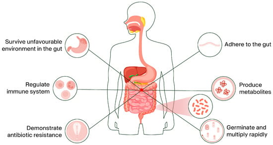
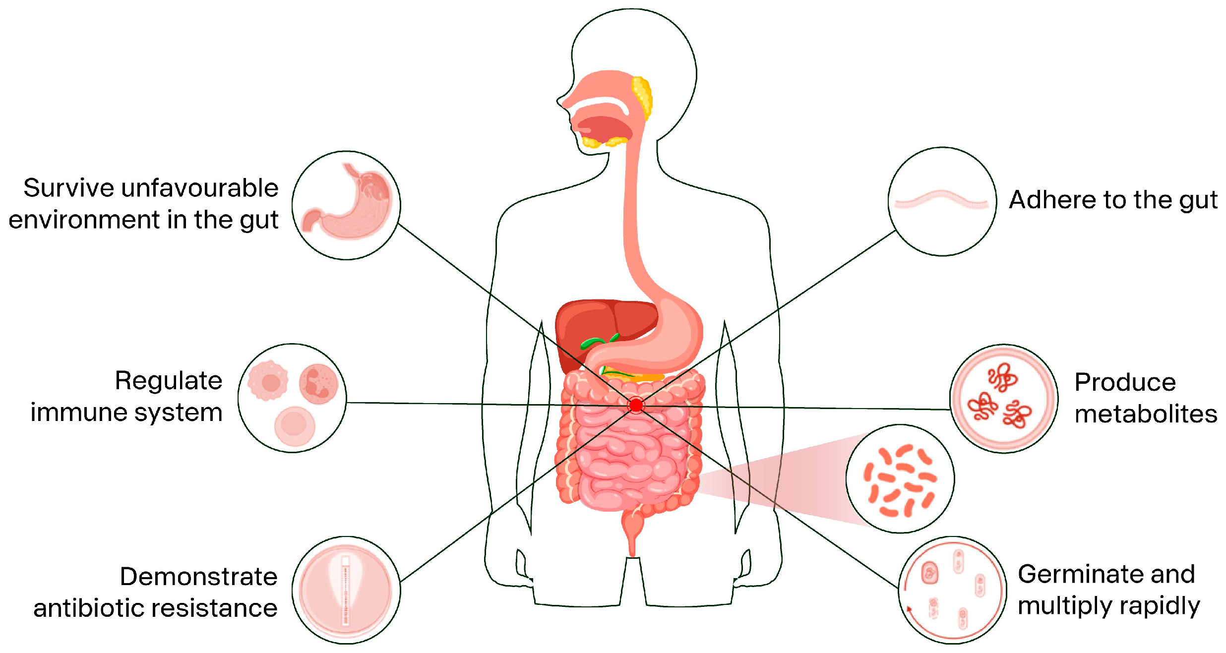

Disclaimer/Publisher’s Note: The statements, opinions and data contained in all publications are solely those of the individual author(s) and contributor(s) and not of MDPI and/or the editor(s). MDPI and/or the editor(s) disclaim responsibility for any injury to people or property resulting from any ideas, methods, instructions or products referred to in the content. |
© 2026 by the authors. Licensee MDPI, Basel, Switzerland. This article is an open access article distributed under the terms and conditions of the Creative Commons Attribution (CC BY) license.