


Abstract
Background: HIV-infected individuals with compromised immune function are particularly vulnerable to atmospheric particulate matter (PM). While Vitamin D is known to modulate inflammation and immune function, its effect on PM-induced immune impairment in HIV-infected individuals remains unclear. Methods: This randomized, double-blind, placebo-controlled trial in Wuhan, China, investigated Vitamin D’s protective effects. In total, 80 participants received daily Vitamin D or a placebo and were followed up every 28 days across four visits. PM exposure was assessed 72 h before each follow-up, with immune cell counts, immunomodulatory factors, and whole-genome DNA methylation profiles measured. Linear mixed-effects models were used to assess the association between PM exposure and cell counts, and to identify CpG sites associated with Vitamin D. Mediating models further evaluated the role of these CpG sites in linking Vitamin D intervention to cytokine changes. Results: From March to July, 2023, 75 participants received 271 follow-ups. Vitamin D supplementation significantly attenuated the negative associations between PM exposure and CD4+T cell and CD19+B cell counts. Compared with the placebo group, the estimated PM-associated changes in CD4+T cell counts differed by 3.37%, 2.21%, and 1.96% per 10 µg/m3 increase in PM1, PM2.5, and PM10 concentrations, respectively, while the corresponding differences for CD19+B cell counts were 4.71%, 2.98%, and 2.65%, respectively. Significant methylation changes were observed at 173 CpG sites related to Vitamin D. Of these, 25 sites—including cg27066321 (PEX14), cg08500112 (PDE4D), and cg25882580 (CACNG6)—were identified as potential mediators of the effects of vitamin D supplementation on TLR4, IL-6, and TNF-α levels, with mediation proportions ranging from 27.98% to 78.78%. Conclusions: Vitamin D supplementation may attenuate PM-associated immune alterations in people living with HIV, with DNA methylation potentially contributing to the underlying mechanisms.
1. Introduction
AIDS remains a major global health burden, with approximately 37.7 million individuals living with HIV by 2020 [1]. HIV-infected individuals have compromised immune functions, leading to higher risks of co-infections, cancers, autoimmune diseases, and death [2,3]. Besides direct viral effects, immune competence in HIV-infected individuals is influenced by age, nutrition, comorbidities, treatment, and environmental exposures [4]. Among these, atmospheric particulate matter (PM) has emerged as a critical environmental factor linked to impaired immune responses and worsened clinical outcomes in this population [5]. Identifying effective interventions to alleviate PM-induced immune dysfunction in HIV-infected individuals is crucial for improving their health outcomes.
Vitamin D plays a critical role in regulating immune cell function, inhibiting inflammatory responses, and modulating oxidative stress. Deficiency in Vitamin D is linked to heightened susceptibility to infections, systemic inflammation, and chronic diseases [6,7]. Conversely, sufficient Vitamin D helps mitigate excessive inflammation and oxidative stress, maintaining immune homeostasis [8]. Inflammation and oxidative stress have been identified as potential biological mechanisms through which PM exposure impairs immune function [9]. Kewcharoenwong et al. demonstrated that PM exposure triggers the inflammatory cytokine IL-8 production by neutrophils, a process that Vitamin D supplementation can mitigate [10]. Additionally, Vitamin D has been shown to inhibit IL-6 production by bronchial epithelial cells, regulate oxidative stress pathways, and alleviate PM2.5-induced inflammatory responses, thereby reducing cellular damage [11]. Despite these cellular-level findings, the efficacy of Vitamin D supplementation in alleviating PM-induced immune impairment in HIV-infected individuals, particularly those with Vitamin D deficiency, remains unclear.
Vitamin D modulates DNA methylation by forming a complex with the Vitamin D receptor, which influences the expression and activity of DNA methyltransferases [12]. Altered DNA methylation has been implicated in immune dysfunction, particularly in the differentiation of immune cells (e.g., CD4+T, CD8+T, and B cells) and the secretion of immunomodulatory factors [13,14]. A randomized controlled trial (RCT) among pregnant women identified significant differences in DNA methylation patterns at delivery and 4–6 weeks postpartum between those supplemented with Vitamin D and the control group. The differentially methylated sites were predominantly involved in immune regulation pathways, suggesting that Vitamin D may influence immune function by regulating DNA methylation [15].
Building on emerging evidence that DNA methylation is a key regulator of immune function, this study investigates whether Vitamin D can mitigate PM-induced immune dysfunction in HIV-infected individuals, with a focus on DNA methylation as a key regulatory mechanism. The findings may offer novel insights into potential therapeutic strategies for enhancing immune resilience in this vulnerable population.
2. Materials and Methods
2.1. Study Design and Participants
We conducted a randomized, double-blind, placebo-controlled study across four administrative districts in Wuhan, China. The study protocol was registered with the Chinese Clinical Trial Registry (ChiCTR2300070301; registration date: 7 April 2023) and received approval from the Wuhan Center for Disease Control and Prevention (CDC) ethics committee (WHCDCIRB-K-2022027; approval date: 8 December 2022). After completing a baseline survey on 28 March 2023 and providing informed consent, 80 participants were randomly assigned at a 1:1 ratio to either the Vitamin D group (2 capsules daily, totaling 4000 IU, from Royal Oak Health Ltd., London, UK.) or the placebo group (2 capsules daily, containing custom-made soybean oil). The dose of 4000 IU/day was selected based on established safety guidelines and previous randomized controlled trials demonstrating its efficacy in increasing serum 25(OH)D levels with acceptable tolerability [16,17,18]. A total of four follow-up visits were conducted, with the final visit taking place on 18 July 2023. The randomization was conducted using a random number generator in R (version 4.2.0). The implementers of the program, data collectors at the four district-level CDCs, and the participants were blinded to the group assignments. Only the study’s project designer was aware of the allocation. Participants were selected according to the following inclusion/exclusion criteria.
The Inclusion Criteria: 1. HIV-infected individuals aged 18–65 years. 2. Stable on the same antiretroviral therapy regimen (tenofovir, lamivudine, and efavirenz) for at least one year. 3. No history of mental disorders, able to live independently, and no language barriers. 4. No chronic underlying diseases such as diabetes, hypertension, malignancies, thyroid disease, or coronary heart disease. Exclusion Criteria included: 1. Disease status progressed to the AIDS stage. 2. Left Wuhan within 15 days before follow-up. 3. Smokers (active smoking behavior within the past three months). 4. BMI > 28 kg/m2. 5. Regular alcohol consumption (at least once a week for the past three months or more than three times in the week before follow-up). 6. Suffering from autoimmune diseases such as systemic lupus erythematosus or rheumatoid arthritis. 7. Missed antiretroviral therapy medications for over seven days in the past 30 days. 8. Recently taking additional Vitamin D, beta-adrenergic blockers, statins, or other anti-inflammatory or antioxidant medications. 9. Currently experiencing active infections, inflammatory diseases, pregnancy, breastfeeding, severe diarrhea, or vomiting.
2.2. Procedures
The follow-up protocol is outlined in Figure S1. From the baseline survey to the fourth follow-up, participants took either Vitamin D or a placebo daily. The first follow-up took place 28 days after the baseline survey, with three additional follow-ups scheduled at four-week intervals. Atmospheric PM exposure was assessed starting on Thursday or Friday, with instruments operating for at least 72 h until the following Monday or Tuesday. During each follow-up, participants underwent physical examinations between 8:00 AM and 11:30 AM on either Monday or Tuesday, which included measurements of height and weight, questionnaire surveys, and fasting blood sample collections. Blood samples were collected using three EDTA anticoagulant tubes and one coagulation tube, with each tube collecting 5 mL of whole blood. This process was conducted by professional physicians from the district-level CDCs, who received standardized training and followed strict protocols to ensure data accuracy and reliability.
2.3. Outcomes
Our primary outcome was the lymphocyte subset counts, measured from blood samples collected at the end of weeks 4, 8, 12, and 16 from the start of the study. Anticoagulated whole blood was analyzed with a flow cytometer to determine CD3+T, CD4+T, CD8+T, CD19+B, natural killer (NK), and lymphocyte (LY) cell counts, utilizing the Agilent CD3/CD8/CD45/CD4 and Agilent CD3/CD16+CD56/CD45/CD19 Assay Kits. In addition, we measured plasma levels of immunomodulatory factors, including Toll-like receptor (TLR)-4, intercellular adhesion molecule (ICAM)-1, IL-6, tumor necrosis factor alpha (TNF-α), and interferon alpha-2 (IFNα2), using ELISA kits (Jianglai, Shanghai, China). Genomic DNA from whole blood was extracted using the DNeasy Blood & Tissue Kit (250, QIAGEN, Hilden, Germany), denatured using the EZ DNA Methylation-Gold Kit, and DNA methylation was assessed using the Illumina Infinium Methylation EPIC BeadChip. All tests followed the manufacturers’ instructions. Due to budget constraints, immunomodulatory factors and whole-genome DNA methylation were only measured in blood samples collected during follow-ups with the highest and lowest PM exposure concentrations for both groups. Serum 25(OH)D levels were measured at baseline and each follow-up to assess the effectiveness of Vitamin D supplementation and verify adherence in the intervention group while ensuring no additional Vitamin D supplementation in the placebo group. The levels of Serum 25(OH)D were quantified using an electrochemiluminescence immunoassay (Roche Diagnostics, Mannheim, Germany). Flow cytometry and serum 25(OH)D measurements were completed within 12 h of blood collection. Plasma samples for ELISA and whole blood samples for DNA methylation analysis were stored at −80 °C until subsequent analyses.
2.4. Exposure Measurements
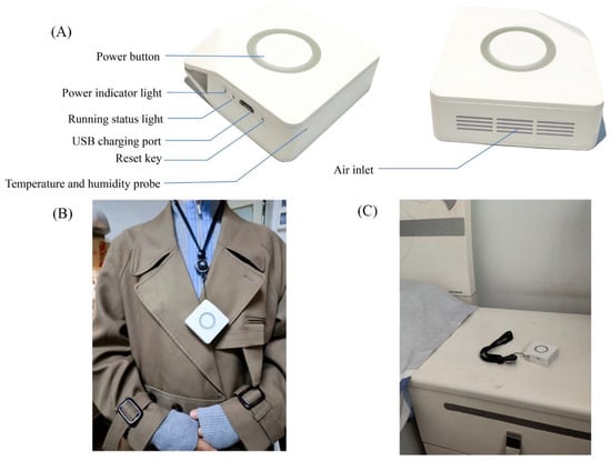
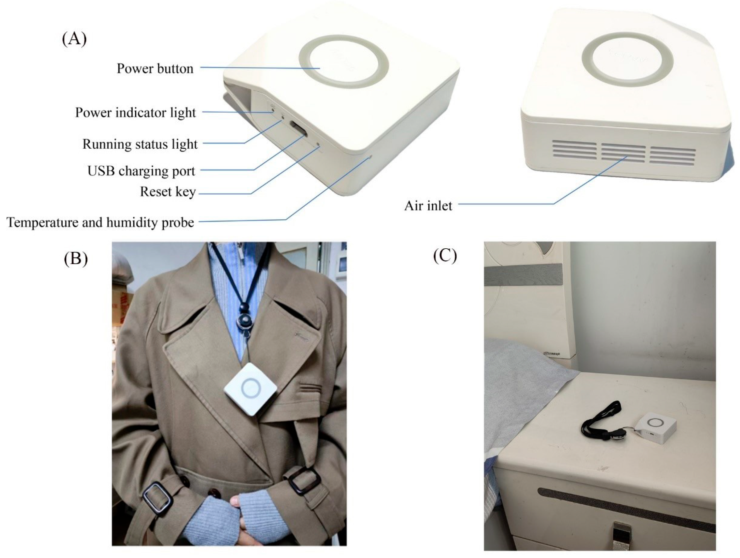
Our team developed an atmospheric PM optical detection instrument called “Air Now” (Figure 1A). The device comprises a laser particle sensor (PMS7003, Plantower, Beijing, China) for fine PM detection and a temperature and humidity sensor (SHT30, Sensirion AG, Stäfa, Switzerland). The “Air Now” operates hourly, with each cycle lasting 30 s: the first 10 s clear residual air, and the remaining 20 s are used for real-time sampling and PM detection. Each operation produces seven data sets, including PM1, PM2.5, PM10, temperature, and relative humidity. Hourly data are obtained by averaging these seven sets. The portable PM monitor was compared with a DustTrak II Aerosol Monitor (Model 8532; TSI Inc., Shoreview, MN, USA), yielding a coefficient of determination of 0.993. The “Air Now” was used to measure environmental exposure conditions within 72 h before blood collection. Participants wore the instrument on their chests during the day (Figure 1B) and placed it bedside at night (Figure 1C) to ensure continuous hourly exposure data collection. Participants were instructed to maintain their usual daily activities without travel restrictions during the monitoring period to capture real-world personal PM exposure.
Figure 1.
Optical detector for atmospheric particulate matter. Note: (A) Structure of the personal PM monitor. (B) Daytime wearing method. (C) Nighttime placement position.
2.5. Covariates
Covariate data were mainly obtained from questionnaire surveys conducted at each follow-up. To ensure accuracy and consistency, all researchers involved in the surveys received standardized training to thoroughly understand the questionnaire content and master effective questioning techniques. Covariables in this study include age (years), sex, BMI, work types (outdoor worker, indoor worker, and mixed work), family income (<¥50,000, ¥50,000–100,000, ¥100,000–150,000, and >¥150,000), education level (junior, senior, and college or above), marital status (unmarried/divorced, and married), physical activity (low, moderate, and high), dietary habits (plant-based, balanced, and meat-based), frequency of drinking (no alcohol, 1–2 times per week, 3–4 times per week), emotional disorder (no, yes), sleep disorder (no, yes), temperature, and relative humidity. Physical activity was assessed using the Metabolic Equivalent of Task (MET) via the short form of the International Physical Activity Questionnaire (IPAQ), classifying participants into low, moderate, or high activity levels based on weekly activity. Sleep disorders were identified using the Athens Insomnia Scale (AIS), with scores exceeding 6 indicating a sleep disorder. Emotional disorders were evaluated using the Hospital Anxiety and Depression Scale, with scores above 11 for either HADS-A or HADS-D indicating emotional disorders.
2.6. Statistical Analysis
Sample size calculation formula is provided in the Supplementary Information Text S1. Continuous variables are presented as mean ± standard deviation, while categorical variables are reported as frequencies (percentages). Immune cell counts and immunomodulatory factors level were natural log-transformed to approximate a normal distribution, satisfying the linear model assumptions. Linear mixed-effects (LME) models were used to evaluate the association between PM exposure and immune cell counts in both groups. LME models were fitted using all available follow-up observations, allowing participants with incomplete repeated measurements to contribute to the analysis. Covariates adjusted in the models included age, sex, BMI, work type, family income, education level, marital status, physical activity, dietary habits, drinking frequency, emotional disorder, sleep disorder, temperature, and relative humidity. Temperature and relative humidity were incorporated into the models as natural spline functions with 3 degrees of freedom. Due to the short duration of the follow-up period, no time trend adjustments were applied. Variance Inflation Factors (VIFs) for all variables were assessed, with results indicating all VIFs were below 2, thus avoiding significant multicollinearity issues.
To explore the lag effect of PM exposure on immune cell counts, multiple exposure windows were analyzed, including 0–6 h, 7–12 h, 13–24 h, 25–48 h, 49–72 h, 0–12 h, 0–24 h, 0–48 h, and 0–72 h. Additionally, the study compared the estimated effect sizes of PM short-term exposure on immune cell counts between the two groups. The formulas used are as follows:
where β1 and β2 represent the effect estimates of PM exposure for the Vitamin D group and placebo group, respectively. se1 and se2 represent the corresponding standard errors. d represents the average difference in effect estimates between the two groups, and L and U represent the lower and upper limits of the 95% CI of the mean difference between groups. For multiple comparisons of group differences, the Benjamini–Hochberg method was used to control the False Discovery Rate (FDR) across all tests involving six immune cell outcomes, three PM fractions, and nine exposure windows. An FDR-adjusted p-value < 0.05 was considered statistically significant. To assess the robustness of the findings, we additionally adjusted the LME models for baseline serum 25(OH)D concentrations, corresponding baseline immune cell counts, and both variables simultaneously. Furthermore, pooled linear mixed-effects models incorporating a PM exposure × vitamin D supplementation interaction term were fitted to evaluate between-group differences in PM-associated immune cell responses while accounting for repeated measurements.
d = β1 − β2
For Genome-wide DNA methylation analysis, raw chip results underwent filtering, correction, and normalization. The filtering criteria included: (1) removal of probes with detected p-value ≥ 0.01 (6039 probes); (2) removal of probes with insufficient bead numbers (19,836 probes); (3) exclusion of non-CpG detection probes (2827 probes); (4) removal of multi-mapping probes (22 probes); (5) removal of probes with single nucleotide polymorphisms (95,116 probes); and (6) exclusion of probes on sex chromosomes X and Y (16,269 probes). The specific site cytosine methylation percentage (β value) is equivalent to the ratio of methylation (M) signal intensity to the sum of methylation (M) and non-methylation (U) signal intensities. The methylation β-value represents the proportion of DNA methylation (ranging from 0 to 1). Subsequently, β value matrix underwent normalization using Beta mixture quantile normalization to adjust for type I and type II probe biases [19]. Next, Singular Value Decomposition Analysis (SVA) was employed to examine batch effects arising from BeadChip Slide and Array, and Combat was applied for the subsequent correction of these batch effects [20]. A total of 64 samples and 726,017 probes were included in the subsequent analysis. M values, which were logit-transformed from the adjusted β values, were used as indicators of methylation levels. The transformation followed the formula:
LME models were used to screen for differential CpG sites associated with Vitamin D intervention. The models were adjusted for sex, age, BMI, work type, education level, physical activity, frequency of drinking, atmospheric PM exposure, and directly measured leukocyte proportions composition (CD8+T, CD4+T, CD19+B, NK cells, monocytes, and granulocytes), with individuals included as random effects. An FDR-adjusted p-value < 0.05 was considered significant for identifying CpG sites significantly associated with Vitamin D intervention. Subsequently, LME models were used to analyze the association between the identified CpG sites and multiple immunomodulatory factors (including TLR4, ICAM-1, IL-6, TNF-α, and IFNα2), with significance defined as FDR-adjusted p-value < 0.05. Finally, mediation analysis explored the role of differential CpG sites in mediating the relationship between Vitamin D intervention and immunomodulatory factors, utilizing the “Mediation” package with 1000 bootstrap iterations to ensure robustness. Mediation proportions were calculated to assess the mediating effects. Additionally, enrichment analysis was performed on genes associated with differential CpG sites using clusterProfiler to identify relevant biological pathways, with significance set at p < 0.05. All statistical analyses were conducted using R software version 4.3.1, with the “minfi”, “IlluminaHumanMethylationEPICanno.ilm10b4.hg19”, “ChAMP”, “sva”, “variancePartition”, “lme4”, “mediation”, and “clusterProfiler” packages used for DNA methylation data processing and analysis.
3. Results
3.1. Participant Recruitment and Follow-Up
From 1 March 2023, to 18 July 2023, 80 HIV-infected individuals were recruited from four administrative districts in Wuhan, China. Five participants had only baseline data and did not complete any follow-up visits, including one who withdrew consent in the vitamin D group, three who relocated, and one who withdrew consent in the placebo group. Consequently, 75 participants (vitamin D: 39; placebo: 36) contributed a total of 271 follow-up observations to the longitudinal analysis. Throughout the study, 74 participants (Vitamin D: 38, placebo: 36) completed the first follow-up, 65 participants (Vitamin D: 35, placebo: 30) completed the second, 69 participants (Vitamin D: 35, placebo: 34) completed the third, and 63 participants (Vitamin D: 30, placebo: 33) completed the fourth follow-up (Figure S2).
3.2. Sample Baseline Characteristics
The basic characteristics of the participants were shown in Table 1. The average age of the 80 participants was 39.12 ± 15.69 years, with an average BMI of 22.32 ± 2.90 kg/m2. The study population was predominantly male (87.5%), with 61.3% indoor workers, 73.8% unmarried or divorced, and 60.0% having a college education or higher. Most participants maintained moderate levels of physical activity (51.3%) and balanced dietary habits (82.5%), with 72.5% never consuming alcohol. There were no statistically significant differences between the two groups regarding age, sex, BMI, work type, household income, education level, marital status, physical activity levels, dietary habits, drinking frequency, PSQI scores, HADS-A scores, HADS-D scores, serum 25(OH)D levels, and various immune cell counts.
Table 1.
Baseline characteristics of the subjects.
3.3. Comparative Analysis of PM Exposure and Immunological Indicators Across Groups
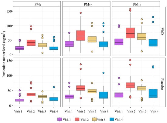
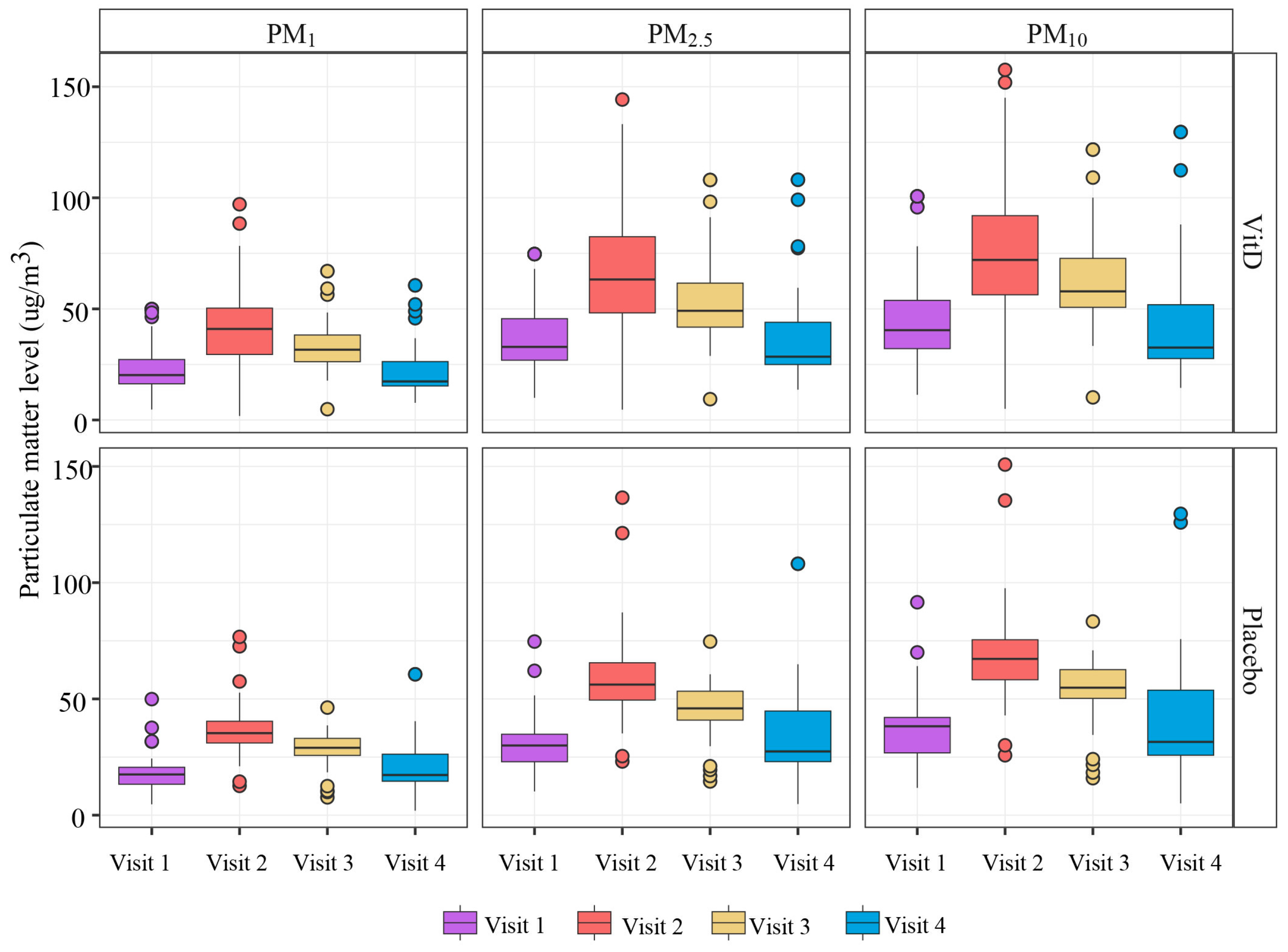
PM concentrations varied across the four follow-up visits, with the highest concentrations observed at the second visit, followed by the third, fourth, and first visits (Figure 2). The average PM2.5 exposure concentrations for the 72 h before the first, second, third, and fourth follow-ups were 37.6 ± 26.8 µg/m3, 68.9 ± 57.7 µg/m3, 53.8 ± 38.7 µg/m3, and 39.8 ± 35.3 µg/m3, respectively. As shown in Figure S3, baseline serum 25(OH)D levels were 16.86 ± 5.50 ng/mL in the Vitamin D group and 19.52 ± 7.37 ng/mL in the placebo group. Serum 25(OH)D levels were significantly higher in the Vitamin D group than in the placebo group at all four follow-ups (p < 0.05). However, there were no significant differences in the counts of CD3+T, CD4+T, CD8+T, CD19+B, NK and LY between the two groups at the any follow-ups (Table S1). Table S2 demonstrates significant differences in TLR4 and IL-6 concentrations between the two groups at the visits during the lowest levels of PM exposure, as well as in TLR4 concentrations at visits during the highest levels of PM exposure (p < 0.05).
Figure 2.
Mean 72-h exposure distribution prior to follow-up for both groups of participants.
3.4. Comparative Effects of PM Exposure on Immune Cell Counts Between Vitamin D and Placebo Groups in HIV-Infected Individuals
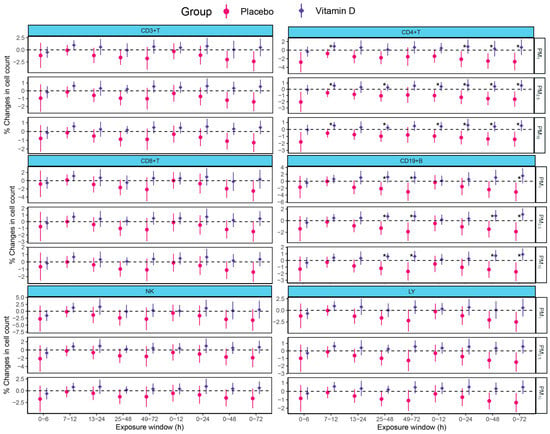
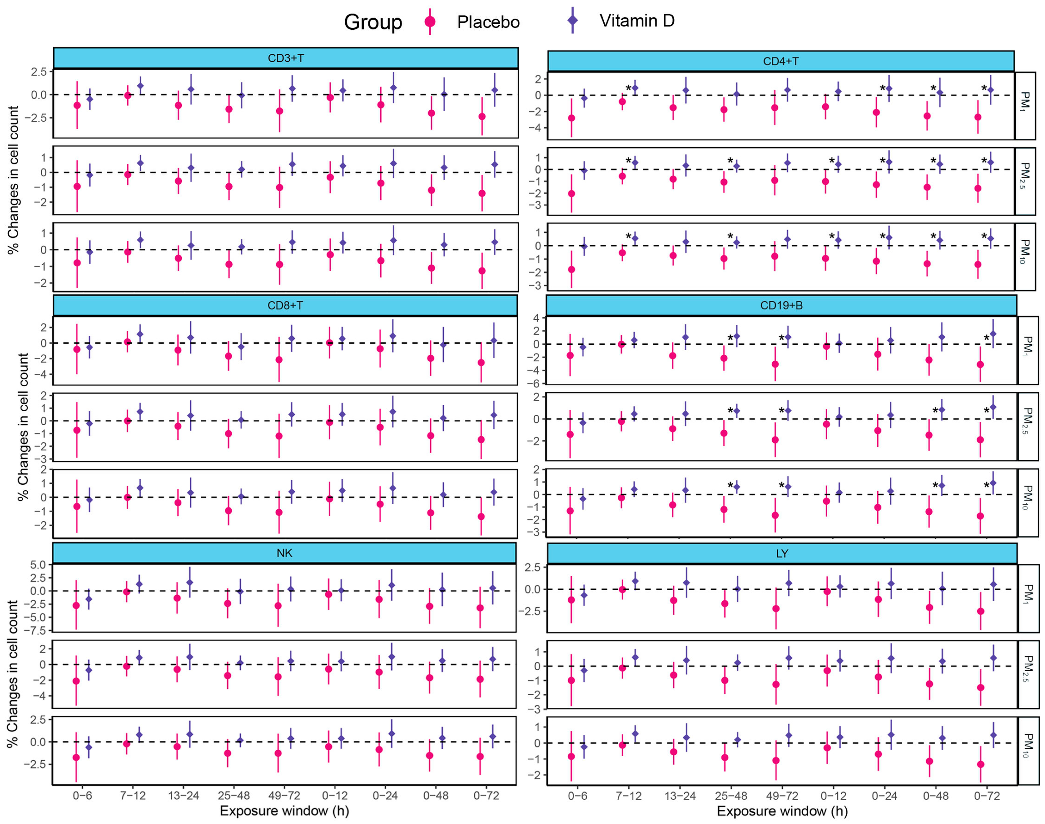
As shown in Figure 3, in the placebo group, PM exposure was negatively correlated with percentage changes in CD3+T (25–48 h, 0–48 h, 0–72 h), CD4+T (0–6 h, 25–48 h, 0–24 h, 0–48 h, 0–72 h), CD19+B (25–48 h, 49–72 h, 0–48 h, 0–72 h), and LY (25–48 h, 0–48 h, 0–72 h) cell counts. During the 0–72 h exposure window, each 10 µg/m3 increase in PM1, PM2.5, and PM10 concentrations was associated with decreases in CD3+T cell counts by 2.36%, 1.41%, and 1.27%; CD4+T cell counts by 2.69%, 1.59%, and 1.41%; CD19+B cell counts by 3.11%, 1.90%, and 1.72%; and LY cell counts by 2.50%, 1.49%, and 1.34%, respectively. Furthermore, significant differences between the two groups were found in the effects of atmospheric PM exposure on CD4+T cell counts (7–12 h, 0–12 h, 25–48 h, 0–24 h, 0–48 h, 0–72 h) and CD19+B cell counts (25–48 h, 49–72 h, 0–48 h, 0–72 h) (FDR-adjusted p < 0.05). Specifically, compared with the placebo group, the estimated PM-associated changes in CD4+T cell counts in the vitamin D group differed by 3.37% (95% CI: 0.58%, 6.16%), 2.21% (95% CI: 0.68%, 3.74%), and 1.96% (95% CI: 0.61%, 3.30%) per 10 µg/m3 increase in PM1, PM2.5, and PM10 concentrations, respectively. The corresponding differences for CD19+B cell counts were 4.71% (95% CI: 1.17%, 8.26%), 2.98% (95% CI: 1.03%, 4.94%), and 2.65% (95% CI: 0.94%, 4.37%), respectively.
Figure 3.
Associations between particulate matter concentrations measured during the preceding 72 h and immune cell counts in the vitamin D and placebo groups. Note: CD3+T, total T lymphocytes; CD4+T, helper T lymphocytes; CD8+T, suppressor T lymphocytes; CD19+B, total B lymphocytes; NK, natural killer cells; LY, lymphocytes. Models were adjusted for age, sex, BMI, work type, family income, education level, marital status, physical activity, dietary habits, drinking frequency, emotional disorder, sleep disorder, temperature, and relative humidity. The horizontal dashed line at zero corresponds to the null effect. * indicates a statistically significant between-group difference in PM-associated regression coefficients (BH-FDR-adjusted p < 0.05).
Additional adjustment for baseline 25(OH)D concentrations, corresponding baseline immune cell counts, or both yielded between-group differences in PM-associated CD4+T and CD19+B cell responses that were broadly consistent in direction and magnitude with the primary analysis. For the 0–72 h exposure window, these differences remained significant after within-outcome BH-FDR correction in all three models (Tables S3–S5). In pooled linear mixed-effects models, nominally significant PM × treatment interactions were observed for CD4+T and CD19+B cell counts with PM2.5 and PM10 during the 0–48 h and 0–72 h exposure windows, as well as for CD19+B cell counts with PM1 during 0–72 h (all unadjusted p < 0.05). However, none of these interactions remained statistically significant after BH-FDR correction (Table S6).
3.5. Association of Differentially Methylated Sites with Immunomodulatory Factors
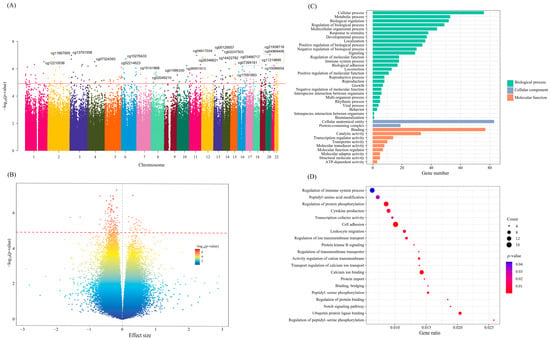
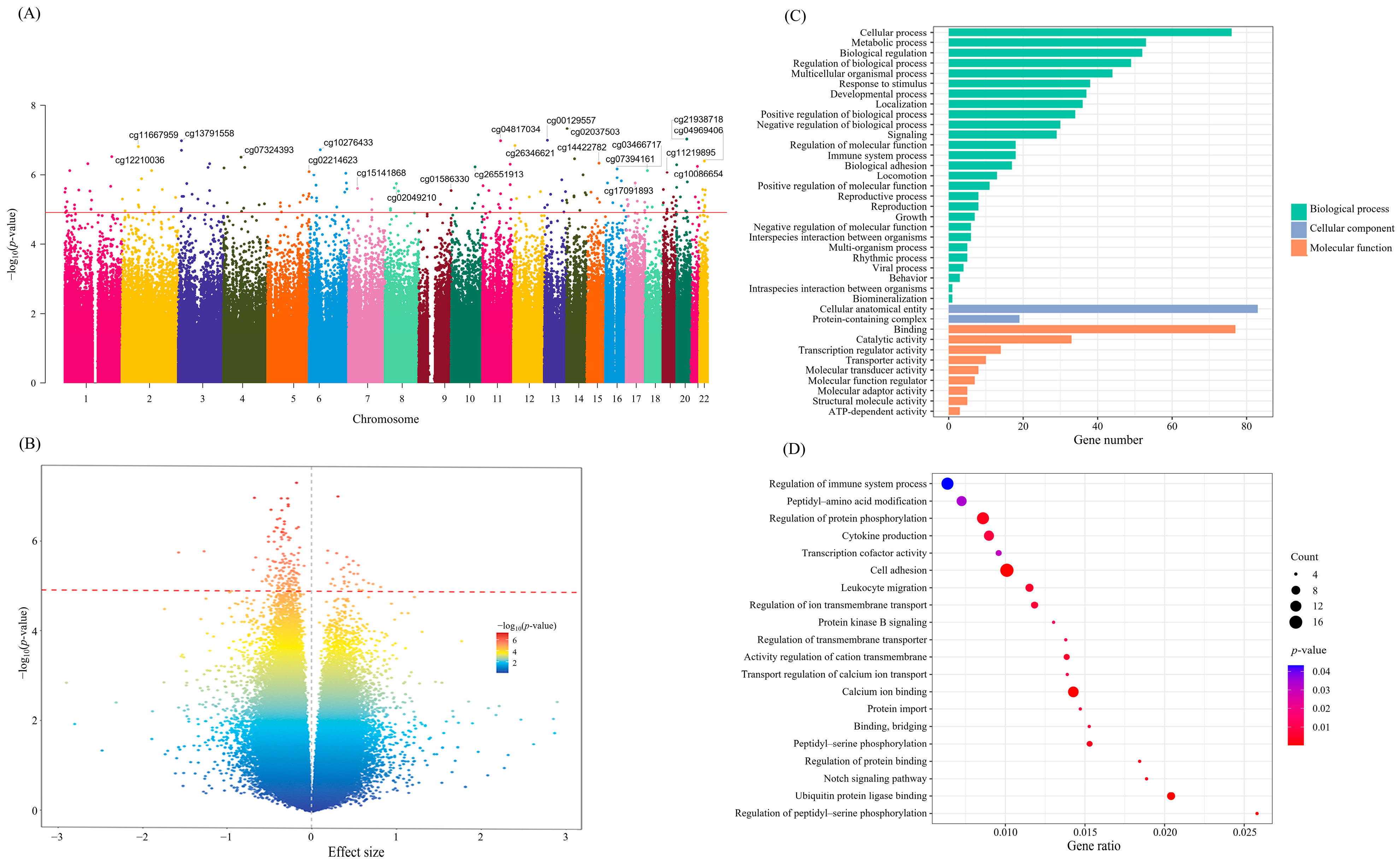
Based on the initial 726,017 CpG sites, a Manhattan plot was generated (Figure 4A); this identified 173 significantly differentially methylated sites between the Vitamin D group and the placebo group (Table S7). A volcano plot highlighted 146 hypomethylated and 27 hypermethylated sites in the Vitamin D group compared to the placebo group (Figure 4B). The top five CpG sites ranked by p-value were cg02037503, cg21938718, cg00129557, cg13791558, and cg04817034. The methylation M-values of 52 CpG sites were positively correlated with TLR4 concentrations, with regression coefficients (β) of 1.55 (95% CI: 1.01, 2.09) for cg23096476, 0.97 (95% CI: 0.56, 1.37) for cg04982488, and 0.67 (95% CI: 0.36, 0.99) for cg27066321. Conversely, methylation levels of 10 CpG sites were negatively correlated with TLR4 concentrations, including cg08558340 (β = −1.14, 95% CI: −1.58, −0.69), cg00830604 (β = −0.68, 95% CI: −1.11, −0.24), and cg26014511 (β = −0.54, 95% CI: −0.99, −0.09) (Table S8). Additionally, the study also identified 3 CpG sites associated with ICAM-1 concentrations, 56 with IL-6, 96 with TNF-α, and 15 with IFNα2. (Tables S9–S12). GO enrichment analysis of the 93 genes annotated by the 137 differentially methylated CpG sites indicated significant involvement in biological pathways related to immune regulation, including cell adhesion, Notch signaling pathway, leukocyte migration, cytokine production, ubiquitin protein ligase binding, regulation of protein phosphorylation, and regulation of immune system processes (Figure 4C,D).
Figure 4.
Identification and enrichment analysis of differentially methylated sites associated with Vitamin D intervention. Note: (A) Manhattan plot of differentially methylated CpG sites associated with Vitamin D intervention. The red dashed line denotes the genome-wide significance threshold. (B) Volcano plot depicting the distribution of differentially methylated CpG sites associated with Vitamin D intervention. The red dashed line marks the genome-wide significance threshold. (C) Annotated gene GO analysis of differentially methylated CpG sites. (D) GO pathway bubble map of annotated genes with differentially methylated CpG sites.
3.6. Mediation of DNA Methylation in the Association Between Vitamin D Intervention and Immunomodulatory Factors
Exploratory mediation analyses identified five CpG sites showing statistically significant estimated indirect effects in the association between vitamin D supplementation and TLR4 concentrations (Table 2). The mediation effects of cg27066321 (β: −0.278, 95% CI: −0.495, −0.091), cg04982488 (β: −0.338, 95% CI: −0.588, −0.145), cg23096476 (β: −0.584, 95% CI: −1.027, −0.214), cg25499646 (β: −0.268, 95% CI: −0.533, −0.021), and cg08558340 (β: −0.475, 95% CI: −0.887, −0.138) accounted for 36.97%, 42.44%, 77.06%, 33.79%, and 63.82% of the total effect, respectively. The methylation levels of 10 CpG sites, including cg08500112, cg17164356, cg05990080, cg14070323, cg23096476, cg11304950, cg11962082, cg09996840, cg25317338, and cg05015022, mediated the reduction in IL-6 levels due to Vitamin D intervention by contributing 34.63%, 37.12%, 34.74%, 27.98%, 78.78%, 36.26%, 62.83%, 47.08%, 50.81%, and 36.92% of the mediation effect, respectively. Furthermore, 12 CpG sites, including cg25882580, cg06709331, and cg23528791, etc., mediated the association between Vitamin D intervention and TNF-α levels, with mediation proportions ranging from 57.09% to 76.86%.
Table 2.
The mediating effect of DNA methylation in the association between Vitamin D intervention and immunomodulatory factors.
4. Discussion
This study identified negative associations between PM concentrations measured during recent exposure windows and CD3+T cell, CD4+T cell, CD19+B cell, and lymphocyte counts. Vitamin D supplementation was associated with attenuated negative PM-related associations for CD4+T cell and CD19+B cell counts.
Previous studies show that PM exposure alters immune cell counts in vulnerable populations. For instance, a cohort of 1397 infants found that a 25 µg/m3 increase in PM2.5 before birth reduced CD4+T cells by 2.27% and increased CD19+B cells by 1.55%, without affecting other cell types [21]. Another study indicated that every 10 µg/m3 increase in PM2.5 and PM10 exposure during early pregnancy decreased CD3+T and CD8+T cell proportions by 2.69% and 1.84% (PM2.5), and by 1.72% and 1.37% (PM10), respectively, in 3-year-old children, with no association observed for CD4+T cell [22]. Our findings align with these results, showing a negative association between PM exposure and CD3+T, CD4+T, CD19+B, and LY cell counts, but no effect on NK cells. These findings suggest that PM exposure may adversely affect immune function by selectively influencing immune cell populations in vulnerable individuals. When interpreting these associations, the observed PM exposure levels should also be considered. Mean 72-h PM2.5 concentrations ranged from 37.6 to 68.9 µg/m3 across follow-up visits, indicating variability in personal exposure during the study period. As PM exposure was measured continuously without predefined high- or low-exposure groups, these findings should be interpreted within the observed exposure range.
Vitamin D supplementation appeared to attenuate the negative associations between PM exposure and CD4+T cell and CD19+B cell counts in individuals living with HIV. This aligns with findings in healthy populations, where Vitamin D modulates immune cell function and inflammation [23]. Unlike transient immune responses in healthy individuals, HIV-infected individuals may benefit more from Vitamin D’s ability to enhance immune resilience and reduce inflammation. Vitamin D exerts protective effects against PM-induced immune dysfunction through its anti-inflammatory and immunomodulatory properties [9]. The active metabolite, 1,25(OH)2D3, suppresses NF-κB signaling in lymphocytes, thereby reducing pro-inflammatory cytokine production and limiting immune dysregulation [24,25,26]. This action helps to counteract the immune dysregulation and cellular damage that often follow PM exposure. Additionally, Vitamin D decreases B cell antigen presentation, dampening T-cell inflammatory responses and promoting immune stability [27]. In HIV-infected individuals, Vitamin D enhances immune defense by inducing antiviral gene expression, downregulating CCR5 on CD4+T cells, and preserving T cell integrity [28]. Through VDR-mediated pathways, it further promotes monocyte-to-macrophage differentiation and enhances antimicrobial responses, underscoring its role in maintaining immune homeostasis under environmental stress [29,30]. These mechanisms may help explain the observed differences in PM-associated immune cell responses between the intervention groups.
The potential influence of antiretroviral therapy (ART) should also be considered when interpreting these findings. Efavirenz-containing regimens have been associated with reduced circulating 25(OH)D concentrations, potentially through altered vitamin D metabolism [31]. In our study, all participants had received the same tenofovir, lamivudine, and efavirenz regimen for at least one year before enrollment, minimizing heterogeneity in ART exposure between groups. Notably, serum 25(OH)D concentrations were consistently higher in the vitamin D group throughout follow-up, indicating a biochemical response to supplementation within this treatment context. This finding is consistent with a previous randomized trial showing that efavirenz use did not diminish the 25(OH)D response to vitamin D supplementation [32]. Nevertheless, the potential influence of ART on immune responses and the generalizability of our findings to other ART regimens remain uncertain. Although our findings suggest that vitamin D supplementation may attenuate PM-associated immune alterations, their direct clinical implications remain uncertain. Nevertheless, these observations provide preliminary evidence to inform future investigations into strategies for protecting immune health in people living with HIV exposed to air pollution. Further studies incorporating clinical outcomes are needed to determine whether these associations translate into meaningful health benefits.
We identified 173 differentially methylated CpG sites linked to Vitamin D supplementation, with 84.39% (146 sites) showing hypomethylation, annotated to 122 genes. These findings suggest that vitamin D supplementation may be associated with changes in the blood DNA methylation landscape. Notably, GO enrichment analysis identified biological processes related to leukocyte migration, cell adhesion, cytokine production, Notch signaling, and immune regulation. These processes are relevant to immune cell trafficking, inflammatory signaling, and immune homeostasis, providing a biological context for the observed methylation differences. Our findings align with Cruz et al., who reported methylation changes at specific CpG sites (cg00523161, cg21165262, cg07145979, and cg13918754) in genes like LPIN1, DYSF, COL23A1, and RNF39 among individuals taking Vitamin D [33], whereas another randomized trial reported differential methylation at a CpG site annotated to GAB2 [15]. LPIN1 has been implicated in metabolic and inflammatory regulation [34,35,36], while GAB2 participates in intracellular signaling pathways associated with immune responses [37,38]. These findings support the biological plausibility of an association between vitamin D supplementation and epigenetic variation in immune-related processes.
At the candidate-gene level, exploratory mediation analyses identified several CpG sites showing potential indirect effects in associations between vitamin D supplementation and immunomodulatory factors. Among these, cg27066321 was annotated to PEX14, a gene involved in peroxisomal protein import and homeostasis. Previous studies have demonstrated the importance of peroxisomal regulation in macrophage redox balance and inflammatory responses [39]. Notably, experimental induction of peroxisomes attenuated lipopolysaccharide -induced production of COX-2 and pro-inflammatory cytokines, including TNF-α, IL-6, and IL-12, whereas PEX14 knockdown enhanced lipopolysaccharide induced inflammatory responses [40]. These findings provide a biological rationale for investigating the potential involvement of PEX14 in TLR4-related immune regulation. Another candidate, cg04982488, was annotated to AFAP1L2, which encodes the adaptor protein XB130. XB130 participates in Src-related intracellular signaling [41], and experimental deletion of XB130 has been associated with enhanced inflammatory responses, including increased NF-κB activity and ERK1/2 phosphorylation in a skin inflammation model [42]. Separately, the p38γ/δ–ERK1/2 signaling axis has been shown to regulate TLR4-induced cytokine production [43], although a direct mechanistic connection between AFAP1L2 and this pathway remains unestablished. In addition, cg08500112, annotated to PDE4D, was identified in the exploratory analysis involving IL-6. The PDE4 family regulates intracellular cAMP signaling, which plays an important role in inflammatory cytokine production [44]. Together, these findings suggest several biologically plausible pathways that may be relevant to the observed epigenetic associations. However, the functional consequences of the identified CpG sites remain uncertain, as gene expression and protein activity were not directly assessed. Further integrated methylation–transcriptomic and experimental studies are needed to validate these candidate mechanisms.
This study offers several strengths, including the use of individual-level PM exposure monitoring to precisely assess its impact on immune function, and the first RCT to investigate genome-wide DNA methylation changes induced by Vitamin D supplementation in HIV-infected individuals. The exploratory mediation analyses identified candidate CpG sites potentially associated with indirect immunomodulatory effects, providing hypotheses for future mechanistic investigations. However, several limitations should be emphasized when interpreting the results. First, the relatively small sample size may limit the accuracy and power of the trial. Increasing the sample size in future studies could enhance the robustness of the findings. Second, the male-to-female ratio of HIV-infected individuals in Wuhan is approximately 10:1, and this study could not recruit participants at a 1:1 gender ratio. However, the gender ratio difference between the two groups was not statistically significant. Future research should aim for a more balanced gender ratio. Third, this study did not measure the expression of genes annotated by differentially methylated CpG sites identified through DNA methylation chips. Future studies could functionally validate these annotated genes. Fourth, although the mediation analyses identified potential indirect effects of DNA methylation, these findings should be interpreted cautiously. DNA methylation was not randomized, and its concurrent measurement with immunomodulatory factors prevented confirmation of temporal precedence. Residual confounding and alternative biological mechanisms cannot be excluded, and causal mediation cannot be established. Finally, PM exposure was assessed within 72 h before each follow-up visit, which captured recent exposure levels but did not account for participants’ long-term exposure histories. Therefore, the observed associations cannot be attributed exclusively to recent PM exposure, as potential contributions from cumulative or long-term exposure cannot be excluded.
5. Conclusions
In conclusion, vitamin D supplementation was associated with attenuated negative associations between recently measured PM concentrations and CD4+T cell and CD19+B cell counts in people living with HIV. Exploratory DNA methylation analyses identified potential epigenetic associations with immunomodulatory responses. These findings provide novel insights into the epigenetic pathways underlying immune dysfunction and offer implications for targeted interventions in vulnerable populations.
Supplementary Materials
The following supporting information can be downloaded at: https://www.mdpi.com/article/10.3390/nu18213710/s1, Supplementary Information Text S1. Figure S1: Follow-up protocol of the study. Figure S2: Participant flow through the trial. Figure S3: Distribution of serum 25(OH)D concentrations in the two study groups during the follow-up period. Table S1: Levels of lymphocyte counts during follow-up of study subjects. Table S2: Distribution of immunomodulatory factors in subjects under the low and high PM exposure visit. Table S3: PM-associated changes in CD4+T and CD19+B cell counts in the vitamin D and placebo groups with additional adjustment for baseline 25(OH)D. Table S4: PM-associated changes in CD4+T and CD19+B cell counts in the vitamin D and placebo groups with additional adjustment for the corresponding baseline immune cell count. Table S5: PM-associated changes in CD4+T and CD19+B cell counts in the vitamin D and placebo groups with additional adjustment for baseline 25(OH)D and the corresponding baseline immune cell count. Table S6: PM-associated changes in CD4+T and CD19+B cell counts and PM-by-treatment interactions. Table S7: Association between Vitamin D Intervention and Whole-Genome DNA Methylation in people living with HIV. Table S8: Association between Vitamin D intervention-related CpGs and TLR4 concentrations (Top 20 ranked by p-value). Table S9: Association between Vitamin D intervention-related CpGs and ICAM-1 Concentrations (Top 20 ranked by p-value). Table S10: Association between Vitamin D intervention-related CpGs and IL-6 Concentrations (Top 20 ranked by p-value). Table S11: Association between Vitamin D intervention-related CpGs and TNF-α concentrations (Top 20 ranked by p-value). Table S12: Association between Vitamin D intervention-related CpGs and IFNα2 concentrations (Top 20 ranked by p-value).
Author Contributions
Conceptualization, H.B. and Y.H.; formal analysis, W.L. and S.P.; data curation, H.B., Z.P., Z.X., Y.T. and S.P.; software, X.Z.; investigation, H.B., Z.P., Z.X. and X.Z.; validation, X.L., X.Z. and Z.X.; writing—original draft preparation, H.B.; writing—review and editing, Y.H., W.L., X.L. and W.L.; supervision, X.L. and W.L.; project administration, W.L.; funding acquisition, W.L. All authors have read and agreed to the published version of the manuscript.
Funding
This research was supported by funding from the National Natural Science Foundation of China (Grant no. 42505176); Natural Science Research of Jiangsu Higher Education Institutions of China (Grant no. 25KJD330003).
Institutional Review Board Statement
The study was conducted in accordance with the Declaration of Helsinki and approved by the Ethics Committee of the Wuhan Center for Disease Control and Prevention (approval number: WHCDCIRB-K-2022027), approval date: 8 December 2022.
Informed Consent Statement
Written informed consent was obtained from all participants involved in the randomized controlled trial.
Data Availability Statement
The datasets used and/or analyzed during the current study are available from the corresponding author on reasonable request.
Conflicts of Interest
The authors declare that they have no conflicts of interest.
Abbreviations
The following abbreviations are used in this manuscript:
| PM | particulate matter |
| PM1 | submicron particulate matter |
| PM2.5 | fine particulate matter |
| PM10 | inhalable particulate matter |
| HIV | human immunodeficiency virus |
| AIDS | acquired immunodeficiency syndrome |
| RCT | randomized controlled trial |
| CDC | Centers for Disease Control and Prevention |
| BMI | body mass index |
| SD | standard deviation |
| CI | confidence interval |
| LME | linear mixed-effects model |
| VIF | variance inflation factor |
| FDR | false discovery rate |
| SVA | singular value decomposition analysis |
| GO | Gene Ontology |
| 25(OH)D | 25-hydroxyvitamin D |
| CD3+T | total T lymphocytes |
| CD4+T | helper T lymphocytes |
| CD8+T | suppressor T lymphocytes |
| CD19+B | total B lymphocytes |
| NK | natural killer cells |
| LY | lymphocytes |
| TLR4 | Toll-like receptor 4 |
| ICAM-1 | intercellular adhesion molecule-1 |
| IL-6 | interleukin-6 |
| TNF-α | tumor necrosis factor-α |
| IFNα2 | interferon alpha-2 |
| MET | Metabolic Equivalent of Task |
| IPAQ | International Physical Activity Questionnaire |
| AIS | Athens Insomnia Scale |
| HADS | Hospital Anxiety and Depression Scale |
| PSQI | Pittsburgh Sleep Quality Index |
References
- Ayano, G.; Duko, B.; Bedaso, A. The Prevalence of Post-Traumatic Stress Disorder among People Living with HIV/AIDS: A Systematic Review and Meta-Analysis. Psychiatr. Q. 2020, 91, 1317–1332. [Google Scholar] [CrossRef] [Scilit] [PubMed]
- Boulougoura, A.; Sereti, I. HIV infection and immune activation: The role of coinfections. Curr. Opin. HIV AIDS 2016, 11, 191–200. [Google Scholar] [CrossRef]
- Epeldegui, M.; Vendrame, E.; Martínez-Maza, O. HIV-associated immune dysfunction and viral infection: Role in the pathogenesis of AIDS-related lymphoma. Immunol. Res. 2010, 48, 72–83. [Google Scholar] [CrossRef] [Scilit] [PubMed]
- Ahn, M.Y.; Jiamsakul, A.; Khusuwan, S.; Khol, V.; Pham, T.T.; Chaiwarith, R.; Avihingsanon, A.; Kumarasamy, N.; Wong, W.W.; Kiertiburanakul, S.; et al. The influence of age-associated comorbidities on responses to combination antiretroviral therapy in older people living with HIV. J. Int. AIDS Soc. 2019, 22, e25228. [Google Scholar] [CrossRef] [Scilit] [PubMed]
- Liang, W.; Wang, X.; Xie, N.; Yan, H.; Ma, H.; Liu, M.; Kong, W.; Zhu, Z.; Bai, W.; Xiang, H. Short-term associations of PM2.5 and PM2.5 constituents with immune biomarkers: A panel study in people living with HIV/AIDS. Environ. Pollut. 2023, 317, 120743. [Google Scholar] [CrossRef] [Scilit] [PubMed]
- Nakhl, S.; Sleilaty, G.; El Samad, S.; Saliba, Y.; Chahine, R.; Farès, N. Association between vitamin D deficiency and lipid and non-lipid markers of cardiovascular diseases in the middle east region. Eur. J. Clin. Nutr. 2019, 73, 850–858. [Google Scholar] [CrossRef] [Scilit] [PubMed]
- Hammami, F.; Koubaa, M.; Mejdoub, Y.; Turki, M.; Ben Ayed, H.; Chakroun, A.; Rekik, K.; Smaoui, F.; Ben Jemaa, M. The association between vitamin D deficiency and extrapulmonary tuberculosis: Case-control study. Tuberculosis 2021, 126, 102034. [Google Scholar] [CrossRef] [Scilit] [PubMed]
- Wimalawansa, S.J. Controlling Chronic Diseases and Acute Infections with Vitamin D Sufficiency. Nutrients 2023, 15, 3623. [Google Scholar] [CrossRef] [Scilit] [PubMed]
- Glencross, D.A.; Ho, T.R.; Camiña, N.; Hawrylowicz, C.M.; Pfeffer, P.E. Air pollution and its effects on the immune system. Free Radic. Biol. Med. 2020, 151, 56–68. [Google Scholar] [CrossRef] [Scilit] [PubMed]
- Kewcharoenwong, C.; Khongmee, A.; Nithichanon, A.; Palaga, T.; Prueksasit, T.; Mudway, I.S.; Hawrylowicz, C.M.; Lertmemongkolchai, G. Vitamin D3 regulates PM-driven primary human neutrophil inflammatory responses. Sci. Rep. 2023, 13, 15850. [Google Scholar] [CrossRef] [Scilit] [PubMed]
- Xin, L.; Che, B.; Zhai, B.; Luo, Q.; Zhang, C.; Wang, J.; Wang, S.; Fan, G.; Liu, Z.; Feng, J.; et al. 1,25-Dihydroxy Vitamin D3 Attenuates the Oxidative Stress-Mediated Inflammation Induced by PM2.5via the p38/NF-κB/NLRP3 Pathway. Inflammation 2019, 42, 702–713. [Google Scholar] [CrossRef] [Scilit] [PubMed]
- Jiao, X.; Wang, L.; Wei, Z.; Liu, B.; Liu, X.; Yu, X. Vitamin D deficiency during pregnancy affects the function of Th1/Th2 cells and methylation of IFN-γ gene in offspring rats. Immunol. Lett. 2019, 212, 98–105. [Google Scholar] [CrossRef] [Scilit] [PubMed]
- Scheer, S.; Runting, J.; Bramhall, M.; Russ, B.; Zaini, A.; Ellemor, J.; Rodrigues, G.; Ng, J.; Zaph, C. The Methyltransferase DOT1L Controls Activation and Lineage Integrity in CD4+ T Cells during Infection and Inflammation. Cell Rep. 2020, 33, 108505. [Google Scholar] [CrossRef] [Scilit] [PubMed]
- Lai, A.Y.; Mav, D.; Shah, R.; Grimm, S.A.; Phadke, D.; Hatzi, K.; Melnick, A.; Geigerman, C.; Sobol, S.E.; Jaye, D.L.; et al. DNA methylation profiling in human B cells reveals immune regulatory elements and epigenetic plasticity at Alu elements during B-cell activation. Genome Res. 2013, 23, 2030–2041. [Google Scholar] [CrossRef] [Scilit] [PubMed]
- Anderson, C.M.; Gillespie, S.L.; Thiele, D.K.; Ralph, J.L.; Ohm, J.E. Effects of Maternal Vitamin D Supplementation on the Maternal and Infant Epigenome. Breastfeed. Med. 2018, 13, 371–380. [Google Scholar] [CrossRef] [Scilit] [PubMed]
- Ross, A.C.; Manson, J.E.; Abrams, S.A.; Aloia, J.F.; Brannon, P.M.; Clinton, S.K.; Durazo-Arvizu, R.A.; Gallagher, J.C.; Gallo, R.L.; Jones, G.; et al. The 2011 Report on Dietary Reference Intakes for Calcium and Vitamin D from the Institute of Medicine: What Clinicians Need to Know. J. Clin. Endocrinol. Metab. 2011, 96, 53–58. [Google Scholar] [CrossRef] [Scilit] [PubMed]
- Pittas, A.G.; Dawson-Hughes, B.; Sheehan, P.; Ware, J.H.; Knowler, W.C.; Aroda, V.R.; Brodsky, I.; Ceglia, L.; Chadha, C.; Chatterjee, R.; et al. Vitamin D Supplementation and Prevention of Type 2 Diabetes. N. Engl. J. Med. 2019, 381, 520–530. [Google Scholar] [CrossRef] [Scilit] [PubMed]
- Overton, E.T.; Chan, E.S.; Brown, T.T.; Tebas, P.; McComsey, G.A.; Melbourne, K.M.; Napoli, A.; Hardin, W.R.; Ribaudo, H.J.; Yin, M.T. Vitamin D and Calcium Attenuate Bone Loss with Antiretroviral Therapy Initiation: A Randomized Trial. Ann. Intern Med. 2015, 162, 815–824. [Google Scholar] [CrossRef] [Scilit] [PubMed]
- Teschendorff, A.E.; Marabita, F.; Lechner, M.; Bartlett, T.; Tegner, J.; Gomez-Cabrero, D.; Beck, S. A beta-mixture quantile normalization method for correcting probe design bias in Illumina Infinium 450 k DNA methylation data. Bioinformatics 2013, 29, 189–196. [Google Scholar] [CrossRef] [Scilit] [PubMed]
- Johnson, W.E.; Li, C.; Rabinovic, A. Adjusting batch effects in microarray expression data using empirical Bayes methods. Biostatistics 2007, 8, 118–127. [Google Scholar] [CrossRef] [Scilit] [PubMed]
- Hertz-Picciotto, I.; Herr, C.E.; Yap, P.S.; Dostál, M.; Shumway, R.H.; Ashwood, P.; Lipsett, M.; Joad, J.P.; Pinkerton, K.E.; Srám, R.J. Air pollution and lymphocyte phenotype proportions in cord blood. Environ. Health Perspect. 2005, 113, 1391–1398. [Google Scholar] [CrossRef] [Scilit] [PubMed]
- Deng, Y.L.; Liao, J.Q.; Zhou, B.; Zhang, W.X.; Liu, C.; Yuan, X.Q.; Chen, P.P.; Miao, Y.; Luo, Q.; Cui, F.P.; et al. Early life exposure to air pollution and cell-mediated immune responses in preschoolers. Chemosphere 2022, 286, 131963. [Google Scholar] [CrossRef] [Scilit] [PubMed]
- Pope, C.A.; Bhatnagar, A.; McCracken, J.P.; Abplanalp, W.; Conklin, D.J.; O’Toole, T. Exposure to Fine Particulate Air Pollution Is Associated with Endothelial Injury and Systemic Inflammation. Circ. Res. 2016, 119, 1204–1214. [Google Scholar] [CrossRef] [Scilit] [PubMed]
- Pfeffer, P.E.; Lu, H.; Mann, E.H.; Chen, Y.H.; Ho, T.R.; Cousins, D.J.; Corrigan, C.; Kelly, F.J.; Mudway, I.S.; Hawrylowicz, C.M. Effects of vitamin D on inflammatory and oxidative stress responses of human bronchial epithelial cells exposed to particulate matter. PLoS ONE 2018, 13, e0200040. [Google Scholar] [CrossRef] [Scilit] [PubMed]
- Mann, E.H.; Ho, T.R.; Pfeffer, P.E.; Matthews, N.C.; Chevretton, E.; Mudway, I.; Kelly, F.J.; Hawrylowicz, C.M. Vitamin D Counteracts an IL-23-Dependent IL-17A+IFN-gamma+ Response Driven by Urban Particulate Matter. Am. J. Respir. Cell Mol. Biol. 2017, 57, 355–366. [Google Scholar] [CrossRef] [Scilit] [PubMed]
- Yu, X.P.; Bellido, T.; Manolagas, S.C. Down-regulation of NF-kappa B protein levels in activated human lymphocytes by 1,25-dihydroxyvitamin D3. Proc. Natl. Acad. Sci. USA 1995, 92, 10990–10994. [Google Scholar] [CrossRef] [Scilit] [PubMed]
- Hewison, M. An update on vitamin D and human immunity. Clin. Endocrinol. 2012, 76, 315–325. [Google Scholar] [CrossRef] [Scilit] [PubMed]
- Aguilar-Jimenez, W.; Villegas-Ospina, S.; Gonzalez, S.; Zapata, W.; Saulle, I.; Garziano, M.; Biasin, M.; Clerici, M.; Rugeles, M.T. Precursor Forms of Vitamin D Reduce HIV-1 Infection In Vitro. J. Acquir. Immune Defic. Syndr. 2016, 73, 497–506. [Google Scholar] [CrossRef] [Scilit] [PubMed]
- Gil, A.; Plaza-Diaz, J.; Mesa, M.D. Vitamin D: Classic and Novel Actions. Ann. Nutr. Metab. 2018, 72, 87–95. [Google Scholar] [CrossRef] [Scilit] [PubMed]
- Calder, P.C.; Carr, A.C.; Gombart, A.F.; Eggersdorfer, M. Optimal Nutritional Status for a Well-Functioning Immune System Is an Important Factor to Protect against Viral Infections. Nutrients 2020, 12, 1181. [Google Scholar] [CrossRef] [Scilit] [PubMed]
- Brown, T.T.; McComsey, G.A. Association between initiation of antiretroviral therapy with efavirenz and decreases in 25-hydroxyvitamin D. Antivir. Ther. 2010, 15, 425–429. [Google Scholar] [CrossRef] [Scilit] [PubMed]
- Havens, P.L.; Mulligan, K.; Hazra, R.; Flynn, P.; Rutledge, B.; Van Loan, M.D.; Lujan-Zilbermann, J.; Kapogiannis, B.G.; Wilson, C.M.; Stephensen, C.B. Serum 25-hydroxyvitamin D response to vitamin D3 supplementation 50,000 IU monthly in youth with HIV-1 infection. J. Clin. Endocrinol. Metab. 2012, 97, 4004–4013. [Google Scholar] [CrossRef] [Scilit] [PubMed]
- Cruz, M.A.D.; Ulfenborg, B.; Blomstrand, P.; Faresjö, M.; Ståhl, F.; Karlsson, S. Characterization of methylation patterns associated with lifestyle factors and vitamin D supplementation in a healthy elderly cohort from Southwest Sweden. Sci. Rep. 2022, 12, 12670. [Google Scholar] [CrossRef] [Scilit] [PubMed]
- Csaki, L.S.; Dwyer, J.R.; Fong, L.G.; Tontonoz, P.; Young, S.G.; Reue, K. Lipins, lipinopathies, and the modulation of cellular lipid storage and signaling. Prog. Lipid Res. 2013, 52, 305–316. [Google Scholar] [CrossRef] [Scilit] [PubMed]
- Reue, K.; Wang, H. Mammalian lipin phosphatidic acid phosphatases in lipid synthesis and beyond: Metabolic and inflammatory disorders. J. Lipid Res. 2019, 60, 728–733. [Google Scholar] [CrossRef] [Scilit] [PubMed]
- Dai, W.; Zheng, P.; Luo, D.; Xie, Q.; Liu, F.; Shao, Q.; Zhao, N.; Qian, K. LPIN1 Is a Regulatory Factor Associated with Immune Response and Inflammation in Sepsis. Front. Immunol. 2022, 13, 820164. [Google Scholar] [CrossRef] [Scilit] [PubMed]
- Liu, Z.; Zhang, X.; Dong, M.; Liu, Z.; Wang, Y.; Yu, H.; Yu, K.; Xu, N.; Liu, W.; Song, H. Analysis of the microRNA and mRNA expression profile of ricin toxin-treated RAW264.7 cells reveals that miR-155-3p suppresses cell inflammation by targeting GAB2. Toxicol. Lett. 2021, 347, 67–77. [Google Scholar] [CrossRef] [Scilit] [PubMed]
- Cheng, J.; Zhong, Y.; Chen, S.; Sun, Y.; Huang, L.; Kang, Y.; Chen, B.; Chen, G.; Wang, F.; Tian, Y.; et al. Gab2 mediates hepatocellular carcinogenesis by integrating multiple signaling pathways. FASEB J. 2017, 31, 5530–5542. [Google Scholar] [CrossRef] [Scilit] [PubMed]
- Ganguli, G.; Pattanaik, K.P.; Jagadeb, M.; Sonawane, A. Mycobacterium tuberculosis Rv3034c regulates mTORC1 and PPAR-γ dependant pexophagy mechanism to control redox levels in macrophages. Cell Microbiol. 2020, 22, e13214. [Google Scholar] [CrossRef] [Scilit] [PubMed]
- Vijayan, V.; Srinu, T.; Karnati, S.; Garikapati, V.; Linke, M.; Kamalyan, L.; Mali, S.R.; Sudan, K.; Kollas, A.; Schmid, T.; et al. A New Immunomodulatory Role for Peroxisomes in Macrophages Activated by the TLR4 Ligand Lipopolysaccharide. J. Immunol. 2017, 198, 2414–2425. [Google Scholar] [CrossRef] [Scilit] [PubMed]
- Xu, J.; Bai, X.H.; Lodyga, M.; Han, B.; Xiao, H.; Keshavjee, S.; Hu, J.; Zhang, H.; Yang, B.B.; Liu, M. XB130, a novel adaptor protein for signal transduction. J. Biol. Chem. 2007, 282, 16401–16412. [Google Scholar] [CrossRef] [Scilit] [PubMed]
- Cho, H.R.; Wang, Y.; Bai, X.; Xiang, Y.Y.; Lu, C.; Post, A.; Al Habeeb, A.; Liu, M. XB130 deficiency enhances carcinogen-induced skin tumorigenesis. Carcinogenesis 2019, 40, 1363–1375. [Google Scholar] [CrossRef] [Scilit] [PubMed]
- Risco, A.; del Fresno, C.; Mambol, A.; Alsina-Beauchamp, D.; MacKenzie, K.F.; Yang, H.T.; Barber, D.F.; Morcelle, C.; Arthur, J.S.; Ley, S.C.; et al. P38γ and p38δ kinases regulate the Toll-like receptor 4 (TLR4)-induced cytokine production by controlling ERK1/2 protein kinase pathway activation. Proc. Natl. Acad. Sci. USA 2012, 109, 11200–11205. [Google Scholar] [CrossRef] [Scilit] [PubMed]
- Blauvelt, A.; Langley, R.G.; Gordon, K.B.; Silverberg, J.I.; Eyerich, K.; Sommer, M.O.A.; Felding, J.; Warren, R.B. Next Generation PDE4 Inhibitors that Selectively Target PDE4B/D Subtypes: A Narrative Review. Dermatol. Ther. 2023, 13, 3031–3042. [Google Scholar] [CrossRef] [Scilit] [PubMed]

Disclaimer/Publisher’s Note: The statements, opinions and data contained in all publications are solely those of the individual author(s) and contributor(s) and not of MDPI and/or the editor(s). MDPI and/or the editor(s) disclaim responsibility for any injury to people or property resulting from any ideas, methods, instructions or products referred to in the content. |
© 2026 by the authors. Licensee MDPI, Basel, Switzerland. This article is an open access article distributed under the terms and conditions of the Creative Commons Attribution (CC BY) license.