


Abstract
Vitamin D has long been recognized for its role in calcium homeostasis and bone metabolism; however, it is now emerging as an important regulator of central nervous system (CNS) function. Recent evidence suggests that vitamin D signaling contributes to the pathogenesis and progression of several neurodegenerative disorders. Vitamin D exerts neuroprotective effects through multiple mechanisms, including regulation of calcium homeostasis, modulation of immune responses, reduction in oxidative stress, stimulation of neurotrophic factors, and maintenance of blood–brain barrier (BBB) integrity. Vitamin D receptors and metabolizing enzymes are widely distributed across several brain regions, highlighting their direct involvement in neuronal function. This review summarizes the biosynthesis, metabolism, and signaling pathways of vitamin D. It explores its role in neurodegenerative diseases such as Alzheimer’s disease (AD), Parkinson’s disease (PD), multiple sclerosis (MS), amyotrophic lateral sclerosis (ALS), stroke, and traumatic brain injury (TBI). Evidence from experimental and clinical studies indicates that vitamin D deficiency is associated with an increased risk and severity of these conditions, while supplementation may provide therapeutic benefits.
1. Introduction
Neurodegenerative disorders are driven by multifactorial processes such as neuroinflammation, oxidative stress, and protein aggregation. Emerging evidence highlights vitamin D as a neuroactive secosteroid hormone with roles extending beyond calcium homeostasis to include regulation of neuronal function and immune responses in the central nervous system (CNS). There are two forms of vitamin D in the body: vitamin D2 and vitamin D3. Vitamin D3 is formed in the skin from a cholesterol derivative by the action of UV radiation from the sun [1]. The human body produces 90% of its vitamin D, which depends on sunlight exposure. Natural sources of vitamin D3 are eggs, cod liver oil, fish oil, and vitamin D3-fortified milk [2]. Vitamin D2 is primarily obtained from plant-based and fungal sources, particularly UV-exposed mushrooms and fortified foods. Additionally, at the beginning of the metabolic activity of vitamin D in the liver, it is hydroxylated at carbon 25, which represents the initial step in the metabolic activation pathway of vitamin D [3].
Vitamin D is synthesized in the skin when 7-dehydrocholesterol absorbs UVB radiation, forming pre-vitamin D3, which later isomerizes to vitamin D3. Vitamin D is subsequently metabolized in the liver to 25-hydroxyvitamin D (25(OH)D) and in the kidneys to the biologically active form 1,25-dihydroxyvitamin D3 (calcitriol), which regulates calcium and phosphate homeostasis and supports skeletal health. Because vitamin D receptors are present in many tissues, vitamin D signaling influences numerous biological pathways, and its deficiency has been associated with several chronic diseases.
After entering target tissues, vitamin D and its metabolites bind to the vitamin D receptor (VDR), a nuclear receptor transcription factor in the cytosol [4]. Moreover, VDR was discovered in the cerebellum, thalamus, hypothalamus, posterior ganglia, hippocampus, olfactory system, and temporal and orbital brain regions. It has been emphasized that the etiopathogenesis of various neurodegenerative diseases, such as multiple sclerosis (MS), Parkinson’s disease (PD), Alzheimer’s disease (AD), and amyotrophic lateral sclerosis (ALS), could be affected by environmental and genetic factors related to vitamin D and VDR concentration [5]. Key vitamin D-mediated signaling pathways and their functional consequences in the CNS are summarized in Table 1. Calcitriol has been shown to exert a strong regulatory effect on nerve growth factor (NGF)-mediated signal transduction, as demonstrated in neuronal cells. Consequently, it may play a crucial role in the formation of brain neurons [6]. Many neurodegenerative diseases have overlapping pathologies, such as inflammation, protein aggregation, oxidative damage, and demyelination. The role of vitamin D in the nervous system has gained more interest in recent years. There are many vitamin D-related enzymes and receptors in the brain, and vitamin D metabolites can bind to glial and neuronal cells, exerting diverse effects [7]. It has been shown that vitamin D deficiency can exacerbate neuropathological changes, and supplementation can improve outcomes. This review discusses the link between vitamin D and various neurodegenerative diseases, including the mechanisms by which vitamin D contributes to their pathogenesis and its application in their management.
However, the relationship between vitamin D status and biological effects may not be strictly linear, and responses may depend on baseline serum concentration. Vitamin D values are also correlated with threshold effects, which suggests a biological ceiling, in which VDR-mediated neuroprotection becomes less responsive once 25-hydroxyvitamin D levels cross 30–50 ng/mL. Beyond this value, various counter-regulatory mechanisms are activated, such as the upregulation of the CYP24A1 enzyme [8,9]. A previous study has demonstrated a non-linear association of the serum vitamin D levels with liver cirrhosis and hepatocellular carcinoma (HCC) [10]. The risks and disease-induced mortality have been linked with a U-shaped association curve. Studies have shown that for all causes of mortality, the risk is primarily elevated at lower vitamin D levels (<30 ng/mL), while higher levels (>70 ng/mL) do not appear to significantly increase mortality risk [10]. These findings suggest that the biological effects of vitamin D may depend not only on deficiency status but also on the concentration range, highlighting the importance of considering threshold effects when evaluating its role in neurological diseases.
Based on previous research, many neurological conditions, including PD, AD, and MS, have been associated with vitamin D deficiency [5]. The immunomodulatory and anti-inflammatory properties of vitamin D are attributed to its neuroprotective properties. Vitamin D may help improve symptoms and potentially slow disease progression by reducing oxidative stress and neuroinflammation, stimulating the production of neurotrophic factors, modulating neurotransmitter production, and protecting the blood–brain barrier (BBB). In older individuals, vitamin D deficiency can be prevented and treated to improve depression and cognitive impairment [11]. Collectively, these findings highlight the multifaceted role of vitamin D in neurodegenerative disorders and support the growing interest in its mechanistic and therapeutic relevance.
Table 1.
Vitamin D-mediated signaling pathways and their functional consequences in the CNS.
2. Vitamin D Biosynthesis, Signaling, and Regulation
Vitamin D is a fat-soluble nutrient, synthesized when 7-dehydrocholesterol is exposed to UV light, the B ring is broken, and it forms pre-D3 [25]. Pre-D3 isomerizes to D3, although tachysterol and lumisterol are produced when exposed to continuous UV light. D3 is linked to DBP and is preferentially extracted from the skin (Figure 1). Whether taken orally or through the skin, vitamin D is converted by the liver and other tissues into its main circulating form, 25(OH)D [26]. The circulating 25-hydroxyvitamin D levels are generally maintained between 20 and 50 ng/mL (with ~30 ng/mL often considered optimal) to ensure an adequate concentration of substrate. It has also been reported that 25-hydroxyvitamin D levels below 20 ng/mL are considered deficient, levels between 20 and 30 ng/mL are considered insufficient, and markedly elevated levels are associated with hypervitaminosis D (vitamin D toxicity) [27]. Consuming high doses of vitamin D (>10,000 IU/day) is linked to hypervitaminosis D, can cause symptoms of hypercalcemia, apathy, and several other linked renal and neurological disorders. The most significant enzyme with 25-hydroxylase activity is CYP2R1. The enzyme CYP27B1 then further metabolizes 25(OH)D to 1,25(OH)2D3, mostly in the kidney. However, it is also present in other organs, including immune system cells, parathyroid gland cells, and various epithelial cells [1,28]. The primary hormonal form of vitamin D, 1,25(OH)2D3, is in charge of most of its biological effects. The major vitamin D-mediated signaling pathways and their functional roles in the CNS are summarized in Table 1. The kidney’s generation of 1,25(OH)2D3 is strictly regulated; parathyroid hormone (PTH) stimulates it, while calcium, phosphate, and FGF23 decrease it (Figure 1) [29]. 1,25(OH)2D3 lowers its own cellular levels primarily by promoting its catabolism via induction of CYP24A1, the 24-hydroxylase. The extrarenal production of 1,25(OH)2D3, as in keratinocytes and macrophages, is regulated differently and is mainly stimulated by cytokines such as tumor necrosis factor alpha (TNFα) and interferon gamma (IFNγ) [30]. The CYP24A1 enzyme hydroxylates 25(OH)D and 1,25(OH)2D3 at position 24 to produce 24, 25(OH)2D and 1, 24, 25(OH)3D, respectively. Although 24, 25(OH)2D and 1, 24, 25(OH)3D have their own biologic functions, this 24-hydroxylation is typically the initial step in the catabolism of these active metabolites to the end product of calcitroic acid [31]. Additionally, CYP24A1 exhibits 23-hydroxylase activity, which produces an alternative product. The ratio of 23-hydroxylase to 24-hydroxylase activity in CYP24A1 varies among species, though in humans, 24-hydroxylase activity is more prevalent. CYP24A1 is expressed as widely as CYP27B1 [32]. In most tissues, 1,25(OH)2D3 induces CYP24A1, a crucial feedback mechanism that prevents vitamin D toxicity. In the kidney, FGF23, calcium, and phosphate activate CYP24A1, while PTH inhibits it. This is the exact reverse of how these hormones and minerals affect CYP27B1. Other tissues, however, do not exhibit this control. Since CYP24A1 is either absent or malfunctioning in macrophages, hypercalcemia and hypercalciuria resulting from elevated 1,25(OH)2D3 can happen in conditions such as granulomatous disorders like sarcoidosis, where macrophage production of 1,25(OH)2D3 is enhanced without CYP24A1 counter-regulation [1,33]. Together, these tightly regulated biosynthetic and catabolic steps maintain vitamin D homeostasis and prevent both deficiency-related dysfunction and toxicity, underscoring the importance of context-dependent vitamin D signaling in CNS physiology. Beyond its biosynthesis and regulatory control, the clinical relevance of vitamin D may also depend on the disease stage. Available evidence suggests that vitamin D may be more effective in preclinical or early symptomatic phases, particularly when deficiency is corrected. In contrast, its impact in advanced disease appears less consistent and may be limited to supportive or symptomatic benefits. The temporal pattern of vitamin D in neurodegenerative disorders seems most crucial during the pre-clinical and early symptomatic stages [34,35]. Research suggests that vitamin D mainly acts as an initial modulator, controlling calcium levels and reactive oxygen species (ROS), and reducing neuroinflammation before irreversible synaptic damage happens [36]. In AD, macrophages clear amyloid-β (Aβ) most effectively during early accumulation. As the disease advances into neurodegeneration, the effectiveness of vitamin D supplementation diminishes due to the influence of the VDR [37]. Therefore, vitamin D plays a crucial role in preventing disease and in early intervention. However, its impact during late stages is probably limited to symptom management rather than altering disease progression.
Figure 1.
The biosynthesis, regulation and metabolic pathway of vitamin D: Vitamin D is produced in the skin by UV exposure or obtained from diet and transported in the blood. It is converted in the liver to 25(OH)D and further activated in the kidney to 1,25(OH)2D3 (calcitriol). FGF23, PTH, and calcium regulate this process. Active vitamin D exerts biological effects on bone, intestine, immune cells, and the brain, supporting calcium homeostasis, immune regulation, and overall physiological functions.
2.1. Neuroprotective Mechanism of Vitamin D
Vitamin D plays a multifaceted neuroprotective role in the CNS by regulating calcium homeostasis, modulating inflammatory responses, exerting antioxidant effects, supporting neurotrophic factors and neurogenesis, influencing neurotransmission and maintaining BBB integrity.
In terms of calcium homeostasis, vitamin D, particularly its active form 1,25-dihydroxycholecalciferol, modulates intracellular calcium levels in neurons by regulating calcium-binding proteins and calcium channels. This regulation helps prevent excitotoxicity caused by excessive calcium influx, a common pathway leading to neuronal injury and death, thus maintaining neuronal health [38]. Dysregulation of calcium homeostasis is implicated in neurodegeneration, including AD, where perturbed intracellular calcium contributes to synaptic dysfunction and neuronal apoptosis [39].
Vitamin D also exerts anti-inflammatory effects by suppressing pro-inflammatory cytokines such as TNF-α and interleukin-6 (IL-6) while promoting anti-inflammatory cytokines like IL-10. This immunomodulatory role reduces neuroinflammation, which is a key contributor to chronic neurodegenerative processes including MS, AD, and PD [40,41]. It acts in glial cells, including astrocytes and microglia, to regulate immune function and mitigate inflammatory damage [42].
Regarding antioxidant properties, vitamin D enhances the expression of antioxidant enzymes such as glutathione peroxidase (GPx) and superoxide dismutase (SOD), which reduce oxidative stress and reactive oxygen species (ROS) accumulation. By modulating oxidative pathways, vitamin D protects neurons from oxidative damage, a critical factor in neuronal degeneration and cognitive decline [43,44].
Vitamin D supports neurotrophic factors, stimulating the synthesis of NGF and brain-derived neurotrophic factor (BDNF), thereby promoting neuronal survival, differentiation, synaptic plasticity, and repair processes. This neurotrophic support aids neural regeneration and functional recovery after injury [45]. It also promotes neural stem cell proliferation and differentiation, contributing to remyelination and neuroprotection in diseases like multiple sclerosis [46].
Modulation of neurotransmission by vitamin D involves regulating the synthesis and release of key neurotransmitters such as dopamine, serotonin, and acetylcholine. These neurotransmitters are vital for cognitive functions and mood regulation, linking vitamin D to psychiatric and neurodegenerative disease prevention [47].
In neurodegenerative disease pathology, vitamin D enhances the clearance of amyloid-beta peptides and reduces tau phosphorylation, mechanisms that mitigate plaque formation and neurofibrillary tangles characteristic of the disease. These actions support the slowing of AD progression through estimated neuroprotective pathways [48]. The association between vascular dysfunction and Alzheimer’s also highlights the role of vitamin D in maintaining BBB integrity, thereby preventing infiltration of neurotoxic substances and immune cells that exacerbate neurodegeneration [49].
Vitamin D supports BBB integrity by regulating tight junction proteins and reducing BBB permeability. This is crucial for maintaining the selective barrier function of the BBB to protect the CNS from harmful agents. Vitamin D decreases leukocyte recruitment into the CNS and counteracts neuroinflammation, promoting neuronal survival and brain homeostasis [50,51]. BBB dysfunction is a known contributor to multiple neurodegenerative disorders; therefore, vitamin D’s role in preserving BBB integrity has therapeutic implications [52].
In summary, vitamin D acts as a critical modulator of multiple neuroprotective mechanisms: it regulates neuronal calcium to prevent excitotoxicity; controls inflammatory and oxidative stress responses to protect neurons; promotes neurotrophic support and neurogenesis; regulates neurotransmitter systems vital for cognition and mood; reduces pathological hallmarks of Alzheimer’s; and maintains BBB integrity to safeguard the brain’s microenvironment. These combined effects underscore the importance of vitamin D in maintaining brain health and preventing or slowing the progression of neurodegenerative diseases [36,53,54,55].
2.2. Vitamin D Signaling in Specific Neurodegenerative Diseases
2.2.1. Alzheimer’s Disease
AD is one of the leading causes of dementia, often characterized by the progressive loss of memory and cognition [56]. Senile plaques are a hallmark of AD and form when Aβ peptides begin to aggregate in the brain [57] (Figure 2). It was anticipated that low vitamin D levels would be linked to a higher risk of AD and compromised cognitive performance. It has been documented that adding vitamin D to Alzheimer’s treatment improves cognitive abilities. The risk of dementia and AD has been found to be considerably increased by vitamin D deficiency. Different clinical studies evaluating this association are summarized in Table 2. According to another study, it was unclear how cognitive function and 25(OH)D3 levels were related [58]. Chronic inflammation and vitamin D insufficiency impair cognitive function and have an impact on neurodegeneration in the elderly population. Decreased vitamin D levels and depression have also been found to be strongly correlated [59]. As people age, vitamin D insufficiency becomes increasingly prevalent and is linked to several disorders. Treatment with vitamin D3 has a favorable major impact on life expectancy and quality of life. Combined treatment with memantine and vitamin D has been reported to improve cognitive function in patients with AD and may provide therapeutic benefits by slowing disease progression [37]. According to another investigation, the neurogenesis process contributes favorably to the substantial protection against neurological disorders that stem-cell application and vitamin D treatment offer. Low vitamin D levels have a major role in the onset and progression of the majority of neurodegenerative disorders, including AD, since it has been found that vitamin D levels are effective in neurological diseases [45]. As a result, it is stressed that vitamin D receptor agonists are crucial to vitamin D-based research and that maintaining vitamin D levels at a specific level throughout life is crucial. According to reports, vitamin D receptor suppression unavoidably causes aging and neurodegeneration and speeds up the process of neurodegeneration in conjunction with Aβ toxicity [60]. It has been shown that long-term vitamin D deficiency promotes neuron death, which in turn leads to aging and neurodegeneration. In another molecular investigation, it was found that IL-34, induced by 1α,25(OH)2D3, comprises a key mechanism in the protective effect of vitamin D in AD [61]. It has also been highlighted that Helicobacter pylori infection plays a significant role in neurodegenerative diseases like PD and MS, including AD, by influencing vitamin D levels through hyperhomocysteinemia [62]. It has been shown that increasing PGC-1α production in the hippocampal regions of AD mice provides substantial protection by significantly reducing Aβ plaque formation through activation of VDRs [63]. Another in vitro study found that resveratrol and vitamin D together suppressed tau phosphorylation. The beneficial effects of vitamin D application (1000 IU/day) on learning mechanisms have been established. Through its antioxidative, anti-inflammatory, and anti-apoptotic properties, vitamin D also offers significant protection against a variety of neurodegenerative disorders [64]. Additionally, recent research demonstrated that vitamin D and its analogs inhibited the synthesis of Aβ and promoted its breakdown in neuroblastoma cells or in the brains of mice lacking vitamin D. They stated that BACE1 and γ-secretases, which produce Aβ, are impacted by this inhibition (Figure 2). As a result, vitamin D’s impact on AD has been shown, and it has been proposed that it could help prevent the condition [65]. Similarly, it has been highlighted that vitamin D’s preventive impact in AD is significant for other neurodegenerative illnesses as well. Through its regulation of intracellular signaling pathways and calcium homeostasis, vitamin D exerts neuroprotective effects that may attenuate neurodegenerative progression. Vitamin deficiencies have also been associated with impaired cognitive function; therefore, supplementation with vitamin B complex, folic acid, and vitamin D has been suggested to support neurological health. In a similar vein, it has been proposed that vitamin D supplements could benefit people with impaired cognitive function [60]. Gezen-Ak and colleagues’ investigation revealed a strong correlation among vitamin D, VDR, and hippocampal tissue, suggesting that vitamin D deficiency may be linked to AD [66]. Similar findings were also documented, showing that vitamin levels and AD are significantly correlated [67]. The biggest difference between the AD and control groups was observed for vitamin C, but there were also noticeable differences in vitamin D, E, folate, A, and B12. These results imply that vitamin deficiencies are major contributors to AD pathogenesis and could be a target for future treatment approaches [68].
Figure 2.
Crosslink between vitamin D deficiency and its regulation in Alzheimer’s Disease: Vitamin D deficiency contributes to Alzheimer’s disease by promoting amyloid-β accumulation, tau hyperphosphorylation, oxidative stress, and chronic neuroinflammation, leading to neuronal cell death and neurodegeneration. The active form of vitamin D, 1,25(OH)2D3 (calcitriol), binds to the vitamin D receptor (VDR) and regulates gene expression via VDR–RXR signaling. This reduces amyloidogenic processing (BACE1 and γ-secretases), decreases tau phosphorylation, suppresses pro-inflammatory cytokines, and enhances antioxidant and neuroprotective responses. It also helps to reduce amyloid plaque formation, and supports neuroprotection and improves cognitive function.
Table 2.
Recent clinical studies on vitamin D in neurodegenerative and neurological disorders.
2.2.2. Parkinson’s Disease
PD, a common neurological disorder expected to affect more elderly adults and render them incapable of performing various activities effectively, has been associated with a deficiency of dopamine in the brain due to a failure of nerve cells to generate enough dopamine in this context [86] Common symptoms of PD include tremors, rigidity of the limbs and trunk, impaired balance and coordination, and generalized slowness of movement (bradykinesia) [87]. Additionally, individuals suffering from PD are also likely to experience dysphagia, as well as depression and sleep problems [88]. PD has been identified as a condition in which dopaminergic neurons in a part of the substantia nigra, identified as pars compacta, fail to perform adequately in providing dopamine, a condition indicative of this disease [89] (Figure 3). The mitochondria in PD are vulnerable to oxidative stress and neuroinflammation, and vitamin D can alleviate this disease in various ways by reducing oxidative stress via pathways including NF-kB and Nrf2, and by safeguarding astrocytes from ROS [90].
Figure 3.
The deficiency of vitamin D and its consequences in the progression of Parkinson’s disease: Vitamin D deficiency contributes to Parkinson’s disease by promoting α-synuclein aggregation, mitochondrial dysfunction, oxidative stress, and neuroinflammation, leading to the loss of dopaminergic neurons and motor dysfunction. The active form of vitamin D, 1,25(OH)2D3 (calcitriol), activates VDR–RXR-mediated transcription, which reduces inflammation and oxidative stress, improves mitochondrial function, and enhances dopaminergic neuron survival and dopamine production.
Chronic neurodegenerative diseases can also be worsened in the absence of adequate levels of vitamin D. The beneficial effect of vitamin D is attributed to its constituent antioxidative defense, calcium homeostasis, immune response, neurotransmission, and detoxification pathways, which might protect against PD [91]. Vitamin D deficiency is thought to lead to the cell death of the substantia nigra’s dopaminergic cells, thereby leading to PD. In a study of PD, researchers observed that patients with PD have significantly lower vitamin D levels [92]. In a study done by Sanchez et al. on rats whose model mimics PD, researchers observed reduced tyrosine hydroxylase staining in the rats’ substantia nigra, whereas BDNF, and GDNF expression increased before treatment [93]. This indicates that 1,25(OH)2D3, a form of vitamin D, would protect the rats’ dopamine-producing cells from damage. A study done on the Chinese population revealed that no correlation is observable between PD sufferers’ genetic variants of the VDR, and PD [94]. The nigrostriatal system is significantly affected in PD, a disease where vitamin D is especially abundant. The mechanisms by which vitamin D regulates phosphatase homeostasis, thereby protecting against the adverse consequences of excessive L–DOPA, are an essential aspect of vitamin D’s importance alongside its neuronal homeostatic, developmental, and maturation pathways [95]. Additionally, an abnormal form of vitamin D acts at the molecular level; upon activation, it can reduce Klotho levels, thereby impairing dopaminergic neurotransmission [96].
To begin with, there is a demonstrated relationship between neurological disorders and variances/mutations in the vitamin D receptor genetics [97]. Moreover, there is a demonstrated relationship between the development and progression of PD and levels of 25-hydroxyvitamin D3 [9]. Vascular endothelial dysfunction and neuroinflammation are commonly observed in PD, and VDRs have been identified in endothelial tissues, suggesting a potential regulatory role of vitamin D in vascular and inflammatory processes. Elevated levels of pro-inflammatory cytokines, including TNF-α and IL-6, have also been shown to contribute significantly to the induction and maintenance of dyskinesia in PD [98].
In other related areas, it is clear that vitamin D may also have a positive effect on matrix metalloproteinases, nerve growth factor, and L-type voltage-gated calcium channels, among other aspects of PD [99]. It therefore becomes apparent that the active form of vitamin D, 1,25(OH)2D3, binds to specific VDRs within the CNS, thereby contributing to its important neurological functions [100].
Vitamin D3, through activation of the VDR, exerts antioxidant and anti-inflammatory effects and may therefore provide neuroprotective benefits in neurodegenerative diseases such as PD. Reduced circulating levels of vitamin D3 and calcidiol have been associated with an increased risk of major neurodegenerative disorders, including PD [101]. Vitamin D is a neuroactive steroid with a significant role in the maintenance and possible amelioration of diseases such as ALS, AD, and PD. The Klotho protein in cerebrospinal fluid may be a useful biomarker for aging-related pathways in PD, as its levels are often decreased and correlate with disease severity [102]. In PD, vitamin D deficiency has been reported to be associated with reduced dopamine levels and α-synuclein accumulation. Experimental data demonstrated that vitamin D treatment is neuroprotective [103], particularly by increasing anti-inflammatory cytokines and reducing pro-inflammatory cytokines and supporting clinical evidence is summarized in Table 2. Astrocytes participate in the pathogenesis of PD, contributing to α-synuclein pathology but can protect neurons through mechanisms including α-synuclein clearance. Astrocytes positive for the vitamin D-activating enzyme CYP27B1 have been identified in PD, and those CYP27B1-expressing astrocytes sequester α-synuclein oligomers, showing a possible neuroprotective function. Overall, vitamin D3 modulates both neuroinflammation and immune function [104].
2.2.3. Multiple Sclerosis
Impaired immunological response to myelin sheaths is a hallmark of MS, a neurodegenerative illness. Neuronal injury results in the formation of sclerosis plaques surrounding brain neurons. Significant advancements have been made in the therapeutic treatment of MS through the introduction of therapies that impact the peripheral nervous system immunity [105]. MS employs an unidentified substance to initiate a T cell-mediated inflammatory response that demyelinates CNS tissues (Figure 4). Some studies have suggested indirect effects mediated by regulatory T cells. MS patients have reduced regulatory T cells. One powerful immunomodulator is vitamin D [106]. In an experimental animal model of MS, some studies have demonstrated that administering the biologically active hormone 1,25-dihydroxyvitamin D prevents the development and progression of MS [107]. Local paracrine or autocrine effects of vitamin D are predicted by the presence of VDR in neurons, microglia, and the CNS [108]. Serological indicators of disease activity were the subject of a randomized, double-blind, placebo-controlled study. Anti-inflammatory cytokine transforming growth factor-β (TGF-β) and IL-2 levels were significantly reduced after 6 months of 1000 IU cholecalciferol application [109]. For MS patients, vitamin D supplementation is advised, and clinical trial evidence supporting this approach is mentioned in Table 2. According to previous reports, high-dose vitamin D supplementation significantly promotes remyelination and neural repair, particularly in demyelination induced by experimental cuprizone administration, through mechanisms involving oligodendrocyte maturation and astrocyte activation. Therefore, vitamin D supplementation has been recommended for patients with MS [110,111]. It has been proposed that the vitamin D3 receptor, which is expressed in oligodendrocytes, could be a novel target for remyelination. Vitamin D-binding protein (DBP) and extracellular gelsolin (GSN) levels were shown to be low in another study, and it was proposed that raising GSN levels could be a novel therapeutic strategy. Inflammatory demyelination and neurodegeneration are the two most significant complications of MS [112]. Thus, vitamin D’s protective effects and its role in immune system regulation are important therapeutic components. The most noteworthy discovery regarding MS is that vitamin D3 stimulates the growth of stem cells and promotes neural stem cells to differentiate into oligodendrocytes, a crucial process for remyelination [113]. Consuming more vitamin D is associated with a decreased incidence of MS. Numerous factors, such as vitamin D deficiency, infections, and hormonal imbalance, contribute to the development of inflammation and neurodegeneration in multiple sclerosis [114]. Because of this, vitamin D insufficiency has been proposed as a key risk factor for MS. 1,25(OH)2D3 offered substantial protection by lowering neuronal loss, inflammation, and demyelination (Figure 4). It has been highlighted that this protection is mostly due to increased Beclin1 expression, a rise in the Bcl-2/Bax ratio, and a decrease in LC3 accumulation, all of which are critical components of apoptosis [54,115].
Figure 4.
Immunopathogenesis of multiple sclerosis (MS) and immunomodulatory role of vitamin D3: Peripheral activation of autoreactive T cells through antigen presentation and co-stimulation (CD28) facilitates their migration across a compromised BBB, resulting in the release of pro-inflammatory cytokines and the activation of microglia and B cells. B cells become plasma cells that make autoantibodies. This activates the complement system and causes immune-mediated demyelination. Inflammatory mediators (ROS, NO, TNF-α, IL-1β) cause oligodendrocyte stress, which disrupts AMPK–mTOR signaling and apoptosis/autophagy pathways, leading to the loss of myelin. Vitamin D3 (cholecalciferol), obtained from dietary sources or sunlight, undergoes hydroxylation in the liver (CYP2R1) and kidney (CYP27B1) to produce 1,25(OH)2D3 (calcitriol), which interacts with the VDR–RXR complex to modulate gene transcription. This signaling stops microglia from being pro-inflammatory, boosts anti-inflammatory cytokines, strengthens the BBB, and helps oligodendrocytes mature and remyelinate.
2.2.4. Amyotrophic Lateral Sclerosis
Progressive motor neuron degeneration in ALS is accompanied by microglial activation, astrogliosis, lymphocyte infiltration, and dendritic cell activation. Additionally, ALS is a progressive motor neuron disease characterized by degeneration of upper and lower motor neurons. ALS exists within a broader motor neuron disease spectrum that includes progressive muscular atrophy (PMA) and primary lateral sclerosis (PLS), and it shares pathological and genetic overlap with frontotemporal dementia (FTD) [116]. ALS-like neurological manifestations have occasionally been reported in hypoparathyroidism, a potentially treatable endocrinopathy. Furthermore, vitamin D administration has been shown to promote axonal regeneration in experimental rat models of ALS [117]. It has been reported that in the experimental rat model of ALS, vitamin D treatment had no effect on the development of leg palsy or the age at which the disease first manifested [117]. For comparison, D3 has been shown to have a neuroprotective effect and reduce inflammation in experimental models of TBI [118]. Vitamin D’s neuroprotective effect can also be explained by its ability to lower ROS levels. Vitamin D supplementation shields neurons from apoptosis and cytotoxicity.
Similarly in AD models, according to reports, Aβ significantly reduces VDR expression, which causes neurodegeneration [119] (Figure 5). Many target genes are impacted by Aβ1–42. These have a complex effect on the regulation of the amyloidogenic process, specifically leading to a general overexpression of genes associated with the tau pathway and a general downregulation of NMDAR, ApoE, TREM2, and 1αOHase genes [120,121]. Altered miRNA expression has been implicated in AD, where several miRNAs regulate amyloid precursor protein (APP) processing and Aβ formation, thereby contributing to Aβ pathogenesis. Several miRNAs directly target the 3′UTR or 5′UTR of APP mRNA and regulate BACE1 expression, thereby influencing Aβ generation and neurodegenerative progression in AD [122]. The vitamin D binding protein complex (VitD-dgVDBP) is deglycosylated by 25-OH vitamin D. Likewise, in AD patients, curcumin restores Aβ phagocytosis by peripheral blood mononuclear cells and Aβ clearance through the activation of important genes such as Toll-like receptors (TLRs), VDR, and MGAT3 [123]. Increased neurological issues in ALS are caused by inadequate intake of vitamin E, riboflavin, pyridoxine, folate, cobalamin, Ca, Zn, Mg, and vitamin D. In diseases like ALS, cognitive deficits are a serious issue. Vitamin D has been identified as a key moderating component in the treatment of cognitive impairment in recent years [124]. Vitamin D deficiency has been shown to have detrimental impacts on quality of life, while vitamin D supplementation has demonstrated beneficial effects on cognitive function in patients with ALS. Additionally, because ALS patients have trabecular bone loss, a multidisciplinary approach should be used in their treatment. Vitamin E and vitamin D offer protection against ALS, while dietary components circulating in the blood pose a risk for the disease [125]. Other substances, including linoleic acid, are also advised to reduce the risk of ALS. It should be noted that the choice of vitamins should be tailored to the individual, even though the delaying effect of the ketogenic diet and vitamin supplementation on motor neuron degeneration and the progression of the disease in ALS has been established. Supplementation has been proposed to prevent vitamin D deficiency-related medical consequences in people with ALS and other neurodegenerative disease groups that exhibit limited mobility. Additionally, some research claims that taking vitamins orally has no beneficial effects on the disease’s prognosis [126,127].
Figure 5.
Pathobiology of amyotrophic lateral sclerosis (ALS) and the potential neuroprotective role of vitamin D: ALS is a progressive neurodegenerative disorder characterized by the degeneration of motor neurons, accompanied by microglial activation, astrocytosis, lymphocyte infiltration, and the pathological aggregation of proteins such as TDP-43, SOD1, and FUS. These changes lead to VDR downregulation, excitotoxicity, oxidative stress, inflammation, and progressive weakness. Vitamin D, which comes from sunlight, food, or supplements and is turned into calcitriol, works through the VDR–RXR pathway to control transcription, stop inflammatory signaling, lower ROS, boost antioxidant defenses, and support neuronal survival and axonal regeneration.
2.2.5. Stroke
Stroke remains a leading cause of death and long-term disability, driven by acute ischemic injury and secondary neurovascular dysfunction, including oxidative stress, BBB disruption, neuroinflammation, and neuronal apoptosis. Beyond its classical roles in calcium homeostasis and bone metabolism, vitamin D signaling has emerged as a key regulator of neurovascular health, owing to the widespread expression of the VDR and the vitamin D-activating enzyme 1α-hydroxylase in neurons, glia, endothelial cells, and immune cells within the brain [97,128].
Preclinical models of cerebral ischemia demonstrate that active vitamin D (1,25-dihydroxyvitamin D3) attenuates infarct size and improves functional recovery through modulation of inflammatory and oxidative pathways. Specifically, vitamin D suppresses pro-inflammatory NF-κB signaling and decreases the levels of TNF-α, IL-1β, and IL-6 while promoting anti-inflammatory mediators and shifting microglia toward a reparative phenotype [129,130] (Figure 6). Vitamin D also enhances endogenous antioxidant defenses by increasing SOD and GPx activity, thereby mitigating ROS-mediated neuronal damage after ischemia–reperfusion injury [43]. Recent mechanistic studies further indicate that vitamin D modulates endoplasmic reticulum (ER) stress and ferroptosis pathways, suggesting additional routes by which it may protect ischemic neurons and support cellular homeostasis post-stroke [131]. A critical aspect of post-stroke pathophysiology is BBB breakdown. Vitamin D supports vascular integrity by preserving key tight junction proteins such as claudin-5 and occludin and by attenuating matrix metalloproteinase (MMP-9) activity—processes known to exacerbate BBB permeability in ischemic stroke [50,132]. Moreover, vitamin D enhances endothelial nitric oxide synthase (eNOS) activity and cerebrovascular reactivity, promoting vascular repair and improved blood flow in the post-ischemic brain [133] (Figure 6). Clinically, vitamin D deficiency is highly prevalent among stroke patients and has been linked with greater ischemic stroke severity and poorer short- and long-term outcomes. Prospective observational data reveal that lower serum 25-hydroxyvitamin D levels correlate with increased lesion severity, worse neurological deficits, and poorer functional recovery based on NIH Stroke Scale and modified Rankin Scale scores [134]. Meta-analyses of observational studies corroborate that low vitamin D status is associated with a significantly elevated risk of ischemic stroke as well as poorer post-stroke prognoses, highlighting its potential role as a prognostic biomarker [135,136]. These findings are further summarized in Table 2. Interventional studies suggest that vitamin D supplementation may improve post-stroke outcomes in select populations, particularly among those who are deficient at baseline. Recent systematic reviews and meta-analyses of randomized and controlled studies show that vitamin D supplementation can lead to meaningful improvements in motor function (e.g., Brunnstrom Recovery Stage), NIHSS scores, and modified Rankin Scale outcomes in rehabilitating ischemic stroke patients [78,137]. However, other trials demonstrate mixed results in functional rehabilitation metrics, indicating heterogeneity in response that may depend on dosing strategies, timing, and patient characteristics [138]. Large, well-powered randomized controlled trials are still needed to define optimal supplementation parameters, including dose, duration, and identification of responder subgroups [139]. Taken together, vitamin D signaling appears to modulate key neurovascular processes after ischemic stroke, including inflammation, oxidative stress, ER stress/ferroptosis, and BBB integrity. While observational evidence consistently links vitamin D deficiency to worse stroke incidence and outcomes, the translational impact of supplementation remains an active area of research. Nonetheless, current evidence supports the clinical rationale for screening and correcting vitamin D deficiency as a potentially beneficial adjunct to stroke prevention and recovery strategies [99].
Figure 6.
Ischemic stroke pathophysiology and neurovascular modulation by vitamin D signaling: Ischemic injury causes the blood–brain barrier (BBB) to break down by increasing the levels of adhesion molecules (VCAM-1, ICAM-1) and matrix metalloproteinases (MMP-2/9). This lets Th1/Th17 cells and monocytes into the CNS. Damage-associated molecular patterns (DAMPs) released from injured neurons activate microglia through TLR2/4, initiating NF-κB/MAPK signaling, NLRP3 inflammasome activation, and caspase-1-mediated IL-1β release, consequently exacerbating neuroinflammation and neurodegeneration. Vitamin D3, which is turned into calcitriol, works through the VDR–RXR complex to control transcriptional programs that lessen post-ischemic injury. This includes stopping pro-inflammatory signaling, boosting antioxidant defenses, and changing the pathways for ER stress and ferroptosis. Vitamin D also protects the BBB by keeping tight junction proteins (claudin-5, occludin, and ZO-1) in place and lowering MMP-9 activity. It also improves endothelial function by boosting eNOS activity and nitric oxide production.
3. Conclusions
Vitamin D signaling plays an important role in maintaining CNS function and protecting against neurodegenerative processes. Through its active form, calcitriol, vitamin D regulates several biological mechanisms such as calcium homeostasis, immune modulation, antioxidant defense, neurotrophic support, and maintenance of BBB integrity. These mechanisms contribute to neuron survival and may help slow the progression of neurological disorders. Increasing evidence suggests that vitamin D deficiency is associated with several neurodegenerative diseases, including AD, PD, MS, ALS and ischemic stroke. Some studies indicate that vitamin D supplementation may provide neuroprotective benefits and improve disease outcomes in certain populations. Recent clinical evidence indicates that while vitamin D exhibits robust mechanistic neuroprotective properties, further large-scale clinical studies are required to better understand the precise mechanisms of vitamin D signaling in the brain and to develop more effective therapeutic strategies. Overall, maintaining appropriate vitamin D levels may represent a promising approach for supporting neurological health and potentially reducing the burden of neurodegenerative diseases.
Author Contributions
N.S. and N.T. conceptualized this review and designed the overall framework of the manuscript. N.S. and N.T. performed the literature survey and drafted the initial version of the manuscript. N.D. contributed to diagram generation as well as to writing this review. E.R., A.J., S.C.T. and B.B. contributed to the critical revision of the manuscript and provided intellectual input on key sections. N.S., N.D. and N.T. supervised this study and contributed to manuscript editing and refinement. All authors have read and agreed to the published version of the manuscript.
Funding
This research study was supported, in part, by the American Heart Association Transformational Project Award: 971566; 24 TPA1304527 and 25 TPA1481771 to Neetu Tyagi.
Institutional Review Board Statement
Not applicable.
Informed Consent Statement
Not applicable.
Data Availability Statement
No new data were created or analyzed in this study.
Conflicts of Interest
The authors declare no conflicts of interest.
References
- Bikle, D.; S. Adams, J.; Christakos, S. Vitamin D: Production, Metabolism, Mechanism of Action, and Clinical Requirements. Primer Metab. Bone Dis. Disord. Miner. Metab. 2013, 235–248. [Google Scholar] [CrossRef] [Scilit]
- Vitamin D—Health Professional Fact Sheet. Available online: https://ods.od.nih.gov/factsheets/VitaminD-HealthProfessional/ (accessed on 24 May 2026).
- Wacker, M.; Holick, M.F. Sunlight and Vitamin D. Dermato-endocrinology 2013, 5, 51–108. [Google Scholar] [CrossRef] [Scilit] [PubMed]
- Voltan, G.; Cannito, M.; Ferrarese, M.; Ceccato, F.; Camozzi, V. Vitamin D: An Overview of Gene Regulation, Ranging from Metabolism to Genomic Effects. Genes 2023, 14, 1691. [Google Scholar] [CrossRef] [Scilit] [PubMed]
- Savran, Z.; Baltaci, S.; Aladağ, T.; Mogulkoc, R.; Baltaci, A. Vitamin D and Neurodegenerative Diseases Such as Multiple Sclerosis (MS), Parkinson’s Disease (PD), Alzheimer’s Disease (AD), and Amyotrophic Lateral Sclerosis (ALS): A Review of Current Literature. Curr. Nutr. Rep. 2025, 14, 77. [Google Scholar] [CrossRef] [Scilit] [PubMed]
- Brown, J.; Bianco, J.I.; McGrath, J.J.; Eyles, D.W. 1,25-Dihydroxyvitamin D3 Induces Nerve Growth Factor, Promotes Neurite Outgrowth and Inhibits Mitosis in Embryonic Rat Hippocampal Neurons. Neurosci. Lett. 2003, 343, 139–143. [Google Scholar] [CrossRef] [Scilit] [PubMed]
- Gadhave, D.; Sugandhi, V.; Jha, S.; Nangare, S.; Gupta, G.; Singh, S.; Dua, K.; Cho, H.; Hansbro, P.; Paudel, K. Neurodegenerative Disorders: Mechanisms of Degeneration and Therapeutic Approaches with Their Clinical Relevance. Ageing Res. Rev. 2024, 99, 102357. [Google Scholar] [CrossRef] [Scilit] [PubMed]
- Sailike, B.; Onzhanova, Z.; Akbay, B.; Tokay, T.; Molnár, F. Vitamin D in Central Nervous System: Implications for Neurological Disorders. Int. J. Mol. Sci. 2024, 25, 7809. [Google Scholar] [CrossRef] [Scilit] [PubMed]
- Akter, R.; Islam, M.; Mosaddek, A.; Akhtar, M.; Afrose, A.; Haque, A.; Azam, F.; Sharmin, S.; Hasan, R.; Hossain, M.; et al. Association of 25-Hydroxyvitamin D Levels and Vitamin D Receptor Gene Polymorphisms with Breast Cancer Risk in Bangladeshi Women. Sci. Rep. 2026, 16, 8176. [Google Scholar] [CrossRef] [Scilit] [PubMed]
- Tseng, Y.-T.; Wang, C.-H.; Wang, J.-D.; Chen, K.-T.; Li, C.-Y. Nonlinear Associations of Serum Vitamin D Levels with Advanced Liver Disease and Mortality: A US Cohort Study. Ther. Adv. Gastroenterol. 2025, 18, 17562848251338669. [Google Scholar] [CrossRef] [Scilit] [PubMed]
- Athanassiou, L.; Mavragani, C.; Koutsilieris, M. The Immunomodulatory Properties of Vitamin D. Mediterr. J. Rheumatol. 2022, 33, 7–13. [Google Scholar] [CrossRef] [Scilit] [PubMed]
- Sun, J. Bringing Vitamin D and the Vitamin D Receptor into the Limelight. Biomolecules 2024, 14, 1094. [Google Scholar] [CrossRef] [Scilit] [PubMed]
- Cui, P.; Lu, W.; Wang, J.; Wang, F.; Zhang, X.; Hou, X.; Xu, F.; Liang, Y.; Chai, G.; Hao, J. Microglia/Macrophages Require Vitamin D Signaling to Restrain Neuroinflammation and Brain Injury in a Murine Ischemic Stroke Model. J. Neuroinflamm. 2023, 20, 63. [Google Scholar] [CrossRef] [Scilit] [PubMed]
- Zhao, H.; Mu, L.; Wang, J.; Chen, R.; Li, Y.; Zhao, L.; Zhao, Y.; Liu, L. Vitamin D Activates Nrf2 to Prevent Nerve Injury and Reduce Brain Damage in Acute Cerebral Infarction. Curr. Med. Sci. 2025, 45, 469–480. [Google Scholar] [CrossRef] [Scilit] [PubMed]
- Li, J.; Cao, Y.; Xu, J.; Li, J.; Lv, C.; Gao, Q.; Zhang, C.; Jin, C.; Wang, R.; Jiao, R.; et al. Vitamin D Improves Cognitive Impairment and Alleviates Ferroptosis via the Nrf2 Signaling Pathway in Aging Mice. Int. J. Mol. Sci. 2023, 24, 15315. [Google Scholar] [CrossRef] [Scilit] [PubMed]
- Zhang, X.; Huang, S.; Xu, H.; Hu, Y.; Gao, L. Research Progress on Plant-Derived Natural Compounds Regulating the MAPK Signaling Pathway for the Prevention and Therapy of Alzheimer’s Disease. Front. Pharmacol. 2025, 16, 1666082. [Google Scholar] [PubMed]
- Abdelhady, E.G.; Abdelaziz, G.M.; Soliman, A.S.; Ahmed, A.A.; Gaber, N.A.A.E. Effects of Vitamin D and Exercise on Depression: Exploring the VEGF-Mediated MAPK/ERK Pathway. Egypt. J. Neurol. Psychiatr. Neurosurg. 2026, 62, 21. [Google Scholar] [CrossRef] [Scilit]
- Huang, X.; Wei, P.; Fang, C.; Yu, M.; Yang, S.; Qiu, L.; Wang, Y.; Xu, A.; Hoo, R.L.C.; Chang, J. Compromised Endothelial Wnt/β-Catenin Signaling Mediates the Blood-Brain Barrier Disruption and Leads to Neuroinflammation in Endotoxemia. J. Neuroinflamm. 2024, 21, 265. [Google Scholar] [CrossRef] [Scilit] [PubMed]
- Gong, Z.; Lao, D.; Wu, Y.; Li, T.; Lv, S.; Mo, X.; Huang, W. Inhibiting PI3K/Akt-Signaling Pathway Improves Neurobehavior Changes in Anti-NMDAR Encephalitis Mice by Ameliorating Blood–Brain Barrier Disruption and Neuronal Damage. Cell. Mol. Neurobiol. 2023, 43, 3623–3637. [Google Scholar] [CrossRef] [Scilit] [PubMed]
- Korewo-Labelle, D.; Karnia, M.; Myślińska, D.; Kaczor, J. Supplementation with Vitamin D3 Protects against Mitochondrial Dysfunction and Loss of BDNF-Mediated Akt Activity in the Hippocampus during Long-Term Dexamethasone Treatment in Rats. Int. J. Mol. Sci. 2023, 24, 13941. [Google Scholar] [CrossRef] [Scilit] [PubMed]
- Shi, Y.; Shi, Y.; Jie, R.; He, J.; Luo, Z.; Li, J. Vitamin D: The Crucial Neuroprotective Factor for Nerve Cells. Neuroscience 2024, 560, 272–285. [Google Scholar] [CrossRef] [Scilit] [PubMed]
- Acharya, M.; Singh, N.; Gupta, G.; Tambuwala, M.M.; Aljabali, A.A.A.; Chellappan, D.K.; Dua, K.; Goyal, R. Vitamin D, Calbindin, and Calcium Signaling: Unraveling the Alzheimer’s Connection. Cell. Signal. 2024, 116, 111043. [Google Scholar] [CrossRef] [Scilit] [PubMed]
- Wei, X.; Zhang, F.; Cheng, D.; Wang, Z.; Xing, N.; Yuan, J.; Zhang, W.; Xing, F. Free Heme Induces Neuroinflammation and Cognitive Impairment by Microglial Activation via the TLR4/MyD88/NF-ΚB Signaling Pathway. Cell Commun. Signal. 2024, 22, 16. [Google Scholar] [CrossRef] [Scilit] [PubMed]
- Jiang, H.; Yang, X.; Wang, Y.; Zhou, C. Vitamin D Protects against Traumatic Brain Injury via Modulating TLR4/MyD88/NF-ΚB Pathway-Mediated Microglial Polarization and Neuroinflammation. BioMed Res. Int. 2022, 2022, 3363036. [Google Scholar] [CrossRef] [Scilit] [PubMed]
- Sun, Y.; Alessandroni, L.; Angeloni, S.; Del Bianco, E.; Sagratini, G. From 7-Dehydrocholesterol to Vitamin D3: Optimization of UV Conversion Procedures toward the Valorization of Fish Waste Matrices. Food Chem. X 2024, 22, 101373. [Google Scholar] [CrossRef] [Scilit] [PubMed]
- Schümmer, T.; Stangl, G.; Wätjen, W. Safety Assessment of Vitamin D and Its Photo-Isomers in UV-Irradiated Baker’s Yeast. Foods 2021, 10, 3142. [Google Scholar] [CrossRef] [Scilit] [PubMed]
- Holick, M.F. Vitamin D Deficiency. N. Engl. J. Med. 2007, 357, 266–281. [Google Scholar] [CrossRef] [Scilit] [PubMed]
- Bikle, D.D. Vitamin D Metabolism, Mechanism of Action, and Clinical Applications. Chem. Biol. 2014, 21, 319–329. [Google Scholar] [CrossRef] [Scilit] [PubMed]
- Portales-Castillo, I.; Simic, P. PTH, FGF-23, Klotho and Vitamin D as Regulators of Calcium and Phosphorus: Genetics, Epigenetics and Beyond. Front. Endocrinol. 2022, 13, 992666. [Google Scholar] [CrossRef] [Scilit]
- Nesterova, G.; Malicdan, M.C.; Yasuda, K.; Sakaki, T.; Vilboux, T.; Ciccone, C.; Horst, R.; Huang, Y.; Golas, G.; Introne, W.; et al. 1,25-(OH)2D-24 Hydroxylase (CYP24A1) Deficiency as a Cause of Nephrolithiasis. Clin. J. Am. Soc. Nephrol. 2013, 8, 649–657. [Google Scholar] [PubMed]
- Dai, B.; David, V.; Alshayeb, H.M.; Showkat, A.; Gyamlani, G.; Horst, R.L.; Wall, B.M.; Quarles, L.D. Assessment of 24,25(OH)2D Levels Does Not Support FGF23-Mediated Catabolism of Vitamin D Metabolites. Kidney Int. 2012, 82, 1061–1070. [Google Scholar] [CrossRef] [Scilit] [PubMed]
- Jones, G.; Prosser, D.; Kaufmann, M. 25-Hydroxyvitamin D-24-Hydroxylase (CYP24A1): Its Important Role in the Degradation of Vitamin D. Arch. Biochem. Biophys. 2011, 523, 9–18. [Google Scholar] [CrossRef] [Scilit] [PubMed]
- Meyer, M.B.; Pike, J.W. Mechanistic Homeostasis of Vitamin D Metabolism in the Kidney through Reciprocal Modulation of Cyp27b1 and Cyp24a1 Expression. J. Steroid Biochem. Mol. Biol. 2020, 196, 105500. [Google Scholar] [CrossRef] [Scilit] [PubMed]
- Barakat, G.; Yassine, N.; Khoury, N.; Ellaboudi, N.; Ramadan, W. Vitamin D and Brain Health: A Systematic Review. Clin. Nutr. Open Sci. 2026, 67, 100646. [Google Scholar] [CrossRef] [Scilit]
- Wang, W.; Li, Y.; Meng, X. Vitamin D and Neurodegenerative Diseases. Heliyon 2023, 9, e12877. [Google Scholar] [CrossRef] [Scilit] [PubMed]
- García Menéndez, S.; Manucha, W. Vitamin D as a Modulator of Neuroinflammation: Implications for Brain Health. Curr. Pharm. Des. 2024, 30, 323–332. [Google Scholar] [CrossRef] [Scilit] [PubMed]
- Chakkera, M.; Ravi, N.; Ramaraju, R.; Vats, A.; Nair, A.; Bandhu, A.; Koirala, D.; Pallapothu, M.; Quintana, M.; Khan, S. The Efficacy of Vitamin D Supplementation in Patients With Alzheimer’s Disease in Preventing Cognitive Decline: A Systematic Review. Cureus 2022, 14, e31710. [Google Scholar] [CrossRef] [Scilit] [PubMed]
- Jiang, Y.; Wang, G.; Jiang, S.; Wang, Y.; Tian, Q.; Li, M. Molecular Mechanisms and Targeted Intervention Strategies of Calcium Overload in Ischemic Stroke. Int. J. Mol. Sci. 2026, 27, 2279. [Google Scholar] [CrossRef] [Scilit] [PubMed]
- Popugaeva, E.; Pchitskaya, E.; Bezprozvanny, I. Dysregulation of Neuronal Calcium Homeostasis in Alzheimer’s Disease—A Therapeutic Opportunity? Biochem. Biophys. Res. Commun. 2017, 483, 998–1004. [Google Scholar] [CrossRef] [Scilit] [PubMed]
- Bhol, N.K.; Bhanjadeo, M.M.; Singh, A.K.; Dash, U.C.; Ojha, R.R.; Majhi, S.; Duttaroy, A.K.; Jena, A.B. The Interplay between Cytokines, Inflammation, and Antioxidants: Mechanistic Insights and Therapeutic Potentials of Various Antioxidants and Anti-Cytokine Compounds. Biomed. Pharmacother. 2024, 178, 117177. [Google Scholar] [CrossRef] [Scilit] [PubMed]
- Fenercioglu, A. The Anti-Inflammatory Roles of Vitamin D for Improving Human Health. Curr. Issues Mol. Biol. 2024, 46, 13514–13525. [Google Scholar] [CrossRef] [Scilit] [PubMed]
- Farina, G.; Crescioli, C. Vitamin D Associated with Exercise Can Be Used as a Promising Tool in Neurodegenerative Disease Protection. Molecules 2025, 30, 3823. [Google Scholar] [CrossRef] [Scilit] [PubMed]
- Dentino, P.; Mora, J.; Zuo, L. Vitamin D Deficiency and Its Role in Pathologies of Oxidative Stress: A Literature Review. Cureus 2025, 17, e90042. [Google Scholar] [CrossRef] [Scilit] [PubMed]
- Vázquez-Lorente, H.; Herrera-Quintana, L.; Jimenez, L.; Fernandez Perea, B.; Plaza-Díaz, J. Antioxidant Functions of Vitamin D and CYP11A1-Derived Vitamin D, Tachysterol, and Lumisterol Metabolites: Mechanisms, Clinical Implications, and Future Directions. Antioxidants 2024, 13, 996. [Google Scholar] [CrossRef] [Scilit] [PubMed]
- Liu, Y.; Zhong, Z.; Xie, J.; Ni, B.; Wu, Y. Neuroprotective Roles of Vitamin D: Bridging the Gap Between Mechanisms and Clinical Applications in Cognitive Decline. Int. J. Mol. Sci. 2025, 26, 7146. [Google Scholar] [CrossRef] [Scilit] [PubMed]
- Hijal, N.; Fouani, M.; Awada, B. Unveiling the Fate and Potential Neuroprotective Role of Neural Stem/Progenitor Cells in Multiple Sclerosis. Front. Neurol. 2024, 15, 1438404. [Google Scholar] [CrossRef] [Scilit] [PubMed]
- Banerjee, A.; Khemka, V.K.; Ganguly, A.; Roy, D.; Ganguly, U.; Chakrabarti, S. Vitamin D and Alzheimer’s Disease: Neurocognition to Therapeutics. Int. J. Alzheimers Dis. 2015, 2015, 192747. [Google Scholar] [CrossRef] [Scilit] [PubMed]
- Chen, J.; Li, Y.; Wang, L.; Liu, Q.; Qiu, B. Neuroprotective Synergy of Vitamin D and Exercise: A Narrative Review of Preclinical and Clinical Evidence on Aging-Related Neuroplasticity and Cognitive Health. Front. Nutr. 2025, 12, 1642363. [Google Scholar] [CrossRef] [Scilit] [PubMed]
- Sehwan, K.; Jung, U.; Kim, S. The Crucial Role of the Blood–Brain Barrier in Neurodegenerative Diseases: Mechanisms of Disruption and Therapeutic Implications. J. Clin. Med. 2025, 14, 386. [Google Scholar] [CrossRef] [Scilit] [PubMed]
- Won, S.; Sayeed, I.; Peterson, B.L.; Wali, B.; Kahn, J.S.; Stein, D.G. Vitamin D Prevents Hypoxia/Reoxygenation-Induced Blood-Brain Barrier Disruption via Vitamin D Receptor-Mediated NF-KB Signaling Pathways. PLoS ONE 2015, 10, e0122821. [Google Scholar] [PubMed]
- Shimizu, F.; Nakamori, M. Blood–Brain Barrier Disruption in Neuroimmunological Disease. Int. J. Mol. Sci. 2024, 25, 10625. [Google Scholar] [CrossRef] [Scilit] [PubMed]
- Rust, R.; Sagare, A.; Zhang, M.; Zlokovic, B.; Kisler, K. The Blood-Brain Barrier as a Treatment Target for Neurodegenerative Disorders. Expert Opin. Drug Deliv. 2025, 22, 673–692. [Google Scholar] [CrossRef] [Scilit] [PubMed]
- Velusamy, T.; Panneerselvam, A.S.; Purushottam, M.; Anusuyadevi, M.; Pal, P.K.; Jain, S.; Essa, M.M.; Guillemin, G.J.; Kandasamy, M. Protective Effect of Antioxidants on Neuronal Dysfunction and Plasticity in Huntington’s Disease. Oxid. Med. Cell. Longev. 2017, 2017, 3279061. [Google Scholar] [CrossRef] [Scilit] [PubMed]
- Gombash, S.E.; Lee, P.W.; Sawdai, E.; Lovett-Racke, A.E. Vitamin D as a Risk Factor for Multiple Sclerosis: Immunoregulatory or Neuroprotective? Front. Neurol. 2022, 13, 796933. [Google Scholar]
- Cui, X.; Eyles, D. Vitamin D and the Central Nervous System: Causative and Preventative Mechanisms in Brain Disorders. Nutrients 2022, 14, 4353. [Google Scholar] [CrossRef] [Scilit] [PubMed]
- Lui, F.; Tsao, J.W. Alzheimer Disease. In StatPearls [Internet]; StatPearls Publishing: Treasure Island, FL, USA, 2024; pp. 1–27. Available online: https://www.ncbi.nlm.nih.gov/sites/books/NBK499922/ (accessed on 14 June 2026).
- Gouras, G.; Olsson, T.; Hansson, O. β-Amyloid Peptides and Amyloid Plaques in Alzheimer’s Disease. Neurotherapeutics 2014, 12, 3–11. [Google Scholar] [CrossRef] [Scilit] [PubMed]
- Littlejohns, T.J.; Henley, W.E.; Lang, I.A.; Annweiler, C.; Beauchet, O.; Chaves, P.H.M.; Fried, L.; Kestenbaum, B.R.; Kuller, L.H.; Langa, K.M.; et al. Vitamin D and the Risk of Dementia and Alzheimer Disease. Neurology 2014, 83, 920–928. [Google Scholar] [CrossRef] [Scilit] [PubMed]
- Kumar, A.; Verma, A.; Chaurasia, R. Vitamin D and Inflammatory Cytokines Association in Mild Cognitive Impaired Subjects. Neurosci. Lett. 2022, 795, 137044. [Google Scholar] [CrossRef] [Scilit] [PubMed]
- Lasoń, W.; Jantas, D.; Leśkiewicz, M.; Regulska, M.; Basta-Kaim, A. The Vitamin D Receptor as a Potential Target for the Treatment of Age-Related Neurodegenerative Diseases Such as Alzheimer’s and Parkinson’s Diseases: A Narrative Review. Cells 2023, 12, 660. [Google Scholar] [CrossRef] [Scilit] [PubMed]
- Zhang, D.; Li, M.; Dong, Y.; Zhang, X.; Liu, X.; Chen, Z.; Zhu, Y.; Wang, H.; Liu, X.; Zhu, J.; et al. 1α,25-Dihydroxyvitamin D3 up-Regulates IL-34 Expression in SH-SY5Y Neural Cells. Innate Immun. 2017, 23, 584–591. [Google Scholar] [CrossRef] [Scilit] [PubMed]
- Kountouras, J.; Doulberis, M.; Papaefthymiou, A.; Polyzos, S.; Zavos, C.; Kazakos, E.; Arapoglou, S.; Kyrailidi, F.; Mouratidou, M.; Boziki, M.; et al. Controlling the Impact of Helicobacter Pylori-Related Hyperhomocysteinemia on Neurodegeneration. Medicina 2023, 59, 504. [Google Scholar] [CrossRef] [Scilit] [PubMed]
- Mota, B.; Sastre, M. The Role of PGC1α in Alzheimer’s Disease and Therapeutic Interventions. Int. J. Mol. Sci. 2021, 22, 5769. [Google Scholar] [CrossRef] [Scilit] [PubMed]
- Cheng, J.; Rui, Y.; Qin, L.; Xu, J.; Han, S.; Linxi, Y.; Yin, X.; Wan, Z. Vitamin D Combined with Resveratrol Prevents Cognitive Decline in SAMP8 Mice. Curr. Alzheimer Res. 2017, 14, 820–833. [Google Scholar] [CrossRef] [Scilit] [PubMed]
- Grimm, M.; Thiel, A.; Lauer, A.; Winkler, J.; Lehmann, J.; Regner, L.; Nelke, C.; Janitschke, D.; Benoist, C.; Streidenberger, O.; et al. Vitamin D and Its Analogues Decrease Amyloid-β (Aβ) Formation and Increase Aβ-Degradation. Int. J. Mol. Sci. 2017, 18, 2764. [Google Scholar] [CrossRef] [Scilit] [PubMed]
- Gezen-Ak, D.; Dursun, E.; Yilmazer, S. Vitamin D Inquiry in Hippocampal Neurons: Consequences of Vitamin D-VDR Pathway Disruption on Calcium Channel and the Vitamin D Requirement. Neurol. Sci. 2013, 34, 1453–1458. [Google Scholar] [CrossRef] [Scilit] [PubMed]
- Landel, V.; Annweiler, C.; Millet, P.; Morello, M.; Feron, F. Vitamin D, Cognition and Alzheimer’s Disease: The Therapeutic Benefit Is in the D-Tails. J. Alzheimer’s Dis. 2024, 53, 419–444. [Google Scholar] [CrossRef] [Scilit] [PubMed]
- Lauer, A.; Grimm, H.; Apel, B.; Golobrodska, N.; Kruse, L.; Ratanski, E.; Schulten, N.; Schwarze, L.; Slawik, T.; Sperlich, S.; et al. Mechanistic Link between Vitamin B12 and Alzheimer’s Disease. Biomolecules 2022, 12, 129. [Google Scholar] [CrossRef] [Scilit] [PubMed]
- Thouvenot, E.; Laplaud, D.; Lebrun-Frenay, C.; Derache, N.; Le Page, E.; Maillart, E.; Froment-Tilikete, C.; Castelnovo, G.; Casez, O.; Coustans, M.; et al. High-Dose Vitamin D in Clinically Isolated Syndrome Typical of Multiple Sclerosis: The D-Lay MS Randomized Clinical Trial. JAMA 2025, 333, 1413–1422. [Google Scholar] [CrossRef] [Scilit] [PubMed]
- Butzkueven, H.; Ponsonby, A.-L.; Stein, M.S.; Lucas, R.M.; Mason, D.; Broadley, S.; Kilpatrick, T.; Lechner-Scott, J.; Barnett, M.; Carroll, W.; et al. Vitamin D Did Not Reduce Multiple Sclerosis Disease Activity after a Clinically Isolated Syndrome. Brain 2024, 147, 1206–1215. [Google Scholar] [CrossRef] [Scilit] [PubMed]
- Zali, A.; Hajyani, S.; Salari, M.; Ebrahimi, M.; Mortazavian, A.; Pakpour, B. Co-Administration of Probiotics and Vitamin D Reduced Disease Severity and Complications in Patients with Parkinson’s Disease: A Randomized Controlled Clinical Trial. Psychopharmacology 2024, 241, 1905–1914. [Google Scholar] [CrossRef] [Scilit] [PubMed]
- Li, D.; Ma, X.; Zhang, W.; Zhong, P.; Li, M.; Liu, S. Impact of Vitamin D3 Supplementation on Motor Functionality and the Immune Response in Parkinson’s Disease Patients with Vitamin D Deficiency. Sci. Rep. 2025, 15, 25154. [Google Scholar] [CrossRef] [Scilit] [PubMed]
- Chen, L.-J.; Sha, S.; Stocker, H.; Brenner, H.; Schöttker, B. The Associations of Serum Vitamin D Status and Vitamin D Supplements Use with All-Cause Dementia, Alzheimer’s Disease, and Vascular Dementia: A UK Biobank Based Prospective Cohort Study. Am. J. Clin. Nutr. 2024, 119, 1052–1064. [Google Scholar] [CrossRef] [Scilit] [PubMed]
- Yong, Y.; Dong, H.; Zhou, Z.; Zhu, Y.; Gu, M.; Li, W. Serum 25-Hydroxyvitamin D Concentrations and Their Impact on All-Cause Mortality in Parkinson’s Disease: Insights from National Health and Nutrition Examination Survey 1999–2020 Data. Front. Nutr. 2024, 11, 1423651. [Google Scholar] [PubMed]
- Lu, Y.; Yamaji, T.; Yasuda, N.; Kuchiba, A.; Shimizu, Y.; Nakano, S.; Yamagishi, K.; Inoue, M.; Tsugane, S.; Sawada, N.; et al. Association between Plasma 25-Hydroxyvitamin D Concentration and Incident Disabling Dementia Risk in Japan: A Case-Cohort Study. J. Neurol. Sci. 2025, 478, 123720. [Google Scholar] [CrossRef] [Scilit] [PubMed]
- Ren, J.-J.; Li, Z.-H.; Zhong, W.-F.; Gao, J.; Chen, P.; Wang, X.-M.; You, F.-F.; Mao, C. Serum 25-Hydroxyvitamin D Concentrations, Sleep Characteristics and the Risk of Incident Dementia. Maturitas 2026, 206, 108864. [Google Scholar] [CrossRef] [Scilit] [PubMed]
- Lönnroos, E.; Ylilauri, M.; Lamberg-Allardt, C.; Manson, J.E.; Nurmi, T.; Uusitupa, M.; Voutilainen, A.; Hantunen, S.; Tuomainen, T.-P.; Virtanen, J.K. The Effect of Vitamin D3 Supplementation on the Incidence of Diagnosed Dementia Among Healthy Older Adults—The Finnish Vitamin D Trial. J. Gerontol. Ser. A 2025, 80, glaf077. [Google Scholar] [CrossRef] [Scilit] [PubMed]
- Borowicz, W.; Ptaszkowska, L.; Malecki, R.; Paprocka-Borowicz, M. The Effect of Vitamin D Supplementation on Functional Outcomes in Patients Undergoing Rehabilitation After an Ischemic Stroke: A Prospective, Single-Blind, Randomized, Placebo-Controlled Study. J. Clin. Med. 2025, 14, 1848. [Google Scholar] [CrossRef] [Scilit] [PubMed]
- Yang, H.; Wu, Q.; Zheng, J. Expression and Clinical Significance of 25-Hydroxyvitamin D, Insulin-like Growth Factor 1, and Beta-2 Microglobulin in Cognitive Dysfunction after Ischemic Stroke in the Elderly. Neuroreport 2025, 36, 127–134. [Google Scholar] [CrossRef] [Scilit] [PubMed]
- Hung, K.-C.; Chang, L.-C.; Lai, Y.-C.; Yew, M.; Liu, P.-H.; Chen, I.-W. Vitamin D Deficiency and Risk of Acute Kidney Injury after Ischemic Stroke: A Propensity-Matched Cohort Study. Front. Nutr. 2026, 12, 1709491. [Google Scholar] [CrossRef] [Scilit] [PubMed]
- Al-Shammri, S.; Chattopadhyay, A.; Raghupathy, R. Vitamin D Supplementation Mediates a Shift toward Anti-Inflammatory Cytokine Response in Multiple Sclerosis. Med. Princ. Pract. 2025, 34, 360–368. [Google Scholar] [CrossRef] [Scilit] [PubMed]
- Asadpour, S.; Mazdeh, M.; Karimi, J.; Khodadadi, I.; Shafiee, G. Vitamin D3 Supplementation Modulates Inflammatory Protein CHI3L1/YKL-40 and Oxidative Stress Status in Multiple Sclerosis. Neuropsychopharmacol. Rep. 2025, 45, e70076. [Google Scholar] [CrossRef] [Scilit] [PubMed]
- Kumar, G.; Hassan Chandrashekara, R.; Nimmakanty, R.; Sushmitha, R.; Harinishri, T.; Sunil, C.; Imran, K. Prospective Cohort Study on Vitamin D Deficiency and Its Association with Multiple Sclerosis Relapses. Bioinformation 2025, 21, 1639–1642. [Google Scholar] [CrossRef] [Scilit] [PubMed]
- Vachova, M.; Stastna, D.; Mazouchová, A.; Hruskova, P.; Uher, T.; Lizrova Preiningerova, J.; Potuznik, P.; Drahota, J.; Havrdova, E. From Sunlight to MS Fight: Impact of Vitamin D Levels on Multiple Sclerosis Activity. Neurol. Sci. 2026, 47, 38. [Google Scholar] [CrossRef] [Scilit] [PubMed]
- Galus, W.; Winder, M.; Owczarek, A.; Walawska-Hrycek, A.; Rzepka, M.; Kaczmarczyk, A.; Siuda, J.; Krzystanek, E. Does Vitamin D Supplementation Slow Brain Volume Loss in Multiple Sclerosis? A 4-Year Observational Study. Nutrients 2025, 17, 2271. [Google Scholar] [CrossRef] [Scilit] [PubMed]
- Parkinson’s Disease | National Institute of Neurological Disorders and Stroke. Available online: https://www.ninds.nih.gov/health-information/disorders/parkinsons-disease (accessed on 24 May 2026).
- Tanner, C.M.; Ostrem, J.L. Parkinson’s Disease. N. Engl. J. Med. 2024, 391, 442–452. [Google Scholar] [CrossRef] [Scilit] [PubMed]
- Santos-García, D.; de Deus Fonticoba, T.; Jesús, S.; Cosgaya, M.; García Caldentey, J.; Caballol, N.; Legarda, I.; Hernández Vara, J.; Cabo, I.; López Manzanares, L.; et al. Dysphagia in Parkinson’s Disease. A 5-Year Follow-up Study. Neurol. Sci. 2025, 46, 2637–2653. [Google Scholar] [CrossRef] [Scilit] [PubMed]
- Ramesh, S.; Perera Molligoda Arachchige, A.S. Depletion of Dopamine in Parkinson’s Disease and Relevant Therapeutic Options: A Review of the Literature. AIMS Neurosci. 2023, 10, 200–231. [Google Scholar] [CrossRef] [Scilit] [PubMed]
- Dias, V.; Junn, E.; Mouradian, M.M. The Role of Oxidative Stress in Parkinson’s Disease. J. Park. Dis. 2013, 3, 461–491. [Google Scholar] [CrossRef] [Scilit] [PubMed]
- Grimm, M.O.W.; Lauer, A.; Janitschke, D.; Grimm, H.; Hartmann, T. The Effects of Vitamin D Deficiency on Neurodegenerative Diseases. In Vitamin D Deficiency; Fedotova, J.O., Ed.; IntechOpen: London, UK, 2019; ISBN 978-1-83880-776-4. [Google Scholar]
- Fullard, M.E.; Duda, J.E. A Review of the Relationship Between Vitamin D and Parkinson Disease Symptoms. Front. Neurol. 2020, 11, 454. [Google Scholar] [CrossRef] [Scilit] [PubMed]
- Chen, Y.; Lian, Y.; Ma, Y.; Wu, C.; Zheng, Y.; Xie, N. The Expression and Significance of Tyrosine Hydroxylase in the Brain Tissue of Parkinson’s Disease Rats. Exp. Ther. Med. 2017, 14, 4813–4816. [Google Scholar] [CrossRef] [Scilit] [PubMed]
- Xia, M.; Zhou, Q. Correlation between 25-hydroxy-vitamin D and Parkinson’s disease. IBRO Neurosci. Rep. 2024, 16, 162–167. [Google Scholar] [CrossRef] [Scilit] [PubMed]
- Lương, K.; Nguyễn, L. Role of Vitamin D in Parkinson’s Disease. Int. Sch. Res. Not. 2012, 2012, 134289. [Google Scholar] [CrossRef] [Scilit] [PubMed]
- Cui, X.; McGrath, J.J.; Burne, T.H.J.; Eyles, D.W. Vitamin D and Schizophrenia: 20 Years On. Mol. Psychiatry 2021, 26, 2708–2720. [Google Scholar] [CrossRef] [Scilit] [PubMed]
- Moretti, R.; Morelli, M.E.; Caruso, P. Vitamin D in Neurological Diseases: A Rationale for a Pathogenic Impact. Int. J. Mol. Sci. 2018, 19, 2245. [Google Scholar] [CrossRef] [Scilit] [PubMed]
- dos Santos Pereira, M.; Abreu, G.H.D.; Rocca, J.; Hamadat, S.; Raisman-Vozari, R.; Michel, P.P.; Del Bel, E. Contributive Role of TNF-α to L-DOPA-Induced Dyskinesia in a Unilateral 6-OHDA Lesion Model of Parkinson’s Disease. Front. Pharmacol. 2021, 11, 617085. [Google Scholar] [PubMed]
- Lasoń, W.; Jantas, D.; Leśkiewicz, M.; Regulska, M.; Basta-Kaim, A. Vitamin D3 and Ischemic Stroke: A Narrative Review. Antioxidants 2022, 11, 2120. [Google Scholar] [CrossRef] [Scilit] [PubMed]
- Ross, A.C.; Taylor, C.L.; Yaktine, A.L.; Del Valle, H.B. Dietary Reference Intakes for Calcium and Vitamin D; The National Academies Press: Washington, DC, USA, 2011; pp. 1–1116. [Google Scholar] [CrossRef] [Scilit] [PubMed]
- Khatoon, R. Unlocking the Potential of Vitamin D: A Comprehensive Exploration of Its Role in Neurological Health and Diseases. Biology 2025, 14, 280. [Google Scholar] [CrossRef] [Scilit] [PubMed]
- Pieri, M.; Morello, M.; Zenobi, R.; Alessandra, T.; Stephan, D.; Verena, L.; Feron, F.; Millet, P. The Influence of Vitamin D on Neurodegeneration and Neurological Disorders: A Rationale for Its Physio-Pathological Actions. Curr. Pharm. Des. 2020, 26, 2475–2491. [Google Scholar] [CrossRef] [Scilit] [PubMed]
- Lima, L.A.R.; Lopes, M.J.P.; Costa, R.O.; Lima, F.A.V.; Neves, K.R.T.; Calou, I.B.F.; Andrade, G.M.; Viana, G.S.B. Vitamin D Protects Dopaminergic Neurons against Neuroinflammation and Oxidative Stress in Hemiparkinsonian Rats. J. Neuroinflamm. 2018, 15, 249. [Google Scholar] [CrossRef] [Scilit] [PubMed]
- Mazzetti, S.; Barichella, M.; Giampietro, F.; Giana, A.; Calogero, A.M.; Amadeo, A.; Palazzi, N.; Comincini, A.; Giaccone, G.; Bramerio, M.; et al. Astrocytes Expressing Vitamin D-Activating Enzyme Identify Parkinson’s Disease. CNS Neurosci. Ther. 2022, 28, 703–713. [Google Scholar] [CrossRef] [Scilit] [PubMed]
- Huang, W.; Chen, W.; Zhang, X. Multiple Sclerosis: Pathology, Diagnosis and Treatments (Review). Exp. Ther. Med. 2017, 13, 3163–3166. [Google Scholar] [CrossRef] [Scilit] [PubMed]
- Galoppin, M.; Kari, S.; Soldati, S.; Pal, A.; Rival, M.; Engelhardt, B.; Astier, A.; Thouvenot, E. Full Spectrum of Vitamin D Immunomodulation in Multiple Sclerosis: Mechanisms and Therapeutic Implications. Brain Commun. 2022, 4, fcac171. [Google Scholar] [CrossRef] [Scilit] [PubMed]
- Sloka, S.; Zhornitsky, S.; Silva, C.; Metz, L.M.; Yong, V.W. 1,25-Dihydroxyvitamin D3 Protects against Immune-Mediated Killing of Neurons in Culture and in Experimental Autoimmune Encephalomyelitis. PLoS ONE 2015, 10, e0144084-. [Google Scholar] [CrossRef] [Scilit] [PubMed]
- Lisakovska, O.; Labudzynskyi, D.; Khomenko, A.; Isaev, D.; Savotchenko, A.; Kasatkina, L.; Savosko, S.; Veliky, M.; Shymanskyi, I. Brain Vitamin D3-Auto/Paracrine System in Relation to Structural, Neurophysiological, and Behavioral Disturbances Associated with Glucocorticoid-Induced Neurotoxicity. Front. Cell. Neurosci. 2023, 17, 1133400. [Google Scholar] [PubMed]
- Argano, C.; Torres, A.; Orlando, V.; Cangialosi, V.; Maggio, D.; Pollicino, C.; Corrao, S. Molecular Insight into the Role of Vitamin D in Immune-Mediated Inflammatory Diseases. Int. J. Mol. Sci. 2025, 26, 4798. [Google Scholar] [CrossRef] [Scilit] [PubMed]
- Al-Otaibi, K.M.; Alghamdi, B.S.; Al-Ghamdi, M.A.; Mansouri, R.A.; Ashraf, G.M.; Omar, U.M. Therapeutic Effect of Combination Vitamin D3 and Siponimod on Remyelination and Modulate Microglia Activation in Cuprizone Mouse Model of Multiple Sclerosis. Front. Behav. Neurosci. 2023, 16, 1068736. [Google Scholar] [CrossRef] [Scilit] [PubMed]
- Gandhi, F.; Jhaveri, S.; Avanthika, C.; Singh, A.; Jain, N.; Gulraiz, A.; Shah, P.; Nasir, F. Impact of Vitamin D Supplementation on Multiple Sclerosis. Cureus 2021, 13, e18487. [Google Scholar] [CrossRef] [Scilit] [PubMed]
- Gomez-Pinedo, U.; Cuevas, J.; Benito-Martin, M.S.; Moreno, L.; Esteban-Garcia, N.; Torre Fuentes, L.; Matias-Guiu, J.A.; Pytel, V.; Montero- Escribano, P.; Matías-Guiu, J. Vitamin D Increases Remyelination by Promoting Oligodendrocyte Lineage Differentiation. Brain Behav. 2019, 10, e01498. [Google Scholar] [CrossRef] [Scilit] [PubMed]
- Sangha, A.; Quon, M.; Pfeffer, G.; Orton, S. The Role of Vitamin D in Neuroprotection in Multiple Sclerosis: An Update. Nutrients 2023, 15, 2978. [Google Scholar] [CrossRef] [Scilit] [PubMed]
- Sintzel, M.B.; Rametta, M.; Reder, A.T. Vitamin D and Multiple Sclerosis: A Comprehensive Review. Neurol. Ther. 2018, 7, 59–85. [Google Scholar] [CrossRef] [Scilit] [PubMed]
- Sartori, A.; Zorzella-Pezavento, S.F.G.; Ishikawa, L.L.W.; Mimura, L.A.N.; Fraga-Silva, T.F.D.C.; Fraga-Silva, T.F.C. Therapeutic and Prophylactic Potential of Vitamin D for Multiple Sclerosis. In A Critical Evaluation of Vitamin D—Clinical Overview; Thatha Gowder, S.J., Ed.; IntechOpen: London, UK, 2017; ISBN 978-953-51-3086-4. [Google Scholar]
- You, J.; Youssef, M.; Santos, J.; Lee, J.; Park, J. Microglia and Astrocytes in Amyotrophic Lateral Sclerosis: Disease-Associated States, Pathological Roles, and Therapeutic Potential. Biology 2023, 12, 1307. [Google Scholar] [CrossRef] [Scilit] [PubMed]
- Gianforcaro, A.; Hamadeh, M.J. Vitamin D as a Potential Therapy in Amyotrophic Lateral Sclerosis. CNS Neurosci. Ther. 2014, 20, 101–111. [Google Scholar] [CrossRef] [Scilit] [PubMed]
- Sharma, S.; Kumar, A.; Choudhary, A.; Sharma, S.; Khurana, L.; Sharma, N.; Kumar, V.; Bisht, A. Neuroprotective Role of Oral Vitamin D Supplementation on Consciousness and Inflammatory Biomarkers in Determining Severity Outcome in Acute Traumatic Brain Injury Patients: A Double-Blind Randomized Clinical Trial. Clin. Drug Investig. 2020, 40, 327–334. [Google Scholar] [CrossRef] [Scilit] [PubMed]
- AlJohri, R.; AlOkail, M.; Haq, S.H. Neuroprotective Role of Vitamin D in Primary Neuronal Cortical Culture. eNeurologicalSci 2019, 14, 43–48. [Google Scholar] [CrossRef] [Scilit] [PubMed]
- Beeg, M.; Battocchio, E.; De Luigi, A.; Colombo, L.; Natale, C.; Cagnotto, A.; Corbelli, A.; Fiordaliso, F.; Diomede, L.; Salmona, M.; et al. Non-Phosphorylated Tau Slows down Aβ1-42 Aggregation, Binds to Aβ1-42 Oligomers and Reduces Aβ1-42 Toxicity. J. Biol. Chem. 2021, 296, 100664. [Google Scholar] [CrossRef] [Scilit] [PubMed]
- Wes, P.; Easton, A.; Corradi, J.; Barten, D.; Devidze, N.; Decarr, L.; Truong, A.; He, A.; Barrezueta, N.; Polson, C.; et al. Tau Overexpression Impacts a Neuroinflammation Gene Expression Network Perturbed in Alzheimer’s Disease. PLoS ONE 2014, 9, e106050. [Google Scholar] [CrossRef] [Scilit] [PubMed]
- Amakiri, N.; Kubosumi, A.; Tran, J.; Reddy, P.H. Amyloid Beta and MicroRNAs in Alzheimer’s Disease. Front. Neurosci. 2019, 13, 430. [Google Scholar] [PubMed]
- Greilberger, J.; Herwig, R. Vitamin D-Deglycosylated Vitamin D Binding Protein Dimer: Positive Synergistic Effects on Recognition, Activation, Phagocytosis and Oxidative Stress on Macrophages. Clin. Lab. 2020, 66, 169. [Google Scholar] [CrossRef] [Scilit] [PubMed]
- Barros, A.; Felipe, M.; Barbosa, I.; Leite, L.; Pedrosa, L. Dietary Intake of Micronutrients and Disease Severity in Patients with Amyotrophic Lateral Sclerosis. Metabolites 2023, 13, 696. [Google Scholar] [CrossRef] [Scilit] [PubMed]
- De Marchi, F.; Saraceno, M.; Sarnelli, M.F.; Virgilio, E.; Cantello, R.; Mazzini, L. Potential Role of Vitamin D Levels in Amyotrophic Lateral Sclerosis Cognitive Impairment. Neurol. Sci. 2023, 44, 2795–2802. [Google Scholar] [CrossRef] [Scilit] [PubMed]
- Xia, K.; Wang, Y.; Zhang, L.; Tang, L.; Zhang, G.; Huang, T.; Huang, N.; Fan, D. Dietary-Derived Essential Nutrients and Amyotrophic Lateral Sclerosis: A Two-Sample Mendelian Randomization Study. Nutrients 2022, 14, 920. [Google Scholar] [CrossRef] [Scilit] [PubMed]
- Veldink, J.H.; Kalmijn, S.; Groeneveld, G.J.; Wunderink, W.; Koster, A.; De Vries, J.H.M.; Van Der Luyt, J.; Wokke, J.H.J.; Van Den Berg, L.H. Intake of Polyunsaturated Fatty Acids and Vitamin E Reduces the Risk of Developing Amyotrophic Lateral Sclerosis. J. Neurol. Neurosurg. Psychiatry 2007, 78, 367–371. [Google Scholar] [CrossRef] [Scilit] [PubMed]
- Lee, G.Y.; Park, C.; Han, S. Vitamin D-Mediated Immunoregulation in Degenerative Diseases: Insights into Cardiovascular, Neurodegenerative and Musculoskeletal Disorders. Nutrients 2026, 18, 629. [Google Scholar] [CrossRef] [Scilit] [PubMed]
- Zhang, Y.; Mu, Y.; Ding, H.; Du, B.; Zhou, M.; Li, Q.; Gong, S.; Zhang, F.; Geng, D.; Wang, Y. 1α,25-Dihydroxyvitamin D3 Promotes Angiogenesis After Cerebral Ischemia Injury in Rats by Upregulating the TGF-β/Smad2/3 Signaling Pathway. Front. Cardiovasc. Med. 2022, 9, 769717. [Google Scholar] [PubMed]
- Yin, X.; Chang, J.; Yang, L.; Jia, L.; Han, J.; Chen, L.; Cao, J.; Chen, X.; Xie, N.; Lin, Y.; et al. The ERK Signaling Pathway in Cerebral Ischemic Stroke: Mechanism and Therapeutic Significance. Biomed. Pharmacother. 2026, 196, 119058. [Google Scholar] [CrossRef] [Scilit] [PubMed]
- Song, T.; Li, J.; Xia, Y.; Hou, S.; Zhang, X.; Wang, Y. 1,25-D3 Ameliorates Ischemic Brain Injury by Alleviating Endoplasmic Reticulum Stress and Ferroptosis: Involvement of Vitamin D Receptor and P53 Signaling. Cell. Signal. 2024, 122, 111331. [Google Scholar] [CrossRef] [Scilit] [PubMed]
- Andjelkovic, A.V.; Situ, M.; Citalan-Madrid, A.F.; Stamatovic, S.M.; Xiang, J.; Keep, R.F. Blood-Brain Barrier Dysfunction in Normal Aging and Neurodegeneration: Mechanisms, Impact, and Treatments. Stroke 2023, 54, 661–672. [Google Scholar] [CrossRef] [Scilit] [PubMed]
- Andrukhova, O.; Slavic, S.; Zeitz, U.; Riesen, S.; Heppelmann, M.; Ambrisko, T.; Markovic, M.; Kuebler, W.; Erben, R. Vitamin D Is a Regulator of Endothelial Nitric Oxide Synthase and Arterial Stiffness in Mice. Mol. Endocrinol. 2013, 28, 53–64. [Google Scholar] [CrossRef] [Scilit] [PubMed]
- Ibhar, A.M.; Riyaz, P.; Jonathan, M.; Alanna, M.; Ayaz, R.; Lucy, F.; Nino, K.; Irina, U.; Craig, H.; Vin, T.; et al. Vitamin D Status Is Associated With Arterial Stiffness and Vascular Dysfunction in Healthy Humans. J. Am. Coll. Cardiol. 2011, 58, 186–192. [Google Scholar] [CrossRef] [Scilit] [PubMed]
- Xiong, J.; Zhao, C.; Li, J.; Li, Y. A Systematic Review and Meta-analysis of the Linkage between Low Vitamin D and the Risk as Well as the Prognosis of Stroke. Brain Behav. 2024, 14, e3577. [Google Scholar] [CrossRef] [Scilit] [PubMed]
- Zhou, R.; Wang, M.; Huang, H.; Li, W.; Hu, Y.; Wu, T. Lower Vitamin D Status Is Associated with an Increased Risk of Ischemic Stroke: A Systematic Review and Meta-Analysis. Nutrients 2018, 10, 277. [Google Scholar] [CrossRef] [Scilit] [PubMed]
- Momosaki, R.; Abo, M.; Urashima, M. Vitamin D Supplementation and Post-Stroke Rehabilitation: A Randomized, Double-Blind, Placebo-Controlled Trial. Nutrients 2019, 11, 1295. [Google Scholar] [CrossRef] [Scilit] [PubMed]
- Tani, T.; Kanai, M.; Kamada, M.; Ide, K.; Fushimi, K. Effectiveness of Rehabilitation Dose Heterogeneity for Improving Functional Outcomes of Patients with Acute Stroke: A Nationwide Observational Study. Sci. Rep. 2025, 16, 2222. [Google Scholar] [CrossRef] [Scilit] [PubMed]
- Marx, W.; Visser, M.; Wallace, C.; Jacka, F.N.; Bayes, J.; Francis, H.; Opie, R.; Hockey, M.; Teasdale, S.B.; Sanchez Villegas, A.; et al. Methodological and Reporting Recommendations for Clinical Trials in Nutritional Psychiatry: Guidelines from the International Society for Nutritional Psychiatry Research. Br. J. Nutr. 2026, 135, 608–620. [Google Scholar] [CrossRef] [Scilit] [PubMed]
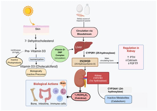
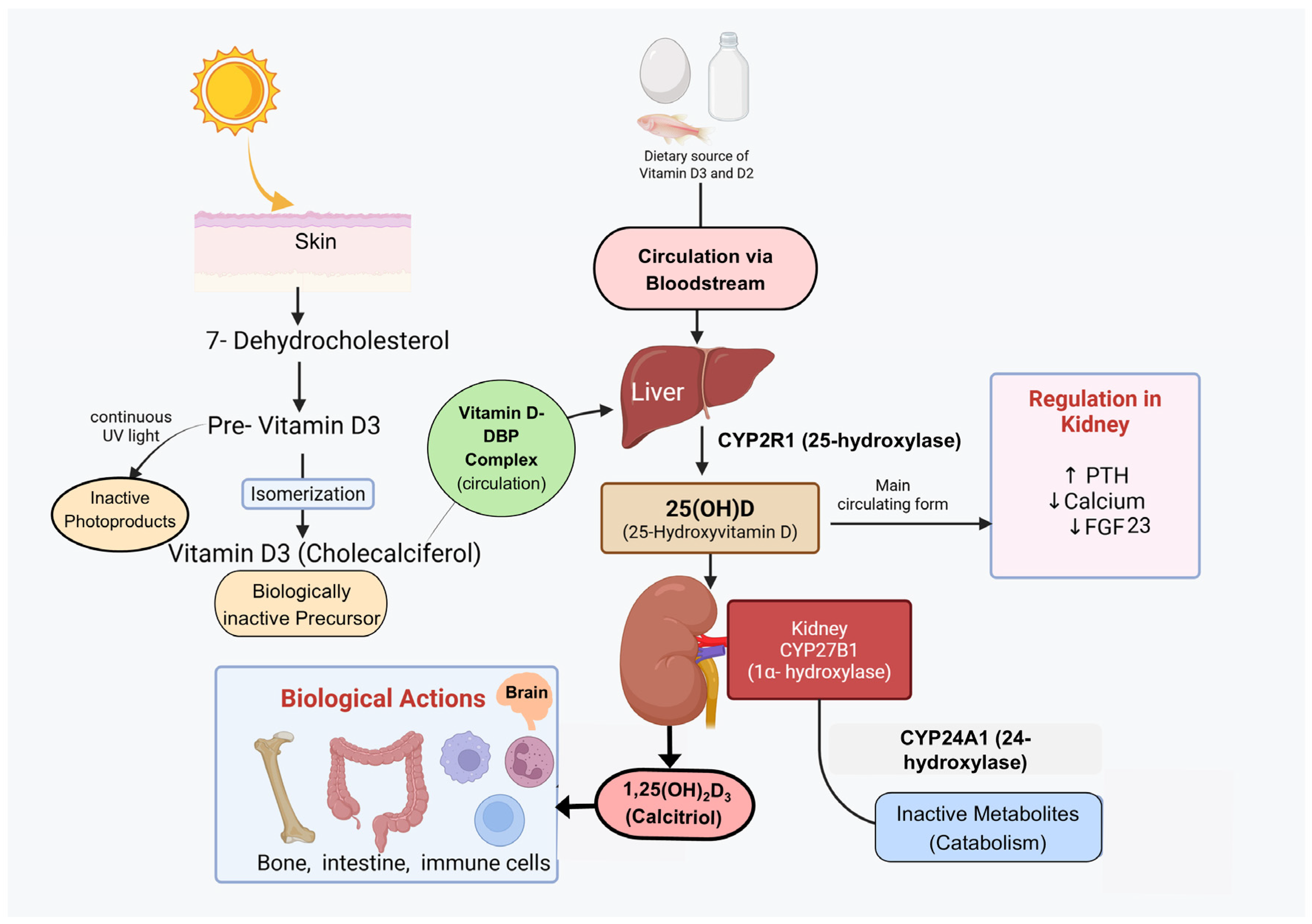
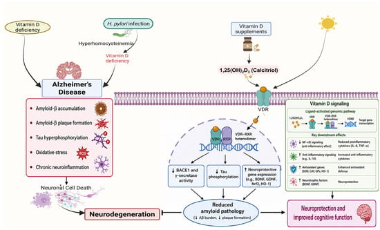
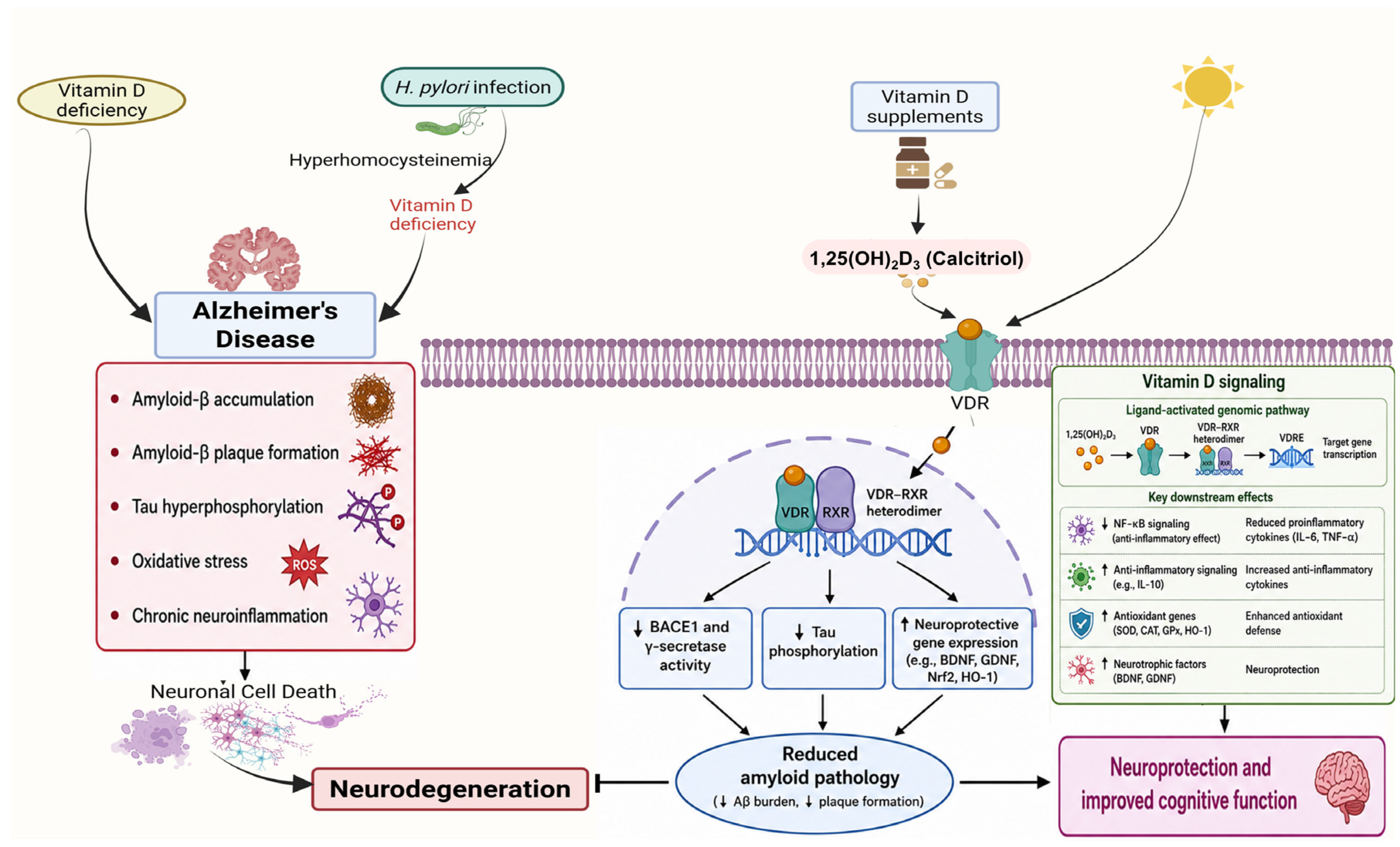
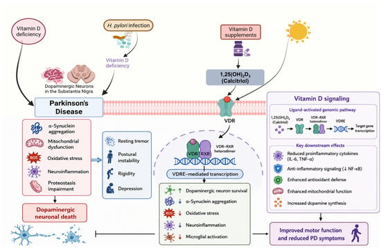
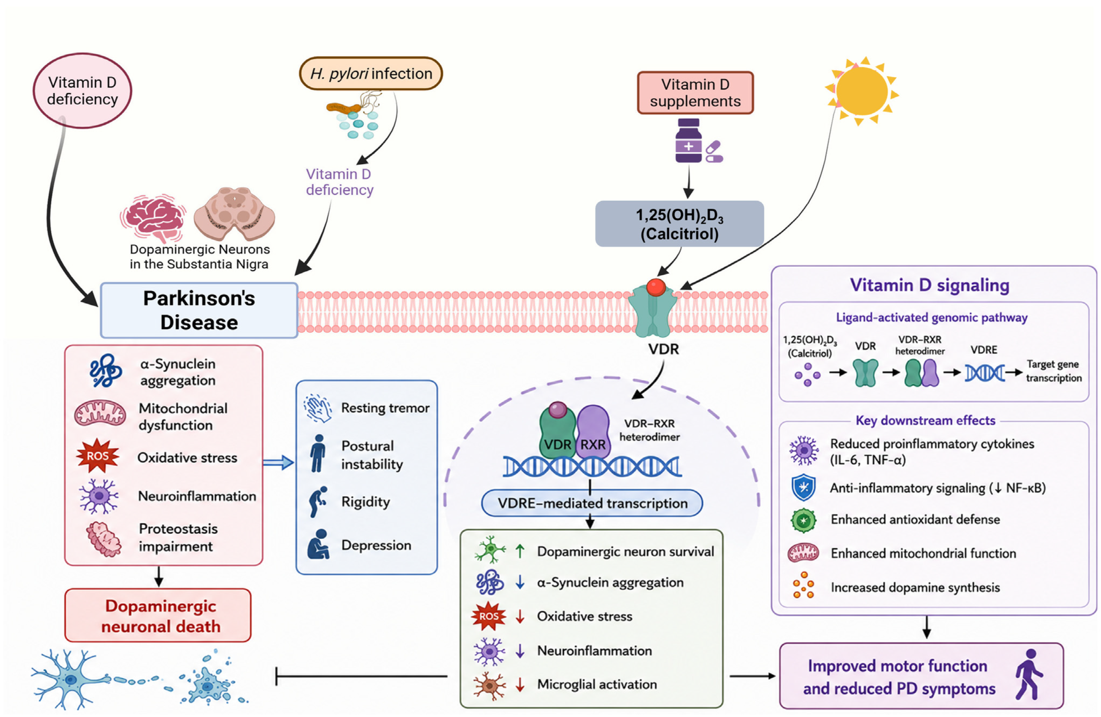
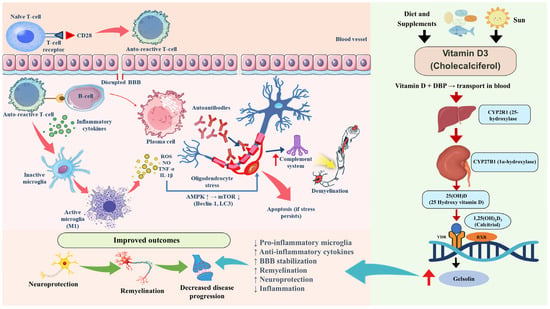
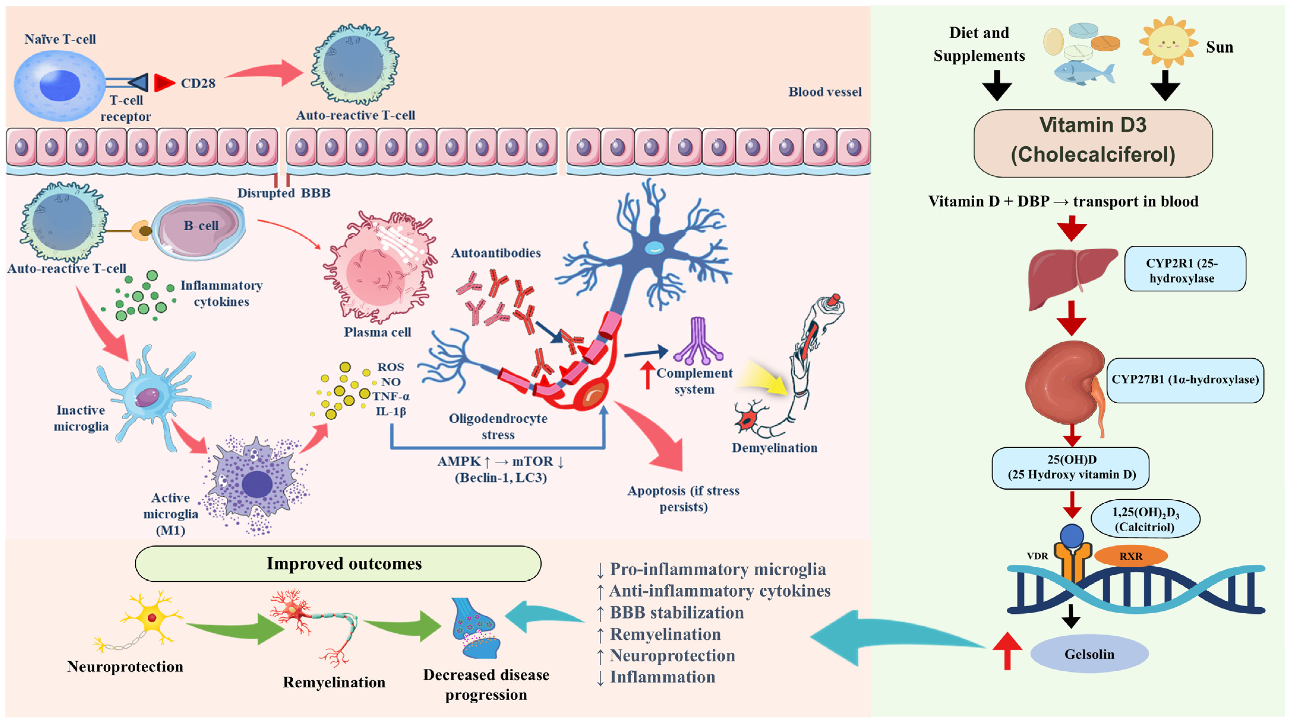
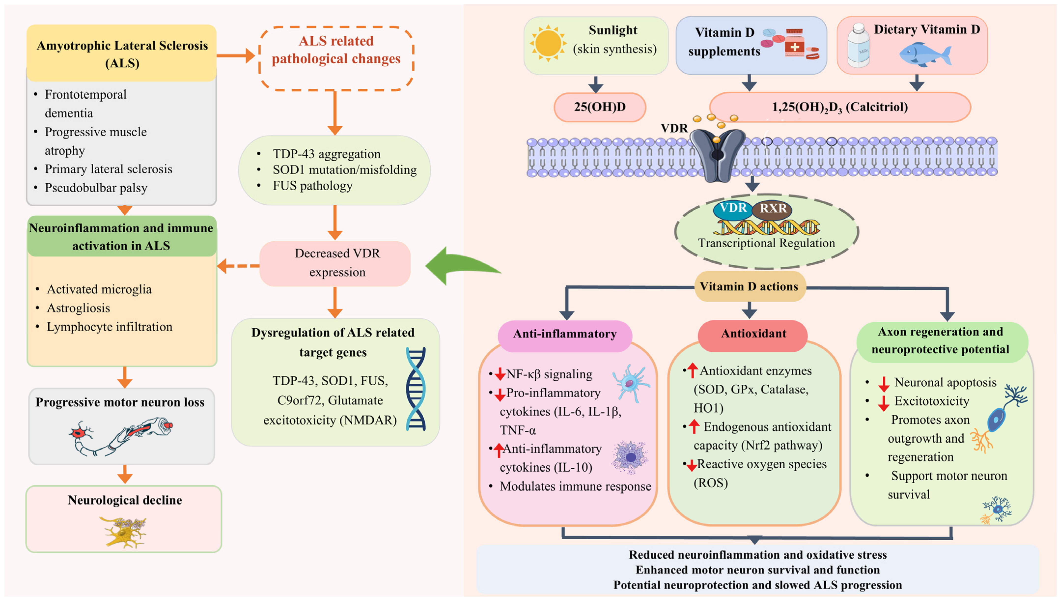
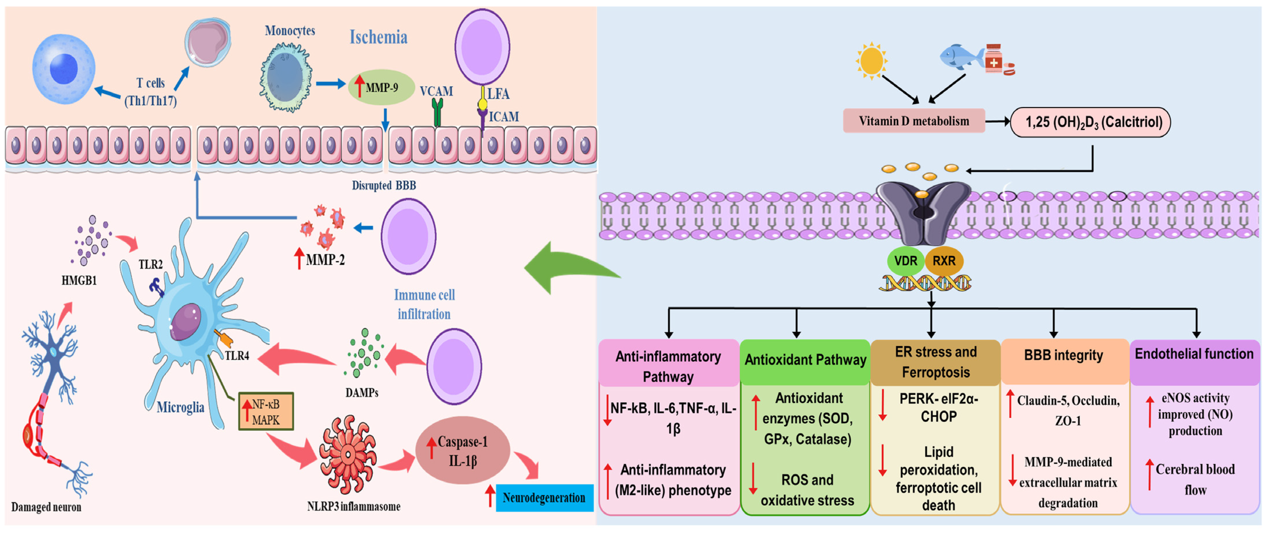

Disclaimer/Publisher’s Note: The statements, opinions and data contained in all publications are solely those of the individual author(s) and contributor(s) and not of MDPI and/or the editor(s). MDPI and/or the editor(s) disclaim responsibility for any injury to people or property resulting from any ideas, methods, instructions or products referred to in the content. |
© 2026 by the authors. Licensee MDPI, Basel, Switzerland. This article is an open access article distributed under the terms and conditions of the Creative Commons Attribution (CC BY) license.