


Abstract
Childhood obesity represents a multifaceted challenge to bone health, influenced by a combination of endocrine, metabolic, and mechanical factors. Excess body fat correlates with an increase in bone mineral density (BMD) yet paradoxically elevates fracture risk due to compromised bone quality and increased mechanical loading on atypical sites. Additionally, subjects with syndromic obesity, as well as individuals with atypical nutritional patterns, including those with eating disorders, show bone fragility through unique genetic and hormonal dysregulations. Emerging evidence underscores the adverse effects of new pharmacological treatments for severe obesity on bone health. Novel drugs, such as glucagon-like peptide-1 (GLP-1) receptor agonists, and bariatric surgery demonstrate potential in achieving weight loss, though limited evidence is available regarding their short- and long-term impacts on skeletal health. This review provides a comprehensive analysis of the mechanisms underlying the impact of childhood obesity on bone health. It critically appraises evidence from in vitro studies, animal models, and clinical research in children with exogenous obesity, syndromic obesity, and eating disorders. It also explores the effects of emerging pharmacological and surgical treatments for severe obesity on skeletal integrity, highlights prevention strategies, and identifies research gaps.
1. Introduction
Obesity has become a significant worldwide concern for public health, with the prevalence of pediatric obesity rising dramatically over recent decades [1]. In Europe, nearly one-third of children are classified as overweight or obese, with severe obesity impacting an estimated 800,000 individuals [1]. This trend represents a significant public health concern, as obesity is closely linked to an increased likelihood of developing chronic illnesses (e.g., type 2 diabetes (T2D), cardiovascular diseases) which can manifest already in childhood [2,3]. Despite the established connections between excess body fat and metabolic and cardiovascular complications, a growing concern associated with obesity is its negative impact on bone health, which can have lasting effects on skeletal development [4]. Understanding the impact of obesity-related pathways on bone is critical to addressing obesity’s broader implications beyond its well-known comorbidities. The common culprit of comorbidities linked to obesity is insulin resistance [5]. The restricted ability of adipose tissue to expand results in an overproduction of free fatty acids that build up in the liver and muscle, impairing insulin signaling by triggering the formation of diacylglycerol and ceramides. This process, coupled with dysregulated adipose tissue lipolysis, further exacerbates insulin resistance, leading to the development of conditions such as fatty liver disease, T2D, and metabolic syndrome. Simultaneously, hyperinsulinemia alters mesenchymal stem cell differentiation, favoring adipogenesis at the expense of osteoblastogenesis, thus reducing bone formation [6]. The connection between obesity and bone health is also influenced by mechanical and inflammatory factors. On one hand, increased body weight imposes mechanical loading that can stimulate cortical bone formation. Conversely, the chronic low-grade inflammation linked to obesity undermines this advantage, negatively affecting bone quality, particularly in trabecular bone-rich areas [7]. This inflammation is characterized by high levels of pro-inflammatory cytokines such as TNF-α and IL-6, which activate the receptor activator of nuclear factor kappa-B ligand (RANKL) [8]. RANKL, produced essentially by osteoblastic lineage cells and immune cells during inflammation, promotes osteoclastogenesis by binding to its receptor, RANK, on osteoclast precursors. Osteoprotegerin (OPG), a decoy receptor for RANKL, regulates this pathway and plays a crucial role in protecting bone. Disruption of the RANKL/RANK/OPG axis has been identified as a contributing factor to bone alterations observed in various congenital and acquired pediatric conditions, including obesity [9,10].
This narrative review aims to explore how excess weight may influence bone health in children with exogenous obesity, syndromic obesity, and eating disorders through evidence from in vitro studies, animal models, and clinical research. In particular, we explored (1) the molecular mechanisms underlying bone remodeling and the clinical and experimental aspects of excess weight as determinants of bone health; (2) the effects of emerging treatments, such as GLP-1 receptor agonists and bariatric surgery (BS); (3) the possible prevention strategies.
2. Material and Methods
2.1. Eligibility Criteria
Manuscripts considered eligible for this review included: i. original published articles and ii. observational or experimental studies.
2.2. Information Sources and Search Strategy
The following keywords were searched in PubMed and ClinicalTrials.gov, covering studies published from January 2000 to December 2024: childhood obesity; bone health; adipokines; inflammation; syndromic obesity; anorexia nervosa; autism spectrum disorders; eating disorders; bariatric surgery; GLP-1 receptor agonists
2.3. Study Selection
Articles were reviewed with regard to the main topics, i.e., pediatric obesity, bone health, in vitro, in vivo, and human studies.
3. In Vitro, In Vivo, and Human Studies
3.1. In Vitro Insights: The Role of Pro-Inflammatory Cytokines in Obesity-Induced Bone Loss
Childhood obesity significantly affects bone metabolism through a complex interplay of inflammatory, endocrine, and cellular mechanisms. Adipose-derived cytokines such as TNF-α, IL-6, lymphotoxin-like inducible protein that competes with glycoprotein D for herpesvirus entry mediator on T cells (LIGHT), and monocyte chemoattractant protein-1 (MCP-1) contribute to a state of persistent low-grade inflammation, which influences bone remodeling by promoting osteoclastogenesis and impairing osteoblastogenesis, ultimately favoring bone resorption and compromising bone health, as highlighted by in vitro studies [4,11] (Figure 1).
Figure 1.
Accumulation of adipose tissue is associated with low-grade chronic inflammation that induces the release of inflammatory factors to activate RANKL-mediated osteoclast differentiation. Abbreviations: IL-1: interleukin 1; IL-6 interleukin 6; MCP1: monocyte chemoattractant protein-1; TNF-α: tumor necrosis factor α; LIGHT: lymphotoxin-like inducible protein that competes with glycoprotein D for herpesvirus entry mediator on T cells; OPG: osteoprotegerin; RANKL: receptor activator of nuclear factor κΒ ligand; TRAF: TNF-receptor-associated factor; NFATc1, nuclear factor of activated T-cells cytoplasmic 1.
Adiponectin and leptin have demonstrated distinct and sometimes opposing roles in both in vivo and in vitro studies. Adiponectin is mainly linked to supporting bone formation and improving osteogenesis by encouraging the proliferation and differentiation of osteoblasts while suppressing osteoclastogenesis. On the other hand, leptin influences bone metabolism through intricate and context-specific mechanisms, engaging both central and peripheral pathways.
At the bone marrow level, obesity-induced changes create an inflammatory microenvironment that drives mesenchymal stem cells (MSCs) toward adipogenesis. Da Silva et al. demonstrated this shift, highlighting its connection to an inflammatory bone marrow microenvironment [12]. Likewise, Cortez et al. reported elevated levels of proinflammatory cytokines, including IL-1, IL-6, and TNF-α, in bone marrow MSCs from two-month-old male Wistar rats subjected to a high-fat diet (HFD) [13]. This pro-inflammatory state enhances osteoclast activity while suppressing osteoblast function, contributing to bone loss and disrupted trabecular architecture [14]. TNF-α has the ability to directly stimulate osteoclast formation from bone marrow macrophages, even when RANKL levels are not elevated [15,16]. This cytokine not only synergizes with low levels of RANKL to amplify osteoclastogenesis but can also induce osteoclast differentiation independently under specific conditions, such as the presence of macrophage-colony stimulating factor (M-CSF) [17]. Moreover, TNF-α stimulates stromal cells to produce M-CSF, which enhances the proliferation and differentiation of osteoclast precursors [11]. Interestingly, TNF-α also plays a role in fracture healing, by facilitating mesenchymal stromal cell recruitment and promoting osteogenic differentiation. While low concentrations (1 ng/mL) enhance osteogenesis, higher levels inhibit these processes, demonstrating a biphasic effect. This highlights the complexity of the role of TNF-α, which depends on local inflammatory conditions and concentration levels [18]. Li et al. discovered that IL-6 is essential in driving MSC senescence and contributing to trabecular bone loss in bone marrow stromal cells in wild-type mice fed an HFD [19]. The treatment with IL-6 antibody reduced the expression of senescence markers (p53 and p21) and decreased senescence-associated β-galactosidase activity. Conversely, the addition of recombinant IL-6 to IL-6 knockout mouse bone marrow stromal cells reversed these effects, inducing senescence via the STAT3/p53/p21 signaling pathway. These results suggest that IL-6 amplifies the senescent characteristics of bone marrow stromal cells, disturbs the equilibrium between osteogenesis and adipogenesis, and hastens bone fragility in mice fed an HFD. Kim et al. demonstrated that MCP-1 induces the formation of osteoclast-like cells that are positive for tartrate-resistant acid phosphatase (TRAP), nuclear factor of activated T-cells, and the calcitonin receptor [20]. However, these cells lack bone-resorbing activity in the absence of RANKL. This indicates that MCP-1 is essential in the initial phases of osteoclast differentiation, facilitating cell fusion and the expression of specific markers, while requiring RANKL to finalize the differentiation into functional, bone-resorbing osteoclasts [20]. On the other hand, MCP-1 has been shown to influence bone metabolism by promoting the differentiation of stromal cells into osteoblasts, enhancing osteoblast proliferation, inhibiting osteoclastogenesis, and mediating these effects through both peripheral mechanisms and hypothalamic pathways [21,22]. Adiponectin has been shown in in vitro studies to promote bone formation and osteogenesis in bone marrow stromal cells and MSCs. This is achieved through pathways such as the Adaptor Protein, Phosphotyrosine Interacting with PH Domain and Leucine Zipper 1 Mitogen-Activated Protein Kinase (APPL1-P38 MAPK) and Adiponectin Receptor 1-P38 MAPK, which enhance the production of Bone Morphogenetic Protein 2, a key osteogenic cytokine [23]. Furthermore, adiponectin stimulates osteoblast proliferation and differentiation, increasing alkaline phosphatase activity, type I collagen, and osteocalcin levels, markers of osteoblastic function, through the AdipoR/c-Jun N-terminal Kinase and AdipoR/P38 pathways [24]. Additionally, in vitro studies demonstrate that adiponectin suppresses osteoclastogenesis and bone resorption through APPL1-mediated suppression of protein kinase B [25].
3.2. In Vivo Models
Leptin has a multifaceted and debated role in bone metabolism in vivo, with its effects varying significantly depending on the context. Research using leptin-deficient mice, such as Ob/Ob models, highlights its complex role, revealing increased trabecular volume and vertebral bone mass in the spine, coupled with a notable decrease in femoral bone mass and a rise in bone marrow fat within the femur [26,27]. Steppan et al. demonstrated that administering leptin to Ob/Ob mice improved skeletal properties, including increased femur length, systemic bone area, BMD, and bone mineral content (BMC) compared to controls [28]. Subsequent research has corroborated leptin’s capacity to enhance bone mass and quality under specific conditions, with central leptin injections in Ob/Ob mice increasing bone formation, BMD, and muscle mass [26]. Similarly, adiponectin influences bone metabolism in a contradictory manner. Adiponectin knockout mice often show reduced bone mineralization and density compared to wild-type mice [29]. However, some studies suggest that adiponectin deficiency protects against trabecular bone loss induced by ovariectomy and can improve bone quality [30,31]. These contrasting roles of leptin and adiponectin highlight their distinct contributions to bone metabolism and their interplay with pathological states like obesity and estrogen deficiency.
Experimental models of obesity, such as those induced by an HFD, have provided further evidence into the impact of obesity on bone metabolism. While HFD-induced obesity is often associated with increased bone mass due to greater mechanical loading, this does not necessarily correlate with improved bone quality. Histomorphometric analyses reveal reduced bone formation, characterized by decreased osteoblast surface, osteoid surface, and osteoid volume, despite an unchanged mineralization process [32]. Supporting this, Ootsuka et al. demonstrated that TNF-α enhances osteoclastogenesis by activating NF-κB and increasing the expression of RANK, RANKL, and colony-stimulating factor-1 receptor in hyperphagic obese rat models [33]. Feng et al. proposed a model in which IL-6 maintains balanced bone remodeling through the modulation of osteoblast and osteoclast activity. In IL-6-deficient mice, there is increased osteoblast activity, leading to enhanced bone formation and immature trabecular structures. However, despite an increased number of osteoclasts, their resorptive activity is impaired due to reduced expression of osteoclastic markers and higher apoptosis rates. The introduction of an HFD compensates for the lack of IL-6 by reducing osteoblast activity, restoring osteoclast function, and suppressing osteoclast apoptosis. This normalization of bone remodeling counteracts the effects of IL-6 deficiency, leading to a more balanced and mature bone structure. The model highlights IL-6’s dual role and the potential of dietary factors to modulate bone metabolism in its absence [34]. HFD-fed mice exhibit decreased trabecular BMD [35], and while overall bone mass may increase, cortical bone quality and mechanical properties are significantly compromised [36]. Further complicating the picture, ketone body metabolism may exacerbate bone resorption in HFD-induced obesity. Increased expression of acetoacetyl-CoA synthetase, stimulated by IL-6 in osteoclasts, enhances ketone utilization, fueling inflammatory and osteoclastic activity [37]. Encouragingly, treatments like combined supplementation with conjugated linoleic acid and calcium have demonstrated positive outcomes in obese mice, mitigating bone loss and increasing the expression of bone formation markers, such as γ-carboxyglutamate-containing protein 2 and collagen α1. This approach also improves metabolic markers and modulates genes related to energy metabolism, insulin, and leptin receptors [38]. Exercise offers another strategy to mitigate obesity-induced bone loss. Studies reveal that moderate-intensity physical activity preserves bone mass, improves BMD, and enhances trabecular microarchitecture. For instance, voluntary wheel running in obese rats counteracts HFD-induced bone deterioration, improving trabecular density and reducing medullary cavity size [39] (Table 1).
Table 1.
In vitro and in vivo studies on the impact of adipose tissue and obesity on bone.
While animal models provide valuable insights into the mechanisms of obesity-induced bone alterations, their relevance to human conditions is limited by several factors. Animal models often exhibit differences in bone architecture, metabolism, and inflammatory responses compared to humans, making direct extrapolation challenging. For instance, the mechanical loading and hormonal environments in rodent models differ significantly from those in children. Furthermore, the rapid skeletal growth in animal models may not fully mimic the gradual skeletal maturation observed in human adolescents. These limitations emphasize the importance of integrating findings from animal models with human studies to ensure clinical relevance.
3.3. The Burden of Exogenous Childhood Obesity on Bone Health: Human Studies
The effect of excess weight on bone health in children and adolescents remains intricate and subject to debate, with studies reporting both beneficial and detrimental effects. Children with obesity experience higher mechanical loads on their bones, resulting in larger bone size, enhanced bone strength, higher vertebral density, increased BMC relative to height, greater stature, and accelerated bone maturation compared to their peers with normal weight [40,41,42,43]. Despite this, obesity leads to a systemic pro-inflammatory state that worsens bone health [44]. This pro-inflammatory state is compounded by nutritional deficiencies, leading to a reduced overall bone mass relative to body weight, decreased total bone density, and lower BMC. These conditions predispose to low-velocity fractures [45,46]. Indeed, a study involving 913,178 patients aged 2 to 19 years reported an increased rate of extremity fractures among overweight, moderately obese, and extremely obese individuals, highlighting a potential decline in bone quality [47]. As mentioned above, the elevated levels of pro-inflammatory cytokines, such as TNF-α, IL-1, IL-6, and LIGHT, associated with obesity promote osteoclastogenesis and impair bone formation. These cytokines stimulate the RANK/RANKL pathway, enhancing bone resorption and contributing to skeletal fragility. Supporting this, Gil-Cosano et al. discovered a strong inverse relationship between IL-6 levels and total body-less head BMC in children with obesity [48]. Erazmus et al. demonstrated that overweight and obese children had lower levels of soluble RANKL compared to normal-weight peers, leading to a higher OPG/RANKL ratio. They also found that children in the lowest quartile of RANKL levels had higher BMI and uric acid. These results suggests that excess fat mass alters the OPG/RANKL system and potentially contributes to cardiometabolic and skeletal health complications [49]. Brunetti et al. emphasize the role of LIGHT in driving spontaneous osteoclastogenesis in peripheral blood mononuclear cells (PBMCs) derived from individuals with obesity. Unlike controls, PBMCs from obese individuals were able to form osteoclasts without the need for external stimulation by RANKL or M-CSF. The addition of anti-LIGHT antibodies reduced osteoclast formation in a manner proportional to the dose, confirming LIGHT’s direct involvement in this process [50]. Elevated leptin levels in subjects affected with obesity further exacerbate bone fragility by negatively correlating with OPG and increasing radial cortical porosity and tibial trabecular thickness [51]. In addition, obese subjects have low adiponectin levels and consequently they have decreased osteoblast activity and bone formation [52,53]. The pro-inflammatory microenvironment also influenced the expression of 11β-hydroxysteroid dehydrogenase type 1 (11β-HSD1) in bone, an enzyme responsible for converting inactive cortisone into active cortisol [54]. 11β-HSD1 is expressed in both osteoblasts and adipocytes and its increased activity may be another mechanism by which obesity leads to detrimental effects on bone health [55]. As described above, insulin resistance also impacts bone metabolism. Studies showed a complex relationship between insulin resistance and OPG. Ugur-Altan et al. observed that reduced OPG levels were linked to elevated HOMA-IR values, showing an inverse relationship between OPG and both fasting insulin and glucose levels [56]. Conversely, Suliburska et al. found higher OPG levels in obese adolescents, positively correlating with insulin resistance [57]. Excess fat mass also influences skeletal maturation. Obese children often exhibit accelerated skeletal maturity and advanced bone age beyond their chronological age [58]. Research conducted by Oh et al. revealed that the accelerated bone age seen in children with obesity was strongly associated with body weight, BMI, and waist circumference percentiles [59]. While this accelerated bone growth initially results in increased bone mass and density, it may predispose children to structural anomalies and fragility in adulthood [60]. Despite these endocrine and developmental alterations, mechanical loading from excess weight initially promotes increased bone mass and density. Clark et al. reported higher BMC and BMD in obese children compared to their normal-weight peers, suggesting a positive effect of fat mass on bone accrual during early life [61]. However, this advantage is counterbalanced by poorer bone quality and a higher incidence of fractures, particularly in the lower extremities, as shown by Kessler et al. and Fornari et al. [47,62] (Table 2).
Table 2.
Human studies on impact of obesity on bone health.
Diets rich in fats and sugary carbonated beverages, but lacking in green leafy vegetables, are commonly linked to obesity and can result in deficiencies of essential micronutrients like vitamin D and calcium [40]. An HFD can interfere with calcium absorption by forming calcium soaps in the intestine, which enhance calcium excretion instead of promoting its absorption [63]. Vitamin D plays a role in regulating leptin and adiponectin levels as well as the production of inflammatory cytokines [64]. Low serum vitamin D levels are frequently observed in children with obesity [65,66]; this is probably due to the sequestration of the liposoluble vitamin D by the subcutaneous fat [66]. Supplementation of vitamin D in obese and overweight subjects may require higher doses than in normal-weight individuals [67,68]. This paradox underscores the fragility underlying increased bone mass in obesity. Engaging in physical activity is crucial for reducing the adverse impact of excess adiposity on bone health. Sedentary lifestyles reduce bone mass accrual and promote tissue absorption by upregulating peroxisome proliferator-activated receptor gamma (PPARγ) in mesenchymal stem cells and RANKL in bone marrow, leading to osteoclast-mediated resorption [69]. Conversely, weight-bearing PA provides mechanical stimuli that promote osteogenesis and inhibits adipogenesis. Behringer’s meta-analysis of 27 studies confirmed that PA, particularly during pubertal growth, enhances BMC accrual [70]. Additionally, irisin, a myokine released during exercise, has been positively associated with BMD in children [71]. These findings emphasize the importance of physical activity in preventing bone fragility and improving skeletal health in children with obesity [72].
4. Bone Health in Syndromic Obesity and Overweight Related to Eating Disorders
4.1. Bone Health in Syndromic Obesity
Syndromic obesity is part of a common spectrum of hypothalamic pathologies characterized by severe early onset obesity caused by dysfunction of the leptin–melanocortin pathway that has a pivotal role in satiety/appetite regulation and in energy expenditure [73]. Syndromic obesity is often associated with malformation, dysmorphic features, and often eating disorders, such as impaired satiety and disruptive food-seeking behavior [73]. Bone health in syndromic obesity is still little studied, but it can be impaired by the same mechanisms described in children with exogenous obesity plus other specific mechanisms (Figure 2).
Figure 2.
The key pathways by which obesity influences bone health in children and adolescents. The left circle (exogenous obesity) highlights factors such as excessive caloric intake, decreased physical activity, chronic low-grade inflammation (e.g., elevated TNF-α, IL-6, LIGHT), altered adipokine profiles (high leptin/low adiponectin), vitamin D deficiency due to sequestration in adipose tissue or calcium deficiency. The top-right circle (neurodevelopmental disorders/eating disorders) emphasizes the roles of restrictive eating (e.g., autism, anorexia nervosa), endocrine dysfunctions (hypogonadotropic hypogonadism, GH resistance, hypercortisolism), and cognitive or behavioral barriers that limit physical activity. The bottom circle (syndromic/monogenic obesity) illustrates genetic disruptions (e.g., leptin–melanocortin pathway), hypogonadism, growth hormone deficiencies, and poor micronutrient intake. Overlapping regions underscore shared pathways—such as inflammation, endocrine alterations, and nutrient deficiencies—demonstrating how these differing etiologies of obesity can converge to compromise bone health. Abbreviations: Ca, calcium; GH, growth hormone; ND, neurodevelopmental; Vit D, vitamin D.
Prader–Willi syndrome (PWS) is the most studied form of syndromic obesity caused by deletion on the paternal chromosome 15q (del15q), maternal uniparental disomy (mUPD), or imprinting defects, with an incidence of about 1/15,000 births. In childhood, individuals with PWS often exhibit normal BMD levels when corrected for their shorter stature [74]. However, during adolescence they may experience a decrease in total BMD and BMC, and in adulthood about 29% to 44% of subjects with PWS may suffer from bone fractures due to the high prevalence of osteoporosis [75]. Possible explanations for the bone impairment include reduced production of sex hormones during puberty and relative growth hormone (GH) deficiency during childhood and adolescence [76]. Additionally, inadequate calcium intake, vitamin D deficiency, limited physical activity, and changes in serum adipokines contribute to disruptions in bone metabolism [77]. Brunetti et al. demonstrated high levels of RANKL and low levels of OPG in both children and adults with PWS compared to sex- and age-matched controls, while sclerostin, an inhibitor of osteoblastogenesis, was found to be higher in PWS children and lower in PWS adults than controls [78]. Consistently with these data, the authors found that BMD was lower in PWS children when compared to healthy controls [78]. Recently, irisin has generated great interest, due to its ability to mimic the effects of physical exercise on bone [71]. Hirsch et al. observed increased levels of salivary irisin in obese individuals with PWS compared to non-obese controls, whereas plasma irisin levels remained similar between the groups [79,80]. Faienza et al. reported that serum irisin levels in individuals with PWS were comparable to those in healthy controls. However, they demonstrate a difference according to genotype underlying the disease [81].
LIGHT levels were found to be significantly higher in both children and adults with PWS than in controls [81]; moreover, serum LIGHT levels negatively correlated with dual-energy X-ray absorptiometry (DXA) parameters.
Alstrom syndrome (ALMS) and Bardet–Biedl syndrome (BBS) are uncommon autosomal recessive disorders, occurring at rates of approximately 1 in 100,000 and 0.7 in 100,000 cases, respectively [82,83]. Both syndromes belong to the category of ciliopathies due to alterations in the molecular structure of the cilium, important for proper functioning of several intracellular signaling pathways [84]. The primary clinical features of both syndromes include early-onset obesity, retinal degeneration progressing to blindness, kidney failure, cardiovascular issues, liver disorders, hypothyroidism, and abnormalities in dental and/or facial development, among others. Patients with BBS frequently exhibit polydactyly, brachydactyly, or syndactyly, as well as intellectual disabilities [82,83]. Bone metabolism can be affected, and osteoporosis is often observed in these subjects even if still little studied [85]. Jeziorni et al. evaluated serum levels of osteocalcin (OC) and urinary deoxypyridinoline (DPD) levels, two bone markers indicating bone formation and bone resorption, respectively, in a cohort of 18 patients (11 with ALMS and 7 with BBS) [86]. They observed reduced levels in the ALMS and BBS groups compared to healthy controls. Additionally, serum OC and urinary DPD values showed a negative correlation with the HOMA-IR index, while a positive correlation was identified between OC and 25-OH vitamin D levels, alongside a negative correlation between RANKL and fasting glucose concentrations [86].
4.2. Bone Health in Eating and Neurodevelopmental Disorders
In children and adolescents with eating disorders like anorexia nervosa (AN), bulimia nervosa (BN), pica, or neurodevelopmental conditions like autism and avoidant/restrictive food intake disorder (ARFID), bone accrual is often compromised. While the link between AN or pica and deficiencies in key nutrients such as calcium and vitamin D is well-established, emerging studies highlight the broader impact of these disorders on bone metabolism and structural skeleton integrity during crucial developmental stages. AN, a psychiatric disorder marked by self-induced undernutrition leading to low body weight, profoundly affects bone health, primarily through hormonal disruptions that impair bone metabolism during critical growth phases. This condition triggers significant hormonal adaptations aimed at survival during chronic energy deficiency, albeit at the expense of skeletal integrity. Adolescents with AN, already experiencing dynamic skeletal development, face substantial long-term risks as these hormonal changes result in reduced BMD and microarchitectural damage. A central factor in bone loss in AN is estrogen deficiency caused by hypogonadotropic hypogonadism. While this condition conserves energy by diverting resources from reproduction during nutritional deficits, the resulting hypoestrogenic state is a major driver of bone mass loss. Estrogen deficiency is marked by heightened bone remodeling activity, elevated markers of bone resorption, and a general reduction in bone mass [87,88]. Furthermore, in AN, the duration of amenorrhea correlates with reductions in spinal and femoral neck BMD [87,89]. Acquired GH resistance represents another critical adaptation that reduces energy expenditure in undernutrition states [90] but also significantly contributes to bone mass loss in AN. GH resistance is marked by elevated GH levels that fail to effectively stimulate the production of insulin-like growth factor 1 (IGF-1) [91]. Persistently low IGF-1 levels in AN impair osteoblast function, further hindering bone accrual during adolescence [92]. Additionally, hyperactivation of the hypothalamic–pituitary–adrenal axis leads to elevated cortisol levels, which adversely affect bone health [93]. Cortisol inhibits calcium absorption in the gut, suppresses osteoblast activity, and accelerates osteoclast-mediated bone resorption [94]. Leptin, an anorexigenic hormone secreted by adipose tissue, is also decreased in individuals with AN [95]. Leptin plays a key role in connecting nutrient availability to hypogonadotropic hypogonadism. In women experiencing functional hypothalamic amenorrhea, treatment with recombinant human leptin has been shown to increase LH pulse frequency within two weeks and enhance menstruation recovery rates compared to a placebo [96]. Additionally, leptin therapy raises osteocalcin and bone-specific alkaline phosphatase levels, markers of bone formation [97], and shows a positive association with BMD and bone microarchitecture in AN [98]. However, leptin treatment has notable side effects, including significant weight loss likely due to appetite suppression [99]. The skeletal effects of these hormonal imbalances are evident in the compromised bone microarchitecture of individuals with AN. Approximately 90% of women with AN exhibit BMD values more than 1 SD below the mean for their age [100,101]. Adolescents with AN display reduced cortical thickness, increased porosity, and impaired trabecular connectivity, structural deficits that persist into adulthood and significantly elevate fracture risks [102,103]. Weight recovery remains the cornerstone of treatment and often leads to notable improvements in BMD [104]. However, even with recovery, bone restoration is frequently incomplete, underscoring the irreversible skeletal damage caused during periods of malnutrition [105].
While substantial evidence links AN to impaired bone health through reductions in BMD and BMC, the link between BN and impaired bone health remains less well-documented, with emerging research providing important insights. Studies indicate that BN patients, particularly those with a previous history of AN, exhibit lower BMD, notably at the spine and hip, compared to healthy controls [106]. This indicates that the lasting impact of previous episodes of AN plays a major role in weakening bone health in individuals with BN. Additionally, factors such as low BMI, reduced fat and lean mass, and menstrual irregularities, including amenorrhea, are identified as predictors of reduced BMD in BN. Other studies [107] suggest that purging behaviors and chronic nutritional deficits in BN may further exacerbate risks to bone health. While the connection between BN alone and skeletal compromise remains less clear, the overlap of risk factors with AN underscores the importance of monitoring bone health in BN patients. ARFID also significantly affects growth and bone health during the various stages of development. Research indicates that children and adolescents with ARFID exhibit lower BMD and shorter stature compared to healthy peers, a trend observed across various studies [108,109]. The dietary constraints inherent to ARFID often result in malnutrition, marked by deficiencies in energy and critical nutrients such as calcium, vitamin D, and proteins, which are essential for bone growth and development [110]. Dinkler and colleagues investigated the prevalence and features of ARFID by analyzing data from the Japan Environment and Children’s Study, a large-scale national birth cohort, which included 6633 children aged 4 to 7 years [108]. The study found that children with ARFID were generally shorter than their peers without the disorder. Height differences were also observed among ARFID subtypes, with those classified as A1, A2, or A3—characterized by low body weight, nutritional deficiencies, or reliance on nutritional supplementation—being shorter compared to those in subtype A4, which involves psychosocial impairments without documented nutritional deficiencies. These findings highlight that ARFID’s impact on height varies by subtype, with the greatest deficits seen in subtypes A1–A3. Studies also reveal that the bone deficits in ARFID are comparable to those observed in anorexia nervosa. For instance, research on children and adolescents with ARFID shows that lumbar spine BMD Z-scores are significantly lower and align closely with those of patients diagnosed with AN [109]. Moreover, as in AN, early endocrine disruptions in ARFID, such as altered levels of GH and IGF-1, further compound its effects on bone accrual and overall growth [111]. Pica, characterized by the ongoing consumption of non-food items or raw substances for a minimum duration of one month, has historically been linked to anemia and deficiencies in micronutrients like iron and zinc [112]. While these relationships are well-documented, recent studies explored the association between pica and nutrients critical for bone metabolism, including vitamin D and calcium. Emerging evidence highlights that individuals with pica, particularly adolescents, may be at increased risk of deficiencies in these nutrients, which are essential for proper skeletal development.
A study conducted in northern Sudan involving 344 adolescents demonstrated a significant association between pica symptoms and vitamin D deficiency. Adolescents with pica had median serum vitamin D levels of 17.8 ng/mL, significantly lower than the 21.2 ng/mL observed in their peers without pica (p = 0.020). Notably, pica symptoms emerged as an independent predictor of lower vitamin D levels, even after adjusting for confounding factors such as age, sex, and BMI, emphasizing the condition’s impact on vitamin D status [113]. In addition, case series have consistently reported calcium deficiencies in individuals with pica. These deficits are often attributed to the ingestion of non-food substances, such as clay or starch, which bind to calcium in the gastrointestinal tract, reducing its bioavailability. Such deficiencies are particularly concerning in children and adolescents, where calcium is critical for bone mineralization and achieving peak bone mass [114]. Autism spectrum disorder (ASD) is a neurodevelopmental condition that notably affects bone health, especially in children and adolescents. Recent studies indicate that individuals with ASD face challenges in achieving optimal bone development due to various factors, such as nutrient deficiencies, restrictive diets, and reduced levels of physical activity. Dietary selectivity, characterized by a significantly restricted range of food choices and commonly observed in children with ASD [115], contributes to inadequate intake of essential nutrients. Decreased intake of calcium and vitamin D appears to be one of the most consistent findings in children with ASD [116]. This deficiency is further exacerbated by the use of restrictive diets, such as gluten-free and casein-free regimens, which are occasionally suggested for their potential link to symptom reduction [117] but often lack sufficient calcium content. Hediger et al. investigated bone development in 75 boys aged 4–8 years with ASD, focusing on second metacarpal bone cortical thickness. Their findings revealed that although bone cortical thickness increased with age, a notable and progressive divergence from standard reference values began to emerge at the age of six. By age eight, boys with ASD showed an average 25–30% deficit in bone cortical thickness compared to reference medians. Importantly, boys adhering to casein-free diets exhibited nearly double the bone cortical thickness deficits (−18.9%) compared to boys on unrestricted diets (−10.5%). Despite these diets’ potential adverse impact, even boys on unrestricted diets had significantly negative deviations [118]. Neumeyer et al. demonstrated that BMD is significantly lower in peripubertal boys with ASD compared to controls. In an early study utilizing DXA to evaluate 18 boys with ASD and 19 controls aged 8–14 years, researchers observed lower BMD Z-scores at the spine, hip, and femoral neck in boys with ASD. Notably, even after adjusting for maturity and BMI, the differences in BMD at the hip and femoral neck remained, indicating that factors beyond growth and body composition play a role in these deficiencies. The study also highlighted reduced vitamin D intake from both dietary sources and serum levels, alongside lower exercise activity among boys with ASD [119]. In a subsequent study, the same group expanded on these findings by evaluating 25 boys with ASD and 24 typically developing controls, aged 8–17 years. They verified that boys with ASD exhibited BMD Z-scores that were 0.7 to 1.2 standard deviations lower at the lumbar spine, femoral neck, and total hip and in the whole body (excluding the head). Additionally, this research identified key dietary and physical activity differences: boys with ASD consumed 16% fewer calories, with a higher proportion of carbohydrates and significantly less animal protein (37% less) and fat (20% less) than controls. Calcium and phosphorus intake were also lower in the ASD group. Interestingly, just 27% of boys with ASD were categorized as “very physically active”, in contrast to 79% of their typically developing peers [120].
5. Prevention at the Core: Strategies for Childhood Obesity and Bone Health
Addressing the complex interplay between childhood obesity and bone health requires a multifaceted approach that encompasses a range of strategies and interventions. This includes early prevention efforts, targeted dietary recommendations, and promotion of physical activity [121]. Growing evidence suggests that the prevention of childhood obesity begins in the preconception period and pregnancy, providing an opportunity to shape the future health outcomes of both mothers and their newborns [122]. Preventive strategies may be further implemented during the breastfeeding period [123]. Human milk’s composition and properties are well-known protective factors [124], and lower-protein formulas are associated with a lower risk of obesity at 6 years [125]. During the postweaning period, diets low in carbohydrates (10–30% of total caloric intake) and low in fat (18–40% of total caloric intake) have demonstrated short-term effectiveness. These dietary approaches are thought to enhance satiety, leading to a reduction in overall caloric consumption [126]. However, adhering to these dietary practices can be particularly challenging in childhood and adolescence. The Mediterranean diet proves to be an effective compromise between acceptability and adequate nutritional intake [127], It is recommended as a protective dietary approach in the most recent guidelines issued by the Italian Society of Pediatric Endocrinology and Diabetology [128]. It has been demonstrated that adherence to the Mediterranean diet is associated with higher levels of circulating vitamin D, a nutrient particularly deficient in obese children and adolescents, due to the significant impact of fat accumulation on its metabolism [129,130]. An inverse association between circulating levels of 25-OH vitamin D and body composition has been observed [131]. Vitamin D deficiency is largely attributed to its lipophilic nature which causes the sequestration in adipose tissue, rendering it unavailable for biological functions [132]. Additionally, obesity is linked to reduced expression of genes that regulate the enzymes 25-hydroxylase and 1α-hydroxylase, both of which are critical for vitamin D metabolism. This leads to compromised production of 25-OH vitamin D in the liver and adipose tissue, resulting in lower serum vitamin D levels [133]. In addition, limited sun exposure, common in sedentary lifestyles associated with obesity, further reduces natural vitamin D synthesis [134]. Impaired renal function and increased vitamin D catabolism due to liver steatosis also contribute to lower 25-OH vitamin D levels, as demonstrated in a meta-analysis examining children with non-alcoholic fatty liver disease [135]. The effects of vitamin D deficiency go beyond its impact on bone health. Poor vitamin D status in obese children aggravates their metabolic profiles, increasing susceptibility to impaired glucose metabolism [130]. Vitamin D plays a pivotal role in insulin secretion and sensitivity, as well as systemic inflammatory response. It directly binds to vitamin D receptors in pancreatic β-cells to enhance insulin release and indirectly raises plasma calcium levels, promoting calcium-dependent insulin secretion [136,137]. The Italian Pediatric Society advises seasonal vitamin D supplementation for children and adolescents with limited sun exposure during the summer months, specifically from late autumn to early spring (November–April) or with specific risk factors, including obesity [138]. However, the effects of vitamin D supplementation on bone health in obese children and adolescents have yielded mixed findings. A recent meta-analysis evaluating the effectiveness of vitamin D supplementation in obese children and adolescents revealed that high doses of vitamin D benefit cardiovascular metabolism and improve insulin resistance; however, no significant impact on bone health was observed [133]. The absence of direct effects on bone health observed in the meta-analysis may be attributed to the unique characteristics of bone formation and growth during adolescence, a period of enhanced skeletal activity. Unlike in adults, parathyroid hormone (PTH) activity in adolescents is influenced by complex physiological mechanisms that are not necessarily tied to bone metabolism. Elevated PTH levels in this age group may reflect these intricate processes, which appear relatively independent of vitamin D supplementation [139]. On the other hand, a recent study conducted by Wang et al. investigated the effects of vitamin D3 supplementation on bone mass in obese pediatric patients. They found that vitamin D3 supplementation improved lipid profiles, glucose metabolism, as well as bone mass development in obese children [140].
Physical activity is essential for bone health [141]. Muscle and bone interact anatomically, mechanically, and via paracrine and endocrine signals [142], mediated by muscle-secreted factors like myokines [143]. Among these factors, irisin is crucial for bone metabolism, as it facilitates the differentiation of bone marrow stromal cells into mature osteoblasts [144]. Achieving an osteogenic response requires deformation of bones beyond their usual thresholds. However, there is limited evidence to determine which sport is most effective for obese children in simultaneously achieving weight loss and promoting bone modeling [141]. However, for children with obesity, joint pain, reduced mobility, and low self-esteem can be barriers to such activities [145].
Starting with low-impact exercises, like swimming or resistance training, helps promote bone health while reducing strain. Gradual progression to more intensive activities can improve fitness and resilience. Incorporating daily movement, such as walking to school or active family outings, and structured, enjoyable programs tailored to individual abilities can encourage consistent participation [146]. The findings underscore the importance of prevention that should begin as early as the prenatal stage, where maternal nutrition and vitamin D levels play a pivotal role in shaping the skeletal development of offspring. Extending into early childhood, breastfeeding and the adoption of balanced dietary practices, such as the Mediterranean diet, are essential to prevent excessive weight gain and for supporting bone mineralization. In children and adolescents already affected by obesity, targeted nutritional strategies are indispensable and supplementation of vitamin D should be considered. Diet must be accompanied by physical activity, the cornerstone of any intervention aimed at improving bone health in obese children and adolescents. Exercise not only enhances bone strength through mechanical loading but also counters sedentary behaviors that exacerbate skeletal fragility (Figure 3).
Figure 3.
Promoting bone health involves a comprehensive approach starting from the preconception period and pregnancy, where maternal diet and lifestyle influence future health outcomes for both mother and child. Breastfeeding plays a key protective role, supported by evidence linking human milk’s composition to better weight regulation and bone health in early childhood. Vitamin D supplementation is crucial, particularly for children and adolescents with obesity, who are at higher risk of vitamin D deficiency due to reduced sun exposure, dietary insufficiency, and metabolic alterations. The Mediterranean diet is highlighted as an effective and sustainable dietary pattern, promoting higher vitamin D levels and providing essential nutrients such as calcium and phosphorus, necessary for bone mineralization. Physical activity, particularly high-impact exercises during puberty, enhances bone strength through mechanical stress and improves overall fitness. Aerobic activities during childhood, combined with resistance training in adolescence, play a significant role in fostering healthy bone development while addressing risk factors such as obesity and sedentary behavior.
6. Impact of Severe Obesity Drug Treatments and Bariatric Surgery on Bone Health
Glucagon-like peptide 1 receptor agonists (GLP-1 RAs) and bariatric surgery represent new possibilities for the treatment of severe obesity [147].
GLP-1 RAs, including liraglutide, exenatide, dulaglutide, and semaglutide, reduce hunger by slowing gastric emptying and targeting the central nervous system [148,149,150,151,152].
Clinical trials on the effects of anti-obesity drugs on bone health are reported in Table 3.
Liraglutide is currently the sole GLP-1 receptor agonist approved in Europe for managing pediatric obesity in children and adolescents aged 12 years and above, with a BMI of 30 kg/m2 or greater, or 27 kg/m2 or greater if accompanied by comorbidities such as T2D or metabolic syndrome [153]. Liraglutide is given via subcutaneous injection, beginning at a dose of 0.6 mg/day and gradually increasing to a maximum of 3.0 mg/day. Several studies have highlighted the effects of GLP-1 on bone metabolism [154,155,156,157,158]. The administration of GLP-1 or its analog, exendin-4, was found to increase BMD and promote the expression of osteoblast markers. These effects were mediated by mechanisms such as activation of the Wnt pathway and an elevated OPG/RANKL ratio in normal, diabetic, and hyperlipidemic rats [154,155]. Exendin-4 also increases BMD by suppressing SOST/sclerostin expression in MLO-Y4 cells, resulting in elevated serum osteocalcin levels, reduced serum sclerostin levels, and increased femoral BMD in T2D rats [156]. In a study by Iepsen et al., involving 37 obese women, it was observed that treatment with long-acting liraglutide over 52 weeks resulted in a 16% increase in bone formation and helped prevent bone loss following weight reduction through a low-calorie diet [158]. Recently, a randomized clinical trial analyzed the effect of a 1-year intervention with either liraglutide (3.0 mg/day), exercise, or the combination of both on BMD in obese subjects undergoing weight loss compared with placebo [158].
The combination of liraglutide and exercise resulted in the most significant reductions in weight and body fat, while preserving hip, spine, and forearm BMD compared to the placebo group. In contrast, liraglutide alone caused a decrease in hip and spine BMD compared to both the placebo and exercise-only groups. These findings underscore the crucial role of physical activity in counteracting the negative impact of liraglutide on bone health during weight loss interventions [158].
The use of bariatric surgery (BS) has shown promising results in severe obese adolescents, with evidence of long-term efficacy and good safety [159]. Laparoscopic sleeve gastrectomy (SG) and Roux-en-Y gastric bypass (RYGB) are the most frequently performed procedures in adolescents. The maintenance of weight achieved after surgery is satisfactory from 3 years up to 12 years postsurgery [160,161]. Moreover, bariatric surgery’s metabolic impact is widely acknowledged, with rates of T2D remission reaching up to 86% in adolescents compared to 53% in adults [162]. The impact of BS on the skeleton during a crucial phase of bone growth and development continues to be a major concern. BS has been associated with reductions in BMD across various skeletal sites. Mitra et al., in a systematic review and meta-analysis, revealed that RYGB and sleeve gastrectomy SG were associated with substantial reductions in lumbar BMD Z-scores [163]. These procedures also led to elevated markers of bone resorption, such as C-terminal telopeptide, indicating intensified bone turnover. These data highlight the importance of pharmacological treatments like GLP-1 receptor agonists as a promising avenue for weight management but require careful consideration of their effects on skeletal health. Combining these treatments with structured physical activity may prevent the reductions in bone density observed during weight loss. Similarly, while bariatric surgery offers substantial weight reduction benefits, its long-term skeletal risks necessitate a rigorous postoperative regimen of supplementation, dietary management, and physical activity.
Table 3.
Clinical trials on the effects of anti-obesity drugs on bone health.
7. Conclusions
The intricate relationship between childhood obesity and bone health reflects a balance between mechanical loading benefits and the detrimental effects of chronic inflammation, suboptimal nutrition, and altered endocrine signals. Although increased body weight can initially yield higher bone mass, this apparent advantage is undermined by poor trabecular architecture, micronutrient deficiencies, and inflammatory pathways that favor bone resorption. Syndromic forms of obesity and excess weight due to eating disorders, including neurodevelopmental conditions such as autism, add further complexity, often compounding bone fragility through hormonal dysregulations, insufficient vitamin intake, and reduced physical activity. Innovations in treatment of severe obesity, ranging from GLP-1 RAs to BS, underscore both the promise and the inherent risks to skeletal integrity. Pharmacological agents may offer balanced reductions in adiposity and preservation of bone mass if integrated with exercise regimens. BS can achieve dramatic weight loss and metabolic improvements but frequently leads to bone demineralization and heightened fracture risk, underscoring the need for structured follow-up and targeted supplementation.
Prevention through the promotion of healthy lifestyles, such as a diet based on the principles of the Mediterranean diet and the practice of regular physical activity, remains the main objective to be pursued in order to preserve bone health. Future research must prioritize the development of longitudinal studies to better understand the short- and long-term effects of pharmacological therapies and BS on skeletal health. These studies should focus on identifying critical windows for intervention and defining strategies to mitigate bone fragility. Innovations in personalized medicine hold promise for integrating individualized nutrition plans, tailored exercise programs, and optimized vitamin supplementation. Additionally, exploring molecular pathways involved in obesity-induced bone alterations, such as the role of adipokines and inflammatory mediators, could uncover novel therapeutic targets. Advanced imaging technologies and biomarkers should be leveraged to monitor changes in bone quality more accurately over time. A multidisciplinary effort is essential, bringing together pediatricians, endocrinologists, nutritionists, and exercise specialists to create holistic, sustainable strategies that address the dual challenges of obesity and bone health. These advancements will ultimately contribute to reducing the burden of obesity-related skeletal complications and improving long-term outcomes for children and adolescents.
Author Contributions
Conceptualization, I.F., G.D., and M.F.F.; methodology, I.F., M.C., and M.F.F.; resources, G.D. and M.F.F.; writing—original draft preparation, I.F., M.C., and R.V.; writing—review and editing, I.F., M.C., R.V., and M.F.F.; supervision, G.D. and M.F.F.; project administration, M.F.F. All authors have read and agreed to the published version of the manuscript.
Funding
This research received no external funding.
Conflicts of Interest
The authors declare no conflicts of interest.
Abbreviations
The following abbreviations are used in this manuscript:
| ALP | Alkaline Phosphatase |
| AN | Anorexia Nervosa |
| APPL1 | Adaptor Protein, Phosphotyrosine Interacting with PH Domain and Leucine Zipper1 |
| ARFID | Avoidant/Restrictive Food Intake Disorder |
| ASD | Autism Spectrum Disorder |
| BMC | Bone Mineral Content |
| BMD | Bone Mineral Density |
| BMSC | Bone Mesenchymal Stem Cell |
| BMI | Body Mass Index |
| BN | Bulimia Nervosa |
| Ca | Calcium |
| CTX | C-terminal Telopeptide |
| DXA | Dual-Energy X-ray Absorptiometry |
| DPD | Deoxypyridinoline |
| GH | Growth Hormone |
| GLP-1 | Glucagon-like Peptide-1 |
| GLP-1R | Glucagon-like Peptide-1 Receptor |
| HFD | High-Fat Diet |
| HOMA-IR | Homeostatic Model Assessment of Insulin Resistance |
| IGF-1 | Insulin-like Growth Factor 1 |
| IL-6 | Interleukin-6 |
| LIGHT | Lymphotoxin-like Inducible Protein that Competes with Glycoprotein D for Herpesvirus Entry Mediator on T Cells |
| MAPK | Mitogen-Activated Protein Kinase |
| MBS | Metabolic and Bariatric Surgery |
| MCP-1 | Monocyte Chemoattractant Protein-1 |
| N/A | Not Available |
| OC | Osteocalcin |
| OPG | Osteoprotegerin |
| PA | Physical Activity |
| P1NP | Procollagen Type 1 N-terminal Propeptide |
| Pica | Persistent Ingestion of Non-food Substances |
| PPARγ | Peroxisome Proliferator-Activated Receptor Gamma |
| PTH | Parathyroid Hormone |
| QUS | Quantitative Ultrasound |
| RANK | Receptor Activator of Nuclear Factor Kappa-B |
| RANKL | Receptor Activator of Nuclear Factor Kappa-B Ligand |
| RYGB | Roux-en-Y Gastric Bypass |
| SG | Sleeve Gastrectomy |
| STAT3 | Signal Transducer and Activator of Transcription 3 |
| T2DM | Type 2 Diabetes Mellitus |
| TGFβ | Transforming Growth Factor-beta |
| TNF-α | Tumor Necrosis Factor-alpha |
| TRAcP5b | Tartrate-resistant Acid Phosphatase 5b |
| WHO | World Health Organization |
References
- World Health Organization. Mapping the Health System Response to Childhood Obesity in the WHO European Region: An Overview and Country Perspectives; World Health Organization: Geneva, Switzerland, 2022; Available online: https://apps.who.int/iris/handle/10665/353747 (accessed on 11 January 2025).
- Ciężki, S.; Odyjewska, E.; Bossowski, A.; Głowińska-Olszewska, B. Not Only Metabolic Complications of Childhood Obesity. Nutrients 2024, 16, 539. [Google Scholar] [CrossRef] [Scilit] [PubMed]
- Faienza, M.F.; Santoro, N.; Lauciello, R.; Calabrò, R.; Giordani, L.; Di Salvo, G.; Ventura, A.; Delvecchio, M.; Perrone, L.; Del Giudice, E.M.; et al. IGF2 Gene Variants and Risk of Hypertension in Obese Children and Adolescents. Pediatr. Res. 2010, 67, 340–344. [Google Scholar] [CrossRef] [Scilit] [PubMed]
- Fintini, D.; Cianfarani, S.; Cofini, M.; Andreoletti, A.; Ubertini, G.M.; Cappa, M.; Manco, M. The Bones of Children With Obesity. Front. Endocrinol. 2020, 11, 200. [Google Scholar] [CrossRef] [Scilit] [PubMed]
- Miniello, V.L.; Faienza, M.F.; Scicchitano, P.; Cortese, F.; Gesualdo, M.; Zito, A.; Basile, M.; Recchia, P.; Leogrande, D.; Viola, D.; et al. Insulin Resistance and Endothelial Function in Children and Adolescents. Int. J. Cardiol. 2014, 174, 343–347. [Google Scholar] [CrossRef] [Scilit] [PubMed]
- Han, L.; Wang, B.; Wang, R.; Gong, S.; Chen, G.; Xu, W. The Shift in the Balance between Osteoblastogenesis and Adipogenesis of Mesenchymal Stem Cells Mediated by Glucocorticoid Receptor. Stem. Cell Res. Ther. 2019, 10, 377. [Google Scholar] [CrossRef] [Scilit] [PubMed]
- Rinonapoli, G.; Pace, V.; Ruggiero, C.; Ceccarini, P.; Bisaccia, M.; Meccariello, L.; Caraffa, A. Obesity and Bone: A Complex Relationship. Int. J. Mol. Sci. 2021, 22, 13662. [Google Scholar] [CrossRef] [Scilit] [PubMed]
- Zhang, Y.-H.; Heulsmann, A.; Tondravi, M.M.; Mukherjee, A.; Abu-Amer, Y. Tumor Necrosis Factor-α (TNF) Stimulates RANKL-Induced Osteoclastogenesis via Coupling of TNF Type 1 Receptor and RANK Signaling Pathways. J. Biol. Chem. 2001, 276, 563–568. [Google Scholar] [CrossRef] [Scilit] [PubMed]
- Brunetti, G.; D’Amato, G.; Chiarito, M.; Tullo, A.; Colaianni, G.; Colucci, S.; Grano, M.; Faienza, M.F. An Update on the Role of RANKL–RANK/Osteoprotegerin and WNT-ß-Catenin Signaling Pathways in Pediatric Diseases. World J. Pediatr. 2019, 15, 4–11. [Google Scholar] [CrossRef] [Scilit] [PubMed]
- Faienza, M.F.; Ventura, A.; Colucci, S.; Cavallo, L.; Grano, M.; Brunetti, G. Bone Fragility in Turner Syndrome: Mechanisms and Prevention Strategies. Front. Endocrinol. 2016, 7, 34. [Google Scholar] [CrossRef] [Scilit]
- Kitaura, H.; Kimura, K.; Ishida, M.; Kohara, H.; Yoshimatsu, M.; Takano-Yamamoto, T. Immunological Reaction in TNF-α-Mediated Osteoclast Formation and Bone Resorption In Vitro and In Vivo. Clin. Dev. Immunol. 2013, 2013, 181849. [Google Scholar] [CrossRef] [Scilit] [PubMed]
- Da Silva, S.V.; Renovato-Martins, M.; Ribeiro-Pereira, C.; Citelli, M.; Barja-Fidalgo, C. Obesity Modifies Bone Marrow Microenvironment and Directs Bone Marrow Mesenchymal Cells to Adipogenesis. Obesity 2016, 24, 2522–2532. [Google Scholar] [CrossRef] [Scilit]
- Cortez, M.; Carmo, L.S.; Rogero, M.M.; Borelli, P.; Fock, R.A. A High-Fat Diet Increases IL-1, IL-6, and TNF-α Production by Increasing NF-κB and Attenuating PPAR-γ Expression in Bone Marrow Mesenchymal Stem Cells. Inflammation 2013, 36, 379–386. [Google Scholar] [CrossRef] [Scilit] [PubMed]
- Patsch, J.M.; Kiefer, F.W.; Varga, P.; Pail, P.; Rauner, M.; Stupphann, D.; Resch, H.; Moser, D.; Zysset, P.K.; Stulnig, T.M.; et al. Increased Bone Resorption and Impaired Bone Microarchitecture in Short-Term and Extended High-Fat Diet–Induced Obesity. Metabolism 2011, 60, 243–249. [Google Scholar] [CrossRef] [Scilit] [PubMed]
- Lam, J.; Takeshita, S.; Barker, J.E.; Kanagawa, O.; Ross, F.P.; Teitelbaum, S.L. TNF-α Induces Osteoclastogenesis by Direct Stimulation of Macrophages Exposed to Permissive Levels of RANK Ligand. J. Clin. Investig. 2000, 106, 1481–1488. [Google Scholar] [CrossRef] [Scilit] [PubMed]
- Azuma, Y.; Kaji, K.; Katogi, R.; Takeshita, S.; Kudo, A. Tumor Necrosis Factor-α Induces Differentiation of and Bone Resorption by Osteoclasts. J. Biol. Chem. 2000, 275, 4858–4864. [Google Scholar] [CrossRef] [Scilit] [PubMed]
- Kim, N.; Kadono, Y.; Takami, M.; Lee, J.; Lee, S.-H.; Okada, F.; Kim, J.H.; Kobayashi, T.; Odgren, P.R.; Nakano, H.; et al. Osteoclast Differentiation Independent of the TRANCE–RANK–TRAF6 Axis. J. Exp. Med. 2005, 202, 589–595. [Google Scholar] [CrossRef] [Scilit] [PubMed]
- Glass, G.E.; Chan, J.K.; Freidin, A.; Feldmann, M.; Horwood, N.J.; Nanchahal, J. TNF-α Promotes Fracture Repair by Augmenting the Recruitment and Differentiation of Muscle-Derived Stromal Cells. Proc. Natl. Acad. Sci. USA 2011, 108, 1585–1590. [Google Scholar] [CrossRef] [Scilit]
- Li, Y.; Lu, L.; Xie, Y.; Chen, X.; Tian, L.; Liang, Y.; Li, H.; Zhang, J.; Liu, Y.; Yu, X. Interleukin-6 Knockout Inhibits Senescence of Bone Mesenchymal Stem Cells in High-Fat Diet-Induced Bone Loss. Front. Endocrinol. 2021, 11, 622950. [Google Scholar] [CrossRef] [Scilit] [PubMed]
- Kim, M.S.; Day, C.J.; Selinger, C.I.; Magno, C.L.; Stephens, S.R.J.; Morrison, N.A. MCP-1-Induced Human Osteoclast-like Cells Are Tartrate-Resistant Acid Phosphatase, NFATc1, and Calcitonin Receptor-Positive but Require Receptor Activator of NFκB Ligand for Bone Resorption. J. Biol. Chem. 2006, 281, 1274–1285. [Google Scholar] [CrossRef] [Scilit] [PubMed]
- Cornish, J.; Callon, K.; Bava, U.; Lin, C.; Naot, D.; Hill, B.; Grey, A.; Broom, N.; Myers, D.; Nicholson, G.; et al. Leptin Directly Regulates Bone Cell Function in Vitro and Reduces Bone Fragility In Vivo. J. Endocrinol. 2002, 175, 405–415. [Google Scholar] [CrossRef] [Scilit] [PubMed]
- Thomas, T.; Gori, F.; Khosla, S.; Jensen, M.D.; Burguera, B.; Riggs, B.L. Leptin Acts on Human Marrow Stromal Cells to Enhance Differentiation to Osteoblasts and to Inhibit Differentiation to Adipocytes. Endocrinology 1999, 140, 1630–1638. [Google Scholar] [CrossRef] [PubMed]
- Xin, X.; Zhou, L.; Reyes, C.M.; Liu, F.; Dong, L.Q. APPL1 Mediates Adiponectin-Stimulated P38 MAPK Activation by Scaffolding the TAK1-MKK3-P38 MAPK Pathway. Am. J. Physiol. Endocrinol. Metab. 2011, 300, E103–E110. [Google Scholar] [CrossRef] [Scilit] [PubMed]
- Luo, X.-H.; Guo, L.-J.; Xie, H.; Yuan, L.-Q.; Wu, X.-P.; Zhou, H.-D.; Liao, E.-Y. Adiponectin Stimulates RANKL and Inhibits OPG Expression in Human Osteoblasts Through the MAPK Signaling Pathway. J. Bone Miner. Res. 2006, 21, 1648–1656. [Google Scholar] [CrossRef] [Scilit] [PubMed]
- Tu, Q.; Zhang, J.; Dong, L.Q.; Saunders, E.; Luo, E.; Tang, J.; Chen, J. Adiponectin Inhibits Osteoclastogenesis and Bone Resorption via APPL1-Mediated Suppression of Akt1. J. Biol. Chem. 2011, 286, 12542–12553. [Google Scholar] [CrossRef] [Scilit]
- Bartell, S.M.; Rayalam, S.; Ambati, S.; Gaddam, D.R.; Hartzell, D.L.; Hamrick, M.; She, J.-X.; Della-Fera, M.A.; Baile, C.A. Central (ICV) Leptin Injection Increases Bone Formation, Bone Mineral Density, Muscle Mass, Serum IGF-1, and the Expression of Osteogenic Genes in Leptin-Deficient Ob/Ob Mice. J. Bone Miner. Res. 2011, 26, 1710–1720. [Google Scholar] [CrossRef] [Scilit]
- Williams, G.A.; Callon, K.E.; Watson, M.; Costa, J.L.; Ding, Y.; Dickinson, M.; Wang, Y.; Naot, D.; Reid, I.R.; Cornish, J. Skeletal Phenotype of the Leptin Receptor–Deficient Db/Db Mouse. J. Bone Miner. Res. 2011, 26, 1698–1709. [Google Scholar] [CrossRef] [Scilit] [PubMed]
- Steppan, C.M.; Crawford, D.T.; Chidsey-Frink, K.L.; Ke, H.; Swick, A.G. Leptin Is a Potent Stimulator of Bone Growth in Ob/Ob Mice. Regul. Pept. 2000, 92, 73–78. [Google Scholar] [CrossRef] [Scilit] [PubMed]
- Naot, D.; Watson, M.; Callon, K.E.; Tuari, D.; Musson, D.S.; Choi, A.J.; Sreenivasan, D.; Fernandez, J.; Tu, P.T.; Dickinson, M.; et al. Reduced Bone Density and Cortical Bone Indices in Female Adiponectin-Knockout Mice. Endocrinology 2016, 157, 3550–3561. [Google Scholar] [CrossRef] [Scilit]
- Pal China, S.; Sanyal, S.; Chattopadhyay, N. Adiponectin Signaling and Its Role in Bone Metabolism. Cytokine 2018, 112, 116–131. [Google Scholar] [CrossRef] [Scilit]
- Wang, F.; Wang, P.; Wu, X.; Dang, S.; Chen, Y.; Ni, Y.; Gao, L.; Lu, S.; Kuang, Y.; Huang, L.; et al. Deficiency of Adiponectin Protects against Ovariectomy-Induced Osteoporosis in Mice. PLoS ONE 2013, 8, e68497. [Google Scholar] [CrossRef] [Scilit]
- Lecka-Czernik, B.; Stechschulte, L.A.; Czernik, P.J.; Dowling, A.R. High Bone Mass in Adult Mice with Diet-Induced Obesity Results from a Combination of Initial Increase in Bone Mass Followed by Attenuation in Bone Formation; Implications for High Bone Mass and Decreased Bone Quality in Obesity. Mol. Cell. Endocrinol. 2015, 410, 35–41. [Google Scholar] [CrossRef] [Scilit] [PubMed]
- Ootsuka, T.; Nakanishi, A.; Tsukamoto, I. Increase in Osteoclastogenesis in an Obese Otsuka Long-Evans Tokushima Fatty Rat Model. Mol. Med. Rep. 2015, 12, 3874–3880. [Google Scholar] [CrossRef] [Scilit] [PubMed]
- Feng, W.; Liu, B.; Liu, D.; Hasegawa, T.; Wang, W.; Han, X.; Cui, J.; Yimin; Oda, K.; Amizuka, N.; et al. Long-Term Administration of High-Fat Diet Corrects Abnormal Bone Remodeling in the Tibiae of Interleukin-6-Deficient Mice. J. Histochem. Cytochem. 2016, 64, 42–53. [Google Scholar] [CrossRef] [Scilit] [PubMed]
- Fujita, Y.; Watanabe, K.; Maki, K. Serum Leptin Levels Negatively Correlate with Trabecular Bone Mineral Density in High-Fat Diet-Induced Obesity Mice. J. Musculoskelet. Neuronal Interact. 2012, 12, 84–94. [Google Scholar] [PubMed]
- Silva, M.J.; Eekhoff, J.D.; Patel, T.; Kenney-Hunt, J.P.; Brodt, M.D.; Steger-May, K.; Scheller, E.L.; Cheverud, J.M. Effects of High-Fat Diet and Body Mass on Bone Morphology and Mechanical Properties in 1100 Advanced Intercross Mice. J. Bone Miner. Res. 2019, 34, 711–725. [Google Scholar] [CrossRef] [Scilit] [PubMed]
- Yamasaki, M.; Hasegawa, S.; Imai, M.; Takahashi, N.; Fukui, T. High-Fat Diet-Induced Obesity Stimulates Ketone Body Utilization in Osteoclasts of the Mouse Bone. Biochem. Biophys. Res. Commun. 2016, 473, 654–661. [Google Scholar] [CrossRef] [Scilit]
- Chaplin, A.; Palou, A.; Serra, F. Body Fat Loss Induced by Calcium in Co-Supplementation with Conjugated Linoleic Acid Is Associated with Increased Expression of Bone Formation Genes in Adult Mice. J. Nutr. Biochem. 2015, 26, 1540–1546. [Google Scholar] [CrossRef] [Scilit]
- Ip, T.Y.; Peterson, J.; Byrner, R.; Tou, J.C. Bone Responses to Body Weight and Moderate Treadmill Exercising in Growing Male Obese (Fa/Fa) and Lean Zucker Rats. J. Musculoskelet. Neuronal Interact. 2009, 9, 155–166. [Google Scholar]
- Kelley, J.C.; Crabtree, N.; Zemel, B.S. Bone Density in the Obese Child: Clinical Considerations and Diagnostic Challenges. Calcif. Tissue Int. 2017, 100, 514–527. [Google Scholar] [CrossRef] [Scilit] [PubMed]
- Bialo, S.R.; Gordon, C.M. Underweight, Overweight, and Pediatric Bone Fragility: Impact and Management. Curr. Osteoporos. Rep. 2014, 12, 319–328. [Google Scholar] [CrossRef] [Scilit] [PubMed]
- Leonard, M.B.; Shults, J.; Wilson, B.A.; Tershakovec, A.M.; Zemel, B.S. Obesity during Childhood and Adolescence Augments Bone Mass and Bone Dimensions. Am. J. Clin. Nutr. 2004, 80, 514–523. [Google Scholar] [CrossRef] [Scilit]
- Pollock, N.K. Childhood Obesity, Bone Development, and Cardiometabolic Risk Factors. Mol. Cell. Endocrinol. 2015, 410, 52–63. [Google Scholar] [CrossRef] [Scilit] [PubMed]
- Palermo, A.; Tuccinardi, D.; Defeudis, G.; Watanabe, M.; D’Onofrio, L.; Lauria Pantano, A.; Napoli, N.; Pozzilli, P.; Manfrini, S. BMI and BMD: The Potential Interplay between Obesity and Bone Fragility. Int. J. Environ. Res. Public Health 2016, 13, 544. [Google Scholar] [CrossRef] [Scilit] [PubMed]
- Lazar-Antman, M.A.; Leet, A.I. Effects of Obesity on Pediatric Fracture Care and Management. J. Bone Jt. Surg. Am. 2012, 94, 855–861. [Google Scholar] [CrossRef] [Scilit] [PubMed]
- Cao, J.J. Effects of Obesity on Bone Metabolism. J. Orthop. Surg. Res. 2011, 6, 30. [Google Scholar] [CrossRef] [Scilit]
- Kessler, J.; Koebnick, C.; Smith, N.; Adams, A. Childhood Obesity Is Associated With Increased Risk of Most Lower Extremity Fractures. Clin. Orthop. Relat. Res. 2013, 471, 1199–1207. [Google Scholar] [CrossRef] [Scilit]
- Gil-Cosano, J.J.; Gracia-Marco, L.; Ubago-Guisado, E.; Labayen, I.; Adelantado-Renau, M.; Cadenas-Sanchez, C.; Mora-Gonzalez, J.; Plaza-Florido, A.; Aguilera, C.M.; Gómez-Vida, J.; et al. Inflammatory Markers and Bone Mass in Children with Overweight/Obesity: The Role of Muscular Fitness. Pediatr. Res. 2020, 87, 42–47. [Google Scholar] [CrossRef] [Scilit] [PubMed]
- Erazmus, M.; Rumińska, M.; Witkowska-Sędek, E.; Kucharska, A.M.; Stelmaszczyk-Emmel, A.; Majcher, A.; Pyrżak, B. Decreased Level of Soluble Receptor Activator of Nuclear Factor-Κβ Ligand (sRANKL) in Overweight and Obese Children. Front. Endocrinol. 2022, 13, 963467. [Google Scholar] [CrossRef] [Scilit]
- Brunetti, G.; Faienza, M.F.; Piacente, L.; Storlino, G.; Oranger, A.; D’Amato, G.; De Filippo, G.; Colucci, S.; Grano, M. Shedding “LIGHT” on the Link between Bone and Fat in Obese Children and Adolescents. Int. J. Mol. Sci. 2020, 21, 4739. [Google Scholar] [CrossRef] [Scilit]
- Dimitri, P.; Jacques, R.M.; Paggiosi, M.; King, D.; Walsh, J.; Taylor, Z.A.; Frangi, A.F.; Bishop, N.; Eastell, R. Leptin May Play a Role in Bone Microstructural Alterations in Obese Children. J. Clin. Endocrinol. Metab. 2015, 100, 594–602. [Google Scholar] [CrossRef] [Scilit] [PubMed]
- Schaffler, M.B.; Kennedy, O.D. Osteocyte Signaling in Bone. Curr. Osteoporos. Rep. 2012, 10, 118–125. [Google Scholar] [CrossRef] [Scilit] [PubMed]
- Arita, Y.; Kihara, S.; Ouchi, N.; Takahashi, M.; Maeda, K.; Miyagawa, J.; Hotta, K.; Shimomura, I.; Nakamura, T.; Miyaoka, K.; et al. Paradoxical Decrease of an Adipose-Specific Protein, Adiponectin, in Obesity. Biochem. Biophys. Res. Commun. 1999, 257, 79–83. [Google Scholar] [CrossRef] [Scilit] [PubMed]
- Cooper, M.S.; Bujalska, I.; Rabbitt, E.; Walker, E.A.; Bland, R.; Sheppard, M.C.; Hewison, M.; Stewart, P.M. Modulation of 11β-Hydroxysteroid Dehydrogenase Isozymes by Proinflammatory Cytokines in Osteoblasts: An Autocrine Switch from Glucocorticoid Inactivation to Activation. J. Bone Miner. Res. 2001, 16, 1037–1044. [Google Scholar] [CrossRef] [Scilit]
- Tomlinson, J.W.; Walker, E.A.; Bujalska, I.J.; Draper, N.; Lavery, G.G.; Cooper, M.S.; Hewison, M.; Stewart, P.M. 11β-Hydroxysteroid Dehydrogenase Type 1: A Tissue-Specific Regulator of Glucocorticoid Response. Endocr. Rev. 2004, 25, 831–866. [Google Scholar] [CrossRef] [Scilit] [PubMed]
- Ugur-Altun, B.; Altun, A.; Gerenli, M.; Tugrul, A. The Relationship between Insulin Resistance Assessed by HOMA-IR and Serum Osteoprotegerin Levels in Obesity. Diabetes Res. Clin. Pract. 2005, 68, 217–222. [Google Scholar] [CrossRef] [Scilit] [PubMed]
- Suliburska, J.; Bogdanski, P.; Gajewska, E.; Kalmus, G.; Sobieska, M.; Samborski, W. The Association of Insulin Resistance with Serum Osteoprotegerin in Obese Adolescents. J. Physiol. Biochem. 2013, 69, 847–853. [Google Scholar] [CrossRef] [Scilit] [PubMed]
- De Leonibus, C.; Marcovecchio, M.L.; Chiarelli, F. Update on Statural Growth and Pubertal Development in Obese Children. Pediatr. Rep. 2012, 4, e35. [Google Scholar] [CrossRef] [Scilit]
- Oh, M.S.; Kim, S.; Lee, J.; Lee, M.S.; Kim, Y.-J.; Kang, K.-S. Factors Associated with Advanced Bone Age in Overweight and Obese Children. Pediatr. Gastroenterol. Hepatol. Nutr. 2020, 23, 89. [Google Scholar] [CrossRef] [Scilit] [PubMed]
- Chaplais, E.; Naughton, G.; Greene, D.; Dutheil, F.; Pereira, B.; Thivel, D.; Courteix, D. Effects of Interventions with a Physical Activity Component on Bone Health in Obese Children and Adolescents: A Systematic Review and Meta-Analysis. J. Bone Miner. Metab. 2018, 36, 12–30. [Google Scholar] [CrossRef] [Scilit]
- Clark, E.M.; Ness, A.R.; Tobias, J.H. Adipose Tissue Stimulates Bone Growth in Prepubertal Children. J. Clin. Endocrinol. Metab. 2006, 91, 2534–2541. [Google Scholar] [CrossRef] [Scilit] [PubMed]
- Fornari, E.D.; Suszter, M.; Roocroft, J.; Bastrom, T.; Edmonds, E.W.; Schlechter, J. Childhood Obesity as a Risk Factor for Lateral Condyle Fractures Over Supracondylar Humerus Fractures. Clin. Orthop. Relat. Res. 2013, 471, 1193–1198. [Google Scholar] [CrossRef] [Scilit]
- Nelson, S.E.; Frantz, J.A.; Ziegler, E.E. Absorption of Fat and Calcium by Infants Fed a Milk-Based Formula Containing Palm Olein. J. Am. Coll. Nutr. 1998, 17, 327–332. [Google Scholar] [CrossRef] [Scilit] [PubMed]
- Zakharova, I.; Klimov, L.; Kuryaninova, V.; Nikitina, I.; Malyavskaya, S.; Dolbnya, S.; Kasyanova, A.; Atanesyan, R.; Stoyan, M.; Todieva, A.; et al. Vitamin D Insufficiency in Overweight and Obese Children and Adolescents. Front. Endocrinol. 2019, 10, 103. [Google Scholar] [CrossRef] [Scilit] [PubMed]
- Cediel, G.; Corvalán, C.; Aguirre, C.; De Romaña, D.L.; Uauy, R. Serum 25-Hydroxyvitamin D Associated with Indicators of Body Fat and Insulin Resistance in Prepubertal Chilean Children. Int. J. Obes. 2016, 40, 147–152. [Google Scholar] [CrossRef] [Scilit] [PubMed]
- Saneei, P.; Salehi-Abargouei, A.; Esmaillzadeh, A. Serum 25-hydroxy Vitamin D Levels in Relation to Body Mass Index: A Systematic Review and Meta-analysis. Obes. Rev. 2013, 14, 393–404. [Google Scholar] [CrossRef] [Scilit] [PubMed]
- Holick, M.F.; Binkley, N.C.; Bischoff-Ferrari, H.A.; Gordon, C.M.; Hanley, D.A.; Heaney, R.P.; Murad, M.H.; Weaver, C.M. Evaluation, Treatment, and Prevention of Vitamin D Deficiency: An Endocrine Society Clinical Practice Guideline. J. Clin. Endocrinol. Metab. 2011, 96, 1911–1930. [Google Scholar] [CrossRef] [Scilit] [PubMed]
- Zhu, L.; Li, S.; Zhong, L.; Xu, S.; Zhu, H. Optimal Vitamin D Supplement Dosage for Improving Insulin Resistance in Children and Adolescents with Overweight/Obesity: A Systematic Review and Network Meta-Analysis. Eur. J. Nutr. 2024, 63, 763–775. [Google Scholar] [CrossRef] [Scilit]
- Pagnotti, G.M.; Styner, M.; Uzer, G.; Patel, V.S.; Wright, L.E.; Ness, K.K.; Guise, T.A.; Rubin, J.; Rubin, C.T. Combating Osteoporosis and Obesity with Exercise: Leveraging Cell Mechanosensitivity. Nat. Rev. Endocrinol. 2019, 15, 339–355. [Google Scholar] [CrossRef] [Scilit] [PubMed]
- Behringer, M.; Gruetzner, S.; McCourt, M.; Mester, J. Effects of Weight-Bearing Activities on Bone Mineral Content and Density in Children and Adolescents: A Meta-Analysis. J. Bone Miner. Res. 2014, 29, 467–478. [Google Scholar] [CrossRef] [Scilit] [PubMed]
- Colaianni, G.; Cuscito, C.; Mongelli, T.; Pignataro, P.; Buccoliero, C.; Liu, P.; Lu, P.; Sartini, L.; Di Comite, M.; Mori, G.; et al. The Myokine Irisin Increases Cortical Bone Mass. Proc. Natl. Acad. Sci. USA 2015, 112, 12157–12162. [Google Scholar] [CrossRef] [Scilit]
- Faienza, M.F.; Lassandro, G.; Chiarito, M.; Valente, F.; Ciaccia, L.; Giordano, P. How Physical Activity across the Lifespan Can Reduce the Impact of Bone Ageing: A Literature Review. Int. J. Environ. Res. Public Health 2020, 17, 1862. [Google Scholar] [CrossRef] [Scilit] [PubMed]
- Kaur, Y.; De Souza, R.J.; Gibson, W.T.; Meyre, D. A Systematic Review of Genetic Syndromes with Obesity. Obes. Rev. 2017, 18, 603–634. [Google Scholar] [CrossRef] [Scilit]
- De Lind Van Wijngaarden, R.F.A.; Festen, D.A.M.; Otten, B.J.; Van Mil, E.G.A.H.; Rotteveel, J.; Odink, R.J.; Van Leeuwen, M.; Haring, D.A.J.P.; Bocca, G.; Mieke Houdijk, E.C.A.; et al. Bone Mineral Density and Effects of Growth Hormone Treatment in Prepubertal Children with Prader-Willi Syndrome: A Randomized Controlled Trial. J. Clin. Endocrinol. Metab. 2009, 94, 3763–3771. [Google Scholar] [CrossRef] [Scilit] [PubMed]
- Van Abswoude, D.H.; Pellikaan, K.; Rosenberg, A.G.W.; Davidse, K.; Coupaye, M.; Høybye, C.; Markovic, T.P.; Grugni, G.; Crinò, A.; Caixàs, A.; et al. Bone Health in Adults with Prader–Willi Syndrome: Clinical Recommendations Based on a Multicenter Cohort Study. J. Clin. Endocrinol. Metab. 2022, 108, 59–84. [Google Scholar] [CrossRef] [Scilit] [PubMed]
- Cassidy, S.B.; Schwartz, S.; Miller, J.L.; Driscoll, D.J. Prader-Willi Syndrome. Genet. Med. 2012, 14, 10–26. [Google Scholar] [CrossRef] [Scilit] [PubMed]
- Duran, A.T.; Wilson, K.S.; Castner, D.M.; Tucker, J.M.; Rubin, D.A. Association between Physical Activity and Bone in Children with Prader-Willi Syndrome. J. Pediatr. Endocrinol. Metab. 2016, 29, 819–826. [Google Scholar] [CrossRef] [Scilit] [PubMed]
- Brunetti, G.; Grugni, G.; Piacente, L.; Delvecchio, M.; Ventura, A.; Giordano, P.; Grano, M.; D’Amato, G.; Laforgia, D.; Crinò, A.; et al. Analysis of Circulating Mediators of Bone Remodeling in Prader–Willi Syndrome. Calcif. Tissue Int. 2018, 102, 635–643. [Google Scholar] [CrossRef] [Scilit]
- Hirsch, H.J.; Gross-Tsur, V.; Sabag, Y.; Nice, S.; Genstil, L.; Benarroch, F.; Constantini, N. Myokine Levels after Resistance Exercise in Young Adults with Prader–Willi Syndrome (PWS). Am. J. Med. Genet. A 2020, 182, 115–121. [Google Scholar] [CrossRef] [Scilit] [PubMed]
- Faienza, M.F.; Brunetti, G.; Grugni, G.; Fintini, D.; Convertino, A.; Pignataro, P.; Crinò, A.; Colucci, S.; Grano, M. The genetic background and vitamin D supplementation can affect irisin levels in Prader-Willi syndrome. J. Endocrinol. Investig. 2021, 44, 2261–2271. [Google Scholar] [CrossRef] [Scilit] [PubMed] [PubMed Central]
- Faienza, M.F.; Brunetti, G.; Fintini, D.; Grugni, G.; Wasniewska, M.G.; Crinò, A.; D’Amato, G.; Piacente, L.; Oranger, A.; Dicarlo, M.; et al. High Levels of LIGHT/TNFSF14 in Patients with Prader–Willi Syndrome. J. Endocrinol. Investig. 2023, 46, 1901–1909. [Google Scholar] [CrossRef] [Scilit] [PubMed]
- Marshall, J.D.; Maffei, P.; Collin, G.B.; Naggert, J.K. Alstrom Syndrome: Genetics and Clinical Overview. Curr. Genom. 2011, 12, 225–235. [Google Scholar] [CrossRef] [Scilit]
- Forsythe, E.; Beales, P.L. Bardet–Biedl Syndrome. Eur. J. Hum. Genet. 2013, 21, 8–13. [Google Scholar] [CrossRef] [Scilit] [PubMed]
- Álvarez-Satta, M.; Castro-Sánchez, S.; Valverde, D. Bardet-Biedl Syndrome as a Chaperonopathy: Dissecting the Major Role of Chaperonin-Like BBS Proteins (BBS6-BBS10-BBS12). Front. Mol. Biosci. 2017, 4, 55. [Google Scholar] [CrossRef] [Scilit]
- Han, J.C.; Reyes-Capo, D.P.; Liu, C.-Y.; Reynolds, J.C.; Turkbey, E.; Turkbey, I.B.; Bryant, J.; Marshall, J.D.; Naggert, J.K.; Gahl, W.A.; et al. Comprehensive Endocrine-Metabolic Evaluation of Patients With Alström Syndrome Compared With BMI-Matched Controls. J. Clin. Endocrinol. Metab. 2018, 103, 2707–2719. [Google Scholar] [CrossRef] [Scilit] [PubMed]
- Jeziorny, K.; Zmyslowska-Polakowska, E.; Wyka, K.; Pyziak-Skupień, A.; Borowiec, M.; Szadkowska, A.; Zmysłowska, A. Identification of Bone Metabolism Disorders in Patients with Alström and Bardet-Biedl Syndromes Based on Markers of Bone Turnover and Mandibular Atrophy. Bone Rep. 2022, 17, 101600. [Google Scholar] [CrossRef] [Scilit] [PubMed]
- Seeman, E. Pathogenesis of Bone Fragility in Women and Men. Lancet 2002, 359, 1841–1850. [Google Scholar] [CrossRef] [Scilit] [PubMed]
- Falahati-Nini, A.; Riggs, B.L.; Atkinson, E.J.; O’Fallon, W.M.; Eastell, R.; Khosla, S. Relative Contributions of Testosterone and Estrogen in Regulating Bone Resorption and Formation in Normal Elderly Men. J. Clin. Investig. 2000, 106, 1553–1560. [Google Scholar] [CrossRef] [Scilit] [PubMed]
- Bass, S.; Delmas, P.D.; Pearce, G.; Hendrich, E.; Tabensky, A.; Seeman, E. The Differing Tempo of Growth in Bone Size, Mass, and Density in Girls Is Region-Specific. J. Clin. Investig. 1999, 104, 795–804. [Google Scholar] [CrossRef] [Scilit] [PubMed]
- Misra, M.; Miller, K.K.; Bjornson, J.; Hackman, A.; Aggarwal, A.; Chung, J.; Ott, M.; Herzog, D.B.; Johnson, M.L.; Klibanski, A. Alterations in Growth Hormone Secretory Dynamics in Adolescent Girls with Anorexia Nervosa and Effects on Bone Metabolism. J. Clin. Endocrinol. Metab. 2003, 88, 5615–5623. [Google Scholar] [CrossRef] [Scilit] [PubMed]
- Counts, D.R.; Gwirtsman, H.; Carlsson, L.M.; Lesem, M.; Cutler, G.B. The Effect of Anorexia Nervosa and Refeeding on Growth Hormone-Binding Protein, the Insulin-like Growth Factors (IGFs), and the IGF-Binding Proteins. J. Clin. Endocrinol. Metab. 1992, 75, 762–767. [Google Scholar] [PubMed]
- Grinspoon, S.K.; Baum, H.B.; Peterson, S.; Klibanski, A. Effects of rhIGF-I Administration on Bone Turnover during Short-Term Fasting. J. Clin. Investig. 1995, 96, 900–906. [Google Scholar] [CrossRef] [Scilit] [PubMed]
- Lawson, E.A.; Donoho, D.; Miller, K.K.; Misra, M.; Meenaghan, E.; Lydecker, J.; Wexler, T.; Herzog, D.B.; Klibanski, A. Hypercortisolemia Is Associated with Severity of Bone Loss and Depression in Hypothalamic Amenorrhea and Anorexia Nervosa. J. Clin. Endocrinol. Metab. 2009, 94, 4710–4716. [Google Scholar] [CrossRef] [Scilit]
- Rauch, A.; Seitz, S.; Baschant, U.; Schilling, A.F.; Illing, A.; Stride, B.; Kirilov, M.; Mandic, V.; Takacz, A.; Schmidt-Ullrich, R.; et al. Glucocorticoids Suppress Bone Formation by Attenuating Osteoblast Differentiation via the Monomeric Glucocorticoid Receptor. Cell Metab. 2010, 11, 517–531. [Google Scholar] [CrossRef] [Scilit] [PubMed]
- Misra, M.; Miller, K.K.; Kuo, K.; Griffin, K.; Stewart, V.; Hunter, E.; Herzog, D.B.; Klibanski, A. Secretory Dynamics of Leptin in Adolescent Girls with Anorexia Nervosa and Healthy Adolescents. Am. J. Physiol. Endocrinol. Metab. 2005, 289, E373–E381. [Google Scholar] [CrossRef] [Scilit] [PubMed]
- LaMarca, A.; Volpe, A. Recombinant Human Leptin in Women with Hypothalamic Amenorrhea. N. Engl. J. Med. 2004, 351, 2343. [Google Scholar] [CrossRef] [Scilit]
- Upadhyay, J.; Farr, O.M.; Mantzoros, C.S. The Role of Leptin in Regulating Bone Metabolism. Metabolism 2015, 64, 105–113. [Google Scholar] [CrossRef] [Scilit] [PubMed]
- Legroux-Gérot, I.; Vignau, J.; Biver, E.; Pigny, P.; Collier, F.; Marchandise, X.; Duquesnoy, B.; Cortet, B. Anorexia nervosa, osteoporosis and circulating leptin: The missing link. Osteoporos. Int. 2010, 21, 1715–1722. [Google Scholar] [CrossRef] [Scilit]
- Fazeli, P.K.; Klibanski, A. Effects of Anorexia Nervosa on Bone Metabolism. Endocr. Rev. 2018, 39, 895–910. [Google Scholar] [CrossRef] [Scilit] [PubMed]
- Miller, K.K. Medical Findings in Outpatients With Anorexia Nervosa. Arch. Intern. Med. 2005, 165, 561. [Google Scholar] [CrossRef] [Scilit] [PubMed]
- Grinspoon, S. Prevalence and Predictive Factors for Regional Osteopenia in Women with Anorexia Nervosa. Ann. Intern. Med. 2000, 133, 790. [Google Scholar] [CrossRef] [Scilit] [PubMed]
- Faje, A.T.; Karim, L.; Taylor, A.; Lee, H.; Miller, K.K.; Mendes, N.; Meenaghan, E.; Goldstein, M.A.; Bouxsein, M.L.; Misra, M.; et al. Adolescent Girls With Anorexia Nervosa Have Impaired Cortical and Trabecular Microarchitecture and Lower Estimated Bone Strength at the Distal Radius. J. Clin. Endocrinol. Metab. 2013, 98, 1923–1929. [Google Scholar] [CrossRef] [Scilit]
- Singhal, V.; Tulsiani, S.; Campoverde, K.J.; Mitchell, D.M.; Slattery, M.; Schorr, M.; Miller, K.K.; Bredella, M.A.; Misra, M.; Klibanski, A. Impaired Bone Strength Estimates at the Distal Tibia and Its Determinants in Adolescents with Anorexia Nervosa. Bone 2018, 106, 61–68. [Google Scholar] [CrossRef] [Scilit] [PubMed]
- Miller, K.K.; Lee, E.E.; Lawson, E.A.; Misra, M.; Minihan, J.; Grinspoon, S.K.; Gleysteen, S.; Mickley, D.; Herzog, D.; Klibanski, A. Determinants of Skeletal Loss and Recovery in Anorexia Nervosa. J. Clin. Endocrinol. Metab. 2006, 91, 2931–2937. [Google Scholar] [CrossRef] [Scilit] [PubMed]
- Lucas, A.R.; Melton, L.J.; Crowson, C.S.; O’Fallon, W.M. Long-Term Fracture Risk Among Women With Anorexia Nervosa: A Population-Based Cohort Study. Mayo Clin. Proc. 1999, 74, 972–977. [Google Scholar] [CrossRef] [Scilit] [PubMed]
- Lopes, M.P.; Robinson, L.; Stubbs, B.; Dos Santos Alvarenga, M.; Araújo Martini, L.; Campbell, I.C.; Schmidt, U. Associations between Bone Mineral Density, Body Composition and Amenorrhoea in Females with Eating Disorders: A Systematic Review and Meta-Analysis. J. Eat. Disord. 2022, 10, 173. [Google Scholar] [CrossRef] [Scilit]
- Morris, J.; Tothill, P.; Gard, M.; McPhail, K.; Hannan, J.; Cowen, S.; Freeman, C. Reduced Bone Mineral Density in Bulimia as Well as Anorexia Nervosa. Eur. Eat. Disord. Rev. 2004, 12, 71–78. [Google Scholar] [CrossRef] [Scilit]
- Dinkler, L.; Yasumitsu-Lovell, K.; Eitoku, M.; Fujieda, M.; Suganuma, N.; Hatakenaka, Y.; Hadjikhani, N.; Bryant-Waugh, R.; Råstam, M.; Gillberg, C. Development of a Parent-Reported Screening Tool for Avoidant/Restrictive Food Intake Disorder (ARFID): Initial Validation and Prevalence in 4–7-Year-Old Japanese Children. Appetite 2022, 168, 105735. [Google Scholar] [CrossRef] [Scilit] [PubMed]
- Alberts, Z.; Fewtrell, M.; Nicholls, D.E.; Biassoni, L.; Easty, M.; Hudson, L.D. Bone Mineral Density in Anorexia Nervosa versus Avoidant Restrictive Food Intake Disorder. Bone 2020, 134, 115307. [Google Scholar] [CrossRef] [Scilit] [PubMed]
- Schmidt, R.; Hiemisch, A.; Kiess, W.; Von Klitzing, K.; Schlensog-Schuster, F.; Hilbert, A. Macro- and Micronutrient Intake in Children with Avoidant/Restrictive Food Intake Disorder. Nutrients 2021, 13, 400. [Google Scholar] [CrossRef] [Scilit]
- Aulinas, A.; Marengi, D.A.; Galbiati, F.; Asanza, E.; Slattery, M.; Mancuso, C.J.; Wons, O.; Micali, N.; Bern, E.; Eddy, K.T.; et al. Medical Comorbidities and Endocrine Dysfunction in Low-weight Females with Avoidant/Restrictive Food Intake Disorder Compared to Anorexia Nervosa and Healthy Controls. Int. J. Eat. Disord. 2020, 53, 631–636. [Google Scholar] [CrossRef] [Scilit]
- Miao, D.; Young, S.L.; Golden, C.D. A Meta-analysis of Pica and Micronutrient Status. Am. J. Hum. Biol. 2015, 27, 84–93. [Google Scholar] [CrossRef] [Scilit] [PubMed]
- Ahmed, M.A.; Al-Nafeesah, A.; AlEed, A.; Adam, I. Serum Level of 25-Hydroxyvitamin D and Symptoms of Pica Among Adolescent School Children in Northern Sudan: A Cross-Sectional Study. Glob. Pediatr. Health. 2024, 11, 2333794X241242564. [Google Scholar] [CrossRef] [Scilit] [PubMed]
- Advani, S.; Kochhar, G.; Chachra, S.; Dhawan, P. Eating Everything except Food (PICA): A Rare Case Report and Review. J. Int. Soc. Prev. Community Dent. 2014, 4, 1. [Google Scholar] [CrossRef] [Scilit] [PubMed]
- McCarty, D.; Reddy, A.; Keigley, Q.; Kim, P.Y.; Cohen, S.; Marino, A.A. Nonspecific Pain Is a Marker for Hypovitaminosis D in Patients Undergoing Evaluation for Sleep Disorders: A Pilot Study. Nat. Sci. Sleep 2013, 5, 37–42. [Google Scholar] [CrossRef] [Scilit] [PubMed]
- Herndon, A.C.; DiGuiseppi, C.; Johnson, S.L.; Leiferman, J.; Reynolds, A. Does Nutritional Intake Differ Between Children with Autism Spectrum Disorders and Children with Typical Development? J. Autism. Dev. Disord. 2009, 39, 212–222. [Google Scholar] [CrossRef] [Scilit] [PubMed]
- Zafirovski, K.; Aleksoska, M.T.; Thomas, J.; Hanna, F. Impact of Gluten-Free and Casein-Free Diet on Behavioural Outcomes and Quality of Life of Autistic Children and Adolescents: A Scoping Review. Children 2024, 11, 862. [Google Scholar] [CrossRef] [Scilit]
- Hediger, M.L.; England, L.J.; Molloy, C.A.; Yu, K.F.; Manning-Courtney, P.; Mills, J.L. Reduced Bone Cortical Thickness in Boys with Autism or Autism Spectrum Disorder. J. Autism. Dev. Disord. 2008, 38, 848–856. [Google Scholar] [CrossRef] [Scilit] [PubMed]
- Neumeyer, A.M.; Gates, A.; Ferrone, C.; Lee, H.; Misra, M. Bone Density in Peripubertal Boys with Autism Spectrum Disorders. J. Autism. Dev. Disord. 2013, 43, 1623–1629. [Google Scholar] [CrossRef] [Scilit]
- Neumeyer, A.M.; Cano Sokoloff, N.; McDonnell, E.I.; Macklin, E.A.; McDougle, C.J.; Holmes, T.M.; Hubbard, J.L.; Misra, M. Nutrition and Bone Density in Boys with Autism Spectrum Disorder. J. Acad. Nutr. Diet. 2018, 118, 865–877. [Google Scholar] [CrossRef] [Scilit] [PubMed]
- Fornari, E.; Brusati, M.; Maffeis, C. Nutritional Strategies for Childhood Obesity Prevention. Life 2021, 11, 532. [Google Scholar] [CrossRef] [Scilit] [PubMed]
- Faienza, M.F.; Urbano, F.; Anaclerio, F.; Moscogiuri, L.A.; Konstantinidou, F.; Stuppia, L.; Gatta, V. Exploring Maternal Diet-Epigenetic-Gut Microbiome Crosstalk as an Intervention Strategy to Counter Early Obesity Programming. Curr. Issues Mol. Biol. 2024, 46, 4358–4378. [Google Scholar] [CrossRef] [Scilit]
- Farella, I.; D’Amato, G.; Orellana-Manzano, A.; Segura, Y.; Vitale, R.; Clodoveo, M.L.; Corbo, F.; Faienza, M.F. “OMICS” in Human Milk: Focus on Biological Effects on Bone Homeostasis. Nutrients 2024, 16, 3921. [Google Scholar] [CrossRef] [Scilit] [PubMed]
- Isganaitis, E. Milky Ways: Effects of Maternal Obesity on Human Milk Composition and Childhood Obesity Risk. Am. J. Clin. Nutr. 2021, 113, 772–774. [Google Scholar] [CrossRef] [Scilit] [PubMed]
- Patro-Gołąb, B.; Zalewski, B.M.; Kouwenhoven, S.M.; Karaś, J.; Koletzko, B.; Bernard Van Goudoever, J.; Szajewska, H. Protein Concentration in Milk Formula, Growth, and Later Risk of Obesity: A Systematic Review. J. Nutr. 2016, 146, 551–564. [Google Scholar] [CrossRef] [Scilit] [PubMed]
- Herouvi, D.; Paltoglou, G.; Soldatou, A.; Kalpia, C.; Karanasios, S.; Karavanaki, K. Lifestyle and Pharmacological Interventions and Treatment Indications for the Management of Obesity in Children and Adolescents. Children 2023, 10, 1230. [Google Scholar] [CrossRef] [Scilit] [PubMed]
- Farella, I.; Miselli, F.; Campanozzi, A.; Grosso, F.M.; Laforgia, N.; Baldassarre, M.E. Mediterranean Diet in Developmental Age: A Narrative Review of Current Evidences and Research Gaps. Children 2022, 9, 906. [Google Scholar] [CrossRef] [Scilit]
- Valerio, G.; Maffeis, C.; Saggese, G.; Ambruzzi, M.A.; Balsamo, A.; Bellone, S.; Bergamini, M.; Bernasconi, S.; Bona, G.; Calcaterra, V.; et al. Diagnosis, Treatment and Prevention of Pediatric Obesity: Consensus Position Statement of the Italian Society for Pediatric Endocrinology and Diabetology and the Italian Society of Pediatrics. Ital. J. Pediatr. 2018, 44, 88. [Google Scholar] [CrossRef] [Scilit]
- D’Innocenzo, S.; Biagi, C.; Lanari, M. Obesity and the Mediterranean Diet: A Review of Evidence of the Role and Sustainability of the Mediterranean Diet. Nutrients 2019, 11, 1306. [Google Scholar] [CrossRef] [Scilit]
- Alemzadeh, R.; Kichler, J.; Babar, G.; Calhoun, M. Hypovitaminosis D in Obese Children and Adolescents: Relationship with Adiposity, Insulin Sensitivity, Ethnicity, and Season. Metabolism 2008, 57, 183–191. [Google Scholar] [CrossRef] [Scilit]
- Lenders, C.M.; Feldman, H.A.; Von Scheven, E.; Merewood, A.; Sweeney, C.; Wilson, D.M.; Lee, P.D.; Abrams, S.H.; Gitelman, S.E.; Wertz, M.S.; et al. Relation of Body Fat Indexes to Vitamin D Status and Deficiency among Obese Adolescents. Am. J. Clin. Nutr. 2009, 90, 459–467. [Google Scholar] [CrossRef] [Scilit] [PubMed]
- Drincic, A.T.; Armas, L.A.G.; Van Diest, E.E.; Heaney, R.P. Volumetric Dilution, Rather Than Sequestration Best Explains the Low Vitamin D Status of Obesity. Obesity 2012, 20, 1444–1448. [Google Scholar] [CrossRef] [Scilit] [PubMed]
- Gou, H.; Wang, Y.; Liu, Y.; Peng, C.; He, W.; Sun, X. Efficacy of Vitamin D Supplementation on Child and Adolescent Overweight/Obesity: A Systematic Review and Meta-Analysis of Randomized Controlled Trials. Eur J. Pediatr. 2022, 182, 255–264. [Google Scholar] [CrossRef] [Scilit] [PubMed]
- Rajakumar, K.; Fernstrom, J.D.; Holick, M.F.; Janosky, J.E.; Greenspan, S.L. Vitamin D Status and Response to Vitamin D3 in Obese vs. Non-obese African American Children. Obesity 2008, 16, 90–95. [Google Scholar] [CrossRef] [Scilit] [PubMed]
- Zhu, S.; Wang, Y.; Luo, F.; Liu, J.; Xiu, L.; Qin, J.; Wang, T.; Yu, N.; Wu, H.; Zou, T. The Level of Vitamin D in Children and Adolescents with Nonalcoholic Fatty Liver Disease: A Meta-Analysis. BioMed Res. Int. 2019, 2019, 7643542. [Google Scholar] [CrossRef] [Scilit] [PubMed]
- Lee, S.; Clark, S.A.; Gill, R.K.; Christakos, S. 1,25-Dihydroxyvitamin D3 and pancreatic beta-cell function: Vitamin D receptors, gene expression, and insulin secretion. Endocrinology 1994, 134, 1602–1610. [Google Scholar] [CrossRef] [Scilit] [PubMed]
- Sooy, K.; Schermerhorn, T.; Noda, M.; Surana, M.; Rhoten, W.B.; Meyer, M.; Fleischer, N.; Sharp, G.W.G.; Christakos, S. Calbindin-D28k Controls [Ca2+] and Insulin Release. J. Biol. Chem. 1999, 274, 34343–34349. [Google Scholar] [CrossRef] [Scilit]
- Saggese, G.; Vierucci, F.; Prodam, F.; Cardinale, F.; Cetin, I.; Chiappini, E.; De’ Angelis, G.L.; Massari, M.; Miraglia Del Giudice, E.; Miraglia Del Giudice, M.; et al. Vitamin D in Pediatric Age: Consensus of the Italian Pediatric Society and the Italian Society of Preventive and Social Pediatrics, Jointly with the Italian Federation of Pediatricians. Ital. J. Pediatr. 2018, 44, 51. [Google Scholar] [CrossRef] [Scilit]
- Asghari, G.; Yuzbashian, E.; Wagner, C.L.; Park, Y.; Mirmiran, P.; Hosseinpanah, F. Daily Vitamin D3 in Overweight and Obese Children and Adolescents: A Randomized Controlled Trial. Eur. J. Nutr. 2021, 60, 2831–2840. [Google Scholar] [CrossRef] [Scilit] [PubMed]
- Wang, F.; Bei, L.; Zhang, X.; Fu, Y. Vitamin D Supplementation Reduces Hyperlipidemia and Improves Bone Mass in Pediatric Obesity. Crit. Rev. Immunol. 2025, 45, 31–39. [Google Scholar] [CrossRef] [Scilit] [PubMed]
- Faienza, M.F.; Urbano, F.; Chiarito, M.; Lassandro, G.; Giordano, P. Musculoskeletal Health in Children and Adolescents. Front. Pediatr. 2023, 11, 1226524. [Google Scholar] [CrossRef] [Scilit] [PubMed]
- Herrmann, M.; Engelke, K.; Ebert, R.; Müller-Deubert, S.; Rudert, M.; Ziouti, F.; Jundt, F.; Felsenberg, D.; Jakob, F. Interactions between Muscle and Bone—Where Physics Meets Biology. Biomolecules 2020, 10, 432. [Google Scholar] [CrossRef] [Scilit] [PubMed]
- Li, G.; Zhang, L.; Wang, D.; AIQudsy, L.; Jiang, J.X.; Xu, H.; Shang, P. Muscle-bone Crosstalk and Potential Therapies for Sarco-osteoporosis. J. Cell. Biochem. 2019, 120, 14262–14273. [Google Scholar] [CrossRef] [Scilit] [PubMed]
- Colaianni, G.; Cuscito, C.; Mongelli, T.; Oranger, A.; Mori, G.; Brunetti, G.; Colucci, S.; Cinti, S.; Grano, M. Irisin Enhances Osteoblast Differentiation In Vitro. Int. J. Endocrinol. 2014, 2014, 902186. [Google Scholar] [CrossRef] [Scilit] [PubMed]
- Valerio, G.; Gallarato, V.; D’Amico, O.; Sticco, M.; Tortorelli, P.; Zito, E.; Nugnes, R.; Mozzillo, E.; Franzese, A. Perceived Difficulty with Physical Tasks, Lifestyle, and Physical Performance in Obese Children. BioMed Res. Int. 2014, 2014, 735764. [Google Scholar] [CrossRef] [Scilit]
- Watson, L.A.; Baker, M.C.; Chadwick, P.M. Kids Just Wanna Have Fun: Children’s Experiences of a Weight Management Programme. Br. J. Health Psychol. 2016, 21, 407–420. [Google Scholar] [CrossRef] [Scilit] [PubMed]
- Bahia, L.; Schaan, C.W.; Sparrenberger, K.; Abreu, G.D.A.; Barufaldi, L.A.; Coutinho, W.; Schaan, B.D. Overview of Meta-Analysis on Prevention and Treatment of Childhood Obesity. J. Pediatr. 2019, 95, 385–400. [Google Scholar] [CrossRef] [Scilit] [PubMed]
- Chen, B.; Zou, Z.; Zhang, X.; Xiao, D.; Li, X. Exenatide for Obesity in Children and Adolescents: Systematic Review and Meta-Analysis. Front. Pharmacol. 2024, 15, 1290184. [Google Scholar] [CrossRef] [Scilit] [PubMed]
- Kelly, A.S.; Auerbach, P.; Barrientos-Perez, M.; Gies, I.; Hale, P.M.; Marcus, C.; Mastrandrea, L.D.; Prabhu, N.; Arslanian, S. A Randomized, Controlled Trial of Liraglutide for Adolescents with Obesity. N. Engl. J. Med. 2020, 382, 2117–2128. [Google Scholar] [CrossRef] [Scilit]
- Bunck, M.C.; Eliasson, B.; Cornér, A.; Heine, R.J.; Shaginian, R.M.; Taskinen, M.-R.; Yki-Järvinen, H.; Smith, U.; Diamant, M. Exenatide Treatment Did Not Affect Bone Mineral Density Despite Body Weight Reduction in Patients with Type 2 Diabetes. Diabetes Obes. Metab. 2011, 13, 374–377. [Google Scholar] [CrossRef] [Scilit]
- Li, R.; Xu, W.; Luo, S.; Xu, H.; Tong, G.; Zeng, L.; Zhu, D.; Weng, J. Effect of exenatide, insulin and pioglitazone on bone metabolism in patients with newly diagnosed type 2 diabetes. Acta Diabetol. 2015, 52, 1083–1091. [Google Scholar] [CrossRef] [Scilit]
- Cai, T.-T.; Li, H.-Q.; Jiang, L.-L.; Wang, H.-Y.; Luo, M.-H.; Su, X.-F.; Ma, J.-H. Effects of GLP-1 Receptor Agonists on Bone Mineral Density in Patients with Type 2 Diabetes Mellitus: A 52-Week Clinical Study. BioMed Res. Int. 2021, 2021, 3361309. [Google Scholar] [CrossRef] [Scilit] [PubMed]
- Hampl, S.E.; Hassink, S.G.; Skinner, A.C.; Armstrong, S.C.; Barlow, S.E.; Bolling, C.F.; Avila Edwards, K.C.; Eneli, I.; Hamre, R.; Joseph, M.M.; et al. Executive Summary: Clinical Practice Guideline for the Evaluation and Treatment of Children and Adolescents With Obesity. Pediatrics 2023, 151, e2022060641. [Google Scholar] [CrossRef] [Scilit] [PubMed]
- Xie, B.; Chen, S.; Xu, Y.; Han, W.; Hu, R.; Chen, M.; Zhang, Y.; Ding, S. The Impact of Glucagon-Like Peptide 1 Receptor Agonists on Bone Metabolism and Its Possible Mechanisms in Osteoporosis Treatment. Front. Pharmacol. 2021, 12, 697442. [Google Scholar] [CrossRef] [Scilit] [PubMed]
- Nuche-Berenguer, B.; Moreno, P.; Esbrit, P.; Dapía, S.; Caeiro, J.R.; Cancelas, J.; Haro-Mora, J.J.; Villanueva-Peñacarrillo, M.L. Effect of GLP-1 Treatment on Bone Turnover in Normal, Type 2 Diabetic, and Insulin-Resistant States. Calcif. Tissue Int. 2009, 84, 453–461. [Google Scholar] [CrossRef] [Scilit]
- Zhao, C.; Liang, J.; Yang, Y.; Yu, M.; Qu, X. The Impact of Glucagon-Like Peptide-1 on Bone Metabolism and Its Possible Mechanisms. Front. Endocrinol. 2017, 8, 98. [Google Scholar] [CrossRef] [Scilit]
- Iepsen, E.W.; Lundgren, J.R.; Hartmann, B.; Pedersen, O.; Hansen, T.; Jørgensen, N.R.; Jensen, J.-E.B.; Holst, J.J.; Madsbad, S.; Torekov, S.S. GLP-1 Receptor Agonist Treatment Increases Bone Formation and Prevents Bone Loss in Weight-Reduced Obese Women. J. Clin. Endocrinol. Metab. 2015, 100, 2909–2917. [Google Scholar] [CrossRef] [Scilit]
- Jensen, S.B.K.; Sørensen, V.; Sandsdal, R.M.; Lehmann, E.W.; Lundgren, J.R.; Juhl, C.R.; Janus, C.; Ternhamar, T.; Stallknecht, B.M.; Holst, J.J.; et al. Bone Health After Exercise Alone, GLP-1 Receptor Agonist Treatment, or Combination Treatment: A Secondary Analysis of a Randomized Clinical Trial. JAMA Netw. Open 2024, 7, e2416775. [Google Scholar] [CrossRef] [Scilit]
- Inge, T.H.; Coley, R.Y.; Bazzano, L.A.; Xanthakos, S.A.; McTigue, K.; Arterburn, D.; Williams, N.; Wellman, R.; Coleman, K.J.; Courcoulas, A.; et al. Comparative Effectiveness of Bariatric Procedures among Adolescents: The PCORnet Bariatric Study. Surg. Obes. Relat. Dis. 2018, 14, 1374–1386. [Google Scholar] [CrossRef] [Scilit] [PubMed]
- Inge, T.H.; Courcoulas, A.P.; Jenkins, T.M.; Michalsky, M.P.; Helmrath, M.A.; Brandt, M.L.; Harmon, C.M.; Zeller, M.H.; Chen, M.K.; Xanthakos, S.A.; et al. Weight Loss and Health Status 3 Years after Bariatric Surgery in Adolescents. N. Engl. J. Med. 2016, 374, 113–123. [Google Scholar] [CrossRef] [Scilit] [PubMed]
- Olbers, T.; Beamish, A.J.; Gronowitz, E.; Flodmark, C.-E.; Dahlgren, J.; Bruze, G.; Ekbom, K.; Friberg, P.; Göthberg, G.; Järvholm, K.; et al. Laparoscopic Roux-En-Y Gastric Bypass in Adolescents with Severe Obesity (AMOS): A Prospective, 5-Year, Swedish Nationwide Study. Lancet Diabetes Endocrinol. 2017, 5, 174–183. [Google Scholar] [CrossRef] [Scilit] [PubMed]
- Inge, T.H.; Courcoulas, A.P.; Jenkins, T.M.; Michalsky, M.P.; Brandt, M.L.; Xanthakos, S.A.; Dixon, J.B.; Harmon, C.M.; Chen, M.K.; Xie, C.; et al. Five-Year Outcomes of Gastric Bypass in Adolescents as Compared with Adults. N. Engl. J. Med. 2019, 380, 2136–2145. [Google Scholar] [CrossRef] [Scilit] [PubMed]
- Mitra, A.T.; Das, B.; Sarraf, K.M.; Ford-Adams, M.; Fehervari, M.; Ashrafian, H. Bone Health Following Paediatric and Adolescent Bariatric Surgery: A Systematic Review and Meta-Analysis. eClinicalMedicine 2024, 69, 102462. [Google Scholar] [CrossRef] [Scilit] [PubMed]
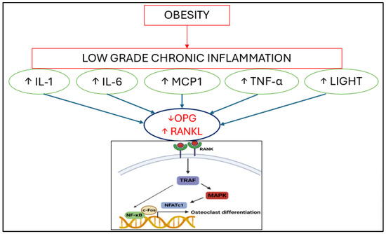
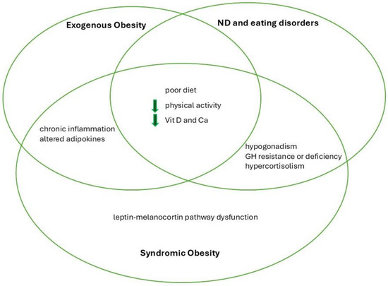
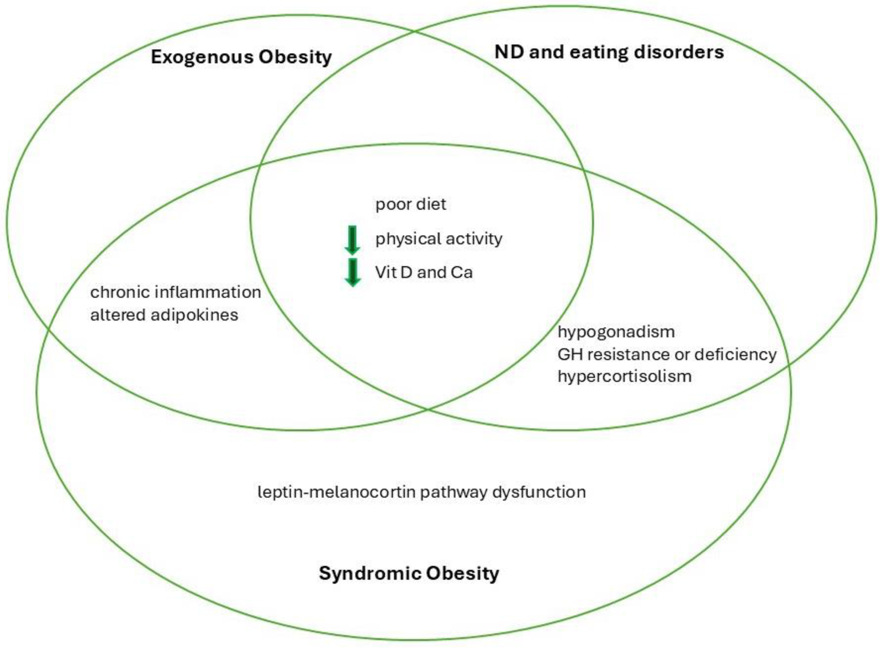
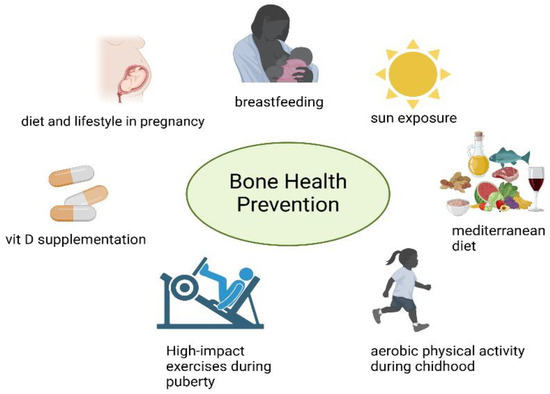
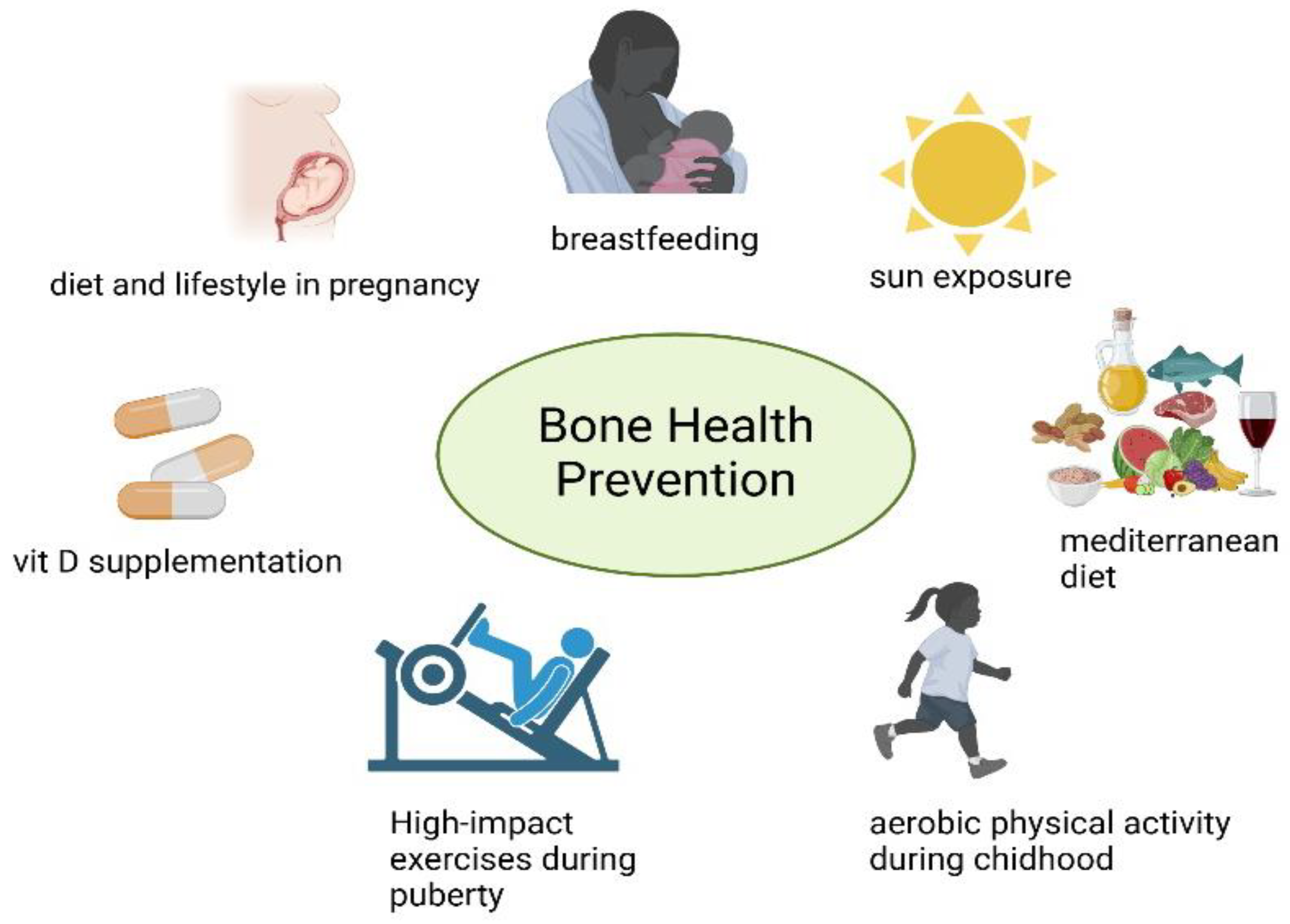

Disclaimer/Publisher’s Note: The statements, opinions and data contained in all publications are solely those of the individual author(s) and contributor(s) and not of MDPI and/or the editor(s). MDPI and/or the editor(s) disclaim responsibility for any injury to people or property resulting from any ideas, methods, instructions or products referred to in the content. |
© 2025 by the authors. Licensee MDPI, Basel, Switzerland. This article is an open access article distributed under the terms and conditions of the Creative Commons Attribution (CC BY) license (https://creativecommons.org/licenses/by/4.0/).