


Abstract
Gypsum ecosystems are constrained by extreme nutrient and water scarcity, where cyanoprokaryota interact with gypsophyte rhizospheres, influencing plant performance and soil biogeochemistry. This study examines three gypsophytes—Herniaria fruticosa, Helianthemum squamatum, and Teucrium libanitis—during winter and spring to characterize rhizospheric and bulk soil properties, assess enzymatic activity and nutrient cycling, identify cyanoprokaryota communities, and determine bioelement accumulation patterns in both Microcoleus chthonoplastes and gypsophytes. Physical, chemical, microbiological, and microscopic analyses were conducted across seasons. β-glucosidase activity showed species-specific responses to water pulses, particularly in Helianthemum squamatum. Seasonal differences in water-soluble C and N distinguished rhizospheres of Teucrium libanitis and Helianthemum squamatum. Key soil drivers included water-holding capacity to −1500 kPa, total and organic carbon, and Cr content. Cyanoprokaryota exhibited both rhizosphere-specific (Gloeocapsa novacekii, Pseudocapsa dubia) and ubiquitous taxa, with Microcoleus chthonoplastes reflecting bioaccumulation strategies. Bioelement accumulation differed between leaves and roots, especially for K, Mn, Zn, Na, Ni, C, and V, while the Sr:Ca ratio emerged as a potential indicator, especially in Herniaria fruticosa. These findings highlight the role of cyanoprokaryota in regulating nutrient availability and enzymatic activity, supporting gypsophyte adaptation and the ecological sustainability and resilience of gypsum ecosystems, and informing conservation and restoration strategies in these neglected ecosystems.
1. Introduction
Gypsiferous soils are mineral substrates formed under arid to semiarid climates [1]. The Iberian Peninsula hosts the highest global diversity of gypsum specialist flora (Gypsophiletalia) [2]. These ecosystems are characterized by strong abiotic constraints, including water scarcity, low nutrient availability, and extreme physicochemical conditions. N is typically the primary limiting nutrient, often co-limited by P, Fe, and other micronutrients. P availability is frequently reduced due to precipitation with Ca, while seasonal water variability strongly affects soil C:N ratios and microbial enzymatic activity [3]. Micronutrients such as Mo and Fe act as cofactors for nitrogenase and biological N fixation in cyanoprokaryota [4], whereas elements such as V may partially substitute for Fe under limiting conditions [5].
Beyond essential nutrients, bioelements play important roles in plant performance and stress tolerance [6]. Some elements can act as stimulants or hormetic modulators, enhancing biological processes at low concentrations [7,8]. Bioelements are therefore considered chemical elements with functional biological roles beyond classical essentiality [9], contributing to nutrient storage, stress tolerance, and detoxification processes [10,11]. In gypsum ecosystems, endemic species may accumulate excess elements in plant tissues as an adaptive strategy to cope with edaphic stress and herbivory [12,13].
Cyanoprokaryota (Cyanobacteria) are extremophiles capable of surviving high UV radiation, salinity, drought, temperature extremes, and metal toxicity [14]. They inhabit free-living and symbiotic niches with plants, animals, and fungi [15], displaying metabolic flexibility—including autotrophy, heterotrophy, and mixotrophy [16]. In symbiotic associations, cyanobacteria provide C and N to their hosts while benefiting from reduced UV flux, moderated microclimate, and lower predation risk [17]. In rhizospheric associations, plant-derived carbon exudates compensate for diminished light availability [17,18,19].
Cyanoprokaryota also remain protected from UVB radiation, which can otherwise reduce photosynthetic performance, damage chloroplast ultrastructure, and disrupt antioxidant systems [20]. They can buffer heat stress, maintain favorable micro-humidity, and sustain metabolic activity under extreme conditions [21], participating in N, P, and C cycling and CO2, H2, and O2 transformations [22]. Additionally, the production of organic acids by cyanoprokaryota can improve soil fertility, enhance nutrient availability, and increase soil water retention [23].
Among cyanoprokaryota, nitrogen-fixing Microcoleus chthonoplastes Thuret ex Gomont synthesizes mucilage around plant roots, protecting nitrogenase from oxygen exposure and influencing soil structure and microenvironmental conditions [24].
Water availability is a key driver of ecosystem functioning in gypsum environments. Seasonal water pulses derived from rainfall, dew, or condensation regulate microbial activity and nutrient cycling [3]. Some gypsophytes, such as Helianthemum squamatum (L.) Dum. Cours can access gypsum crystallization water [25], while cyanoprokaryota may also influence water dynamics, although this remains under debate [26,27].
Despite their ecological relevance, the interactions between seasonal microbial dynamics, cyanoprokaryota communities, and bioelement cycling in gypsophyte rhizospheres remain poorly understood. In particular, the role of water pulses in regulating microbial processes and plant–soil interactions is still unclear, limiting our understanding of ecosystem functioning and resilience in gypsum habitats.
This study examines three gypsophytes—Herniaria fruticosa subsp. erecta (Willk.) Batt., H.squamatum, and Teucrium libanitis Schreb.—during winter and spring. The objectives are to: (i) characterize the physical, chemical, and microbiological properties of gypsophyte rhizospheric and bulk soils across two seasons; (ii) evaluate the response of soil enzymatic activity and nutrient cycling to seasonal water pulses; (iii) identify cyanoprokaryota communities associated with gypsophyte rhizospheres and non-rhizospheric soils; and (iv) determine the bioelements in M. chthonoplastes, bioelements in rhizospheric and non-rhizospheric soils, and gypsophytes’ bioelement accumulation strategies.
Based on this framework, we hypothesize that (H1) seasonal water pulses activate microbial processes regulating C, N, and P cycling in gypsophyte rhizospheres; (H2) cyanoprokaryota exhibit both ubiquitous and rhizosphere-specific associations with gypsophyte species; and (H3) cyanoprokaryota influence bioelement availability and plant elemental strategies, contributing to gypsophyte adaptation and the ecological sustainability of gypsum ecosystems.
2. Materials and Methods
2.1. Study Site
The study was conducted in the municipality of Fortuna (38°14′06.90″ N, 1°11′24.54″ W), at the foothills of the Sierra de la Pila Regional Park (Murcia, SE Spain), an area under multiple environmental protection designations. The study hill is capped by dolomites producing large detached blocks and a rugged relief. Geologically, the site belongs to the external zones of the Betic Cordillera, within the Subbetic system. The dominant lithology consists of Triassic gypsum interbedded with dolomites and carnioles, with gypsum outcrops displaying varied morphologies and colors (black, reddish, whitish) and diverse crystalline structures. Fossil ammonites and bivalves from the Cretaceous are present.
Soils show intense gypsum accumulation from Triassic gypsum marls and deposits, classified as Petric Gypsisols or Hypergypsic Gypsisols [28]. Lithic Leptosols [28] dominate the upper slopes, where primary gypsum occurs at <10 cm depth, while Nudulitic Leptosols [28] appear where gypsum reaches the surface as well-developed multicolored crystals.
The climate is semiarid Mediterranean, with a mean annual temperature of 18 °C and mean annual precipitation of 278 mm (SIAM: http://siam.imida.es, accessed on 14 December 2022). Meteorological data from the Fortuna station (38°09′39.79″ N, 1°09′09.43″ W; 197 m a.s.l.) preceding each sampling date are summarized in Table S1.
Vegetation belongs to the Gypsophiletalia [2], specifically the habitat “Yesos del Sureste Semiárido” in northern Murcia and southern Alicante. In the study area, this is represented by the community Teucrio verticillati–Thymetum pallescentis Bellot, Esteve & Rivas Goday [29,30], with 1% cover. The dominant gypsophytes are T. libanitis, H. fruticosa subsp. erecta, and H. squamatum, with T. libanitis also diagnostic for the association. Other species include Fumana ericoides (Cav.) Gand., Helianthemum syriacum (Jacq.) Dum. Cours., and Rosmarinus officinalis L. Stipa tenacissima L. occurs at low abundance, delimiting the gypsiferous area. Lower slopes host abandoned vineyards and almond and olive orchards.
Surfaces lacking vascular vegetation are covered by biological soil crusts (biocrusts), which stabilize soil, reduce erosion, enhance primary productivity, and improve water retention and infiltration [31]. Biocrusts include cyanobacteria, algae, lichens, and mosses, all highly stress-tolerant. Lichen–moss communities in gypsum substrates are well documented [32,33], whereas cyanobacterial communities remain comparatively less studied [34]. Dominant lichens include Acarospora placodiiformis Magnusson, Fulgensia poeltii Llimona, and Lecidea circinarioides Casares & Hafellner [35], while common cyanoprokaryota include Gloeocapsa biformis Ercegovic, Gloeocapsa violascea (Corda) Rabenhorst, and M. chthonoplastes, often forming incipient lichens with hyphae [36].
2.2. Sampling Procedures
The sampling area was oriented N 85° E with a 15–20% slope, along a 65-m transect at 480 m a.s.l., near Loma de Planes. Two campaigns were conducted: December 2011 (D1) and May 2012 (D2). Sampling included three replicated blocks in both non-rhizospheric soil (control, 0) and rhizospheric soil under the gypsophytes H. fruticosa (Hf), H. squamatum (Hs), and T. libanitis (Tl). For each species, three individuals per block were sampled (n = 9), with above- and below-ground biomass separated for leaf and root analyses.
Rhizospheric soil was collected directly beneath each individual along the dichotomous root pattern to a depth of 30 cm. Non-rhizospheric soil was sampled at the same depth in adjacent vegetation-free areas after removing the surface biocrust. Soil samples were transported in plastic bags to the laboratory. Each sample was divided: one portion was sieved (2 mm) and stored at 4 °C for microbiological and biochemical analyses; the other was air-dried and sieved (2 mm) for remaining analyses.
2.3. Analyses
2.3.1. Soil
Soil pH, electrical conductivity, and gypsum content were measured following Porta [37] (Table 1). Water retention at −33 kPa (field capacity) and −1500 kPa (permanent wilting point or water-holding capacity at 15 atm) was determined according to Dirksen [38], with available water content calculated. Carbonates, water-soluble carbon (WSC), water-soluble N (WNS), total organic C (TOC), total C (TC), total nitrogen (TN), soil moisture, microbial biomass C, and respiration were measured following Fernández et al. [39]. Enzyme activities—dehydrogenase [40], urease and protease [41], alkaline phosphatase, arylsulfatase, and β-glucosidase [42] followed Díaz-Pereira et al. [3]. Water-soluble carbohydrates (1:5 w:v extract) were determined following Brink et al. [43].
Table 1.
Soil pH, electrical conductivity (EC, mScm−1), and gypsum content (G, g kg−1) in function of non-rhizospheric soil (0) and in the rhizosphere soil of the three gypsophytes species (Hf: H. fruticosa, Hs: H. squamatum, and Tl: T. libanitis) for the different sampling dates.
Total bioelements were quantified by ICP-OES (Thermo ICAP 6500 DUO, Thermo Electron Manufacturing Ltd., Cambridge, UK) after HNO3:H2O2 (4:1 v:v) microwave digestion at CEBAS-CSIC (Murcia, Spain). Major elements (g kg−1) included Al, Ca, K, Na, Mg, P, and S; trace elements (mg kg−1) included As, Bi, B, Cd, Cr, Cu, Fe, Li, Mn, Ni, Pb, Sr, Ti, Tl, V, and Zn. Elements below detection limits were Be, Co, Mo, Sb, Se (<0.5 mg kg−1), and Cd (<0.1 mg kg−1).
2.3.2. Gypsophytes
Leaves and roots were washed with distilled water, oven-dried at ≤50 °C to constant mass, milled, and analyzed by ICP-MS as above. Therefore, the measured metal concentrations represent total metals associated with the roots. C and N concentrations were determined using an automated CN analyzer (CEBAS-CSIC). Elements quantified included Al, B, Cr, Cu, Fe, Li, Mn, Ni, Pb, Sr, Ti, Tl, V, and Zn (mg kg−1) and Ca, K, Mg, Na, P, and S (g kg−1). Elements below detection limits were As, Be, Bi, Cd, Co, Mo, Sb, Se (<0.5 mg kg−1)—diagnostic thresholds of S, Ca, and Mg following Merlo et al. and Baker [12,44].
2.3.3. Cyanoprokaryota
Cyanoprokaryota were collected from rhizospheric and non-rhizospheric soils. Morphological examination of subaerial cyanobacteria was performed on field-collected material, as thalli are often coated with mineral particles. For detailed taxonomic analysis, isolates were cultured on agar-solidified BG11 medium [45] at 20 °C under 70 µmol m−2 s−1 light (16 h:8 h L:D). Observations were made using a LAN OPTICS stereomicroscope and an Olympus BX41 compound microscope. Species richness was assessed as the number of taxa, and abundance was scored semi-quantitatively as + (<5%), ++ (5–15%), +++ (15–30%) [3].
Freeze-dried biomass of cultured M. chthonoplastes was analyzed by ICP-MS for Al, B, Be, Ca, Cd, Co, Cr, Cu, Fe, K, Li, Mg, Mn, Mo, Na, Ni, P, Pb, Rb, S, Si, Sr, Ti, and Zn. Macroelements were expressed in g kg−1 DW and trace elements in mg kg−1 DW. Elements below detection limits included As, Bi, La, Sb, Se, Tl, and V (<0.01 mg kg−1).
2.3.4. Bioaccumulation and Translocation Factors
The bioconcentration (or bioaccumulation) factor (BCF) and translocation factor (TF) were calculated according to commonly used approaches [46] for each gypsophyte species and bioelement.
BCF = Element Concentration in Root/Element Concentration in rhizospheric soil. The value = 1 for the BF indicates equilibrium, values > 1 indicate bioaccumulation, while values < 1 indicate that it does not occur.
TF = Element Concentration in Leaf/Element Concentration in Root. The value = 1 for the TF indicates equilibrium, values > 1 indicate translocation, while values < 1 indicate that it does not occur.
2.4. Statistical Analyses
Normality and homoscedasticity were assessed using Kolmogorov–Smirnov and Levene tests. Data were ln-transformed when necessary. Effects of species (S), sampling date (D), and their interaction (S × D) were tested by two-way ANOVA, followed by Tukey’s post hoc test for pairwise comparisons when significant (p < 0.05); non-normal variables were analyzed with Kruskal–Wallis tests. Spearman correlations assessed relationships among parameters.
Stepwise discriminant function analysis (DFA) identified variables that best discriminated among gypsophytes. Similarities between non-rhizospheric soil, rhizospheric soil by gypsophyte, and M. chthonoplastes were evaluated using dendrograms generated from centroid averages (similarity scale: 0 = highest, 25 = lowest). Analyses were performed in IBM SPSS v.28.0 (Windows).
3. Results
3.1. Soil Properties and Bioelements
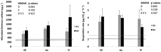
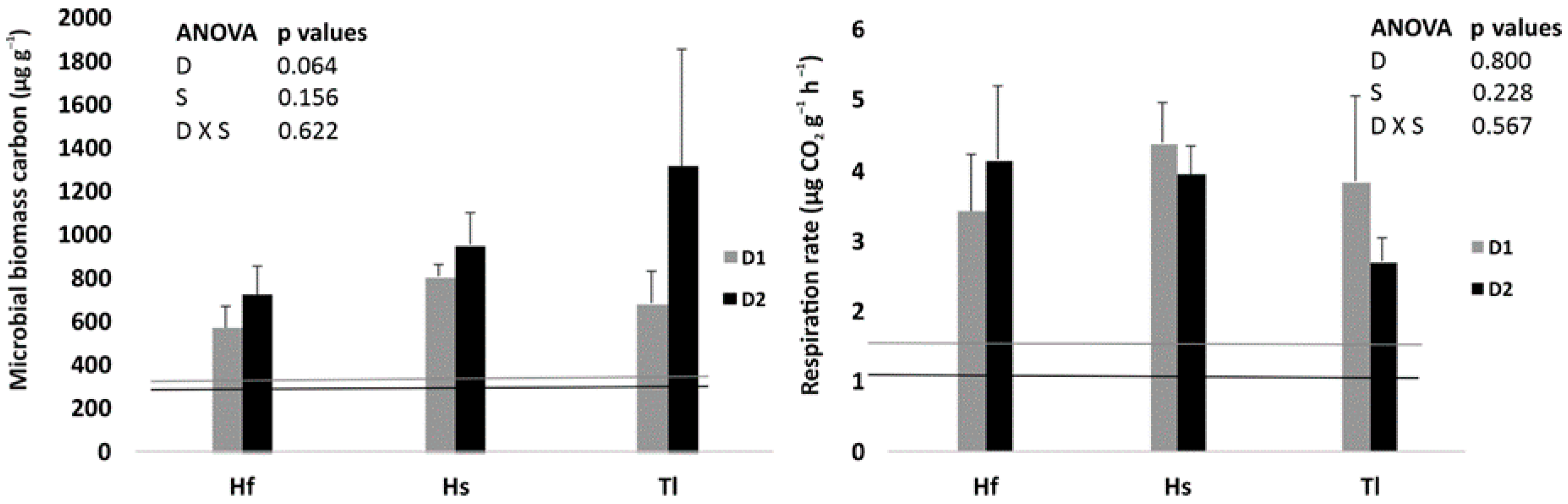
Significant differences were detected between non-rhizospheric soil (without associated plants or biocrusts) and rhizospheric soil across the three gypsophytes. This is why non-rhizospheric soil values are excluded in the statistical analyses to magnify differences among rhizospheric soils of gypsophytes, and in the figures, only the mean values for each property or bioelement of the non-rhizospheric soil are shown as color-coded reference or control lines according to the sampling. Microbial biomass C and basal respiration did not differ significantly among the rhizospheric soils of the three species or between sampling dates (Figure 1).
Figure 1.
Microbial biomass carbon and basal respiration rate in rhizospheric soils of the three gypsophytes (Hf: H. fruticosa; Hs: H. squamatum; Tl: T. libanitis) at two sampling dates (D1: winter; D2: spring). Lines indicate the corresponding non-rhizospheric soil values for the same sampling periods (light-gray lines correspond to D1, and dark-gray lines to D2).
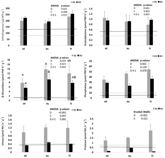
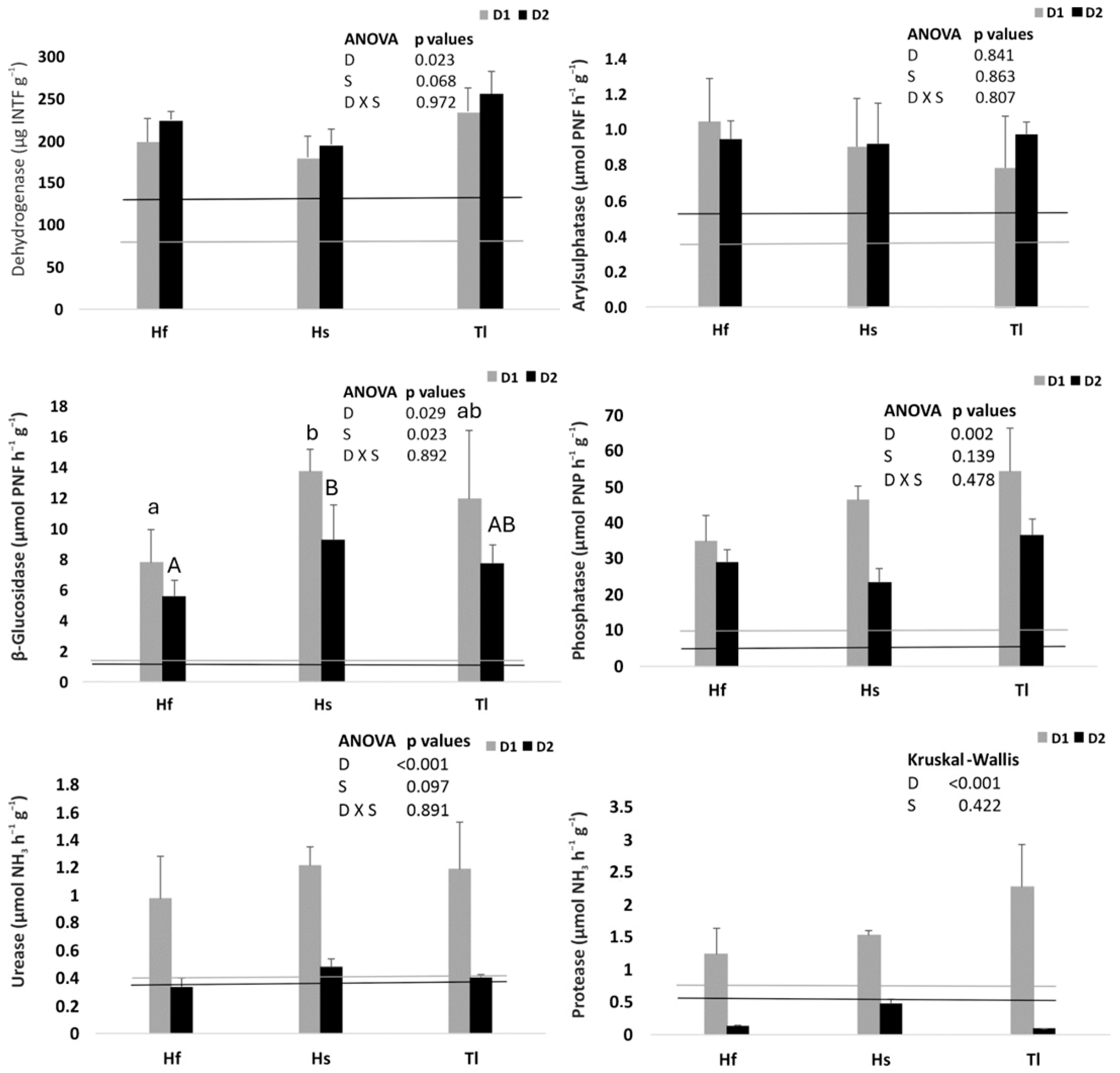
Enzyme activities varied significantly with sampling date for all enzymes except arylsulfatase (Figure 2). Generally, the highest activities occurred in December (D1), coinciding with greater antecedent precipitation and higher gravimetric soil moisture (Table S1), whereas dehydrogenase peaked in May (D2). Species-level differences were observed only for β-glucosidase, with the lowest activity in Hf, the highest in Hs, and intermediate in Tl.
Figure 2.
Enzyme activities in rhizospheric soils of the three gypsophytes (Hf: H. fruticosa; Hs: H. squamatum; Tl: T. libanitis) at two sampling dates (D1: winter; D2: spring). Lines indicate the corresponding non-rhizospheric soil values for the same sampling periods (light-gray lines correspond to D1, and dark-gray lines to D2). Different lower letters (sampling D = 1) and capital letters (sampling D = 2) indicate statistically significant differences at p < 0.05 (Tukey’s test) between Hf, Hs, and Tl.
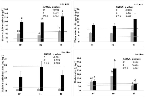
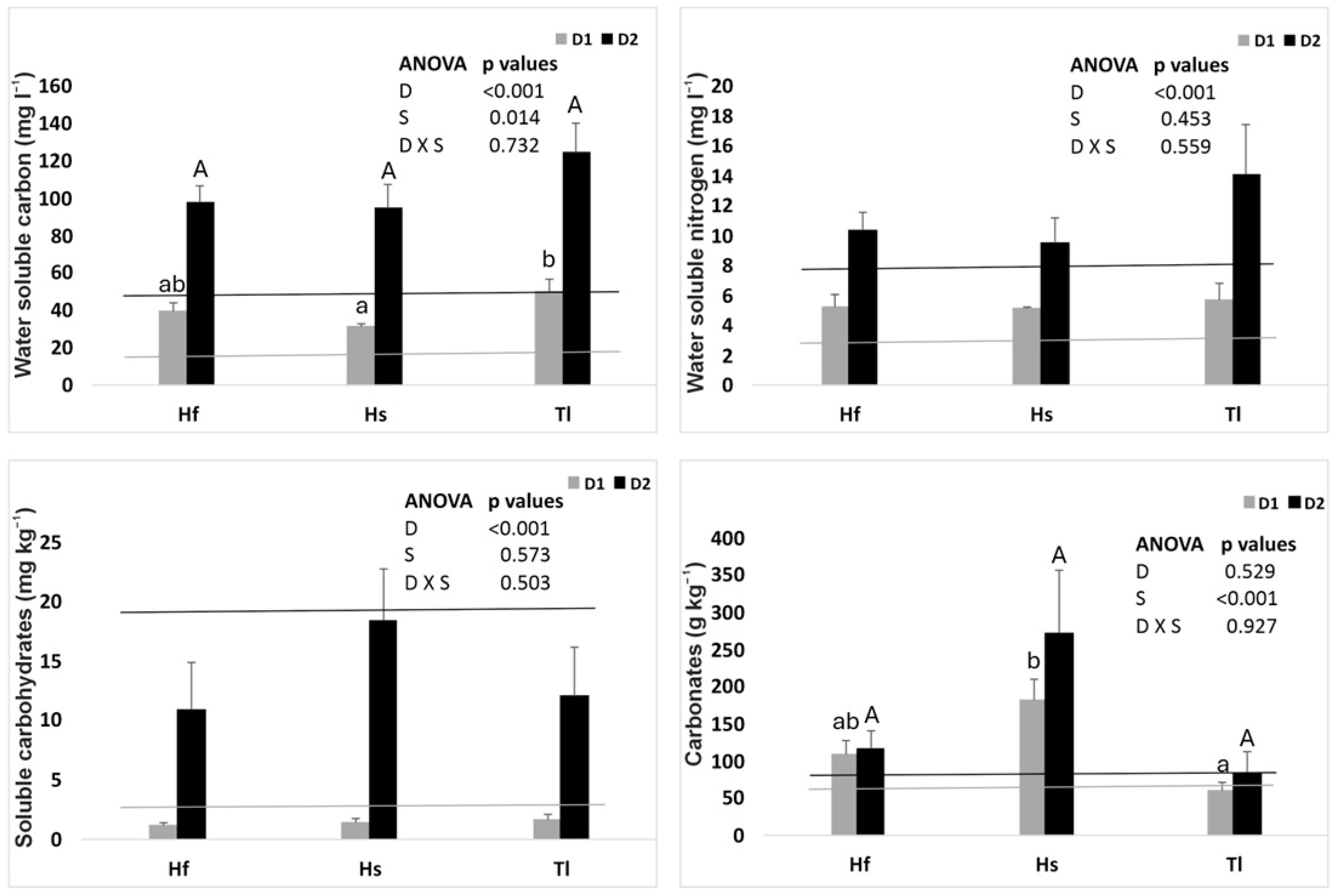
Water-soluble C and N differed significantly between dates, being higher in D2 (May) (Figure 3). Across species, soluble C was lowest in Hs, highest in Tl, and intermediate in Hf. Water-soluble carbohydrates were high in non-rhizospheric soil, exceeded in D1 by Tl, and in D2 by Hs. Carbonate content was consistently highest in Hs rhizospheres (Figure 3).
Figure 3.
Water-soluble carbon (WSC) and water-soluble nitrogen (WSN) in rhizospheric soils of the three gypsophytes (upper panels). Soluble carbohydrates and carbonates in rhizospheric soils of the three gypsophytes (lower panels). Hf: H. fruticosa; Hs: H. squamatum; Tl: T. libanitis. Sampling dates: D1 (winter), D2 (spring). Lines indicate the corresponding non-rhizospheric soil values for the same sampling periods (light-gray lines correspond to D1, and dark-gray lines to D2). Different lower letters (sampling D = 1) and capital letters (sampling D = 2) indicate statistically significant differences at p < 0.05 (Tukey’s test) between Hf, Hs, and Tl.
Functionally, β-glucosidase activity was highest in Hs rhizospheres. Soluble C and N increased sharply in spring (D2), accompanied by elevated dehydrogenase activity and water-soluble carbohydrates (Figure 2 and Figure 3).
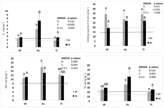
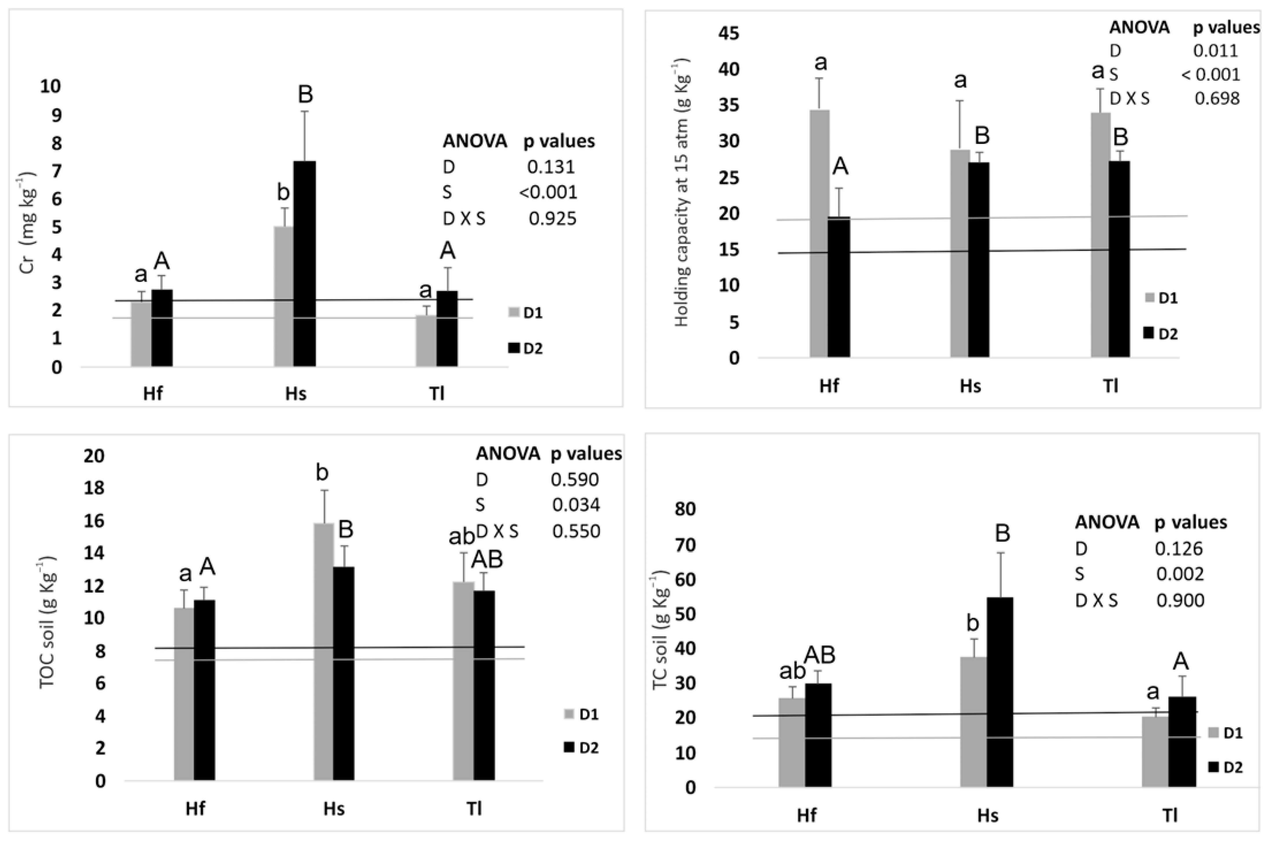
Discriminant function analysis (DFA) using all soil properties and bioelements identified Cr, pF 15 atm (−1500 kPa), TOC, and TC as the variables consistently driving group separation (Figure 4; Table S2A,B, Figure S1A,B). Excluding non-rhizospheric soil improved classification success while retaining the same discriminating variables.
Figure 4.
Discriminant variables obtained considering all chemical elements and soil properties analyzed in rhizospheric soils. The variables selected were chromium (Cr), water-retention capacity at permanent wilting point or holding capacity (pF 15 atm or −1500 KPa), total soil organic carbon (TOC), and total soil carbon (TC). Sampling dates: D1 (winter), D2 (spring). Lines indicate the corresponding non-rhizospheric soil values for the same sampling periods (light-gray lines correspond to D1, and dark-gray lines to D2). Rhizospheres: Hf (H. fruticosa); Hs (H. squamatum); Tl (T. libanitis). Different lower letters (sampling D = 1) and capital letters (sampling D = 2) indicate statistically significant differences at p < 0.05 (Tukey’s test) between Hf, Hs, and Tl.
3.2. Presence of Cyanoprokaryota in Soils
Nine cyanoprokaryota taxa were identified in rhizospheric and non-rhizospheric soils (Table 2). Coccoid forms (Chroococcales, 88.8%) dominated over filamentous forms (Oscillatoriales, 11.2%). Gloeocapsa was the most species-rich genus (five species). The most abundant taxa were Asterocapsa salina Hindák et Smarda, G. violascea, and M. chthonoplastes, while G. biformis and Chroococcus giganteus W. West were least abundant.
Table 2.
Cyanoprokaryota species in the non-rhizospheric soil (0) and in the rhizosphere soil of the three gypsophytes species (Hf: H. fruticosa, Hs: H. squamatum, and Tl: T. libanitis) for the different sampling dates.
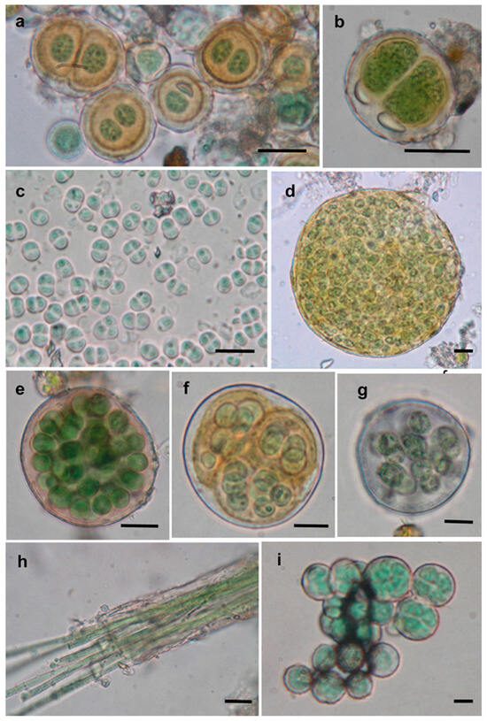
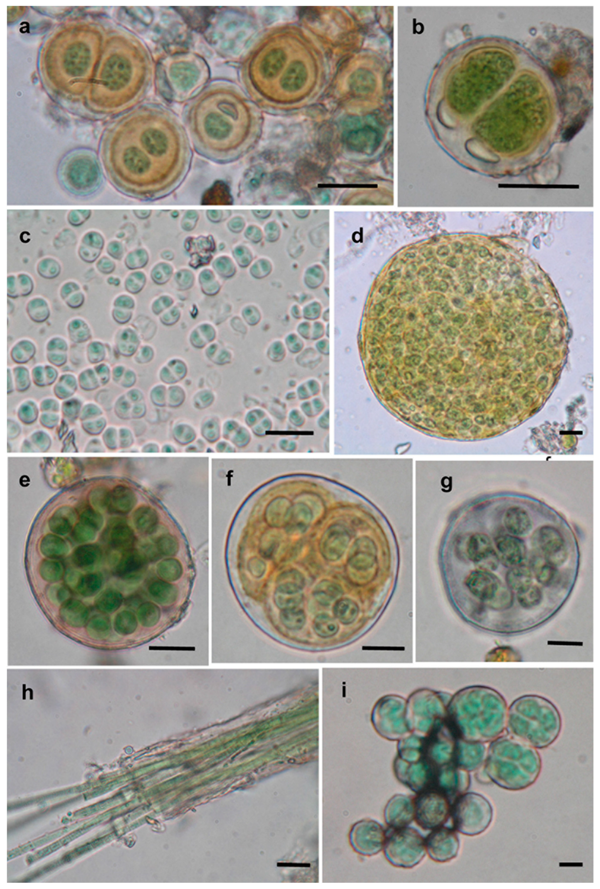
Species richness was higher in rhizospheric than non-rhizospheric soils. Among rhizospheres, Hf supported the highest richness (9 species in spring, 7 in winter), followed by Tl (7 and 6 species) and Hs (6 and 7 species). In spring, cyanoprokaryota were particularly abundant in Hf rhizospheres; in winter, pronounced differences were observed among Hf, Tl, and Hs. Several taxa were ubiquitous across soils and seasons, including A. salina, G. violascea, and M. chthonoplastes (Figure 5a,g,h).
Figure 5.
(a–i) Light micrographs (scale bar: 10 µm) of: (a) Asterocapsa salina; (b) Chroococcus giganteus; (c) Gloeocapsa biformis; (d) Gloeocapsa kützingiana; (e) Gloeocapsa novacekii; (f) Gloeocapsa rupestris; (g) Gloeocapsa violascea; (h) Microcoleus chthonoplastes; (i) Pseudocapsa dubia.
Other taxa displayed spatial or seasonal specificity: C. giganteus occurred only in Hf rhizospheres in both seasons (Figure 5b). G. biformis was restricted to Hf rhizospheres in spring (Figure 5c). Gloeocapsa kützingiana Nägeli appeared in winter in non-rhizospheric soil and Hs and Hf rhizospheres; in spring, it was present in all rhizospheres but absent from non-rhizospheric soil (Figure 5d). Gloeocapsa novacekii Komárek et Anagnostidis and Gloeocapsa rupestris Kützing were found only in rhizospheres (Figure 5e,f). Pseudocapsa dubia Ercegovic occurred exclusively in rhizospheres in both seasons (Figure 5i).
3.2.1. Bioelement Composition in Microcoleus chthonoplastes
In M. chthonoplastes, bioelement concentrations followed the order: Fe > Mn > B > Si > Cu > Zn > Al > Mo > Pb > Sr > Ti > Rb > Na > Ni > P > K > Cr > Co > S > Ca > Mg > Cd > Li > Be. Fe (1924.11 ± 459.78 mg kg−1) and Mn (716.42 ± 205.52 mg kg−1) were particularly high, followed by B, Si, and Cu.
DFA of bioelements in soils and M. chthonoplastes identified Al, B, Fe, Na, and P as canonical variables separating biomass from soils, with soils clustering together opposite M. chthonoplastes (Table S2A,B, Figure S1A,B; Table S3A, Figure S2A). Including leaves or roots shifted canonical variables, with leaves discriminated by B, Cu, Mn, Na, and Zn, and roots by K, Zn, and Cu (Table S3B,C, Figure S2B,C). Dendrograms indicated that non-rhizospheric soils clustered with Hf and Tl rhizospheres, while Hs rhizospheres and M. chthonoplastes formed distinct groups.
3.2.2. Bioelements in Gypsophytes
All species accumulated higher S and Ca in roots than in leaves, with sampling date and species affecting concentrations (Ca nearly significant, p = 0.055). Hf exhibited the highest Mg concentrations in both organs (Table S2A,B, Figure S3).
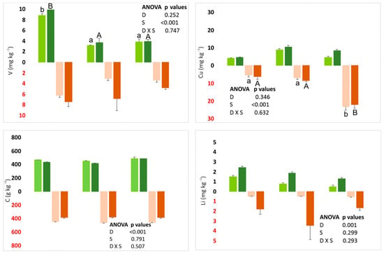
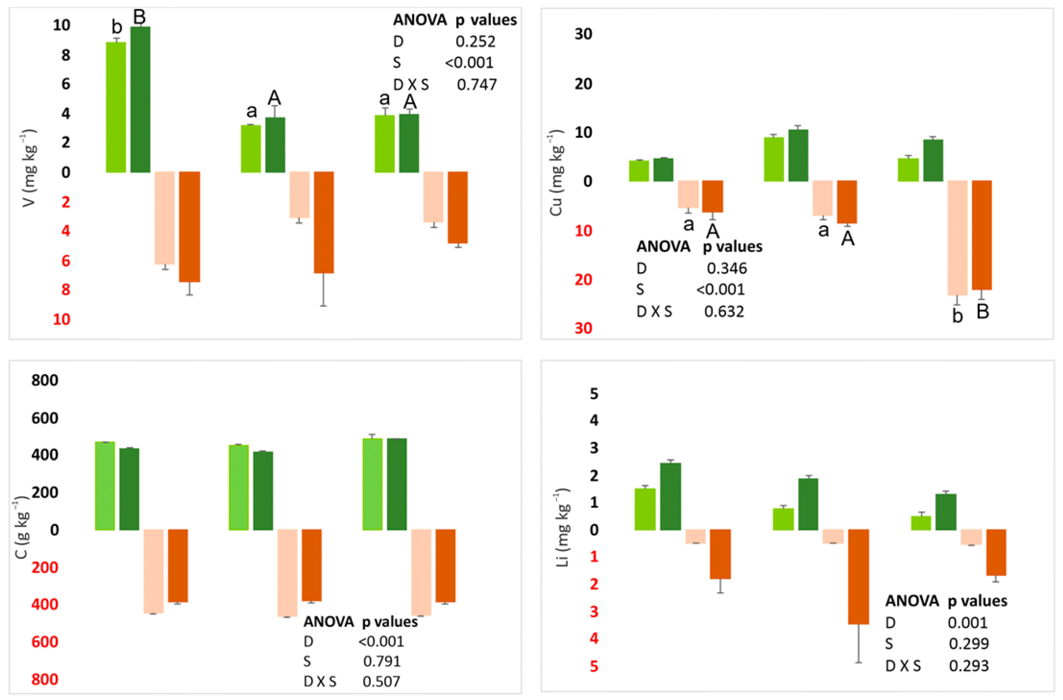
DFA integrating leaf, root, and rhizospheric bioelements identified canonical variables:
Leaves: N, Ca, Ni, C, V
Figure 6.
Discriminant variables, listed from highest to lowest weight, for each gypsophyte: N (leaves), Ca (leaves), Ni (leaves), C (leaves), V (leaves), Cu (roots), C (roots), and Li (roots). Green: leaves; brown: roots; light colors: D1; dark colors: D2. Hf = H. fruticosa; Hs = H. squamatum; Tl = T. libanitis. ANOVA tables presented above or below each panel indicate whether the variable corresponds to aerial (leaves) or underground (roots) organs. Different lower letters (sampling D = 1) and capital letters (sampling D = 2) indicate statistically significant differences at p < 0.05 (Tukey’s test) between Hf, Hs, and Tl.
Excluding rhizospheric soil, canonical variables shifted to:
Leaves: K, Ni, S, Sr, C, V
Roots: K, Cu, S, Tl (Table S4B, Figure S4B)
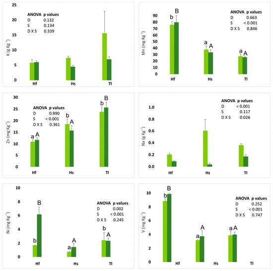
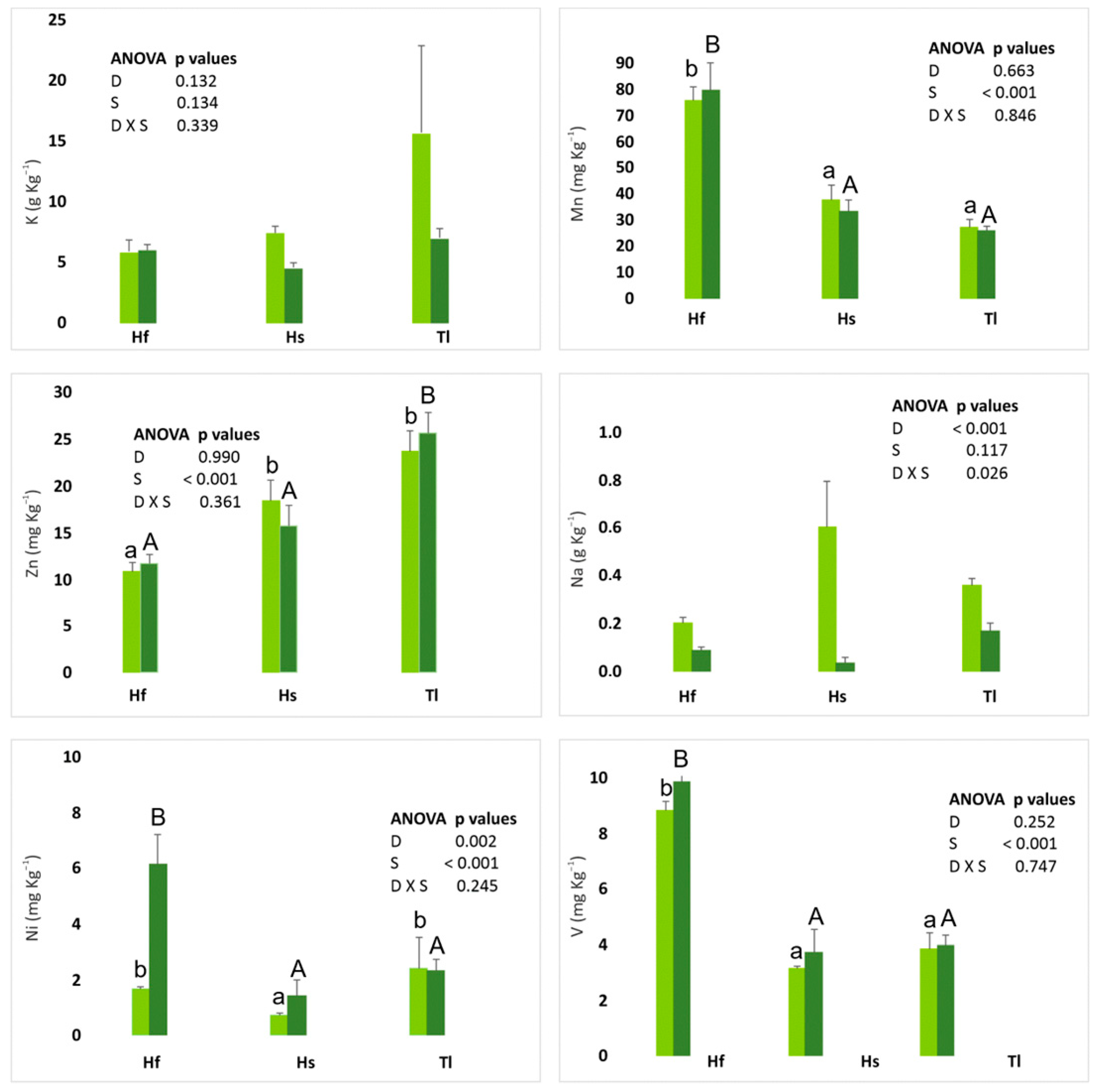
Leaf variables displayed the strongest discriminating power. DFA with leaves only highlighted K, Mn, Zn, Na, Ni, C, and V as the most important discriminators (Figure 7; Table S4C, Figure S4C).
Figure 7.
Discriminant variables, listed from highest to lowest weight, for leaves of the gypsophyte species. Light colors: D1; dark colors: D2. Hf = H. fruticosa; Hs = H. squamatum; Tl = T. libanitis. Different lower letters (sampling D = 1) and capital letters (sampling D = 2) indicate statistically significant differences at p < 0.05 (Tukey’s test) between Hf, Hs, and Tl.
3.2.3. Bioaccumulation Factors and Gypsophyte Strategies
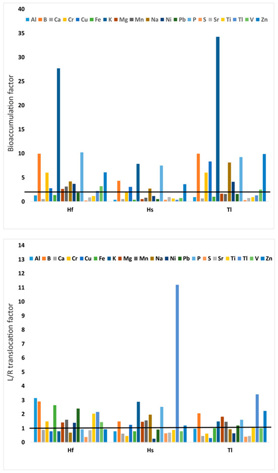
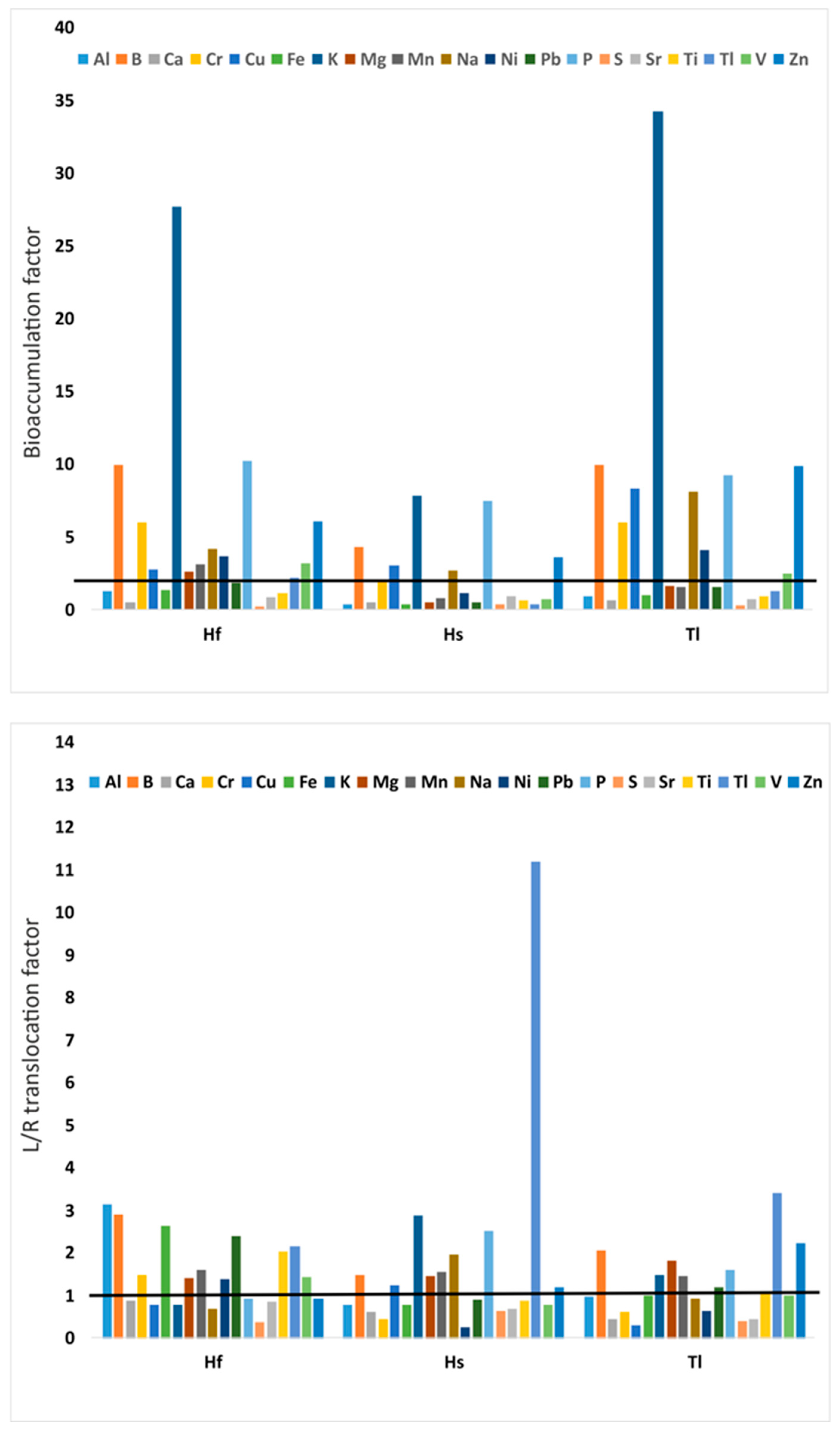
Bioaccumulation or bioconcentration factor (R/Srizospheric) and leaf-to-root (L/R) translocation factor are shown in Figure 8. Hs bioaccumulated the fewest elements (8), whereas Hf and Tl accumulated 16 and 14 of 19 elements, respectively. K showed particularly high bioaccumulation in Hf and Tl, with Tl exhibiting high L/R ratios for several elements. The bioaccumulation values of Hs are approximately half of those of Hf. Minimum values range from 0.2 to 0.3 for S, with maximum values of 34.3 for K, and a predominance of values between 1.5 and 10. The lowest bioaccumulation values are observed for S, Ca, and Sr.
Figure 8.
Bioaccumulation factors of bioelements (upper panels) and translocation factor from leaves (L) to roots (R) (lower panels). Hf = H. fruticosa; Hs = H. squamatum; Tl = T. libanitis. A solid black line is shown at the value of 1 for the bioaccumulation and translocation factors, indicating equilibrium. For each element, values > 1 indicate bioaccumulation or translocation, while values < 1 indicate that it does not occur.
The three studied species show the same pattern regarding the translocated elements (10 out of 19). Translocation values range from 0.3 to 11.2. No translocation was observed for Ca, S, or Sr in any case.
Potential nutritional strategies include:
Hs: higher rhizospheric element concentrations but lower plant accumulation.
Hf and Tl: lower rhizospheric concentrations but higher accumulation, indicating similar acquisition strategies.
Leaf C:N ratios differed among species: Hf = 52, Hs = 42, Tl = 38; root C:N was highest in Hs (59), versus Tl (23) and Hf (25), reflecting adaptive responses to N limitation. Rhizospheric soil C:N ranged 27–41, with no significant interspecific differences.
Soil Sr:Ca ratios were highest in non-rhizospheric soil (96), followed by Hs rhizosphere (57), and intermediate (78) in Hf and Tl. Leaf Sr:Ca ratios did not differ (108–117), while root Sr:Ca was highest in Hf (127) and lowest in Tl (97). Sr and Ca were strongly correlated in Hf (r = 0.734, p < 0.01) and Hs roots (r = 0.608, p < 0.05) but not in Tl (r = 0.028).
4. Discussion
4.1. Gypsiferous Soils
Gypsum rhizospheric soils remain largely unexplored; most cyanobacterial research has focused on biocrusts rather than direct plant–rhizosphere interactions [47]. In highly arid environments, biocrusts mechanistically enhance plant growth by stabilizing soil surfaces, reducing erosion, and increasing local nutrient and water retention [48]. Vertical transfer experiments demonstrate that biocrusts release soluble carbon and nutrients into subsurface layers, where dissolved metabolites function as osmolytes, modulating soil osmotic potential [31].
Cyanobacteria display strong physiological plasticity. Proteomic analyses of Synechocystis sp. PCC 6803 reveals large-scale protein redistribution under multiple abiotic stresses, suggesting rapid metabolic reprogramming [49]. Secondary metabolites from cyanobacteria mitigate oxidative stress, heavy-metal toxicity, and pathogen pressure, thereby improving host plant performance. Symbiotic associations are species-specific and reciprocal: plants provide carbon and rhizosphere stabilization, while cyanobacteria contribute photosynthates and bioactive metabolites [19,50].
Cyanobacteria also act as geomicrobiological engineers in gypsum soils. Chasmoendolithic coccoid forms mechanically fracture the substrate through cycles of mucilage swelling and desiccation, while chemical dissolution of minerals via excreted organic acids allows endolithic penetration [34]. Gypsophytes likely become established in these areas previously influenced by cyanoprokaryota, and rhizospheric-level interactions can be established, thereby achieving a mechanism of adaptation to extreme drought conditions. Prior studies report cyanoprokaryota in the rhizospheres of gypsophytes (H. fruticosa, H. squamatum, T. balthazaris) [3], emphasizing their role in bioelement accumulation [12,51]. Cyanobacterial surfaces—rich in polysaccharides, proteins, and lipids—serve as high-capacity metal-binding matrices [52,53]. Aluminum, while potentially toxic, can stimulate plant metabolic activity at low concentrations [54].
Semiarid gypsum ecosystems rely on multi-domain microbial consortia, where metabolic redundancy and structural diversity enable persistence under extreme water and nutrient limitation [55]. Cyanoprokaryota modulate rhizospheric water and nutrient dynamics by increasing organic matter, enhancing nutrient retention, and facilitating microscale water redistribution [19,56,57]. These microbial interactions are natural mechanisms for ecosystem sustainability. Cyanoprokaryota metabolism contributes not only to climate change mitigation but also to reducing the rate of carbon turnover, enhancing carbon retention in the soil. Seasonal climatic fluctuations modulate carbon-cycle enzyme activity, N transformations, and general microbial metabolism, reflecting the tight coupling of microbial processes to environmental conditions [58,59].
Microbial biomass C and basal respiration lacked consistent seasonal patterns, though biomass C increased in winter, coinciding with moisture pulses. The initial sampling captured activation of C, P, and particularly N cycles, aligned with pre-spring moisture inputs. Comparison with our data in other gypsum subtypes [3] shows comparable dehydrogenase and β-glucosidase activity, along with elevated alkaline phosphatase and arylsulfatase. The EPS produced by cyanobacteria in the rhizosphere increases the soil’s water retention at field capacity and permanent wilting capacity [3]. Water-holding capacity to −1500 kPa may serve as a mechanistic proxy for cyanoprokaryota–soil functional interactions.
In the spring sampling, the pH of gypsiferous rhizospheres and non-rhizospheric soils decreased compared to the winter sampling date (Table 1). This may be caused by soil microbiota participating in this acidification [60], as a greater decrease in pH was observed in non-rhizospheric soils. Gypsophytes, through their exudates, can also contribute to this acidification, as shown in the work carried out on the roots of Ononis tridentata, which also contributed directly to this acidification [60].
4.2. Bioelements
4.2.1. Role of Bioelements in Microcoleus chthonoplastes
Cyanobacteria can accumulate a wide spectrum of trace metals—including Al, As, Cd, Cr, Cu, Co, Hg, Ni, Pb, Ti, and Zn—through adsorption, active transport, and intracellular sequestration [61]. They are intrinsically enriched in Mg, Mn, Co, Zn, Mo, Cu, and Fe because these metals act as structural or redox cofactors in central metabolic pathways. While elevated concentrations disrupt photosynthesis, respiration, and cellular redox balance [62], cyanobacteria possess multiple tolerance mechanisms: adsorption to extracellular polysaccharides (EPS), metal chelation, controlled uptake, and vacuolar-like compartmentalization [63].
The elemental profile of M. chthonoplastes aligns with previous findings of Fe- and Mn-dominated signatures [64], although Fe concentrations were markedly higher and Mn lower than expected. Both Mn and Fe are mechanistically essential for oxygenic photosynthesis, P turnover, and regulation of oxidative stress via metalloenzymes and ROS-scavenging systems [65]. Their relative proportions suggest differential control of redox-active processes under gypsum-derived stress.
Bo was the third most abundant element, consistent with its role in stabilizing nitrogenase complexes and strengthening cell-wall polysaccharides [65]. Si ranked fourth, supporting its function in cell envelope architecture and mineral-organic interface formation.
Cu was the fifth most abundant element, falling within the general concentration ranges reported for cyanobacteria [62,63,66,67]. Cu acts as a catalytic center in proteins related to photosynthetic electron transport, nitrification–denitrification pathways, and oxidative stress regulation [65]. Zn and Ni concentrations were lower than those typically found in related species [64]. Like Cu, these elements participate in redox homeostasis, ROS detoxification, and stress-induced enzyme activation [65].
Several detected elements—including Al, Be, Cd, Co, Cr, Li, Ni, Pb, Rb, Si, Sr, and Ti—were absent from the BG-11 culture medium, indicating uptake directly from gypsum substrates and/or trace minerals such as clays and Fe–Mn oxides, as well as organic complexes. The particularly high concentrations of Si and Al support an EPS-mediated sorption mechanism [52] combined with structural incorporation into the cell envelope. Laboratory evidence from Synechococcus sp. WH7803 shows that Al can enhance growth and chlorophyll a production [68], suggesting that similar mechanisms could operate in M. chthonoplastes. Co and Ni function as essential cofactors in metalloenzymes involved in C and N metabolism [69]. Cd and Co also trigger EPS production and biofilm-associated stress responses; low Cd levels can stimulate biofilm expansion by activating detoxification pathways [70].
V was not detected in M. chthonoplastes, despite its presence in soils and plants. V accumulated most strongly in the rhizospheres of H. squamatum and in leaves and roots of H. fruticosa. Such enrichment may arise from microbially facilitated redox cycling in the rhizosphere, which generates bioavailable vanadyl species that can stimulate microbial growth under nutrient-limited conditions [71].
Finally, the detection of Co in M. chthonoplastes, but not in BG-11 medium, where it is absent, confirms acquisition from trace minerals such as clays and Fe–Mn oxides, as well as organic complexes. This supports a mechanism of bioaccumulation driven by EPS-mediated sorption and selective uptake pathways characteristic of metal-tolerant cyanobacteria.
4.2.2. Role of Bioelements in Gypsophytes
Essential bioelements in gypsophytes include B, Cu, halogens, Fe, Mn, Mo, Ni, and Zn, while Al, Co, La, Li, Rb, Se, Si, Ag, Sn, Sr, and V act as beneficial elements that modulate physiological performance without being strictly required [72]. Cr, abundant in rocky substrates such as gypsiferous soils, can integrate into plant defense pathways: its redox-active forms interact with cellular antioxidants and structural polymers, limiting herbivory and pathogen invasion [73]. Cr exhibits a hormetic dose–response pattern, whereby low concentrations stimulate enzyme activity and high concentrations inhibit it through oxidative and protein-denaturing mechanisms [8].
H. squamatum may experience enhanced mycorrhization—consistent with reports for the genus [74]—which could mechanistically elevate β-glucosidase activity in its rhizosphere by accelerating carbon turnover and increasing microbial enzymatic demand. Elevated Cr concentrations in H. squamatum rhizospheres co-occurred with higher total and organic C, suggesting that organic ligands and microbial metabolites modulate Cr speciation and, consequently, β-glucosidase activity through seasonal shifts in redox and carbon availability.
Elemental concentrations in H. squamatum tissues largely aligned with [12], but higher levels of C, Ca, P, B, Cr, Cu, Ni, Pb, Zn, and notably Al, Fe, and Ti (six-fold increase) indicate intensified uptake or tighter internal retention under local gypsum-soil conditions. Conversely, reduced N, K, Mg, Na, S, Li, Mn, and Sr suggest constraints on nutrient acquisition or physiological downregulation. Although H. squamatum is categorized as an indicator species (IN, additive nutritional strategy [12]), our elemental profiles are consistent with an EX strategy, where internal concentrations remain buffered against external fluctuations. T. libanitis also expressed EX-like homeostasis, whereas H. fruticosa maintained an IN strategy.
Incorporating rhizosphere data revealed additional discriminant elements among species: leaf N and Ca, and root Li. The functional role of Li in plants remains unresolved, but it displays hormetic effects on growth and can partially substitute for Ca, Mg, or K in structural and signaling pathways; Li is also an essential micronutrient for humans [72].
Tl also emerged as a key discriminant. Tl accumulated most strongly in leaves and roots of H. fruticosa and in rhizospheres of H. squamatum, while T. libanitis exhibited the lowest concentrations. Tl is naturally present in gypsum and associated sulfates [75], and its redox-active behavior may induce hormesis in Tl-hyperaccumulating plants, enhancing stress resilience [76]. Cyanoprokaryota such as Synechococcus PCC 8806 possess high Tl tolerance [77,78], suggesting that microbial metal-handling mechanisms may influence Tl distributions in rhizospheres.
Leaf-element discriminants further revealed species-specific biogeochemical strategies: H. fruticosa was enriched in Mn, V, and Ni. These elements are cofactors in oxidative and redox-driven metabolic pathways and may indicate enhanced cyanoprokaryota colonization, given the reliance of many cyanobacteria on Mn-dependent photosystems and Ni-dependent enzymes. The presence of Ni–Fe hydrogenases, which become catalytically active in darkness [79], suggests metabolic flexibility linking cyanoprokaryota activity with diel cycles. In contrast, H. squamatum displayed the highest Na accumulation, consistent with elevated rhizospheric salinity and with Na partially substituting for K under K-limited conditions [80].
4.2.3. Accumulation Factors and Gypsophyte Strategies
Cyanoprokaryota in the rhizosphere can mechanistically enhance N use efficiency (NUE) by increasing the turnover of organic N, supplying ammonium through diazotrophic activity, and promoting microbial immobilization–mineralization cycles that stabilize high soil C:N ratios—patterns typical of nutrient-limited ecosystems [81]. Several cyanobacteria also precipitate intracellular carbonates through biologically induced mineralization, where photosynthetic CO2 uptake elevates pH and drives carbonate supersaturation [82].
Gypsophytes are recognized as Sr accumulators [55]. The presence of Sr in M. chthonoplastes, despite its absence from BG-11 medium, combined with known microbe–Sr–carbonate associations [83], indicates that cyanoprokaryota may strongly regulate Sr mobility and partitioning within rhizospheres—particularly in H. fruticosa. Although carbonate concentrations were highest in H. squamatum rhizospheres, Sr behavior is likely governed by Sr–Ca substitution pathways: Sr2+ readily replaces Ca2+ in plant structural pools and can enter roots through Ca-selective channels. Another study conducted in Sicily on the nutritional strategies of gypsophytes reported the accumulation of high concentrations of Sr in the leaves of the narrow gypsophile Erysimum metlesicsii, cited as a remarkable finding [84].
Comparable ionic mimicry may occur for other elements, exemplified by Tl+ entering cells through K+ channels due to similar ionic radii and hydration energies [77]. These mechanistic analogies suggest that cyanoprokaryota-mediated carbonate formation and ion-transport pathways jointly influence the biogeochemical cycling of Sr and other trace elements in gypsum soils.
4.3. Limitations of the Study, Future Research Perspectives, and Practical Recommendations
While this study provides valuable insights into cyanoprokaryota–gypsophyte interactions and their effects on bioelement dynamics in gypsiferous rhizospheres, several limitations should be acknowledged. First, the study was conducted over two specific months within winter and spring, which may not capture the full seasonal variability of soil and plant processes in this gypsum subtype. Second, the spatial scope of sampling was limited to selected gypsum soils in southeastern Spain, and caution should be exercised when extrapolating these findings to other gypsum ecosystems. Addressing these limitations in future research would strengthen the generalizability of the results.
Future studies could expand on the current findings by:
(i) investigating cyanoprokaryota–gypsophyte interactions across additional seasons, including summer and autumn, to capture full annual variability; (ii) conducting long-term monitoring to assess temporal dynamics of bioelement cycling and plant adaptation; (iii) exploring the role of other cyanoprokaryota modulating rhizospheric nutrient dynamics; and (iv) testing the effectiveness of these interactions under simulated climate change scenarios, including altered precipitation and temperature regimes. Such approaches would provide a more comprehensive understanding of biotic–abiotic interactions in gypsum soils and their ecological implications.
In summary, our results demonstrate that cyanoprokaryota–gypsophyte interactions play a critical role in modulating bioelement dynamics and enhancing plant adaptation in semiarid gypsiferous soils. Species-specific responses of soil enzymes and nutrients to seasonal water availability highlight the importance of microbial–plant associations in maintaining soil fertility under nutrient- and water-limited conditions. By linking these interactions to plant adaptive strategies, this study provides a mechanistic understanding of how gypsum ecosystems function and sustain vegetation in extreme environments. These findings not only address the objectives out-lined at the beginning of the study but also provide a solid foundation for practical applications in the conservation and management of semiarid gypsum landscapes, as well as guidance for future research on microbial–plant–soil interactions.
5. Conclusions
Winter water pulses trigger mechanistic activation of C, N, and P cycles in gypsophyte rhizospheres. β-glucosidase activity is particularly prominent in H. squamatum, indicating enhanced cellulose degradation, while spring conditions highlight differentiation in water-soluble C and N between T. libanitis and H. squamatum. Key soil traits driving rhizospheric differentiation include Cr content, total and organic C, and water-holding capacity to −1500 kPa, reflecting mechanistic links between soil chemistry and microbial–plant interactions. The characterization of physical, chemical, and microbiological properties of gypsophyte rhizospheres revealed clear seasonal differentiation in soil nutrient availability and enzymatic activity, consistent with objectives (i) and (ii).
Cyanoprokaryota are bioagents and a valuable biological resource for sustainable development, exhibiting both ubiquitous and rhizosphere-specific distributions. Widespread taxa (A. salina, G. violascea, M. chthonoplastes) co-occur with rhizosphere specialists (G. novacekii, P. dubia). M. chthonoplastes selectively accumulates Al, B, Fe, Na, and P, paralleling the elemental strategy of H. squamatum, suggesting mechanistic coupling of microbial bioelement dynamics and plant nutrient acquisition. The identification of cyanoprokaryota communities, including both ubiquitous and rhizosphere-specific taxa, directly addresses objective (iii).
Gypsophyte ecological strategies are strongly mediated by bioelement profiles. H. squamatum concentrates elements in its rhizosphere, enhancing soil–microbe–plant interactions, whereas H. fruticosa and T. libanitis rely on higher tissue bioaccumulation for internal homeostasis. Also, M. chthonoplastes reflects bioaccumulation strategies. Leaf-level differentiation is mainly associated with K, Mn, Zn, Na, Ni, C, and V, reflecting functional partitioning of essential and beneficial elements. The Sr:Ca ratio emerges as a mechanistic indicator of cyanoprokaryota presence, particularly in H. fruticosa, linking microbial activity with plant elemental status. The observed relationships between microbial bioelement dynamics and plant accumulation strategies fulfill objective (iv).
Together, these findings confirm our hypotheses that seasonal water pulses regulate nutrient cycling (H1), that cyanoprokaryota exhibit both ubiquitous and rhizosphere-specific associations (H2), and that microbial activity influences plant bioelement strategies and adaptation to gypsum soils (H3). This study highlights the mechanistic role of microbial–plant interactions in promoting the ecological sustainability and resilience of gypsum ecosystems demonstrates that the associations between cyanoprokaryota and gypsophytes constitute a key component of the functional sustainability of gypsum ecosystems by contributing to soil stability, nutrient cycling, and resilience to climatic disturbances. These habitats are included in multiple conservation protection frameworks and represent unique ecosystems offering important ecosystem services.
Supplementary Materials
The following supporting information can be downloaded at: https://www.mdpi.com/article/10.3390/su18073384/s1, Table S1. Multivariate test for overall significance of discrimination (a), tests of residual roots (b) and classification results (c) for the scenarios A (Non-rhizospheric and rhizospheric soil properties and elements) and B (Rhizospheric soil properties and elements). Table S2. Multivariate test for overall significance of discrimination (a), tests of residual roots (b) and classification results (original and predicted group) (c). 0 = non-rhizospheric soil, 1 = Hf = H. fruticosa, 2 = Hs = H. squamatum, 3 = Tl = T. libanitis, and 4 = Mc = M. chthonoplastes for the scenarios A (Rhizospheric and non-rhizospheric soils and M. chthonoplastes), B (Leaves of gypsophytes and M. chthonoplastes) and C (Roots of gypsophytes and M. chthonoplastes). Table S3. Multivariate test for overall significance of discrimination (a), tests of residual roots (b) and classification results (original and predicted group) (c) for the scenarios A (Rhizospheric soils of gypsopytes and leaves and roots), B (B Leaves and roots from gypsophytes) and C (Leaves from gypsophytes). Figure S1. Outputs of canonical discriminant functions for gypsophyte specie. Species with group centroid. S = 0 non-rhizospheric soil, S = 1 = Herniaria fruticosa (Hf), S = 2 = Helianthemum squamatum (Hs), and S = 3 = Teucrium libanitis (Tl). Upper figure (A) with all the rhizospheric and non rhizospheric mining data. In the lower figure (B) only the rhizospheric soil mining data for gypsophytes. Figure S2. Outputs of canonical discriminant functions for gypsophyte specie and cyanoprokaryota M. chthonoplastes. Species with group centroid. S = 0 non-rhizospheric soil, S = 1 = H. fruticosa (Hf), S = 2 = H. squamatum (Hs), S = 3 = T. libanitis (Tl), S = 4 = M. chthonoplastes. Upper figure (A) with all soils (rhizospheric and non rhizospheric) plus M. chthonoplastes bioelements data. In the middle figure (B) only with bioelements in rhizospheric soils data, leaves of gypsophytes plus M. chthonoplastes data. In the lower figure (C) only the biolements from rhizospheric soil data, roots of gypsophytes plus M. chthonoplastes data. Figure S3. S, Ca and Mg bioelements in leaves and roots or three gypsophyte for each data. Green colors indicate leaves, brown colors indicate roots, light colors are first sample data, and dark colors are data from the second sample. Hf = H. fruticosa, Hs = H. squamatum and Tl = T. libanitis. The Anova table located at the top or bottom of each figure indicates whether it refers to the aerial part of the gypsophyte (leaves), or the underground part of the gypsophyte (roots). Different lower letters (sampling D = 1) and capital letters (sampling D = 2) indicate statistically significant differences at p < 0.05 (Tukey’s test) between Hf, Hs and Tl. Figure S4. Canonical discriminant functions for gypsophyte specie groups with group centroid. S = 1 = H. fruticosa (Hf), S = 2 = H. squamatum (Hs), and S = 3 = T. libanitis (Tl). Upper figure (A) bioelements from rizhospheric soils and leaves and roots of gypsophytes. In the middle figure (B) only bioelements data from leaves and roots of gypsophytes. In the lower figure (C) only bioelements from the leaves of gypsophytes.
Author Contributions
E.D.-P. and P.M.-S.: Conceptualization, Formal analysis. Methodology and Resources. A.D.A.: Methodology and Resources (related to cyanoprokaryota). E.D.-P., P.M.-S., and A.D.A.: Investigation, Visualization, Writing—original draft, and Writing—review and editing. All authors have read and agreed to the published version of the manuscript.
Funding
National Government of Spain (CICYT CGL2009-12582-C02-02) and Valencian Generalitat (AICO/2019/258, 2019-2021).
Institutional Review Board Statement
Not applicable.
Informed Consent Statement
Not applicable.
Data Availability Statement
The original contributions presented in this study are included in the article/Supplementary Materials. Further inquiries can be directed to the corresponding authors.
Acknowledgments
The authors would like to thank all those who at some point have participated in a specific analysis, and especially the General Analysis Services of CEBAS (CSIC), UMH, and UM. Thanks to several bilingual colleagues for their review of the English. The authors thank the reviewers for their valuable ideas and suggestions. We thank Andrés Marín for his assistance with the graphical editing.
Conflicts of Interest
The authors declare no conflicts of interest.
References
- FAO. Soil Map of the World, Revised Legend; World Soil Resources Report; FAO: Rome, Italy, 1988; Volume 60, p. 119. [Google Scholar]
- Escudero, A. 1520 Vegetación gipsícola mediterránea (Gypsophiletalia). In Bases Ecológicas Preliminares Para la Conservación de los Tipos de Hábitats de Interés Comunitario en España; Ministerio de Medio Ambiente y Medio Rural y Marino: Madrid, Spain, 2009; p. 78. [Google Scholar]
- Díaz-Pereira, E.; Marín, P.; Asencio, A.D. Effects of drought and water pulses on microbial functionality and the role of cyanoprokaryota in the rhizospheres of gypsophytes. Sci. Total Environ. 2019, 691, 919–932. [Google Scholar] [CrossRef] [Scilit] [PubMed]
- Sadvakasova, A.K.; Kossalbayev, B.D.; Token, A.I.; Bauenova, M.O.; Wang, J.; Zayadan, B.K.; Balouch, H.; Alwasel, S.; Leong, Y.K.; Chang, J.S.; et al. Influence of Mo and Fe on Photosynthetic and Nitrogenase Activities of Nitrogen-Fixing Cyanobacteria under Nitrogen Starvation. Cells 2022, 11, 904. [Google Scholar] [CrossRef] [Scilit] [PubMed]
- Pilbeam, D.J. Vanadium. In Handbook of Plant Nutrition, 2nd ed.; Barker, A.V., Pilbeam, D.J., Eds.; CRC Press: Boca Raton, FL, USA, 2015; pp. 711–722. [Google Scholar]
- Singhal, R.K.; Fahad, S.; Kumar, P.; Choyal, P.; Javed, T.; Jinger, D.; Singh, P.; Saha, D.; Prathibha; Bose, B.; et al. Beneficial elements: New Players in improving nutrient use efficiency and abiotic stress tolerance. Plant Growth Regul. 2022, 100, 237–265. [Google Scholar] [CrossRef] [Scilit]
- Liu, J.; Zhou, L.; Ke, Z.; Li, G.; Shi, R.; Tan, Y. Beneficial effects of aluminum enrichment on nitrogen-fixing cyanobacteria in the South China Sea. Mar. Pollut. Bull. 2018, 129, 142–150. [Google Scholar] [CrossRef] [Scilit]
- Morkunas, I.; Woźniak, A.; Mai, V.C.; Rucińska-Sobkowiak, R.; Jeandet, P. The Role of Heavy Metals in Plant Response to Biotic Stress. Molecules 2018, 23, 2320. [Google Scholar] [CrossRef] [Scilit]
- Maret, W.; Blower, P. Teaching the chemical elements in biochemistry: Elemental biology and metallomics. Biochem. Mol. Biol. Educ. 2022, 50, 283–289. [Google Scholar] [CrossRef] [Scilit]
- Küpper, H.; Kroneck, P.M. Heavy metal uptake by plants and cyanobacteria. In Metal Ions In Biological Systems; CRC Press: Boca Raton, FL, USA, 2005; Volume 44, pp. 97–144. [Google Scholar] [PubMed]
- Canfora, L.; Vendramin, E.; Vittori Antisari, L.; Lo Papa, G.; Dazzi, C.; Benedetti, A.; Lavazzo, P.; Adamo, P.; Pinzari, F. Compartmentalization of gypsum and halite associated with cyanobacteria in saline soil crusts. FEMS Microbiol. Ecol. 2016, 92, fiw080. [Google Scholar] [CrossRef] [Scilit]
- Merlo, M.E.; Garrido-Becerra, J.A.; Mota, J.F.; Salmerón-Sánchez, E.; Martínez-Hernández, F.; Mendoza-Fernández, A.; Pérez-García, F.J. Threshold ionic contents for defining the nutritional strategies of gypsophile flora. Ecol. Indic. 2019, 97, 247–259. [Google Scholar] [CrossRef] [Scilit]
- Cera, A.; Montserrat-Martí, G.; Drenovsky, R.E.; Ourry, A.; Brunel-Muguet, S.; Palacio, S. Gypsum endemics accumulate excess nutrients in leaves as a potential constitutive strategy to grow in grazed extreme soils. Physiol. Plant. 2022, 174, e13738. [Google Scholar] [CrossRef] [Scilit]
- Mandal, S.; Rath, J. Secondary metabolites of cyanobacteria and drug development. In Extremophilic Cyanobacteria for Novel Drug Development; Mandal, S., Rath, J., Eds.; Springer: Cham, Switzerland, 2015; pp. 23–43. [Google Scholar]
- Adams, D.G. Symbiotic interactions. In The Ecology of Cyanobacteria: Their Diversity in Time and Space; Whitton, B.A., Potts, M., Eds.; Academic Publishers: London, UK, 2000; pp. 523–561. [Google Scholar]
- Stal, L.J.; Van Gemerden, H.; Krumbein, W.E. Structure and development of a benthic marine microbial mat. FEMS Microbiol. Ecol. 1985, 1, 111–125. [Google Scholar] [CrossRef]
- Adams, D.G.; Duggan, P.S.; Jackson, O. Cyanobacterial symbioses. In Ecology of Cyanobacteria II; Whitton, B., Ed.; Springer: Dordrecht, The Netherlands, 2012; pp. 593–647. [Google Scholar] [CrossRef] [Scilit]
- Smith, A.J. Modes of cyanobacterial carbon metabolism. Ann. Inst. Pasteur Microbiol. 1983, 134, 93–113. [Google Scholar] [CrossRef] [Scilit] [PubMed]
- Kollmen, J.; Strieth, D. The Beneficial Effects of Cyanobacterial Co-Culture on Plant Growth. Life 2022, 12, 223. [Google Scholar] [CrossRef] [Scilit] [PubMed]
- Xie, J.; Li, Y.; Zhai, C.; Li, C.; Lan, Z. CO2 absorption by alkaline soils and its implication to the global carbon cycle. Environ. Geol. 2009, 56, 953–961. [Google Scholar] [CrossRef] [Scilit]
- Lange, O.L. Photosynthesis of soil crust biota as dependent on environmental factors. In Biological Soil Crusts; Belnap, J., Lange, O.L., Eds.; Ecological Studies, 150; Springer: Berlin/Heidelberg, Germany, 2003; pp. 217–240. [Google Scholar]
- Wilson, L.T. Cyanobacteria: A potential nitrogen source in the rice fields. Texas Rice 2006, 6, e10. [Google Scholar]
- Saadantia, H.; Riahi, H. Cyanobacteria from paddy fields in Iran as a biofertilizer in rice plants. Plant Soil Environ. 2009, 5, 207–212. [Google Scholar]
- Gunawardana, D.; Sashika, A.; Pathmalal, M. Legacy of New Normal Plastics and New Nitrogen in the Cyanotoxin Footprint in Mangrove Ecosystems. Phycology 2023, 3, 106–126. [Google Scholar] [CrossRef] [Scilit]
- Palacio, S.; Azorín, J.; Montserrat-Martí, G.; Ferrio, J.P. The crystallization water of gypsum rocks is a relevant water source for plants. Nat. Comms. 2014, 5, 4660. [Google Scholar] [CrossRef] [Scilit]
- Huang, W.; Ertekin, E.; Wang, T.; Cruz, L.; Dailey, M.; DiRuggiero, J.; Kisailus, D. Mechanism of water extraction from gypsum rock by desert colonizing microorganisms. Proc. Natl. Acad. Sci. USA 2020, 117, 10681–10687. [Google Scholar] [CrossRef] [Scilit]
- Wierzchos, J.; Artieda, O.; Ascaso, C.; Nieto García, F.; Vítek, P.; Azua-Bustos, A.; Fairén, A.G. Crystalline water in gypsum is unavailable for cyanobacteria in laboratory experiments and in natural desert endolithic habitats. Proc. Natl. Acad. Sci. USA 2020, 117, 27786–27787. [Google Scholar] [CrossRef] [Scilit]
- IUSS Working Group WRB. Base Referencial Mundial Del Recurso Suelo 2014. Actualización 2015. Sistema Internacional de Clasificación de Suelos Para la Nomenclatura de Suelos y la Creación de Leyendas de Mapas de Suelos; Informes Sobre Recursos Mundiales de Suelos 106; FAO: Rome, Italy, 2015. [Google Scholar]
- Rivas-Goday, S.; Esteve, F. Nuevas comunidades de tomillares del sudeste árido ibérico. Anal. Inst. Bot. Cavanilles 1968, 23, 7–78. [Google Scholar]
- Alcaraz, F.; Barreña, J.A.; Clemente, M.; González-Garnés, A.J.; López-Bernal, J.; Rivera, D.; Ríos, S. Manual de Interpretación de los Hábitats Naturales y Seminaturales de la Región de Murcia; Dirección General del Medio Natural, Consejería de Desarrollo Sostenible y Ordenación del Territorio: Murcia, Spain, 2008.
- Young, K.E.; Ferrenberg, S.; Reibold, R.; Reed, S.C.; Swenson, T.; Northen, T.; Darrouzet-Nardi, A. Vertical movement of soluble carbon and nutrients from biocrusts to subsurface mineral soils. Geoderma 2022, 405, 115495. [Google Scholar] [CrossRef] [Scilit]
- Guerra, J.; Ros, R.M.; Cano, M.J.; Casares, M. Gypsiferous outcrops in SE Spain, refuges of rare, vulnerable and endangered bryophytes and lichens. Cryptogam. Bryol. Lichenol. 1995, 16, 125–135. [Google Scholar]
- Egea, J.M.; Alonso, F.L. Patrones de distribución de la flora liquénica xerófila del sureste de España. Acta Bot. Malacit. 1996, 21, 35–47. [Google Scholar] [CrossRef] [Scilit]
- Domínguez, S.G.; Asencio, A.D. Distribution of chasmoendolithic cyanobacteria in gypsiferous soils from semi-arid environments (SE Spain) by chemical and physical parameters. Nova Hedwig. 2011, 92, 1–27. [Google Scholar] [CrossRef] [Scilit]
- Atienza, V.; Segarra-Moragues, J.G.; Laguna-Lumbreras, E. Propuesta de microrreservas vegetales: Una alternativa para la conservación de líquenes en la Comunidad Valenciana. Bot. Complut. 2001, 25, 115–128. [Google Scholar]
- Asencio, A.D. Personal Communication; University Miguel Hernández: Elche, Spain, 2026. [Google Scholar]
- Porta, J. Methodologies for the analysis and characterization of gypsum in soils: A review. Geoderma 1998, 87, 31–46. [Google Scholar] [CrossRef] [Scilit]
- Dirksen, C. Soil Physics Measurements; GeoEcology Peperback; Catena Verlag: Reiskirchen, Germany, 1999. [Google Scholar]
- Fernández, C.; Alonso, C.; Babín, M.M.; Carbonell, J.; Pro, G.; Tarazona, J.V. Ecotoxicological assessment of doxycycline in aged pig manure using multispecies soil systems. Sci. Total Environ. 2004, 323, 63–69. [Google Scholar] [CrossRef] [Scilit]
- García, C.; Hernández, T.; Costa, F. Potential use of dehydrogenase activity as an index of microbial activity in degraded soils. Commun. Soil Sci. Plant. Anal. 1997, 28, 123–134. [Google Scholar] [CrossRef] [Scilit]
- Nannipieri, P.; Ceccanti, B.; Cervelli, S.; Matarese, E. Extraction of phosphatase, urease, proteases, organic-carbon, and nitrogen from soil. Soil Sci. Soc. Am. J. 1980, 44, 1011–1016. [Google Scholar] [CrossRef] [Scilit]
- Tabatabai, M.A. Soil enzymes. In Methods of Soil Analysis. Part 2. Microbiological and Biochemical Properties, 2nd ed.; Klute, A., Ed.; Soil Science Society of America Inc.: Madison, WI, USA, 1994; pp. 788–826. [Google Scholar]
- Brink, R.H.; Dubach, P.; Lynch, D.L. Measurements of carbohydrates in soil hydrolyzates with anthrone. Soil Sci. 1960, 89, 157–166. [Google Scholar] [CrossRef] [Scilit]
- Baker, A.J. Accumulators and excluders-strategies in the response of plants to heavy metals. J. Plant Nutr. 1981, 3, 643–654. [Google Scholar] [CrossRef] [Scilit]
- Rippka, R.; Deruelles, J.; Waterbury, J.B.; Herdman, M.; Stanier, R.Y. Generic assignments, strain histories and properties of pure cultures of cyanobacteria. J. Gen. Microbiol. 1979, 111, 1–61. [Google Scholar] [CrossRef] [Scilit]
- Huang, W.; Zhang, C.; Zhu, B.; Liu, X.; Xiao, H.; Liu, S.; Shao, H. Systematic evaluation of plant metals/metalloids accumulation efficiency: A global synthesis of bioaccumulation and translocation factors. Front. Plant Sci. 2025, 16, 1602951. [Google Scholar] [CrossRef] [Scilit]
- Nelson, C.; Giraldo-Silva, A.; Garcia-Pichel, F. A symbiotic nutrient exchange within the cyanosphere microbiome of the biocrust cyanobacterium, Microcoleus vaginatus. ISME J. 2021, 15, 282–292. [Google Scholar] [CrossRef] [Scilit]
- Zhang, Y.; Aradottir, A.L.; Serpe, M.; Boeken, B. Interactions of Biological Soil Crusts with Vascular Plants. In Biological Soil Crusts: An Organizing Principle in Drylands; Weber, B., Büdel, B., Belnap, J., Eds.; Ecological Studies, 226; Springer: Cham, Switzerland, 2016; pp. 385–406. [Google Scholar] [CrossRef] [Scilit]
- Wang, Y.; Ge, H.; Xiao, Z.; Huang, C.; Wang, G.; Duan, X.; Zheng, L.; Dong, J.; Huang, X.; Zhang, Y.; et al. Spatial Proteome Reorganization of a Photosynthetic Model Cyanobacterium in Response to Abiotic Stresses. J. Proteome Res. 2023, 22, 1255–1269. [Google Scholar] [CrossRef] [Scilit]
- Álvarez, C.; Jiménez-Ríos, L.; Iniesta-Pallarés, M.; Jurado-Flores, A.; Molina-Heredia, F.P.; Ng, C.K.Y.; Mariscal, V. Symbiosis between cyanobacteria and plants: From molecular studies to agronomic applications. J. Exp. Bot. 2023, 74, 6145–6157. [Google Scholar] [CrossRef] [Scilit]
- Singh, J.S.; Kumar, A.; Rai, A.N.; Singh, D.P. Cyanobacteria: A Precious Bio-resource in Agriculture, Ecosystem, and Environmental Sustainability. Front. Microbiol. 2016, 7, 529. [Google Scholar] [CrossRef] [Scilit]
- Al-Thani, R.F. Cyanomatrix and Cyanofilm. J. Res. Dev. 2015, 3, 123. [Google Scholar]
- Al-Amin, A.; Parvin, F.; Chakraborty, J.; Kim, Y. Cyanobacteria mediated heavy metal removal: A review on mechanism, biosynthesis, and removal capability. Environ. Technol. Rev. 2021, 10, 44–57. [Google Scholar] [CrossRef] [Scilit]
- Brown, P.H.; Zhao, F.J.; Dobermann, A. What is a plant nutrient? Changing definitions to advance science and innovation in plant nutrition. Plant Soil 2022, 476, 11–23. [Google Scholar] [CrossRef] [Scilit]
- Mota, J.F.; Garrido-Becerra, J.A.; Merlo, M.E.; Medina-Cazorla, J.M.; Sánchez-Gómez, P. The edaphism: Gypsum, dolomite and serpentine flora and vegetation. In The Vegetation of the Iberian Peninsula; Loidi, J., Ed.; Plant and Vegetation 13; Springer International Publishing: Berlin/Heidelberg, Germany, 2017; pp. 277–354. [Google Scholar]
- Chittora, D.; Mukesh, M.; Tansukh, B.; Prashant, S. Cyanobacteria as a source of biofertilizers for sustainable agriculture. Biochem. Biophys. Rep. 2020, 22, 100737. [Google Scholar] [CrossRef] [Scilit] [PubMed]
- Ayangbenro, A.S.; Babalola, O.O. Reclamation of arid and semi-arid soils: The role of plant growth-promoting archaea and bacteria. Curr. Plant Biol. 2021, 25, 100173. [Google Scholar] [CrossRef] [Scilit]
- Montesinos-Navarro, A. Nitrogen transfer between plant species with different temporal N-demand. Ecol. Lett. 2023, 26, 1676–1686. [Google Scholar] [CrossRef] [Scilit]
- Gaur, J.P.; Rai, L.C. Heavy Metal Tolerance in Algae. In Algal Adaptation to Environmental Stresses: Physiological, Biochemical and Molecular Mechanisms; Rai, L.C., Gaur, J.P., Eds.; Springer: Berlin/Heidelberg, Germany, 2001; pp. 363–388. [Google Scholar] [CrossRef] [Scilit]
- de la Puente, L.; Echevarría, L.; Igual, J.M.; Ferrio, J.M.; Palacio, S. Changes in soil microbiota alter root exudation and rhizosphere pH of the gypsum endemic Ononis tridentata L. Plant Soil. 2024, 505, 581–594. [Google Scholar] [CrossRef] [Scilit]
- Chakravorty, M.; Nanda, M.; Bisht, B.; Sharma, R.; Kumar, S.; Mishra, A.; Vlaskin, M.S.; Chauhan, P.K.; Kumar, V. Heavy metal tolerance in microalgae: Detoxification mechanisms and applications. Aquat. Toxicol. 2023, 260, 106555. [Google Scholar] [CrossRef] [Scilit]
- Arguelles, E.D.L.R. Proximate analysis, antibacterial activity, total phenolic content and antioxidant capacity of a green microalga Scenedesmus quadricauda (Turpin) Brébisson. Asian J. Microbiol.Biotechnol. Environ. Sci. 2018, 20, 150–158. [Google Scholar]
- Mota, R.; Pereira, S.B.; Meazzini, M.; Fernandes, R.; Santos, A.; Evans, C.A.; De Philippis, R.; Wright, P.C.; Tamagnini, P. Effects of heavy metals on Cyanothece sp. CCY 0110 growth, extracellular polymeric substances (EPS) production, ultrastructure and protein profiles. J. Proteom. 2015, 120, 75–94. [Google Scholar] [CrossRef] [Scilit]
- Rajeshwari, K.R.; Rajashekhar, M. Biochemical Composition of Seven Species of Cyanobacteria Isolated from Different Aquatic Habitats of Western Ghats, Southern India. Braz. Arch. Biol. Technol. 2011, 54, 849–857. [Google Scholar] [CrossRef] [Scilit]
- Hemkemeyer, M.; Schwalb, S.A.; Heinze, S.; Joergensen, R.G.; Wichern, F. Functions of elements in soil microorganisms. Microbiol. Res. 2021, 252, 126832. [Google Scholar] [CrossRef] [Scilit]
- Martínez-Goss, M.R.; Arguelles, E.D.L.R.; Sapin, A.B.; Almeda, R.A. Chemical composition and in vitro antioxidant and antibacterial properties of the edible cyanobacterium Nostoc commune Vaucher. Philipp. Sci. Lett. 2021, 14, 25–35. [Google Scholar]
- Subhashini, R.; Kumar, K.; Kannaiyan, S. Intrinsic antibiotic resistance and biochemical characteristics of Anabaena azollae isolated from Azolla—Cultures. Indian J. Microbiol. 2003, 43, 165–169. [Google Scholar]
- Shi, R.; Li, G.; Zhou, L.; Liu, J.; Tan, Y. The increasing aluminum content affects the growth, cellular chlorophyll a and oxidation stress of cyanobacteria Synechococcus sp. WH7803. Oceanol. Hydrobiol. Stud. 2015, 44, 343–351. [Google Scholar] [CrossRef] [Scilit]
- Huertas, M.; López-Maury, L.; Giner-Lamia, J.; Sánchez-Riego, A.; Florencio, F. Metals in cyanobacteria: Analysis of the copper, nickel, cobalt and arsenic homeostasis mechanisms. Life 2014, 4, 865–886. [Google Scholar] [CrossRef] [Scilit] [PubMed]
- Dutta, A.; Mandal, A.; Chanda, P.; Misra, S.; Mukherjee, J.; Das, R. Biosorption of cadmium and cobalt by intertidal multicomponent biofilms. Mar. Pollut. Bull. 2022, 185, 114318. [Google Scholar] [CrossRef] [Scilit]
- Wu, Z.Z.; Yang, J.Y.; Huang, Y.; Zhang, Y.X. Effect of vanadium on Nicotiana tabacum L. grown in vanadium-loaded soil. Arch. Agron. Soil. Sci. 2023, 69, 2799–2813. [Google Scholar] [CrossRef] [Scilit]
- Kaur, H.; Kaur, H.; Kaur, H.; Srivastava, S. The beneficial roles of trace and ultratrace elements in plants. Plant Growth Regul. 2023, 100, 219–236. [Google Scholar] [CrossRef] [Scilit]
- Sharma, A.; Kapoor, D.; Wang, J.; Shahzad, B.; Kumar, V.; Bali, A.S.; Jasrotia, S.; Zheng, B.; Yuan, H.; Yan, D. Chromium Bioaccumulation and Its Impacts on Plants: An Overview. Plants 2020, 9, 100. [Google Scholar] [CrossRef] [Scilit]
- Wang, B.; Qui, Y.L. Phylogenetic Distribution and Evolution of Mycorrhizas in Land Plants. Mycorrhiza 2006, 16, 299–363. [Google Scholar] [CrossRef] [Scilit]
- Johan, Z.; Mantienne, J. Thallium-rich mineralization at Jas Roux, Hautes-Alpes, France: A complex epithermal, sediment-hosted, ore-forming system. J. Czech. Geol. Soc. 2000, 45, 63–77. [Google Scholar]
- Corzo Remigio, A.; Pošćić, F.; Nkrumah, P.N.; Edraki, M.; Spiers, K.M.; Brueckner, D.; van der Ent, A. Comprehensive insights in thallium ecophysiology in the hyperaccumulator Biscutella laevigata. Sci. Total Environ. 2022, 383, 155899. [Google Scholar] [CrossRef] [Scilit]
- Zhang, Q.; Rickaby, R.E.M. Interactions of thallium with marine phytoplankton. Geochim. Cosmochim. Acta 2020, 276, 1–13. [Google Scholar] [CrossRef] [Scilit]
- Minnikova, T.; Kolesnikov, S.; Khoroshaev, D.; Tsepina, N.; Evstegneeva, N.; Timoshenko, A. Assessment of the Health of Soils Contaminated with Ag, Bi, Tl, and Te by the Intensity of Microbiological Activity. Life 2023, 13, 1592. [Google Scholar] [CrossRef] [Scilit] [PubMed]
- De Rosa, E.; Checchetto, V.; Franchin, C.; Bergantino, E.; Berto, P.; Szabò, I.; Giacometti, G.M.; Arrigoni, G.; Costantini, P. [NiFe]-hydrogenase is essential for cyanobacterium Synechocystis sp. PCC 6803 aerobic growth in the dark. Sci. Rep. 2015, 28, 12424. [Google Scholar] [CrossRef] [Scilit] [PubMed]
- Subbarao, G.V.; Ito, O.; Berry, W.L.; Wheeler, R.M. Sodium—A Functional Plant Nutrient. Crit. Rev. Plant Sci. 2003, 22, 391–416. [Google Scholar] [CrossRef] [Scilit]
- Camenzind, T.; Haettenschwiler, S.; Treseder, K.K.; Lehmann, A.; Rillig, M.C. Nutrient limitation of soil microbial processes in tropical forests. Ecol. Monogr. 2018, 88, 4–21. [Google Scholar] [CrossRef] [Scilit]
- Cam, N.; Georgelin, T.; Jaber, M.; Lambert, J.F.; Benzerara, K. In vitro synthesis of amorphous Mg-, Ca-, Sr- and Ba-carbonates: What do we learn about intracellular calcification by cyanobacteria? Geochim. Cosmochim. Acta 2016, 161, 36–49. [Google Scholar] [CrossRef] [Scilit]
- Su, Z.; Deng, Z.; Wang, Y.; Ji, C.; Li, F.; Yang, G.; Huang, L. Effects of the Sr/Ca ratio on the bioremediation of strontium based on microbially-induced carbonate precipitation. J. Environ. Chem. Eng. 2022, 11, 108990. [Google Scholar] [CrossRef] [Scilit]
- Mendoza-Fernández, A.J.; Merlo, E.; Musarella, C.M.; Salmerón-Sánchez, E.; Martínez-Hernández, F.; Pérez-García, F.J.; Spampinato, G.; Mota, J. Elemental Screening and Nutritional Strategies of Gypsophile Flora in Sicily. Plants 2025, 14, 804. [Google Scholar] [CrossRef] [Scilit]

Disclaimer/Publisher’s Note: The statements, opinions and data contained in all publications are solely those of the individual author(s) and contributor(s) and not of MDPI and/or the editor(s). MDPI and/or the editor(s) disclaim responsibility for any injury to people or property resulting from any ideas, methods, instructions or products referred to in the content. |
© 2026 by the authors. Licensee MDPI, Basel, Switzerland. This article is an open access article distributed under the terms and conditions of the Creative Commons Attribution (CC BY) license.