


Abstract
Diffuse pollution affects urban water bodies worldwide, hindering management and restoration and increasing the need for complementary detection tools and sustainable bioremediation. This pilot study evaluated an adapted microalgae growth-inhibition assay (based on OECD Test No. 201) in a closed system and assessed microalgae-assisted sediment bioaugmentation in a lotic system. Two aquatic environments in Costa Rica’s Greater Metropolitan Area were studied: La Paz Park lagoons (closed) and the Toyogres River (lotic). In the lagoons, pollution levels and ecological impacts were analyzed during dry and rainy seasons using physicochemical analysis, microscopy, and 16S rRNA amplicon sequencing. Growth inhibition of Chlorella sp., Scenedesmus sp., and Anabaena sp. was tested with lagoon water samples. Chlorella sp. was subsequently immobilized in clay matrices and used for sediment bioaugmentation in the river. In the lagoons, chemical analyses of composite samples showed organic contaminants (VOCs, BTEX, trihalomethanes, antibiotics, and neonicotinoids) below the limits of quantification, whereas several metals were quantified at seasonally variable concentrations; aerobic conditions prevailed and microscopy indicated a sparse microbial community. The microalgal bioassay detected sublethal growth inhibition, which was transient in the dry season and persistent in the rainy season. In the river, Chlorella sp. bioaugmentation was associated with reduced sediment toxicity in the dry season but not in the rainy season, and changes in the abundance and diversity of sediment microorganisms were not statistically significant. In conclusion, microalgae-based bioassays are a promising complement to chemical analyses for pollution monitoring, and microalgal bioaugmentation warrants further replicated study as a sediment restoration strategy.
1. Introduction
Diffuse pollution in urban environments represents one of the most complex challenges for sustainable water management. Unlike point sources, which are easily identifiable and controllable, diffuse pollution originates from multiple everyday activities, such as urban runoff, the use of fertilizers and pesticides in agricultural areas, and the atmospheric deposition of pollutants [1]. These inputs, which are difficult to trace and mitigate, deteriorate the quality of surface and groundwater, affecting the health of aquatic ecosystems, the availability of water resources for consumption, and human health [2].
Continuous monitoring of water bodies exposed to diffuse pollution remains a global challenge, making it necessary to develop complementary tools for the assessment of water bodies. Microalgae, due to their sensitivity to environmental changes and rapid response to contaminants, have been widely used as bioindicators for water quality assessment in aquatic ecosystems [3,4,5].
Beyond their role as bioindicators, microalgae also serve as effective agents for the bioremediation of contaminated aquatic environments [6,7]. Their ability to absorb nutrients, heavy metals, and other pollutants makes them efficient tools for wastewater treatment and ecosystem restoration [8]. Specifically, species such as Chlorella sp. have demonstrated effectiveness in removing nutrients and heavy metals, contributing to improving water quality and restoring biodiversity in impacted water bodies [9].
The application of microalgae in bioremediation offers significant advantages, including integration into existing treatment systems, potentially lower operational costs compared to conventional methods, and the possibility of obtaining biomass for different purposes [10]. To optimize the process, it is crucial to select suitable matrices for microalgae immobilization [11].
This study aims to evaluate the applicability of an ecotoxicological bioassay based on microalgae growth inhibition as a monitoring tool for impacted water bodies, and to assess the potential of immobilized Chlorella sp. for the bioremediation of sediments in urban contaminated water bodies. The research was conducted in two aquatic systems with contrasting hydrological characteristics in Costa Rica’s Greater Metropolitan Area [12], the most densely populated region in the country, and one facing major challenges from diffuse urban pollution. These systems, representative of many others in urbanized regions worldwide [13,14,15], are affected by urban runoff, uncontrolled discharges, and the accumulation of contaminated sediments [16]. Unlike the standard OECD Test No. 201, which evaluates one species over a concentration series under controlled laboratory conditions, this study adapts the growth-inhibition assay to a single dilution of environmental samples and to sediment extracts, and combines it with 16S rRNA amplicon profiling. In addition, clay immobilized microalgae have been tested mainly in wastewater treatment systems, whereas here clay-immobilized Chlorella sp. was deployed in situ in river sediments and monitored with the same bioassay. We hypothesized that microalgae-based bioassays can detect sublethal effects of low-level contamination and that Chlorella sp. bioaugmentation can reduce sediment toxicity. Our findings contribute to the development of sustainable biotechnological tools to complement the monitoring and restoration of urban aquatic ecosystems impacted by diffuse pollution.
2. Materials and Methods
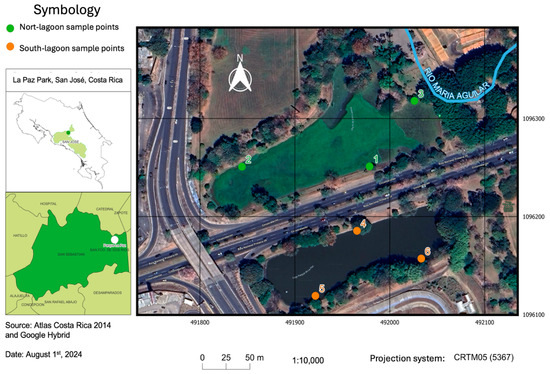
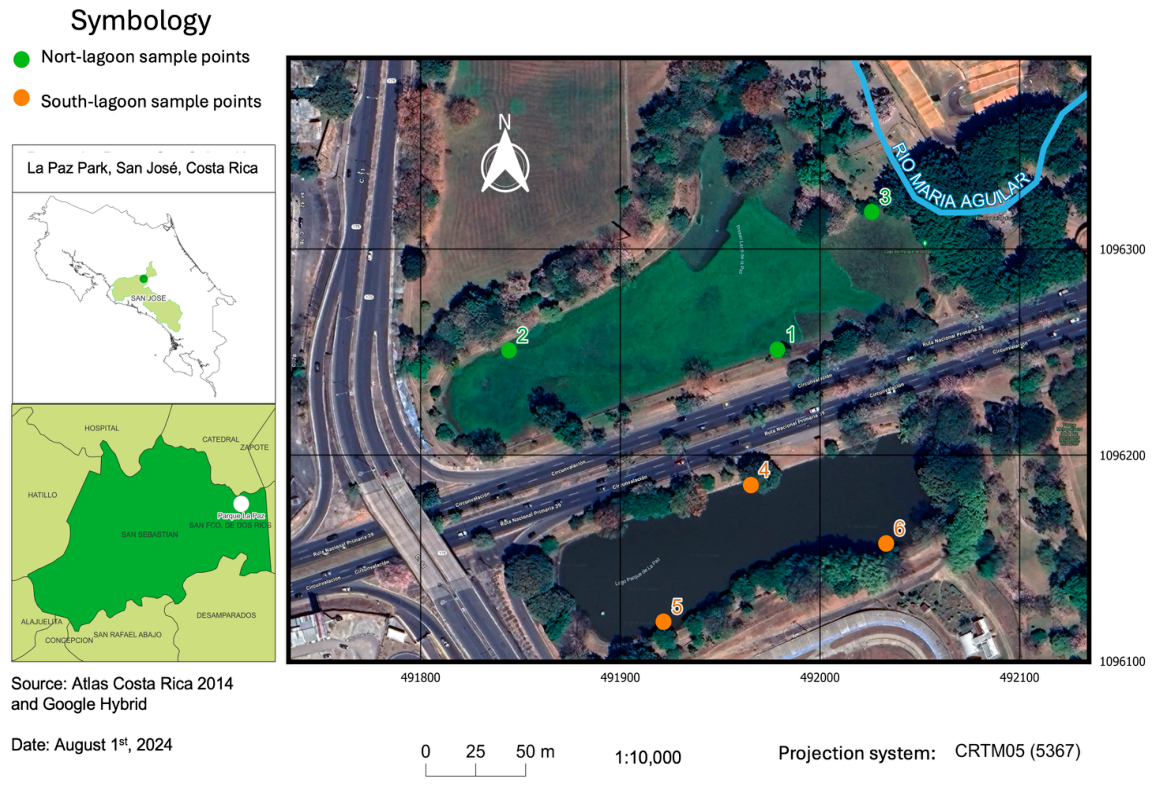
Study sites and sampling. This study was conducted in two urban water bodies within the Greater Metropolitan Area of Costa Rica: the lagoons at La Paz Park and the Toyogres River. The first site (coordinates 9.914268, −84.073507) represents a closed system with no surface inflow or outflow, which can nevertheless receive pollutant inputs from atmospheric deposition, rainfall runoff from green and paved areas, groundwater, and animals. This site was selected for the evaluation of an ecotoxicological assay, given the expected absence of high contaminant levels (Figure 1). Sampling was conducted during both the dry and rainy seasons to analyse seasonal variations in water quality and assess the applicability of the proposed methodologies under different hydrological conditions. In Costa Rica, the dry season (December–April) has average temperatures ranging from 25 °C to 35 °C, with relative humidity between 50% and 70%. During the rainy season (May–November), temperatures range from 22 °C to 30 °C, and humidity levels rise between 80% and 95%, especially during the months of maximum precipitation (September and October) [17]. In each season (3 February 2023, dry; 26 September 2023, rainy), one 1-L water sample was collected from each of six sampling points (three per lagoon; Figure 1) over the entire water column (approximately 60 cm deep), using sterile glass containers (BOECO, Hamburg, Germany), and immediately stored at 4 °C in a refrigerated transport unit.
Figure 1.
Geographical location of the La Paz lagoons in the Central Valley, San José, Costa Rica, showing sampling points 1 to 3 (north lagoon, green dots) and 4 to 6 (south lagoon, orange dots).
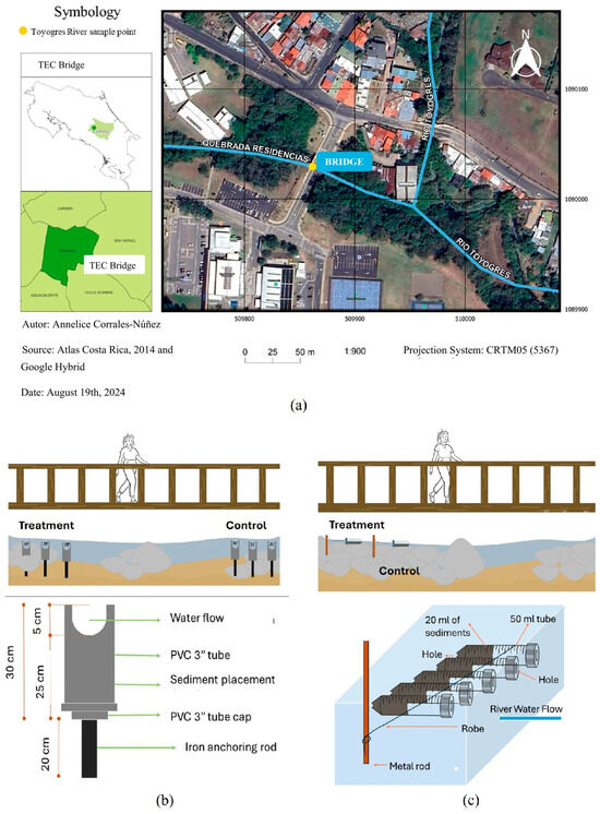
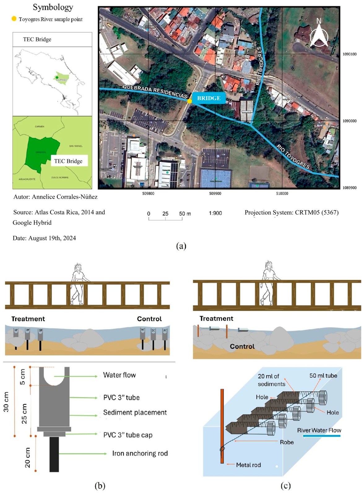
The Toyogres River (coordinates 9.857974, −83.910033), on the other hand, is a lotic system exposed to diffuse discharges from domestic and agricultural sources [16]. This site was chosen to evaluate the effects of sediment bioremediation via bioaugmentation with immobilized microalgae (Figure 2).
Figure 2.
Geographical location and experimental setup in the Toyogres River, Cartago, Costa Rica. (a) Location of the sampling for bioremediation experiments at the Toyogres River, situated under the bridge near the Tecnológico de Costa Rica (TEC) campus (yellow dot). (b) Schematic representation of the experimental units for bioaugmentation tests and their placement beneath the bridge. Each unit consisted of a 3″ PVC tube containing sediment and anchored to the riverbed with an iron rod. (c) Detail of the experimental units designed for biodiversity impact assessment, showing sediment distribution inside the tubes and their orientation with respect to the water flow.
Chemical analysis. 500 mL aliquots were taken for comprehensive chemical analysis, including assessments of metals, volatile organic compounds (VOCs), BTEX compounds, trihalomethanes, and antibiotics. The complete list of analytes, with their limits of quantification (LOQ) and the results for each season, is provided in Supplementary Table S1. Subsamples from the six sampling points were combined into one composite sample per season. LOQs ranged from 0.0001 to 0.05 mg/L for trace metals and phosphorus, from 0.5 to 2.1 mg/L for major elements (Ca, Mg, Na, K, S, and silica), from 0.5 to 5 μg/L for VOCs, BTEX, and trihalomethanes, and were 0.005 μg/L for antibiotics and neonicotinoids. Results were not corrected for recovery factors. The analyses were performed by an internationally accredited external laboratory, which certified the results; the simplified test reports do not include method blanks or recovery data, which are held by the laboratory. The analyses were performed at an external laboratory AGQ Laboratories reports A-23_013094 and A-23_121108, following standardized methodologies (IT-399, SM 3125 Ed 23, EPA 200.8, EPA 6020 B, IT-383, EPA 8260 D, PE-678).
Microalgae strains and culture conditions. Three strains of photosynthetic microorganisms (two green microalgae and one cyanobacterium, hereafter collectively referred to as microalgae for simplicity) from the Microalgae Laboratory of the Technological Institute of Costa Rica were used in this study, Chlorella sp. MT02, Scenedesmus sp. MT33, and Anabaena sp. MT64, and are registered with the National Commission for the Management of Biodiversity (CONAGEBIO) of Costa Rica. Cultures were maintained in BG-11 medium (Phytotechnology Laboratories Shawnee Mission, KS, USA), in an acclimatized room, under controlled conditions, within an orbital shaker (Lab Companion, Jeio Tech, Des Plaines, IL, USA) at 25 °C and 100 rpm. Illumination was provided by 18 W LED lamps with an intensity of 7000 lux (approximately 100–130 μmol photons m−2 s−1, estimated from the illuminance using conversion factors of 0.014–0.019 μmol m−2 s−1 per lux for white LED light; photon flux was not measured directly), following a 16:8 h light:dark photoperiod.
Ecotoxicological assay. Water toxicity to Chlorella sp. MT02, Scenedesmus sp. MT33, and Anabaena sp. MT64 was evaluated using an assay adapted from OECD Test No. 201 (freshwater alga and cyanobacteria growth inhibition test). The assay was designed as a screening tool for environmental samples rather than as a regulatory test and differed from the standard protocol in that each water sample was tested at a single 1:1 dilution with BG-11 medium, without range-finding or a concentration series and, therefore, without EC50 determination; tests were carried out in six-well plates; and the inoculum density, illumination, and temperature were those routinely used in the laboratory, which may differ from the values recommended in the guideline. Cells were grown to the logarithmic phase and harvested to prepare inocula at a concentration of 1 × 10⁶ cells/mL, determined by direct counting in a Neubauer chamber (BOECO, Hamburg, Germany) under an optical microscope (model DM 750, Leica Microsystems, Wetzlar, Germany) at 400× magnification. Each inoculum was prepared in 250 mL Erlenmeyer flasks (Kimax, Kimble Chase, Vineland, NJ, USA) containing 100 mL of BG-11 medium (Phytotech Labs, Lenexa, KS, USA). For each strain, two flat-bottom six-well plates (Corning® Costar®, Corning, NY, USA) were used: one for the toxicity assay and one for growth control. In the control plate, all wells contained 5 mL of BG-11 medium and 250 μL of microalgae inoculum. In the assay plate, five wells contained a 1:1 mixture of BG-11 and the water sample (2.5 mL each, used as five replicates), while in the sixth well the water sample was replaced with 2.5 mL of sterile water to verify that dilution alone did not affect growth (dilution control). Then all wells were inoculated with 250 μL of the respective microalgae inoculum. Baseline cell counts were recorded immediately (time 0 h), followed by additional counts at 24, 48, and 72 h using a Neubauer chamber (BOECO, Germany) and an optical microscope (LEICA DM 750, 400×). The percent inhibition of the average specific growth rate (%Ir) was calculated for each replicate as in OECD Test No. 201 (Equation (2)): %Ir = (μC − μT)/μC × 100, where μC is the mean average specific growth rate in the growth control (BG-11 only) and μT is the average specific growth rate of the treatment replicate. Because %Ir is normalized to μC, it is not bounded between 0 and 100%: values above 100% occur when the treatment replicate shows a negative growth rate (net cell loss), very large values occur when μC approaches zero, and negative values indicate a growth rate higher than that of the control.
Microscopic analysis of water samples. Water samples were examined under an optical microscope (model DM 750, Leica Microsystems) at 40× magnification, and four microscopic fields were observed per sample. Microalgae, protozoa, and larval forms were identified by morphology, and bacteria were observed in Gram-stained smears (Supplementary Figure S1). The abundance of each group was assessed semi-quantitatively from the fields examined and described as sparse, low, or abundant.
DNA extraction. For DNA extraction, 500 mL of each water sample was filtered using cellulose acetate filters with a pore size of 0.20 µm and a diameter of 47 mm (Sartorius, Göttingen, Germany). The filters were then stored in 50 mL conical tubes at −80 °C until further processing. Genomic DNA was extracted using the PowerSoil Pro Kit (Qiagen, Germantown, MD, USA) following the manufacturer’s protocol.
Sequencing. The DNA samples were sent to Macrogen Corp (Beotkkot-ro Geumcheon-g, Seoul, Republic of Korea) for sequencing of the 16S rRNA gene, targeting the V3–V4 hypervariable region. Amplification was carried out using the universal primers Bakt_341F (5′-CCTACGGGNGGCWGCAG-3′) and Bakt_805R (5′-GACTACHVGGGTATCTAATCC-3′), following the methodology described by Klindworth et al. (2013) [18]. Sequencing was performed on the MiSeq platform (Illumina, San Diego, CA, USA), utilizing a paired-end approach with the Nextera XT Index Kit V2 (Illumina, San Diego, CA, USA), generating 300 bp paired-end raw reads.
Bioinformatics and amplicon sequence analysis. Sequencing data were analyzed using PUDU (Pipeline for Universal Diversity Unveiling) [19]. The sequencing data from 24 files, corresponding to forward and reverse raw reads from 12 water samples, were processed using the DADA2 R package (v1.30.0). Initial quality control involved truncating forward reads at position 280 and reverse reads at position 260 to remove low-quality bases. Reads were then filtered to remove sequences with ambiguous bases or more than the maximum number of expected errors, error models were learned from the data, and the number of reads retained at each processing step was tracked. Following this, sequences underwent quality filtering, deduplication, and denoising to infer amplicon sequence variants (ASVs) with improved accuracy [20]. The remaining high-quality sequences were merged into a single ASV count table, representing unique sequence variants across all samples. Chimeric sequences were identified and removed before taxonomic assignment to prevent misinterpretation of microbial diversity. Taxonomic classification was performed using the assignTaxonomy() function, which applies a naïve Bayesian classifier as described by [20]. The SILVA rRNA database v138.1 (silva_nr99_v138.1_train_set.fa) was used as the reference training set for taxonomy assignment. Based on the classified ASVs, abundance bar plots and prevalence plots were generated at different taxonomic levels using the ggplot2 R package (v3.5.1) to visualize microbial community composition and distribution across samples. Relative abundances of the six samples collected in each season were combined to represent the lagoons as a whole. The resulting high-quality, non-redundant ASV dataset provided a robust representation of the microbial communities present in the sampled water bodies at family and genus level.
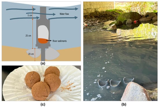
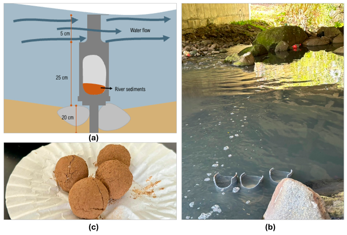
Bioaugmentation with Chlorella sp. in river sediments. To evaluate the potential of Chlorella sp. for the bioremediation of contaminated sediments, a bioaugmentation experiment was conducted in the Toyogres River. PVC structures anchored to the riverbed were used to contain sediments (200 mL per chamber) extracted directly from the river, while preserving natural flow dynamics (Figure 2b and Figure 3a,b). Clay spheres were used as the inoculum carriers for microalgae. Chlorella sp. cultures were grown in BG-11 medium to a concentration of 5 × 10⁶ cells/mL. Local clay was sterilized and manually shaped into spheres (~10 g each) before being dried in an oven (Excalibur EXC10EL, Excalibur Dehydrators, Sacramento, CA, USA; 60 °C, 24 h), reducing their weight to under 8 g (Figure 3c). Once cooled, each sphere was carefully inoculated with 2 mL of microalgae culture, achieving an effective final concentration of 5 × 10⁴ cells/mL when combined with 200 mL of sediment. Each sediment chamber was filled with 200 mL of homogenized sediment, and an aliquot was reserved for sediment monitoring, as will be described ahead. Six chambers were deployed in each season: three treatment chambers, which received a clay sphere inoculated with Chlorella sp., and three control chambers, which received a non-inoculated clay sphere to ensure comparable conditions. A new sphere was added to each chamber at every inoculation event (weeks 0, 2, 5, and 6; see Section 3.2), and sediment was sampled weekly for six weeks. All chambers were securely anchored in the river to maintain natural water flow over the sediment. The chambers were arranged in a line across the width of the river, perpendicular to the direction of flow, with ample spacing between control and treatment chambers, so that the current would not carry inoculum from one chamber to another.
Figure 3.
Sediment retention chambers and clay spheres used for bioaugmentation. (a) Diagram of the chambers; (b) in situ placement in the Toyogres River; and (c) dehydrated clay spheres for microalgae immobilization.
Monitoring of the Chlorella sp.-inoculated river sediments. To evaluate whether sediment inoculation with Chlorella sp. reduced sediment toxicity, the adapted ecotoxicological assay described earlier was performed, substituting the water sample with a sediment sample prepared as follows. At the beginning of the bioaugmentation assay (baseline) and weekly thereafter for six weeks, 5 mL of sediments were collected from each chamber and mixed with 10 mL of BG-11 medium. The mixture was gently stirred, left to settle to allow sand and heavier particles to sediment, and centrifuged. Five replicate 2.5 mL aliquots of the supernatant were transferred to culture wells containing 2.5 mL of BG-11 medium and inoculated with 250 μL of Chlorella sp. at a concentration of 1 × 10⁶ cells/mL, ensuring a final cell concentration of 5 × 10⁴ cells/mL. Cell counts were performed under a light microscope (Leica DM750, 400×) at time 0 and after 24 h to determine growth inhibition levels. The five wells of each chamber were treated as technical replicates, and the three chambers of each treatment as the experimental replicates. The same procedure was applied for Scenedesmus sp. and Anabaena sp., to compare the response of a second green microalga and a cyanobacterium. The dilution of the sediment sample (approximately 1:20 relative to the chamber content) and the centrifugation step minimized the contribution of Chlorella sp. cells already present in the sediment, and cell counts at time 0 were recorded for each well, so that growth was assessed from the change in cell density.
Biodiversity impact assessment. To assess the impact of bioaugmentation with Chlorella sp. on the biodiversity of the Toyogres River sediments, approximately 3 L of sediment were collected from a single sampling point (Figure 2) to minimize variability among samples. Stones and other coarse materials were removed. All samples were obtained from a single batch, ensuring a homogeneous baseline for comparison between treatments. 20 mL of the processed sediment was distributed into six 50 mL Falcon™ tubes (each containing 20 mL of sediment), which served as sediment retention chambers. These tubes were independent of the chambers used for the sediment toxicity experiment. Small perforations (5 mm) were made on both ends of each tube to allow water flow while preventing material loss. Two experimental treatments were established, each with three replicates: a control group without microalgae inoculation and a treatment group inoculated with Chlorella sp. The chambers were anchored in a stable riverbed area, selected for accessibility and the presence of stones that facilitated structural fixation, near a vehicular bridge (Figure 2c). The inoculum consisted of Chlorella sp. at a concentration of 1.84 × 10⁶ cells/mL. A 560 μL aliquot of the inoculum was added to each treatment tube at the beginning of the experiment (week 0), with additional weekly applications over three consecutive weeks. To monitor microbial community changes, sediment samples (1–2 mL) were collected from each tube weekly using sterile spatulas and 2 mL Eppendorf tubes. A 100 μL subsample of the sediment-water mixture was analysed under an optical microscope (LEICA DM 750, 400×). All observable organisms, including diatoms, protozoa, nematodes, and larval forms, were classified into coarse morphological groups and enumerated within six randomly selected microscopic fields per sample. The obtained counts were used to determine total organism abundance and calculate the Shannon-Wiener diversity index (H’). Abundance was defined as the total number of individual organisms observed, while H’ was computed based on the relative proportion of each taxonomic group. The means and standard deviations of abundance and diversity were then compared between the control and treated groups to evaluate whether Chlorella sp. inoculation influenced the composition and structure of the sediment community of microscopic organisms.
Statistical analysis. All experiments were performed in triplicate, except for the biodiversity study in Toyogres River sediments, which had three replicate tubes per treatment. Results are expressed as mean ± standard deviation (SD). Normal distribution and homogeneity of variances were tested, and statistical comparisons between control and treatment groups were conducted using one-way ANOVA and Student’s t-tests in R (v4.4.2), with significance set at p < 0.05. Analyses were performed in RStudio version 9.5.191.709.709, and graphical results were plotted using ggplot2 to assess variability between treatments.
3. Results
3.1. Evaluation of the Lagoons of the La Paz Park
The original reports in Spanish were provided by the accredited laboratory that conducted the chemical analyses of the water samples retrieved from the two lagoons during the dry and rainy seasons (see Data Availability). Based on these reports, we compiled Supplementary Files S1 and S2, which present the results translated into English. As expected, all volatile organic compounds, BTEX, trihalomethanes, antibiotics, and neonicotinoid insecticides were below their limits of quantification (0.5–5 μg/L for VOCs, BTEX, and trihalomethanes; 0.005 μg/L for antibiotics and neonicotinoids) in both seasons. In contrast, most metals were quantified in the composite samples (Supplementary Table S1). Aluminum (0.304 and 1.120 mg/L), iron (0.175 and 1.757 mg/L), and manganese (0.009 and 0.347 mg/L) were considerably higher in the rainy season, whereas copper (0.0147 and 0.0044 mg/L), boron (0.054 and 0.013 mg/L), strontium (0.211 and 0.146 mg/L), and potassium (19 and 1.7 mg/L) were higher in the dry season (values for dry and rainy seasons, respectively). Notably, strontium and potassium exhibited unusual concentrations compared to baseline levels typically reported in similar ecosystems. Zinc was also quantified (0.0763 and 0.0355 mg/L). These metal concentrations could contribute to the growth inhibition observed in the bioassays, and no reference values for the protection of aquatic life were applied in this study; the absence of acute toxicity therefore cannot be asserted from the chemical data alone [21]. Regarding nutrient-related elements, phosphorus and sulfur concentrations were considerably higher in the dry season (4.34 and 12 mg/L) than in the rainy season (0.123 and 2.2 mg/L, the latter close to the LOQ of 2.0 mg/L). Additionally, optical microscopy analysis of water quality revealed a sparse presence of microalgae and a low bacterial load, with all detected organisms exhibiting a relatively homogeneous morphology (Supplementary Figure S1). The presence of protozoa or larval forms was minimal or absent. Furthermore, dissolved oxygen (DO) levels were measured at an average of 3.8 mg/L at a depth of 0.5 m, indicating aerobic conditions.
In the microalgae growth inhibition bioassays, no significant differences (p > 0.05) were observed between the diluted control (sterile water and BG-11, 1:1) and the growth control (BG-11 only). These results confirm that the dilution process did not affect microalgal growth, and support that the observed growth inhibition effects were attributable to water quality rather than methodological artifacts.
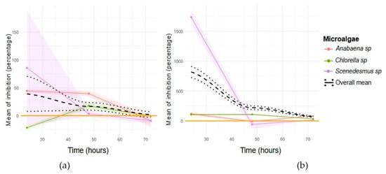
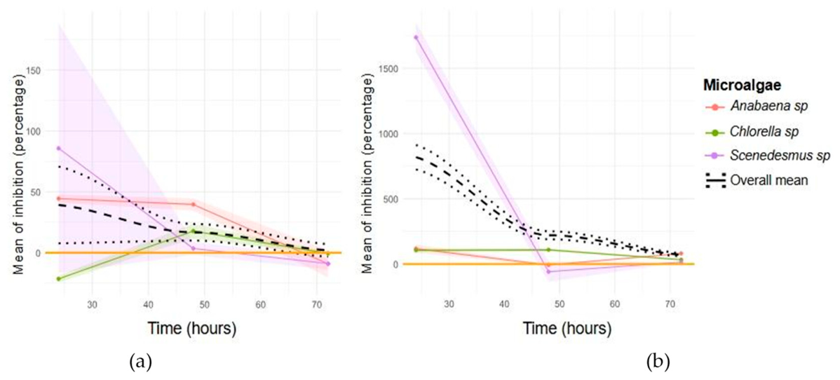
During the dry season (Figure 4a), the microalgae growth inhibition bioassay revealed an initial phase of latency in the tested species (Chlorella sp., Scenedesmus sp., and Anabaena sp.), followed by a transition to exponential growth. Compared to cultures maintained in BG-11 medium, all three species experienced significant inhibition in the first hours of exposure to contaminated water. However, after 72 h, this inhibition effect subsided, with Anabaena sp. exhibiting the most pronounced recovery. These results suggest that, despite initial inhibition, the tested strains may acclimate to the conditions of the water samples, which is consistent with their potential for bioremediation [22].
Figure 4.
Microalgal growth inhibition in La Paz Park lagoons. (a) Inhibition results for the dry season and (b) rainy season, over 72 h of exposure. Coloured shades indicate the standard deviation. Inhibition is the percent inhibition of the average specific growth rate (OECD Test No. 201, Equation (2)); values above 100% indicate a negative growth rate (net cell loss) in the sample, and negative values indicate a growth rate higher than that of the control.
During the rainy season (Figure 4b), the microalgae growth profiles showed a similar trend; however, initial growth inhibition was more severe and persisted throughout the 72-h test. While Scenedesmus sp. managed to overcome inhibition at 48 h, this effect did not sustain at 72 h. Conversely, Chlorella sp. and Anabaena sp. remained under high inhibition levels throughout the test period, with only a slight reduction at 72 h in some cases. Growth rates and inhibition percentages recorded during the rainy season suggest that, even under increased pollutant loads from stormwater runoff, microalgae exhibit a certain degree of acclimatization. However, the persistence of residual toxicity was more pronounced compared to the dry season.
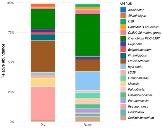
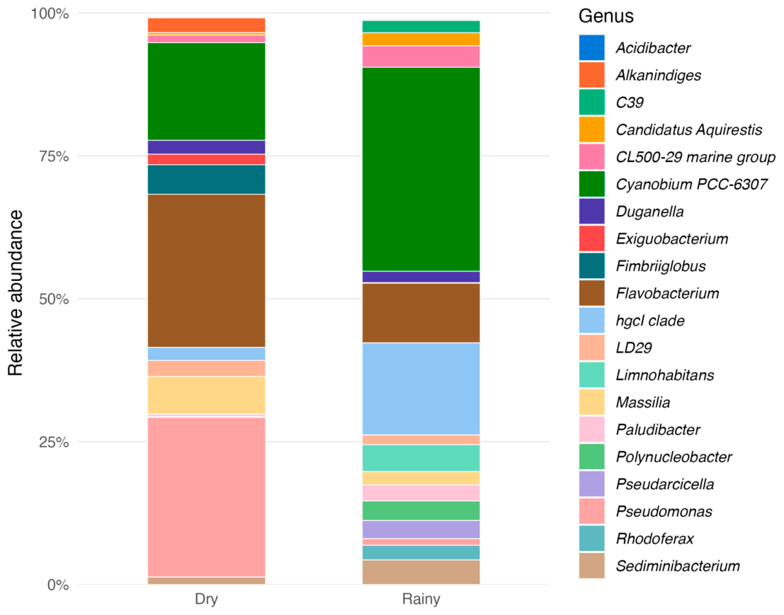
The bioassay results coincided with a marked seasonal shift in the relative composition of the bacterial community, as revealed by 16S rRNA amplicon sequencing. Taxonomic profiles at both the family and genus levels (Figure 5 and Figure S2) illustrate distinct changes in microbial composition between seasons. During the dry season, the community was dominated by Pseudomonas and Flavobacterium, which together accounted for more than half of the displayed relative abundance. Cyanobium PCC-6307 was also well represented, whereas Massilia, Fimbriiglobus, Duganella, and the hgcI clade occurred in lower proportions.
Figure 5.
Relative abundance of dominant bacterial genera in La Paz Park lagoons at dry and rainy seasons. Each bar represents the six samples collected in each season (three per lagoon), combined to represent both lagoons as a whole, and shows the genus-level taxonomic composition.
During the rainy season, the taxonomic profile changed markedly. Cyanobium PCC-6307 became the dominant taxon, and the relative abundance of the hgcI clade increased substantially. In contrast, Pseudomonas and Flavobacterium decreased compared with the dry season. Several freshwater-associated taxa, including Limnohabitans, Polynucleobacter, Sediminibacterium, Pseudarcicella, Paludibacter, and Rhodoferax, also showed greater representation during the rainy season. Conversely, Fimbriiglobus, Alkanindiges, Exiguobacterium, and Massilia declined, while Duganella remained at a relatively low proportion and did not exhibit a substantial seasonal increase.
Overall, the bacterial community shifted from a heterotroph-dominated structure during the dry season to a more taxonomically even and functionally diverse community in the rainy season. This suggests a restructuring of the microbial community in response to seasonal shifts.
3.2. Effect of Chlorella sp. Bioaugmentation on Sediment Toxicity
Chlorella sp. was selected as the bioaugmentation agent, mainly because its unicellular morphology makes it easy to count under the microscope, which reduces counting uncertainty, and because it was the simplest strain to handle, although it did not show the best recovery in the lagoon bioassays, and its potential for sediment bioremediation in the Toyogres River was assessed. Inoculations were performed at weeks 0, 2, 5, and 6, monitoring sediment quality over time. The intermittent application of inoculations allowed for an evaluation of the response time to treatment and the potential regression of sediment quality when treatment was discontinued.
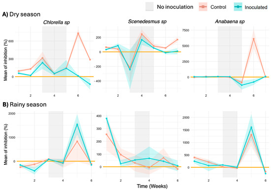
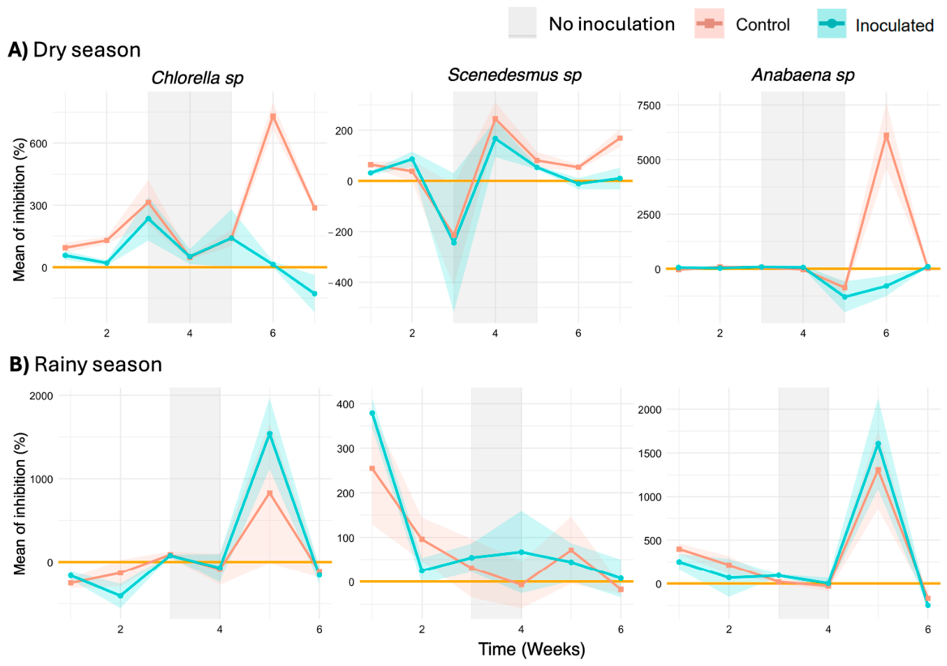
The ANOVA analysis revealed significant differences across sampling weeks in both dry and rainy seasons (Figure 6), indicating high temporal variability in sediment toxicity. During the dry season, treatment effects were statistically significant for Chlorella sp. (p < 0.001), Scenedesmus sp. (p < 0.001), and Anabaena sp. (p = 0.026). Significant variation across weeks was also observed for all species (p = 0.004 for Chlorella sp., p < 0.001 for Scenedesmus sp., and p = 0.004 for Anabaena sp.), which is consistent with a bioremediation effect of Chlorella sp. bioaugmentation. In contrast, during the rainy season, while week-to-week variation remained significant (p < 0.001 for all species), treatment effects were not significant (p = 0.447 for Chlorella sp., p = 0.278 for Scenedesmus sp., and p = 0.660 for Anabaena sp.), possibly related to increased flow rates associated with rainfall runoff (river discharge was not measured). This higher flow may have diluted contaminants or exceeded the capacity of Chlorella sp. to improve sediment quality effectively.
Figure 6.
Effect of Chlorella sp. bioaugmentation on sediment toxicity. Inhibition values for Chlorella sp., Scenedesmus sp., and Anabaena sp. in dry and rainy seasons; grey shading indicates periods without treatment, other colored shades indicate the standard deviation. Inhibition is the percent inhibition of the average specific growth rate (OECD Test No. 201, Equation (2)); values above 100% indicate a negative growth rate (net cell loss) in the sample, and negative values indicate a growth rate higher than that of the control.
During the dry season, inhibition values for microalgal growth progressively decreased following the application of Chlorella sp. (Figure 6). A noticeable increase in inhibition was observed when inoculations were discontinued at weeks 3 and 4, which suggests that periodic and sustained bioaugmentation may be needed to maintain sediment recovery. Anabaena sp. maintained a relatively stable growth rate but exhibited a reduction in inhibition toward the end of the treatment, suggesting higher sensitivity to environmental conditions. Conversely, Scenedesmus sp. followed a similar trend to Chlorella sp., though its differences relative to the control group were less pronounced.
In contrast, during the rainy season, greater variability in microalgal growth inhibition was observed, with similar behaviours between the control and Chlorella sp.-inoculated chambers (Figure 6). The lack of significant differences between treatments is compatible with the hypothesis that increased river flow may disperse contaminants and reduce the effectiveness of bioaugmentation under these conditions, although river discharge was not measured.
3.3. Impact of Bioaugmentation on Sediment Biodiversity
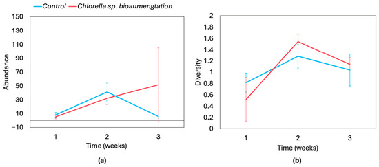
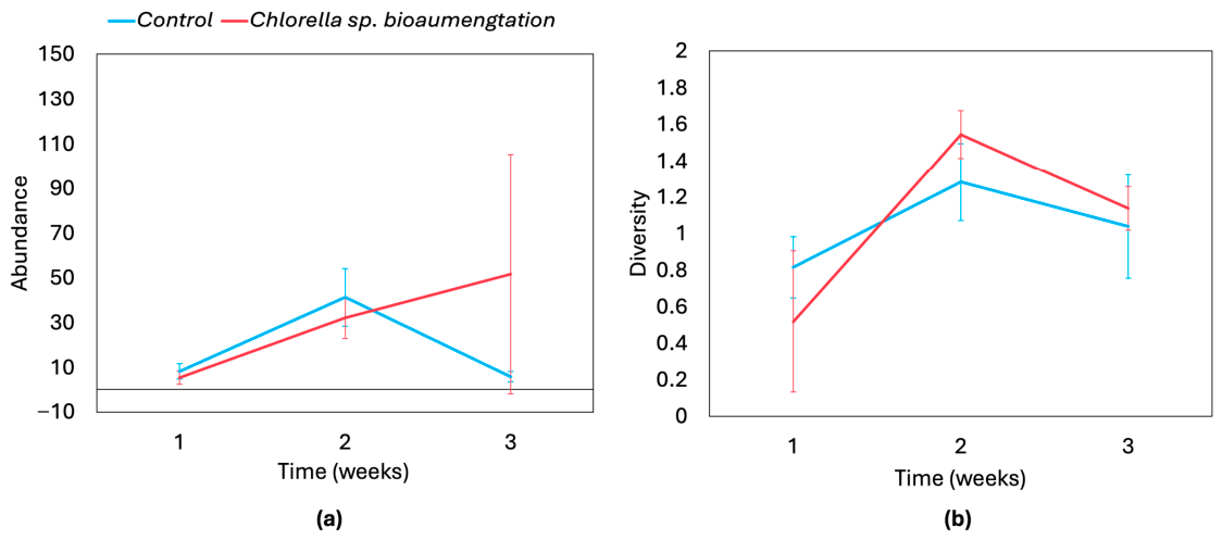
The results indicate a temporal shift in both the total abundance of microscopic organisms and Shannon-Wiener biodiversity index values, with differences between treated and control sediments (Figure 7). During the initial sampling period, both metrics were relatively low; however, control systems exhibited slightly higher biodiversity than treated sediments. This suggests that the introduction of Chlorella sp. inoculum may have temporarily suppressed some native sediment microorganisms, potentially due to competition for nutrients or secondary metabolite production [23].
Figure 7.
Biodiversity response to Chlorella sp. bioaugmentation in Toyogres River sediments. Temporal changes in (a) total abundance of microscopic organisms and (b) the Shannon–Wiener diversity index, mean ± SD (n = 3).
By the second week, both total abundance and biodiversity showed an apparent increase in treated sediments compared to controls, suggesting a potential trend toward a more balanced microbial distribution and improved community structuring [24]. However, these differences were not statistically significant due to the high variability observed among replicates. By the third week, a similar tendency was observed, with a slight decline in microbial abundance in control systems and apparent recovery in sediments treated with Chlorella sp., although again without statistical support. At the end of the experiment, biodiversity indices and the total abundance of microscopic organisms appeared higher in bioaugmented sediments than in controls, yet these trends did not reach statistical significance. These observations therefore represent tendencies rather than confirmed effects, indicating that while Chlorella sp. may influence microbial community dynamics, further replicated studies would be required to substantiate its role as a bioremediation agent in urban river sediments.
4. Discussion
The physicochemical analyses of water samples from the lagoons at La Paz Park revealed that organic contaminants were below the limits of quantification, although several metals were quantified, consistent with the fact that this is a closed water body with no surface inflow or outflow, which limits, but does not eliminate, pollutant inputs from rainfall runoff and animals. However, its location in a highly urbanized area exposes it to constant atmospheric deposition of airborne pollutants, contributing to some degree of contamination [1].
During the dry season, microalgae growth inhibition bioassays showed that initial inhibition was overcome after 72 h. This suggests that, despite the presence of potentially toxic compounds, microalgae were able to acclimate to the water conditions and resume growth. In the rainy season, however, inhibition was more severe at the start of the experiment, but all species exhibited a gradual reduction in inhibition over time. This could be due to an increased pollutant load from runoff, leading to higher toxicity and synergistic effects among multiple contaminants [25,26].
The bioassay results indicate that water from the lagoons at La Paz Park initially exhibited significant toxicity for the microalgae used in this study. However, microalgae were able to recover and grow after a period of acclimatization, suggesting that the toxic effects were transient and that self-remediation potential exists. These findings align with previous research showing that microalgae can adapt to polluted environments through detoxification mechanisms and the bioaccumulation of contaminants [7,8,11,27,28].
The 16S rRNA amplicon profiles and the microscopic observations are compatible with a stressed system, as counts of microscopic organisms were low and the dominant taxa have been reported in contaminated or stressed ecosystems; however, relative abundance data cannot establish absolute abundance, and no qPCR, flow cytometry, or total cell counts were available to confirm this [29,30].
At the genus level, the bacterial profiles indicate pronounced seasonal compositional turnover rather than a single, persistent contamination signature. During the dry season, Pseudomonas and Flavobacterium together represented more than half of the displayed reads, whereas the rainy-season profile was dominated by Cyanobium PCC-6307 and the hgcI clade, accompanied by a broader representation of typically freshwater-associated genera [31]. Comparable studies have shown that in freshwater systems, bacterial community composition has been linked to seasonal variation in temperature, precipitation, and nutrient availability, while research in a sewage-polluted river has shown that seasonal dynamics also influence community responses to wastewater inputs [31,32]. The observed community turnover may therefore reflect environmental filtering and seasonal hydrological influences, although the present data do not resolve the relative contributions of these processes and anthropogenic inputs.
The dry-season dominance of Pseudomonas and Flavobacterium is consistent with a greater contribution of metabolically versatile heterotrophs under conditions of longer water residence times and the accumulation of locally produced or deposited organic matter. In eutrophic lakes, increases in Flavobacteria have been closely associated with resource availability and periods of high bacterial production [33]. The predominance of these genera suggests a seasonal shift in bacterial community composition, potentially linked to the availability of organic matter during the dry season.
The rainy-season increase in Cyanobium PCC-6307 indicates a stronger relative contribution of freshwater picocyanobacteria [34]. Members of this taxon are widely distributed in lakes, reservoirs, and rivers, and their dynamics can be influenced by temperature, nutrient ratios, mixing, and light availability [32,33,35]. The higher relative abundance of Cyanobium PCC-6307 may reflect changes in temperature, nutrient availability, mixing, and light conditions that influence picocyanobacterial dynamics in freshwater systems [32,33,36]. Its concurrent increase with the hgcI clade is notable, given their distinct environmental associations. In the Meuse River, both taxa were positively correlated with temperature, whereas Cyanobium was positively associated with total suspended matter, and the hgcI clade showed a negative relationship [29]. Their joint increase in the present study may therefore reflect different responses to rainy-season conditions. Together with the greater representation of Limnohabitans and Polynucleobacter, this pattern suggests that seasonal changes favored several characteristic freshwater bacterioplankton lineages [37,38,39]. Changes in Sediminibacterium, Pseudarcicella, Paludibacter, and Rhodoferax may likewise reflect seasonal variation in hydrology and resource availability.
The findings suggest that microalgae growth inhibition bioassays can be a sensitive complementary tool for detecting sublethal effects of contamination in urban water bodies with low pollutant loads, allowing for the identification of toxic effects even when conventional chemical analyses do not indicate high concentrations of specific substances [40].
Bioaugmentation trials in the Toyogres River provided preliminary evidence on the potential of microalgae in sediment bioremediation. During the dry season, sediment quality improved with the periodic application of Chlorella sp., reflected in a progressive reduction of microalgal growth inhibition in bioassays and a non-significant tendency toward higher diversity of microscopic organisms. These results are compatible with the hypothesis that microalgae may contribute to the recovery of contaminated sediments by modifying microbial community structure and reducing the toxicity of present compounds. However, discontinuing treatments at specific points in the trial was followed by an increase in inhibition, suggesting that regular and consistent applications may be necessary to maintain the bioremediation effect.
During the rainy season, the positive effects of bioaugmentation were less evident possibly because of high flow variability and greater pollutant dispersion [16]. The lack of significant differences between treatments and controls during this period suggests that river hydrodynamics may play an important role in bioremediation efficiency, possibly requiring complementary strategies to enhance inoculum retention in sediments and prolong its effect.
The biodiversity analysis of Toyogres River sediments revealed that bioaugmentation with Chlorella sp. was associated with changes in the abundance and diversity of microscopic organisms that were not statistically significant and varied over time. During the first few weeks, inoculation appeared to temporarily suppress some native microorganisms, possibly due to resource competition or secondary metabolite production by microalgae. However, by week two, a non-significant increase in diversity was observed in treated sediments, which might indicate a more balanced taxonomic distribution. At the end of the experiment, abundance appeared higher in treated than in control sediments, but neither abundance nor the diversity index differed significantly, so these observations should be regarded as trends.
Limitations
This study has several limitations that restrict the extrapolation of its findings. First, it was based on a single lagoon complex and a single river site, without replication at the system level, and it should therefore be regarded as a pilot study. Second, no river discharge or contemporaneous pollutant data were available, so the proposed influence of runoff and flow on growth inhibition and on bioaugmentation efficiency remains hypothetical. Third, chemical analyses were performed on one composite sample per season, and unmeasured or unregulated pollutants cannot be ruled out as contributors to the observed growth inhibition. Fourth, the 16S rRNA amplicon data describe relative abundances only, and no multivariate analysis relating community composition to chemical or hydrological variables was possible, so the seasonal patterns remain correlative. Finally, the microalgal bioassay is an adapted version of OECD Test No. 201 with a single dilution, so no EC50 values could be derived and the results should be interpreted as a screening of relative growth inhibition.
5. Conclusions
The adapted microalgae growth inhibition bioassay showed potential as a complementary screening tool for detecting sublethal effects of contamination in urban water bodies, even when chemical analyses did not indicate elevated concentrations of organic pollutants.
16S rRNA amplicon sequencing revealed seasonal shifts in bacterial community composition in the La Paz Park lagoons, from dry-season dominance of Pseudomonas and Flavobacterium to greater relative abundance of Cyanobium PCC-6307 and the hgcI clade during the rainy season. These findings highlight the importance of accounting for seasonal community variation when assessing urban water quality and support the use of 16S rRNA amplicon profiling to complement physicochemical measurements and microalgal bioassays.
Bioaugmentation with Chlorella sp. in the sediments of the Toyogres River was associated with a reduction in sediment toxicity during the dry season, whereas the apparent increases in the abundance and diversity of microscopic organisms were not statistically significant. However, no treatment effect was detected in the rainy season, possibly because of high flow variability, suggesting the need for strategies to improve inoculum retention in sediments. Replicated studies at multiple sites, with hydrological monitoring, are needed to confirm these preliminary results.
Supplementary Materials
The following supporting information can be downloaded at https://www.mdpi.com/article/10.3390/su182110903/s1, File S1: Simplified test report of the chemical analyses performed at La Paz Lagoons during the dry season; File S2: Simplified test report of the chemical analyses performed at La Paz Lagoons during the rainy season; Figure S1: Gram staining of water samples from La Paz lagoons; Figure S2: Relative abundance of dominant bacterial families during the rainy and dry seasons at La Paz Lagoons; Table S1: Analytes, limits of quantification, and concentrations in the composite samples of La Paz lagoons.
Author Contributions
Conceptualization, M.C.-R., A.C.-N., R.G.-W., F.M.-V. and O.R.-S.; methodology, M.C.-R., A.C.-N., R.G.-W., F.M.-V. and O.R.-S.; software, A.C.-N. and A.M.-M.; validation, R.G.-W. and O.R.-S.; formal analysis, M.C.-R. and S.F.-B.; investigation, M.C.-R., A.C.-N., R.G.-W., F.M.-V., S.F.-B., A.M.-M. and O.R.-S.; resources, M.C.-R.; data curation, M.C.-R. and A.M.-M.; writing—original draft preparation, M.C.-R. and F.M.-V.; writing—review and editing, A.C.-N., A.M.-M., F.M.-V., M.C.-R., O.R.-S., R.G.-W. and S.F.-B.; visualization, A.C.-N., A.M.-M., F.M.-V., M.C.-R., O.R.-S., R.G.-W. and S.F.-B.; supervision, M.C.-R.; project administration, M.C.-R.; funding acquisition, M.C.-R., O.R.-S. and R.G.-W. All authors have read and agreed to the published version of the manuscript.
Funding
This research was funded by the Vicerectorate of Research and Extension (VIE) of Instituto Tecnológico de Costa Rica, grant number 1510185 (Project 1510185: Development of an inter-institutional strategy for the reduction of emerging pollutants in water bodies of the GAM, Costa Rica). The APC were funded by the Vicerectorate of Research and Extension (VIE) of Instituto Tecnológico de Costa Rica.
Institutional Review Board Statement
Not applicable.
Informed Consent Statement
Not applicable.
Data Availability Statement
The raw 16S rRNA gene sequencing data generated in this study have been deposited in the NCBI Sequence Read Archive (SRA) under BioProject accession number PRJNA1514957, and the original reports or the chemical analyses, in Spanish, are available at https://doi.org/10.5281/zenodo.22033013.
Acknowledgments
During the preparation of this manuscript/study, the authors used Google Translate (Google LLC; https://translate.google.com; accessed 21 November 2025) for the purposes of translating the laboratory reports from Spanish to English. The authors have reviewed the output and take full responsibility for the content of this publication.
Conflicts of Interest
The authors declare no conflicts of interest.
References
- Saxena, V. Water Quality, Air Pollution, and Climate Change: Investigating the Environmental Impacts of Industrialization and Urbanization. Water Air Soil Pollut. 2025, 236, 73. [Google Scholar] [CrossRef] [Scilit]
- Guan, Y.; Zhang, N.; Chu, C.; Xiao, Y.; Niu, R.; Shao, C. Health Impact Assessment of the Surface Water Pollution in China. Sci. Total Environ. 2024, 933, 173040. [Google Scholar] [CrossRef] [Scilit]
- Omar, W.M.W. Perspectives on the Use of Algae as Biological Indicators for Monitoring and Protecting Aquatic Environments, with Special Reference to Malaysian Freshwater Ecosystems. Trop. Life Sci. Res. 2010, 21, 51–67. [Google Scholar]
- Ahmed, S. Algae as Bioindicators of Aquatic Pollution. J. Adv. Res. Altern. Energy Environ. Ecol. 2024, 11, 1–17. [Google Scholar] [CrossRef] [Scilit]
- Mulenga, M.; Monde, C.; Johnson, T.; Ouma, K.O.; Syampungani, S. Advances in the Integration of Microalgal Communities for Biomonitoring of Metal Pollution in Aquatic Ecosystems of Sub-Saharan Africa. Environ. Sci. Pollut. Res. 2024, 31, 40795–40817. [Google Scholar] [CrossRef] [Scilit]
- Chieti, M.G.; Petrucciani, A.; Mollo, L.; Gerotto, C.; Eusebi, A.L.; Fatone, F.; Norici, A.; González-Camejo, J. Acclimated Green Microalgae Consortium to Treat Sewage in an Alternative Urban WWTP in a Coastal Area of Central Italy. Sci. Total Environ. 2024, 945, 174056. [Google Scholar] [CrossRef] [Scilit]
- Thoré, E.S.J.; Muylaert, K.; Bertram, M.G.; Brodin, T. Microalgae. Curr. Biol. 2023, 33, R91–R95. [Google Scholar] [CrossRef] [Scilit]
- Machado, A.A.; Valiaparampil, J.G.; Lavanya, M. Unlocking the Potential of Algae for Heavy Metal Remediation. Water Air Soil Pollut. 2024, 235, 629. [Google Scholar] [CrossRef] [Scilit]
- Faruque, M.O.; Uddin, S.; Hossain, M.M.; Hossain, S.M.Z.; Shafiquzzaman, M.; Razzak, S.A. A Comprehensive Review on Microalgae-Driven Heavy Metals Removal from Industrial Wastewater Using Living and Nonliving Microalgae. J. Hazard. Mater. Adv. 2024, 16, 100492. [Google Scholar] [CrossRef] [Scilit]
- Ali, S.S.; Hassan, L.H.S.; El-Sheekh, M. Microalgae-Mediated Bioremediation: Current Trends and Opportunities—A Review. Arch. Microbiol. 2024, 206, 343. [Google Scholar] [CrossRef] [Scilit]
- Severo, I.A.; de Avila Azevedo, O.G.; da Silva, P.A.S.; Jacob-Furlan, B.; Mariano, A.B.; Ordonez, J.C.; Vargas, J.V.C. Wastewater Treatment Process Using Immobilized Microalgae. Water Sci. Technol. 2024, 90, 1306–1320. [Google Scholar] [CrossRef] [Scilit]
- van Lidth de Jeude, M.; Schütte, O.; Quesada, F. The Vicious Circle of Social Segregation and Spatial Fragmentation in Costa Rica’s Greater Metropolitan Area. Habitat Int. 2016, 54, 65–73. [Google Scholar] [CrossRef] [Scilit]
- Francis, R.A. Urban Rivers: Novel Ecosystems, New Challenges. WIREs Water 2014, 1, 19–29. [Google Scholar] [CrossRef] [Scilit]
- Padowski, J.C.; Gorelick, S.M. Global Analysis of Urban Surface Water Supply Vulnerability. Environ. Res. Lett. 2014, 9, 104004, Erratum in Environ. Res. Lett. 2014, 9, 119501. https://doi.org/10.1088/1748-9326/9/11/119501. [Google Scholar] [CrossRef] [Scilit]
- Costadone, L. Urban Lakes: Ecosystem Services and Management. Ph.D. Thesis, Portland State University, Portland, OR, USA, 2021. [Google Scholar]
- Camacho-Jiménez, R.d.l.Á. Plan de Gestión Comunitaria Para La Microcuenca Del Río Toyogres: Estrategias Para La Reducción de La Contaminación En Ríos de Oreamuno y Cartago, Costa Rica. Licentiate Thesis, Instituto Tecnológico de Costa Rica, Cartago, Costa Rica, 2024. [Google Scholar]
- Instituto Meteorológico Nacional El Clima y las Regiones Climáticas de Costa Rica. Available online: https://www.imn.ac.cr/documents/10179/31165/clima-regiones-climat.pdf/cb3b55c3-f358-495a-b66c-90e677e35f57 (accessed on 6 March 2025).
- Klindworth, A.; Pruesse, E.; Schweer, T.; Peplies, J.; Quast, C.; Horn, M.; Glöckner, F.O. Evaluation of General 16S Ribosomal RNA Gene PCR Primers for Classical and Next-Generation Sequencing-Based Diversity Studies. Nucleic Acids Res. 2013, 41, e1. [Google Scholar] [CrossRef] [Scilit]
- Medaglia-Mata, A.; Rojas-Rodríguez, P.; Bystrý, V.; Guillén-Watson, R.; Gómez-Espinoza, O.; Núñez-Montero, K. PUDU (Pipeline for Universal Diversity Unveiling): An Accessible End-to-End Workflow for Taxonomic Profiling and Ecological Visualization of Environmental Microbiomes across Amplicon, Shotgun, and Long-Read Sequencing. Front. Bioinform. 2026, 6, 1909327. [Google Scholar] [CrossRef] [Scilit]
- Wang, Q.; Garrity, G.M.; Tiedje, J.M.; Cole, J.R. Naive Bayesian Classifier for Rapid Assignment of RRNA Sequences into the New Bacterial Taxonomy. Appl. Environ. Microbiol. 2007, 73, 5261–5267. [Google Scholar] [CrossRef] [Scilit]
- Calvo-Brenes, G.; Quirós-Bustos, N.; Robles-Chaves, D.; Caballero-Chavarría, A.; Navarro-Garro, A.; Araya-Rodríguez, F. Elaboración de Un Modelo Para Predecir La Vulnerabilidad de Metales Pesados En Agua y Sedimentos En Distintas Regiones Del País [Development of a Model to Predict the Vulnerability of Heavy Metals in Water and Sediments in Different Regions of the Country]; Instituto Tecnológico de Costa Rica: Cartago, Costa Rica, 2023. [Google Scholar]
- Abdelfattah, A.; Ali, S.S.; Ramadan, H.; El-Aswar, E.I.; Eltawab, R.; Ho, S.H.; Elsamahy, T.; Li, S.; El-Sheekh, M.M.; Schagerl, M.; et al. Microalgae-Based Wastewater Treatment: Mechanisms, Challenges, Recent Advances, and Future Prospects. Environ. Sci. Ecotechnology 2023, 13, 100205. [Google Scholar] [CrossRef] [Scilit]
- Alazaiza, M.Y.D.; He, S.; Su, D.; Abu Amr, S.S.; Toh, P.Y.; Bashir, M.J.K. Sewage Water Treatment Using Chlorella Vulgaris Microalgae for Simultaneous Nutrient Separation and Biomass Production. Separations 2023, 10, 229. [Google Scholar] [CrossRef] [Scilit]
- Pickering, L.; Castro-Gutierrez, V.; Holden, B.; Haley, J.; Jarvis, P.; Campo, P.; Hassard, F. How Bioaugmentation for Pesticide Removal Influences the Microbial Community in Biologically Active Sand Filters. Chemosphere 2024, 363, 142956. [Google Scholar] [CrossRef] [Scilit]
- Werbowski, L.M.; Gilbreath, A.N.; Munno, K.; Zhu, X.; Grbic, J.; Wu, T.; Sutton, R.; Sedlak, M.D.; Deshpande, A.D.; Rochman, C.M. Urban Stormwater Runoff: A Major Pathway for Anthropogenic Particles, Black Rubbery Fragments, and Other Types of Microplastics to Urban Receiving Waters. ACS ES T Water 2021, 1, 1420–1428. [Google Scholar] [CrossRef] [Scilit]
- Zanoletti, A.; Bontempi, E. Editorial: Urban Runoff of Pollutants and Their Treatment. Front. Environ. Chem. 2023, 4, 1151859. [Google Scholar] [CrossRef] [Scilit]
- Yousif, Y.I.D.; Mohamed, E.S.; El-Gendy, A.S. Using Chlorella Vulgaris for Nutrient Removal from Hydroponic Wastewater: Experimental Investigation and Economic Assessment. Water Sci. Technol. 2022, 85, 3240–3258. [Google Scholar] [CrossRef] [Scilit]
- Fahimah, N.; Salami, I.R.S.; Oginawati, K.; Yapfrine, S.J.; Supriatin, A.; Thaher, Y.N. Mapping and Identifying Heavy Metals in Water Use as Chemicals of Potential Concerns in Upper Watershed. Glob. J. Environ. Sci. Manag. 2023, 9, 765–788. [Google Scholar] [CrossRef]
- Adams, S.M. Biomarker/Bioindicator Response Profiles of Organisms Can Help Differentiate between Sources of Anthropogenic Stressors in Aquatic Ecosystems. Biomarkers 2001, 6, 33–44. [Google Scholar] [CrossRef] [Scilit]
- Qin, Y.; Hou, J.; Deng, M.; Liu, Q.; Wu, C.; Ji, Y.; He, X. Bacterial Abundance and Diversity in Pond Water Supplied with Different Feeds. Sci. Rep. 2016, 6, 35232. [Google Scholar] [CrossRef] [Scilit]
- Barberoux, V.; Anzil, A.; Meinertzhagen, L.; Nguyen-Dinh, T.; Servais, P.; George, I.F. Spatio–Temporal Dynamics of Bacterial Community Composition in a Western European Watershed, the Meuse River Watershed. FEMS Microbiol. Ecol. 2025, 101, 22. [Google Scholar] [CrossRef] [Scilit]
- Marmen, S.; Fadeev, E.; Al Ashhab, A.; Benet-Perelberg, A.; Naor, A.; Patil, H.J.; Cytryn, E.; Viner-Mozzini, Y.; Sukenik, A.; Lalzar, M.; et al. Seasonal Dynamics Are the Major Driver of Microbial Diversity and Composition in Intensive Freshwater Aquaculture. Front. Microbiol. 2021, 12, 679743. [Google Scholar] [CrossRef] [Scilit]
- Eiler, A.; Bertilsson, S. Flavobacteria Blooms in Four Eutrophic Lakes: Linking Population Dynamics of Freshwater Bacterioplankton to Resource Availability. Appl. Environ. Microbiol. 2007, 73, 3511–3518. [Google Scholar] [CrossRef] [Scilit]
- Huber, P.; Cornejo-Castillo, F.M.; Ferrera, I.; Sánchez, P.; Logares, R.; Metz, S.; Balagué, V.; Acinas, S.G.; Gasol, J.M.; Unrein, F. Primer Design for an Accurate View of Picocyanobacterial Community Structure by Using High-Throughput Sequencing. Appl. Environ. Microbiol. 2019, 85, e02659-18. [Google Scholar] [CrossRef] [Scilit]
- Barsanti, L.; Vismara, R.; Passarelli, V.; Gualtieri, P. Paramylon (β-1,3-Glucan) Content in Wild Type and WZSL Mutant of Euglena Gracilis. Effects of Growth Conditions. J. Appl. Phycol. 2001, 13, 59–65. [Google Scholar] [CrossRef] [Scilit]
- Paruch, L.; Paruch, A.M.; Eiken, H.G.; Sørheim, R. Faecal Pollution Affects Abundance and Diversity of Aquatic Microbial Community in Anthropo-Zoogenically Influenced Lotic Ecosystems. Sci. Rep. 2019, 9, 19469. [Google Scholar] [CrossRef] [Scilit]
- Jezberová, J.; Jezbera, J.; Znachor, P.; Nedoma, J.; Kasalický, V.; Šimek, K. The Limnohabitans Genus Harbors Generalistic and Opportunistic Subtypes: Evidence from Spatiotemporal Succession in a Canyon-Shaped Reservoir. Appl. Environ. Microbiol. 2017, 83, e01530-17. [Google Scholar] [CrossRef] [Scilit]
- Nuy, J.K.; Hoetzinger, M.; Hahn, M.W.; Beisser, D.; Boenigk, J. Ecological Differentiation in Two Major Freshwater Bacterial Taxa Along Environmental Gradients. Front. Microbiol. 2020, 11, 509874. [Google Scholar] [CrossRef] [Scilit]
- Newton, R.J.; Jones, S.E.; Helmus, M.R.; McMahon, K.D. Phylogenetic Ecology of the Freshwater Actinobacteria AcI Lineage. Appl. Environ. Microbiol. 2007, 73, 7169–7176. [Google Scholar] [CrossRef] [Scilit]
- Holt, E.A.; Miller, S.W. Bioindicators: Using Organisms to Measure Environmental Impacts. Nat. Educ. Knowl. 2010, 3, 8. [Google Scholar]
Disclaimer/Publisher’s Note: The statements, opinions and data contained in all publications are solely those of the individual author(s) and contributor(s) and not of MDPI and/or the editor(s). MDPI and/or the editor(s) disclaim responsibility for any injury to people or property resulting from any ideas, methods, instructions or products referred to in the content. |
© 2026 by the authors. Licensee MDPI, Basel, Switzerland. This article is an open access article distributed under the terms and conditions of the Creative Commons Attribution (CC BY) license.