


Abstract
Background: Ivabradine, an inhibitor of funny current channels, is used in the management of sinus tachycardia. This work assessed the efficacy of ivabradine as a possible prophylactic candidate for acute liver injury (ALI) in mice. Methods: ALI was induced via acetaminophen (APAP) injection. Male Swiss Albino mice were allocated to six groups randomly, as follows: control, ivabradine control, APAP, NAC + APAP, ivabradine 10 + APAP, and ivabradine 20 + APAP. Ivabradine and NAC were given orally once daily for three successive days before the APAP challenge (with the final dose given 2 h before APAP). Different biochemical, pathological, immunohistochemical, and molecular assessments were adopted to evaluate the efficacy of ivabradine. Results: The significant elevation of the serum liver injury markers ALT, AST, and ALP, together with the significant histopathological alterations observed in hepatic tissues, confirmed the occurrence of APAP-induced ALI. APAP induced lipid peroxidation and depleted hepatic antioxidants, which were associated with significantly restrained endogenous antioxidative defense mechanisms of the hepatocytes, such as SIRT1/Nrf2. Additionally, APAP downregulated AMPK while inducing mTOR expression. Interestingly, pretreatment with ivabradine markedly attenuated APAP-induced hepatic injury, which was associated with restoration of hepatic SIRT1 and Nrf2 expression alongside favorable modulation of AMPK/mTOR signaling, along with significant reductions in hepatic MCP-1, CD68, and caspase-3 levels. Conclusions: Ivabradine exerts significant prophylactic hepatoprotection against APAP-provoked ALI in mice. These protective actions are associated with favorable modulation of SIRT1/Nrf2/Keap1 and AMPK/mTOR signaling alterations, though direct causal mediation remains to be confirmed via specific pathway-inhibition studies. Ivabradine may serve as a promising prophylactic candidate against drug-induced ALI.
1. Introduction
Acute liver injury (ALI) is a serious liver disease with high mortality, distinguished pathologically by massive hepatic necrosis with extensive loss of parenchyma, intense oxidative stress, and sustained inflammation. Acetaminophen (APAP) is commonly used safely as an antipyretic and analgesic. Overdose of APAP is associated with severe hepatotoxicity and acute hepatic injury. Indeed, across many countries, it stands as the main cause of acute liver failure. The underlying pathogenesis of APAP-induced ALI is driven largely by oxidative stress and mitochondrial dysfunction [1,2].
Within the therapeutic dose range, the liver converts APAP into harmless metabolites via glucuronidation and sulfation. However, an acute toxic overdose rapidly saturates these major conjugation pathways, shifting the metabolic load toward cytochrome P450-mediated bioactivation (predominantly CYP2E1). This enzymatic shift yields an excessive amount of the highly reactive and cytotoxic intermediate N-acetyl-p-benzoquinone imine (NAPQI). Under physiological conditions, NAPQI is efficiently detoxified and cleared from hepatocytes by reduced glutathione (GSH). Upon toxic challenge, excessive NAPQI production rapidly depletes endogenous hepatic GSH stores; once GSH is exhausted, unconjugated NAPQI covalently binds to cellular proteins to form APAP-protein adducts. These protein adducts trigger severe mitochondrial dysfunction, oxidative stress, lipid peroxidation, and centrilobular hepatocellular necrosis. The effects of APAP-induced hepatotoxicity (liver damage) and centrilobular necrosis (cell death in the center of a liver lobule) can be worsened by increased levels of ROS (reactive oxygen species). ROS increases the rate at which lipids undergo peroxidation (damage to fats), causes DNA fragmentation (damage to genetic material), increases the amount of inflammation in the body, and ultimately leads to cell death [3,4]. Furthermore, many investigations have confirmed the central role of cytoprotective pathways, such as silent information regulator sirtuin 1 (SIRT1)/nuclear factor erythroid 2-related factor 2 (Nrf2), in the modulation of oxidative stress-driven injury during APAP toxicity [1,5,6].
Recent research has highlighted that adenosine monophosphate-activated protein kinase (AMPK) is an important metabolic sensor for cells to regulate various functions such as cell proliferation and autophagy. When there are stressful environmental changes, AMPK can be activated and have a dual effect on determining what will happen to the cell; it can either promote cell survival or cell death through apoptosis, based upon the type of change experienced by the cell. In addition to regulating how cells respond to stress, AMPK also negatively impacts the mechanistic target of rapamycin (mTOR) signaling pathway, which leads to the induction of autophagy [7,8].
Ivabradine specifically inhibits the activity of the funny current (If) channels that are hyperpolarization-activated cyclic nucleotide-gated in the sinoatrial node tissue. The primary use for Ivabradine is to manage chronic heart failure, stable angina, and inappropriate sinus tachycardia. It reduces heart rate without influencing other cardiac ionic channels (such as calcium or potassium). Ivabradine, in contrast to β-adrenoceptor blockers, does not depress myocardial contractility, making it suitable for use in patients with heart failure. Notably, clinical trial data have demonstrated that ivabradine can reduce the seriousness of heart failure, potentially through anti-inflammatory and antioxidant mechanisms [9]. Recent studies revealed new aspects of the therapeutic potential of ivabradine as cardioprotection against doxorubicin-induced cardiotoxicity, diabetic cardiomyopathy, as well as neuroprotection and protection against carrageenan-induced inflammatory pain [10,11,12,13,14].
This study aimed to investigate whether prophylactic administration of ivabradine mitigates APAP-induced ALI in a rodent model. Additionally, we sought to explore the concomitant modulation of oxidative stress responses and cellular survival networks, specifically focusing on the SIRT1/Nrf2/Keap1 and AMPK/mTOR pathways, to elucidate the multifaceted functional correlates underlying ivabradine’s hepatoprotective potential.
2. Materials and Methods
2.1. Animals
Forty-eight adult male Swiss Albino mice (weight: 25 ± 5 g, age: 8–10 weeks) were obtained from the breeding animal house facility at the Urology and Nephrology Center, Mansoura University, Egypt. Animals were housed in polypropylene cages (4 mice per cage containing clean wood-shaving bedding (changed twice weekly). The housing room was maintained under strictly controlled environmental conditions: temperature of 21 ± 2 °C, relative humidity of 55 ± 10%, and a regular 12-h light/dark cycle (lights on at 07:00 a.m.). Mice were fed a standard commercial pelleted rodent diet (~20% crude protein, 5% crude fat, 4.5% crude fiber, and balanced micronutrients) and had ad libitum access to filtered drinking water. Mice were acclimatized for two weeks before the start of the experiment under the same housing conditions. Animals were handled according to the instructions of the Animal Care and Use Committee, Mansoura University, Egypt, and approved by the Faculty of Pharmacy Research Ethics Committee (approval number: MU-ACUC (PHARM.PhD.24.01.34). This was in agreement with the principles of Laboratory Animal Care (NIH publication no. 85-23, revised 1985).
2.2. Drugs and Chemicals
Acetaminophen (APAP) was administered as Injectmol® (Pharma-Tech, Cairo, Egypt; 500 mg/50 mL vial for intravenous (i.v.) infusion, 10 mg/mL; pH 5.5–6.5; isotonic according to the manufacturer). The commercial solution was administered undiluted and unmodified by intraperitoneal (i.p.) injection at 400 mg/kg, corresponding to a volume of 40 mL/kg (approximately 1 mL for a 25-g mouse). Ivabradine hydrochloride (Procoralan® film-coated tablets, Servier Egypt Industries Limited, Cairo, Egypt) and N-acetylcysteine (NAC; effervescent granules, Sedico Pharmaceuticals, 6th of October City, Egypt) were finely pulverized, freshly suspended in an aqueous solution of 0.5% carboxymethylcellulose (CMC), and administered via oral gavage at a dosing volume of 10 mL/kg. Commercial tablet and granule formulations contain standard inert excipients (such as lactose monohydrate, magnesium stearate, citric acid, and sodium bicarbonate). All other chemicals and reagents utilized were of analytical grade.
2.3. Experimental Protocol
The sample size was calculated a priori using G*Power (version 3.1.9.7; F test, one-way ANOVA, six groups). Based on a previous study of APAP-induced serum transaminase changes [4], an effect size of f = 0.75, α = 0.05, and power of 80% indicated a minimum of 29 mice in total (about five per group). A total of 48 mice (n = 8 per group) were used. Only healthy male mice with body weights between 20 and 30 g exhibiting normal physical activity were included. Predefined exclusion criteria included pre-existing physical defects, severe weight loss (>10%) during acclimatization, or accidental death prior to endpoint assessment. No animals were excluded, and zero mortality occurred during the experimental period (n = 8 per group included in all final analyses). Mice were assigned to six experimental groups (n = 8 each) using a computer-generated random number sequence (GraphPad QuickCalcs, GraphPad Software Inc., San Diego, CA, USA). Group allocation, drug preparation and administration, and sample collection were performed by the same investigator, so blinding was not possible at these stages, and statistical analysis was not blinded. Samples were assigned randomized codes before biochemical, histological, and immunohistochemical assessment. ALI was stimulated by sole i.p. injection of APAP (400 mg/kg) as formerly stated [4,15]. The study used six experimental groups of mice: Control—mice were given only a 0.5% CMC-Na solution. Ivabradine 20—mice were administered only ivabradine (20 mg/kg). APAP—mice were administered only APAP. NAC + APAP—mice were administered NAC (400 mg/kg) [16] prior to being administered APAP. Ivabradine 10 + APAP—mice were administered ivabradine (10 mg/kg) prior to being administered APAP. Ivabradine 20 + APAP—mice were administered ivabradine (20 mg/kg) prior to being challenged with APAP.
The dose regimen for ivabradine was selected based on previous reports [10,17] demonstrating its potency, antioxidant, anti-inflammatory, and metabolic regulatory efficacy against tissue and inflammatory injury models without causing hemodynamic compromise. The dose of APAP (400 mg/kg, i.p.) is widely used to induce acute hepatocellular necrosis and glutathione depletion in mice [4,15].
Mice had free access to food and water on days 1–3 and were not fasted before APAP administration. On days 1–3, mice received a once-daily oral gavage starting at 09:00 a.m. of vehicle (0.5% CMC), ivabradine (10 or 20 mg/kg), or NAC (400 mg/kg) at 10 mL/kg. On day 3, APAP (400 mg/kg, 40 mL/kg, i.p.) or an equal volume of sterile 0.9% saline was administered 2 h after each animal’s final oral dose, at approximately 11:00 a.m. (4 h after lights on at 07:00 a.m.). Food remained available after the injection and was removed 12 h before sample collection (about 12 h after APAP), with water available throughout. All groups were handled identically. Animals were euthanized 24 h after APAP administration on day 4 for blood and liver collection.
2.4. Euthanasia, Blood Sampling, and Tissue Collection
At the end of the experimental period (24 h post-APAP or vehicle administration), mice were deeply anesthetized with sodium thiopental (40 mg/kg, i.p.). Terminal blood collection (approximate volume: 0.8–1.0 mL per mouse) was performed from the retro-orbital venous plexus using capillary tubes. Immediately following blood collection, animals were euthanized via cervical dislocation while still under deep anesthesia, adhering strictly to institutional animal care guidelines. Death was confirmed for each animal by verifying respiratory arrest, bilateral absence of pedal and corneal reflexes, and cardiac standstill. Blood samples were allowed to sit at room temperature for 30 min so that a clot formed, then centrifuged at 2000× g for 15 min at 4 °C to produce serum, which was stored at −80 °C for biochemical analyses. Immediately following confirmation of death, liver tissues were rapidly excised, rinsed with ice-cold physiological saline, blotted dry, and weighed. A portion of the left hepatic lobe was fixed in 10% (v/v) neutral buffered formalin for histopathological and immunohistochemical evaluations. A portion of the right lobe was weighed, homogenized in phosphate-buffered saline (PBS; 10% w/v), and centrifuged at 10,000× g for 15 min at 4 °C. The resulting supernatant was aliquoted and stored at −80 °C for biochemical assays and enzyme-linked immunosorbent assay (ELISA) analyses. An additional segment of the right median lobe was rapidly snap-frozen in liquid nitrogen and preserved at −80 °C for downstream quantitative RT-PCR and Western blot analyses. All biochemical, molecular, immunohistochemical, and histopathological analyses were performed by investigators blinded to the group allocation.
2.5. Assessment of Serum Biochemical Markers of Liver Injury
Serum activities of aminotransferases (ALT and AST) and alkaline phosphatase (ALP) were determined using standard colorimetric assay kits (Biodiagnostic, Giza, Egypt) on a semi-automated clinical chemistry spectrophotometer (Prietest Touch Plus, Robonik India Pvt. Ltd., Mumbai, India). Prior to testing, all serum samples were visually and spectrophotometrically assessed for hemolysis (absorbance at 540 nm), and hemolyzed specimens were excluded to prevent analytical interference. To eliminate run order bias and batch effects, coded serum samples were randomized across testing runs and analyzed in duplicate by an investigator blinded to group allocations. Mean values from duplicate measurements were utilized for final statistical calculations.
2.6. Hepatic Oxidant/Antioxidant Parameters
Hepatic GSH and MDA levels were assessed using standard colorimetric assay kits (Biodiagnostic, Giza, Egypt) according to the manufacturer’s protocols. Absorbance measurements were performed on a double-beam UV-Vis spectrophotometer (Spectro UVD-2950, Labomed, Inc., Culver City, CA, USA).
2.7. Hepatic Kelch-like ECH-Associated Protein 1 (Keap1) and Monocyte Chemoattractant Protein-1 (MCP-1) Expressions
The amount of Keap1 in the liver (Cat #CSB-EL012147MO) and MCP-1 (Cat #CSB-E07430m) was measured using commercially produced ELISA kits (Cusabio, Houston, TX, USA), according to the instructions provided by each manufacturer.
2.8. Western Blot Analysis for Hepatic AMPK, p-AMPK, mTOR, and p-mTOR Expressions
Samples were homogenized using the ReadyPrep™ Protein Extraction Kit (Bio-Rad, Mississauga, ON, Canada). Protein concentrations were quantified with the Bradford Protein Assay Kit (SK3041, Bio Basic Inc., Markham, ON, Canada). Equal protein amounts (20 μg per sample) were resolved by SDS–polyacrylamide gel electrophoresis and subsequently transferred onto PVDF membranes. Membranes were blocked for 1 h at room temperature in TBST containing 3% bovine serum albumin (BSA), then incubated overnight at 4 °C with primary antibodies against AMPK (Cat# ab80039, 62 kDa, 1:1000), phosphorylated AMPK (Thr172) (Cat# ab133448, 62 kDa, 1:1000), mTOR (Cat# ab2732, 289 kDa, 1:1000), phosphorylated mTOR (Ser2448) (Cat# ab109268, 289 kDa, 1:1000), and β-actin (Cat# ab8227, 42 kDa, 1:1000), all sourced from Abcam (Cambridge, UK). After washing, the membranes were incubated with horseradish peroxidase-conjugated secondary antibody (Novus Biologicals, Centennial, CO, USA). Protein bands were visualized using Clarity™ Western ECL Substrate (Bio-Rad, Mississauga, ON, Canada) and captured with a ChemiDoc™ MP Imaging System (Bio-Rad, Mississauga, ON, Canada). All chemiluminescent signals were acquired within the linear dynamic range using non-saturating exposure times, and signal intensities were quantified using Image Lab™ Software (version 5.1, Bio-Rad Laboratories Inc., Hercules, CA, USA) to ensure accurate quantification. For Western blot analysis, three biological samples per group (n = 3) were randomly selected from the eight animals per group for assessment of total AMPK and total mTOR. p-AMPK and p-mTOR were assessed in two biological samples per group (n = 2). Total protein concentrations were determined, and equal amounts were resolved by SDS-PAGE. Densitometric intensity levels of both total and phosphorylated target proteins (AMPK, p-AMPK, mTOR, and p-mTOR) were normalized to β-actin as the internal loading control.
2.9. Quantitative Real-Time Polymerase Chain Reaction (qRT-PCR)
Total RNA was isolated from hepatic tissue samples (n = 5) using the RNeasy Mini Kit (Qiagen, Hilden, Germany) according to the manufacturer’s protocol. RNA yield and purity were spectrophotometrically assessed, confirming A260/A280 absorbance ratios between 1.6 and 2.3. One microgram of total RNA was reverse-transcribed into complementary DNA (cDNA) using the RevertAid First Strand cDNA Synthesis Kit (Thermo Scientific, Rockford, IL, USA).
Quantitative real-time PCR was performed using Thermo Scientific Maxima SYBR Green/Fluorescein qPCR Master Mix (2X) (Thermo Fisher Scientific, Waltham, MA, USA) on a Rotor-Gene Q real-time PCR cycler (Qiagen, Hilden, Germany). Each sample was subjected to real-time PCR in technical duplicates, and the mean threshold cycle (Ct) values were used for subsequent quantitative analysis. Sequence specificity and target homology for Mus musculus genes were validated via NCBI Primer-BLAST (https://blast.ncbi.nlm.nih.gov/Blast.cgi), and post-cycling melting curve analysis confirmed single-product amplification without primer-dimers. Relative expression levels of target genes were calculated using the 2−∆∆ct method, with glyceraldehyde 3-phosphate dehydrogenase (GAPDH) serving as the stable internal reference gene.
The primer sequences were as follows:
- SIRT1: Forward 5′-GAATTCTTAACCAGCATTGGGAACTTTAGC-3′, Reverse 5′-GGATCCTTGGAGGAAGATAATCCAGTCA-3′
- Nrf2: Forward 5′-GCCTTCCTCTGCTGCCATTAGTC-3′, Reverse 5′-TCATTGAACTCCACCGTGCCTTC-3′
- GAPDH: Forward 5′-ACAGCAACAGGGTGGTGGAC-3′, Reverse 5′-TTTGAGGGTGCAGCGAACTT-3′
2.10. Histopathological and Immunohistochemical Examination
2.10.1. Histopathological Staining Protocol
To prepare the liver samples for histological examination, the samples were fixed, processed so that they could be embedded in paraffin, and then cut into 4-micrometer sections. Following this preparation, the sections were stained using hematoxylin and eosin (H&E). Tissue sections were evaluated blindly by a pathologist who examined ten randomly selected high-power fields (100× and 400× magnification) per animal (n = 8 per group). Histopathological liver injury was quantified using a semi-quantitative scoring system based on clear percentage ranges of centrilobular necrosis and structural degeneration [18] as follows:
- Grade 0: Normal liver histology with intact hepatic lobules and no necrosis (0% affected).
- Grade 1: Minimal/mild focal injury; necrotic changes affecting <25% of hepatic lobules.
- Grade 2: Mild to moderate multifocal injury; necrotic changes affecting 25% to 50% of hepatic lobules.
- Grade 3: Moderate to severe multifocal injury; necrotic changes affecting 51% to 75% of hepatic lobules.
- Grade 4: Severe widespread injury; necrotic changes affecting >75% of hepatic lobules.
- Grade 5: Massive diffuse hepatocellular disintegration and parenchymal loss.
Slides were photographed using MVV5000CL digital eyepieces installed on an MEIJI MX5200L microscope (Meiji Techno Co., Ltd., Saitama, Japan) and Future WinJoe software version 1.6.5., using a 10× or 40× objective.
2.10.2. Immunohistochemical Staining Protocol
Paraffin-embedded liver sections (4-µm thickness) were deparaffinized in xylene and rehydrated through a graded ethanol series. Heat-induced epitope retrieval (HIER) was carried out in 10 mM Tris-EDTA buffer (pH 9.0) at 95 °C for 20 min in a microwave oven, followed by cooling at room temperature for 20 min. Endogenous peroxidase activity was quenched by incubating sections in 3% H2O2 in methanol for 10 min. Non-specific binding was blocked with 5% normal goat serum in PBS for 1 h at room temperature.
Sections were then incubated overnight at 4 °C with primary antibodies against Nrf2 (Cat #ab211777, 1:250 dilution; Abcam, UK), CD68 (Cat #ab125212, 1:150 dilution; Abcam, UK), and caspase-3 (Cat #ab184787, 1:1000 dilution; Abcam, UK). Negative controls were processed identically by omitting the primary antibody.
After washing three times with PBS, sections were incubated with biotinylated goat anti-rabbit secondary antibody (1:200; Vector Laboratories, Newark, CA, USA) for 30 min at room temperature, followed by horseradish peroxidase-conjugated streptavidin (1:300) for 30 min. Immunoreactivity was visualized using 3,3′-diaminobenzidine (DAB) chromogen substrate (Vector Laboratories, Newark, CA, USA) for exactly 3 min, and sections were counterstained with Mayer’s hematoxylin for 30 s.
Quantitative evaluation was performed by a pathologist blinded to group allocations. For each mouse tissue section (n = 8 animals per group), 10 non-overlapping fields centered around centrilobular regions were systematically selected at 100× and 400× magnifications. High-resolution digital micrographs were imported into ImageJ software (version 1.54, FIJI, NIH, Bethesda, MD, USA). The DAB signal was isolated using the Ruifrok and Johnston color deconvolution plugin (Version 1.8). A standardized hue/saturation/brightness thresholding value was maintained across all images to capture brown DAB-specific staining. The quantified endpoint for Nrf2, CD68, and caspase-3 was expressed as the DAB-positive immunoreactive area fraction (%) relative to total tissue area per ROI [19,20].
2.11. Statistical Analysis
Continuous quantitative data are presented as mean ± standard error of the mean (SEM). Data normality was evaluated via the Shapiro–Wilk test, and homogeneity of variance (homoscedasticity) was evaluated using Brown–Forsythe and Bartlett’s tests. For parametric data meeting both normality and homoscedasticity assumptions, group comparisons were done via one-way analysis of variance (ANOVA) followed by Tukey- Kramer’s post hoc test for multiple comparisons. In case of violated variance homogeneity assumption, Welch’s ANOVA followed by Dunnett’s T3 post hoc test was utilized. Ordinal semi-quantitative data (histopathological injury scores) were summarized as median with interquartile range (IQR) and assessed via the non-parametric Kruskal–Wallis test followed by Dunn’s post hoc test. Statistical analyses and graphing were performed using GraphPad Prism software (version 8.0, GraphPad Software Inc., San Diego, CA, USA). Statistical significance was defined at p < 0.05.
3. Results
3.1. Effect of Ivabradine on Hepatic Histopathological Alterations Induced by APAP
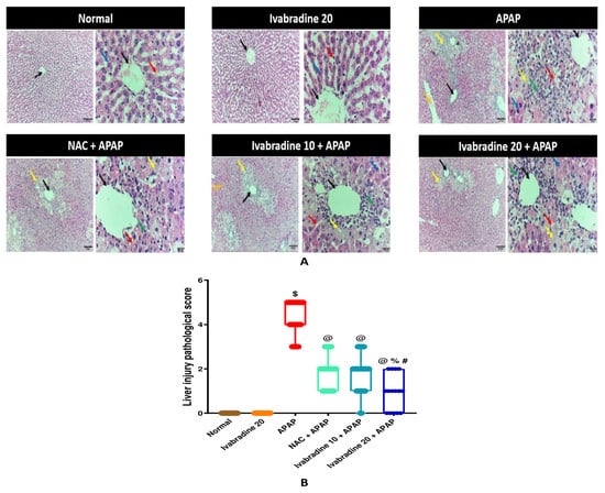
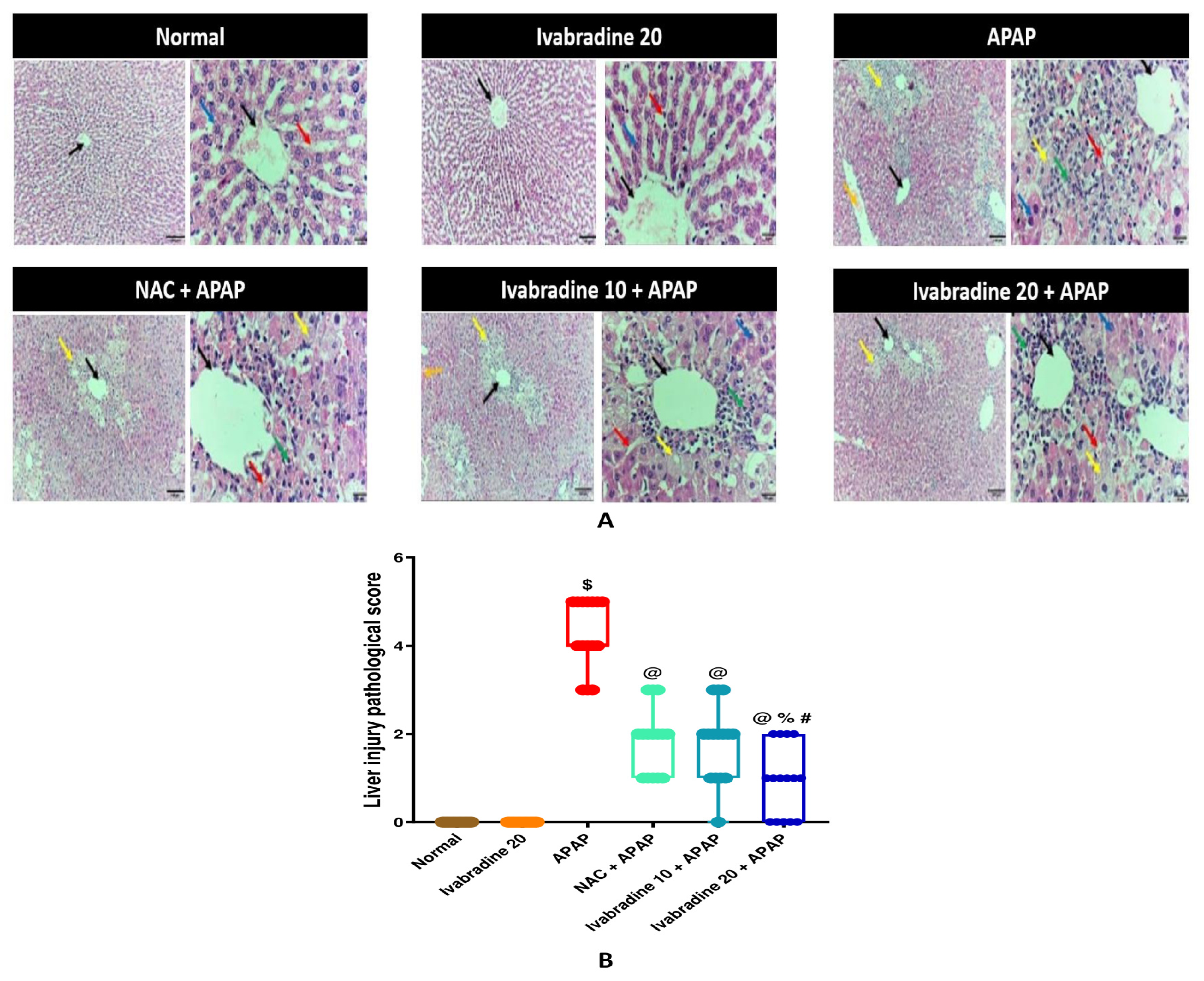
Microscopic evaluation of H&E-stained sections displayed well-defined different histological profiles among treatment groups (Figure 1). Control groups (normal and ivabradine 20 alone) exhibited preserved hepatic architecture with normal polyhedral hepatocytes arranged in cords around central veins (median histopathological score = 0, IQR: 0–0). Conversely, the APAP group displayed severe ALI characterized by extensive centrilobular necrosis, portal vessel congestion, cellular vacuolation, and dense perivascular inflammatory cell infiltration (median histopathological score = 4, IQR: 4–5; p < 0.05 vs. Normal). Pretreatment with NAC significantly attenuated structural damage (median histopathological score = 2, IQR: 1–2; p < 0.05 vs. APAP). Similarly, ivabradine pretreatment reduced liver injury in a graded, dose-related fashion: the lower dose showed moderate tissue preservation (median histopathological score = 2, IQR: 1–2; p < 0.05 vs. APAP), whereas the higher dose (20 mg/kg) demonstrated marked histological protection with minimal inflammatory cell infiltration and limited sinusoidal congestion (median histopathological score = 1, IQR: 0–2; p < 0.05 vs. APAP) bringing tissue structural alterations in a protective direction comparable to the observed effects of NAC (median score = 2, IQR: 1–2).
Figure 1.
Effect of ivabradine on hepatic histopathological alterations induced by APAP. Representative micrographs (stained with h & E) are presented for each experimental group. Histopathological scoring was performed on all animals in each group, and the displayed images illustrate representative findings rather than the median histopathological score of individual animals. (A) Control groups (Normal and Ivabradine 20) exhibited preserved hepatic architecture with polyhedral hepatocytes arranged in strands around central veins, normal sinusoids containing Kupffer cells, and no significant pathology. APAP group displayed profound liver damage marked by intense perivascular inflammatory cell infiltration, portal vessel congestion, severe vacuolation, and centrilobular necrosis. NAC + APAP group showed mild improvements compared to APAP, with mild sinusoidal congestion, few perivascular inflammatory cells, and mild perivascular hepatocyte degeneration. Ivabradine 10 + APAP group exhibited moderate pathology, including portal and sinusoidal congestion, moderate perivascular hepatocyte degeneration, and inflammatory cell infiltration. Ivabradine 20 + APAP group demonstrated minimal changes, consisting of minimal perivascular hepatocyte degeneration, minimal inflammatory cell infiltration, and minimal sinusoidal congestion. Black arrow: central vein, red arrow: blood sinusoids with Kupffer cells, blue arrow: polyhedral hepatocytes, green arrow: inflammatory cells, yellow arrow: centrilobular necrosis, orange arrow: portal vein congestion. Image magnification: 100× with scale bar 100 µm and 400× with scale bar 20 µm, respectively. (B) Effect of ivabradine on liver injury pathological score. Mice received ivabradine (10 and 20 mg/kg/day, orally) and NAC (400 mg/kg/day, orally) for 3 days prior to acetaminophen (400 mg/kg, single i.p. injection). Values from each data point were represented as individual points above box plots (median, interquartile range, and min-to-max range; n = 8) and then assayed via Kruskal–Wallis test tracked by Dunn’s post hoc, p < 0.05. $ vs. normal; @ vs. APAP; % vs. NAC + APAP; # vs. Ivabradine 10 + APAP. Original images are provided in the Supplementary Materials.
3.2. Influence of Ivabradine on Serum Biochemical Markers of Liver Injury in APAP-Induced ALI
Administration of APAP significantly increased ALT, AST, and ALP activities compared to controls. Pre-administration with either NAC or ivabradine (at both tested doses) significantly reduced ALT, AST, and ALP activities in comparison to the APAP group. However, the high-dose ivabradine group (20 mg/kg) exhibited significantly higher serum liver enzyme activities than the NAC-pretreated group (Table 1).
Table 1.
Influence of ivabradine on serum biochemical markers of liver injury in APAP-induced ALI. Mice received ivabradine (10 and 20 mg/kg/day, orally) and NAC (400 mg/kg/day, orally) for 3 days prior to acetaminophen (400 mg/kg, single i.p. injection). Data are presented as means ± SEM (n = 8). ANOVA followed by Tukey–Kramer’s test was used for analysis. Significantly different $ vs. normal (p ˂ 0.0001); @ vs. APAP (p ˂ 0.0001); % vs. NAC + APAP (p ˂ 0.0001); # vs. Ivabradine 10 + APAP (p ˂ 0.0001).
3.3. Effect of Ivabradine on Hepatic Oxidant/Antioxidant Status in APAP-Induced ALI
As presented in Table 2, APAP challenge induced severe oxidative stress, as evidenced by a marked, statistically significant elevation in hepatic MDA content alongside a pronounced exhaustion of reduced GSH levels relative to controls (p < 0.05). Pretreatment with ivabradine or NAC significantly attenuated APAP-induced lipid peroxidation and preserved hepatic GSH reserves compared to the APAP-treated group (p < 0.05). Notably, no statistically significant differences were observed in hepatic MDA and GSH levels between the high-dose ivabradine group (20 mg/kg) and the NAC reference group (p > 0.05). Furthermore, administration of ivabradine alone (20 mg/kg) produced no statistically significant shifts in baseline MDA or GSH levels compared to normal control mice (p > 0.05).
Table 2.
Effect of ivabradine on hepatic oxidant/antioxidant status in APAP-induced ALI. Mice received ivabradine (10 and 20 mg/kg/day, orally) and NAC (400 mg/kg/day, orally) for 3 days prior to acetaminophen (400 mg/kg, single i.p. injection). Data are presented as means ± SEM (n = 8). Data were statistically analyzed using ANOVA followed by Tukey–Kramer’s test. Significantly different $ vs. normal (p ˂ 0.0001); @ vs. APAP (p ˂ 0.0001); % vs. NAC + APAP (p ˂ 0.0001).
3.4. Effect of Ivabradine on Hepatic Nrf2 and Keap1 Expression During APAP-Induced ALI
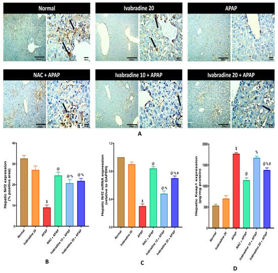
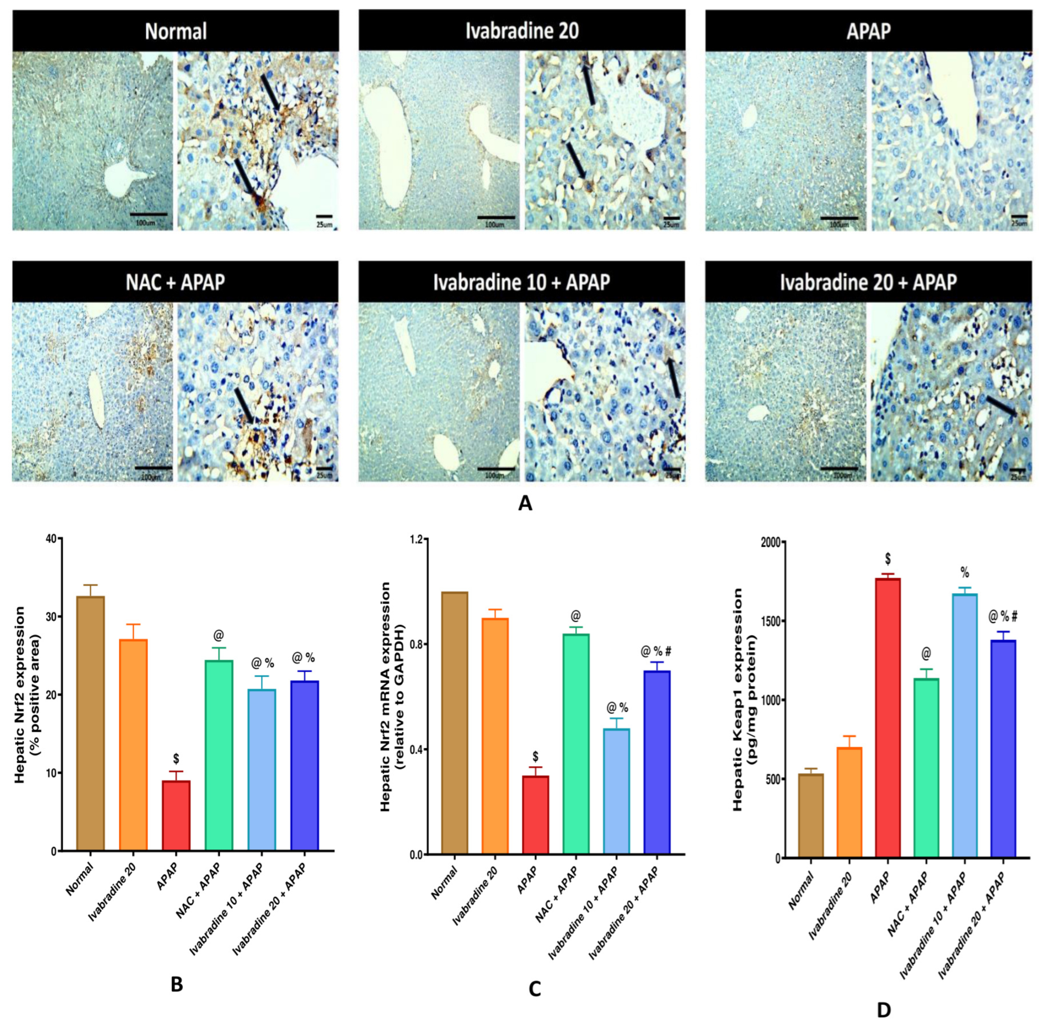
The APAP group exhibited a marked reduction in hepatic Nrf2 DAB-positive area fraction (%) and Nrf2 mRNA expression compared with the control group. This was accompanied by a substantial increase in hepatic Keap1expression. Pretreatment with ivabradine (10 or 20 mg/kg) or NAC markedly increased hepatic Nrf2 immunoreactivity and Nrf2 mRNA expression compared with the APAP group. In addition, NAC and ivabradine (20 mg/kg) significantly reduced hepatic Keap1 expression, whereas ivabradine (10 mg/kg) produced a non-significant reduction compared with the APAP group (Figure 2).
Figure 2.
Effect of ivabradine on hepatic Nrf2 (A,B) immunoreactivity, (C) mRNA expression, and (D) Keap1 level during APAP-induced ALI. Mice received ivabradine (10 and 20 mg/kg/day, orally) and NAC (400 mg/kg/day, orally) for 3 days prior to acetaminophen (400 mg/kg, single i.p. injection). Data are presented as means ± SEM (n = 8 per group, except for qRT-PCR analysis where n = 5 randomly selected samples per group were analyzed). ANOVA followed by Tukey–Kramer’s test was used for data analysis. Image magnification: 100× with scale bar 100 µm and 400× with scale bar 25 µm, respectively. Significantly different $ vs. normal (p ˂ 0.0001); @ vs. APAP (p ˂ 0.0001); % vs. NAC + APAP (p ˂ 0.0001); # vs. Ivabradine 10 + APAP (p = 0.0002). Original images are provided in the Supplementary Materials.
3.5. Effect of Ivabradine on Hepatic SIRT1 mRNA Expression During APAP-Induced ALI
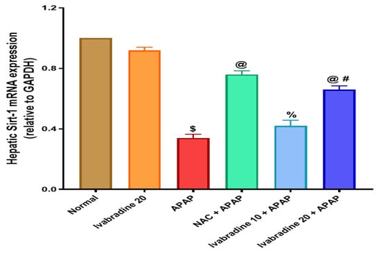
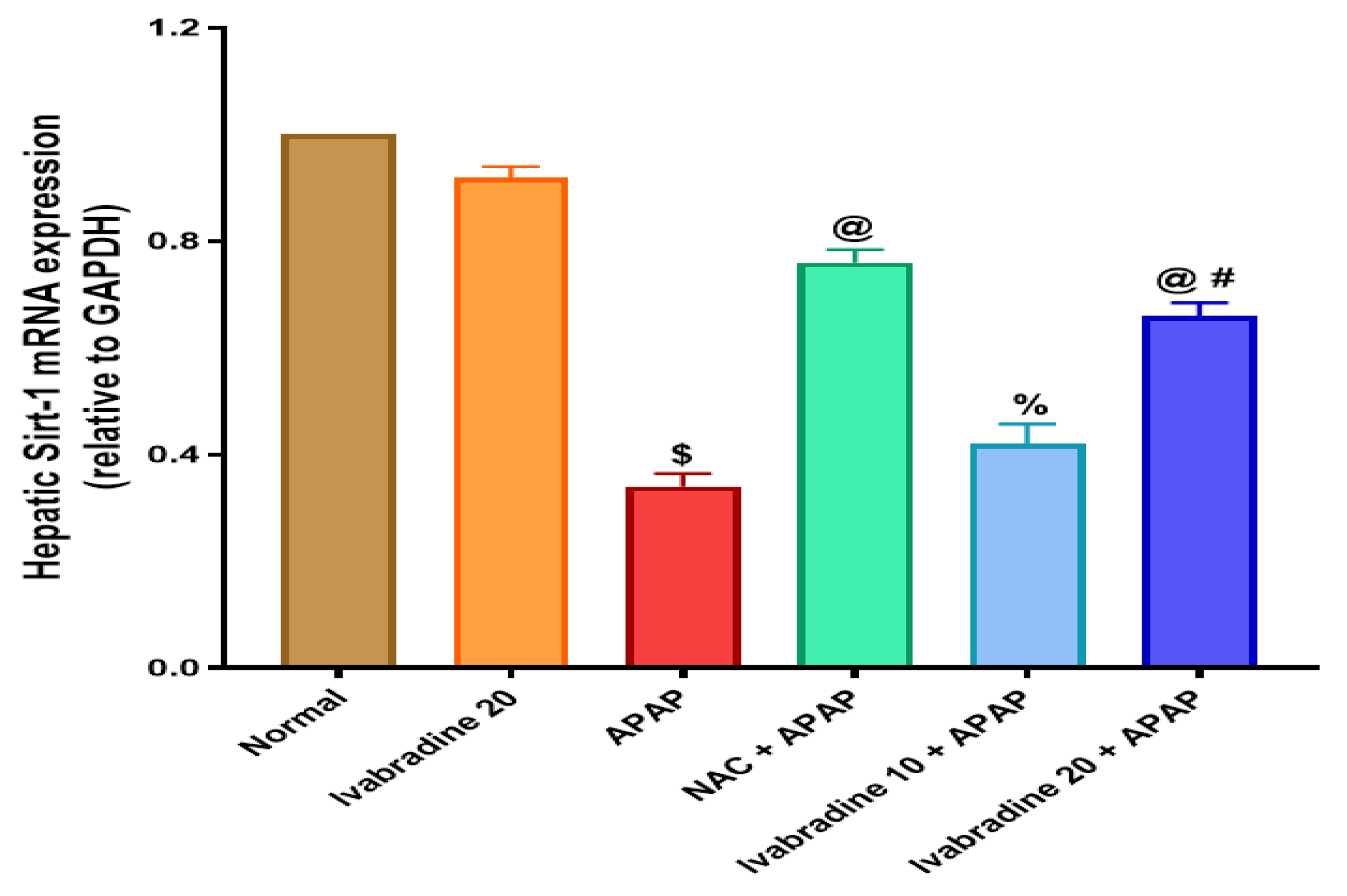
Upon administration of APAP, there was a significant decrease in hepatic SIRT1 mRNA expression in comparison to the normal control. Pretreatment with NAC and a high dose of ivabradine markedly enhanced hepatic SIRT1 mRNA expression in comparison to the APAP group. However, a low dose of ivabradine preserved hepatic SIRT1 mRNA expression, but in a non-significant manner, matching the APAP group (Figure 3).
Figure 3.
Effect of ivabradine on hepatic SIRT1 mRNA expression during APAP-induced ALI. Mice received ivabradine (10 and 20 mg/kg/day, orally) and NAC (400 mg/kg/day, orally) for 3 days prior to acetaminophen (400 mg/kg, single i.p. injection). Data are presented as means ± SEM (n = 5). ANOVA followed by Tukey–Kramer’s test was utilized for data analysis. Significantly different $ vs. normal (p ˂ 0.0001); @ vs. APAP (p ˂ 0.0001); % vs. NAC + APAP (p ˂ 0.0001); # vs. Ivabradine 10 + APAP (p ˂ 0.0001).
3.6. Effect of Ivabradine on Hepatic AMPK, p-AMPK, mTOR, and p-mTOR Expression During APAP-Induced ALI
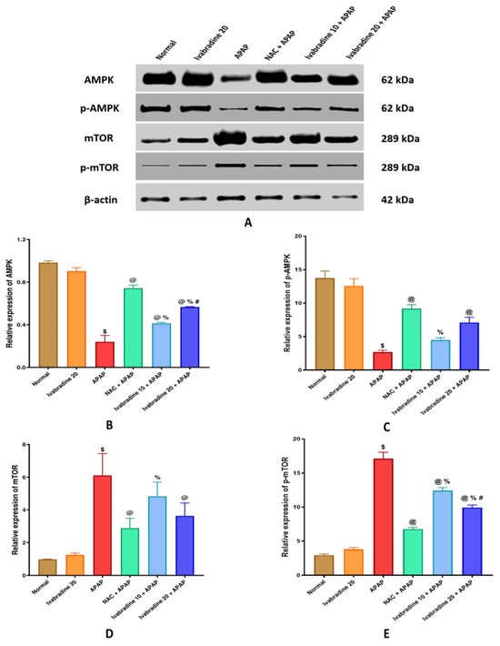
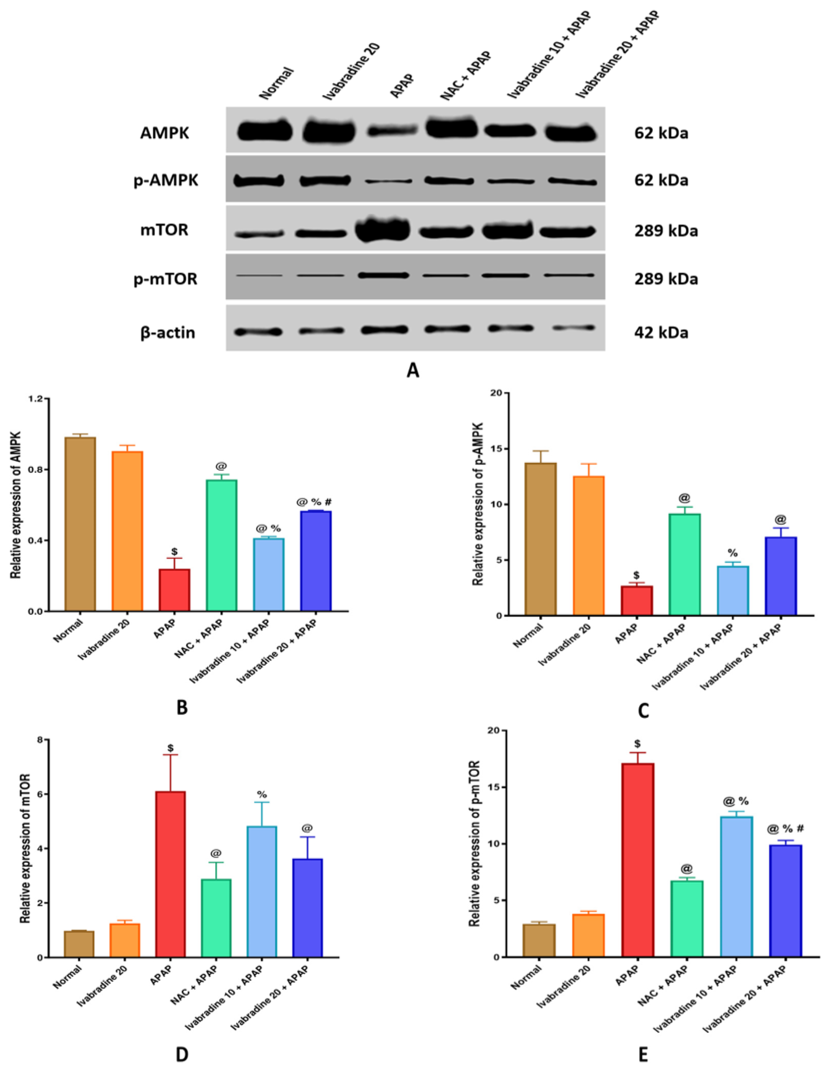
APAP group revealed a significant reduction in hepatic AMPK and p-AMPK expression in comparison to the normal group. Pretreatment with both doses of ivabradine (10 and 20 mg/kg), as well as NAC, markedly enhanced hepatic AMPK expression in comparison to the APAP group. However, while both NAC and ivabradine at 20 mg/kg significantly restored hepatic p-AMPK expression, the 10 mg/kg dose of ivabradine produced only a modest, non-significant improvement (Figure 4).
Figure 4.
Effect of ivabradine on hepatic (A,B) AMPK, (A,C) p-AMPK, (A,D) mTOR, and (A,E) p-mTOR expressions during APAP-induced ALI. Mice received ivabradine (10 and 20 mg/kg/day, orally) and NAC (400 mg/kg/day, orally) for 3 days prior to acetaminophen (400 mg/kg, single i.p. injection). Data are expressed as mean ± SEM. Biological sample sizes were n = 3 per group for total AMPK and mTOR, and n = 2 per group for p-AMPK and p-mTOR. Data were statistically analyzed using ANOVA followed by Tukey–Kramer’s test. Significantly different $ vs. normal (p ˂ 0.0001); @ vs. APAP (p ˂ 0.0001); % vs. NAC + APAP (p ˂ 0.0001); # vs. Ivabradine 10 + APAP (p ˂ 0.0001). Original images are provided in the Supplementary Materials.
On the contrary, APAP administration significantly increased hepatic mTOR and p-mTOR expressions compared with the normal group. Pretreatment with NAC and high-dose ivabradine (20 mg/kg) significantly reduced hepatic mTOR expression compared with the APAP group, whereas the low dose of ivabradine (10 mg/kg) produced only a non-significant reduction. Regarding p-mTOR, both doses of ivabradine and NAC significantly attenuated its expression compared with the APAP group; however, the effect of ivabradine was more pronounced at the 20 mg/kg dose (Figure 4).
3.7. Effect of Ivabradine on Hepatic CD68 Immunoreactivity and MCP-1 Level During APAP-Induced ALI
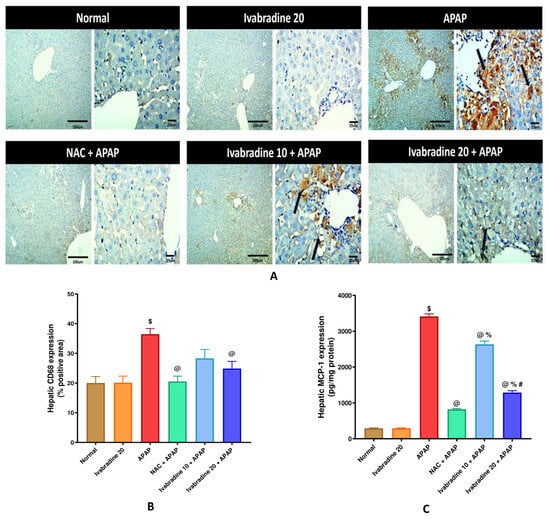
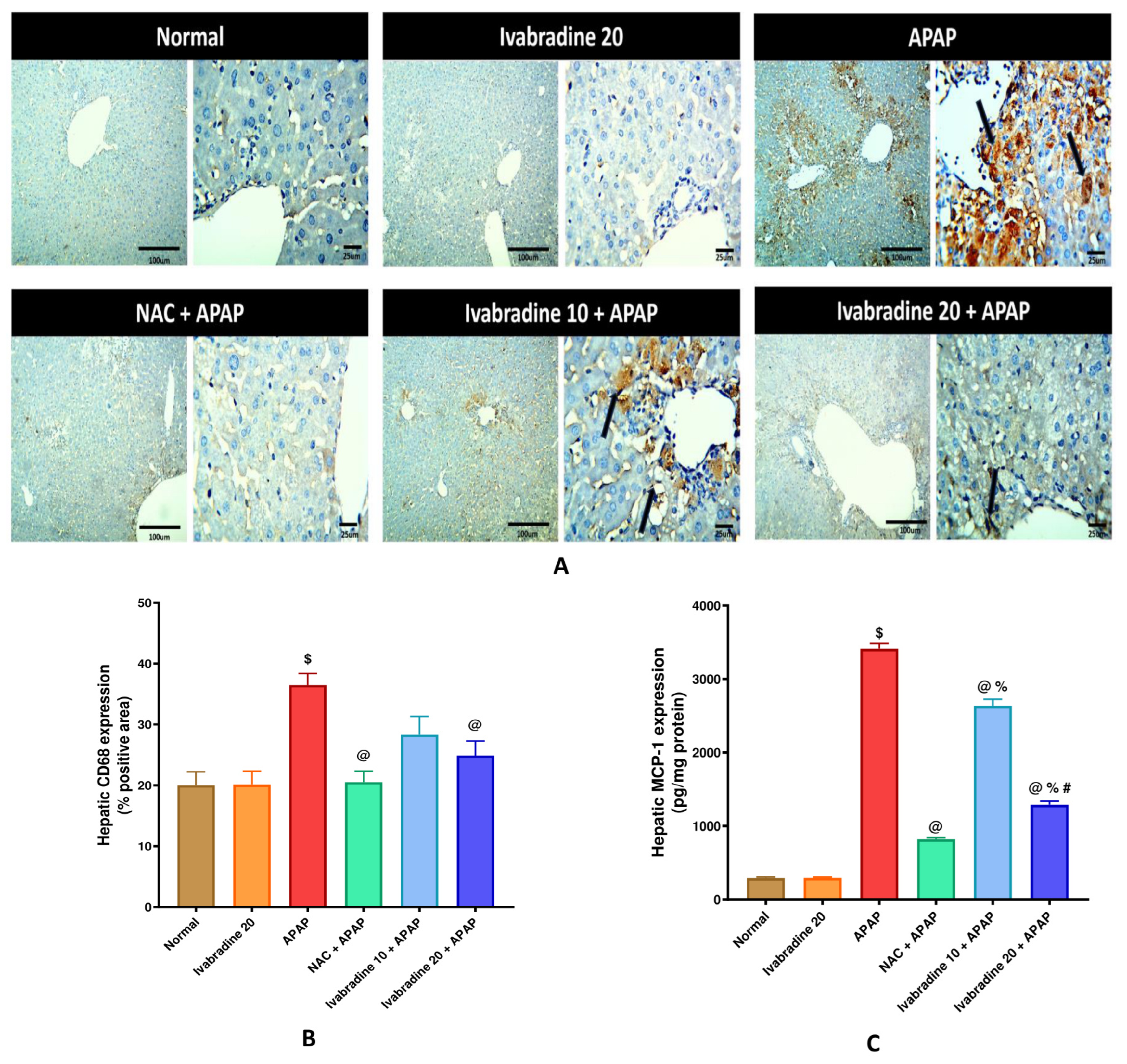
APAP induced a significant elevation in hepatic CD68 DAB-positive area fraction (%) and MCP-1 level compared with normal control. Pretreatment with high-dose ivabradine (20 mg/kg) as well as NAC demonstrated a prompt reduction in CD68 immunoreactivity compared to the APAP group, whereas ivabradine (10 mg/kg) reduced CD68 immunoreactivity in a non-significant manner. Both ivabradine doses as well as NAC demonstrated a significant reduction in hepatic MCP-1 level compared to the APAP group (Figure 5).
Figure 5.
Effect of ivabradine on hepatic (A,B) CD68 immunoreactivity and (C) MCP-1 levels during APAP-induced ALI. Mice received ivabradine (10 and 20 mg/kg/day, orally) and NAC (400 mg/kg/day, orally) for 3 days prior to acetaminophen (400 mg/kg, single i.p. injection). Data are presented as means ± SEM (n = 8) and analyzed via ANOVA followed by Tukey–Kramer’s test. Image magnification: 100× with scale bar 100 µm and 400× with scale bar 25 µm, respectively. Significantly different $ vs. normal (p ˂ 0.0001); @ vs. APAP (p ˂ 0.0001); % vs. NAC + APAP (p ˂ 0.0001); # vs. Ivabradine 10 + APAP (p ˂ 0.0001). Original images are provided in the Supplementary Materials.
3.8. Effect of Ivabradine on Hepatic Caspase-3 Immunoreactivity During APAP-Induced ALI
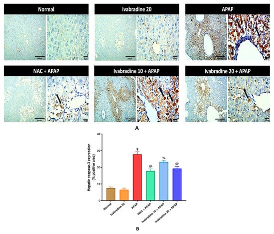
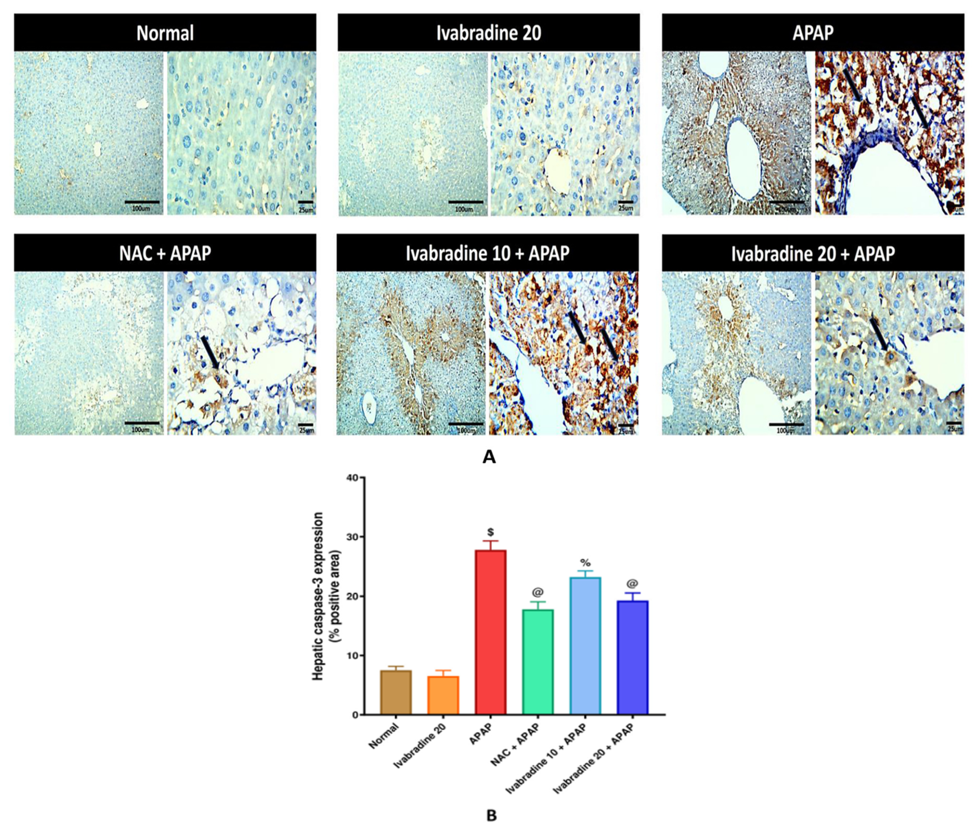
Injection of APAP prompted a marked increase in hepatic caspase-3 immunoreactivity relative to normal mice, predominantly localized within centrilobular regions exhibiting structural degeneration. Pretreatment with a high dose of ivabradine and NAC demonstrated a significant decrease in hepatic caspase-3 immunoreactive area (%) in relation to the APAP group; however, the low dose of ivabradine reduced hepatic caspase-3 immunoreactivity but in a non-significant manner compared to the APAP group (Figure 6).
Figure 6.
Effect of ivabradine on hepatic caspase-3 immunoreactivity during APAP-induced ALI (A,B). Mice received ivabradine (10 and 20 mg/kg/day, orally) and NAC (400 mg/kg/day, orally) for 3 days prior to acetaminophen (400 mg/kg, single i.p. injection). Data are presented as means ± SEM (n = 8) and analyzed via ANOVA followed by Tukey–Kramer’s test. Image magnification: 100× with scale bar 100 µm and 400× with scale bar 25 µm, respectively. Significantly different $ vs. normal (p ˂ 0.0001); @ vs. APAP (p ˂ 0.0001); % vs. NAC + APAP (p ˂ 0.0001). Original images are provided in the Supplementary Materials.
4. Discussion
Although APAP is among the most widely used analgesics worldwide, overdose can cause severe acute hepatocellular injury [21]. Accordingly, this study was designed to investigate the protective effects of ivabradine against APAP-induced ALI in mice and to explore the molecular pathways associated with its effects. Our findings revealed a hepatoprotective effect of ivabradine pretreatment, associated with favorable modulation of hepatic SIRT1/Nrf2/Keap1 and AMPK/mTOR signaling. NAC was included as an experimental prophylactic control rather than as a model of its conventional clinical use following APAP overdose. Both interventions were administered before APAP challenge, with the final pretreatment dose given 2 h before APAP administration, allowing their protective effects to be evaluated under the same experimental conditions. However, significant differences remained between high-dose ivabradine and NAC in serum liver enzyme activities, indicating that the observed effects were not identical across all assessed parameters.
The 3-day pretreatment protocol was selected to allow sufficient time for potential pharmacological and molecular adaptations before the acute toxic challenge. It was hypothesized that this pretreatment period might permit tissue exposure to ivabradine and modulation of endogenous cytoprotective pathways, including SIRT1- and Nrf2-associated signaling. However, because plasma concentrations and hepatic accumulation of ivabradine were not measured, the achievement of steady-state tissue levels cannot be confirmed. The present findings demonstrate protection in a prophylactic mouse model of APAP-induced ALI and do not establish efficacy when ivabradine is administered after liver injury has developed. Any potential clinical relevance, including its possible role in individuals at increased risk of drug-induced liver injury, remains hypothetical and requires dedicated investigation.
Previous reports have demonstrated the injurious impact of APAP administration on the liver tissue. High doses of APAP result in notable necrosis of hepatic cells and congestion of the liver parenchyma [15,18]. Similarly, our study revealed an intense perivascular inflammatory cell infiltration, congestion of the portal vessels, and severe vacuolation and centrilobular necrosis of hepatocytes in the APAP group. Interestingly, ivabradine successfully counteracted APAP-induced pathological lesions and preserved the hepatic architecture in a way that may be in the same direction as NAC results. To confirm the histopathological results, the biochemical estimation of serum biochemical parameters indicated lowered levels of transaminases and ALP in ivabradine-pretreated groups. This clearly points to the hepatoprotective potential of ivabradine.
Notably, while NAC produced marked numerical improvements in several serum biomarkers, high-dose ivabradine (20 mg/kg) also significantly reduced the histopathological injury score compared to the APAP group (median score = 1 vs. 2 in the NAC group). However, significant differences remained in serum liver enzyme activities between the high-dose ivabradine and NAC groups, indicating that high-dose ivabradine did not achieve the same degree of improvement in liver injury markers as NAC. This apparent discrepancy may reflect the different biological information provided by biochemical and histopathological assessments. Serum transaminases primarily reflect hepatocyte membrane integrity, whereas histopathological scoring integrates multiple structural features, including inflammatory infiltration, sinusoidal congestion, degeneration, and necrosis. Moreover, the representative photomicrographs illustrate typical findings from each group, whereas the histopathological scores were derived from the evaluation of all animals.
Oxidative stress plays an important role in mediating the pathogenesis of APAP-induced ALI. The depletion of GSH by NAPQI (a highly reactive metabolite of APAP) results in excessive ROS formation, creation of oxidative stress, and exacerbation of hepatic injury [1,6,15,22]. Our results confirmed GSH depletion following APAP administration, accompanied by an elevation in the lipid peroxidation marker, MDA. Ivabradine pretreatment diminished the restriction in the antioxidant capacity of the liver and consequently lowered the oxidative burden in the hepatic tissue. Several studies have reported the antioxidant efficacy of ivabradine as mentioned against doxorubicin-induced cardiotoxicity, against classical seizure models, and during endothelial inflammation [10,13,23]. However, the exact mechanistic pathway by which ivabradine preserved the oxidant/antioxidant balance is still unclear.
Growing lines of evidence have highlighted the vital protective role of the Nrf2 signaling pathway in defending against oxidative stress-induced hepatic injuries, including APAP-associated ALI [6,24,25,26]. Under physiological conditions, Keap1 acts as a sensor that binds Nrf2 in the cytoplasm, facilitating its continuous ubiquitination and proteasomal degradation. Upon acute oxidative stress, Keap1 inactivation normally leads to Nrf2 stabilization, accumulation, and subsequent nuclear translocation to drive antioxidant response element (ARE)-dependent gene expression [22]. However, following APAP exposure, Nrf2 signaling exhibits dynamic, time-dependent kinetics. At the 24-h post-APAP time point evaluated in the present study, severe late-stage oxidative burden, extensive intracellular GSH depletion, and ongoing hepatocellular necrosis result in a marked reduction in hepatic Nrf2 mRNA expression and immunoreactivity, accompanied by elevated Keap1 levels, reflecting an exhausted endogenous antioxidant capacity.
In line with previous reports investigating protective interventions against APAP hepatotoxicity [1,6,22,27], our data confirmed this suppressed Nrf2 expression and elevated Keap1 pattern 24 h after APAP challenge. Pretreatment with ivabradine significantly preserved hepatic Nrf2 mRNA expression and immunoreactivity while limiting Keap1 elevation. Rather than implying a direct causal feedback mechanism, these observations indicate that ivabradine pretreatment is compatible with the preservation and favorable modulation of hepatic redox-regulatory signaling, helping to maintain cellular redox homeostasis and endogenous antioxidant defenses during acute toxic challenge.
Another important modulator of the antioxidant defense system is SIRT1. SIRT1, Keap1, and Nrf2 constitute an interconnected cytoprotective signaling network that plays a central role in cellular adaptation to oxidative stress. Importantly, SIRT1 positively regulates the Nrf2-dependent pathway through several mechanisms. SIRT1 enhances Nrf2 stability and transcriptional activity, as well as its nuclear retention and DNA-binding capacity [25,28]. Moreover, SIRT1 represses oxidative stress and mitochondrial dysfunction, thereby preserving the integrity of the Keap1–Nrf2 antioxidant defense system [5,22]. Consequently, downregulation of SIRT1 during severe hepatic injury is frequently associated with impaired Nrf2 signaling and attenuation of antioxidant responses. Alongside, SIRT1 activation is closely associated with repression of inflammatory responses in hepatocytes [29,30]. The current study revealed a significant decline in SIRT1 mRNA expression following APAP challenge, a result that is in agreement with previous studies [31,32,33].
These findings are consistent with previous reports demonstrating that APAP suppresses both SIRT1 and AMPK signaling during hepatocyte injury, whereas pharmacological activation of these pathways attenuates APAP-induced hepatotoxicity [34]. Contrarily, ivabradine-pretreated groups showed higher SIRT1 mRNA expression compared to the APAP group. Correspondingly, previous studies have demonstrated significant upregulation of AMPK and SIRT1 following ivabradine administration in models of myocardial dysfunction in mice [35,36]. In this context, it may be acceptable to presume that the ability of ivabradine to improve SIRT1/Nrf2 may be associated with its observed antioxidant hepatoprotective activity and is associated with the regulation of oxidative stress and hepatocellular injury. It is important to emphasize that while ivabradine pretreatment restored SIRT1/Nrf2 expression and modulated AMPK/mTOR activation ratios, these findings reflect molecular and downstream biochemical associations rather than direct mechanistic causality. Without specific gene-silencing or pharmacologic loss-of-function experiments targeting these individual signaling pathways, our data demonstrate that ivabradine creates a cellular environment compatible with redox preservation and metabolic regulation during APAP challenge, without establishing these pathways as indispensable drivers of protection.
Evidence revealed that AMPK-induced liver regeneration was inhibited by APAP administration in mice [37,38]. On the same line, our study demonstrated a substantial reduction in hepatic AMPK expression as well as elevation of hepatic mTOR in comparison to normal mice following APAP injection, which matches the observations of previous studies [39,40]. Ivabradine administration improved hepatic AMPK expression, with the higher dose producing a greater response than the lower dose. The restoration of AMPK expression by ivabradine is consistent with previous reports implicating AMPK activation in protection against APAP-induced liver injury [34,41]. However, the present findings demonstrate an association rather than direct mechanistic evidence that AMPK mediates the hepatoprotective effects of ivabradine. Furthermore, higher doses of ivabradine demonstrated a significant reduction in hepatic mTOR expression relative to the APAP group. Correspondingly, previous studies have reported that ivabradine attenuates hyperactivated PI3K/Akt/mTOR signaling in models of myocardial injury and transverse aortic constriction [36,42]. While mTOR is a key regulator of nutrient sensing, cell survival, and autophagy-related signaling, our study evaluated changes in total mTOR and p-mTOR protein levels rather than directly assessing autophagic flux. The restoration of AMPK accompanied by mTOR suppression reflects favorable metabolic tuning and cellular stress preservation during APAP challenge.
Given that both NAC and ivabradine attenuated oxidative stress, it is plausible that the observed hepatoprotection involves overlapping downstream pathways, including restoration of SIRT1/Nrf2 and AMPK signaling together with suppression of inflammatory and apoptotic mediators. Similar convergence of antioxidant-dependent signaling pathways has been described previously in experimental liver injury models [43]. However, these shared mechanisms were not directly investigated in the present study and should therefore be interpreted as potential downstream associations rather than definitive mechanistic pathways.
Accumulating evidence indicates that inflammatory cell recruitment plays a pivotal role in APAP-induced hepatotoxicity. Following APAP overdose, hepatocellular necrosis triggers the release of damage-associated molecular patterns (DAMPs), which stimulate chemokine production and immune cell infiltration. Among these mediators, MCP-1 is markedly upregulated in the injured liver, promoting the recruitment of circulating monocytes to sites of hepatic damage. Concurrently, increased hepatic immunoreactivity of CD68 reflects enhanced activation and accumulation of macrophages, including resident Kupffer cells and infiltrating monocyte-derived macrophages. These inflammatory cells further amplify liver injury through the release of pro-inflammatory cytokines, reactive oxygen species, and proteolytic enzymes.
Consistent with previous reports, the present findings support the notion that APAP-induced liver injury is associated with pronounced upregulation of hepatic MCP-1 levels and CD68-positive area (%), highlighting the contribution of macrophage-driven inflammatory responses to the progression of hepatic damage [38,44,45]. Our results revealed a significant reduction in hepatic MCP-1 expression following the administration of ivabradine, which complies with the results of prior investigations [17,46].
To the best of our knowledge, this is the first study to demonstrate that ivabradine significantly reduces hepatic CD68 expression, reflecting a reduction in CD68-positive macrophage accumulation and infiltration within the injured liver. It is important to acknowledge the nuanced and dynamic dual role played by macrophages during APAP-induced liver injury. Following acute APAP challenge, the initial recruitment of resident Kupffer cells and infiltrating monocyte-derived macrophages promotes sterile inflammation and tissue breakdown via pro-inflammatory cytokines and ROS release. However, during later recovery phases, distinct macrophage phenotypic populations transition toward a pro-resolving function, promoting phagocytic debris clearance, tissue remodeling, and hepatic regeneration [38,45]. The observed decrease in CD68 immunoreactivity following ivabradine pretreatment primarily aligns with diminished early monocyte/macrophage recruitment during the acute injury phase, reflecting decreased inflammatory cell burden in the hepatic parenchyma.
Although oncotic necrosis is widely recognized as the primary mode of cell death during APAP hepatotoxicity, apoptotic signaling pathways, including elevated caspase-3 immunoreactivity, are frequently activated in response to severe oxidative stress and cellular damage [47,48]. In the current study, APAP administration induced a significant elevation in hepatic caspase-3 immunoreactivity, concentrated primarily in centrilobular areas exhibiting severe tissue injury, which matches the results of previous studies [49,50,51].
While APAP-induced acute liver toxicity is predominantly characterized by oncotic necrosis secondary to severe mitochondrial dysfunction, apoptotic cascades are concurrently activated as part of the overall tissue injury response. In the present study, ivabradine administration significantly suppressed hepatic caspase-3 immunoreactivity alongside inflammatory markers (MCP-1 and CD68). However, it is important to emphasize that these decreases in MCP-1 expression, CD68, and caspase-3 immunoreactivity likely represent secondary downstream phenomena resulting from the primary attenuation of hepatocellular necrosis and overall tissue damage, rather than direct cell-death pathway inhibition.
In conclusion, the present study demonstrates that prophylactic administration of ivabradine exerts a significant hepatoprotective effect against APAP-induced ALI in mice. These protective actions are closely associated with the mitigation of oxidative stress, suppression of CD68-positive macrophage accumulation, and reduction of caspase-3-mediated pro-apoptotic signaling, alongside favorable modulation of the SIRT1/Nrf2/Keap1 and AMPK/mTOR signaling pathways. However, these findings establish protection only within the prophylactic experimental model and do not demonstrate therapeutic efficacy after the onset of liver injury or establish clinical benefit in humans. Furthermore, the observed molecular changes indicate associations rather than definitive causal mechanisms. Future studies incorporating pathway-specific interventions, pharmacokinetic assessments, and post-exposure treatment protocols are warranted to clarify the underlying mechanisms and determine whether these findings have potential clinical relevance.
5. Study Limitations
Despite these promising findings, several limitations of the present study should be acknowledged. First, ivabradine was evaluated using a 3-day prophylactic pretreatment model in male mice at a single acute time point. While this effectively establishes the biological proof-of-concept for its cytoprotective potential, it precludes direct extrapolation to emergency clinical settings of established APAP overdose, long-term liver regeneration, or potential gender-specific variations. Second, while significant modulation of the SIRT1/Nrf2, AMPK/mTOR, and apoptotic (caspase-3) axes was demonstrated, these findings represent functional associations rather than definitive causal mechanisms, as pathway-specific inhibitors, knockout models, HCN channel-independent explorations, and direct autophagic flux markers were not performed. Third, pharmacodynamic and pharmacokinetic parameters, including plasma drug concentrations, hepatic accumulation, and heart rate assessments, were not evaluated.
Author Contributions
S.H.A.: investigation, methodology, validation, resources, formal analysis, writing—original draft, funding acquisition; M.M.S.: conceptualization, methodology, data curation, writing—review and editing, visualization, supervision; M.G.: data curation, supervision; W.M.E.: methodology, pathological and immunohistochemical analysis; D.S.E.-A.: conceptualization, formal analysis, data curation, writing—review and editing, visualization, supervision. All authors have read and agreed to the published version of the manuscript.
Funding
This research received no external funding.
Institutional Review Board Statement
Any experiment that was done with animals complied with the guidelines of the Mansoura University Animal Care and Use Committee, and the Faculty of Pharmacy Research Ethics Committee gave its ethical approval (Approval No.: MU-ACUC (PHARM.PhD.24.01.34) on 12 January 2024. The experiment was done according to the National Institutes of Health (NIH) guidelines for the Care and Use of Laboratory Animals (Publication No. 85-23, revised 1985). Reporting the animal experiment follows the ARRIVE guidelines.
Informed Consent Statement
Not applicable.
Data Availability Statement
The original contributions presented in this study are included in the article/Supplementary Materials. Further inquiries can be directed to the corresponding author(s).
Conflicts of Interest
The authors declare no conflicts of interest.
Abbreviations
The following abbreviations are used in this manuscript:
| AKT | Protein kinase B |
| ALI | Acute liver injury |
| ALP | Alkaline phosphatase |
| ALT | Alanine aminotransferase |
| AMPK | 5′ adenosine monophosphate-activated protein kinase |
| p-AMPK | Phosphorylated 5′ adenosine monophosphate-activated protein kinase |
| ANOVA | Analysis of variance |
| APAP | Acetaminophen |
| ARE | Antioxidant response element |
| ARRIVE | Animal Research: Reporting of In Vivo Experiments |
| AST | Aspartate aminotransferase |
| BSA | Bovine serum albumin |
| CD68 | Cluster of differentiation 68 |
| cDNA | Complementary deoxyribonucleic acid |
| CMC-Na | Sodium carboxymethylcellulose |
| CYP | Cytochrome P450 |
| DAB | 3,3′-Diaminobenzidine |
| DAMPs | Damage-associated molecular patterns |
| DNA | Deoxyribonucleic acid |
| ECL | Enhanced chemiluminescence |
| ELISA | Enzyme-linked immunosorbent assay |
| FIJI | Fiji Is Just ImageJ (ImageJ distribution) |
| GAPDH | Glyceraldehyde-3-phosphate dehydrogenase |
| GSH | Reduced glutathione |
| H&E | Hematoxylin and eosin |
| HCN | Hyperpolarization-activated cyclic nucleotide-gated |
| i.p. | Intraperitoneal |
| IHC | Immunohistochemistry |
| i.v. | Intravenous |
| Iƒ | Funny current |
| kDa | Kilodalton |
| Keap1 | Kelch-like ECH-associated protein 1 |
| MCP-1 | Monocyte chemoattractant protein-1 |
| MDA | Malondialdehyde |
| mRNA | Messenger ribonucleic acid |
| mTOR | Mammalian target of rapamycin |
| p-mTOR | Phosphorylated mammalian target of rapamycin |
| MU-ACUC | Mansoura University Animal Care and Use Committee |
| NAC | N-acetylcysteine |
| NAPQI | N-acetyl-p-benzoquinone imine |
| NIH | National Institutes of Health |
| Nrf2 | Nuclear factor erythroid 2-related factor 2 |
| p70S6K | Ribosomal protein S6 kinase beta-1 |
| PBS | Phosphate-buffered saline |
| PI3K | Phosphoinositide 3-kinase |
| PVDF | Polyvinylidene difluoride |
| qRT-PCR | Quantitative real-time polymerase chain reaction |
| RNA | Ribonucleic acid |
| ROI | Region of interest |
| ROS | Reactive oxygen species |
| rpm | Revolutions per minute |
| SDS-PAGE | Sodium dodecyl sulfate–polyacrylamide gel electrophoresis |
| SEM | Standard error of the mean |
| SIRT1 | Silent information regulator sirtuin 1 |
| TBST | Tris-buffered saline with Tween 20 |
| U/L | Units per litre |
| v/v | Volume per volume |
| w/v | Weight per volume |
References
- Ibrahim, S.R.M.; El-Agamy, D.S.; Abdallah, H.M.; Ahmed, N.; Elkablawy, M.A.; Mohamed, G.A. Protective activity of tovophyllin A, a xanthone isolated from Garcinia mangostana pericarps, against acetaminophen-induced liver damage: Role of Nrf2 activation. Food Funct. 2018, 9, 3291–3300. [Google Scholar] [CrossRef] [Scilit] [PubMed]
- Yan, M.; Huo, Y.; Yin, S.; Hu, H. Mechanisms of acetaminophen-induced liver injury and its implications for therapeutic interventions. Redox Biol. 2018, 17, 274–283. [Google Scholar] [CrossRef] [Scilit] [PubMed]
- Farag, M.; Mohamed, M.; Youssef, E. Assessment of hepatic function, oxidant/antioxidant status, and histopathological changes in rats treated with atorvastatin: Effect of dose and acute intoxication with acetaminophen. Hum. Exp. Toxicol. 2015, 34, 828–837. [Google Scholar] [CrossRef] [Scilit] [PubMed]
- Sriphoosanaphan, S.; Rattanachaisit, P.; Somanawat, K.; Wanpiyarat, N.; Komolmit, P.; Werawatganon, D. Calcitriol Protects against Acetaminophen-Induced Hepatotoxicity in Mice. Biomedicines 2023, 11, 1534. [Google Scholar] [CrossRef] [Scilit] [PubMed]
- Alqahtani, Q.H.; Fadda, L.M.; Alhusaini, A.M.; Hasan, I.H.; Ali, H.M. Involvement of Nrf2, JAK and COX pathways in acetaminophen-induced nephropathy: Role of some antioxidants. Saudi Pharm. J. 2023, 31, 101752. [Google Scholar] [CrossRef] [Scilit] [PubMed]
- Mohamed, G.A.; El-Agamy, D.S.; Abdallah, H.M.; Sindi, I.A.; Almogaddam, M.A.; Alzain, A.A.; Andijani, Y.S.; Ibrahim, S.R.M. Kaempferol sophoroside glucoside mitigates acetaminophen-induced hepatotoxicity: Role of Nrf2/NF-κB and JNK/ASK-1 signaling pathways. Heliyon 2024, 10, e31448. [Google Scholar] [CrossRef] [Scilit] [PubMed]
- Deng, X.; Li, Y.; Chen, Y.; Hu, Q.; Zhang, W.; Chen, L.; Lu, X.; Zeng, J.; Ma, X.; Efferth, T. Paeoniflorin protects hepatocytes from APAP-induced damage through launching autophagy via the MAPK/mTOR signaling pathway. Cell. Mol. Biol. Lett. 2024, 29, 119. [Google Scholar] [CrossRef] [Scilit] [PubMed]
- Li, X.; Zhou, J.; Ling, Y.; Tan, Y.; Zhang, J.; Wang, X.; Li, F.; Jiang, S.; Zhang, S.; Yu, K.; et al. Matrine induces autophagic cell death by triggering ROS/AMPK/mTOR axis and apoptosis in multiple myeloma. Biomed. Pharmacother. 2024, 175, 116738. [Google Scholar] [CrossRef] [Scilit] [PubMed]
- Su, Y.; Ma, T.; Wang, Z.; Dong, B.; Tai, C.; Wang, H.; Zhang, F.; Yan, C.; Chen, W.; Xu, Y.; et al. Efficacy of early initiation of ivabradine treatment in patients with acute heart failure: Rationale and design of SHIFT-AHF trial. ESC Heart Fail. 2020, 7, 4465–4471. [Google Scholar] [CrossRef] [Scilit] [PubMed]
- Cavalcante, T.M.B.; De Melo, J.D.M.A.; Lopes, L.B.; Bessa, M.C.; Santos, J.G.; Vasconcelos, L.C.; Vieira Neto, A.E.; Borges, L.T.N.; Fonteles, M.M.F.; Chaves Filho, A.J.M.; et al. Ivabradine possesses anticonvulsant and neuroprotective action in mice. Biomed. Pharmacother. 2019, 109, 2499–2512. [Google Scholar] [CrossRef] [Scilit] [PubMed]
- Miyake, S.; Higuchi, H.; Honda-Wakasugi, Y.; Fujimoto, M.; Kawai, H.; Nagatsuka, H.; Maeda, S.; Miyawaki, T. Locally injected ivabradine inhibits carrageenan-induced pain and inflammatory responses via hyperpolarization-activated cyclic nucleotide-gated (HCN) channels. PLoS ONE 2019, 14, e0217209. [Google Scholar] [CrossRef] [Scilit] [PubMed]
- Zuo, G.; Ren, X.; Qian, X.; Ye, P.; Luo, J.; Gao, X.; Zhang, J.; Chen, S. Inhibition of JNK and p38 MAPK-mediated inflammation and apoptosis by ivabradine improves cardiac function in streptozotocin-induced diabetic cardiomyopathy. J. Cell. Physiol. 2019, 234, 1925–1936. [Google Scholar] [CrossRef] [Scilit] [PubMed]
- Al-Kuraishy, H.M.; Issa, H.K.; Al-Gareeb, A.I.; El-Bouseary, M.M.; Youssef, A.; Abdelaziz, A.S.; Khalifa, H.A.; Batiha, G.E. The role of ivabradine in doxorubicin-induced cardiotoxicity: Exploring of underlying argument. Inflammopharmacology 2022, 30, 2441–2446. [Google Scholar] [CrossRef] [Scilit] [PubMed]
- Sripusanapan, A.; Piriyakulthorn, C.; Apaijai, N.; Chattipakorn, S.C.; Chattipakorn, N. Ivabradine ameliorates doxorubicin-induced cardiotoxicity through improving mitochondrial function and cardiac calcium homeostasis. Biochem. Pharmacol. 2025, 236, 116881. [Google Scholar] [CrossRef] [Scilit] [PubMed]
- Altowijri, M.A.; Abdelmageed, M.E.; El-Gamal, R.; Saeedi, T.; El-Agamy, D.S. Pristimerin dampens acetaminophen-induced hepatotoxicity; The role of NF-κB/iNOS/COX-II/cytokines, PI3K/AKT, and BAX/BCL-2/caspase-3 signaling pathways. Pharmaceutics 2025, 17, 1003. [Google Scholar] [CrossRef] [Scilit] [PubMed]
- Owumi, S.E.; Andrus, J.P.; Herzenberg, L.A.; Herzenberg, L.A. Co-administration of N-Acetylcysteine and Acetaminophen Efficiently Blocks Acetaminophen Toxicity. Drug Dev. Res. 2015, 76, 251–258. [Google Scholar] [CrossRef] [Scilit] [PubMed]
- Custodis, F.; Baumhäkel, M.; Schlimmer, N.; List, F.; Gensch, C.; Böhm, M.; Laufs, U. Heart rate reduction by ivabradine reduces oxidative stress, improves endothelial function, and prevents atherosclerosis in apolipoprotein E-deficient mice. Circulation 2008, 117, 2377–2387. [Google Scholar] [CrossRef] [Scilit] [PubMed]
- Muhammad-Azam, F.; Nur-Fazila, S.H.; Ain-Fatin, R.; Noordin, M.M.; Yimer, N. Histopathological changes of acetaminophen-induced liver injury and subsequent liver regeneration in BALB/C and ICR mice. Vet. World 2019, 12, 1682–1688. [Google Scholar] [CrossRef] [Scilit] [PubMed]
- Guesdon, J.L.; Ternynck, T.; Avrameas, S. The use of avidin-biotin interaction in immunoenzymatic techniques. J. Histochem. Cytochem. 1979, 27, 1131–1139. [Google Scholar] [CrossRef] [Scilit] [PubMed]
- Helps, S.C.; Thornton, E.; Kleinig, T.J.; Manavis, J.; Vink, R. Automatic nonsubjective estimation of antigen content visualized by immunohistochemistry using color deconvolution. Appl. Immunohistochem. Mol. Morphol. 2012, 20, 82–90. [Google Scholar] [CrossRef] [Scilit] [PubMed]
- Agrawal, S.; Murray, B.P.; Khazaeni, B. Acetaminophen Toxicity. In StatPearls; StatPearls Publishing: Treasure Island, FL, USA, 2025. Available online: http://www.ncbi.nlm.nih.gov/books/NBK441917/ (accessed on 9 November 2026).
- Altowijri, M.A.; Abdelmageed, M.E.; El-Gamal, R.; El-Agamy, D.S. The protective effects of neferine against paracetamol-induced liver injury are associated with the activation of SIRT1/Nrf2/HO-1 signaling pathway and inhibition of NF-kappa B/TNF-alpha/iNOS/COX-II cascade. Drug Chem. Toxicol. 2025, 48, 1394–1409. [Google Scholar] [CrossRef] [Scilit] [PubMed]
- Li, B.; Zhang, J.; Wang, Z.; Chen, S. Ivabradine Prevents Low Shear Stress Induced Endothelial Inflammation and Oxidative Stress via mTOR/eNOS Pathway. PLoS ONE 2016, 11, e0149694. [Google Scholar] [CrossRef] [Scilit] [PubMed]
- Lv, H.; Xiao, Q.; Zhou, J.; Feng, H.; Liu, G.; Ci, X. Licochalcone A Upregulates Nrf2 Antioxidant Pathway and Thereby Alleviates Acetaminophen-Induced Hepatotoxicity. Front. Pharmacol. 2018, 9, 147. [Google Scholar] [CrossRef] [Scilit] [PubMed]
- Zhou, J.; Zheng, Q.; Chen, Z. The Nrf2 Pathway in Liver Diseases. Front. Cell Dev. Biol. 2022, 10, 826204. [Google Scholar] [CrossRef] [Scilit] [PubMed]
- Fuertes-Agudo, M.; Luque-Tévar, M.; Cucarella, C.; Martín-Sanz, P.; Casado, M. Advances in Understanding the Role of NRF2 in Liver Pathophysiology and Its Relationship with Hepatic-Specific Cyclooxygenase-2 Expression. Antioxidants 2023, 12, 1491. [Google Scholar] [CrossRef] [Scilit] [PubMed]
- Qin, H.; Chen, R.; Xie, Y.; Liu, H.; Xiao, Y.; Li, L. Kummerowia striata extract protects paracetamol-induced liver injury by modulating the S1P/Nrf2/Keap1 pathway. PLoS ONE 2025, 20, e0329837. [Google Scholar] [CrossRef] [Scilit] [PubMed]
- Zuo, F.; Yang, J.; Wu, Q.; Yang, Y.; Zhou, H.; Sun, Y.; Xu, T. SIRT1 in Liver Diseases: Mechanistic Insights and Therapeutic Prospects. Int. J. Biol. Sci. 2025, 21, 7086–7103. [Google Scholar] [CrossRef] [Scilit] [PubMed]
- Lei, Y.; Lei, X.; Zhu, A.; Xie, S.; Zhang, T.; Wang, C.; Song, A.; Wang, X.; Shu, G.; Deng, X. Ethanol Extract of Rosa rugosa Ameliorates Acetaminophen-Induced Liver Injury via Upregulating Sirt1 and Subsequent Potentiation of LKB1/AMPK/Nrf2 Cascade in Hepatocytes. Molecules 2023, 28, 7307. [Google Scholar] [CrossRef] [Scilit] [PubMed]
- Zhao, L.; Zhang, X.; Chen, Z.; Lai, Y.; Xu, J.; Zhou, R.; Ma, P.; Cai, W.; Zeng, Y.; Wu, X.; et al. Cynarin alleviates acetaminophen-induced acute liver injury through the activation of Keap1/Nrf2-mediated lipid peroxidation defense via the AMPK/SIRT3 signaling pathway. Food Funct. 2024, 15, 4954–4969. [Google Scholar] [CrossRef] [Scilit] [PubMed]
- Rada, P.; Pardo, V.; Mobasher, M.A.; García-Martínez, I.; Ruiz, L.; González-Rodríguez, Á.; Sanchez-Ramos, C.; Muntané, J.; Alemany, S.; James, L.P.; et al. SIRT1 Controls Acetaminophen Hepatotoxicity by Modulating Inflammation and Oxidative Stress. Antioxid. Redox Signal. 2018, 28, 1187–1208. [Google Scholar] [CrossRef] [Scilit] [PubMed]
- Liu, X.; Zhao, H.; Luo, C.; Du, D.; Huang, J.; Ming, Q.; Jin, F.; Wang, D.; Huang, W. Acetaminophen Responsive miR-19b Modulates SIRT1/Nrf2 Signaling Pathway in Drug-Induced Hepatotoxicity. Toxicol. Sci. 2019, 170, 476–488. [Google Scholar] [CrossRef] [Scilit] [PubMed]
- Yang, R.; Song, C.; Chen, J.; Zhou, L.; Jiang, X.; Cao, X.; Sun, Y.; Zhang, Q. Limonin ameliorates acetaminophen-induced hepatotoxicity by activating Nrf2 antioxidative pathway and inhibiting NF-κB inflammatory response via upregulating Sirt1. Phytomedicine 2020, 69, 153211. [Google Scholar] [CrossRef] [Scilit] [PubMed]
- Njeka Wojnarova, L.; Kutinova Canova, N.; Arora, M.; Farghali, H. Differentiated modulation of signaling molecules AMPK and SIRT1 in experimentally drug-induced hepatocyte injury. Biomed. Pap. Med. Fac. Univ. Palacky Olomouc Czech Repub. 2023, 167, 50–60. [Google Scholar] [CrossRef] [Scilit] [PubMed]
- Zhu, X.; Hua, T.; Wu, M.; Wu, J.; Hong, J.; Yang, M. Cardioprotective effect of ivabradine via the AMPK/SIRT1/PGC-1αsignaling pathway in myocardial ischemia/reperfusion injury induced in H9c2 cell. BIOCELL 2020, 44, 431–441. [Google Scholar] [CrossRef] [Scilit]
- Dai, Y.; Chen, Y.; Wei, G.; Zha, L.; Li, X. Ivabradine protects rats against myocardial infarction through reinforcing autophagy via inhibiting PI3K/AKT/mTOR/p70S6K pathway. Bioengineered 2021, 12, 1826–1837. [Google Scholar] [CrossRef] [Scilit] [PubMed]
- Wu, J.; Zhou, F.; Fan, G.; Liu, J.; Wang, Y.; Xue, X.; Lyu, X.; Lin, S.; Li, X. Ferulic acid ameliorates acetaminophen-induced acute liver injury by promoting AMPK-mediated protective autophagy. IUBMB Life 2022, 74, 880–895. [Google Scholar] [CrossRef] [Scilit] [PubMed]
- Xu, L.; Wang, H. A dual role of inflammation in acetaminophen-induced liver injury. Liver Res. 2023, 7, 9–15. [Google Scholar] [CrossRef] [Scilit] [PubMed]
- Shi, C.; Xue, W.; Han, B.; Yang, F.; Yin, Y.; Hu, C. Acetaminophen aggravates fat accumulation in NAFLD by inhibiting autophagy via the AMPK/mTOR pathway. Eur. J. Pharmacol. 2019, 850, 15–22. [Google Scholar] [CrossRef] [Scilit] [PubMed]
- Xu, Q.; Fan, Y.; Loor, J.J.; Liang, Y.; Sun, X.; Jia, H.; Zhao, C.; Xu, C. Cardamonin Reduces Acetaminophen-Induced Acute Liver Injury in Mice via Activating Autophagy and NFE2L2 Signaling. Front. Pharmacol. 2020, 11, 601716. [Google Scholar] [CrossRef] [Scilit] [PubMed]
- Xu, J.; Zhao, L.; Zhang, X.; Ying, K.; Zhou, R.; Cai, W.; Wu, X.; Jiang, H.; Xu, Q.; Miao, D.; et al. Salidroside ameliorates acetaminophen-induced acute liver injury through the inhibition of endoplasmic reticulum stress-mediated ferroptosis by activating the AMPK/SIRT1 pathway. Ecotoxicol. Environ. Saf. 2023, 262, 115331. [Google Scholar] [CrossRef] [Scilit] [PubMed]
- Yu, Y.; Hu, Z.; Li, B.; Wang, Z.; Chen, S. Ivabradine improved left ventricular function and pressure overload-induced cardiomyocyte apoptosis in a transverse aortic constriction mouse model. Mol. Cell. Biochem. 2019, 450, 25–34. [Google Scholar] [CrossRef] [Scilit] [PubMed]
- Farghali, H.; Kemelo, M.K.; Canová, N.K. SIRT1 Modulators in Experimentally Induced Liver Injury. Oxidative Med. Cell. Longev. 2019, 2019, 8765954. [Google Scholar] [CrossRef] [Scilit] [PubMed]
- Dambach, D.M.; Watson, L.M.; Gray, K.R.; Durham, S.K.; Laskin, D.L. Role of CCR2 in macrophage migration into the liver during acetaminophen-induced hepatotoxicity in the mouse. Hepatology 2002, 35, 1093–1103. [Google Scholar] [CrossRef] [Scilit] [PubMed]
- Tsuji, Y.; Kuramochi, M.; Golbar, H.M.; Izawa, T.; Kuwamura, M.; Yamate, J. Acetaminophen-Induced Rat Hepatotoxicity Based on M1/M2-Macrophage Polarization, in Possible Relation to Damage-Associated Molecular Patterns and Autophagy. Int. J. Mol. Sci. 2020, 21, 8998. [Google Scholar] [CrossRef] [Scilit] [PubMed]
- Speranza, L.; Franceschelli, S.; Riccioni, G. The biological effects of ivabradine in cardiovascular disease. Molecules 2012, 17, 4924–4935. [Google Scholar] [CrossRef] [Scilit] [PubMed]
- Sharma, S.; Singh, R.L.; Kakkar, P. Modulation of Bax/Bcl-2 and caspases by probiotics during acetaminophen induced apoptosis in primary hepatocytes. Food Chem. Toxicol. 2011, 49, 770–779. [Google Scholar] [CrossRef] [Scilit] [PubMed]
- Dong, D.; Xu, L.; Han, X.; Qi, Y.; Xu, Y.; Yin, L.; Liu, K.; Peng, J. Effects of the total saponins from Rosa laevigata Michx fruit against acetaminophen-induced liver damage in mice via induction of autophagy and suppression of inflammation and apoptosis. Molecules 2014, 19, 7189–7206. [Google Scholar] [CrossRef] [Scilit] [PubMed]
- Ge, Z.; Wang, C.; Zhang, J.; Li, X.; Hu, J. Tempol Protects Against Acetaminophen Induced Acute Hepatotoxicity by Inhibiting Oxidative Stress and Apoptosis. Front. Physiol. 2019, 10, 660. [Google Scholar] [CrossRef] [Scilit] [PubMed]
- Hussain, S.; Ashafaq, M.; Alshahrani, S.; Siddiqui, R.; Ahmed, R.A.; Khuwaja, G.; Islam, F. Cinnamon oil against acetaminophen-induced acute liver toxicity by attenuating inflammation, oxidative stress and apoptosis. Toxicol. Rep. 2020, 7, 1296–1304. [Google Scholar] [CrossRef] [Scilit] [PubMed]
- Li-Sha, G.; Li, L.; De-Pu, Z.; Zhe-Wei, S.; Xiaohong, G.; Guang-Yi, C.; Jia, L.; Jia-Feng, L.; Maoping, C.; Yue-Chun, L. Ivabradine Treatment Reduces Cardiomyocyte Apoptosis in a Murine Model of Chronic Viral Myocarditis. Front. Pharmacol. 2018, 9, 182. [Google Scholar] [CrossRef] [Scilit] [PubMed]

Disclaimer/Publisher’s Note: The statements, opinions and data contained in all publications are solely those of the individual author(s) and contributor(s) and not of MDPI and/or the editor(s). MDPI and/or the editor(s) disclaim responsibility for any injury to people or property resulting from any ideas, methods, instructions or products referred to in the content. |
© 2026 by the authors. Licensee MDPI, Basel, Switzerland. This article is an open access article distributed under the terms and conditions of the Creative Commons Attribution (CC BY) license.