


Abstract
Perfluorooctanoic acid (PFOA) and perfluorooctane sulfonic acid (PFOS) are persistent environmental contaminants with potential adverse effects on human health, but their involvement in age-related macular degeneration (AMD) remains poorly understood. Here, we integrated transcriptomics, network toxicology, machine learning, Mendelian randomization (MR), gut microbiome analysis, molecular docking, and molecular dynamics simulations to investigate potential molecular targets linking PFOA/PFOS exposure to AMD. A total of 97 potential PFOA/PFOS-AMD targets were identified, and integrative machine learning analysis prioritized SLC1A4, ADAM17, EGFR, and MAPK10 as core genes. MR analysis suggested associations between genetically predicted SLC1A4 and ADAM17 expression and AMD, while molecular docking and molecular dynamics simulations suggested potential interactions between PFOA/PFOS and these candidate targets. Functional analyses implicated inflammatory responses, oxidative stress, and amino acid metabolic dysregulation. GutMGene analysis identified gut bacteria and microbial metabolites potentially linked to inflammatory genes. Collectively, these findings prioritize SLC1A4 and ADAM17 as candidate targets and provide a hypothesis-generating framework for experimental studies of the possible ocular effects of PFOA/PFOS.
1. Introduction
Per- and polyfluoroalkyl substances (PFAS) represent a class of highly fluorinated aliphatic compounds characterized by exceptional physicochemical stability. Owing to their surface activity and resistance to heat and degradation, PFAS have been used across diverse industrial and consumer applications, and widespread human exposure has been documented in population cohorts [1]. These compounds readily infiltrate global ecosystems through wastewater discharge, atmospheric transport, and bioaccumulation. Among them, perfluorooctanoic acid (PFOA) and perfluorooctanesulfonic acid (PFOS) have drawn particular attention because of their extensive production and demonstrated environmental persistence and toxicity [2]. They are persistent organic pollutants of international regulatory concern—including listing under the Stockholm Convention—and have become serious environmental contaminants due to extreme persistence and transboundary mobility [3].
Age-related macular degeneration (AMD) causes irreversible vision loss or blindness [4] and is a major cause of vision loss in older adults, ranking among the leading causes of blindness in elderly populations in developed countries [5]. Its pathogenesis results from complex interactions between genetics and environmental risk factors [6]. With aging, increased production of reactive oxygen species coupled with impaired clearance can contribute to cumulative oxidative damage in the retina. Over time, oxidative stress and altered antioxidant responses have been linked to impaired function of retinal pigment epithelial (RPE) cells and reduced support for photoreceptors in molecular and transcriptomic studies [7,8]. Concurrently, environmental factors associated with AMD (e.g., smoking) can exacerbate oxidative stress and compromise blood supply [8]. Clarifying environmental contributors to AMD therefore remains of public-health interest [8]; research on therapeutic candidates, including natural products, has been reviewed separately in relation to AMD management [9].
Current research findings on environmental toxins and AMD remain limited and isolated. It has been noted that human visual impairment is closely associated with perfluoroalkyl and polyfluoroalkyl substances [10]. Studies indicate that perfluorooctanoic acid (PFOA) exposure can induce inflammatory responses in retinal pigment epithelial cells [11]. Furthermore, epidemiological research has identified an association between PFOA/PFOS exposure and AMD. Chen et al. [12] examined high-detection-level serum perfluoroalkyl substances (PFAS), including PFOA and PFOS, in 1605 participants recruited during the 2005–2008 National Health and Nutrition Examination Survey (NHANES). They identified an association between PFOS exposure and AMD risk.
However, the association between PFOA/PFOS and AMD is still known mainly from epidemiology [12]. This study integrates predicted toxicological targets with AMD-associated genes, uses machine learning to prioritize candidates, tests genetically predicted expression of those genes against AMD by Mendelian randomization, and examines possible PFOA/PFOS-protein interactions by docking and molecular dynamics. A gut-microbiota database search is included as a complementary hypothesis. The aim is to nominate targets for experimental testing.
2. Materials and Methods
2.1. Toxicity Prediction and Target Identification
The molecular structures and SMILES strings of PFOA and PFOS were obtained from the PubChem database (https://pubchem.ncbi.nlm.nih.gov/ (accessed on 27 August 2025)). The ADMETLab 3.0 platform was then used to predict their physicochemical and toxicological properties. Potential molecular targets were identified through the ChEMBL, SwissTargetPrediction, and PharmMapper databases, restricting the search to Homo sapiens to ensure biological relevance. The UniProt database (https://www.uniprot.org/ (accessed on 27 August 2025)) was used for cross-validation and standardization of target information. Predicted targets from ChEMBL, SwissTargetPrediction, and PharmMapper were integrated, deduplicated, and merged for PFOA and PFOS into a single predicted-target list for downstream intersection analyses; this list is not a stand-alone relational toxicant database.
2.2. Acquisition of Disease-Associated Genes
The Gene Expression Omnibus (GEO) contains high-throughput gene expression data [13]. GSE99248 (Illumina HiSeq 2000, GPL11154; retina and RPE-choroid-sclera; 15 control and 16 AMD samples in the processed matrix) and GSE1719 (Affymetrix HG-U95Av2, GPL8300; cultured dermal fibroblasts; 18 control and 18 AMD-associated samples) were used to construct the training set. GSE99248 contains the ocular tissue affected in AMD. GSE1719 contains cultured dermal fibroblasts. Expression matrices were merged on shared gene symbols, corrected with ComBat (sva, par.prior = TRUE), and each dataset was also analyzed separately. Principal component analysis was performed before and after correction. The corrected matrix was used for limma and WGCNA. The same 67 samples formed the machine learning training set (33 control and 34 AMD; GSE1719, 18 control and 18 AMD; GSE99248, 15 control and 16 AMD). AMD-related symbols were also taken from GeneCards, OMIM, and GEO (2089 rows; 2041 unique symbols). The validation set was a separate matrix and was not used for model fitting. It contained 296 samples (151 control and 145 AMD), kept as three labeled groups of 91 (50 control, 41 AMD), 87 (46 control, 41 AMD), and 118 (55 control, 63 AMD). The first and second groups share 41 sample identifiers, so the 296 rows are not 296 unique donors. No training set sample appears in the validation set.
2.3. Differential Expression Analysis
Differential expression between AMD and Control samples on the ComBat-corrected training matrix was analyzed with the limma package in R software (version 4.5.1), fitting a linear model (lmFit) followed by empirical Bayes moderation (eBayes). Genes with |log2 fold change| > 0.585 and Benjamini–Hochberg adjusted p-value (adj.p.Val) < 0.05 were considered significantly differentially expressed (46 genes). As a sensitivity analysis, the same contrast and thresholds were applied to GSE1719 alone and to GSE99248 after a log2(expression + 1) transformation. These two matrices were also merged and fitted with the dataset as a covariate instead of ComBat.
2.4. Weighted Gene Co-Expression Network Analysis (WGCNA)
Weighted gene co-expression network analysis (WGCNA) was performed on the ComBat-corrected training matrix after between-array normalization and low-variance filtering (gene standard deviation > 0.5). Soft-thresholding power was selected with pickSoftThreshold over powers 1–20 using a scale-free topology fit criterion of R2 ≥ 0.90; the selected power was 9. An adjacency matrix was converted to a topological overlap matrix (TOM), modules were detected by dynamic tree cutting (minimum module size = 30), and closely related modules were merged (merge cutHeight = 0.25). Module–trait correlations were calculated for Normal and Disease status. The blue, greenyellow, and royalblue modules (1796 genes) were used for the subsequent intersection.
2.5. Protein-Protein Interaction (PPI) Network and Core Target Identification
Intersection analysis between toxin-related and AMD-related targets was performed using the Venny tool (version 2.1.0). The overlapping targets were uploaded to the STRING database (https://cn.string-db.org/ (accessed on 27 August 2025)) to construct a PPI network, which was visualized in Cytoscape 3.10.3. Key hub genes and subnetworks were identified using the CytoHubba plugin (version 0.1).
2.6. Functional Enrichment Analysis
Gene Ontology (GO) and Kyoto Encyclopedia of Genes and Genomes (KEGG) enrichment analyses were conducted using the DAVID platform (https://davidbioinformatics.nih.gov/ (accessed on 27 August 2025)) and the clusterProfiler package in R software (version 4.5.1). Biological process (BP), cellular component (CC), and molecular function (MF) categories were analyzed, with significance thresholds set at p < 0.05.
2.7. Machine Learning for Key Target Recognition
The machine learning input was the 97-gene PFOA/PFOS-AMD intersection. After genes absent from the training matrix were dropped, 88 features remained. Models were fit only on the training set (67 samples: 33 control and 34 AMD). We fit 128 algorithms, including Lasso, ridge, elastic net, random forest, gradient boosting, support vector machines, naive Bayes, partial least squares, stepwise GLM, LDA, XGBoost, and stacked combinations. Hyperparameters were chosen using 10-fold cross-validation on the training set. Ranking used the mean AUC on the three groups in the validation set, excluding the training set. Training AUCs were higher than the validation AUCs. Lasso+RF had the higher mean validation AUC (0.590; 0.519, 0.640, and 0.611). Stepglm[both]+RF was lower (0.560; 0.563, 0.548, and 0.570) and returned a smaller gene set, which was used to prioritize candidates for MR and docking. Differential-expression and WGCNA filters were applied before cross-validation. Sensitivity and specificity were calculated at a score threshold of 0.5. For the four-gene logistic model fit to the same 67 training samples, the apparent AUC was 0.787 (2000-bootstrap 95% interval 0.669–0.891). At the Youden threshold of predicted probability 0.495, apparent sensitivity was 0.735 (25/34) and specificity was 0.758 (25/33).
2.8. Functional Prediction of Toxin-Disease Genes and Construction of miRNA-TF-mRNA Regulatory Networks
To systematically investigate the functional roles of toxin-disease-associated genes in disease pathogenesis, comprehensive functional and pathway prediction analyses were conducted using GeneMANIA (http://genemania.org/ (accessed on 27 August 2025)). Simultaneously, by integrating three prediction databases (Starbase, miRTarBase, and TargetScan), we identified miRNAs targeting toxin- and disease-associated genes, from which 23 miRNAs with the strongest disease association were selected. Furthermore, using the NetworkAnalyst database (https://www.networkanalyst.ca/ (accessed on 27 August 2025)), we identified the most strongly associated transcription factors (TFs) among toxin- and disease-associated genes. Finally, based on the above data, a visual miRNA-TF-mRNA regulatory network was constructed using Cytoscape 3.10.3, elucidating the potential molecular mechanisms linking toxins and diseases.
2.9. Mendelian Randomization Analysis of Core Genes and Human Protein Atlas (HPA) Expression-Pattern Assessment
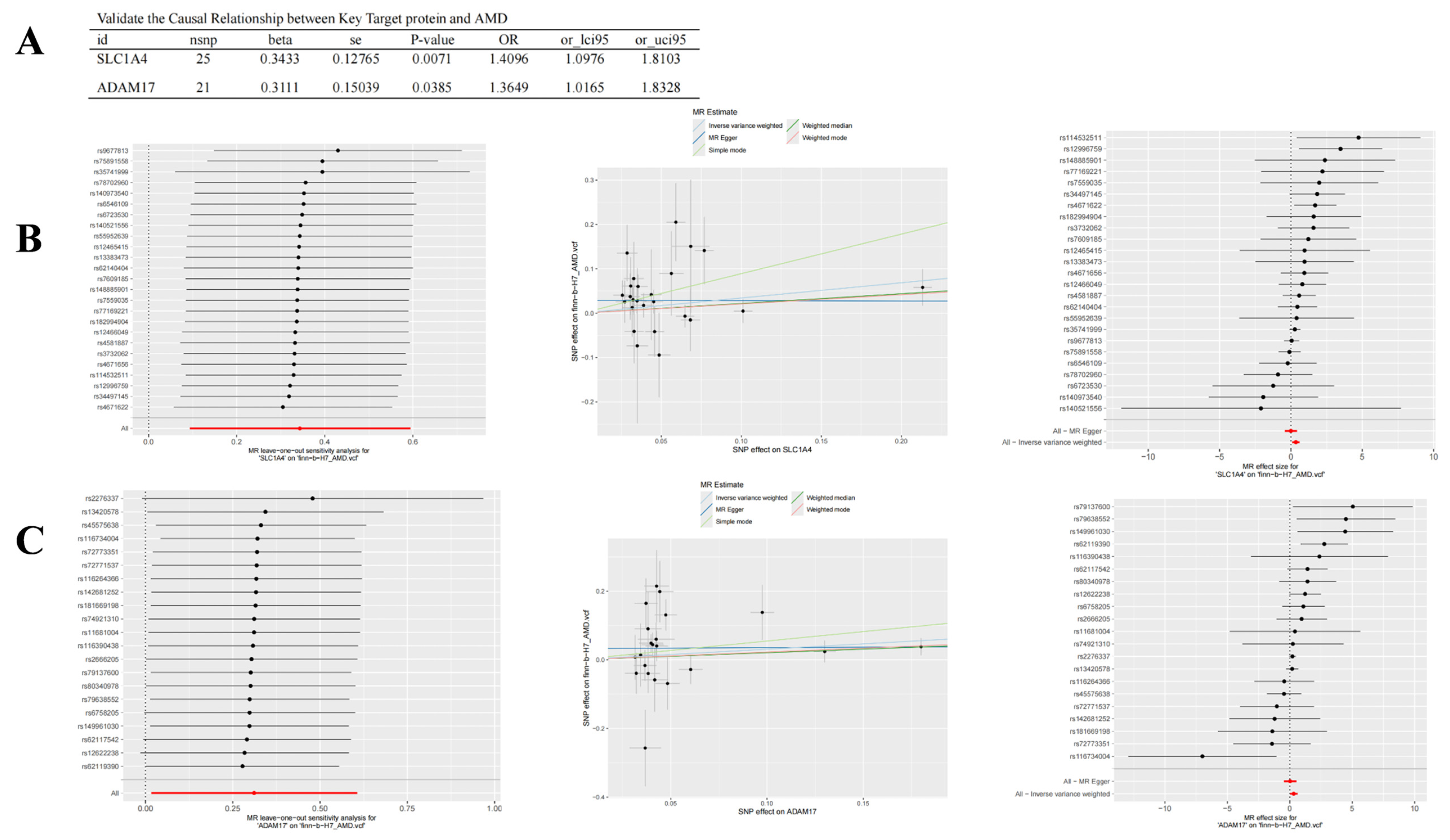
Two-sample Mendelian randomization was used to test genetically predicted gene expression against AMD. It does not test PFOA/PFOS exposure. Cis-eQTL instruments came from eQTLGen (FDR < 0.05). SNPs were retained at p < 1 × 10⁻⁵, clumped at r2 < 0.1 within 500 kb using the 1000 Genomes European reference, and required F > 10. Instrument SNPs were required to have F > 10. IVW used 25 SNPs for SLC1A4 and 21 SNPs for ADAM17. The outcome was FinnGen finn-b-H7_AMD (3763 cases and 205,359 controls, European ancestry). Among machine learning genes, instruments were available for SLC1A4, ADAM17, HSP90AA1, HSPA5, and NQO1. EGFR and MAPK10 had no qualifying instruments. Relevance was assessed by F > 10. Independence was based on European-ancestry germline instruments. Exclusion restriction was assessed with the MR-Egger intercept, Cochran’s Q, and the weighted median. Primary estimation used IVW. MR-Egger, weighted median, weighted mode, and simple mode were sensitivity analyses. Heterogeneity and the Egger intercept were estimated with TwoSampleMR v0.5.6. MR-PRESSO was run with the MRPRESSO package (version 0.6.4) on the same harmonized instruments, using 1000 permutations and a significance threshold of 0.05. A gene was included only when at least four complete instruments were available. Leave-one-out, scatter, and forest plots are shown for the two IVW-significant genes.
Additionally, by integrating the Human Protein Atlas (HPA) database (https://www.proteinatlas.org/ (accessed on 1 September 2025)) and employing single-cell distribution analysis, we focused on observing gene expression in vision-related cell types such as Müller glia cells, photoreceptor cells, and bipolar cells. UMap visualization was employed to analyze gene distribution patterns across different cell populations. Fluorescence microscopy images were inspected to assess subcellular localization patterns (for example, nucleus, cytoplasm, and endoplasmic reticulum).
2.10. Molecular Docking and Molecular Dynamics (MD) Simulations
Ligand coordinates for PFOA (PubChem CID 9554) and PFOS (PubChem CID 23677927) were taken from PubChem. Protein coordinates were PDB 7P4I, a cryo-electron microscopy structure of engineered human ASCT1/SLC1A4, and PDB 1BKC, the catalytic domain of ADAM17. Solvent and non-target organic molecules were removed before docking. Each complex was docked in its own active-site box.PFOA-SLC1A4 was centered at 106.163, 159.334, 128.251 Å. PFOA–ADAM17 was centered at 0.860, 49.345, 20.160 Å. PFOS–ADAM17 was centered at 11.784, 49.549, 20.160 Å. PFOS–SLC1A4 was centered at 136.344, 136.309, 133.448 Å. AutoDock Vina (version 1.2.5) was then run in a 40 Å × 40 Å × 40 Å box around that center, with num_modes = 30 and energy_range = 5. No reference ligand, decoy or negative-control compound was docked. A Vina score below 0 kcal/mol was treated only as a favorable pose under the scoring function, not as a measured affinity. One-hundred-nanosecond molecular dynamics were performed using GROMACS 2025.2, the CHARMM36 force field, and the TIP3P water model. Trajectory stability was not interpreted as binding affinity or as functional modulation.
2.11. Gut Microbiota Analysis
As an exploratory secondary branch complementary to the SLC1A4/ADAM17-centered main line and given prior links between gut microbiota and ophthalmic disease together with gut absorption/persistence of PFOA/PFOS [14], we used the gut microbial gene database (gutMGene; https://bio-computing.hrbmu.edu.cn/gutMGene/#/home (accessed on 1 September 2025)) to retrieve host-gene-based microbe associations [15]. Under the human species filter, we nominated gut microbes linked in gutMGene to host genes that overlap predicted PFOA/PFOS targets. Metabolites associated with these microbes were retrieved and filtered by host species = human, association source = literature association, and availability of a PubChem CID. Corresponding structures were submitted to SEA (https://sea.bkslab.org/ (accessed on 1 September 2025)) and SwissTargetPrediction (https://swisstargetprediction.ch/ (accessed on 1 September 2025)) to predict metabolite–host targets; outputs from the two tools were collated and deduplicated. Genes overlapping among (i) SEA/SwissTargetPrediction metabolite targets, (ii) gutMGene human host-gene annotations, and (iii) the PFOA/PFOS predicted-target list were retained for network visualization and GO/KEGG enrichment. Because the PFOA/PFOS target list was used both to nominate microbes and to filter metabolite targets, overlapping signals may partly reflect this shared input rather than independent biological evidence. No AMD-patient microbiome or metabolomic measurements were analyzed; therefore, these associations remain hypothesis-generating. Networks linking toxins, gut microbiota, metabolites, and host genes were constructed using STRING (https://cn.string-db.org/, accessed on 1 September 2025) and Cytoscape 3.10.3.
2.12. Use of Generative AI in Figure Preparation
During the preparation of this manuscript, the authors used GPT-6, accessed through ChatGPT (OpenAI, San Francisco, CA, USA), solely to assist in creating initial drafts of selected conceptual schematic elements in Figures 1–3 and 7. These AI-assisted drafts were critically reviewed, substantially edited, and refined for scientific accuracy by the authors, who take full responsibility for the final figures. Generative AI was not used to generate or modify research data or quantitative results, or to formulate the study conclusions.
3. Results
3.1. Toxicity Assessment and Target Identification of PFOA and PFOS
ADMETLab 3.0 predicted eye-irritation and eye-corrosion endpoints for PFOA and PFOS (Supplementary Materials Figure S1A,B). SwissTargetPrediction, ChEMBL, and PharmMapper were restricted to Homo sapiens and merged into 439 targets. Overlap with the AMD-gene list gave the 97 candidates listed in Supplementary File S1.
3.2. Identification of AMD-Associated Genes
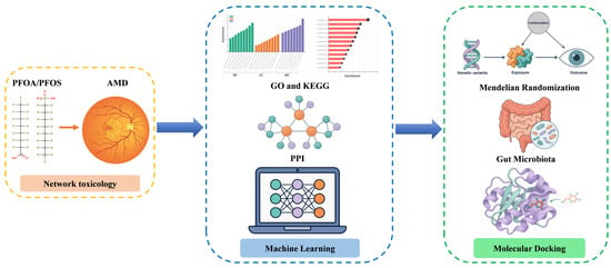
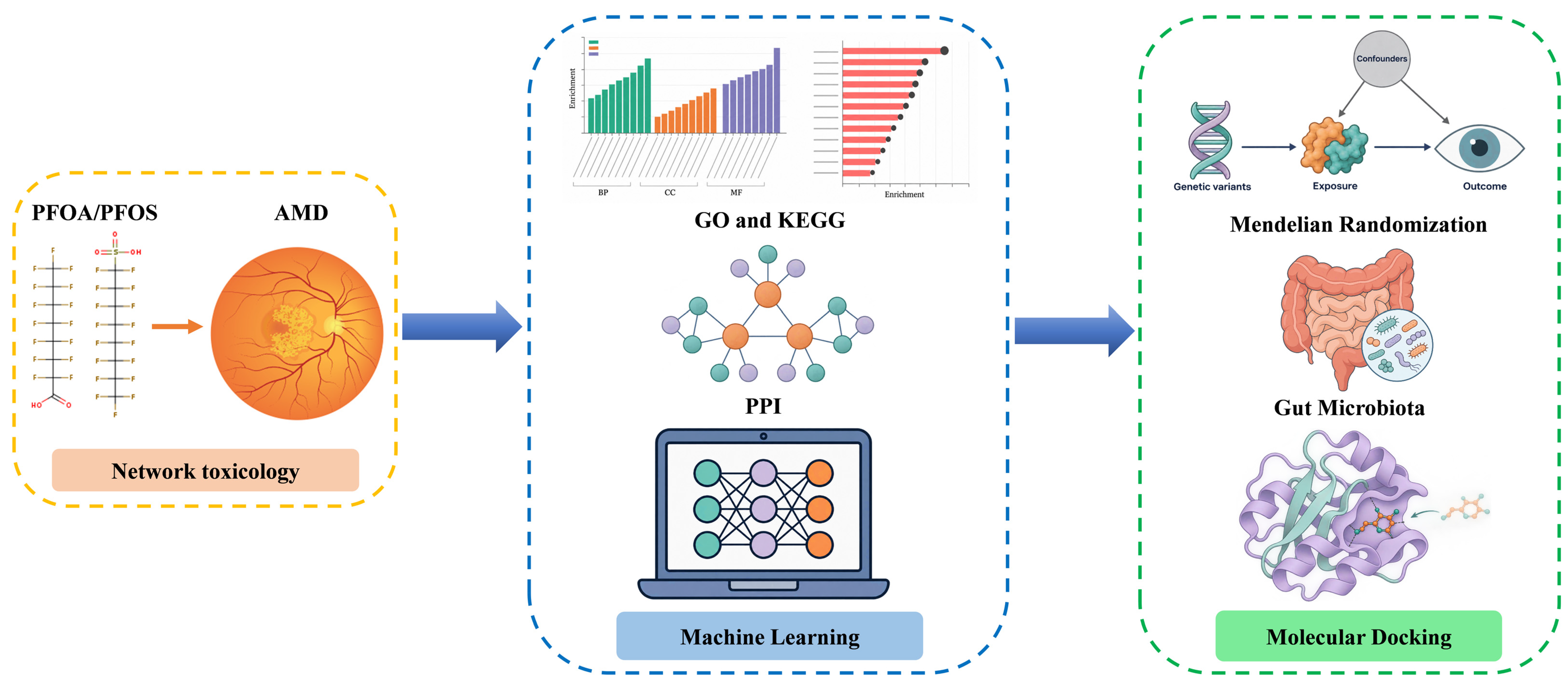
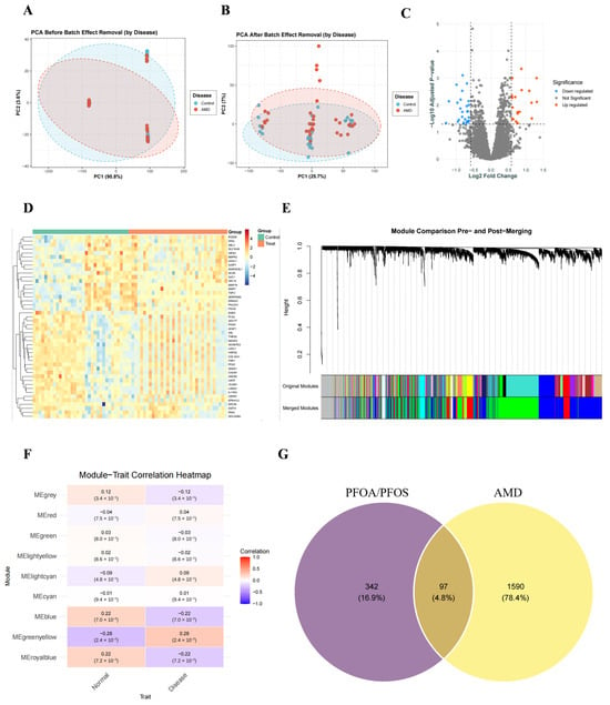
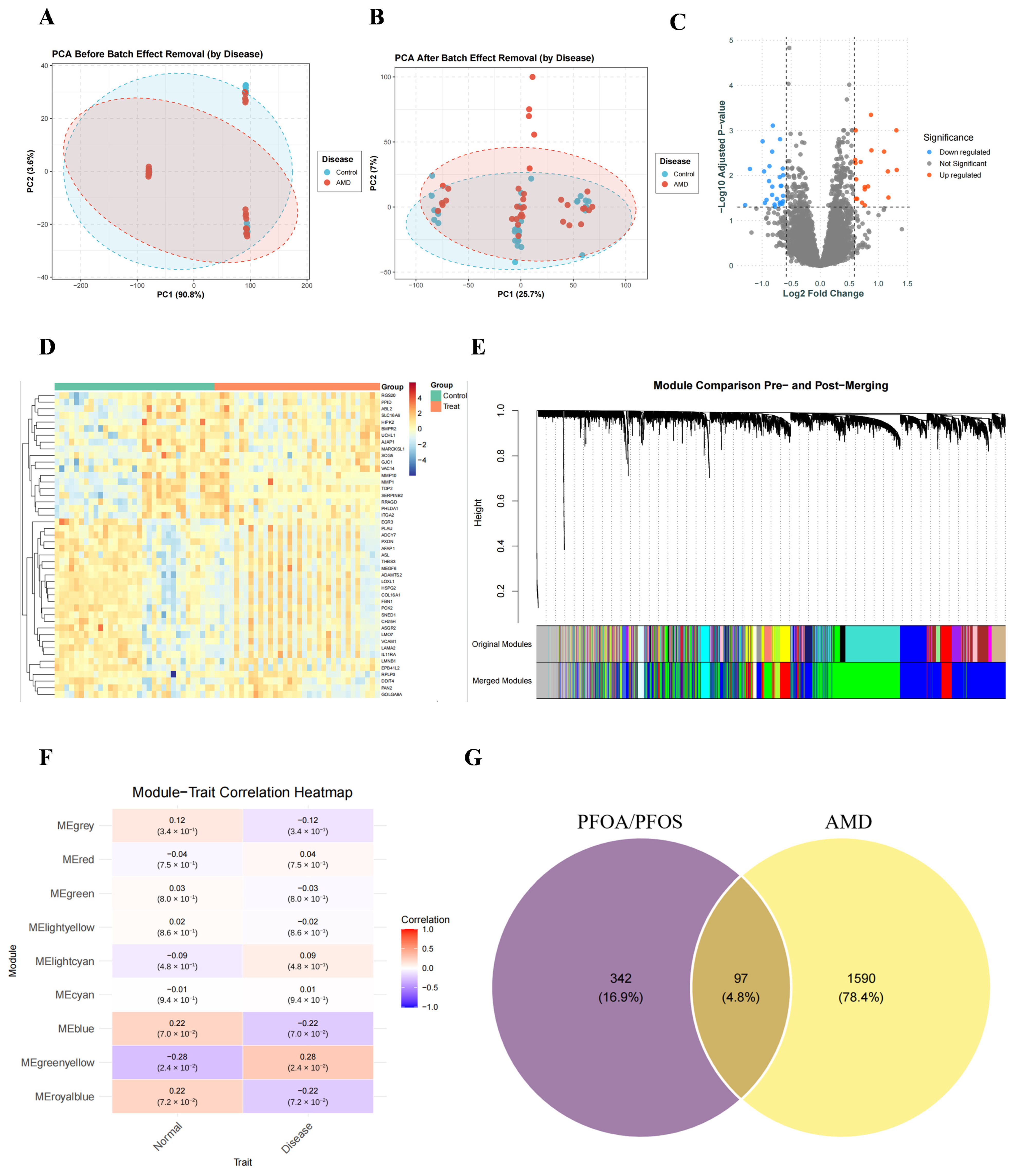
The workflow is summarized in Figure 1. GSE99248 and GSE1719 were merged and corrected with ComBat (Figure 2A,B). Limma identified 46 differentially expressed genes (Figure 2C,D), and WGCNA was run with soft-threshold power 9 (Figure 2E,F). Intersection of the 439 predicted PFOA/PFOS targets with the AMD-gene table gave 97 genes (Figure 2G; Supplementary File S1). Ninety-five were found in GeneCards and five in OMIM. The AMD-versus-control comparison was repeated within GSE99248 and within GSE1719. Of the 46 genes, 39 remained significant in GSE1719 and one, GOLGA8A, remained significant in GSE99248. No gene met the thresholds in both datasets. With the dataset fitted as a covariate instead of ComBat, 33 of the 46 genes remained significant.
Figure 1.
Overall analytical workflow. Left: network toxicology analysis of PFOA/PFOS in relation to AMD. Middle: GO and KEGG enrichment analyses, protein–protein interaction (PPI) network analysis, and machine learning. Right: Mendelian randomization, gut microbiota analysis, and molecular docking (Created with BioRender. Situ, B. (2026) https://BioRender.com/a79fvlp).
Figure 2.
Identification of PFOA/PFOS and AMD-related target genes: (A) PCA scatter plot before ComBat correction, with samples colored by batch (GSE99248 vs. GSE1719), showing clear batch-driven separation. (B) PCA after ComBat correction: left, samples colored by batch, showing improved batch mixing; right, samples colored by disease status (AMD vs. Control), in which the two disease groups were not clearly separated along PC1-PC2. (C) Volcano plot showing DEGs based on logFC and significance. Red dots are upregulated, blue dots are downregulated, and gray dots are nonsignificant. (D) Heatmap shows DEG expression patterns across samples. Red indicates upregulation, blue indicates downregulation. (E) Gene dendrogram from WGCNA shows hierarchical clustering based on co-expression. Module colors in the lower panel represent different gene modules. (F) Module-trait relationships heatmap shows correlations between WGCNA-identified modules and sample traits (Normal vs. Disease). Values in boxes indicate correlation coefficients and p-values. (G) Intersection of predicted PFOA/PFOS targets and AMD-associated genes.
3.3. Protein-Protein Interaction (PPI) Network and Functional Enrichment Analysis
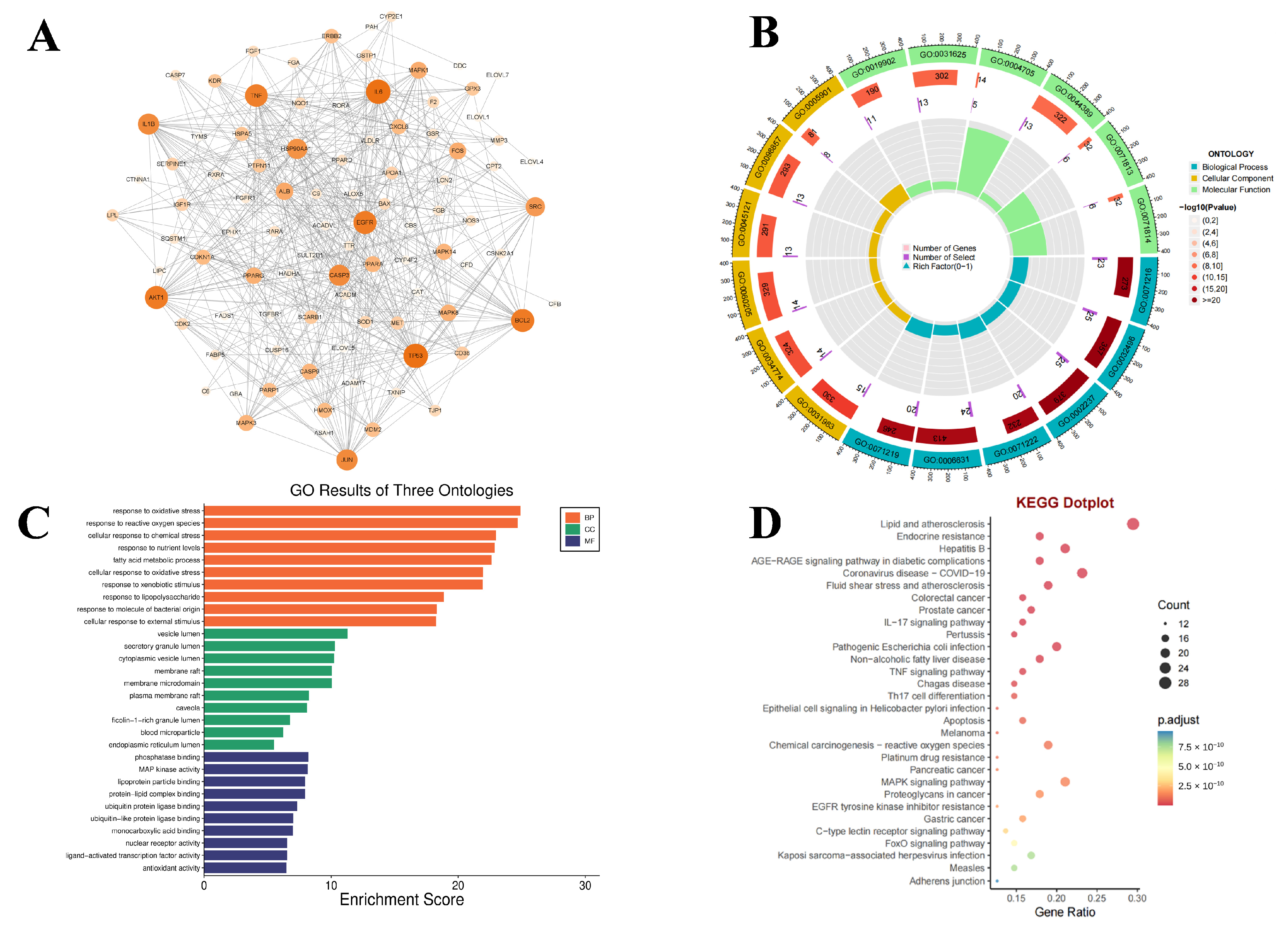
The 97 overlapping targets were uploaded to STRING and visualized in Cytoscape 3.10.3 (Figure 3A). DAVID returned 2158 GO terms at p < 0.05 (1929 biological processes, 56 cellular components and 173 molecular functions), dominated by oxidative stress, response to reactive oxygen species and chemical stress (Figure 3B,C). KEGG terms included lipid metabolism, atherosclerosis, and endocrine resistance (Figure 3D). These pathways are also common in AMD biology. Because the input genes come from the overlap of predicted targets and disease-associated genes, the enrichment is compatible with AMD but does not by itself define a PFOA/PFOS-specific mechanism.
Figure 3.
PPI, GO, and KEGG enrichment analysis of the 97 overlapping PFOA/PFOS–AMD targets. (A) STRING-derived PPI network visualized in Cytoscape 3.10.3. Nodes are proteins labeled by gene symbol; node size and fill color (light to dark orange) both scale with degree (connectivity), highlighting hub proteins. Edges represent STRING protein-protein associations and are drawn at uniform width (edge thickness is not used as a quantitative scale). (B) Circular GO enrichment plot. Outer colored sectors denote GO terms by ontology (BP, cyan/blue; CC, yellow; MF, green) with GO IDs labeled; the outer numeric scale indicates gene counts associated with each term. Colored blocks report the Number of Genes and are shaded by −log10(p-value) (pale to dark red = higher significance). Purple numerals show the Number of Selected Genes from the input list; innermost ontology-colored radial bars show Rich Factor (0–1). (C) Horizontal bar chart of top enriched GO terms for BP (orange), CC (green), and MF (purple/blue); bar length indicates Enrichment Score. (D) KEGG enrichment dot plot: x-axis, Gene Ratio; dot size, gene Count; dot color, adjusted p-value (p.adjust; redder = more significant).
3.4. Identification of Core Genes Using Machine Learning and Prediction of Core Gene Functions and Construction of miRNA-TF-mRNA Regulatory Networks
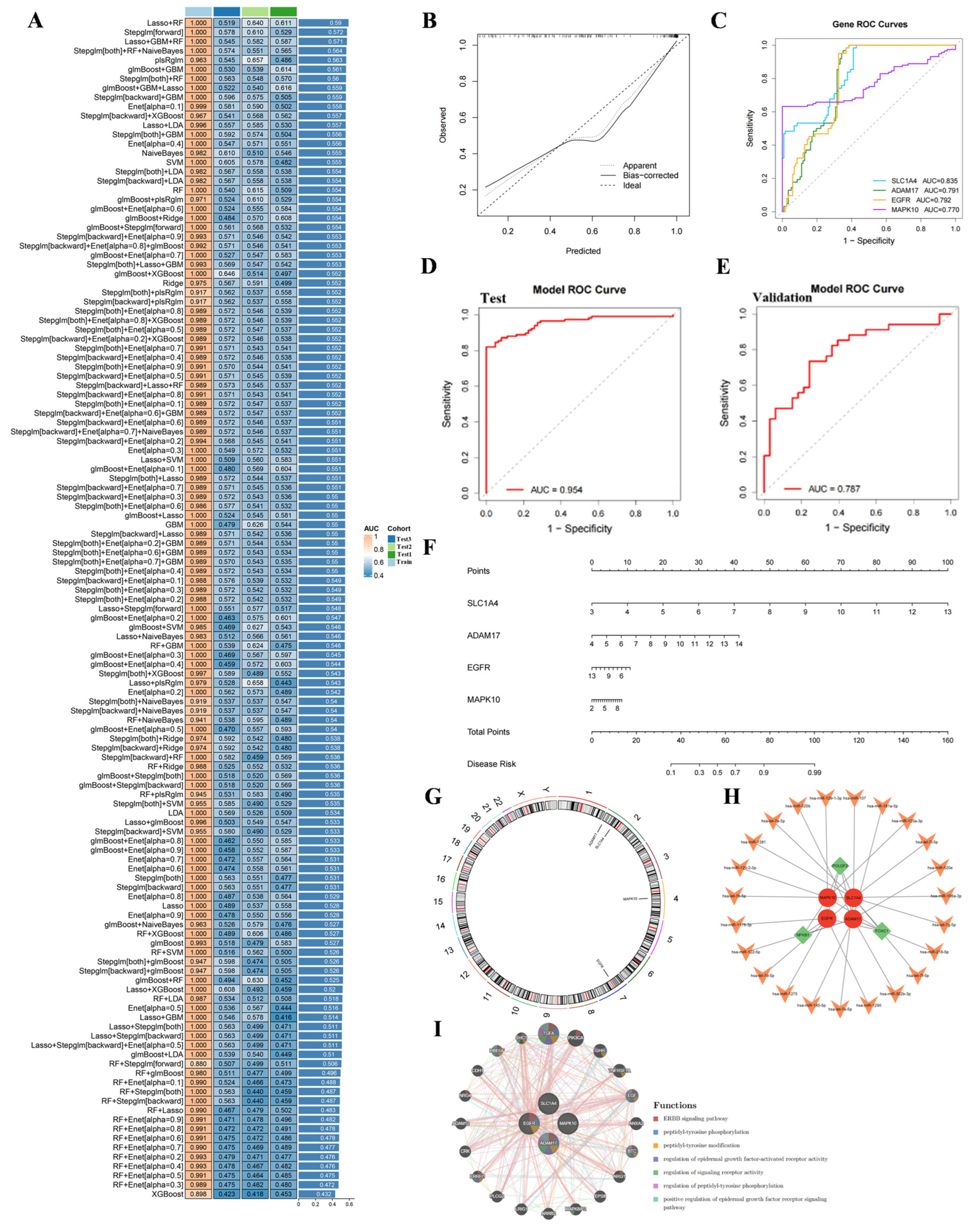
One hundred twenty-eight models were compared on the 88 training set features (Figure 4A). Mean AUCs below use only the three groups in the validation set. Lasso+RF had the higher mean (0.590) and selected RXRA, SLC1A4, EGFR, MDM2, PTPN11, RORA, TYMS, CTNNA1, HSP90AA1, NQO1, HSPA5, CYP2E1, JUN, SERPINE1, and TJP1. Stepglm[both]+RF had a mean of 0.560 and selected SLC1A4, ADAM17, EGFR, MAPK10, CBS, HSP90AA1, NQO1, and HSPA5. The overlap was SLC1A4, EGFR, HSP90AA1, NQO1, and HSPA5. ADAM17 and MAPK10 were not selected by Lasso+RF. On the three validation groups (91, 87, and 118 samples), AUCs for Stepglm[both]+RF were 0.563, 0.548, and 0.570. At a threshold of 0.5, sensitivity and specificity were 0.415 and 0.480, 0.537 and 0.500, and 0.603 and 0.491. Lasso+RF AUCs on the same groups were 0.519, 0.640, and 0.611. The four-gene ROC on the training set is shown in Figure 4E. The nomogram (Figure 4F) is an illustration of that training set fit. Predicted miRNA and transcription-factor links are shown in Figure 4H, and GeneMANIA results are shown in Figure 4I.
Figure 4.
Machine learning comparison. (A) AUC heatmap for the training set and the three groups in the validation set. (B) Calibration curve. (C) ROC curves for SLC1A4, ADAM17, EGFR, and MAPK10. (D) ROC curve of Stepglm[both]+SVM on the training set (AUC = 0.954). (E) ROC curve of the four-gene logistic model on the training set (AUC = 0.787). (F) Nomogram. (G) Chromosomal locations. (H) miRNA-transcription factor-mRNA network. (I) GeneMANIA network.
3.5. Mendelian Randomization and HPA Biological-Plausibility Assessment of Hub Genes
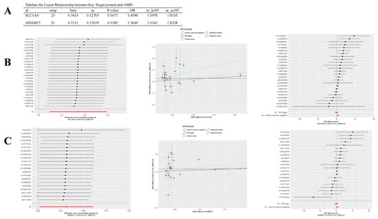
Mendelian randomization asked whether genetically predicted expression of the instrumented genes was associated with AMD. The exposure was the cis-eQTL instrument, and the outcome was FinnGen AMD. For SLC1A4, IVW gave OR 1.410 (95% CI 1.098–1.810, p = 0.0072, 25 SNPs). The weighted median was OR 1.245 (0.899–1.724, p = 0.186), MR-Egger was OR 0.993 (0.656–1.502, p = 0.973), and the weighted mode was p = 0.155. The Egger intercept was 0.029 (p = 0.051). Cochran’s Q for IVW was 25.12 on 24 degrees of freedom (p = 0.399). For ADAM17, IVW gave OR 1.365 (1.016–1.833, p = 0.039, 21 SNPs). The weighted median was OR 1.231 (0.959–1.581, p = 0.102), MR-Egger was OR 1.026 (0.629–1.673, p = 0.920), and the weighted mode was p = 0.090. The Egger intercept was 0.033 (p = 0.174), but heterogeneity was present (IVW Q = 37.19 on 20 df, p = 0.011). HSP90AA1, HSPA5, and NQO1 were not IVW-significant; EGFR and MAPK10 had no qualifying instruments (Table S1). MR-PRESSO global tests did not meet the 0.05 threshold: SLC1A4 RSS = 27.06, p = 0.440 (25 SNPs); ADAM17 RSS = 41.12, p = 0.061 (21 SNPs); HSP90AA1 RSS = 25.17, p = 0.098 (14 SNPs); and NQO1 RSS = 13.98, p = 0.567 (10 SNPs). No outlier-corrected estimate was obtained. HSPA5 had two instruments and was not included. Sensitivity plots are shown in Figure 5. HPA annotations show where the proteins have been observed (Figure S2).
Figure 5.
Mendelian randomization sensitivity analyses of genetically predicted SLC1A4 and ADAM17 expression against AMD liability. (A) IVW summaries. (B,C) Leave-one-out, scatter, and forest plots.
3.6. Predicted Binding Modes and Conformational Stability of PFOA/PFOS–Protein Complexes
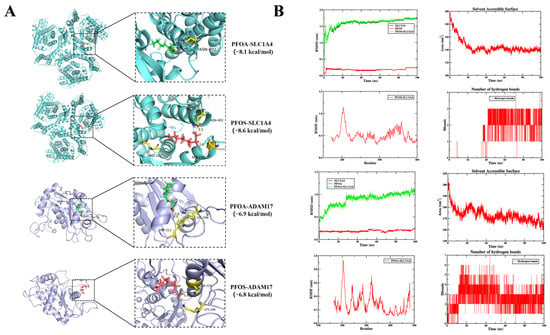
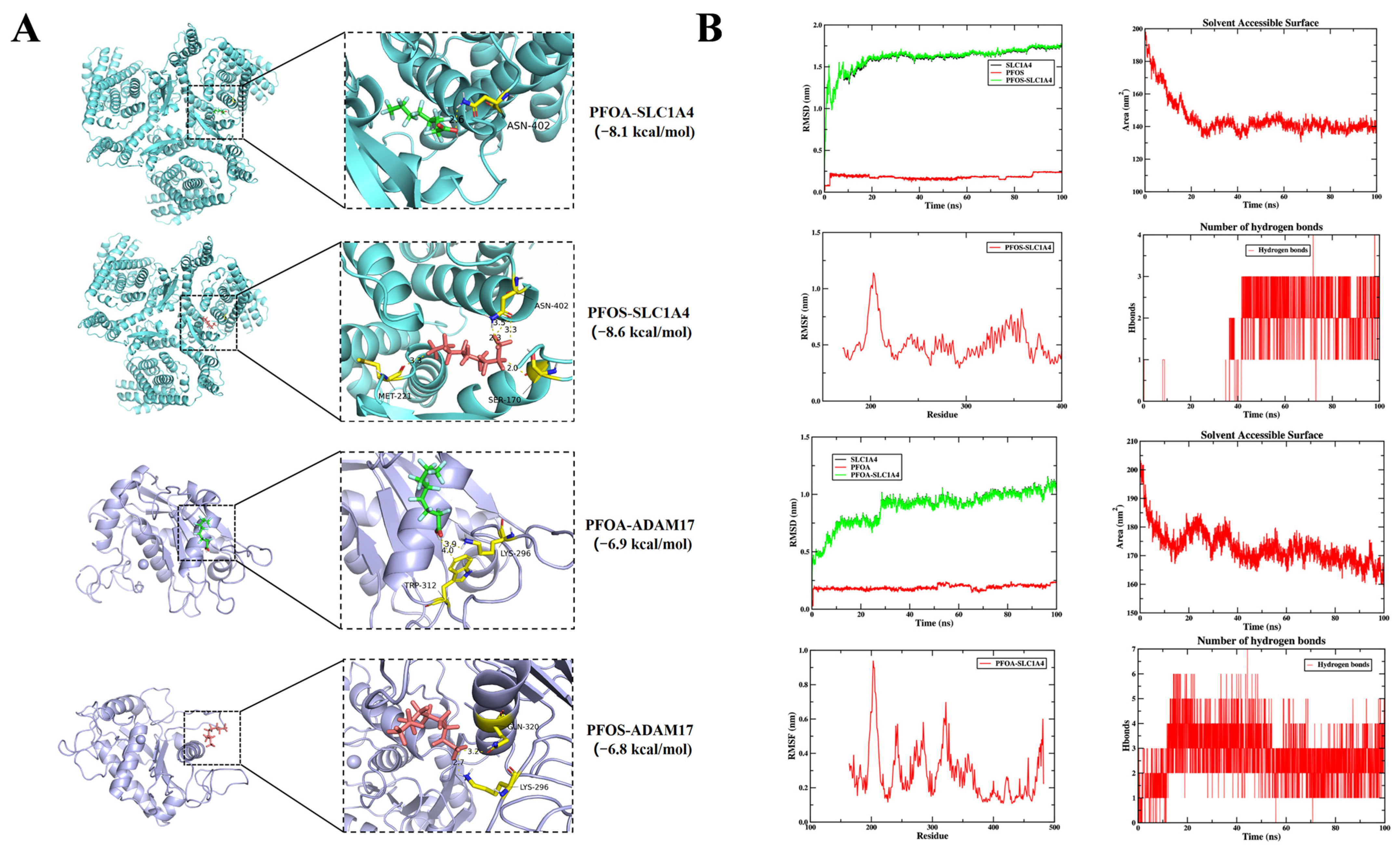
Docking was run for hypothesis generation inside the pocket-centered boxes described in Methods 2.10. Against PDB 7P4I (SLC1A4) and PDB 1BKC (ADAM17 catalytic domain), the top Vina scores were −8.1 kcal/mol for PFOA–SLC1A4, −8.6 kcal/mol for PFOS–SLC1A4, −6.9 kcal/mol for PFOA–ADAM17, and −6.8 kcal/mol for PFOS–ADAM17. Annotated contacts included ASN-402 for PFOA–SLC1A4; ASN-402, MET-221 and SER-170 for PFOS–SLC1A4; TRP-312 and LYS-296 for PFOA–ADAM17; and GLN-320 and LYS-296 for PFOS–ADAM17 (Figure 6A). One-hundred-nanosecond trajectories for the SLC1A4 complexes (Figure 6B) showed RMSD equilibration and limited SASA fluctuation. The scores were not compared with measured serum or ocular concentrations, and no experiment in this study tested whether PFOA or PFOS changes the activity of either protein.
Figure 6.
Predicted interactions of PFOA/PFOS with SLC1A4 and ADAM17. (A) Docking poses and annotated contacts for PDB 7P4I and PDB 1BKC. In panel A, green represents PFOA, red represents PFOS, cyan represents SLC1A4, and purple represents ADAM17. (B) RMSD, SASA, RMSF, and hydrogen-bond counts over 100 ns for the SLC1A4 complexes.
3.7. Exploratory Gut Microbiota-Metabolite-Host Gene Associations Related to Predicted PFOA/PFOS Targets
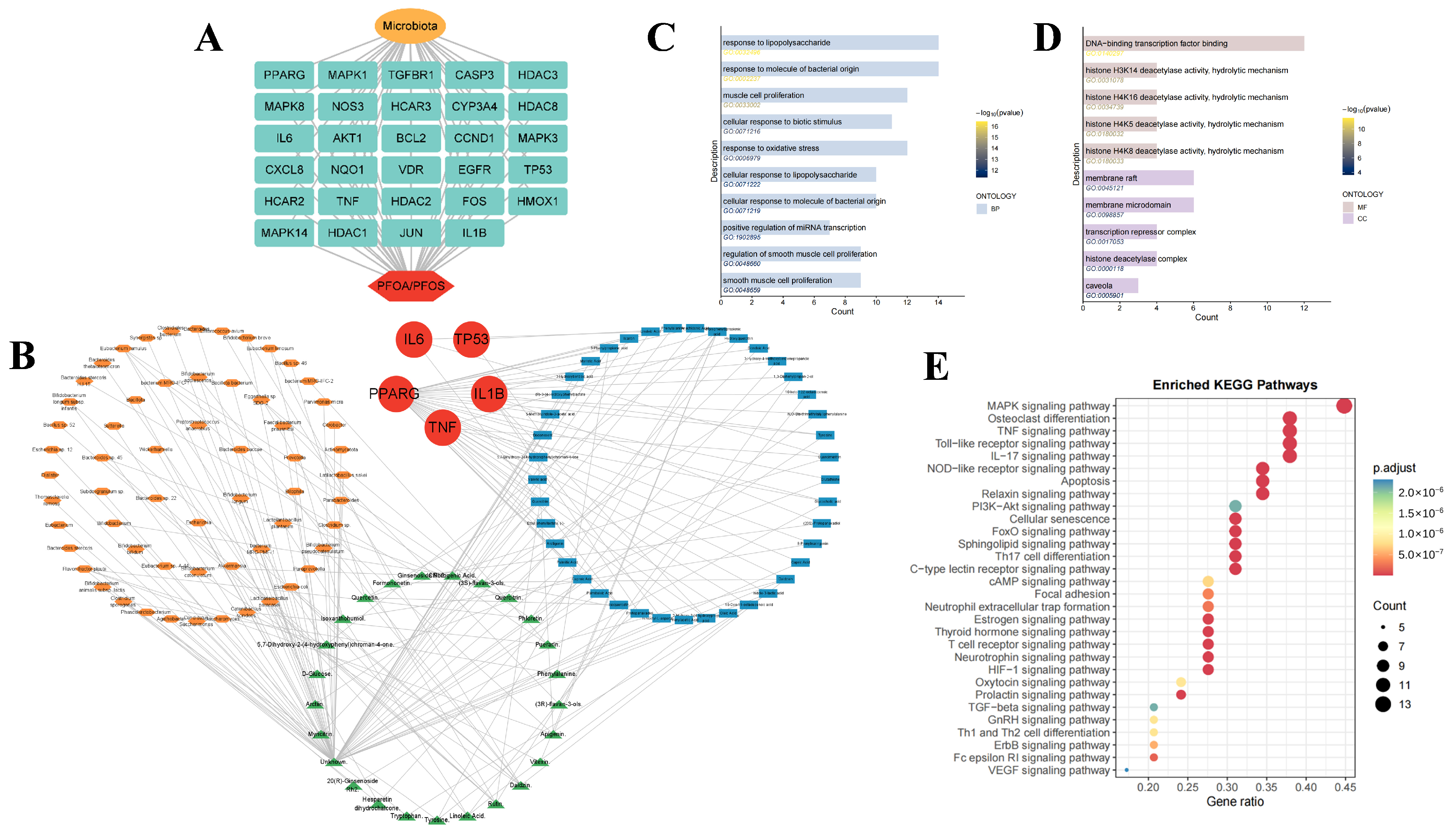
As an exploratory, database-driven secondary branch separate from the SLC1A4/ADAM17 main line, we examined putative gut microbiota-metabolite-host gene links related to predicted PFOA/PFOS targets. Using gutMGene, we retrieved microbes linked to host genes that overlap predicted PFOA/PFOS targets and compiled associated metabolites (human; literature association; PubChem CID). After collating and deduplicating SEA and SwissTargetPrediction outputs, we obtained 1518 predicted metabolite-associated human targets. A three-way intersection with gutMGene human host-gene annotations and the PFOA/PFOS predicted-target list yielded 29 overlapping genes, which were used to construct an exploratory toxin-microbe-gene network (Figure 7A). Four taxa linked in gutMGene to host genes overlapping predicted PFOA/PFOS targets were highlighted (Figure 7B): Enterococcus faecalis, Streptococcus salivarius, Turicibacter, and Lacticaseibacillus paracasei, annotated in gutMGene with IL1B and/or PPARG. These taxa were associated with 29 curated metabolites (Enterococcus faecalis: 9; Streptococcus salivarius: 1; Turicibacter: 15; Lacticaseibacillus paracasei: 4). GO and KEGG enrichment of the overlapping genes suggested associations with response to lipopolysaccharide and molecules of bacterial origin (Figure 7C,D), membrane raft/microdomain terms (Figure 7C,D), transcription factor binding (Figure 7C,D), and inflammation-related pathways including IL-17 and TNF signaling (Figure 7E). Because toxin-target lists were reused in filtering, and because no AMD-patient microbiome or metabolome data were analyzed, these database-derived associations are hypothesis-generating and do not demonstrate that gut microbiota mediate PFOA/PFOS-related AMD.
Figure 7.
Gut microbiota-metabolite-host gene associations related to predicted PFOA/PFOS targets. (A) Overlap of metabolite targets, gutMGene host genes, and PFOA/PFOS targets. (B) Linked taxa. (C) GO biological process terms. (D) GO molecular function and cellular component terms. (E) KEGG pathways.
4. Discussion
This study uses network toxicology to examine possible links between PFOA/PFOS and AMD. SLC1A4 and ADAM17 were prioritized as core candidates. Docking and molecular dynamics suggested possible interactions with these proteins, relevant to amino acid metabolism, inflammatory signaling, and oxidative stress.
Although epidemiological studies have associated PFOA/PFOS exposure with AMD [16], and PFOA can induce inflammatory responses in cultured retinal pigment epithelial cells, the molecular links remain poorly understood. We integrated transcriptomics, network toxicology, machine learning, Mendelian randomization, an exploratory gut-microbiota analysis, molecular docking, and molecular dynamics to prioritize candidates. Three levels of evidence were considered: association of the candidate genes with AMD, predicted contact between PFOA/PFOS and the encoded proteins, and direct evidence that exposure causes AMD. This study addresses the first two. The SLC1A4/ADAM17 results form the main line. The microbiota analysis is a secondary, hypothesis-generating branch and does not independently confirm that main line.
Computational toxicology can screen possible links between pollutants and disease and has been applied to other exposures [17,18,19]. Integration of transcriptomic and database-derived targets with protein-interaction and machine learning analyses identified four genes connected to both the predicted PFOA/PFOS targets and AMD: SLC1A4, ADAM17, EGFR, and MAPK10. For genes with suitable cis-eQTL instruments, MR supported associations of genetically predicted SLC1A4 and ADAM17 expression with AMD (OR 1.410 and 1.365). EGFR and MAPK10 lacked suitable instruments, and the other tested genes were not significant. These MR results concern gene expression rather than measured PFOA/PFOS exposure. Docking and molecular dynamics provided predicted poses of PFOA/PFOS with SLC1A4 and ADAM17 [20]. GO and KEGG analyses showed enrichment for oxidative stress and lipid-related pathways, consistent with known AMD mechanisms [21,22].
Recent studies in the male reproductive system provide additional context for investigating PFAS-related molecular toxicity. Musella et al. combined analyses of sperm nuclear basic proteins and DNA protection with docking predictions of potential PFOA/PFOS interactions with protamine P1 and DNA [23]. Marinaro et al. reported reduced seminal antioxidant activity and increased lipoperoxides in men from a PFAS-contaminated area relative to a comparison population, alongside analyses of sperm nuclear basic protein–associated oxidative DNA damage [24]. These findings support further investigation of PFAS-associated oxidative alterations and possible macromolecular interactions. However, neither study examined retinal tissue, AMD, or mediation through SLC1A4 or ADAM17. Their relevance to the present study is therefore contextual rather than confirmatory, and extrapolation to retinal pathology requires direct investigation in appropriate experimental models.
SLC1A4 is a Na+-dependent neutral amino acid transporter and contributes to amino acid homeostasis in the retina and RPE [25,26]. Disturbance of amino acid metabolism, including glutamine, glutamate, and alanine pathways, has been linked to retinal pathology and AMD [27,28]. SLC36A4, another amino acid transporter, regulates the amino acid pool in RPE lysosomes [29]. These observations provide the biological context for examining SLC1A4. ADAM17 is a protease involved in inflammation, angiogenesis, cell adhesion, and migration [30,31]. It is expressed in the postnatal retina and is required for ocular morphogenesis [32,33]. The ADAM17/TNF pathway protects Drosophila retinal cells from age- and activity-related degeneration [34]. Together, these reports support a hypothesis that altered amino acid transport and ADAM17-related inflammatory signaling could contribute to RPE imbalance and can be tested experimentally.
PFOA and PFOS persist in the environment and can accumulate in humans through drinking water and the food chain [35,36]. Their long environmental persistence coincides with the age-related occurrence of AMD [37]. Absorbed compounds may reach the eye, where slow choroidal blood flow and the high metabolic demand of photoreceptors and the RPE could favor retention. Gut dysbiosis has been linked to ocular disease through a gut-eye axis [38]. Separately from the SLC1A4/ADAM17 analysis, we constructed an exploratory toxin-microbiota-metabolite-host gene network from gutMGene and metabolite-target prediction (Figure 7). IL1B and PPARG linked predicted targets to taxa such as Turicibacter and Lacticaseibacillus paracasei. These database associations suggest that PFOA/PFOS-related host targets may intersect microbe- and metabolite-linked pathways. They do not show that PFOA/PFOS cause AMD by disrupting the microbiota, and they do not support a treatment claim. The same target list was used both to nominate microbes and to filter metabolite targets, and no AMD-patient microbiome was analyzed.
The SLC1A4/ADAM17 analysis provides a computational lead relating PFOA/PFOS to amino acid metabolism and inflammatory signaling, with predicted docking support. The microbiota branch is hypothesis-generating rather than a demonstrated mediating mechanism. Several limitations should be noted. Training AUCs were often near 1.0, whereas the external mean AUC of the retained model was about 0.560. After batch correction, separation of AMD and control samples on the first principal components remained modest. The gut analysis reused the toxin-target list and did not include an AMD microbiome. Docking and molecular dynamics suggest possible binding poses but do not prove binding. Mendelian randomization concerns genetically predicted gene expression rather than measured PFOA/PFOS exposure; weighted-median and MR-Egger analyses did not confirm the IVW estimates, and MR-PRESSO did not yield an outlier-corrected estimate. Discovery sample sizes are limited, and experimental follow-up in retinal models is needed.
5. Conclusions
This study prioritizes SLC1A4 and ADAM17 as candidate targets for the possible ocular effects of PFOA/PFOS. The findings are hypothesis-generating and are intended to guide experimental follow-up rather than clinical prediction or treatment.
Supplementary Materials
The following supporting information can be downloaded at: https://www.mdpi.com/article/10.3390/jox16070223/s1, Figure S1: Molecular Structure and Toxicity Prediction of Toxins (A. Molecular formula of PFOA/PFOS; B. Prediction of toxicity of PFOA/PFOS). The corresponding relationships of the three labels (green, yellow, red) are as follows: excellent, medium and poor; Figure S2: Biological plausibility/expression pattern support for hub targets associated with AMD in the HPA database; Table S1: Inverse-variance weighted (IVW) two-sample Mendelian randomization estimates for gene exposures with available cis-eQTL instruments and AMD outcome (finn-b-H7_AMD). Primary findings are SLC1A4 and ADAM17; HSP90AA1, HSPA5, and NQO1 were tested but non-significant. EGFR and MAPK10 lacked suitable instruments and are not listed; File S1: 97 Candidate Targets Identified by Intersecting PFOA/PFOS Targets with AMD-Associated Genes. References [39,40,41] are cited in the Supplementary Materials.
Author Contributions
Z.G. and X.C.: Investigation, Methodology, Writing-original draft, Writing-review and editing. L.Z.: Formal analysis, Writing-review and editing. Y.L.: Formal analysis, Writing-review and editing. J.L.: Formal analysis, Writing-review and editing. Y.Q. and H.Z.: Project administration, Software, Formal Analysis, Writing-review and editing. All authors have read and agreed to the published version of the manuscript.
Funding
This study was supported by the National Natural Science Foundation of China (grant No. 82171036 & 82471083).
Institutional Review Board Statement
Not applicable.
Informed Consent Statement
Not applicable.
Data Availability Statement
The data analyzed in this study were obtained from publicly available resources. Transcriptomic data are available from GEO (https://www.ncbi.nlm.nih.gov/geo/ (accessed on 27 August 2025)) under GSE99248, GSE1719, and GSE29801. Cis-eQTL summary statistics are from eQTLGen (https://www.eqtlgen.org/cis-eqtls.html (accessed on 1 September 2025)). AMD GWAS summary statistics are from FinnGen finn-b-H7_AMD (https://gwas.mrcieu.ac.uk/). Supplementary File S1 lists the 97 genes and their GeneCards or OMIM membership.
Acknowledgments
During the preparation of this manuscript, the authors used ChatGPT (OpenAI, GPT-6) to generate schematic illustrations for Figure 1. The authors have reviewed and edited the output and take full responsibility for the content of this publication.
Conflicts of Interest
The authors declare no conflicting interests.
References
- Papadopoulou, E.; Stratakis, N.; Basagana, X.; Brantsaeter, A.L.; Casas, M.; Fossati, S.; Grazuleviciene, R.; Smastuen Haug, L.; Heude, B.; Maitre, L.; et al. Prenatal and postnatal exposure to PFAS and cardiometabolic factors and inflammation status in children from six European cohorts. Environ. Int. 2021, 157, 106853. [Google Scholar] [CrossRef] [Scilit] [PubMed]
- Zango, Z.U.; Khoo, K.S.; Garba, A.; Kadir, H.A.; Usman, F.; Zango, M.U.; Da Oh, W.; Lim, J.W. A review on superior advanced oxidation and photocatalytic degradation techniques for perfluorooctanoic acid (PFOA) elimination from wastewater. Environ. Res. 2023, 221, 115326. [Google Scholar] [CrossRef] [Scilit] [PubMed]
- Shi, W.; Zhang, Z.; Li, M.; Dong, H.; Li, J. Reproductive toxicity of PFOA, PFOS and their substitutes: A review based on epidemiological and toxicological evidence. Environ. Res. 2024, 250, 118485. [Google Scholar] [CrossRef] [Scilit] [PubMed]
- Guymer, R.H.; Campbell, T.G. Age-related macular degeneration. Lancet 2023, 401, 1459–1472. [Google Scholar] [CrossRef] [Scilit] [PubMed]
- Jonas, J.B.; Cheung, C.M.G.; Panda-Jonas, S. Updates on the Epidemiology of Age-Related Macular Degeneration. Asia Pac. J. Ophthalmol. 2017, 6, 493–497. [Google Scholar] [CrossRef] [Scilit] [PubMed]
- Qu, S.; Lin, H.; Pfeiffer, N.; Grus, F.H. Age-Related Macular Degeneration and Mitochondria-Associated Autoantibodies: A Review of the Specific Pathogenesis and Therapeutic Strategies. Int. J. Mol. Sci. 2024, 25, 1624. [Google Scholar] [CrossRef] [Scilit] [PubMed]
- Kaidonis, G.; Lamy, R.; Wu, J.; Yang, D.; Psaras, C.; Doan, T.; Stewart, J.M. Aqueous Fluid Transcriptome Profiling Differentiates Between Non-Neovascular and Neovascular AMD. Investig. Ophthalmol. Vis. Sci. 2023, 64, 26. [Google Scholar] [CrossRef] [Scilit] [PubMed]
- Fleckenstein, M.; Schmitz-Valckenberg, S.; Chakravarthy, U. Age-Related Macular Degeneration: A Review. JAMA 2024, 331, 147–157. [Google Scholar] [PubMed]
- Wang, D.; Chen, Y.; Li, J.; Wu, E.; Tang, T.; Singla, R.K.; Shen, B.; Zhang, M. Natural products for the treatment of age-related macular degeneration. Phytomedicine 2024, 130, 155522. [Google Scholar] [CrossRef] [Scilit] [PubMed]
- Yuan, M.; Zhang, T.; Sun, Y.; Yi, S.; Han, G.; Wei, P.; Chen, X.; Wang, Y.; Zhu, L. Microglia-Mediated Pathological Retinal Angiogenesis Leading to Visual Impairment in Mice Exposed to Perfluorooctanoic Acid. Environ. Sci. Technol. 2025, 59, 15680–15691. [Google Scholar] [CrossRef] [Scilit] [PubMed]
- Tien, P.T.; Lin, H.J.; Tsai, Y.Y.; Lim, Y.P.; Chen, C.S.; Chang, C.Y.; Lin, C.J.; Chen, J.J.; Wu, S.M.; Huang, Y.J.; et al. Perfluorooctanoic acid in indoor particulate matter triggers oxidative stress and inflammation in corneal and retinal cells. Sci. Rep. 2020, 10, 15702. [Google Scholar] [CrossRef] [Scilit] [PubMed]
- Chen, X.; Li, J.; Xu, N.; Li, X.; Li, J.; Guo, Q.; Zhang, J.; Miao, H.; Huang, L. Serum lipids mediate the association of per- and polyfluoroalkyl substances exposure and age-related macular degeneration. PLoS ONE 2025, 20, e0317678. [Google Scholar] [CrossRef] [Scilit] [PubMed]
- Clough, E.; Barrett, T.; Wilhite, S.E.; Ledoux, P.; Evangelista, C.; Kim, I.F.; Tomashevsky, M.; Marshall, K.A.; Phillippy, K.H.; Sherman, P.M.; et al. NCBI GEO: Archive for gene expression and epigenomics data sets: 23-year update. Nucleic Acids Res. 2024, 52, D138–D144. [Google Scholar] [CrossRef] [Scilit] [PubMed]
- Zhang, W.; Tian, Y.; Chen, B.; Xu, S.; Wu, L. PFOA/PFOS Facilitated Intestinal Fatty Acid Absorption by Activating the PPARalpha Pathway: Insights from Organoids Model. Environ. Health 2024, 2, 85–94. [Google Scholar] [CrossRef] [Scilit] [PubMed]
- Qi, C.; He, G.; Qian, K.; Guan, S.; Li, Z.; Liang, S.; Liu, J.; Ke, X.; Zhang, S.; Lu, M.; et al. gutMGene v2.0, an updated comprehensive database for target genes of gut microbes and microbial metabolites. Nucleic Acids Res. 2025, 53, D783–D788. [Google Scholar] [CrossRef] [Scilit] [PubMed]
- Yuan, M.; Yi, S.; Wang, X.; Han, G.; Wei, P.; Lv, Z.; Gui, B.; Chen, X.; Wang, Y.; Zhu, L. Promoted Translocation of Perfluorooctanoic Acid across the Blood-Retinal Barrier due to its Inhibition of Tight Junction Assembly by Antagonizing LPAR1. Environ. Sci. Technol. 2025, 59, 4807–4819. [Google Scholar] [CrossRef] [Scilit] [PubMed]
- Feng, C.; Yan, W.; Mei, Z.; Luo, X. Exploring the toxicological impact of bisphenol a exposure on psoriasis through network toxicology, machine learning, and multi-dimensional bioinformatics analysis. J. Environ. Manag. 2025, 385, 125708. [Google Scholar] [CrossRef] [Scilit] [PubMed]
- Song, S.; Huang, L.; Zhou, X.; Yu, J. Exploring the toxicological network in diabetic microvascular disease. Int. J. Surg. 2025, 111, 3895–3907. [Google Scholar] [CrossRef] [Scilit] [PubMed]
- Guo, Y.; Liu, Y.; Chen, Y.; Du, S.; Zheng, Y.; Wang, L. The mechanisms of environmental pollutant acetyl tributyl citrate induced oral squamous cell carcinoma using network toxicology, molecular docking and molecular dynamics simulation. Int. J. Surg. 2025, 111, 7873–7885. [Google Scholar] [CrossRef] [Scilit] [PubMed]
- Saad, M.I.; Jenkins, B.J. The protease ADAM17 at the crossroads of disease: Revisiting its significance in inflammation, cancer, and beyond. FEBS J. 2024, 291, 10–24. [Google Scholar] [CrossRef] [Scilit] [PubMed]
- Fleckenstein, M.; Keenan, T.D.L.; Guymer, R.H.; Chakravarthy, U.; Schmitz-Valckenberg, S.; Klaver, C.C.; Wong, W.T.; Chew, E.Y. Age-related macular degeneration. Nat. Rev. Dis. Primers 2021, 7, 31. [Google Scholar] [PubMed]
- Tan, L.X.; Germer, C.J.; La Cunza, N.; Lakkaraju, A. Complement activation, lipid metabolism, and mitochondrial injury: Converging pathways in age-related macular degeneration. Redox Biol. 2020, 37, 101781. [Google Scholar] [CrossRef] [Scilit] [PubMed]
- Musella, F.; Guarnieri, M.G.; Amore, S.; Montano, L.; Bertola, F.; Micali, S.; Busardo, F.P.; Di Giovanni, C.; Lettieri, G.; Piscopo, M. Molecular and Computational Studies Reveal That Per- and Polyfluoroalkyl Substances Can Impair Protamine-DNA Interaction, Potentially Inducing DNA Damage. Biomolecules 2025, 15, 1279. [Google Scholar] [CrossRef] [Scilit] [PubMed]
- Marinaro, C.; Bianchi, A.R.; Guerretti, V.; Barricelli, G.; Berman, B.; Bertola, F.; Micali, S.; Busardo, F.P.; Di Giorgi, A.; De Maio, A.; et al. Molecular Alterations in Semen of Per- and Polyfluoroalkyl Substance Exposed Subjects: Association Between DNA Integrity, Antioxidant Capacity and Lipoperoxides. Antioxidants 2025, 14, 792. [Google Scholar] [CrossRef] [Scilit] [PubMed]
- Elazar, D.; Alvarez, N.; Drobeck, S.; Gunn, T.M. SLC1A4 and Serine Homeostasis: Implications for Neurodevelopmental and Neurodegenerative Disorders. Int. J. Mol. Sci. 2025, 26, 2104. [Google Scholar] [CrossRef] [Scilit] [PubMed]
- Hansman, D.S.; Du, J.; Casson, R.J.; Peet, D.J. Eye on the horizon: The metabolic landscape of the RPE in aging and disease. Prog. Retin. Eye Res. 2025, 104, 101306. [Google Scholar] [CrossRef] [Scilit] [PubMed]
- Xia, M.; Zhang, F. Amino Acids Metabolism in Retinopathy: From Clinical and Basic Research Perspective. Metabolites 2022, 12, 1244. [Google Scholar] [CrossRef] [Scilit] [PubMed]
- Hou, X.W.; Wang, Y.; Pan, C.W. Metabolomics in Age-Related Macular Degeneration: A Systematic Review. Investig. Ophthalmol. Vis. Sci. 2020, 61, 13. [Google Scholar] [CrossRef] [Scilit] [PubMed]
- Shang, P.; Valapala, M.; Grebe, R.; Hose, S.; Ghosh, S.; Bhutto, I.A.; Handa, J.T.; Lutty, G.A.; Lu, L.; Wan, J.; et al. The amino acid transporter SLC36A4 regulates the amino acid pool in retinal pigmented epithelial cells and mediates the mechanistic target of rapamycin, complex 1 signaling. Aging Cell 2017, 16, 349–359. [Google Scholar] [CrossRef] [Scilit] [PubMed]
- Caolo, V.; Swennen, G.; Chalaris, A.; Wagenaar, A.; Verbruggen, S.; Rose-John, S.; Molin, D.G.; Vooijs, M.; Post, M.J. ADAM10 and ADAM17 have opposite roles during sprouting angiogenesis. Angiogenesis 2015, 18, 13–22. [Google Scholar] [CrossRef] [Scilit] [PubMed]
- Wang, X.; Wang, T.; Kaneko, S.; Kriukov, E.; Lam, E.; Szczepan, M.; Chen, J.; Gregg, A.; Wang, X.; Fernandez-Gonzalez, A.; et al. Photoreceptors inhibit pathological retinal angiogenesis through transcriptional regulation of Adam17 via c-Fos. Angiogenesis 2024, 27, 379–395. [Google Scholar] [CrossRef] [Scilit] [PubMed]
- Toonen, J.A.; Ronchetti, A.; Sidjanin, D.J. A Disintegrin and Metalloproteinase10 (ADAM10) Regulates NOTCH Signaling during Early Retinal Development. PLoS ONE 2016, 11, e0156184. [Google Scholar] [CrossRef] [Scilit] [PubMed]
- Sel, S.; Kalinski, T.; Enssen, I.; Kaiser, M.; Nass, N.; Trau, S.; Wollensak, G.; Brauer, L.; Jager, K.; Paulsen, F. Expression analysis of ADAM17 during mouse eye development. Ann. Anat. 2012, 194, 334–338. [Google Scholar] [CrossRef] [Scilit] [PubMed]
- Muliyil, S.; Levet, C.; Dusterhoft, S.; Dulloo, I.; Cowley, S.A.; Freeman, M. ADAM17-triggered TNF signalling protects the ageing Drosophila retina from lipid droplet-mediated degeneration. EMBO J. 2020, 39, e104415. [Google Scholar] [CrossRef] [Scilit] [PubMed]
- Wee, S.Y.; Aris, A.Z. Environmental impacts, exposure pathways, and health effects of PFOA and PFOS. Ecotoxicol. Environ. Saf. 2023, 267, 115663. [Google Scholar] [CrossRef] [Scilit] [PubMed]
- Cheng, X.; Wei, Y.; Zhang, Z.; Wang, F.; He, J.; Wang, R.; Xu, Y.; Keerman, M.; Zhang, S.; Zhang, Y.; et al. Plasma PFOA and PFOS Levels, DNA Methylation, and Blood Lipid Levels: A Pilot Study. Environ. Sci. Technol. 2022, 56, 17039–17051. [Google Scholar] [CrossRef] [Scilit] [PubMed]
- Collaborators GBDGA. Global burden of vision impairment due to age-related macular degeneration, 1990–2021, with forecasts to 2050, a systematic analysis for the Global Burden of Disease Study 2021. Lancet Glob. Health 2025, 13, e1175–e1190. [Google Scholar] [CrossRef] [Scilit] [PubMed]
- Jin, Q.; Wang, S.; Yao, Y.; Jiang, Q.; Li, K. The gut-eye axis: From brain neurodegenerative diseases to age-related macular degeneration. Neural Regen. Res. 2025, 20, 2741–2757. [Google Scholar] [CrossRef] [Scilit] [PubMed]
- Daina, A.; Michielin, O.; Zoete, V. SwissTargetPrediction: Updated data and new features for efficient prediction of protein targets of small molecules. Nucleic Acids Res. 2019, 47, W357–W364. [Google Scholar] [CrossRef] [Scilit] [PubMed]
- Karlsson, M.; Zhang, C.; Méar, L.; Zhong, W.; Digre, A.; Katona, B.; Sjöstedt, E.; Butler, L.; Odeberg, J.; Dusart, P.; et al. A single-cell type transcriptomics map of human tissues. Sci. Adv. 2021, 7, eabh2169. [Google Scholar] [CrossRef] [Scilit] [PubMed]
- Thul, P.J.; Åkesson, L.; Wiking, M.; Mahdessian, D.; Geladaki, A.; Ait Blal, H.; Alm, T.; Asplund, A.; Björk, L.; Breckels, L.M.; et al. A subcellular map of the human proteome. Science 2017, 356, eaal3321. [Google Scholar] [CrossRef] [Scilit] [PubMed]
Disclaimer/Publisher’s Note: The statements, opinions and data contained in all publications are solely those of the individual author(s) and contributor(s) and not of MDPI and/or the editor(s). MDPI and/or the editor(s) disclaim responsibility for any injury to people or property resulting from any ideas, methods, instructions or products referred to in the content. |
© 2026 by the authors. Licensee MDPI, Basel, Switzerland. This article is an open access article distributed under the terms and conditions of the Creative Commons Attribution (CC BY) license.