


Abstract
Background/Objectives: Chest tubes are inserted in almost all children undergoing cardiac surgery and managed at the bedside until removal. Management spans the drainage system, tube patency, and the criteria and timing used to judge readiness for removal. This review assessed the effectiveness and safety of these strategies, as well as their implications for nursing practice. Methods: Five databases and two trial registries were searched up to 20 May 2026. Eligible studies compared a chest-tube management intervention with an alternative, usual care, or a pre-intervention baseline. Two reviewers independently undertook selection, extraction, and risk-of-bias assessment (RoB 2, ROBINS-I). Randomised and non-randomised evidence were synthesised separately without pooling; certainty was assessed using GRADE. The review was prospectively registered in PROSPERO (CRD420261397281) and received no external funding. Results: Eight studies including up to 6567 children were included; seven, four of them randomised trials, contributed to the comparative synthesis, and one surveillance cohort was synthesised descriptively. Shorter duration was reported with more frequent assessment of an unchanged removal threshold, a smaller-calibre silicone drain, and protocol-driven removal pathways. A high-vacuum drainage system combined with smaller-calibre chest tubes (12 Ch versus 16 Ch) was associated with fewer residual pleural effusions requiring drainage than a conventional underwater-seal system with larger-calibre tubes (risk ratio 0.23, 95% CI 0.08 to 0.66); however, the independent effects of suction configuration and tube calibre could not be distinguished. Apart from that reduction, post-removal events were uncommon, and available studies did not demonstrate a clear increase or decrease in clinically important complications; however, the remaining safety estimates were imprecise and did not exclude clinically important benefit or harm. Certainty was low or very low for all outcomes. Conclusions: Interventions acting at different points of the management pathway were associated with shorter chest-tube duration, though their mechanisms and magnitudes varied. Structured removal criteria and more frequent assessment are potentially modifiable, but their effects cannot be separated from those of drain and drainage-system characteristics. Safety after removal could not be established in either direction, and complication rates varied with outcome definition and surveillance method. For nursing practice, the findings are consistent with an explicit, documented removal decision reviewed at defined intervals, with defined post-removal observation given the uncertain safety of earlier removal.
1. Introduction
Congenital heart disease affects approximately eight to nine per 1000 live births [1,2] and is the most common group of birth defects. About one in four affected infants has a critical lesion requiring surgery or another procedure within the first year of life [3], and congenital heart disease accounts for a substantial proportion of birth-defect-related hospital admissions and healthcare resource use among children [4]. Transcatheter techniques have replaced open operations for selected defects, but many lesions continue to require surgical repair, reconstruction, or palliation, particularly when treatment involves cardiopulmonary bypass, intracardiac reconstruction, or staged management of specific physiologies. Chest tubes are placed intraoperatively in almost all such procedures, and one or more mediastinal or pleural tubes routinely remain in place during early postoperative recovery [5].
Chest tubes are essential during postoperative recovery because blood and fluid may collect in the mediastinal or pleural spaces after cardiac surgery, while air may accumulate in the pleural space. Mediastinal drainage facilitates the evacuation of postoperative blood and fluid and helps reduce the risk of retained collections, pericardial effusion, and tamponade [6,7]. Pleural drainage removes air and fluid, restores negative pleural pressure, and permits lung re-expansion. Effective drainage therefore contributes to haemodynamic and respiratory stability during early postoperative recovery [8].
Effective drainage depends on appropriate management of the chest tube and drainage system from insertion until removal [9]. The system must permit the evacuation of air and fluid while preventing retrograde flow. Water-seal systems provide a one-way mechanism that prevents air or fluid from returning to the pleural space, while regulated negative pressure may be applied when clinically indicated [8]. Tube position, calibre, material, and drainage-system configuration may influence resistance to flow, drainage performance, the risk of occlusion, and patient discomfort [10]. Continued drainage also depends on tube patency, which may be impaired by blood clots, fibrinous material, kinking, compression, malposition, or dependent tubing [6]. A reduction in drainage output should not be interpreted in isolation because it may indicate either resolution of the collection and readiness for removal or impaired drainage from a persistent or accumulating collection [11]. Drainage output should therefore be interpreted together with assessment of tube patency and drainage-system integrity and the child’s haemodynamic, respiratory, and radiological assessment used when clinically indicated [10].
Practices intended to preserve or restore patency vary and remain incompletely standardised. They include inspecting and repositioning tubing, correcting dependent loops, applying prescribed suction, and using manual tube-clearance techniques. Milking and stripping have traditionally been used to mobilise intraluminal material; however, evidence supporting their routine use is limited, and stripping may generate substantial negative intrathoracic pressure [12,13]. Regulated suction differs from manual manipulation in both purpose and mechanism and should therefore be evaluated separately. More broadly, evidence supporting chest-drain management remains inconsistent, even in adult cardiac and thoracic surgical populations [14,15].
Maintaining chest-tube patency must be balanced against timely removal once continued drainage is no longer necessary. While a chest tube remains in place, it may cause pain, restrict handling and mobilisation, interfere with feeding and parental holding, increase monitoring requirements, and delay progression through recovery, creating a clear incentive for removal when clinically appropriate [11]. Conversely, premature removal may allow fluid or air to reaccumulate, potentially leading to additional imaging, respiratory or haemodynamic compromise, chest-tube reinsertion, or another invasive procedure. Evidence from adult cardiac surgery demonstrates this clinical trade-off. A systematic review and meta-analysis of nine studies involving 8557 adults found that removal on the first postoperative day, compared with removal on the second postoperative day or later, was associated with an approximately one-day reduction in hospital stay but a higher risk of pericardial effusion (risk ratio 1.85, 95% CI 1.15 to 2.95), together with a possible increase in pleural effusion whose confidence interval included no difference (risk ratio 1.19, 95% CI 0.99 to 1.42) [7]. Direct extrapolation of these findings to children is uncertain because paediatric patients differ in anatomy, physiology, operative complexity, drainage volumes, tube sizes, weight-based removal thresholds, and post-removal monitoring practices.
Evidence guiding postoperative chest-tube management in children remains limited, and removal practices and criteria vary both between and within paediatric cardiac centres [5]. A multicentre survey reported removal thresholds ranging from 1 to 10 mL/kg/day and found no consensus regarding the interval over which drainage output should be assessed [5]. An analysis of nine centres found up to fourfold variation in postoperative chest-tube duration after comparable benchmark operations, even after adjustment for age, weight, surgical complexity, and cardiopulmonary bypass duration [16]. Centres with shorter chest-tube durations also had shorter intensive care unit and hospital stays, without higher observed rates of chest-tube reinsertion or readmission [16]. These observational findings identify potentially modifiable differences in practice but do not establish that practice differences caused the variation in outcomes. The key issue is not a single threshold but a structured framework for decision-making: how the system is configured, how patency is maintained and checked, how drainage output is interpreted, and what criteria and timing are used to determine readiness for removal.
Postoperative chest-tube management is multidisciplinary, but nurses undertake much of the continuous bedside care. According to published paediatric guidance, bedside nurses assess the tube, insertion site, connections, and drainage system; maintain system integrity and patency; measure and document drainage; identify obstruction, air leakage, or bleeding; communicate drainage trends relevant to readiness for removal during handover and multidisciplinary rounds; and escalate concerns, including signs of deterioration after removal [11]. More broadly, the recent cardiology literature describes advanced practice nursing roles in clinical assessment, care coordination, documentation, education, evidence-based practice, and multidisciplinary collaboration [17], and nurse-led interventions have been compared with usual care for cardiovascular risk-factor management in adults with coronary artery disease [18]. Although this broader literature does not specifically address postoperative chest-tube management in children, it supports the importance of structured nursing assessment and coordination within cardiovascular care. The effectiveness and safety of chest-tube management strategies are therefore directly relevant to paediatric critical care nursing.
Although individual studies have examined specific components of postoperative chest-tube management, the evidence remains fragmented across drainage-system and tube characteristics, patency-maintenance strategies, and removal criteria and timing. Differences in study design, intervention definitions, drainage thresholds, assessment intervals, and reported outcomes make it difficult to determine which approaches are effective and safe for children undergoing cardiac surgery. Searches of PROSPERO and the Cochrane Database of Systematic Reviews identified no completed or ongoing systematic review covering this scope. This review evaluates the effectiveness and safety of postoperative chest-tube management in children, focusing on drainage-system features, bedside maintenance and monitoring, and the criteria and timing for tube removal.
2. Materials and Methods
2.1. Design and Registration
This systematic review was conducted in accordance with the Cochrane Handbook for Systematic Reviews of Interventions [19] and is reported in line with the PRISMA 2020 statement and its PRISMA-S extension [20,21]; the completed PRISMA 2020 checklist is provided in File S1. The protocol was prospectively registered in PROSPERO on 16 May 2026 (CRD420261397281) and is available through the registry record. The conduct of the review differed from the registered protocol in several respects. Interventions limited to analgesia or procedural care at the time of removal, originally specified as a separate domain, were not included, although procedural pain and analgesic use were retained as outcomes. The search used free-text terms across three concept blocks rather than the four planned controlled-vocabulary blocks and included no publication-date restriction. Risk ratios replaced odds ratios, the subgroup analysis by surgical complexity was not undertaken, and post-removal outcomes were not restricted to a 72 h window. One cohort, grouped by the day of actual chest-tube removal, was subsequently reclassified to the descriptive synthesis (Section 2.2).
2.2. Eligibility Criteria
Eligibility criteria were established using the PICOS framework, encompassing Population, Intervention, Comparison, Outcomes, and Study design. Postoperative chest-tube management was viewed as an ongoing clinical process, from maintaining drainage-system function and tube patency to evaluating readiness for removal. Interventions could occur at various points and through different mechanisms within this pathway, each aimed at influencing chest-tube duration, drainage efficacy, removal safety, or related outcomes. These interventions were characterised by their primary management focus and summarised separately if their mechanisms, comparators, clinical goals, or outcome measures differed significantly, thereby preventing meaningful combination.
Population. Eligible participants were children and adolescents from birth to less than 18 years of age undergoing cardiac surgery for congenital or acquired heart disease and requiring at least one postoperative chest tube, either mediastinal or pleural. Eligibility was not restricted according to the use of cardiopulmonary bypass. Studies involving mixed adult and paediatric populations were eligible only if data for participants aged less than 18 years were reported separately or could be extracted unambiguously.
Intervention. Any intervention intended to modify postoperative chest-tube management was considered eligible. Interventions concerned exclusively with analgesia or procedural care during chest-tube removal were excluded; however, procedural pain and analgesic use were considered eligible outcomes when reported as part of another eligible chest-tube management intervention. Eligible interventions included changes to chest-tube type, including size or material; drainage-system configuration, including suction versus water-seal drainage; bedside tube-patency techniques, such as milking, stripping, regulated suction, or other clearance methods; and strategies used to determine readiness for tube removal. Removal strategies included scheduled removal times; drainage-volume thresholds, expressed either as absolute or weight-based values; and structured removal protocols.
Comparison. Eligible comparators included various alternatives, such as different types, calibres, or materials of chest tubes; alternative drainage-system configurations; distinct strategies for maintaining patency or clearing tubes; cases with no active tube manipulation or different patency-maintenance methods; later or routine removal timings; different drainage-volume thresholds; or removals based on typical clinical judgement instead of structured algorithms. A predefined pre-intervention baseline within the same clinical environment was also considered, especially when the intervention was introduced as a unit-level quality improvement or practice change. Studies were excluded if the comparator was not described in enough detail to clarify the comparison.
Outcomes. The primary outcome was chest-tube duration, defined by each study as the interval from chest-tube placement or completion of surgery to the removal of the final chest tube. Secondary outcomes included chest-tube reinsertion, replacement, or another interventional procedure; residual or recurrent pericardial or pleural effusion, including effusion requiring intervention or readmission; pneumothorax after chest-tube removal; procedural pain and analgesic use; haemodynamic effects associated with patency-maintenance strategies; and intensive care unit and hospital length of stay. Outcome reporting was not used as a study-level eligibility criterion.
A single clinical event could satisfy more than one outcome definition because a procedure performed to drain a postoperative collection could constitute both a complication and the intervention used to manage it. Complications and subsequent procedures were therefore treated as distinct outcomes. An effusion managed by chest-tube reinsertion contributed once to the effusion outcome and once to the reinsertion outcome when both were reported by the source study, but no event contributed more than once to the same outcome.
Study design. Randomised controlled trials and non-randomised studies of interventions were eligible because chest-tube management strategies may be evaluated through both patient-level comparisons and unit-level changes in clinical practice. Eligible non-randomised designs included quasi-experimental studies, controlled before-and-after studies, single-group pre-intervention–post-intervention studies with a defined baseline period, interrupted time-series studies, and comparative cohort studies. Case reports, editorials, narrative reviews, and conference abstracts without extractable data were excluded. Studies conducted exclusively in participants aged 18 years or older were excluded, as were studies in which chest drainage was unrelated to postoperative cardiac surgical care.
Non-comparative studies, and studies in which groups were defined by the day on which the chest tube was actually removed rather than by an assigned or protocolised management strategy, were not used to estimate intervention effects or comparative safety, because removal timing in such studies reflects each child’s clinical course. Such studies were eligible for a separate descriptive synthesis when they applied protocolised surveillance for post-removal complications and so provided outcome-ascertainment information unavailable from the comparative studies; they were not assessed with ROBINS-I or rated with GRADE, and their limitations are reported narratively.
2.3. Search Strategy
A comprehensive systematic search was conducted across five electronic databases: MEDLINE via PubMed, Scopus, Web of Science Core Collection, CINAHL, and the Cochrane Library. ClinicalTrials.gov and the World Health Organisation International Clinical Trials Registry Platform were additionally searched to identify completed and ongoing trials. The electronic search was supplemented by citation searching of the reference lists of all included studies and relevant reviews identified during screening. The complete search strategy for each database and trial registry, with the date of the search and the number of records retrieved, is provided in File S2.
A preliminary, limited search of MEDLINE and CINAHL was conducted to identify relevant keywords. The final strategy combined three concept blocks with the Boolean operator AND: paediatric population terms; cardiac surgical and congenital heart disease terms, including named procedures and lesions; and chest-drain terms. Terms within each block were combined with OR. Only free-text terms were used, and intervention and outcome terms were not included, so that studies of postoperative chest drainage in this population would be retrieved regardless of the intervention evaluated or the outcomes reported. The same three conceptual blocks were used across databases as detailed in File S2.
Due to resource limitations, the search was restricted to English-language publications. No study-design filter was applied, and no restrictions were imposed on publication date or geographical location. The final search was conducted on 20 May 2026. PROSPERO and the Cochrane Database of Systematic Reviews were also searched manually on the same date to identify existing or ongoing reviews addressing this scope.
2.4. Study Selection and Data Extraction
All identified citations were imported into EndNote (version 21; Clarivate, Philadelphia, PA, USA) and transferred to Covidence [22], in which duplicate records were removed, and screening was carried out. The titles and abstracts of the remaining records were screened against the predefined eligibility criteria, followed by the full-text assessment of potentially relevant reports. Reasons for exclusion were recorded at the full-text stage. Title and abstract screening and full-text eligibility assessment were conducted independently by two reviewers (R.E. and E.E.). The same reviewers independently extracted data and then compared the completed extraction records. Disagreements were resolved through discussion and, when necessary, consultation with a third reviewer (A.K.).
Extracted data included study design and setting; participant demographics and clinical characteristics; chest-tube, intervention, and comparator details; removal criteria; outcome definitions and follow-up; and effect estimates with measures of precision. Study authors were not contacted; all data were obtained from the published reports and their supplementary materials.
For each eligible outcome, all reported comparative results were considered. Where multiple analyses or reports described the same outcome and participant cohort, the most complete result with the most appropriate denominator and observation period was used.
For binary outcomes, event counts and corresponding denominators were extracted. When events were reported only as percentages, counts were reconstructed only if they could be derived unambiguously and were consistent with the other reported data; no reconstruction was undertaken when rounding allowed more than one value.
2.5. Assessment of Risk of Bias
The risk of bias in the included evidence was evaluated using design-specific, domain-based assessment tools. Two reviewers (R.E. and E.E.) completed the assessments independently, with disagreements resolved through discussion and, when necessary, consultation with a third reviewer (A.K.). Domain-level and overall judgments were visualised as traffic-light plots using robvis(web application; https://mcguinlu.shinyapps.io/robvis, accessed on 1 November 2026) [23].
Randomised trial results were assessed using the Cochrane Risk of Bias 2 tool (version of 22 August 2019) [24] to evaluate the risk of bias in the assignment to the intervention. The assessment covered bias arising from the randomisation process, deviations from intended interventions, missing outcome data, measurement of the outcome, and selection of the reported result. Each domain and the overall result were judged as low risk of bias, some concerns, or high risk of bias.
The results of the non-randomised comparative studies were assessed using the 2016 version of the Risk Of Bias In Non-randomised Studies of Interventions tool [25]. Each assessment was framed against a hypothetical pragmatic randomised trial comparing the intervention with the relevant comparator. The seven domains considered to be sources of bias were confounding, participant selection, classification of interventions, deviations from intended interventions, missing data, measurement of outcomes, and selection of the reported result. Domain-level and overall judgments were classified as low, moderate, serious, or critical risk of bias.
Potential confounding factors were specified before the ROBINS-I assessments were completed and included calendar time and secular trends; concurrent changes in perioperative or enhanced-recovery practice; age, weight, and operative complexity; and confounding by indication associated with clinical instability, postoperative bleeding, and prolonged chest-tube duration. Results judged to be at critical risk of bias were to be excluded from effect synthesis and reported separately; no result met this threshold.
2.6. Synthesis
Randomised and non-randomised evidence were synthesised separately because the designs differed in susceptibility to bias, intervention implementation, and comparator structure; the two streams were not combined into a single effect estimate or statement of effect. Within each design stream, studies were grouped by outcome and by management focus. A subgroup comparison of neonates with older infants and children was prespecified.
Meta-analysis was deemed inappropriate due to substantial clinical and methodological heterogeneity among the included studies. The studies evaluated different chest-tube characteristics, drainage-system configurations, patency-maintenance practices, and removal protocols, using varied comparators, outcome definitions, and assessment periods. Results were therefore synthesised without meta-analysis, in accordance with the SWiM guideline [26]. Findings were presented using structured evidence tables, individual study-level estimates, and narrative interpretation informed by study design, risk of bias, and certainty of evidence.
For binary outcomes, study-level risk ratios with 95% confidence intervals were calculated using the Katz logarithmic method when compatible event counts and denominators were available. A continuity correction of 0.5 was applied when one group reported no events. Risk ratios were not calculated when the numerator and denominator represented different populations or non-comparable observation periods.
Because chest-tube duration was reported using different summary measures and measures of dispersion were inconsistently available, relative differences were calculated descriptively as the comparator value minus the intervention value, divided by the comparator value and multiplied by 100. Positive values indicated a lower reported duration in the intervention group. These differences were not pooled and were not considered directly comparable across different summary measures or observation periods.
Reports arising from overlapping populations were linked and were not treated as independent. Bates et al. [27] and Sheth et al. [28] represented successive phases of the same multicentre programme; therefore, their participants were counted once, and no estimate combined the two reports. Effect estimates and confidence intervals generated by the review team were calculated using Python 3 and independently cross-checked against the source reports; all p values were reported by the original study authors. No sensitivity analyses were conducted because no outcomes were pooled.
2.7. Certainty of the Evidence
Certainty of evidence was assessed separately for randomised and non-randomised evidence for each outcome using the GRADE approach [29]. For non-randomised evidence, ROBINS-I judgements were integrated into the GRADE assessment in accordance with published guidance [30], avoiding duplicate consideration of confounding and selection bias. Certainty was evaluated across risk of bias, inconsistency, indirectness, imprecision, and publication bias. Because no outcomes were pooled, imprecision was assessed using study-level confidence intervals, event counts, and the potential for the estimates to support different clinical decisions. Publication bias was considered qualitatively because fewer than 10 studies contributed to every outcome. Outcome-specific ratings and supporting rationales are presented in Section 3.8.
3. Results
3.1. Study Selection
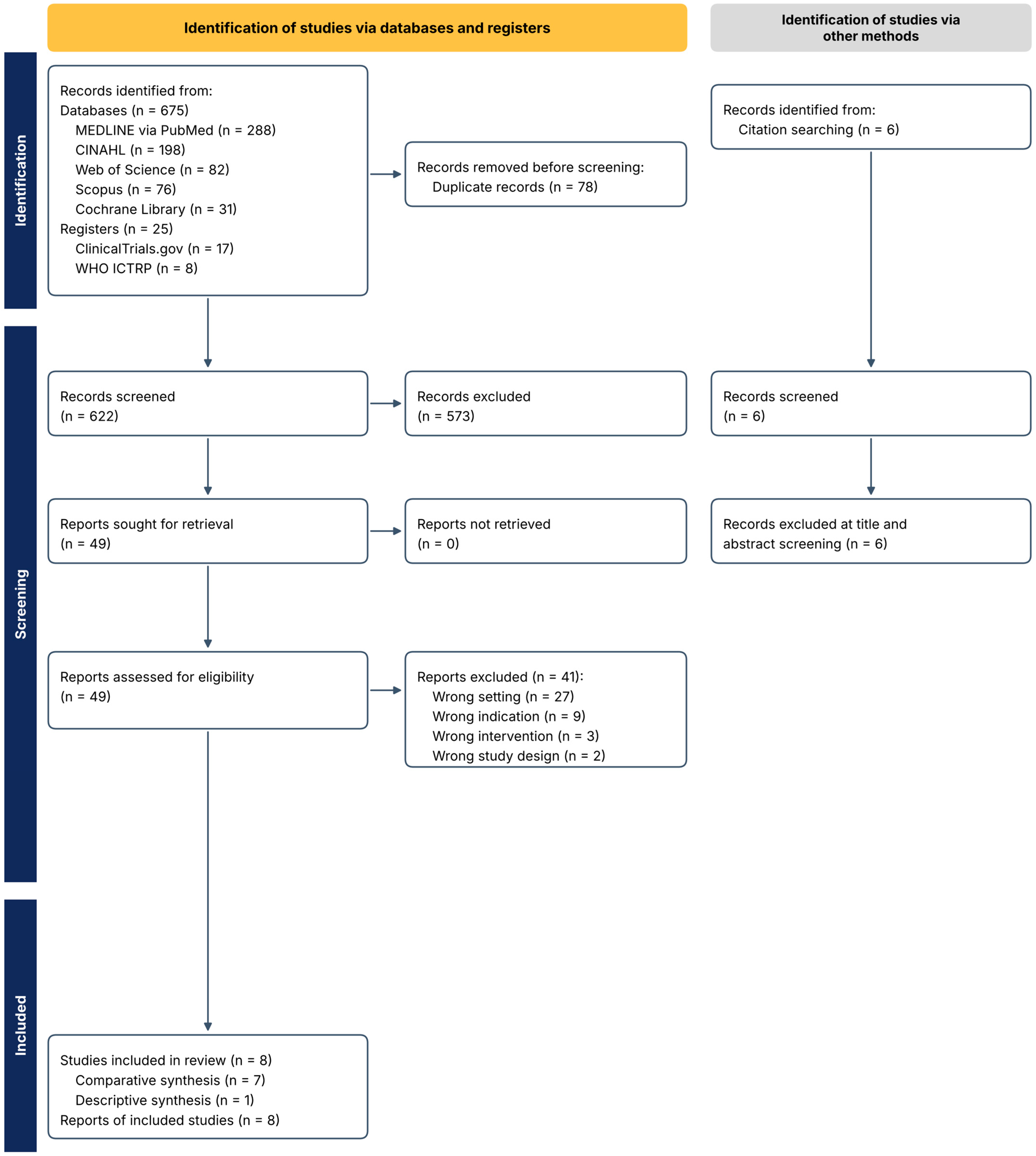
A total of 675 records were identified across five databases (MEDLINE via PubMed, n = 288; CINAHL, n = 198; Web of Science, n = 82; Scopus, n = 76; Cochrane Library, n = 31), and six further records were identified by citation searching. Searches of the two trial registries identified 25 records (ClinicalTrials.gov, n = 17; WHO ICTRP, n = 8). After removal of 78 duplicates, 622 records (597 from databases and 25 from trial registries) were screened by title and abstract, of which 573 were excluded; the six records from citation searching were screened separately and also excluded at this stage. Forty-nine reports were assessed in full, of which 41 were excluded for the following reasons: wrong setting (n = 27), wrong indication (n = 9), wrong intervention (n = 3), and wrong study design (n = 2). Eight studies were included: seven contributed to the comparative synthesis and one to the descriptive synthesis. The study selection process is presented in Figure 1.
Figure 1.
PRISMA 2020 flow diagram of study identification, screening, eligibility assessment, and inclusion.
3.2. Characteristics of Included Studies
The eight included studies were published between 2005 and 2025 and conducted in six countries: Australia, Italy, Turkey, Saudi Arabia, South Korea, and the United States. Four were randomised controlled trials, three were before-and-after quality-improvement studies, and one was a retrospective cohort that was synthesised descriptively. After counting the overlapping collaborative cohort once, the eight studies included up to 6567 children, of whom 6405 were in the seven comparative studies; for randomised trials, the number randomised was used.
The total comprised 546 children randomised in Newcomb et al. [31], 200 in Vida et al. [32], 90 in Gökgöz et al. [33], 227 randomised in Jijeh et al. [34], 300 in Bertrandt et al. [35], 5042 in Sheth et al. [28], and 162 in Kim et al. [36]. Bates et al. [27] was not added separately because its participants were included in the later report by Sheth et al., which covered the nine original (Pioneer) centres and eight newly participating (Spread) centres.
The randomised trials compared high-vacuum drainage combined with smaller-calibre chest tubes against conventional underwater-seal drainage with larger-calibre tubes [31], a smaller-calibre flexible silicone drain with a rigid polyvinyl chloride drain [32], nurse-performed milking or regulated suction with no active patency-maintenance technique [33], and an 8 h with a 24 h interval for assessing drainage against a predefined weight-indexed removal threshold [34]. The non-randomised comparative evidence evaluated structured removal protocols: a single-centre removal algorithm with weight-banded thresholds and a rounding tool [35] and a nine-centre collaborative that adopted the removal criteria of a model centre [27] with its subsequent sustainment and spread to 17 centres [28]. A single-centre retrospective cohort, synthesised descriptively, reported post-removal pericardial effusion under scheduled echocardiographic surveillance [36]. Full characteristics, interventions, comparators, and findings are presented in Table 1 for the randomised trials and Table 2 for the non-randomised studies.
Table 1.
The characteristics and findings of the four included randomised controlled trials.
Table 2.
The characteristics and findings of the three non-randomised comparative studies and one descriptive surveillance cohort.
Comparison of the participating centres and study periods showed that the institution reported by Bertrandt et al. [35] was not included in the original nine-centre collaborative reported by Bates et al. [27] but subsequently participated as one of the eight Spread centres in Sheth et al. [28]. Participant overlap was excluded because the Bertrandt et al. cohort ended in July 2018, whereas data collection for the Spread cohort began in May 2019.
3.3. Risk of Bias
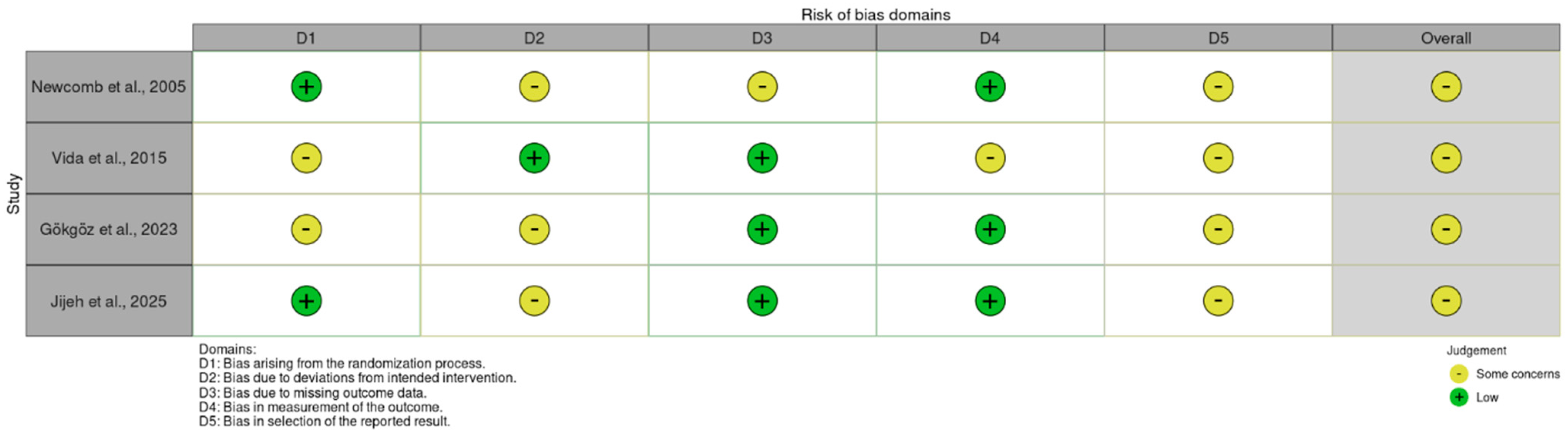
All four randomised trial results were judged to raise some concerns overall, with no result judged to be at high risk of bias. Concerns most frequently related to deviations from intended interventions and selection of the reported result, reflecting the open nature of the interventions, incomplete information on whether awareness of allocation influenced subsequent care, and the absence of accessible prespecified analysis plans. Domain-level judgements for each trial are presented in Figure 2.
Figure 2.
Risk-of-bias judgements for the four included randomised trials (Newcomb et al. [31], Vida et al. [32], Gökgöz et al. [33], and Jijeh et al. [34]) using the Cochrane Risk of Bias 2 tool. D1, bias arising from the randomisation process; D2, bias due to deviations from intended interventions; D3, bias due to missing outcome data; D4, bias in the measurement of the outcome; D5, bias in the selection of the reported result.
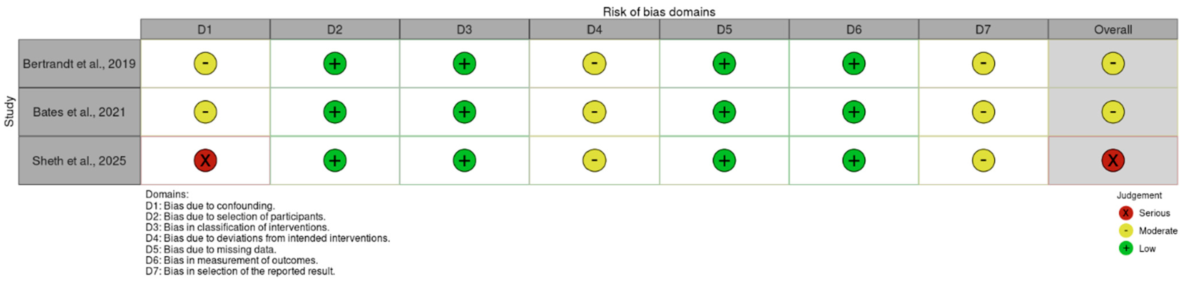
Among the three non-randomised comparative studies, Bertrandt et al. [35] and Bates et al. [27] were judged to be at moderate overall risk of bias, whereas Sheth et al. [28] was judged to be at serious overall risk. Confounding was the principal concern, particularly secular changes during extended implementation periods. No result was judged to be at critical risk of bias. Domain-level judgements are presented in Figure 3.
Figure 3.
Risk-of-bias judgements for the three non-randomised comparative studies (Bertrandt et al. [35], Bates et al. [27], and Sheth et al. [28]) using ROBINS-I. D1, bias due to confounding; D2, bias in the selection of participants; D3, bias in the classification of interventions; D4, bias due to deviations from intended interventions; D5, bias due to missing data; D6, bias in the measurement of outcomes; D7, bias in the selection of the reported result.
3.4. Primary Outcome: Chest-Tube Duration
Three randomised comparisons reported chest-tube duration, with substantial differences in two. Assessing the same weight-indexed removal threshold every 8 rather than every 24 h reduced median duration from 4.9 to 3.0 days, a descriptive relative difference of 38.8% (p < 0.0001) [34]. Similarly, use of a smaller-calibre flexible silicone drain rather than a rigid polyvinyl chloride drain reduced median duration from 40 to 23 h, a difference of 42.5% (p = 0.002) [32]. The third comparison found nearly identical durations of 42 h with high-vacuum drainage and 43 h with conventional underwater-seal drainage. Although the 2.3% difference was statistically significant (p < 0.01), it represented only 1 h. The high-vacuum group also received smaller tubes, with median calibres of 12 versus 16 Ch; therefore, the difference cannot be attributed to the drainage system alone [31].
The three quality-improvement studies also reported shorter duration, although their comparisons were made across successive calendar periods rather than between concurrent groups. Mean duration decreased from 61 to 47 h after introduction of a single-centre removal algorithm [35], while the aggregate geometric mean across the nine original centres of a multicentre collaborative decreased from 72.6 to 61.3 h [27]. During the collaborative’s sustainment and spread phase, mean duration decreased from 91.9 to 70.5 h at the same nine centres. The authors reported a 22.6% reduction, whereas application of the review formula to the rounded published values yielded 23.3%. Mean duration also decreased from 73.1 to 66.0 h at the eight newly participating centres, whose patients were not represented in Bates et al. [27].
3.5. Post-Removal Safety and Reintervention
Post-removal safety was assessed through residual or recurrent effusion, chest-tube reinsertion or replacement, additional interventional procedures, pneumothorax, and readmission. Events were uncommon across the studies, resulting in imprecise estimates. The most informative evidence came from one comparison of combined drainage strategies (high-vacuum drainage with smaller-calibre tubes versus underwater-seal drainage with larger-calibre tubes), in which the observed difference was both substantial and statistically supported.
Residual pleural effusion requiring drainage occurred in four of 237 children managed with a high-vacuum drainage system and smaller-calibre chest tubes and 18 of 241 managed with conventional underwater-seal drainage and larger-calibre tubes (1.7% versus 7.5%; RR 0.23, 95% CI 0.08–0.66; p = 0.003), with no corresponding difference in pericardial effusion requiring drainage [31]. Because drainage-system configuration and tube calibre differed simultaneously between groups, the independent contribution of each component to this finding could not be determined. Mortality, reported by the trial as an additional safety outcome rather than a prespecified review outcome, was similar between groups (2/237 versus 3/241; RR 0.68, 95% CI 0.11–4.02), although one of the five deaths was attributed to a drain-related injury involving the left internal thoracic artery.
Drain material showed a similar, though less certain, pattern. No child receiving a flexible silicone drain required an additional interventional procedure, compared with six of 100 receiving a rigid polyvinyl chloride drain. The review-calculated risk ratio was 0.08 (95% CI 0.004–1.35), whereas the trial reported p = 0.04. Because these values were derived using different methods, the findings should be interpreted cautiously [32]. Children in the silicone-drain group also reported lower pain scores while the drain remained in place and at the time of removal. This was the only patient-level pain assessment in the review and was a secondary outcome of a device comparison rather than the focus of a study designed to evaluate pain. No study reported analgesic consumption.
Post-removal events were even less frequent in the removal-timing trial. Among 215 children assessed at 8 or 24 h intervals, there was one chest-tube reinsertion, four instances of fluid reaccumulation, and seven pneumothoraces; none differed significantly between groups [34]. All events were managed conservatively except in one child, who required reinsertion and contributed to the pneumothorax count; therefore, these outcomes were not independent. The trial was designed assuming a 3% reinsertion rate, whereas the observed rate was 0.5% across both groups, resulting in wide confidence intervals.
At the programme level, no statistically detectable change in reintervention was observed. Chest-tube replacement occurred in 26 of 1312 children during the intervention phase of the multicentre collaborative and 18 of 997 at baseline. Readmission for pleural effusion occurred in six of 1312 and four of 997 children, respectively [27]. Neither outcome differed significantly during the sustainment and spread phase [28]. The confidence intervals remained compatible with clinically important effects in either direction. In the single-centre study, reinsertion counts were reported over unequal periods and for a denominator population different from the cohort contributing to the duration outcome, precluding calculation of a valid risk ratio [35].
In the descriptive cohort, scheduled echocardiographic follow-up detected pericardial effusion in 21 of 162 children (13.0%), of which 19 were identified before discharge and two subsequently; three (1.9%) were moderate or greater. The two effusions identified after discharge prompted readmission, and all resolved without invasive treatment [36]. The authors also reported effusion by the day on which the tube was removed (15 of 87 children with removal on or before the first postoperative day and six of 75 with later removal; p = 0.069); as specified in Section 2.2, this contrast was not treated as an estimate of the effect of removal timing.
3.6. Haemodynamic Effects and Length of Stay
Two additional outcomes addressed clinical recovery rather than post-removal safety. Haemodynamic effects were examined only in the patency-maintenance trial. No between-group differences in heart rate, blood pressure, or bleeding volume were reported. Heart rate changed significantly over time within the no-manipulation group (p = 0.003), but not within the regulated-suction (p = 0.781) or nurse-performed milking (p = 0.070) groups. However, no group-by-time interaction was reported; therefore, differential heart-rate effects among the three strategies were not established [33].
Length-of-stay findings varied by study design. In the randomised trial, removal timing did not affect intensive care unit stay (medians 3.4 and 3.5 days) or hospital stay (medians 9.3 and 9.5 days) [34]. In contrast, both outcomes decreased after implementation in the quality-improvement studies. At the single centre, cardiac intensive care unit stay decreased from 141 to 89 h, and hospital stay decreased from 266 to 156 h [35]. Across the multicentre collaborative, geometric mean hospital stay decreased from 9.2 to 8.3 days [27]. However, length of stay was itself a target of concurrent quality-improvement activity in both programmes, limiting attribution of these changes specifically to chest-tube management.
3.7. Neonatal Subgroup
The prespecified subgroup analysis comparing neonates with older infants and children could not be conducted because no study reported sufficiently detailed age-stratified outcomes. The sustainment and spread study reported that the reduction in chest-tube duration did not reach statistical significance in neonatal operations [28].
3.8. Certainty of the Evidence
Certainty ratings for the seven outcomes judged most important are presented in Table 3. No outcome reached moderate certainty. Residual pleural effusion requiring drainage, the only randomised estimate in the review whose confidence interval excluded no effect, was rated low because the high-vacuum drainage system was delivered with smaller-calibre chest tubes; the comparison was therefore downgraded for indirectness as well as for risk of bias. All other outcomes were rated low or very low, principally because of risk of bias and imprecision, with additional indirectness for selected outcomes. The very low ratings arise where a single study at serious risk of bias contributed the estimate, or where events were too few for the confidence interval to exclude clinically important benefit or harm.
Table 3.
The certainty of the evidence for the main outcomes.
4. Discussion
4.1. Principal Findings
This review synthesised eight studies evaluating postoperative chest-tube management in children undergoing cardiac surgery. Interventions implemented at different points in the management pathway were associated with shorter chest-tube duration, but their mechanisms, effect magnitudes, and certainty of evidence differed. The largest reductions were reported with more frequent assessment of an unchanged removal threshold, use of a smaller-calibre flexible drain, and implementation of structured removal pathways. Taken together, these findings suggest that chest-tube duration may be influenced not only by the child’s clinical condition and drainage characteristics but also by how frequently readiness for removal is assessed and how explicitly the decision is structured. However, the available evidence does not establish the independent effect of removal decision-making or separate it reliably from the effects of tube and drainage-system characteristics.
The most direct evidence regarding assessment frequency came from Jijeh et al. [34]. The drain, drainage system, and weight-indexed removal threshold were identical in both groups, and only the interval at which readiness for removal was assessed differed; earlier recognition that the threshold had been met is therefore a plausible explanation for the almost two-day reduction in median duration, suggesting that infrequent reassessment may prolong chest-tube placement even when the removal criterion itself is appropriate. However, this mechanism was not measured directly, and the open-label design means that related changes in clinical decision-making or care cannot be excluded. To isolate the effect of assessment frequency, future trials should retain the same drain, drainage system, and weight-indexed removal criterion; randomise only the interval at which readiness for removal is assessed; and apply an identical post-removal surveillance strategy in both groups.
Newcomb et al. [31] reported a different pattern. A bundled intervention combining a high-vacuum drainage system with smaller-calibre tubes left chest-tube duration essentially unchanged but was associated with fewer residual pleural effusions requiring drainage (risk ratio 0.23). This finding is compatible with improved evacuation efficiency. Contemporary reviews indicate that drainage performance may be influenced by tube calibre and configuration, fluid viscosity, and the pressure characteristics of the drainage system [8,10]. The Newcomb comparison therefore provides evidence about the combined drainage strategy rather than the independent effectiveness of high-vacuum drainage, and its certainty was downgraded for this reason.
Vida et al. [32] provided further evidence that drain characteristics may influence postoperative recovery, reporting markedly shorter chest-tube duration and lower pain scores during drainage and removal with a smaller-calibre flexible silicone drain. These findings are consistent with the broader chest-tube literature indicating that smaller-calibre tubes may reduce discomfort, although drainage performance also depends on tube design, fluid viscosity, and the potential for obstruction [8,10]. Whether improved tolerance contributed to earlier removal remains uncertain because pain was not assessed as a mediator of the removal decision. Moreover, findings from one specific drain design should not be generalised to all smaller-calibre tubes because material, flexibility, internal configuration, and resistance to flow may differ.
The quality-improvement studies targeted removal decision-making more directly by introducing weight-banded thresholds and a rounding tool [35] and by disseminating explicit removal criteria developed at a model centre [27,28]. These interventions were associated with shorter chest-tube duration without a detectable increase in chest-tube replacement or readmission. The findings are consistent with contemporary calls to replace variable, tradition-based chest-drain practices with explicit and evidence-informed management processes [6,9]. However, the programmes introduced several components simultaneously, including removal criteria, documentation tools, scheduled review, and multidisciplinary implementation; consequently, the independent contribution of each component could not be determined. They therefore show that structured removal processes were feasible, but not that the protocols alone caused the observed improvements.
These findings may help explain, but do not fully account for, the centre-level variation in chest-tube duration [16] and in removal thresholds [5] described in the Introduction. The present review extends this literature by identifying several practice-related components that may contribute to variation, including assessment frequency, explicit removal criteria, structured documentation, and tube or drainage-system characteristics. Nevertheless, residual centre-level variation cannot be attributed solely to chest-tube management. Patient case mix, surgical physiology, staffing, enhanced-recovery pathways, discharge processes, and surveillance intensity may also influence chest-tube duration and length of stay.
The non-significant reduction in chest-tube duration for neonatal operations in the sustainment and spread study [28] should be interpreted as insufficient evidence rather than as evidence that intervention effects differ by age, and future studies should prespecify neonatal and age-stratified analyses to determine whether effects vary by age, weight, surgical complexity, or cardiac physiology.
4.2. Post-Removal Safety and the Ascertainment of Harm
Evidence concerning the safety of earlier chest-tube removal depended substantially on how harm was defined and ascertained. The quality-improvement studies primarily identified pleural effusions through hospital readmission and reported such events in fewer than 1% of children [27,28]. The randomised trials assessed radiographically detected effusions or effusions requiring drainage [31,34], whereas Kim et al. [36] used scheduled echocardiography before discharge and during follow-up and detected postoperative pericardial effusion in 13.0% of children. These estimates represent clinically different outcomes and should not be compared directly. Readmission-based ascertainment captures events severe enough to prompt renewed hospital contact, whereas protocolised imaging can identify small, asymptomatic collections that might otherwise remain undetected. Differences in surveillance intensity and outcome definition therefore probably contributed substantially to the observed variation in complication rates. However, other explanations remain possible because the studies also differed in surgical population, removal practice, duration of follow-up, and clinical thresholds for intervention; notably, the cohort undergoing scheduled echocardiography was limited to children undergoing atrial or ventricular septal defect closure [36].
The higher frequency of effusion detected through systematic surveillance should not be interpreted as evidence that earlier removal caused greater clinically important harm than was observed in the programme-level studies. Kim et al. reported more effusions in children whose tubes were removed on or before the first postoperative day, but because the day of removal reflected each child’s clinical course, this contrast cannot show whether earlier removal causes effusion [36]. Scheduled echocardiography identified predominantly small, self-limiting collections, but this finding does not establish that all detected effusions were clinically unimportant. The clinically relevant question is not simply whether a post-removal collection is detected, but whether it results in symptoms, respiratory or haemodynamic deterioration, prolonged hospitalisation, readmission, or intervention. The available studies were not designed or sufficiently powered to distinguish these outcomes reliably. The randomised evidence could not resolve this uncertainty, because the only trial comparing removal schedules observed far fewer reinsertions than its sample-size calculation assumed [34].
This uncertainty also limits comparison with adult cardiac-surgery evidence, in which earlier chest-tube removal was associated with shorter hospital stay but a higher risk of pericardial effusion [7]. Any apparent difference between the adult and paediatric findings may reflect biological, anatomical, or procedural differences, but variation in outcome ascertainment is also likely to contribute. Adult studies generally evaluated pericardial or pleural effusion as an outcome in its own right, whereas the paediatric quality-improvement studies primarily captured pleural effusion when it resulted in hospital readmission [27,28]. In addition, the adult evidence examined removal timing directly, whereas three of the four paediatric randomised trials evaluated drain characteristics, drainage-system configuration, or patency-maintenance strategies rather than removal timing. Future safety studies should use identical, prespecified surveillance across comparison groups and distinguish imaging-detected collections from symptomatic events, intervention, reinsertion, and readmission. Studies should also be adequately powered for uncommon but clinically important complications, enabling clearer assessment of whether shorter chest-tube duration alters post-removal harm.
Future studies should also use standardised definitions, a clearly defined starting point for chest-tube duration, appropriate measures of dispersion, and complete numerical reporting. A paediatric critical care core outcome set [37,38], supplemented by chest-tube-specific outcomes, could improve comparability.
4.3. Implications for Nursing Practice
The implications for nursing practice centre on the reliability of assessment, documentation, communication, and surveillance throughout the postoperative chest-tube management pathway. Although removal decisions are multidisciplinary, nurses play a central role in monitoring drainage trends, assessing tube and drainage-system function, documenting whether predefined criteria appear to have been met, communicating findings to support multidisciplinary removal decisions, and promptly escalating concerns. These responsibilities are consistent with the broader cardiology literature describing advanced practice nursing contributions to clinical assessment, care coordination, evidence-based practice, documentation, education, and multidisciplinary communication [17]. The findings of this review support an explicit and regularly reviewed decision process more strongly than any specific removal threshold. Quality-improvement programmes suggest that written criteria, structured documentation, and routine review during multidisciplinary rounds are potentially transferable components of practice, although their individual effects and applicability across settings remain uncertain because they were implemented as parts of multicomponent interventions [27,28,35].
Evidence concerning patency-maintenance techniques was particularly limited: the only trial did not establish the comparative haemodynamic effects of regulated suction, nurse-performed milking, and no active manipulation [33], and no included study evaluated manual stripping, despite previous evidence that stripping may generate substantial negative intrathoracic pressure [12,13]. Future comparative studies should evaluate patency-maintenance techniques using direct between-group analyses and clinically meaningful outcomes, including drainage effectiveness, retained collections, pain, haemodynamic deterioration, and reintervention, rather than relying primarily on isolated physiological measures. Local protocols should therefore specify which patency-maintenance techniques are permitted, their indications, required monitoring, and criteria for escalation, without assuming that an absence of demonstrated harm establishes safety or benefit. Post-removal care should similarly include defined observation and escalation criteria, clinically indicated imaging, and education for families before discharge. Routine echocardiographic surveillance after removal was reported by only one descriptive cohort [36], so whether it improves clinically important outcomes, and which surveillance strategy is optimal, could not be determined. Whether the lower pain scores observed with smaller-calibre flexible drains [32] translate into better mobilisation, feeding, parental holding, respiratory function, or family experience is unknown. Because a child’s pain may itself influence how often nurses reassess the drain and when they escalate concerns, future trials should measure pain with validated, age-appropriate instruments throughout drainage and at removal, and examine it as a potential modifier of nursing assessment and escalation rather than only as a secondary endpoint. Future studies should include these patient- and family-centred outcomes, together with nursing workload, protocol adherence, and timeliness of escalation, to determine whether shorter chest-tube duration produces meaningful recovery benefits.
These implications concern the structure and reliability of care rather than the superiority of any particular intervention, and local protocols should be implemented with prospective monitoring of chest-tube duration, drainage effectiveness, clinically important post-removal complications, reinsertion, and readmission.
4.4. Limitations
The main limitations arose from the small and heterogeneous evidence base. Only eight studies were included; most outcomes relied on a single comparison; and the interventions, comparators, outcome definitions, summary measures, and assessment periods differed substantially. These differences precluded meta-analysis, and the reported effect magnitudes should not be interpreted as comparative rankings of the interventions.
Risk of bias and imprecision further limited interpretation. All randomised trials raised some concerns under RoB 2, while the non-randomised comparative studies were at moderate or serious risk of bias. The before-and-after studies were vulnerable to secular trends and concurrent changes in perioperative care, staffing, documentation, and surveillance; in the descriptive cohort, grouping by the day of removal was confounded by each child’s clinical course. Post-removal events were uncommon, and confidence intervals frequently remained compatible with clinically important benefit and harm. Consequently, observed associations cannot be interpreted as causal effects, and non-significant findings do not establish equivalent safety.
The restriction to English-language publications and the limited grey-literature search may have introduced language, availability, or publication bias, and the use of free-text terms without MeSH or subject headings may have reduced the sensitivity of the search; formal assessment of publication bias was not possible because fewer than 10 studies contributed to any outcome. Transferability was also limited because age-stratified data were insufficient for the planned neonatal analysis, and evidence from specialised paediatric cardiac centres may not apply to neonates; children with complex cardiac physiology; or centres with different staffing, equipment, enhanced-recovery pathways, and surveillance practices.
5. Conclusions
Several clinically distinct chest-tube management strategies were associated with shorter chest-tube duration after paediatric cardiac surgery, but their independent effects and comparative safety remain uncertain. For nursing and multidisciplinary practice, the findings are consistent with, but do not establish the benefit of, an explicit, consistently documented removal process reviewed at defined, locally agreed intervals; given the uncertain safety of earlier removal, predefined post-removal observation remains prudent. However, the low- or very-low-certainty evidence does not support universal adoption of a particular drain, drainage-system configuration, patency-maintenance technique, removal threshold, assessment interval, or imaging strategy.
Supplementary Materials
The following supporting information can be downloaded at https://www.mdpi.com/article/10.3390/nursrep16110405/s1, File S1: Completed PRISMA 2020 checklist; File S2: Search strategy for each database and trial registry.
Author Contributions
Conceptualization, R.E., E.E. and A.K.; methodology, R.E. and A.K.; investigation and screening, R.E. and E.E.; data curation, R.E. and E.E.; adjudication of disagreements, R.E. and A.K.; formal analysis, R.E.; writing—original draft preparation, R.E.; writing—review and editing, R.E. and A.K.; supervision, R.E. All authors have read and agreed to the published version of the manuscript.
Funding
This research received no external funding.
Institutional Review Board Statement
Not applicable.
Informed Consent Statement
Not applicable.
Data Availability Statement
No new participant-level data were generated. All data analysed in this review were obtained from the published reports of the included studies. Review-level data supporting the findings, including study characteristics, extracted outcome data, risk-of-bias assessments, and certainty judgements, are provided in the manuscript and Supplementary Materials.
Public Involvement Statement
No public involvement in any aspect of this research.
Guidelines and Standards Statement
This manuscript was prepared in accordance with the PRISMA 2020 statement for systematic reviews [21], with the search reported according to the PRISMA-S extension [20] and the synthesis reported according to the SWiM guideline [26].
Use of Artificial Intelligence
Generative artificial intelligence Cloud Opus 5.5 was used under the authors’ direction to support language editing and drafting refinement. The authors independently verified all scientific content, references, calculations, interpretations, and conclusions and take full responsibility for the final manuscript.
Conflicts of Interest
The authors declare no conflicts of interest.
Abbreviations
The following abbreviations are used in this manuscript:
| CI | confidence interval |
| GRADE | Grading of Recommendations Assessment, Development and Evaluation |
| ICU | intensive care unit |
| PRISMA | Preferred Reporting Items for Systematic Reviews and Meta-Analyses |
| RCT | randomised controlled trial |
| RR | risk ratio |
| ROBINS-I | Risk Of Bias In Non-randomised Studies of Interventions |
| SWiM | Synthesis Without Meta-analysis |
| RoB 2 | Risk of Bias 2 tool |
References
- Hoffman, J.I.; Kaplan, S. The Incidence of Congenital Heart Disease. J. Am. Coll. Cardiol. 2002, 39, 1890–1900. [Google Scholar] [CrossRef] [Scilit] [PubMed]
- van der Linde, D.; Konings, E.E.M.; Slager, M.A.; Witsenburg, M.; Helbing, W.A.; Takkenberg, J.J.M.; Roos-Hesselink, J.W. Birth Prevalence of Congenital Heart Disease Worldwide: A Systematic Review and Meta-Analysis. J. Am. Coll. Cardiol. 2011, 58, 2241–2247. [Google Scholar] [CrossRef] [Scilit] [PubMed]
- Centers for Disease Control and Prevention. Data and Statistics on Congenital Heart Defects. Available online: https://www.cdc.gov/heart-defects/data/index.html (accessed on 17 September 2026).
- Arth, A.C.; Tinker, S.C.; Simeone, R.M.; Ailes, E.C.; Cragan, J.D.; Grosse, S.D. Inpatient Hospitalization Costs Associated with Birth Defects among Persons of All Ages—United States, 2013. MMWR Morb. Mortal. Wkly. Rep. 2017, 66, 41–46. [Google Scholar] [CrossRef] [Scilit] [PubMed]
- Taylor, A.C.; Bates, K.E.; Kipps, A.K. Variability in Paediatric Cardiac Postoperative Chest Tube Management. Cardiol. Young 2018, 28, 1471–1474. [Google Scholar] [CrossRef] [Scilit] [PubMed]
- Lobdell, K.W.; Perrault, L.P.; Drgastin, R.H.; Brunelli, A.; Cerfolio, R.J.; Engelman, D.T.; ERAS Cardiac Society Working Group. Drainology: Leveraging Research in Chest-Drain Management to Enhance Recovery after Cardiothoracic Surgery. JTCVS Tech 2024, 25, 226–240. [Google Scholar] [CrossRef] [Scilit] [PubMed]
- Rakab, M.S.; Awashra, A.; Radwan, N.; Shubietah, A.; Maamoun, A.; Mattar, M.; Manasrah, A.; Ghanem, A.; Elbaz, A.; Emara, A.; et al. Chest Tube Removal after Cardiac Surgery in First vs. Second Post-Operative Day: A Systematic Review and Meta-Analysis. J. Cardiothorac. Surg. 2025, 21, 33. [Google Scholar] [CrossRef] [Scilit] [PubMed]
- Sorino, C.; Feller-Kopman, D.; Mei, F.; Mondoni, M.; Agati, S.; Marchetti, G.; Rahman, N.M. Chest Tubes and Pleural Drainage: History and Current Status in Pleural Disease Management. J. Clin. Med. 2024, 13, 6331. [Google Scholar] [CrossRef] [Scilit] [PubMed]
- Lobdell, K.W.; Engelman, D.T. Chest Tube Management: Past, Present, and Future Directions for Developing Evidence-Based Best Practices. Innov. Technol. Tech. Cardiothorac. Vasc. Surg. 2023, 18, 41–48. [Google Scholar] [CrossRef] [Scilit] [PubMed]
- Anderson, D.; Chen, S.A.; Godoy, L.A.; Brown, L.M.; Cooke, D.T. Comprehensive Review of Chest Tube Management: A Review. JAMA Surg. 2022, 157, 269–274. [Google Scholar] [CrossRef] [Scilit] [PubMed]
- The Royal Children’s Hospital Melbourne. Chest Drain Management. Available online: https://www.rch.org.au/rchcpg/hospital_clinical_guideline_index/chest_drain_management/ (accessed on 17 September 2026).
- Duncan, C.; Erickson, R. Pressures Associated with Chest Tube Stripping. Heart Lung 1982, 11, 166–171. [Google Scholar] [PubMed]
- Day, T.G.; Perring, R.R.; Gofton, K. Is Manipulation of Mediastinal Chest Drains Useful or Harmful after Cardiac Surgery? Interact. Cardiovasc. Thorac. Surg. 2008, 7, 888–890. [Google Scholar] [CrossRef] [Scilit] [PubMed]
- Baumann, M.H. What Size Chest Tube? What Drainage System Is Ideal? And Other Chest Tube Management Questions. Curr. Opin. Pulm. Med. 2003, 9, 276–281. [Google Scholar] [CrossRef] [Scilit] [PubMed]
- Tang, A.T.; Velissaris, T.J.; Weeden, D.F. An Evidence-based Approach to Drainage of the Pleural Cavity: Evaluation of Best Practice. J. Eval. Clin. Pract. 2002, 8, 333–340. [Google Scholar] [CrossRef] [Scilit] [PubMed]
- Bates, K.E.; Madsen, N.L.; Khadr, L.; Gao, Z.; Crawford, K.; Gaies, M.; Graupe, M.; Hanke, S.P.; Hlavacek, A.M.; Morell, E.; et al. Center Variation in Chest Tube Duration and Length of Stay after Congenital Heart Surgery. Ann. Thorac. Surg. 2020, 110, 221–227. [Google Scholar] [CrossRef] [Scilit] [PubMed]
- Sguanci, M.; Mancin, S.; Carù, V.; Simonelli, N.; Cangelosi, G.; Morales Palomares, S.; Ferrara, G.; Lo Cascio, A. Exploring the Role of Advanced Practice Nurses in Cardiology: A Scoping Review. Int. Nurs. Rev. 2025, 72, e70054. [Google Scholar] [CrossRef] [Scilit] [PubMed]
- Yang, J.; Gao, Y.; Yu, C.P.; Zhang, B.; Zou, L.J.; Yan, Z. Nurse-Led Intervention versus Usual Care on the Cardiovascular Risk Factors in Patients with Coronary Artery Disease: A Meta-Analysis Including Data from Randomized Trials and Observational Studies. BMC Cardiovasc. Disord. 2025, 26, 39. [Google Scholar] [CrossRef] [Scilit] [PubMed]
- Higgins, J.P.T.; Thomas, J.; Chandler, J.; Cumpston, M.; Li, T.; Page, M.J.; Welch, V.A. (Eds.) Cochrane Handbook for Systematic Reviews of Interventions; Version 6.5; Cochrane: London, UK, 2024; Available online: https://www.cochrane.org/authors/handbooks-and-manuals/handbook (accessed on 15 September 2026).
- Rethlefsen, M.L.; Kirtley, S.; Waffenschmidt, S.; Ayala, A.P.; Moher, D.; Page, M.J.; Koffel, J.B.; PRISMA-S Group. PRISMA-S: An Extension to the PRISMA Statement for Reporting Literature Searches in Systematic Reviews. Syst. Rev. 2021, 10, 39. [Google Scholar] [CrossRef] [Scilit] [PubMed]
- Page, M.J.; McKenzie, J.E.; Bossuyt, P.M.; Boutron, I.; Hoffmann, T.C.; Mulrow, C.D.; Shamseer, L.; Tetzlaff, J.M.; Akl, E.A.; Brennan, S.E.; et al. The PRISMA 2020 Statement: An Updated Guideline for Reporting Systematic Reviews. BMJ 2021, 372, n71. [Google Scholar] [CrossRef] [Scilit] [PubMed]
- Veritas Health Innovation. Covidence Systematic Review Software; Veritas Health Innovation: Melbourne, Australia; Available online: https://www.covidence.org (accessed on 1 November 2026).
- McGuinness, L.A.; Higgins, J.P. Risk-of-bias VISualization (robvis): An R Package and Shiny Web App for Visualizing Risk-of-bias Assessments. Res. Synth. Methods 2021, 12, 55–61. [Google Scholar] [CrossRef] [Scilit] [PubMed]
- Sterne, J.A.; Savović, J.; Page, M.J.; Elbers, R.G.; Blencowe, N.S.; Boutron, I.; Cates, C.J.; Cheng, H.-Y.; Corbett, M.S.; Eldridge, S.M.; et al. RoB 2: A Revised Tool for Assessing Risk of Bias in Randomised Trials. BMJ 2019, 366, l4898. [Google Scholar] [CrossRef] [Scilit] [PubMed]
- Sterne, J.A.; Hernán, M.A.; Reeves, B.C.; Savović, J.; Berkman, N.D.; Viswanathan, M.; Henry, D.; Altman, D.G.; Ansari, M.T.; Boutron, I.; et al. ROBINS-I: A Tool for Assessing Risk of Bias in Non-Randomised Studies of Interventions. BMJ 2016, 355, i4919. [Google Scholar] [CrossRef] [Scilit] [PubMed]
- Campbell, M.; McKenzie, J.E.; Sowden, A.; Katikireddi, S.V.; Brennan, S.E.; Ellis, S.; Hartmann-Boyce, J.; Ryan, R.; Shepperd, S.; Thomas, J.; et al. Synthesis without Meta-Analysis (SWiM) in Systematic Reviews: Reporting Guideline. BMJ 2020, 368, l6890. [Google Scholar] [CrossRef] [Scilit] [PubMed]
- Bates, K.E.; Connelly, C.; Khadr, L.; Graupe, M.; Hlavacek, A.M.; Morell, E.; Pasquali, S.K.; Russell, J.L.; Schachtner, S.K.; Strohacker, C.; et al. Successful Reduction of Postoperative Chest Tube Duration and Length of Stay after Congenital Heart Surgery: A Multicenter Collaborative Improvement Project. J. Am. Heart Assoc. 2021, 10, e020730. [Google Scholar] [CrossRef] [Scilit] [PubMed]
- Sheth, S.S.; Tignor, A.; Connelly, C.; Madsen, N.L.; Graupe, M.; Bates, K.E.; Figueroa, M.; Greiten, L.E.; Hart, S.A.; Jensen, M.; et al. Sustaining and Spreading Success: Expanding a Multicentre Quality Improvement Project to Reduce Postoperative Chest Tube Duration after Paediatric CHD. Cardiol. Young 2025, 35, 1154–1160. [Google Scholar] [CrossRef] [Scilit] [PubMed]
- Guyatt, G.H.; Oxman, A.D.; Vist, G.E.; Kunz, R.; Falck-Ytter, Y.; Alonso-Coello, P.; Schünemann, H.J.; GRADE Working Group. GRADE: An Emerging Consensus on Rating Quality of Evidence and Strength of Recommendations. BMJ 2008, 336, 924–926. [Google Scholar] [CrossRef] [Scilit] [PubMed]
- Schünemann, H.J.; Cuello, C.; Akl, E.A.; Mustafa, R.A.; Meerpohl, J.J.; Thayer, K.; Morgan, R.L.; Gartlehner, G.; Kunz, R.; Katikireddi, S.V.; et al. GRADE Guidelines: 18. How ROBINS-I and Other Tools to Assess Risk of Bias in Nonrandomized Studies Should Be Used to Rate the Certainty of a Body of Evidence. J. Clin. Epidemiol. 2019, 111, 105–114. [Google Scholar] [CrossRef] [Scilit] [PubMed]
- Newcomb, A.E.; Alphonso, N.; Nørgaard, M.A.; Cochrane, A.D.; Karl, T.R.; Brizard, C.P. High-Vacuum Drains Rival Conventional Underwater-Seal Drains after Pediatric Heart Surgery. Eur. J. Cardiothorac. Surg. 2005, 27, 395–400. [Google Scholar] [CrossRef] [Scilit] [PubMed]
- Vida, V.L.; Gallo, M.; Barzon, E.; Olivato, V.; De Franceschi, M.; Guariento, A.; Padalino, M.; Stellin, G. A Single Institution Evaluation of the Performance of Two Different Chest Drainage Systems in Pediatric Patients after Surgery for Congenital Heart Disease. Thorac. Cardiovasc. Surg. 2015, 63, 404–408. [Google Scholar] [CrossRef] [Scilit] [PubMed]
- Gökgöz, G.; Karabacak, Ü.; Kan Öntürk, Z. The Effects of Milking and Suction Drain Management Methods in Paediatric Patients after Cardiac Surgery. Int. J. Nurs. Pract. 2023, 29, e13122. [Google Scholar] [CrossRef] [Scilit] [PubMed]
- Jijeh, A.M.; Shaath, G.A.; Ismail, S.R.; Kabbani, M.S.; Hijazi, O.M.; Altaweel, H.; Hamada, H.; Qadi, A.; Fatima, A.; Abdrabo, A.; et al. Optimizing Recovery: Early versus Delayed Chest Tube Removal in Pediatric Cardiac Surgery Patients: A Randomized Controlled Trial. Crit. Care Explor. 2025, 7, e1271. [Google Scholar] [CrossRef] [Scilit] [PubMed]
- Bertrandt, R.A.; Saudek, D.M.; Scott, J.P.; Madrzak, M.; Miranda, M.B.; Ghanayem, N.S.; Woods, R.K. Chest Tube Removal Algorithm Is Associated with Decreased Chest Tube Duration in Pediatric Cardiac Surgical Patients. J. Thorac. Cardiovasc. Surg. 2019, 158, 1209–1217. [Google Scholar] [CrossRef] [Scilit] [PubMed]
- Kim, Y.E.; Jung, H.; Cho, J.Y.; Kim, Y.H.; Hyun, M.C.; Lee, Y. Does Early Drain Removal Affect Postoperative Pericardial Effusion after Congenital Cardiac Surgery? Korean J. Thorac. Cardiovasc. Surg. 2020, 53, 16–21. [Google Scholar] [CrossRef] [Scilit] [PubMed]
- Pinto, N.P.; Maddux, A.B.; Dervan, L.A.; Woodruff, A.G.; Jarvis, J.M.; Nett, S.; Killien, E.Y.; Graham, R.J.; Choong, K.; Luckett, P.M.; et al. A Core Outcome Measurement Set for Pediatric Critical Care. Pediatr. Crit. Care Med. 2022, 23, 893–907. [Google Scholar] [CrossRef] [Scilit] [PubMed]
- Fink, E.L.; Jarvis, J.M.; Maddux, A.B.; Pinto, N.; Galyean, P.; Olson, L.M.; Zickmund, S.; Ringwood, M.; Sorenson, S.; Dean, J.M.; et al. Development of a Core Outcome Set for Pediatric Critical Care Outcomes Research. Contemp. Clin. Trials 2020, 91, 105968. [Google Scholar] [CrossRef] [Scilit] [PubMed]

Disclaimer/Publisher’s Note: The statements, opinions and data contained in all publications are solely those of the individual author(s) and contributor(s) and not of MDPI and/or the editor(s). MDPI and/or the editor(s) disclaim responsibility for any injury to people or property resulting from any ideas, methods, instructions or products referred to in the content. |
© 2026 by the authors. Licensee MDPI, Basel, Switzerland. This article is an open access article distributed under the terms and conditions of the Creative Commons Attribution (CC BY) license.