


Abstract
Background: Dominant β-thalassemia is a rare disorder caused by highly unstable β-globin variants that result in ineffective erythropoiesis and variable clinical phenotypes. Codon 121 lies within a functionally important region of the β-globin chain, and mutations at this site are exceptionally rare. The HBB:c.364G>T (GAA>TAA) variant introduces a premature stop codon, leading to production of a truncated β-globin chain. We describe the clinical, hematological, and molecular findings of a Ukrainian family carrying this previously reported rare variant, with particular emphasis on intrafamilial phenotypic variability. Case report presentation: Clinical history, hematological parameters, and molecular findings were evaluated in four affected individuals from three generations of the same family. Genetic analysis identified the heterozygous nonsense mutation HBB:c.364G>T (codon 121, GAA>TAA) in all affected members with variable clinical severity. Two adults had previously undergone splenectomy, whereas two pediatric patients remain under clinical follow-up. A history of premature death in another affected relative suggests the potential for severe disease-related complications. All patients had microcytic hypochromic anemia with elevated HbA2 levels. Adult carriers demonstrated anisopoikilocytosis, circulating erythroblasts, and Howell–Jolly bodies. Compared with the adults, pediatric patients exhibited lower hemoglobin levels and higher HbF levels, consistent with age-related hematologic differences and physiological persistence of fetal hemoglobin. Conclusions: This report provides additional clinical and molecular characterization of the rare HBB:c.364G>T variant and highlights the phenotypic variability observed among affected members of the same family. These findings emphasize the importance of molecular confirmation and careful clinical assessment in patients with suspected dominant β-thalassemia.
1. Introduction
B-thalassemias are among the most prevalent genetic disorders [1]. These inherited blood disorders result from anomalies in the synthesis of β-globin chains of hemoglobin. These anomalies occur due to point mutations or, less frequently, deletions in the beta globin gene located on chromosome 11. Consequently, there is a reduced (β+) or missing (β0) production of the β-chains of hemoglobin (Hb). This leads to a diverse range of phenotypes, varying from significant anemia to individuals with no clinical symptoms [2].
Typically, β-thalassemia follows an autosomal recessive pattern of inheritance. This means that an individual needs to inherit two copies of the mutated gene (one from each parent) to express the full-blown disorder. DNA sequencing reveals new and different thalassemia mutations and variants, and the molecular underpinnings of these clinical changes are documented in databases that are already in place [2,3]. In rare cases, dominant mutations have also been found that can cause symptomatic β-thalassemia with individuals carrying a single copy of the mutated gene [4]. The molecular abnormalities vary greatly, but they all result in the production of β-globin chains that are extremely unstable and often cannot be detected at the protein level. These unstable β-chains form deposits and, combined with an excess of α chains, worsen the ineffective production of red blood cells, resulting in severe phenotypes of the condition [5].
This case report discusses a rare dominant mutation occurring at codon 121 of the β-globin gene, where GAA, encoding glutamic acid, is changed to TAA, a termination codon. This results in the formation of a truncated β-chain consisting of 120 residues, which is prone to rapid degradation, leading to the manifestation of this type of β-thalassemia [6]. The hematologic profile of heterozygotes exhibits moderately severe characteristics of β-thalassemia, such as the presence of inclusion bodies, elevated levels of HbA2 and HbF, splenomegaly, significant phenotypic variation, hemoglobin levels ranging from 9.0 to 10.0 g/dL, mean corpuscular volume (MCV) between 60 and 65 fl, and mean corpuscular hemoglobin (MCH) between 18 and 22 pg [6].
The β-globin codon 121 (GAA>TAA; HBB.364G>T) nonsense mutation represents a rare cause of dominantly inherited β-thalassemia. It was first characterized by Kazazian et al. as a de novo mutation occurring during paternal spermatogenesis in a child of Greek Cypriot and Polish origin [7]. Subsequent reports have identified this variant in individuals and families from different ethnic backgrounds, including British [1,8], Czech [9], Dutch [10], Japanese [11] and Chinese [12] populations, supporting the occurrence of this rare mutation across geographically diverse groups.
Unlike the typical heterozygous β-thalassemia carrier state, which is generally associated with mild microcytic anemia [13], dominant β-thalassemia can result in a clinically significant phenotype despite the presence of only one affected allele. Approximately 40 mutations associated with dominantly inherited β-thalassemia have been described, with nearly two-thirds located within exon 3 of the β-globin gene [14]. In the present report, we describe four affected individuals from three generations of a Ukrainian family carrying the previously reported HBB:c.364G>T variant. Within this family, one member passed away prematurely, two others have had a splenectomy, and there is ongoing monitoring of two young children. Given the rarity of this variant, additional genotype–phenotype descriptions are important to better define its clinical spectrum and intrafamilial variability.
2. Case Report Presentation
A Ukrainian family (mother, son, and two grandsons) was referred for evaluation of anemia. All four individuals exhibited clinical and hematological features consistent with thalassemia intermedia; however, molecular characterization had not been previously performed and was conducted for the first time in the present study.
The 60-year-old female presented with microcytic hypochromic anemia, an elevated red cell distribution width (RDW) of 18.2%, and a serum ferritin level of 532 ng/mL. High-performance liquid chromatography (HPLC) analysis revealed elevated HbA2 levels, while the detailed hematological parameters are presented in Table 1. The Naked Eye Single Tube Red Cell Osmotic Fragility Test (NESTROFT) was positive, while the sickling test was negative. Peripheral blood smear examination demonstrated marked abnormalities, including hypochromia, anisopoikilocytosis, microcytosis, basophilic stippling, circulating erythroblasts, and Howell–Jolly bodies. The proband had undergone splenectomy at a young age. Her family history was notable for the death of her brother during childhood due to anemia. Molecular analysis of the α- and β-globin genes identified a heterozygous β-globin mutation at codon 121 (GAA→TAA), resulting from a G>T substitution (HBB:c.364G>T). This nonsense mutation introduces a premature termination codon, leading to a truncated β-globin chain and is classified as a β0-dominant β-thalassemia mutation.
Table 1.
Hematological and biochemical findings for the four studied members.
Her 42-year-old son was also referred for hematological evaluation, which revealed microcytic hypochromic anemia, an elevated RDW of 19%, and a serum ferritin level of 253 ng/mL. The detailed hematological parameters are summarized in Table 1. Osmotic fragility testing was positive. High-performance liquid chromatography (HPLC) analysis demonstrated elevated HbA2 levels (5%) and HbF levels of 0.5%, while the sickling test was negative.
Peripheral blood smear examination revealed hypochromia, anisopoikilocytosis, microcytosis, target cells, and circulating erythroblasts. The patient had also undergone splenectomy. Molecular analysis confirmed the presence of the same heterozygous β-globin codon 121 mutation (GAA→TAA; HBB:c.364G>T) previously identified in his mother, resulting in a premature termination codon and a dominant β-thalassemia phenotype.
The first grandson, aged 7 years, also underwent hematological and molecular evaluation. His hematological assessment revealed microcytic hypochromic anemia, with the detailed parameters summarized in Table 1. Peripheral blood smear examination demonstrated hypochromia, microcytosis, anisopoikilocytosis, and circulating erythroblasts. HPLC analysis showed elevated HbA2 levels (5.1%) and HbF levels (1.5%), while the sickling test was negative. Molecular analysis confirmed that the child carried the same familial heterozygous β-globin codon 121 mutation (GAA→TAA; HBB:c.364G>T), consistent with a dominant β-thalassemia phenotype. The NESTROFT was positive.
The family’s second affected child, a 16-month-old boy, also underwent hematological and molecular evaluation. Laboratory findings revealed microcytic hypochromic anemia, with the detailed hematological parameters presented in Table 1. Hemoglobin analysis demonstrated a markedly elevated HbF level of 17%, while HbA2 was 4.0%.
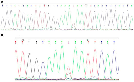
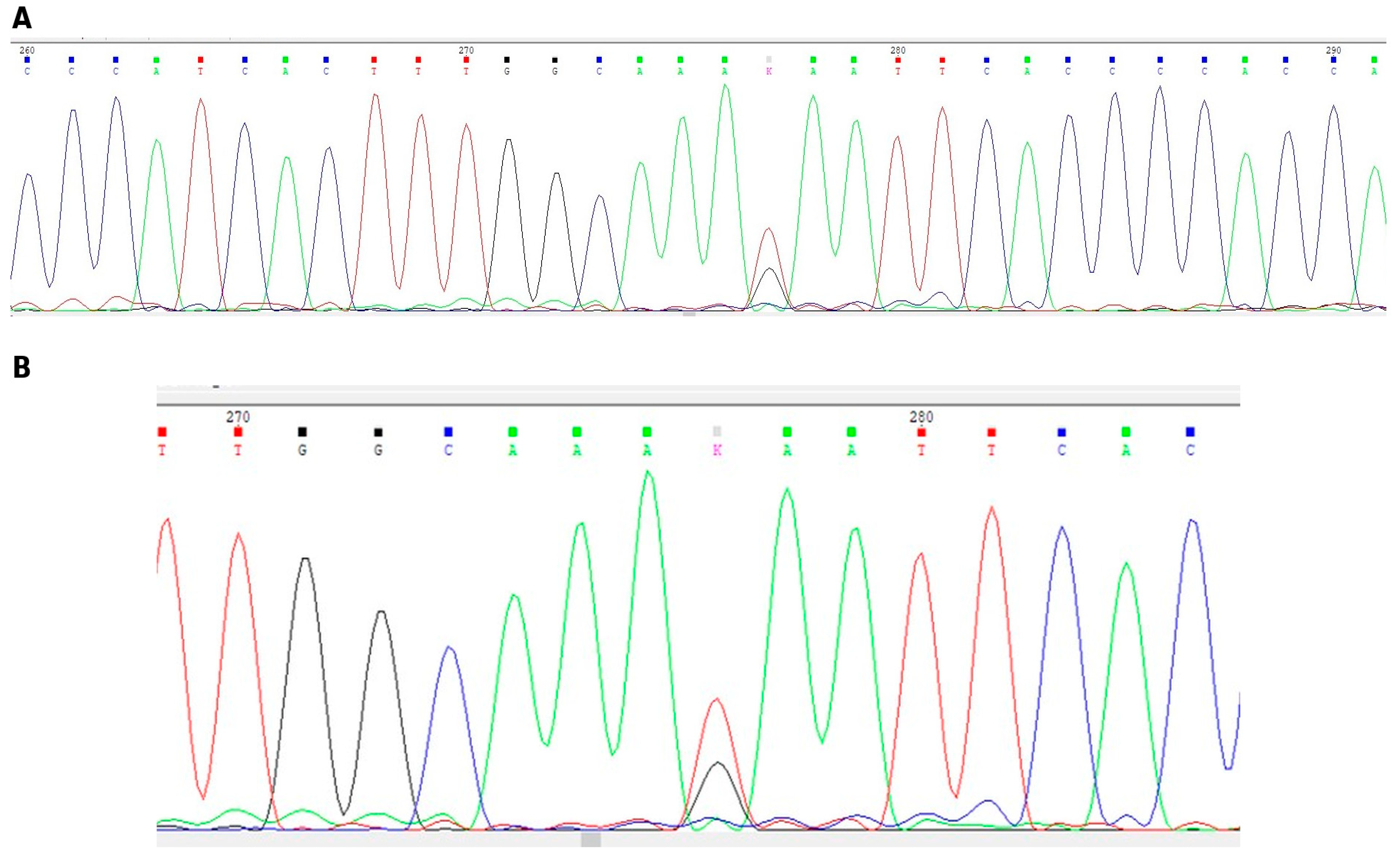
Molecular testing confirmed the presence of the same heterozygous HBB:c.364G>T (GAA>TAA) variant identified in the other affected family members. Representative Sanger sequencing electropherograms illustrating the region of interest and the heterozygous nucleotide position are presented in Figure 1. Exon 3 of the HBB gene was selectively amplified using the following primers:
Figure 1.
Sanger sequencing electropherograms of the HBB gene. (A) Extended sequencing electropherogram showing the surrounding nucleotide sequence. (Β) Enlarged view of the sequence region containing the heterozygous nucleotide substitution. The variant was reported as HBB:c.364G>T (codon 121, GAA>TAA). The colored peaks represent the four nucleotide bases: adenine (A, green), thymine (T, red), cytosine (C, blue), and guanine (G, black).
Forward: 5′-TAT CAT GCC TCT TTG CAC CAT TC-3′
Reverse: 5′-AAT GCA CTG ACC TCC CAC ATT CC-3′
Sequencing was performed with the dideoxy termination method [15], using the above primers and the Applied Biosystems BigDye Terminator v3.1 Cycle Sequencing Kit (Thermo Fisher Scientific, Waltham, MA, USA). The sequencing reactions were analyzed on the Applied Biosystems SeqStudio genetic analyzer (Thermo Fisher Scientific, Waltham, MA, USA). Genomic DNA was isolated from peripheral whole blood, using the Maxwell RSC Whole Blood DNA Kit (Promega Corporation, Fitchburg, WI, USA) and the automated extractor Promega Maxwell RSC (Promega Corporation, Fitchburg, WI, USA), according to the manufacturer’s instructions.
The hematological parameters and molecular findings of the four affected family members are summarized in Table 1.
3. Discussion
The present report describes a Ukrainian family carrying the previously reported rare HBB:c.364G>T (codon 121, GAA>TAA) nonsense variant associated with dominantly inherited β-thalassemia. The mutation was detected in four family members across three generations, with clinical manifestations ranging from splenectomy-requiring disease in adults to moderate anemia under surveillance in pediatric carriers. These findings contribute to the growing body of literature on genotype–phenotype correlations in non-classical β-thalassemia and underscore the importance of molecular diagnostics in identifying rare, clinically significant hemoglobin disorders.
The molecular basis underlying this dominant phenotype is related to the ability of exon 3 nonsense and frameshift mutations to escape nonsense-mediated mRNA decay (NMD), a quality-control mechanism that normally eliminates transcripts containing premature termination codons [16,17]. Hall and Thein demonstrated that individuals heterozygous for the β-codon 121 (G>T) mutation showed comparable levels of mutant and normal β-globin transcripts, in contrast to exon 2 nonsense mutations in which mutant mRNA was nearly absent [8]. Escape from NMD allows translation of a truncated 120-amino-acid β-globin chain, which is highly unstable and contributes to globin chain precipitation and ineffective erythropoiesis [8,18,19].
The truncated β-globin product fails to form functional hemoglobin tetramers and precipitates intracellularly along with excess α-globin chains. Ho et al. confirmed via immunoelectron microscopy that the intra-erythroblastic inclusion bodies characteristic of dominant β-thalassemia due to the codon 121 G>T mutation contain both precipitated α-globin and β-globin chains [18]. Adams et al. isolated the truncated protein and found it constitutes only 0.05–0.1% of total non-α-globin protein, consistent with extreme post-translational instability [18]. These inclusions overwhelm the erythroid cell’s proteolytic capacity—normally maintained by α-hemoglobin-stabilizing protein (AHSP), the ubiquitin–proteasome system, and autophagy—leading to accelerated apoptosis of erythroid precursors, ineffective erythropoiesis, and chronic hemolytic anemia even in the heterozygous state [13,20,21,22].
The marked phenotypic variability observed in this family is consistent with the broad clinical spectrum of dominant β-thalassemia [14]. Similar phenotypic variability among carriers of the same codon 121 (G>T) variant was previously reported by Giordano et al. in four unrelated Dutch families [10]. Despite sharing the same HBB:c.364G>T mutation, affected family members demonstrated variable clinical expression. The pediatric patients presented with lower hemoglobin levels compared with the adults; however, this observation should be interpreted cautiously, as both adult patients had previously undergone splenectomy, potentially modifying their hematological phenotypes. This intrafamilial variability may reflect the combined contribution of age-dependent compensatory mechanisms, differences in HbF expression, and additional genetic modifiers affecting globin chain imbalance and ineffective erythropoiesis. Long-term follow-up of the pediatric patients will be required to further characterize the natural evolution of the disease.
The hematological phenotype was characterized by microcytic hypochromic anemia with elevated HbA2 levels. Adult patients additionally showed marked red blood cell morphological abnormalities, including anisopoikilocytosis and erythroblastosis, while Howell–Jolly bodies were observed as a consequence of previous splenectomy. These findings are consistent with dominant β-thalassemia, which is typically associated with ineffective erythropoiesis, hemolysis, splenomegaly, reticulocytosis, erythroid expansion, and the presence of abnormal globin chain precipitates [14,20].
Several genetic modifiers may contribute to the phenotypic diversity observed among individuals carrying the same β-globin mutation. Variants affecting fetal hemoglobin regulation, particularly within the BCL11A and HBS1L-MYB loci, have been shown to modify disease severity in β-thalassemia through modulation of γ-globin expression and HbF production [13,23,24]. In addition, factors influencing globin chain imbalance and stability, including co-inherited α-thalassemia and variation in α-hemoglobin stabilizing protein (AHSP) expression, may affect the degree of ineffective erythropoiesis [13,21,25]. In the present family, α-globin gene analysis excluded co-inherited α-thalassemia; however, other genetic modifiers were not evaluated and may partly explain the observed intrafamilial variability.
Since its initial description, the β-globin codon 121 (GAA>TAA; HBB:c.364G>T) nonsense mutation has been reported only rarely, with affected individuals and families identified across different geographic and ethnic backgrounds (Table 2). The mutation was first characterized as a de novo event occurring during paternal spermatogenesis in a child of Greek Cypriot and Polish origin [7]. Subsequent reports have documented the presence of this variant in unrelated families from several European populations, including British, Czech, and Dutch families, as well as in individuals of Asian origin [1,8,9,10,11,12,18,19].
Table 2.
Summary of published cases and families carrying the HBB:c.364G>T (codon 121, GAA>TAA; p.Glu121Ter) mutation and related mutations associated with dominant β-thalassemia. The table summarizes the reported patients, ethnic background, and key clinical and molecular findings from the literature.
The occurrence of HBB:c.364G>T on different β-globin gene cluster haplotypes suggests that this variant has arisen through independent mutational events rather than dissemination from a single founder [11]. The identification of the mutation in the present Ukrainian family further expands the known geographic distribution of this rare dominant β-thalassemia variant and highlights the importance of molecular characterization in patients with atypical β-thalassemia phenotypes.
The diagnosis of dominant β-thalassemia poses unique challenges. Unstable β-globin variants are often difficult to detect using conventional hemoglobin electrophoresis or high-performance liquid chromatography, as the abnormal globin chains may undergo rapid degradation and may not be present as detectable hemoglobin fractions. Consequently, affected heterozygous individuals may initially be misclassified as having typical β-thalassemia trait, particularly when presenting with microcytic hypochromic anemia and increased HbA2 levels. A high index of clinical suspicion is required when the clinical phenotype is disproportionately severe for a simple carrier state, especially in the presence of features suggestive of thalassemia intermedia, such as significant anemia, splenomegaly, jaundice, or complications related to ineffective erythropoiesis [26].
Management of dominant β-thalassemia has traditionally relied on supportive measures including transfusion therapy, splenectomy, and iron chelation. Splenectomy can raise hemoglobin levels by 1–2 g/dL in non-transfusion-dependent thalassemia; however, accumulating evidence has highlighted significant long-term risks [25]. Splenectomized patients with non-transfusion-dependent thalassemia have a 4–5-fold increased risk of venous thromboembolism, pulmonary hypertension, leg ulcers, and silent cerebral infarction compared with non-splenectomized patients [25,27]. The hypercoagulable state results from decreased splenic scavenging of procoagulant red blood cells and activated platelets, and is further exacerbated by high counts of nucleated red blood cells and platelets [25]. Current recommendations restrict splenectomy to patients with an inability to receive transfusion and iron-chelation therapy, or those with clinically symptomatic splenomegaly or hypersplenism, with appropriate vaccination, antibiotic prophylaxis, and thromboprophylaxis for high-risk patients [25].
Several limitations should be acknowledged. Although this report provides molecular confirmation of a rare dominant β-thalassemia mutation across multiple generations, the limited number of affected family members restricts broader conclusions regarding genotype–phenotype associations. Comprehensive evaluation of additional genetic modifiers, including BCL11A and HBS1L-MYB polymorphisms as well as AHSP expression, was not performed and could have provided further insight into the observed intrafamilial phenotypic variability. Although α-globin gene analysis excluded co-inherited α-thalassemia, other genetic and environmental factors influencing disease severity cannot be excluded. Long-term follow-up, particularly of the pediatric affected individuals, will be important to better define the clinical course of this rare variant.
4. Conclusions
We describe a Ukrainian family carrying the rare dominantly inherited β-globin codon 121 (GAA>TAA; HBB:c.364G>T) nonsense variant, further expanding the reported geographic distribution of this variant. The identification of the same variant in four affected individuals across three generations highlights the considerable intrafamilial phenotypic variability associated with dominant β-thalassemia. The molecular defects responsible for β-thalassemia phenotypes are highly heterogeneous and can result in variable clinical manifestations. This report highlights the importance of considering dominant β-thalassemia in individuals with a phenotype disproportionate to the typical carrier state and emphasizes the role of molecular testing in achieving an accurate diagnosis, guiding clinical follow-up, and providing appropriate genetic counseling.
Author Contributions
Conceptualization: T.M.V., E.Z. and S.T.; Methodology: T.M.V., E.Z. and S.T.; Investigation: T.M.V., E.Z., E.Y., E.E.D., G.M., H.A., E.V., M.D. and S.T.; Data Curation: T.M.V., E.Z. and S.T.; Formal Analysis: T.M.V., E.Z. and S.T.; Writing—Original Draft Preparation: T.M.V., E.Z. and S.T.; Writing—Review and Editing: T.M.V., E.Z., E.Y., E.E.D., G.M., H.A., E.V., M.D. and S.T.; Visualization: T.M.V., E.Z. and S.T.; Supervision: S.T. All authors have read and agreed to the published version of the manuscript.
Funding
The authors received no external funding.
Institutional Review Board Statement
Ethical review and approval were not required for this case report in accordance with local legislation and institutional requirements.
Informed Consent Statement
Informed consent was obtained from all subjects involved in this case report.
Data Availability Statement
The data presented in this study are available on request from the corresponding author. The data are not publicly available due to privacy and ethical restrictions to protect patient confidentiality.
Acknowledgments
During the preparation of this manuscript, the authors used ChatGPT (OpenAI) for the purpose of image editing and visual enhancement. The authors have reviewed and edited the output and take full responsibility for the content of this publication.
Conflicts of Interest
The authors declare no conflicts of interest.
Abbreviations
The following abbreviations are used in this manuscript:
| AHSP | α-Hemoglobin–stabilizing protein |
| DNA | Deoxyribonucleic acid |
| Hb | Hemoglobin |
| HbA2 | Hemoglobin A2 |
| HbF | Fetal hemoglobin |
| HBB | Hemoglobin subunit β-gene |
| HPLC | High-performance liquid chromatography |
| IVS | Intervening sequence |
| MCH | Mean corpuscular hemoglobin |
| MCV | Mean corpuscular volume |
| mRNA | Messenger ribonucleic acid |
| NESTROFT | Naked Eye Single Tube Red Cell Osmotic Fragility Test |
| NGS | Next-generation sequencing |
| NMD | Nonsense-mediated mRNA decay |
| RBC | Red blood cell (count) |
| RDW | Red cell distribution width |
References
- Thein, S.L.; Hesketh, C.; Taylor, P.; Temperley, I.J.; Hutchinson, R.M.; Old, J.M.; Wood, W.G.; Clegg, J.B.; Weatherall, D.J. Molecular basis for dominantly inherited inclusion body β-thalassemia. Proc. Natl. Acad. Sci. USA 1990, 87, 3924–3928. [Google Scholar] [CrossRef] [Scilit] [PubMed]
- Giardine, B.; Borg, J.; Viennas, E.; Pavlidis, C.; Moradkhani, K.; Joly, P.; Bartsakoulia, M.; Riemer, C.; Miller, W.; Tzimas, G.; et al. Updates of the HbVar database of human hemoglobin variants and thalassemia mutations. Nucleic Acids Res. 2014, 42, D1063–D1069. [Google Scholar] [CrossRef] [Scilit] [PubMed]
- Hardison, R.C.; Chui, D.H.K.; Giardine, B.; Riemer, C.; Patrinos, G.P.; Anagnou, N.; Miller, W.; Wajcman, H. HbVar: A relational database of human hemoglobin variants and thalassemia mutations at the globin gene server. Hum. Mutat. 2002, 19, 225–233. [Google Scholar] [CrossRef] [Scilit] [PubMed]
- Galanello, R.; Origa, R. Beta-thalassemia. Orphanet J. Rare Dis. 2010, 5, 11. [Google Scholar] [CrossRef] [Scilit] [PubMed]
- Efremov, G.D. Dominantly inherited β-thalassemia. Hemoglobin 2007, 31, 193–207. [Google Scholar] [CrossRef] [Scilit] [PubMed]
- Huisman, T.H.J.; Carver, M.F.H.; Baysal, E. A Syllabus of Thalassemia Mutations; The Sickle Cell Anemia Foundation: Augusta, GA, USA, 1997. [Google Scholar]
- Kazazian, H.H., Jr.; Orkin, S.H.; Boehm, C.D.; Goff, S.C.; Wong, C.; Dowling, C.E.; Newburger, P.; Knowlton, R.; Brown, V.; Doniskeller, H. Characterization of a spontaneous mutation to a β-thalassemia allele. Am. J. Hum. Genet. 1986, 38, 860–867. [Google Scholar] [PubMed]
- Hall, G.W.; Thein, S.L. Nonsense codon mutations in the terminal exon of the beta-globin gene are not associated with a reduction in beta-mRNA accumulation: A mechanism for the phenotype of dominant beta-thalassemia. Blood 1994, 83, 2031–2037. [Google Scholar] [CrossRef] [Scilit]
- Indrak, K.; Brabec, V.; Indrakova, J.; Chrobak, L.; Sakalova, A.; Jarosova, M.; Cermak, J.; Fei, Y.-J.; Kutlar, F.; Gu, Y.-C.; et al. Molecular characterization of β-thalassemia in Czechoslovakia. Hum. Genet. 1992, 88, 399–404. [Google Scholar] [CrossRef] [Scilit]
- Giordano, P.C.; Harteveld, C.L.; Michiels, J.J.; Terpstra, W.; Schelfhout, L.J.D.M.; Appel, I.M.; Batelaan, D.; van Delft, P.; Plug, R.J.; Bernini, L.F. Phenotype variability of the dominant β-thalassemia induced in four Dutch families by the rare cd121 (G → T) mutation. Ann. Hematol. 1998, 77, 249–255. [Google Scholar] [CrossRef] [Scilit] [PubMed]
- Yamamoto, K.; Yamamoto, K.; Hattori, Y.; Yamashiro, Y.; Hoshitani, M.; Morishita, M.; Ohba, Y.; Katahira, H.; Karasawa, M.; Omine, M.; et al. Two β-thalassemia mutations in Japan: Codon 121 (GAA → TAA) and IVS-I-130 (G → C). Hemoglobin 1992, 16, 295–302. [Google Scholar] [CrossRef] [Scilit] [PubMed]
- Huang, S.W.; Liu, X.M.; Li, G.F.; Su, L.; Wu, X.; Wang, R.L. Spectrum of β-thalassemia mutations in Guizhou Province, PR China, including first observation of codon 121 (GAA>TAA) in Chinese population. Clin. Biochem. 2013, 46, 1865–1868. [Google Scholar] [CrossRef] [Scilit] [PubMed]
- Taher, A.T.; Musallam, K.M.; Cappellini, M.D. β-Thalassemias. N. Engl. J. Med. 2021, 384, 727–743. [Google Scholar] [CrossRef] [Scilit] [PubMed]
- Croteau, S.E.; Luo, H.Y.; Lehmann, L.E.; Chui, D.H.K.; Neufeld, E.J. Novel dominant β-thalassemia: Hb Boston-Kuwait [Codon 139/140(+T)]. Pediatr. Blood Cancer 2013, 60, E131–E134. [Google Scholar] [CrossRef] [Scilit] [PubMed]
- Sanger, F.; Nicklen, S.; Coulsen, R. DNA Sequencing with Chain Terminating Inhibitors. Proc. Natl. Acad. Sci. USA 1977, 74, 5463–5467. [Google Scholar] [CrossRef] [Scilit] [PubMed]
- Pereira, F.J.C.; Teixeira, A.; Kong, J.; Barbosa, C.; Silva, A.L.; Marques-Ramos, A.; Liebhaber, S.A.; Romão, L. Resistance of mRNAs with AUG-proximal nonsense mutations to nonsense-mediated decay reflects variables of mRNA structure and translational activity. Nucleic Acids Res. 2015, 43, 6528–6544. [Google Scholar] [CrossRef] [Scilit] [PubMed]
- Neu-Yilik, G.; Amthor, B.; Gehring, N.H.; Bahri, S.; Paidassi, H.; Hentze, M.W.; Kulozik, A.E. Mechanism of escape from nonsense-mediated mRNA decay of human β-globin transcripts with nonsense mutations in the first exon. RNA 2011, 17, 843–854. [Google Scholar] [CrossRef] [Scilit] [PubMed]
- Adams, J.G., III; Steinberg, M.H.; Kazazian, H.H., Jr. Isolation and characterization of the translation product of a β-globin gene nonsense mutation (β121 GAA→TAA). Br. J. Haematol. 1990, 75, 561–567. [Google Scholar] [CrossRef] [Scilit] [PubMed]
- Ho, P.J.; Wickramasinghe, S.N.; Rees, D.C.; Lee, M.J.; Eden, A.; Thein, S.L. Erythroblastic inclusions in dominantly inherited β-thalassemias. Blood 1997, 89, 322–328. [Google Scholar] [CrossRef] [Scilit]
- Olivieri, N.F. The β-thalassemias. N. Engl. J. Med. 1999, 341, 99–109. [Google Scholar] [CrossRef] [Scilit] [PubMed]
- Kong, Y.; Zhou, S.; Kihm, A.J.; Katein, A.M.; Yu, X.; Gell, D.A.; Mackay, J.P.; Adachi, K.; Foster-Brown, L.; Louden, C.S.; et al. Loss of α-hemoglobin–stabilizing protein impairs erythropoiesis and exacerbates β-thalassemia. J. Clin. Investig. 2004, 114, 1457–1466. [Google Scholar] [CrossRef] [Scilit]
- Arlet, J.B.; Ribeil, J.A.; Guillem, F.; Negre, O.; Hazoume, A.; Marcion, G.; Beuzard, Y.; Dussiot, M.; Moura, I.C.; Demarest, S.; et al. HSP70 sequestration by free α-globin promotes ineffective erythropoiesis in β-thalassaemia. Nature 2014, 514, 242–246. [Google Scholar] [CrossRef] [Scilit] [PubMed]
- Orkin, S.H. The fetal-to-adult hemoglobin switch—Mechanism and therapy. N. Engl. J. Med. 2025, 392, 2135–2149. [Google Scholar] [CrossRef] [Scilit] [PubMed]
- Galanello, R.; Sanna, S.; Perseu, L.; Sollaino, M.C.; Satta, S.; Lai, M.E.; Barella, S.; Uda, M.; Usala, G.; Abecasis, G.R.; et al. Amelioration of Sardinian β0-thalassemia by genetic modifiers. Blood 2009, 114, 3935–3937. [Google Scholar] [CrossRef] [Scilit] [PubMed]
- Taher, A.T.; Weatherall, D.J.; Cappellini, M.D. Thalassaemia. Lancet 2018, 391, 155–167. [Google Scholar] [CrossRef] [Scilit] [PubMed]
- Rizzuto, V.; Koopmann, T.T.; Blanco-Álvarez, A.; Tazón-Vega, B.; Idrizovic, A.; Díaz de Heredia, C.; Del Orbe, R.; Pampliega, M.V.; Velasco, P.; Beneitez, D.; et al. Usefulness of NGS for diagnosis of dominant beta-thalassemia and unstable hemoglobinopathies in five clinical cases. Front. Physiol. 2021, 12, 628236. [Google Scholar] [CrossRef] [Scilit] [PubMed]
- Sharma, A.; Easow Mathew, M.; Puri, L. Splenectomy for people with thalassaemia major or intermedia. Cochrane Database Syst. Rev. 2019, 9, CD010517. [Google Scholar] [CrossRef] [Scilit] [PubMed]

Disclaimer/Publisher’s Note: The statements, opinions and data contained in all publications are solely those of the individual author(s) and contributor(s) and not of MDPI and/or the editor(s). MDPI and/or the editor(s) disclaim responsibility for any injury to people or property resulting from any ideas, methods, instructions or products referred to in the content. |
© 2026 by the authors. Licensee MDPI, Basel, Switzerland. This article is an open access article distributed under the terms and conditions of the Creative Commons Attribution (CC BY) license.