


Highlights
What are the main findings?
- Tailored vascular reconstruction enabled complete (R0) resection of a complex retrohepatic IVC leiomyosarcoma involving both renal veins.
- Combined prosthetic caval reconstruction and right renal autotransplantation preserved bilateral renal function with no evidence of disease recurrence at 14 months.
What are the implications of the main findings?
- Multidisciplinary management in experienced centers is essential for complex IVC leiomyosarcoma surgery.
- Individualized reconstructive strategies may expand nephron-sparing options in selected patients requiring extensive caval resection.
Abstract
Background: Inferior vena cava (IVC) leiomyosarcoma is a rare vascular malignancy for which complete surgical resection remains the only potentially curative treatment. Surgical management is technically demanding, particularly when the tumor involves the renal veins and retrohepatic IVC, requiring complex vascular reconstruction while preserving renal function. Case Presentation: We report the case of a 60-year-old woman with a localized high-grade IVC leiomyosarcoma involving the retrohepatic cava and both renal vein ostia, with cranial intraluminal extension toward the right atrium. Following multidisciplinary evaluation, the patient underwent en bloc tumor resection with reconstruction of the IVC using a 20mm ringed polytetrafluoroethylene (PTFE) graft. Left renal venous drainage was restored through prosthetic reconstruction using a tubular pericardial conduit anastomosed to the prosthetic graft, while renal function on the right side was preserved by renal autotransplantation into the right iliac fossa. Histopathological examination confirmed a high-grade leiomyosarcoma with negative surgical margins (R0 resection). Adjuvant chemotherapy with doxorubicin and dacarbazine was initiated but discontinued after two cycles because of cardiotoxicity. Fourteen months after surgery, the patient remains disease-free with preserved renal function. Conclusions: Radical resection combined with tailored vascular reconstruction remains the cornerstone of treatment for localized IVC leiomyosarcoma. When the renal veins are involved, combining prosthetic caval reconstruction with renal autotransplantation may represent a safe and feasible strategy to preserve renal function while achieving complete oncological resection.
1. Introduction
Leiomyosarcoma of the inferior vena cava (IVC) is an exceptionally rare vascular malignancy arising from the smooth muscle cells of the venous wall and represents the most common primary malignancy of the inferior vena cava [1,2,3,4,5,6]. It predominantly affects women in the sixth decade of life and is characterized by slow growth, resulting in an insidious clinical presentation and delayed diagnosis [2,5,7].
Tumor growth may be extraluminal, intraluminal, or mixed, with the extraluminal pattern being the most common [1,2,8]. Clinical manifestations are nonspecific and largely depend on tumor location and extent. Abdominal pain is the most frequent presenting symptom, whereas lower extremity edema, venous thrombosis, and Budd–Chiari syndrome may occur in more advanced disease [1,3,6].
According to the anatomical classification proposed by Kulaylat et al., IVC leiomyosarcomas are categorized into three segments: segment I (infrarenal), segment II (between the renal and hepatic veins), and segment III (suprahepatic, with or without intracardiac extension) [8,9,10]. Segment II is the most commonly affected.
Given the rarity of this disease, current evidence is limited to case reports and small retrospective series, and no standardized treatment guidelines have been established [6,7,11]. Complete surgical resection with negative margins (R0 resection) remains the only potentially curative treatment, although the optimal strategy for caval reconstruction and renal preservation continues to be debated [5,6,7,11,12].
Herein, we present the case of a patient with IVC leiomyosarcoma who underwent en bloc tumor resection, prosthetic caval reconstruction, and right renal autotransplantation, followed by a review of the current literature focusing on surgical management and renal preservation strategies.
2. Case Report
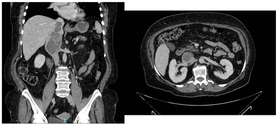
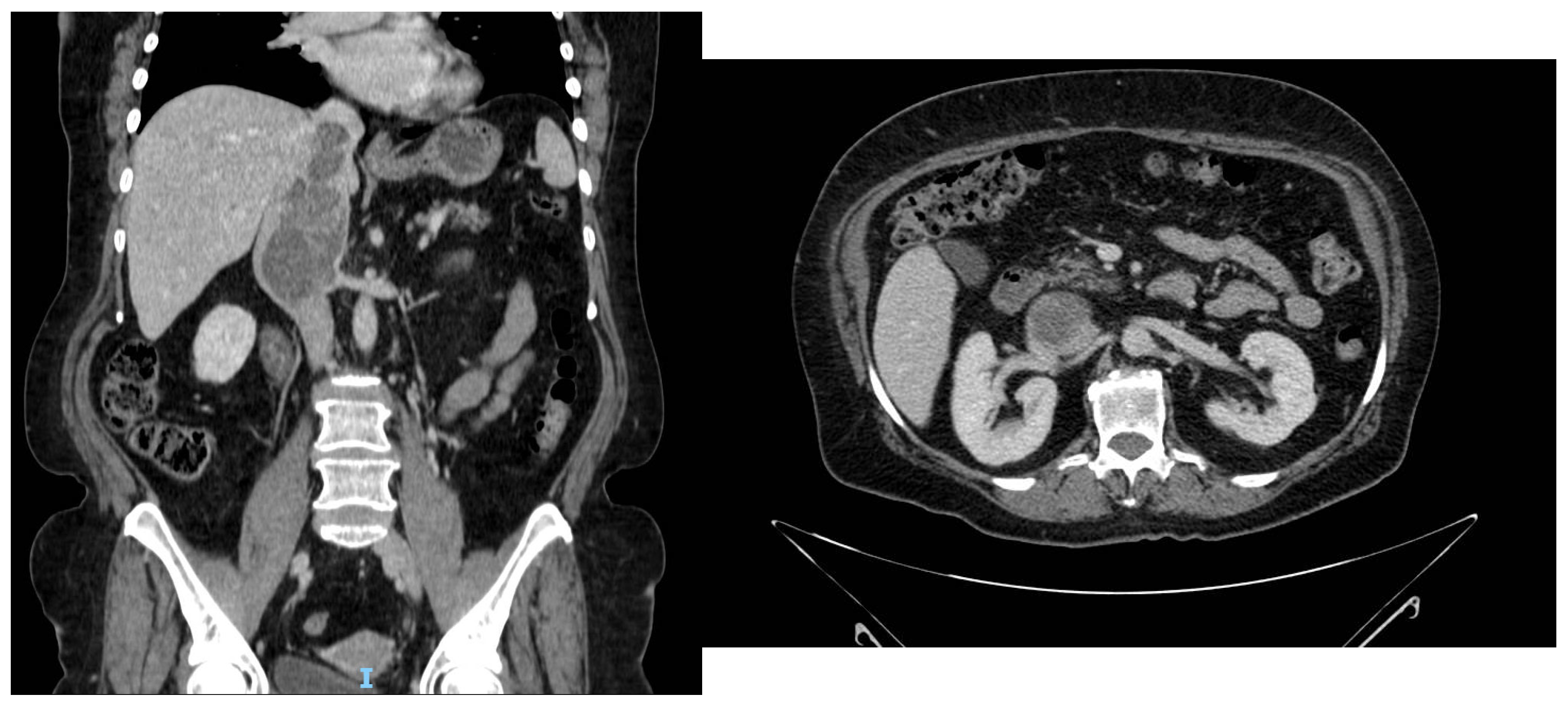
A 60-year-old woman was referred to our institution after the incidental detection of an abdominal mass on ultrasonography performed for nonspecific abdominal discomfort. Contrast-enhanced computed tomography (CT) demonstrated a 5 × 4 × 12 cm intraluminal mass arising from the inferior vena cava (IVC), extending cranially beyond the level of the hepatic veins and caudally to the ostia of both renal veins, highly suggestive of leiomyosarcoma (Figure 1). Transthoracic echocardiography showed cranial tumor extension protruding into the right atrium. No evidence of distant metastatic disease was identified.
Figure 1.
Preoperative contrast-enhanced CT demonstrating the intraluminal IVC tumor with cranial extension toward the suprahepatic cava and caudal involvement of both renal vein ostia.
The case was discussed at a multidisciplinary tumor board involving vascular surgeons, hepatobiliary surgeons, cardiac surgeons, urologists, and radiologists. Given the high radiological suspicion of IVC leiomyosarcoma and the absence of metastatic disease, preoperative biopsy was not performed, as histological confirmation was unlikely to modify the initial management strategy and could have delayed a potentially curative surgical approach. The patient was scheduled for upfront surgical resection.
Surgery was performed through a median sternotomy with pericardiotomy, allowing intrapericardial control of the IVC, followed by a continuous midline laparotomy. Intraoperative inspection revealed a large hypervascular tumor arising from the retrohepatic IVC and extending beyond the ostia of both renal veins. Because preoperative imaging suggested suprahepatic caval wall invasion with intracardiac extension, the initial surgical strategy included the possibility of cardiopulmonary bypass with hepatic vascular exclusion if complete vascular control could not be achieved.
After division of the hepatic ligaments, meticulous circumferential dissection of the retrohepatic and suprahepatic IVC was performed. The infrarenal IVC and both renal veins were individually encircled with vessel loops, followed by complete exposure of the hepatic veins. Careful intraoperative assessment demonstrated that, despite extensive intraluminal tumor extension, the wall of the suprahepatic IVC remained free of tumor infiltration. Distal displacement of the intraluminal tumor using a milking maneuver allowed safe placement of the proximal vascular clamp below the hepatic veins, thereby avoiding cardiopulmonary bypass and substantially simplifying the planned procedure.
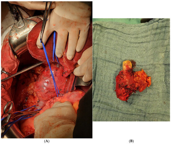
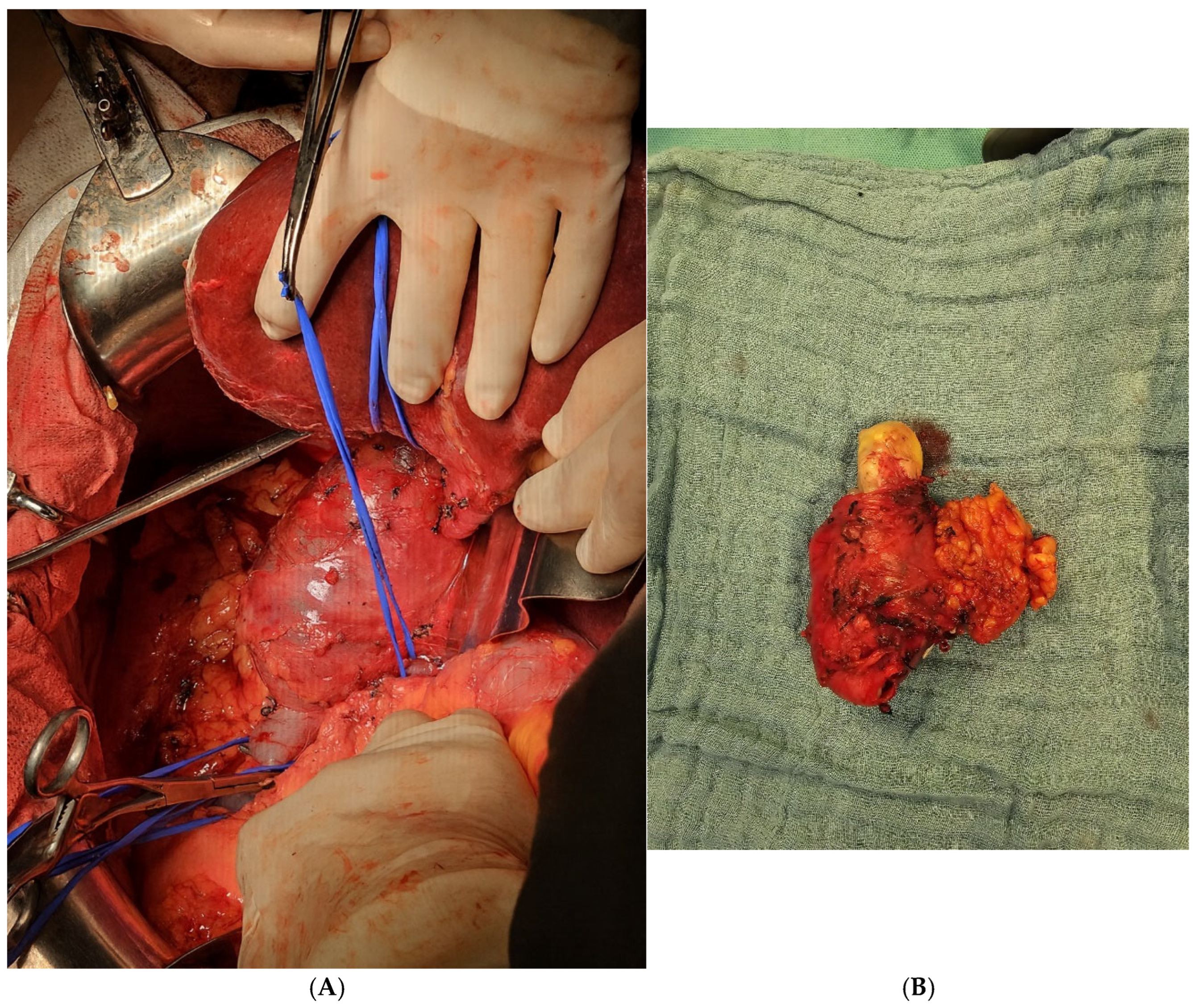
To preserve renal function while achieving complete oncological resection, the right kidney was explanted for subsequent autotransplantation. The left renal vein was divided after vascular control while preserving arterial inflow, allowing temporary venous drainage through the adrenal and gonadal collateral veins. After clamping the infrarenal IVC and the cava immediately below the hepatic veins, the IVC was transected, and the lumbar tributaries were ligated. Following complete vascular control (Figure 2A), the tumor was resected en bloc together with the involved segment of the inferior vena cava and the surrounding pericaval tissue (Figure 2B).
Figure 2.
(A) Intraoperative view after right nephrectomy and immediately before en bloc tumor resection. Blue vessel loops encircle the retrohepatic inferior vena cava (IVC), infrarenal IVC, and left renal vein, providing complete vascular control prior to caval resection. The yellow arrow indicates the right renal vein stump. (B) Surgical specimen following en bloc resection of the inferior vena cava leiomyosarcoma.
IVC continuity was restored using a 20mm ringed polytetrafluoroethylene (PTFE) graft. To facilitate preservation of the left kidney, a tubular pericardial conduit was fashioned and sutured to the prosthetic graft, allowing tension-free reconstruction of the left renal vein by end-to-side anastomosis. Anastomotic patency was assessed intraoperatively by direct surgical inspection, and postoperative Doppler ultrasonography confirmed adequate vascular flow.
The explanted right kidney underwent bench preparation before autotransplantation. Owing to the short length of the right renal vein, venous extension was achieved using a spiral conduit fashioned from the native IVC, facilitating a tension-free venous anastomosis. The renal artery was spatulated, and the kidney was subsequently autotransplanted into the right iliac fossa with end-to-side vascular anastomoses to the external iliac vessels. Ureteroneocystostomy was performed over a double-J ureteral stent. Warm ischemia time was 1 min and 42 s, whereas cold ischemia time was 6 h.
The postoperative course was complicated by transient renal dysfunction associated with mildly elevated bilateral intrarenal arterial resistive indices on Doppler ultrasonography. Renal function progressively recovered with conservative management, accompanied by normalization of Doppler findings.
Histopathological examination confirmed a high-grade leiomyosarcoma of the IVC. Immunohistochemistry demonstrated diffuse positivity for desmin, caldesmon, smooth muscle actin, and HHF35. Surgical margins were negative (R0 resection).
Following multidisciplinary review, adjuvant chemotherapy with doxorubicin and dacarbazine was recommended due to the high-risk pathological features of the tumor. Treatment was discontinued after the second cycle because of cardiotoxicity. At 14 months of follow-up, the patient remains disease-free without further oncological treatment and has preserved renal function.
3. Discussion
Complete surgical resection remains the cornerstone of treatment for localized inferior vena cava (IVC) leiomyosarcoma. However, because of the rarity of this malignancy, no standardized management algorithm has been established, and current recommendations are largely based on case reports and small retrospective series [6,7,11]. Consequently, surgical management must be individualized according to tumor location, vascular involvement, and the possibility of achieving complete oncological resection while preserving organ function. The complexity of these procedures highlights the importance of management within experienced multidisciplinary centers. Recent evidence has emphasized the impact of institutional experience on outcomes after retroperitoneal sarcoma surgery, supporting the centralization of these technically demanding procedures [13].
For localized disease, en bloc resection with negative margins (R0) represents the only potentially curative treatment and remains the most important prognostic factor [5,6,7,11,12]. In our patient, the absence of distant metastatic disease supported an upfront surgical approach with curative intent. The main surgical challenge was not only achieving oncological radicality but also maintaining renal function in the setting of extensive caval involvement affecting both renal vein ostia.
Management of the IVC following tumor resection depends largely on the affected anatomical segment. Reconstruction is generally unnecessary for isolated infrarenal (segment I) tumors because extensive collateral venous circulation is usually sufficient to maintain lower extremity venous drainage [5,11]. In contrast, tumors involving the suprahepatic IVC (segment III) frequently require more extensive surgical approaches, including cardiopulmonary bypass and/or hepatic vascular exclusion [5]. Segment II tumors, which are the most frequently encountered [2,10,14], deserve particular consideration because involvement of the renal veins often necessitates complex reconstructive strategies aimed at preserving renal function while achieving complete oncological resection [2,5].
In the present case, a 20mm ringed PTFE graft was used for caval replacement, followed by six months of postoperative anticoagulation.
Preservation of renal function represented one of the principal objectives of our surgical strategy. Although left renal vein ligation may occasionally be tolerated due to collateral venous drainage, reconstruction may be preferred when renal preservation is a priority [10,11]. An alternative strategy consists of end-to-side anastomosis of the left renal vein to the inferior mesenteric vein, thereby avoiding caval reconstruction, although this technique has only been described in isolated cases [15]. More commonly, reconstruction of the left renal vein by direct anastomosis to the caval prosthesis provides reliable venous drainage while maintaining physiological renal outflow [11].
In carefully selected patients with isolated involvement of the renal vein ostium, nephron-sparing strategies should be considered. Reported alternatives include right renal autotransplantation following caval reconstruction or bifurcated prosthetic grafts allowing bilateral renal vein reimplantation [10,11,14]. In the present case, preservation of bilateral renal function was considered a major surgical objective. Rather than relying on bilateral renal vein reconstruction onto a single prosthetic graft, two complementary strategies were adopted. The left renal vein was reconstructed using a customized pericardial conduit anastomosed to the PTFE graft, whereas the right kidney underwent autotransplantation into the right iliac fossa after venous lengthening with an autologous caval patch. This individualized approach provided independent venous drainage for each kidney while minimizing the potential consequences of prosthetic complications. Although complex caval reconstruction and renal autotransplantation have each been described individually in patients with IVC leiomyosarcoma, reports combining both techniques as part of a tailored strategy to preserve bilateral renal function remain extremely limited [9,10,16].
A particularly relevant aspect of the present case was the discrepancy between the preoperative imaging findings and the intraoperative assessment. Computed tomography and transthoracic echocardiography suggested suprahepatic IVC extension with intracardiac involvement, raising the possibility of requiring cardiopulmonary bypass and hepatic vascular exclusion. However, meticulous circumferential dissection demonstrated that the suprahepatic caval wall was free of tumor infiltration. This finding allowed distal displacement of the tumor and safe clamping below the hepatic veins, thereby avoiding cardiopulmonary bypass without compromising oncological radicality. This finding highlights the importance of definitive intraoperative assessment in tailoring the surgical strategy beyond what preoperative imaging alone may suggest.
The role of adjuvant therapy remains controversial. Although both chemotherapy and radiotherapy have demonstrated some activity, their impact on long-term survival remains uncertain, and treatment is frequently limited by toxicity [3,5,7,9]. In the present case, adjuvant treatment was discussed at the multidisciplinary sarcoma board. Considering the high-risk characteristics of the tumor, including high-grade histology, frequent mitotic activity with atypical mitoses, and extensive vascular involvement, adjuvant chemotherapy with doxorubicin and dacarbazine was recommended, whereas radiotherapy was not considered appropriate because of concerns regarding toxicity. Treatment was discontinued after the second cycle because of cardiotoxicity, underscoring the importance of individualized multidisciplinary decision-making.
As with all single-case reports, the generalizability of our findings is inherently limited. Nevertheless, this case illustrates that, in selected patients with complex segment II IVC leiomyosarcoma, meticulous multidisciplinary planning, individualized vascular reconstruction, and careful intraoperative reassessment may allow radical oncological resection while preserving bilateral renal function.
4. Conclusions
Inferior vena cava leiomyosarcoma remains a rare and technically demanding malignancy for which complete surgical resection remains the cornerstone of potentially curative treatment. This case demonstrates that meticulous multidisciplinary planning, individualized intraoperative decision-making, and tailored vascular reconstruction can facilitate radical oncological resection while preserving bilateral renal function, even in highly complex segment II tumors. Careful reassessment of the extent of caval wall involvement during surgery may allow tailoring of the surgical strategy and avoidance of unnecessary additional procedures without compromising oncological outcomes. Although our experience is limited to a single case, the combined use of customized IVC reconstruction and right renal autotransplantation may represent a feasible surgical strategy for selected patients requiring extensive caval resection and complex renal venous reconstruction.
Author Contributions
Conceptualization, C.G.-R., N.M.U. and Á.T.S.; methodology, C.G.-R., C.L.S., J.A.G.F., J.C.P. and Á.T.S.; validation, N.M.U., C.L.S., J.A.G.F., J.C.P. and Á.T.S.; formal analysis, C.G.-R.; investigation, C.G.-R., N.M.U., S.J.Á., C.G.A., C.L.S., J.A.G.F., J.C.P., A.R.A. and Á.T.S.; resources, Á.T.S.; data curation, C.G.-R.; writing—original draft preparation, C.G.-R.; writing—review and editing, all authors; visualization, C.G.-R.; supervision, N.M.U. and Á.T.S.; project administration, C.G.-R. All authors have read and agreed to the published version of the manuscript.
Funding
This research received no external funding.
Informed Consent Statement
Written informed consent has been obtained from the patient to publish this paper.
Data Availability Statement
The data supporting the findings of this case report are not publicly available because they contain information that could compromise patient privacy. De-identified data may be made available by the corresponding author upon reasonable request, subject to institutional approval and applicable ethical and legal requirements.
Acknowledgments
During the preparation of this manuscript, the authors used ChatGPT (OpenAI, GPT-5.5) to assist with the translation, linguistic refinement, and improvement of the scientific writing. All generated content was critically reviewed, edited, and verified by the authors, who take full responsibility for the accuracy, interpretation, and integrity of the final manuscript.
Conflicts of Interest
The authors declare no conflicts of interest.
Abbreviations
The following abbreviations are used in this manuscript:
| CT | Computed Tomography |
| IVC | Inferior Vena Cava |
| PTFE | Polytetrafluoroethylene |
| R0 | Microscopically Margin-Negative Resection |
References
- Rusu, C.B.; Gorbatâi, L.; Szatmari, L.; Koren, R.; Bungărdean, C.I.; Feciche, B.O.; Bumbuluţ, C.; Society, B.G.M.C.; Andraş, I.M.; Rahotă, R.; et al. Leiomyosarcoma of the inferior vena cava: Our experience and a review of the literature. Rom. J. Morphol. Embryol. 2020, 61, 227–233. [Google Scholar] [CrossRef] [Scilit] [PubMed]
- Pérez-de-Villar, J.M.; Arjona-Sánchez, A.; Rufián-Andújar, B.; Valenzuela-Molina, F.; Sánchez-Hidalgo, J.M.; Rodríguez-Ortiz, L.; Casado-Adam, A.; Viyuela-García, C.; Rufián-Peña, S.; Caro-Cuenca, T.; et al. Tratamiento quirúrgico de los leiomiosarcomas de vena cava: Serie de casos en un hospital de tercer nivel y revisión de la literatura. Cir. Esp. 2022, 100, 481–487. [Google Scholar] [CrossRef] [Scilit]
- López-Ruiz, J.A.; Tallón-Aguilar, L.; Marenco-de la Cuadra, B.; López-Pérez, J.; Oliva-Mompeán, F.; Padillo-Ruiz, J. Leiomiosarcoma de vena cava inferior: Caso clínico y revisión bibliográfica. Cir. Cir. 2017, 85, 361–365. [Google Scholar] [CrossRef] [Scilit] [PubMed]
- Li, F.; Li, C.; Guo, W.; Meng, X. A worldwide systematic review of primary leiomyosarcoma of the inferior vena cava. Asian J. Surg. 2023, 46, 5983–5984. [Google Scholar] [CrossRef] [Scilit] [PubMed]
- Mastoraki, A.; Leotsakos, G.; Mastoraki, S.; Papanikolaou, I.S.; Danias, N.; Smyrniotis, V.; Arkadopoulos, N. Challenging diagnostic and therapeutic modalities for leiomyosarcoma of the inferior vena cava. Int. J. Surg. 2015, 13, 92–95. [Google Scholar] [CrossRef] [Scilit] [PubMed]
- Wachtel, H.; Gupta, M.; Bartlett, E.K.; Jackson, B.M.; Kelz, R.R.; Karakousis, G.C.; Fraker, D.L.; Roses, R.E. Outcomes after resection of leiomyosarcomas of the inferior vena cava: A pooled data analysis of 377 cases. Surg. Oncol. 2015, 24, 21–27. [Google Scholar] [CrossRef] [Scilit] [PubMed]
- Saikia, J.; Rastogi, S.; Barwad, A.; Dhamija, E.; Pandey, R.; Bhoriwal, S.; Deo, S.; Kumar, S. A systematic review of the current management approaches in leiomyosarcoma of the inferior vena cava: Results from analysis of 118 cases. Asian Cardiovasc. Thorac. Ann. 2022, 30, 349–363. [Google Scholar] [CrossRef] [Scilit] [PubMed]
- Kulaylat, M.N.; Karakousis, C.P.; Doerr, R.J.; Karamanoukian, H.L.; O’Brien, J.; Peer, R. Leiomyosarcoma of the inferior vena cava: A clinicopathologic review and report of three cases. J. Surg. Oncol. 1997, 65, 205–217. [Google Scholar] [CrossRef]
- Fernandez, H.T.; Kim, P.T.W.; Anthony, T.L.; Hamman, B.L.; Goldstein, R.M.; Testa, G. Inferior vena cava reconstruction for leiomyosarcoma of zone I–III requiring complete hepatectomy and bilateral nephrectomy with autotransplantation. J. Surg. Oncol. 2015, 112, 481–485. [Google Scholar] [CrossRef] [Scilit] [PubMed]
- Kieffer, E.; Alaoui, M.; Piette, J.C.; Cacoub, P.; Chiche, L. Leiomyosarcoma of the inferior vena cava: Experience in 22 cases. Ann. Surg. 2006, 244, 289–295. [Google Scholar] [PubMed]
- Teixeira, F.J.R.; Couto Netto, S.D.; Perina, A.L.D.F.; Torricelli, F.C.M.; Ragazzo Teixeira, L.; Zerati, A.E.; Ferreira, F.d.O.; Akaishi, E.H.; Nahas, W.C.; Utiyama, E.M. Leiomyosarcoma of the inferior vena cava: Survival rate following radical resection. Oncol. Lett. 2017, 14, 3909–3916. [Google Scholar] [CrossRef] [Scilit] [PubMed]
- Mingoli, A.; Feldhaus, R.J.; Cavallaro, A.; Stipa, S. Leiomyosarcoma of the inferior vena cava: Analysis and search of world literature on 141 patients and report of three new cases. J. Vasc. Surg. 1991, 14, 688–699. [Google Scholar] [CrossRef] [Scilit]
- Samà, L.; Kumar, S.; Ruspi, L.; Sicoli, F.; D’AMato, V.; Mintemur, Ö.; Renne, S.L.; Quagliuolo, V.L.; Cananzi, F.C. Learning curve in retroperitoneal sarcoma surgery. Eur. J. Surg. Oncol. 2024, 50, 108612. [Google Scholar] [CrossRef] [Scilit] [PubMed]
- Kyriazi, M.A.; Stafyla, V.K.; Chatzinikolaou, I.; Koureas, A.; Chatziioannou, A.; Kondi-Paphiti, A.; Arkadopoulos, N.; Smyrniotis, V. Surgical challenges in the treatment of leiomyosarcoma of the inferior vena cava: Analysis of two cases and brief review of the literature. Ann. Vasc. Surg. 2010, 24, 826.e13–826.e17. [Google Scholar] [CrossRef] [Scilit] [PubMed]
- Arkadopoulos, N.; Karmaniolou, I.; Ekonomopoulos, N.; Vassiliu, P.; Smyrniotis, V. Combination of total abdominal inferior vena cava resection with a novel technique of left renal outflow restoration. Surgery 2012, 152, 142–143. [Google Scholar] [CrossRef] [Scilit] [PubMed]
- Kraybill, W.G.; Callery, M.P.; Heiken, J.P.; Flye, M.W. Radical resection of tumors of the inferior vena cava with vascular reconstruction and kidney autotransplantation. Surgery 1997, 121, 31–36. [Google Scholar] [CrossRef] [Scilit] [PubMed]
Disclaimer/Publisher’s Note: The statements, opinions and data contained in all publications are solely those of the individual author(s) and contributor(s) and not of MDPI and/or the editor(s). MDPI and/or the editor(s) disclaim responsibility for any injury to people or property resulting from any ideas, methods, instructions or products referred to in the content. |
© 2026 by the authors. Licensee MDPI, Basel, Switzerland. This article is an open access article distributed under the terms and conditions of the Creative Commons Attribution (CC BY) license.