Case Report

Living-Donor Renal Vein Reconstruction with Banked Deceased-Donor Iliac Vein: A Case Report

1
Department of General Surgery, Rabin Medical Center, Petah Tikva 49100, Israel
2
Gray Faculty of Medicine, Tel Aviv University, Tel Aviv 69978, Israel
3
Department of Gastro-Enterology, Shiba Medical Center, Ramat-Gan 5265601, Israel
4
Department of Transplantation Surgery, Beilinson Medical Center (Rabin Campus), Clalit Health Services, Gray Faculty of Medicine and Health Sciences, Tel Aviv University, Derech Ze’ev Jabotinsky 39, Petah Tikva 4941492, Israel
*
Author to whom correspondence should be addressed.
†
These authors contributed equally to this work.
Surg. Tech. Dev. 2026, 15(3), 35; https://doi.org/10.3390/std15030035
Submission received: 10 June 2026 / Revised: 24 July 2026 / Accepted: 18 August 2026 / Published: 18 August 2026

Abstract

Background: The right renal vein is about half the length of the right renal artery and is shortened further by endovascular stapling at laparoscopic donor nephrectomy. Right-sided living-donor grafts carry roughly twice the adjusted risk of delayed graft function and of early graft loss, a penalty absent from deceased donation, where the inferior vena cava accompanies the graft. The deficit is one of venous length, not of the organ; extension is the remedy, and no guideline specifies how the conduit should be obtained. Methods and cases: Three consecutive recipients of right living-donor kidneys had the renal vein extended with a deceased-donor iliac vein. Conduits were recovered at multiorgan retrieval from ABO-identical or ABO-compatible donors, immersed in University of Wisconsin solution at 4 °C, and used within seven days; elective right donor nephrectomy was booked to follow a suitable retrieval within that window. Extension was an end-to-end back-table anastomosis with continuous 5-0 or 6-0 polypropylene, converting a short-vein implantation into a routine end-to-side anastomosis to the external iliac vein. All three grafts functioned immediately, without venous thrombosis, technical graft loss, or delayed graft function; the last serum creatinine was 0.91, 1.22, and 1.42 mg/dL. Conclusions. A calibre- and ABO-matched deceased-donor iliac vein, held in University of Wisconsin solution at 4 °C and used within seven days, makes venous extension a scheduled step of an elective right living-donor operation and requires no cryopreservation infrastructure. Where a donor’s safety mandates right nephrectomy, a short right renal vein need not preclude donation. Three cases establish feasibility; the low thrombotic risk of venous extension rests on published series rather than on this report.

1. Introduction

Living donation offers superior graft survival and shorter waiting times than deceased donation [1], and the left kidney is generally preferred because its longer renal vein permits a tension-free anastomosis. The right renal vein measures 21–71 mm, approximately half the length of the accompanying artery [2], and is foreshortened further by endovascular stapling at laparoscopic nephrectomy [3,4].
The resulting penalty is documented rather than theoretical: right-sided living-donor grafts carry an adjusted odds ratio of 2.01 (95% CI 1.31–3.09) for delayed graft function and an adjusted hazard ratio of 2.02 (95% CI 1.06–3.86) for graft loss within 30 days, with no excess thereafter [5], the former being the firmer of the two associations, and a systematic review concurs [6]. The penalty is confined to living donation: in one registry, right-sided implantation predicted technical graft failure among 4372 living-donor recipients (p = 0.03) but not among 5346 deceased-donor recipients (p = 0.16) [7,8,9], the difference being that deceased donation recovers the inferior vena cava with the graft. The disadvantage is therefore one of venous length, not of the organ, which identifies extension as the remedy.
Right procurement remains unavoidable in some donors for safety reasons [7,8]. Extension options are numerous—gonadal [10,11], saphenous [12], internal jugular [13], femoral [14], iliac allograft [15,16,17], cryopreserved [18], synthetic [19], or graft-free [20,21,22]—yet none is standardised, and KDIGO 2020 addresses operative planning without any recommendation on venous extension [23]. For autologous conduits the constraint is anatomical; for deceased-donor conduits it is logistical, and where no cryopreserved vascular allograft bank exists a calibre- and blood-group-matched vein must nonetheless be in hand on the day an elective operation is scheduled.
We report the use of refrigerated, short-window deceased-donor iliac vein for this purpose in three consecutive recipients, and set out the scheduling arrangement it requires.

2. Methods

2.1. Study Design

This is a retrospective case series of three recipients who underwent right living-donor kidney transplantation with deceased-donor iliac vein extension at Rabin Medical Center. It is reported in accordance with the PROCESS 2023 guideline and the Declaration of Helsinki. Institutional review board approval was obtained (IRB RMC-0808-25 approved 24 May 2026), and individual consent was waived owing to the retrospective, de-identified design.
The three cases arose within a consecutive institutional experience of seven right living-donor transplants performed between January 2015 and December 2025. The remaining four are not described. The denominator is given to define the setting, not as an outcome series.

2.2. Donor Evaluation and Laterality

Donors were evaluated by cross-sectional angiography and split renal function assessment. The kidney with superior anticipated function and simpler vasculature was retained by the donor; the right kidney was procured only when mandated by donor safety—left vascular multiplicity, asymmetric split function favouring the left, or anatomical variants. All nephrectomies were laparoscopic. In all three cases, right procurement was mandated on donor-safety grounds and was not elective.

2.3. Donor Nephrectomy

All nephrectomies were laparoscopic and followed our published standard living-donor protocol for positioning, port configuration, ureteric handling, and vascular stapling [4]; only right-specific steps are described here. The right adrenal gland is preserved and its small branches individually clipped, since the right adrenal vein drains directly into the inferior vena cava. The right renal vein receives no gonadal or adrenal tributary and is dissected circumferentially to the caval wall for maximal caval-side length without compromising donor safety. The artery is stapled first; the vein is then stapled flush to the cava while the assistant elevates the kidney cephalad and laterally. Usable venous length was 2–3 cm in all three donors, consistent with the reported anatomical range [2].

2.4. Conduit Procurement, Preservation, and Scheduling

The conduit is a common or external iliac vein recovered at multiorgan retrieval and selected to match the diameter of the right renal vein. It is immersed in University of Wisconsin solution at 4 °C in a dedicated vessel refrigerator and used within seven days. Elective right living-donor nephrectomy and transplantation are therefore booked to follow a retrieval from an ABO-identical or ABO-compatible donor within that window, so that a calibre- and blood-group-matched conduit is in hand before the donor is anaesthetised.
Cryopreservation was not used. Vessels processed under tissue-bank standards and held in the vapour phase of liquid nitrogen have shelf lives measured in years, which decouples conduit availability from the timing of retrieval and is the preferable arrangement wherever such a facility exists; our institution does not operate a vascular tissue establishment due to—regulatory licencing and absence of a controlled-rate freezer. Hypothermic storage in University of Wisconsin solution requires only a dedicated refrigerator, at the cost of a seven-day window that is absorbed by scheduling.

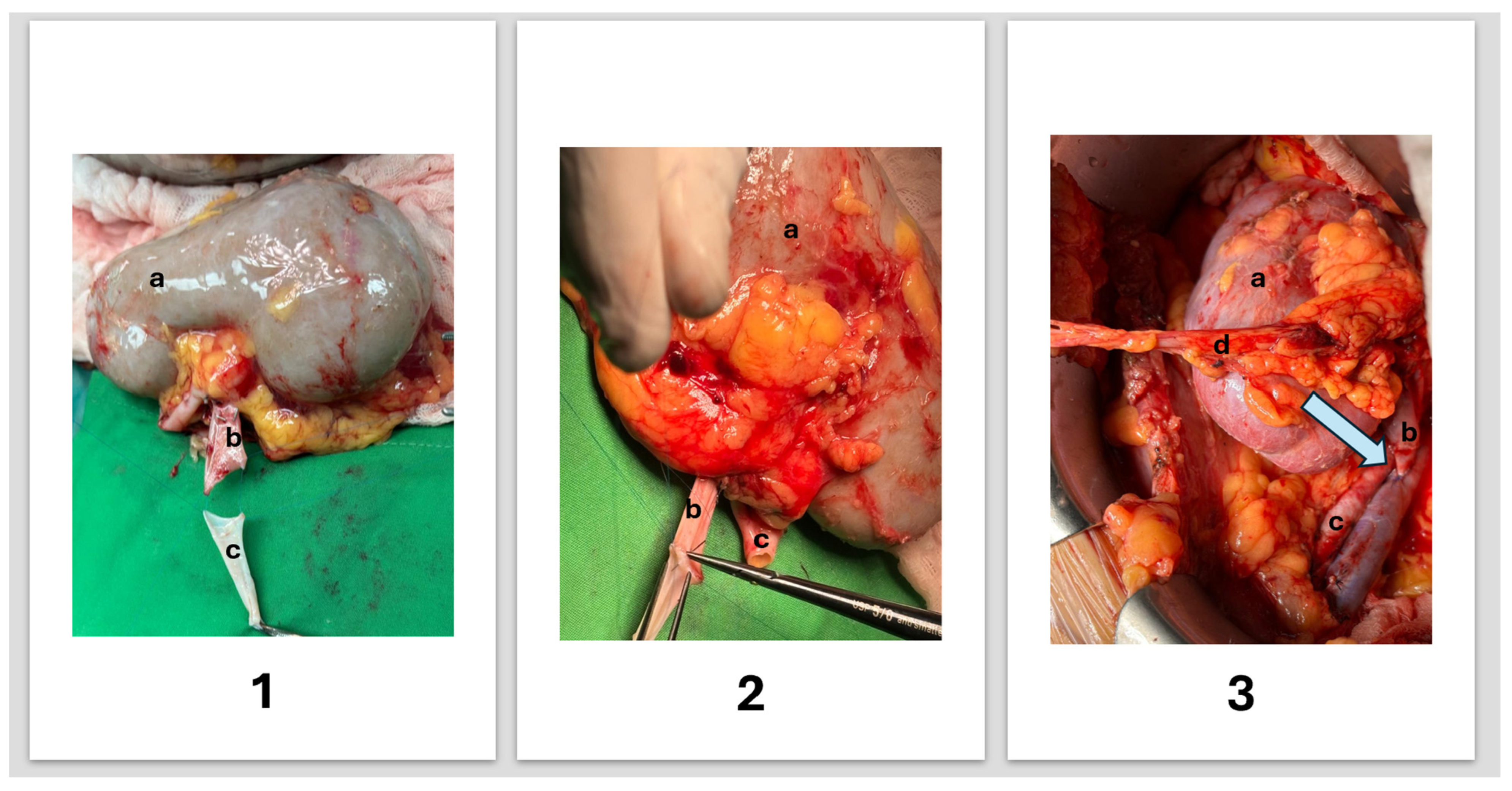
2.5. Back-Table Reconstruction (Figure 1)

The reconstruction is performed under hypothermic conditions. The venous staple line is excised and the vein inspected for intimal injury. The iliac segment is flushed, its side branches ligated, and it is trimmed to 3–5 cm according to recipient anatomy. The extension is an end-to-end anastomosis with continuous 5-0 or 6-0 polypropylene, posterior wall first and with repeated flushing, checked for a widely patent lumen, absence of leak, and no kinking on rotation. The same conduit and the same anastomosis were used in all three cases.

2.6. Recipient Implantation

The graft is implanted extraperitoneally in the iliac fossa. The extended renal vein is anastomosed end-to-side to the external iliac vein, converting a difficult short-vein implantation into a routine venous anastomosis, and the renal artery to the external or internal iliac artery according to recipient anatomy. After sequential release of the venous and then the arterial clamp, the graft is assessed for uniform coloration, prompt venous filling, and urine production. Urinary continuity is restored with a stented Lich–Gregoir ureteroneocystostomy.
Figure 1. Schematic illustration of right living-donor kidney procurement, back-table venous reconstruction, and recipient implantation. (1) Back-table preparation: (1a) right donor kidney following procurement; (1b) short right renal vein from the living donor; (1c) deceased-donor iliac vein graft prepared for extension. (2) Back-table reconstruction: (2a) kidney before reconstruction; (2b) right renal vein after extension with an end-to-end anastomosis; (2c) right renal artery preserved for implantation. (3) Recipient implantation: the blue arrow indicate the venous anastomosis (3a) kidney after venous reconstruction; (3b) extended renal vein anastomosed end-to-side to the external iliac vein; (3c) renal artery anastomosed end-to-side to the external iliac artery; (3d) ureter before ureteroneocystostomy. RV, right renal vein; RRA, right renal artery; CIVG, cadaveric iliac vein graft; EIV, external iliac vein; EIA, external iliac artery.
Std 15 00035 g001

2.7. Immunosuppression and Anticoagulation

Induction was with basiliximab or anti-thymocyte globulin according to immunological risk; maintenance comprised tacrolimus, mycophenolate mofetil, and prednisone. Intraoperative unfractionated heparin and postoperative low-molecular-weight heparin were given per institutional protocol.

2.8. Data, Definitions, and Analysis

Data were extracted from the electronic medical record onto a standardised form and are reported as recorded; missing values are shown as such and were not imputed. Delayed graft function was defined as the need for dialysis within the first seven postoperative days, and primary non-function as a graft that never achieved function. Vascular complications comprised renal vein or artery thrombosis, anastomotic stenosis, or haemorrhage requiring intervention; surgical complications were graded by the Clavien–Dindo classification. Cold ischaemia time was defined as the interval from cold perfusion of the graft to reperfusion in the recipient. Follow-up was censored at the last documented contact.
No statistical analysis was performed. With three cases, no proportion, confidence interval, or hypothesis test carries interpretable information; the single quantitative statement made in the Discussion is an exact binomial bound on an unobserved event rate.

3. Case Presentation

3.1. Case 1

A 38-year-old man with diabetic nephropathy, hypertension, and obesity (BMI 28.7) received a kidney from a 51-year-old unrelated altruistic donor (BMI 30.3) after six months of haemodialysis; PRA was negative. Donor imaging demonstrated three or more left renal vessels, mandating right nephrectomy. The graft had two renal arteries, one vein, and one ureter. Operative time was 156 min and cold ischaemia three hours. The short right renal vein was extended with a banked cadaveric iliac vein graft and anastomosed end-to-side to the external iliac vein, with a single iliac arterial anastomosis. A small lower-polar accessory artery was deliberately left unreconstructed, because revascularisation was technically demanding and threatened the ureteric blood supply. Induction was with basiliximab. The graft perfused immediately with prompt urine output. The postoperative course included a wound haematoma on day 5, hyperglycaemia, hypertension, and a transient creatinine rise, all Clavien–Dindo grade I–II, without readmission or reoperation. At 60 months, serum creatinine was 0.91 mg/dL with a functioning graft.

3.2. Case 2

An 18-year-old man with dysplastic kidneys and bilateral vesicoureteral reflux, hyperparathyroidism, thrombocytopenia, and splenomegaly (BMI 20.7) was transplanted pre-emptively from his 25-year-old brother (BMI 19.8). Right nephrectomy was mandated by three or more left renal vessels. The graft had a single artery, vein, and ureter. Operative time was 113 min and cold ischaemia was 1.5 h. The renal vein was extended with a preserved iliac vein graft and implanted end-to-side to the external iliac vein. Induction was with thymoglobulin. The graft functioned immediately. Mild hydronephrosis was observed; the recipient underwent a planned concurrent allogeneic haematopoietic stem-cell transplant on postoperative day 11 and was readmitted for a creatinine rise in the context of COVID-19 infection. All complications were Clavien–Dindo grade I–II. At 60 months, serum creatinine was 1.22 mg/dL with a functioning graft. The haematological disease and concurrent stem-cell transplant are specific to this recipient and unrelated to the venous reconstruction.

3.3. Case 3

A 67-year-old woman with autosomal dominant polycystic kidney disease, hypertension, borderline diabetes, and proteinuria received a kidney from a 46-year-old unrelated altruistic donor (BMI 25.7) after five years of haemodialysis; PRA was negative. Right nephrectomy was mandated by three or more left renal vessels. The graft had a single artery, vein, and ureter. Operative time was 141 min and cold ischaemia 1.5 h. The renal vein was extended with a banked cadaveric iliac vein graft and implanted end-to-side to the external iliac vein. Induction was with thymoglobulin. The graft functioned immediately. The postoperative course included an Escherichia coli urinary tract infection, transient hypertension, and left shoulder pain with a cardiac cause excluded; all complications were Clavien–Dindo grade I–II. At 60 months, serum creatinine was 1.42 mg/dL with a functioning graft.

3.4. Summary and Donor Outcomes

In all three recipients, the graft perfused immediately. There was no venous thrombosis, anastomotic stenosis, haemorrhage requiring intervention, delayed graft function, primary non-function, reoperation, graft nephrectomy, or return to dialysis, and all complications were Clavien–Dindo grade I–II. No donor died, developed end-stage kidney disease, or required renal replacement therapy. The case 1 donor developed a trocar-site incisional hernia requiring surgical repair (Clavien–Dindo IIIb), and the case 2 donor a wound dehiscence managed with local care and antibiotics. Most recent donor serum creatinine ranged from 0.72 to 1.55 mg/dL; albuminuria was absent except in the case 2 donor, whose urinary albumin-to-creatinine ratio was 63.7 mg/g, Table 1.

4. Discussion

These reconstructions address a problem whose magnitude and mechanism are established but whose management is not: the living-donor penalty [5,6] is absent from deceased donation, where the vena cava accompanies the graft [5,7,8,9], which localises the problem to venous length, yet KDIGO 2020 makes no recommendation on extension [23].

4.1. Position of the Technique

The contribution of this report is logistical rather than technical. A conduit held in University of Wisconsin solution at 4 °C for up to seven days is sufficient to convert venous extension from an intraoperative improvisation into a planned step of an elective operation, and requires a dedicated refrigerator rather than a vascular tissue establishment. For a centre without one, this is the difference between an available conduit and none: a donor whose safety mandates right nephrectomy cannot be scheduled around the chance arrival of a vessel, and improvising at the back table is precisely the circumstance in which a surgeon elects instead to decline the donor. Reported conduit choice is therefore, in part, an artefact of conduit availability.
The underlying technique is established and is positioned as such. End-to-end interposition of an allogeneic venous conduit into a short renal vein was described in 1988 [24], and deceased-donor vein extension for right living-donor kidneys has since been reported in series of up to 38 recipients [17]. Cryopreserved vascular allograft banking is likewise long-standing, with iliac artery and vein allografts processed and stored under tissue-bank standards for years; that arrangement decouples conduit availability from the timing of retrieval altogether and, where a facility exists, removes the scheduling problem addressed here.

4.2. Alternative Conduits

Autologous conduits avoid immunological exposure but are anatomically constrained: on the right the gonadal vein does not accompany the renal vein as it does on the left [10,11]; saphenous vein is substantially calibre-mismatched [12]; internal jugular vein is frequently stenosed after prolonged dialysis, excluding the recipients most likely to need it [13]; and superficial femoral vein adds an operative site and a risk of lower-limb oedema [14]. Synthetic conduits are immediately available but thrombogenic in a low-flow circuit and carry an infection risk under immunosuppression [19,25].
A further option arises from the same retrieval and deserves explicit mention. The deceased donor’s own right renal vein, taken with a cuff of inferior vena cava, can be opened and reconfigured as a spiral to yield a conduit of renal-vein calibre. This material cannot be obtained from the living donor, in whom the vein is stapled flush to the cava, but it is available whenever the iliac vein is and requires no additional recovery. We used iliac vein because it provides adequate length as a straight segment closed by a single circumferential suture line, whereas a spiral adds a longitudinal closure to the interposition anastomosis; the choice was one of simplicity, not of demonstrated superiority [26].
No randomised comparison of conduits exists and none is likely; our position is bounded accordingly. For an elective and therefore schedulable operation, a calibre- and ABO-matched deceased-donor vein imposes no additional incision, no size mismatch, and no prosthetic material, at the cost of the immunological exposure discussed below.

4.3. What Three Cases Can and Cannot Say

Scepticism about the thrombotic behaviour of an interposed venous conduit is reasonable and cannot be addressed with our data. Transplant renal vein thrombosis is reported in 0.1–8.2% of kidney transplants, the lower end deriving from selected living-donor series [27]. Whichever value is assumed, three recipients cannot discriminate between them: with no events in three cases, the exact one-sided 95% upper bound on the underlying rate is 63%. The absence of thrombosis here is uninformative and we draw no inference from it.
The relevant evidence is aggregate, and weaker than it is customarily presented. In the comparative deceased-donor series most often cited, none of 76 recipients of an extended right renal vein sustained a technical vascular complication against 2.2% implanted without extension [28]; the upper 95% bound for 0/76 is approximately 4.7% and therefore contains 2.2%, so no difference is demonstrated. In 38 right living-donor transplants with deceased-donor vein extension, there was no venous thrombosis, stricture, or anastomotic failure [17], and cryopreserved extension has been reported to shorten implantation time for right kidneys to that of left kidneys [18] in a single-centre non-randomised comparison. The mechanism is consistent: a lengthened, tension-free vein converts a difficult anastomosis into a routine one and shortens the warm interval. Tension, not the conduit, is the thrombogenic variable. These series support that extension is not evidently harmful, not that it is protective.

4.4. Immunological Exposure

A deceased-donor venous conduit is allogeneic tissue and its immunological cost is real. Cryopreserved cadaveric vascular allografts have produced broad HLA class I and II sensitisation [29]; third-party vessel allografts elicited de novo donor-specific antibodies in approximately one in six recipients in the largest reported experience, without a demonstrable reduction in graft survival [26]; and minor HLA-antibody formation has been described after cadaveric extension [16]. One observation bears on conduit choice: recipients of venous grafts alone appear less likely to sensitise than recipients of arterial grafts [26]. Decellularised vein allografts appear to abolish allosensitisation [30] and are the logical development of the technique.
We did not perform systematic post-transplant DSA surveillance in these three recipients. In a report whose subject is deceased-donor venous allograft, this is a material limitation and not a peripheral one: the principal known hazard of the technique is precisely the one we did not measure. We recommend that routine HLA-antibody monitoring accompany any use of third-party venous extenders.

4.5. Selective Non-Reconstruction of a Minor Polar Artery

In case 1, a small lower-polar accessory artery was deliberately left unreconstructed because revascularisation threatened the ureteric blood supply, and the graft achieved normal function. This is reported as an observation, not a recommendation; the judgement was specific to that anatomy.

4.6. Limitations

This is a retrospective, uncontrolled, single-centre series of three cases. No comparison group exists, no inference of safety is available, and no statistical analysis is possible. Post-transplant DSA was not assessed. The three cases arose within a consecutive institutional experience of seven right living-donor transplants; the remaining four are not described, and that denominator is reported for transparency rather than as an outcome series. The two donor complications are reported for the same reason, and this series is not designed to address comparative donor safety. The claim is bounded accordingly: that this conduit and this scheduling protocol are feasible without cryopreservation infrastructure, and that the published series are compatible with their safety.

Author Contributions

S.H. and F.K. conceived the study; S.H., M.A., R.H., N.Z. and A.P. collected the data; A.G. and N.Z. drafted the surgical technique; S.H. and F.K. drafted the manuscript; E.N. and F.K. supervised and critically revised it. All authors have read and agreed to the published version of the manuscript.

Funding

This research received no external funding.

Institutional Review Board Statement

This study was approved by the institutional review board of Rabin Medical Center (IRB RMC-0808-25; approved 24 May 2026) and was conducted in accordance with the Declaration of Helsinki.

Informed Consent Statement

Owing to the retrospective, de-identified design, the requirement for individual informed consent was waived by the review board.

Data Availability Statement

The datasets generated and analysed during the current study are available from the corresponding author on reasonable request.

Conflicts of Interest

The authors declare no conflicts of interest.

References

  1. EAU Guidelines Office. EAU Guidelines on Renal Transplantation. In Proceedings of the EAU Annual Congress Paris 2024; European Association of Urology: Arnhem, The Netherlands, 2024; Available online: https://uroweb.org/guidelines/renal-transplantation (accessed on 6 January 2026).
  2. Janschek, E.C.; Rothe, A.U.; Hölzenbein, T.J.; Langer, F.; Brugger, P.C.; Pokorny, H.; Domenig, C.M.; Rasoul-Rockenschaub, S.; Mühlbacher, F. Anatomic Basis of Right Renal Vein Extension for Cadaveric Kidney Transplantation. Urology 2004, 63, 660–664. [Google Scholar] [CrossRef] [Scilit] [PubMed]
  3. Han, D.J.; Han, Y.; Kim, Y.H.; Song, K.B.; Chung, Y.S.; Choi, B.H.; Kwon, T.-W.; Cho, Y.-P. Renal Vein Extension During Living-Donor Kidney Transplantation in the Era of Hand-Assisted Laparoscopic Living-Donor Nephrectomy. Transplantation 2015, 99, 786–790. [Google Scholar] [CrossRef] [Scilit] [PubMed]
  4. Kanani, F.; Kozin, M.; Ben Avraham, Y.; Avitan, E.; Gurevich, M.; Nesher, E.; Gravetz, A. Laparoscopic Living Donor Nephrectomy: Learning Curve Analysis Through 1446 Cases and Outcomes from 200 Consecutive Mastery-Phase Procedures—How I Do It. J. Clin. Med. 2026, 15, 1363. [Google Scholar] [CrossRef] [Scilit] [PubMed]
  5. Dobrijevic, E.L.K.; Au, E.H.K.; Rogers, N.M.; Clayton, P.A.; Wong, G.; Allen, R.D.M. Association Between Side of Living Kidney Donation and Post-Transplant Outcomes. Transpl. Int. 2022, 35, 10117. [Google Scholar] [CrossRef] [Scilit] [PubMed]
  6. Carolan, C.; Tingle, S.J.; Thompson, E.R.; Sen, G.; Wilson, C.H. Comparing Outcomes in Right Versus Left Kidney Transplantation: A Systematic Review and Meta-Analysis. Clin. Transplant. 2021, 35, e14475. [Google Scholar] [CrossRef] [Scilit] [PubMed]
  7. Brunschot, D.M.D.Ö.; van Laarhoven, C.J.H.M.; van der Jagt, M.F.P.; Hoitsma, A.J.; Warlé, M.C. Is the Reluctance for the Implantation of Right Donor Kidneys Justified? World J. Surg. 2015, 40, 471–478. [Google Scholar] [CrossRef] [Scilit] [PubMed]
  8. Johnson, D.W.; Mudge, D.W.; Kaisar, M.O.; Campbell, S.B.; Hawley, C.M.; Isbel, N.M.; Wall, D.; Griffin, A.; Preston, J.; Nicol, D.L. Deceased Donor Renal Transplantation—Does Side Matter? Nephrol. Dial. Transplant. 2006, 21, 2583–2588. [Google Scholar] [CrossRef] [Scilit] [PubMed]
  9. Kulkarni, S.; Wei, G.; Jiang, W.; Lopez, L.A.; Parikh, C.R.; Hall, I.E. Outcomes from Right Versus Left Deceased-Donor Kidney Transplants: A US National Cohort Study. Am. J. Kidney Dis. 2019, 75, 725–735. [Google Scholar] [CrossRef] [Scilit] [PubMed]
  10. Viet, K.N.; Hoang, T.; Hai, D.D.; Dinh, D.T.; Bao, N.N.; Le Nguyen, V.; Duc, H.D. Efficacy of Lengthening Right Renal Veins Using Accompanying Gonadal Veins in Living Donor Kidney Transplantation. World J. Urol. 2024, 42, 407. [Google Scholar] [CrossRef] [Scilit] [PubMed]
  11. Feng, J.; Huang, C.; Fan, M.; Wang, P.; Xiao, Y.; Zhang, G. Renal Vein Lengthening Using Gonadal Vein Reduces Surgical Difficulty in Living-Donor Kidney Transplantation. World J. Surg. 2011, 36, 468–472. [Google Scholar] [CrossRef] [Scilit] [PubMed]
  12. Shimizu, T.; Omoto, K.; Iida, S.; Inui, M.; Tsukuda, F.; Toma, H.; Iizuka, J.; Ishida, H.; Tanabe, K. Living Related Renal Transplantation Using a Saphenous Vein Graft: A Case Report. Transplant. Proc. 2018, 50, 2562–2564. [Google Scholar] [CrossRef] [Scilit] [PubMed]
  13. Lu, T.; Yi, S.G.; Bismuth, J.; Sattar, N.; Bidwell, S.; Gaber, A.O.; Knight, R.J. Short- and Midterm Results for Internal Jugular Vein Extension for Short Right Renal Vein Kidney Transplant. Clin. Transplant. 2018, 32, e13178. [Google Scholar] [CrossRef] [Scilit] [PubMed]
  14. Alcocer, F.; Zazueta, E.; Montes de Oca, J. The Superficial Femoral Vein: A Valuable Conduit for a Short Renal Vein in Kidney Transplantation. Transplant. Proc. 2009, 41, 4392–4393. [Google Scholar] [CrossRef] [Scilit] [PubMed]
  15. Del-Río, E.N.; De-León, C.P.; Villa, M.C.; Espín, P.C.; Quintero-Quintero, M.J.; Vilatobá, M.; Contreras, A.G.; Martínez, R.C. Use of Iliac Allograft from Cadaveric Donor as a Rescue Technique in Living Donor Kidney Transplant: Two Case Reports. Transplant. Proc. 2020, 52, 1102–1105. [Google Scholar] [CrossRef] [Scilit] [PubMed]
  16. Goel, M.C.; Flechner, S.M.; El-Jack, M.; Veniro, J.; Kingman, L.; Modlin, C.; Cook, D.J. Salvage of Compromised Renal Vessels in Kidney Transplantation Using Third-Party Cadaveric Extenders: Impact on Posttransplant Anti-HLA Antibody Formation. Transplantation 2004, 77, 1899–1902. [Google Scholar] [CrossRef] [Scilit] [PubMed]
  17. Meinders, A.M.; Knight, R.; Eagar, T.N.; Hobeika, M.; Podder, H.; Gaber, A.O.; Yi, S.G. Deceased Donor Vein Extension Grafts for Right Living Donor Kidney Transplantation. Clin. Transplant. 2023, 37, e14963. [Google Scholar] [CrossRef] [Scilit] [PubMed]
  18. Fallani, G.; Maroni, L.; Bonatti, C.; Comai, G.; Buzzi, M.; Cuna, V.; Vasuri, F.; Caputo, F.; Prosperi, E.; Pisani, F.; et al. Renal Vessel Extension with Cryopreserved Vascular Grafts: Overcoming Surgical Pitfalls in Living Donor Kidney Transplant. Transpl. Int. 2023, 36, 11060. [Google Scholar] [CrossRef] [Scilit] [PubMed]
  19. Kamel, M.H.; Thomas, A.A.; Mohan, P.; Hickey, D.P. Renal Vessel Reconstruction in Kidney Transplantation Using a Polytetrafluoroethylene (PTFE) Vascular Graft. Nephrol. Dial. Transplant. 2007, 22, 1030–1032. [Google Scholar] [CrossRef] [Scilit] [PubMed]
  20. Ciudin, A.; Musquera, M.; Huguet, J.; Peri, L.; Alvarez-Vijande, J.; Ribal, M.; Alcaraz, A. Transposition of Iliac Vessels in Implantation of Right Living Donor Kidneys. Transplant. Proc. 2012, 44, 2945–2948. [Google Scholar] [CrossRef] [Scilit] [PubMed]
  21. Valeriani, G.; Cerbone, V.; Russo, E.; Sciano, D.; De Rosa, P. Bench Surgery in Right Kidney Transplantation. Transplant. Proc. 2010, 42, 1120–1122. [Google Scholar] [CrossRef] [Scilit] [PubMed]
  22. Prieto Millán, S.M.; Estradé Suárez, O.J.; Llorente García, A.I.; Sanz Casero, J.; Carlevaris Fernández, A.; Olano Grasa, I.; García-Olavarri Rodríguez, J.; Martínez González, B.; Padilla Nieva, J.; Lecumberri Castaños, D. Extension of the Right Renal Vein in Deceased Donor Kidney Transplantation: A Literature Review and Analysis at Our Center. Transplant. Proc. 2025, 57, 10–12. [Google Scholar] [CrossRef] [Scilit] [PubMed]
  23. Kidney Disease: Improving Global Outcomes (KDIGO) Kidney Transplant Candidate Work Group. KDIGO 2020 Clinical Practice Guideline on the Evaluation and Management of Candidates for Kidney Transplantation. Transplantation 2020, 104, S11–S103. [CrossRef] [Scilit] [PubMed]
  24. Stratta, R.J.; D’Alessandro, A.M.; Belzer, F.O. Renal Vein Reconstruction with Interposition Allografts in Cadaveric Renal Transplantation. Transpl. Int. 1988, 1, 86–90. [Google Scholar] [CrossRef] [Scilit] [PubMed]
  25. Puche-Sanz, I.; Pascual-Geler, M.; Vázquez-Alonso, F.; Hernández-Vidaña, A.M.; Flores-Martín, J.F.; Espejo-Maldonado, E.; Cózar-Olmo, J.M. Right Renal Vein Extension With Cryopreserved External Iliac Artery Allografts in Living-Donor Kidney Transplantations. Urology 2013, 82, 1440–1443. [Google Scholar] [CrossRef] [Scilit] [PubMed]
  26. Garg, N.; Hidalgo, L.G.; Ellis, T.M.; Redfield, R.R.; Parajuli, S.; Mezrich, J.D.; Kaufman, D.B.; Astor, B.C.; Djamali, A.; Mandelbrot, D.A. Third-Party Vessel Allografts in Kidney and Pancreas Transplantation: Utilization, De Novo DSAs, and Outcomes. Am. J. Transplant. 2020, 20, 3443–3450. [Google Scholar] [CrossRef] [Scilit] [PubMed]
  27. Surianarayanan, V.; Hoather, T.J.; Tingle, S.J.; Thompson, E.R.; Hanley, J.; Wilson, C.H. Interventions for Preventing Thrombosis in Solid Organ Transplant Recipients. Cochrane Database Syst. Rev. 2021, 3, CD011557. [Google Scholar] [CrossRef] [Scilit] [PubMed]
  28. Benedetti, E.; Fryer, J.; Matas, A.J.; Payne, W.D.; Gillingham, K.J.; Najarian, J.S.; Dunn, D.L.; Sutherland, D.E.R. Kidney Transplant Outcome With and Without Right Renal Vein Extension. Clin. Transplant. 1994, 8, 416–417. [Google Scholar] [CrossRef] [Scilit]
  29. Lopez-Cepero, M.; Sanders, C.E.; Buggs, J.; Bowers, V. Sensitization of Renal Transplant Candidates by Cryopreserved Cadaveric Venous or Arterial Allografts. Transplantation 2002, 73, 817–819. [Google Scholar] [CrossRef] [Scilit] [PubMed]
  30. Madden, R.; Lipkowitz, G.; Benedetto, B.; Kurbanov, A.; Miller, M.; Bow, L. Decellularized Cadaver Vein Allografts Used for Hemodialysis Access Do Not Cause Allosensitization or Preclude Kidney Transplantation. Am. J. Kidney Dis. 2002, 40, 1240–1243. [Google Scholar] [CrossRef] [Scilit] [PubMed]

Table 1. Right living-donor kidney transplants with deceased-donor iliac vein extension.
Case 1Case 2Case 3
Year201920222021
Recipient age/sex38/M18/M67/F
ESRD aetiologyDiabetic nephropathyDysplastic kidneys + bilateral VURADPKD
Recipient BMI28.720.7NR
Pre-transplant dialysisHD, 0.5 yPre-emptiveHD, 5 y
PRANegativeNegativeNegative
Donor age/relationship51/unrelated altruistic25/brother46/unrelated altruistic
Indication for right nephrectomyLeft multiple vessels (≥3)Left multiple vessels (≥3)Left multiple vessels (≥3)
Graft arteries/veins/ureters2/1/11/1/11/1/1
Venous conduitDeceased-donor iliac veinDeceased-donor iliac veinDeceased-donor iliac vein
Venous anastomosisExtended RV → EIV, end-to-sideExtended RV → EIV, end-to-sideExtended RV → EIV, end-to-side
Operative time (min)156113141
Cold ischaemia (h)31.51.5
InductionBasiliximabThymoglobulinThymoglobulin
Immediate graft functionYesYesYes
Vascular complicationNoneNoneNone
Maximum Clavien–Dindo gradeIIIIII
Follow-up (months)783647
Last serum creatinine (mg/dL)0.911.221.42
ADPKD, autosomal dominant polycystic kidney disease; BMI, body mass index; EIV, external iliac vein; ESRD, end-stage renal disease; HD, haemodialysis; NR, not recorded; PRA, panel-reactive antibody; RV, renal vein; VUR, vesicoureteral reflux.
Disclaimer/Publisher’s Note: The statements, opinions and data contained in all publications are solely those of the individual author(s) and contributor(s) and not of MDPI and/or the editor(s). MDPI and/or the editor(s) disclaim responsibility for any injury to people or property resulting from any ideas, methods, instructions or products referred to in the content.