


Abstract
Lilium sp. is one of the most commercially important ornamental species; however, improving its ornamental quality remains a challenge for production systems. In this context, nanoparticles have been proposed as an innovative alternative to optimize the agronomic performance of ornamental crops. The objective of this study was to determine how the foliar application of selenium nanoparticles (SeNPs) and titanium dioxide nanoparticles (TiO2NPs) modifies the physiological response and stem morphoanatomy of Lilium cv. Concador. Plants were grown under greenhouse conditions using conventional agronomic management for commercial Lilium production. We evaluated vegetative growth variables, relative chlorophyll content (SPAD), root biomass, flowering characteristics, and quantitative stem anatomy using histological, morphometric, and multivariate statistical analyses. Most agronomic variables, including plant height, number of leaves, shoot biomass, and flower opening, did not differ significantly between treatments, indicating that the nanoparticles did not compromise vegetative development. In contrast, root biomass and stem anatomical organization responded differently depending on nanoparticle type and applied dose. SeNPs, particularly at the highest dose, promoted greater development of parenchyma and vascular tissue, while TiO2NPs induced dose-dependent anatomical responses, with less parenchyma expansion and less pronounced vascular remodeling. Taken together, these results demonstrate that foliar application of nanoparticles induces early remodeling of the stem’s functional architecture without affecting vegetative growth and show that quantitative anatomy is a sensitive tool for detecting early responses to nano-inputs in ornamental species.
1. Introduction
Lilium sp. is a perennial, herbaceous, bulbous plant in the family Liliaceae, native to China and widely cultivated as an ornamental in East Asia, Europe, and North America [1]. Due to its beautiful flowers and varied colors, it is highly valued in the international cut-flower market [2]. Its high ornamental value has encouraged exploration of management strategies to improve plant performance and flower quality [3]. Among these strategies, selenium nanoparticles (SeNPs) and titanium dioxide nanoparticles (TiO2NPs) have emerged as potential nano-inputs that can modulate plant physiological responses [4,5]. SeNPs have been associated with regulating antioxidant metabolism, including increased activity of superoxide dismutase, catalase, ascorbate peroxidase, and glutathione peroxidase, as well as changes in secondary metabolite accumulation [6]. In contrast, TiO2NPs have been related to changes in photosynthetic performance, biomass accumulation, mineral uptake, and peroxidase activity [7,8]. Thus, SeNPs and TiO2NPs may influence plant performance through different physiological pathways. Importantly, nanoparticle responses depend on plant species, nanoparticle type, dose, and growing conditions [9]; therefore, their effects may not necessarily increase vegetative growth but may instead be expressed through physiological adjustments and structural modifications [5,6,7,8].
Previous studies on Lilium have demonstrated physiological and agronomic responses to nanoparticle application. Sánchez-Granados et al. [9] previously evaluated the foliar application of SeNPs and TiO2NPs in Lilium ‘Sunny Oriental’, reporting treatment-dependent responses in vegetative and reproductive variables. Building on this background, the present study continues this line of research by evaluating the same nanoparticles in a different cultivar, Lilium cv. ‘Concador’, with the aim of expanding knowledge regarding the response of Lilium to these nano-inputs. The data reported in the present study were generated from an independent experiment and do not overlap with the dataset previously reported for Lilium ‘Sunny Oriental’ [9]. Furthermore, the experimental approach was broadened to include quantitative histological and morphometric analyses of the stem, enabling the assessment of structural responses not considered in the previous study. Quantitative changes in parenchymal and vascular tissues could provide insights into structural responses that do not necessarily manifest as changes in conventional growth variables. Thus, this study not only evaluates the response of a different cultivar but also extends the previous approach by integrating agronomic and physiological variables with quantitative analysis of stem anatomy.
Based on this background, it was hypothesized that foliar application of SeNPs and TiO2NPs induces dose-dependent physiological and anatomical responses in Lilium cv. Concador, without necessarily increasing vegetative growth. Consequently, the objective of this study was to determine the effect of different concentrations of SeNPs and TiO2NPs on the physiological response and quantitative morphoanatomy of the stem of Lilium cv. Concador. The main contribution of this study is integrating conventional agronomic and physiological evaluations with quantitative morphometric analysis of stem anatomy to determine whether anatomical responses can reveal effects of nanoparticle application not evident in vegetative growth variables.
2. Materials and Methods
2.1. Establishment and Management of the Crop
The experiment was conducted in a greenhouse at the Institute of Agricultural Sciences of the Autonomous University of the State of Hidalgo, located in the municipality of Tulancingo, Hidalgo, Mexico (20°03′39.93″ N, 98°23′00.41″ W). The experimental site is located at an altitude of approximately 2242 m above sea level and has a temperate, semi-arid climate, with an average annual temperature of 14 °C and an average rainfall of 500 mm [10]. Commercial Lilium cv. Concador bulbs, 18/20 mm in diameter, were used as plant material. The bulbs were planted in 10 L polyethylene bags using a 1:1 (v/v) mixture of perlite and peat moss as a substrate. The pH and electrical conductivity of the substrate were monitored from the start of the experiment and periodically throughout the crop cycle using a LAQUAtwin 4M Kit® portable meter (HORIBA, Ltd., Kyoto, Japan), recording a stable pH of 5.5 and an electrical conductivity of 1.2 dS m−2. Fertilization was carried out once a week, and irrigation was performed three times a week (as a drench), applying 200 mL per plant using a manual multipurpose pump. A 25% Steiner universal solution was used for the first four weeks after planting, and a 45% solution thereafter. The fertilizers used were Ultrasol® Inicial (SQM, Santiago, Chile) (15-30-15) and Calcium Nitrate® (SQM, Santiago, Chile) (15-00-00-26 CaO). From the fifth week onward, the fertilizer was supplemented with Sulmag® (SQM, Santiago, Chile) (16 MgO and 12S) and SOP®(SQM, Santiago, Chile) (51 K2O and 18S).
2.2. Application of Treatments
The nanoparticle-based treatments were formulated using commercially available selenium and titanium dioxide nanomaterials, obtained from a Mexican supplier. Other studies have extensively described the structural and physicochemical properties of these materials [11,12] (Supplementary Material). For treatment preparation, each nanomaterial was suspended in deionized water at two concentrations (0.70 and 1.42 mL L−1) [9], yielding 200 mL of individual working solutions. The concentrations were selected based on previous studies demonstrating their effectiveness and absence of toxicity [13,14]. In particular, preliminary tests were carried out and, based on the results, two levels were selected: a minimum (0.70 mL L−1) and a maximum (1.42 mL L−1), both within the safe and effective range [15,16]. An additional treatment without nanoparticles was included as a negative control and was processed identically to the nanoparticle treatments in all subsequent steps. Hereafter, treatments are referred to as SeD1 and SeD2 for the lowest and highest selenium nanoparticle doses, respectively; TiD1 and TiD2 for the corresponding titanium dioxide nanoparticle doses; and T0 for the untreated control.
Prior to application, a surfactant adjuvant (AGREX®, Agroenzymas México S.A. de C.V., Tlalnepantla, Estado de México, Mexico) was added to each suspension at a rate of 2 mL L−1 to promote uniform coverage of the leaf surface. The suspensions were homogenized by ultrasonic stirring in a laboratory ultrasonic bath (Branson 5800, Branson Ultrasonics Corporation, Danbury, CT, USA) (40 kHz, 100 W) for 35 min at room temperature (25 °C). Immediately after homogenization, the solutions were applied directly to the foliage using hand sprayers at approximately 3.2 mL per plant. The applications were made twice during the crop cycle, corresponding to 46 and 69 days after planting.
2.3. Agronomic Variables
Plant performance under selenium nanoparticle and titanium dioxide treatments was characterized through an integrated assessment of vegetative vigor, reproductive behavior, and biomass distribution. The assessment encompassed plant structural attributes, indicators of photosynthetic status, floral dynamics, and the distribution of fresh and dry matter between aerial and subterranean organs. Morphological characteristics were recorded 194 days after sowing (DAS). Total plant size was determined by measuring the distance from the basal region of the stem to the terminal growth point with a flexible measuring tape (PRO-8-R®, Truper, Mexico City, Mexico), while stem thickness was measured at a fixed position with precision calipers (14388®, CALDI-6MP, Truper, Jilotepec, Estado de México, Mexico). Leaf greenness, as an indirect indicator of chlorophyll status, was estimated on the same date using a portable optical sensor (SPAD-502®, Konica Minolta Sensing, Inc., Sakai, Osaka, Japan), averaging multiple readings from randomly selected leaves of each plant. Reproductive development was monitored from the onset of floral primordia, observed at 111 DAS. In this stage, bud production per plant was quantified, and the elongation of each bud along its longitudinal axis was measured using a measuring tape (PRO-8-R®, Truper, Mexico City, Mexico). Once flowering began (122 DAS), plants were regularly inspected to document button-open duration, defined as the time from anthesis to visible senescence [15]. The proportion of buds that reached full opening was calculated based on systematic records collected during the flowering period. At the end of the experimental cycle (194 DAS), the plants were harvested and processed for biomass determination at the Soil Laboratory of the Institute of Agricultural Sciences (UAEH). The remaining substrate was carefully removed from the roots and bulbs by washing with a continuous water flow. Subsequently, the plants were fractionated into roots, bulbs, bulblets, stems, and leaves. Each fraction was immediately weighed to obtain the fresh mass using an analytical balance (PW124®, Adam Equipment Co. Ltd., Milton Keynes, United Kingdom), then dehydrated in a forced-air oven (LW-201C®, The Grieve Corporation, Round Lake, IL, USA) at 60 °C for 72 h. After drying, the samples were weighed again to quantify dry-matter accumulation in each plant organ.
2.4. Histological Analysis
Stem samples corresponding to different experimental treatments were fixed in FAA solution (formaldehyde, glacial acetic acid, and 70% ethanol; 1:1:18 v/v) at 4 °C for 48 h. Subsequently, the samples were dehydrated using a graded ethanol series (70%, 85%, 95%, and 100%). Once dehydration was complete, the tissues were cleared in ethanol, benzene, and xylene. They were then infiltrated and embedded in molten paraffin at 65 °C to obtain 35 µm histological sections. During preliminary optimization of the histological protocol for Lilium stem tissue, thinner sections were initially tested; however, they were difficult to manipulate and frequently lost structural integrity during sectioning and subsequent processing. Therefore, section thickness was adjusted empirically, and 35 µm was selected because it provided adequate mechanical integrity and handling while maintaining sufficient tissue definition for the identification and quantitative morphometric analysis of parenchymal and vascular structures.
The sections, mounted on glass slides, were placed in a glass container and incubated in an oven for 5 min to remove paraffin residue. They were then immersed in xylene for 6 min to complete deparaffinization. The sections were then rehydrated using a descending series of ethanol treatments: two consecutive 2 min immersions in 100% ethanol, followed by two 2 min immersions in 95% ethanol, one 2 min immersion in 80% ethanol, and a final 2 min immersion in 70% ethanol. After rehydration, the slides were placed in a rack, and 50% ethanol was added using a pipette. They were then left to stand for 5 min. The excess alcohol was removed, and the sections were stained with safranin, applying the stain sequentially between slides every 10 s, with a 1 min exposure time per section. After staining, the excess stain was removed, and the sections were rinsed three times with distilled water. Finally, the sections were stained with Alcian blue following the same sequential application scheme, with an exposure time of 4 min per slide. After staining, the sections were rinsed three times with distilled water, and staining quality was verified by preliminary observation under an Olympus BX51M optical microscope (Olympus Corporation, Tokyo, Japan). The sections were then dehydrated by immersion in 95% ethanol for 1.5 min, followed by 100% ethanol for 1.5 min, and were microscopically evaluated for tissue differentiation using a staining procedure adapted from the protocol described in [16]. For histological evaluation, four independent stems were collected from four plants per treatment (n = 4 biological replicates per treatment). Each stem was processed independently for histological sectioning following the procedure described below.
2.5. Statistical Analysis
2.5.1. Analysis of Physiological Variables
Statistical analyses were performed in Python 3.10.18. We screened data for coding errors, missing values, duplicates, and influential observations. We assessed model assumptions using the Shapiro–Wilk test for residual normality and the Brown–Forsythe test for homogeneity of variances (Supplementary Material). All analyzed variables met these assump-tions after correction of a verified data-entry error (Shapiro–Wilk, p = 0.115–0.567; Brown–Forsythe, p = 0.294–0.833). We evaluated treatment effects using two-way ANOVA, with treatment (five levels), sampling time (46 and 69 days), and their interaction as fixed factors. We used Type II sums of squares, with statistical significance set at α = 0.05. We followed significant treatment effects with Tukey’s HSD test, and used compact letter displays to indicate homogeneous groups. Partial eta squared (η2p) was calculated as a measure of effect size (Supplementary Material). We assessed relationships among physiological variables using Pearson correlations. We examined multivariate responses using principal component analysis (PCA) after Z-score standardization and used treatment centroids and 95% confidence ellipses for visualization. We also displayed standardized treatment means as a heatmap. We conducted analyses using NumPy (version 1.26.4), pandas (version 2.2.2), SciPy (version 1.13.1), statsmodels (version 0.14.2), scikit-learn (version 1.5.1), Matplotlib (version 3.9.0), and Seaborn (version 0.13.2) (Supplementary Material).
2.5.2. Histologic Image Acquisition, Processing, Morphometric Analysis, and Statistical Analysis
We analyzed six independent micrographs per treatment (n = 6 biological replicates per treatment; 30 micrographs in total), with each micrograph representing an independent biological replicate. Digital micrographs were acquired using a fixed 4× objective under standardized illumination and acquisition conditions. Images were recorded at a resolution of 1392 × 1040 pixels, with the 1392-pixel image width corresponding to a 4.8 mm field of view, resulting in a spatial calibration of 3.45 µm pixel−1. We performed morphometric analyses on the original full-resolution images using Fiji (version 2.16.0; ImageJ version 1.54p). The spatial resolution and image contrast allowed us to distinguish individual parenchyma cell boundaries for cell-level morphometric measurements. Prior to analysis, images were spatially calibrated and converted to 8-bit grayscale, followed by threshold-based segmentation and binary conversion. The Fill Holes and Watershed functions were applied when required to improve the separation and delineation of individual cellular profiles. Particle-based morphometric measurements were subsequently obtained from the segmented images, with area and linear dimensions expressed in µm2 and µm, respectively.
Morphometric measurements were obtained using Analyze Particles (particle size: 200–5000 µm2; circularity: 0.00–1.00), excluding objects touching image borders. Parenchyma analyses included cell area, average cell size, occupied area, perimeter, major and minor axes, circularity, solidity, and Feret and minimum Feret diameters. Intercellular spaces were characterized by void area and area percentage, whereas vascular tissue was evaluated using vessel area, occupied area, major and minor axes, circularity, aspect ratio, solidity, and vascular ratio. Statistical analyses were performed in Python 3.10.18. We assessed normality and homogeneity of variances using the Shapiro–Wilk and Levene tests, respectively. Variables meeting parametric assumptions were analyzed using one-way ANOVA, followed by Tukey’s HSD test when significant. Otherwise, Kruskal–Wallis tests followed by Dunn’s test with Holm correction were used. Effect sizes were estimated using eta squared (η2) or epsilon squared (ε2), as appropriate. The effects of nanoparticle type (selenium vs. titanium), dose (D1 vs. D2), and their interaction were additionally evaluated by two-way factorial ANOVA. We examined multivariate patterns by PCA after variable standardization and assessed associations among morphometric variables using Pearson correlations. We set statistical significance at α = 0.05. Because the morphometric assessment comprised multiple anatomical descriptors and was intended as an exploratory analysis, we did not apply a global correction across the full set of morphometric outcomes. We controlled multiplicity only within each response variable for post hoc pairwise comparisons. Accordingly, we interpreted individual morphometric effects cautiously and primarily in the context of consistent patterns across related anatomical traits.
3. Results
3.1. Growth and Physiological Responses to Selenium and Titanium Treatments
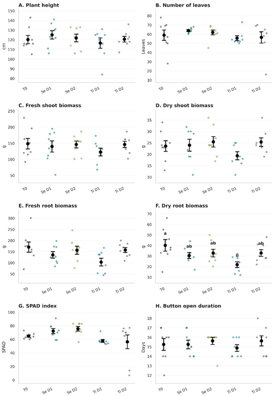
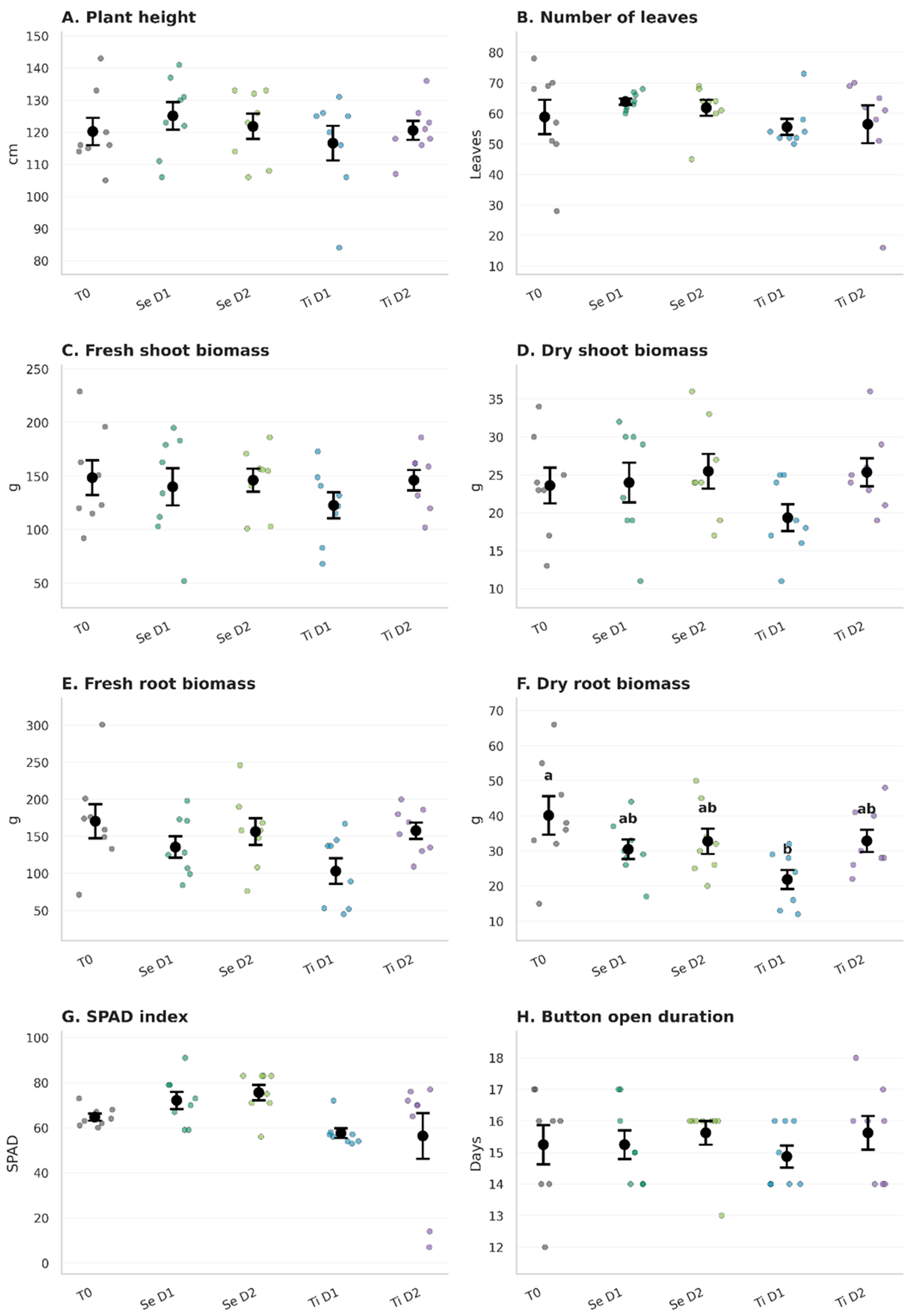
Figure 1 summarizes physiological responses to selenium and titanium treatments, with complete statistical results provided in the Supplementary Material (Tables S1 and S2). Overall, treatment had limited effects on vegetative growth, as plant height, number of leaves, fresh and dry aboveground biomass, fresh belowground biomass, and button open duration did not differ significantly among treatments (p > 0.05).
Figure 1.
Physiological responses of plants subjected to selenium and titanium treatments. Panels show (A) plant height, (B) number of leaves, (C) fresh aboveground biomass, (D) dry aboveground biomass, (E) fresh belowground biomass, (F) dry belowground biomass, (G) SPAD chlorophyll index, and (H) Button open duration. Individual observations are represented by points, and bars indicate mean ± standard error (SE). Different lowercase letters indicate significant differences among treatments according to Tukey’s HSD test (p < 0.05); letters are shown only when significant pairwise differences were detected.
Dry belowground biomass showed the clearest treatment response (Figure 1F), with a significant treatment effect (F4,30 = 3.101, p = 0.030, η2p = 0.293). Tukey’s HSD test showed significantly lower dry belowground biomass in TiD1 than in the untreated control (T0), whereas SeD1, SeD2, and TiD2 did not differ significantly from T0 or from each other. Fresh belowground biomass showed a similar numerical tendency but did not reach statistical significance (F4,30 = 2.329, p = 0.079).
Leaf chlorophyll status, estimated using the SPAD index, was also affected by treatment (F4,30 = 2.962, p = 0.036, η2p = 0.283; Figure 1G). However, Tukey’s HSD test did not identify significant pairwise differences among treatments. Thus, although the overall model indicated variation in SPAD values among treatments, no individual treatment was statistically distinguishable after adjustment for multiple comparisons.
Sampling time did not significantly affect any of the evaluated physiological variables (p = 0.190–0.930), and no significant treatment × sampling time interactions were detected (p = 0.172–0.594), indicating that treatment responses were generally consistent between 46 and 69 days after application.
3.2. Univariate Anatomical Responses to Nanoparticle Treatments
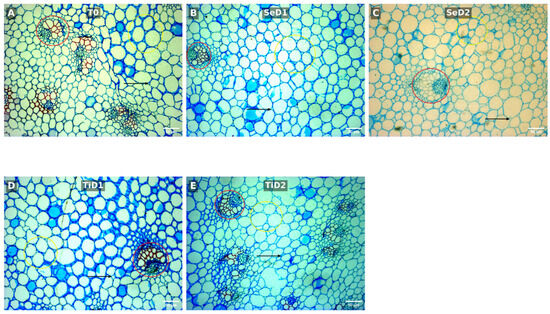
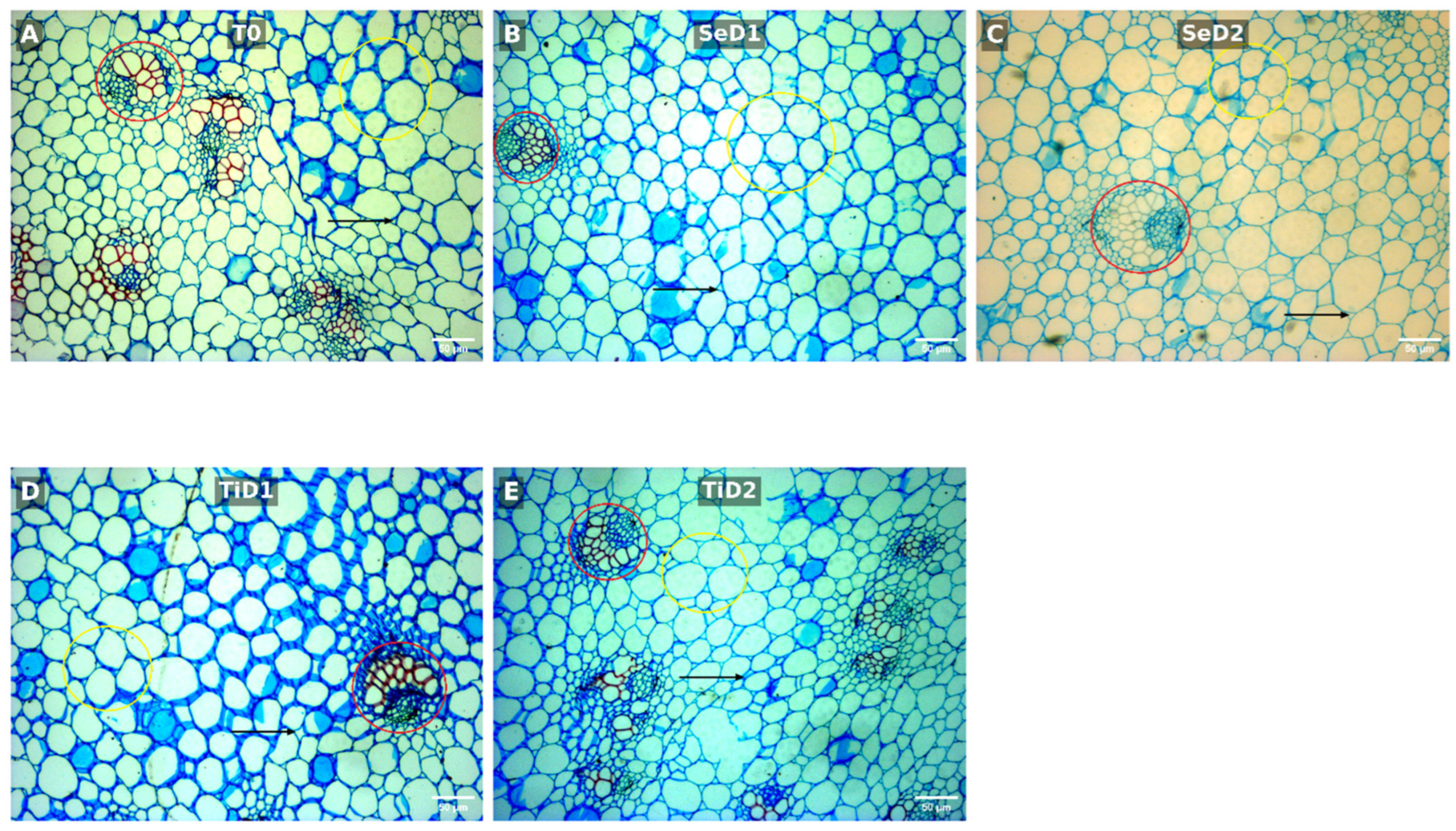
Figure 2 shows representative micrographs of the parenchyma, intercellular spaces, and vascular tissue evaluated across treatments. Complete morphometric data and univariate statistical results are provided in Supplementary Material (Tables S4 and S5).
Figure 2.
Representative micrographs of transverse stem sections from Lilium cv. Concador subjected to the different nanoparticle treatments: (A) T0, control treatment; (B) SeD1, low-dose SeNP treatment; (C) SeD2, high-dose SeNP treatment; (D) TiD1, low-dose TiO2NP treatment; and (E) TiD2, high-dose TiO2NP treatment. One representative field is shown for each treatment; quantitative morphometric analyses were based on six independent biological replicates per treatment (n = 6; 30 micrographs in total). The anatomical regions evaluated by morphometric analysis are indicated: vascular bundles (red circles), parenchyma cells (yellow circles), and intercellular spaces (arrows). Images were acquired at 4× objective magnification. Scale bar = 50 μm.
Parenchyma morphology varied by treatment, particularly in tissue occupation and cell dimensions. Parenchyma area percentage differed significantly among treatments (p = 0.0069, η2 = 0.4908), with the highest value observed in SeD2 (54.41 ± 5.72%) and the lowest in TiD1 (33.40 ± 9.08%). Post hoc comparisons showed that SeD2 had a significantly greater area percentage than SeD1 (p = 0.0337), TiD1 (p = 0.0052), and TiD2 (p = 0.0266). Treatment also affected minor axis length (p = 0.0305, η2 = 0.3993), ranging from 24.82 ± 2.84 µm in SeD2 to 19.04 ± 0.92 µm in TiD2, with a significant difference between these treatments (p = 0.0256). A similar response was observed for minimum Feret diameter (p = 0.0491, η2 = 0.3658), which was greater in SeD2 (25.42 ± 2.87 µm) than in TiD2 (20.09 ± 1.00 µm; p = 0.0386). Other parenchyma traits, including average cell size, perimeter, major axis, circularity, solidity, and Feret diameter, did not show significant overall treatment effects (p > 0.05).
Intercellular spaces also exhibited significant treatment responses. Absolute intercellular space area differed among treatments (p = 0.0375, η2 = 0.3851), with the highest value in SeD2 (143,746.88 ± 30,160.47 µm2) and the lowest in TiD2 (74,636.84 ± 12,046.51 µm2); these treatments differed significantly in the post hoc comparison (p = 0.0173). The percentage of tissue occupied by intercellular spaces also differed significantly (Kruskal–Wallis, p = 0.0039, ε2 = 0.5698). In this case, TiD1 showed the highest proportional area (31.15 ± 2.58%), whereas T0 showed the lowest (20.14 ± 3.04%). Dunn’s post hoc test identified significant differences between TiD1 and SeD2 (p = 0.0147) and between TiD1 and T0 (p = 0.0178), indicating a distinct response of proportional intercellular space under the lower titanium dose.
The most pronounced anatomical responses were observed in vascular tissue. Vascular bundle area differed significantly among treatments (p = 0.0019, η2 = 0.5584), ranging from 7351.42 ± 3686.82 µm2 in SeD1 to 16,271.44 ± 4224.61 µm2 in SeD2. Vascular bundle area was significantly greater in SeD2 than in SeD1 (p = 0.0009), TiD1 (p = 0.0202), and TiD2 (p = 0.0313). The major axis showed a comparable response (p = 0.0064, η2 = 0.4950), with values ranging from 105.30 ± 34.07 µm in SeD1 to 162.87 ± 17.64 µm in SeD2; the significant pairwise difference occurred between SeD2 and SeD1 (p = 0.0028). Similarly, minor axis length differed among treatments (p = 0.0059, η2 = 0.4999), with SeD2 showing the highest value (125.61 ± 24.17 µm) and SeD1 the lowest (84.67 ± 16.54 µm; p = 0.0027).
Vascular ratio followed the same treatment pattern (p = 0.0019, η2 = 0.5584), with the highest value in SeD2 (20.09 ± 5.22) and the lowest in SeD1 (9.08 ± 4.55). SeD2 differed significantly from SeD1 (p = 0.0009), TiD1 (p = 0.0202), and TiD2 (p = 0.0313). In contrast, vascular area percentage, circularity, aspect ratio, and solidity did not differ significantly among treatments (p > 0.05). Overall, SeD2 showed a consistent pattern of higher values across several related parenchyma and vascular descriptors, whereas titanium treatments exhibited distinct responses, particularly in parenchyma dimensions and proportional intercellular space. Given the exploratory nature of the morphometric survey and the number of descriptors evaluated, these patterns were interpreted collectively rather than based on individual significant variables.
3.3. Effects of Nanoparticle Type, Dose and Their Interaction
Factorial ANOVA revealed that the anatomical response to nanoparticle application was influenced by nanoparticle type, dose, and their interaction, although the relative contribution of these factors differed between parenchyma and vascular tissue (Table S5, Supplementary Material).
In parenchyma, nanoparticle type significantly affected average cell size (p = 0.0324), area percentage (p = 0.0064), minor axis length (p = 0.0161), and minimum Feret diameter (p = 0.0191). Dose had a more restricted effect and significantly influenced only parenchyma area percentage (p = 0.0091). A significant nanoparticle type × dose interaction was detected for total parenchyma area (p = 0.0146), indicating that changes in this trait with increasing dose differed between selenium and titanium nanoparticles. Together with the treatment-level comparisons described above, these results indicate that parenchyma responses were primarily associated with nanoparticle composition, with additional dose-dependent effects on tissue occupation and total area.
A different pattern was observed in vascular tissue. Nanoparticle type alone did not significantly affect any of the evaluated vascular traits, whereas dose significantly influenced vascular bundle area (p = 0.0029), major axis (p = 0.0101), minor axis (p = 0.0058), and vascular ratio (p = 0.0029). Importantly, significant nanoparticle type × dose interactions were detected for vascular bundle area (p = 0.0053), major axis (p = 0.0105), minor axis (p = 0.0159), and vascular ratio (p = 0.0053). Thus, the effect of increasing nanoparticle dose on vascular dimensions depended on the material applied rather than representing a uniform dose response. This interaction was consistent with the treatment-level analysis, in which SeD2 showed the greatest vascular bundle area and dimensions, whereas TiD2 did not exhibit a comparable increase.
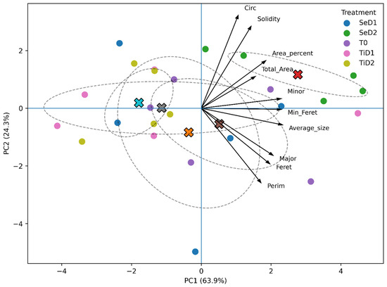
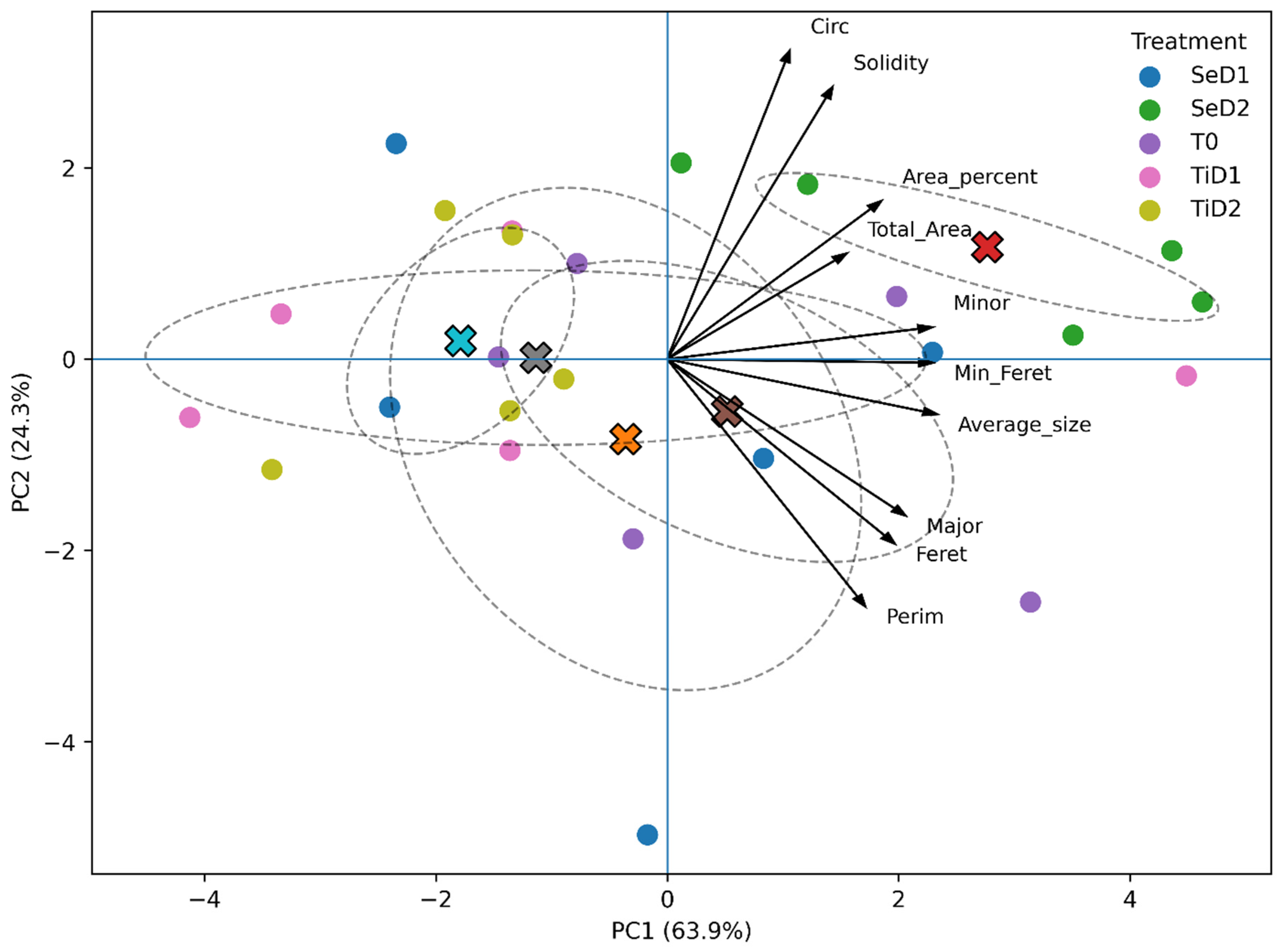
Multivariate analyses provided complementary support for these anatomical patterns (Figures S1 and S2, Supplementary Material). For parenchyma traits, the first two principal components explained 88.24% of the total variation (PC1 = 63.90%; PC2 = 24.34%, Figure 3). PC1 was predominantly associated with cell-size-related variables, including average size, minor axis, and minimum Feret diameter, and SeD2 tended to separate from the remaining treatments along the positive side of this component. This pattern was consistent with the greater parenchyma area percentage and cell dimensions observed for SeD2 in the univariate analyses.
Figure 3.
Principal component analysis (PCA) of parenchyma cell morphometric traits. PCA biplot based on ten morphometric variables measured in parenchyma cells. Colored circles represent individual observations grouped by treatment, while crosses (×) indicate treatment centroids: red, SeD2; blue, TiD2; gray, TiD1; orange, SeD1; and brown, T0. Dashed ellipses represent the dispersion of observations within each treatment. Arrows indicate the direction and contribution (loadings) of individual morphometric variables to the principal components. PC1 and PC2 explain 63.90% and 24.34% of the total variance, respectively.
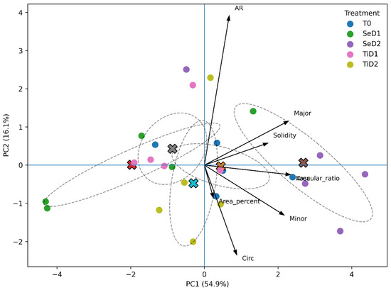
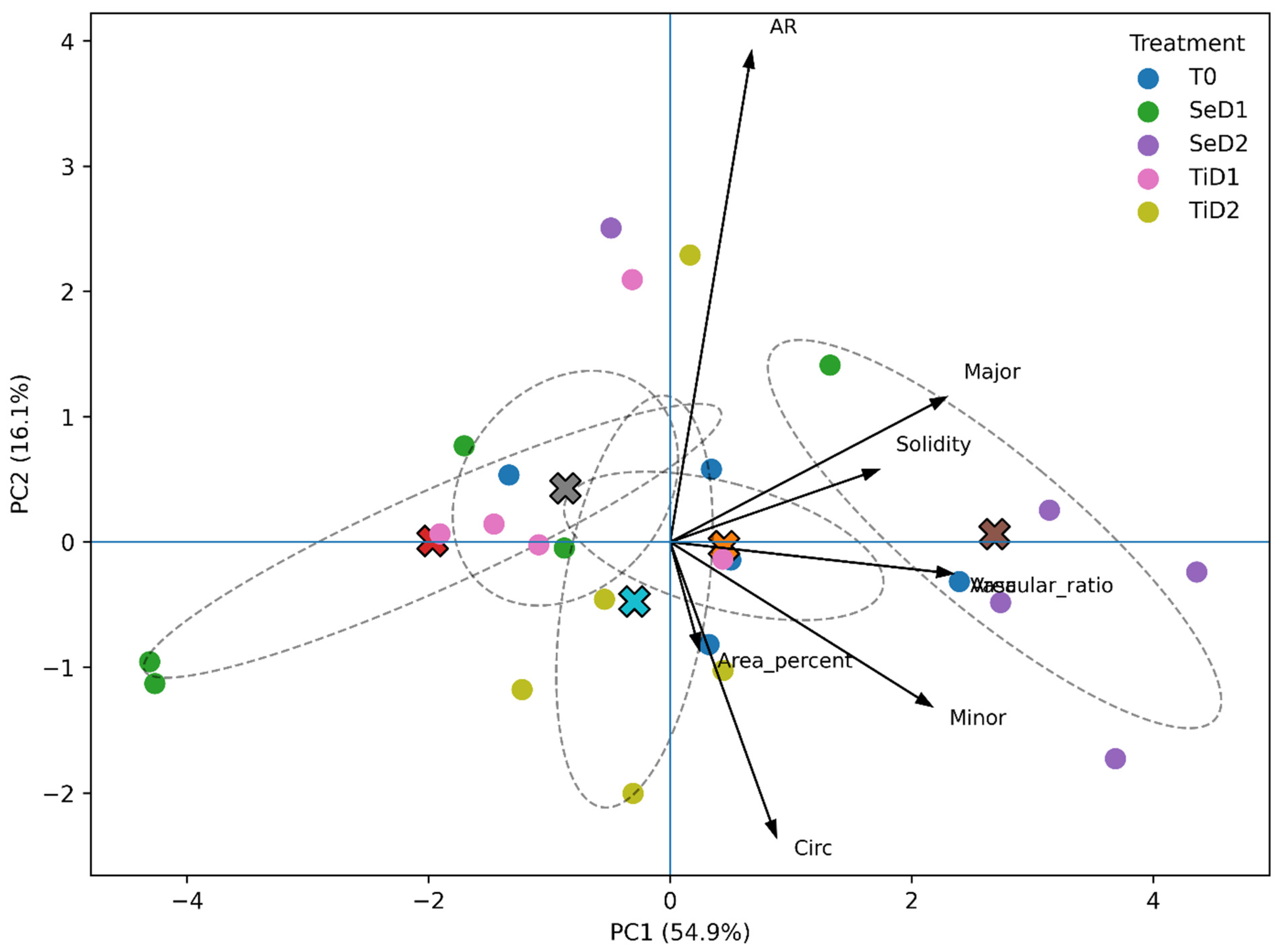
For vascular tissue (Figure 4), PC1 and PC2 explained 54.86% and 16.07% of the total variation, respectively, accounting for 70.93% of the cumulative variance. Vascular bundle area, vascular ratio, major axis, and minor axis contributed strongly to PC1, whereas aspect ratio was primarily associated with PC2. SeD2 was positioned toward the positive side of PC1, consistent with its larger vascular bundle dimensions. Thus, both the factorial and multivariate analyses indicated that the anatomical response was not simply proportional to nanoparticle dose but depended on the combination of nanoparticle composition and concentration.
Figure 4.
Principal component analysis (PCA) of vascular tissue morphometric traits. PCA biplot based on vascular bundle morphometric variables. Colored circles represent individual observations grouped by treatment, while crosses (×) indicate treatment centroids: red, SeD2; blue, TiD2; gray, TiD1; orange, SeD1; and brown, T0. Dashed ellipses represent the dispersion of observations within each treatment. Arrows indicate the direction and contribution (loadings) of individual morphometric variables to the principal components. PC1 and PC2 account for 54.86% and 16.07% of the total variance, respectively.
4. Discussion
The results are consistent with the hypothesis that foliar application of SeNPs and TiO2NPs induces dose-dependent physiological and anatomical responses in Lilium cv. Concador without necessarily increasing vegetative growth. Variables such as plant height, number of leaves, shoot biomass, and duration of flower opening did not differ statistically between treatments, indicating that neither nanoparticle type significantly affected overall vegetative or reproductive development under the evaluated conditions (Figure 1). Notably, this growth stability contrasted with the treatment-dependent responses observed in root biomass and stem anatomy, suggesting that nanoparticle effects were more clearly manifested at the belowground and tissue-organization levels than in overall plant growth [17]. Among the physiological variables evaluated, relative chlorophyll content and root biomass showed the greatest sensitivity to nanoparticle application (Table S2). Although ANOVA detected a significant treatment effect on SPAD values, multiple comparisons did not identify differences between individual treatments (Table S1), suggesting that the set of applications modified the chlorophyll-related physiological state, but that the magnitude of individual responses was relatively small after statistical correction (Figure S2). This behavior is consistent with the sensitivity of chlorophyll metabolism to small modifications of the cellular redox state [18]. Selenium has been widely described as an element that preserves chloroplast integrity, decreases lipid peroxidation, and maintains photosystem II stability by strengthening the antioxidant system [19]. Rather than drastically increasing chlorophyll concentration, selenium tends to preserve photosynthetic machinery by reducing oxidative pigment degradation, consistent with the overall stability observed in the SPAD values in this study [20,21]. TiO2 has shown variable effects on chlorophyll content, depending on foliar absorption, photocatalytic properties, and the applied dose [22].
In contrast, the most evident physiological response was observed in the root system. Root dry biomass decreased significantly only in the TiD1 treatment, while both selenium treatments and the high-dose titanium dioxide (TiD2) maintained values statistically comparable to the control (Figure 1). Interestingly, fresh root biomass showed a similar trend, though it did not reach statistical significance despite a large effect size (Table S2). These results indicate that titanium nanoparticles induced a dose-dependent response in root biomass accumulation, with low doses producing a greater adverse effect than high doses [23]. This nonlinear response has been documented for different types of nanoparticles and has been associated with hormetic or compensatory processes rather than proportional concentration-dependent effects. However, this study did not measure oxidative signaling, antioxidant activity, or carbon allocation. Therefore, the mechanisms underlying the contrasting responses of TiD1 and TiD2 remain to be determined [24,25,26]. Consequently, the observed response is more consistent with a treatment-dependent adjustment than with generalized phytotoxicity (Figure S5). The response of root biomass and stem anatomy following foliar application suggests that the treatments’ effects were not restricted to the application site. Foliar-absorbed nanoparticles or their dissolved ionic species may be redistributed through vascular pathways, while nanoparticle perception in leaves may also trigger systemic signals that affect distant tissues [15]. In the present study, the treatment-dependent response of root biomass together with the remodeling of stem parenchymal and vascular tissues is consistent with a systemic response to foliar application. However, nanoparticle uptake, elemental accumulation, and translocation were not directly quantified; therefore, the present results cannot distinguish between direct nanoparticle redistribution and indirect systemic signaling. Future studies combining quantitative anatomy with elemental localization and translocation analyses will be necessary to determine the mechanism underlying these responses.
Among the tissues analyzed, cortical parenchyma showed the most evident anatomical response to nanoparticle application, highlighting its high plasticity in response to changes in the cellular environment. Unlike highly specialized tissues, parenchyma cells retain a significant capacity for cell wall expansion and remodeling, allowing them to respond rapidly to changes in the plant’s physiological state [27]. In this study, changes in tissue occupancy percentage, minor axis, and minimum Feret diameter indicate treatment-dependent differences in cell size and spatial tissue organization (Figure S5). However, the cellular processes underlying these morphometric changes were not directly evaluated.
The observed response clearly depended on both nanoparticle type and applied dose (Table S4). While SeD2 promoted a consistent increase in variables associated with cell occupancy and size, titanium treatments, particularly TiD2, showed lower values in most morphometric descriptors (Figure 2). The contrasting morphometric profiles of SeD2 and TiD2 indicate that stem anatomical remodeling was dependent on both nanoparticle composition and dose. SeD2 was consistently associated with greater parenchymal occupation and vascular dimensions, whereas TiO2 treatments produced less pronounced or contrasting structural responses. Importantly, these anatomical differences occurred without corresponding increases in shoot biomass, indicating that nanoparticle-induced remodeling involved changes in internal tissue organization rather than proportional enhancement of whole-plant growth [28,29,30]. Notably, the increase in parenchyma occupancy percentage observed under SeD2 was not accompanied by increases in shoot biomass (Figure S5). This result suggests that anatomical modifications do not necessarily imply greater organ growth but rather an internal reorganization of the organ’s cellular architecture [31]. In ornamental species, greater parenchymal development could contribute to temporary water and carbohydrate storage and, consequently, to stem turgor maintenance [32]. However, this study did not evaluate postharvest water relations, vase life, or wilt resistance; therefore, the functional significance of the observed parenchymal remodeling for postharvest performance remains to be experimentally determined.
Although the present study did not evaluate postharvest performance, the observed anatomical reorganization is consistent with functions previously attributed to parenchyma in herbaceous species [33]. In ornamental species such as Lilium, stem firmness, flower bud hydration, and uniformity of flower opening are relevant attributes of commercial quality [34]. Therefore, determining whether vascular remodeling contributes to these functional traits represents an important direction for future research [35]. In contrast to the storage and water-regulation functions associated with parenchyma, vascular tissue primarily transports water, mineral nutrients, and photoassimilates and also contributes to mechanical support of the stem [36]. In this study, the modifications observed in vascular bundle area, vascular ratio, and major and minor axis dimensions indicate a treatment-dependent reorganization of the conducting system, particularly under SeD2, which showed the highest values for these variables (Figure S6). Together, these modifications indicate coordinated remodeling of the vascular tissue rather than an isolated change in a single morphometric descriptor [37].
The morphometric analysis should be interpreted as exploratory because multiple anatomical descriptors were evaluated independently without applying a global correction across the complete family of outcomes. Although post hoc comparisons were adjusted within each variable, testing multiple morphometric endpoints increases the probability of type I error. Consequently, isolated significant descriptors, particularly those close to the significance threshold, should be interpreted cautiously. Greater emphasis should therefore be placed on coherent responses observed across related anatomical traits, such as the concurrent changes in vascular bundle area, major and minor axes, and vascular ratio, rather than on individual significant variables alone.
The greater vascular dimensions observed under SeD2 indicate a treatment-dependent remodeling of the conducting tissue. Redox and hormonal signaling have been associated with vascular differentiation [38]; however, these pathways were not evaluated in the present study. Therefore, the observed morphometric pattern supports structural remodeling of vascular tissues but does not establish the biochemical or molecular mechanism responsible for this response [39,40]. This interpretation was also supported by principal component analysis, in which SeD2 was grouped with variables associated with increased vascular bundle area, vascular ratio, and major and minor axis dimensions, indicating that the observed remodeling corresponded to an integrated structural response and not to isolated modifications of a single morphometric descriptor (Figure 3 and Figure 4). Greater vascular dimensions could influence water and photoassimilate transport [41]; however, this study did not measure hydraulic conductivity or postharvest water relations. Therefore, the functional consequences of vascular remodeling for stem hydration, floral development, and postharvest performance remain hypothetical and should be evaluated directly in future studies [42].
In addition to providing evidence on Lilium’s response to selenium and titanium nanoparticles, this study constitutes, to our knowledge, the first analysis to integrate physiological evaluation with quantitative anatomical morphometry of the stem in this species. This approach demonstrated that the chemical composition of the nanomaterial and the applied dose determine distinct structural response patterns, information that could hardly be obtained solely from conventional agronomic variables. Consequently, the results provide a basis for future studies aimed at relating these anatomical modifications to functional variables, such as hydraulic conductivity, photosynthetic efficiency, vase life, and tolerance to biotic or abiotic stresses. Overall, the results broaden our understanding of how nano-inputs interact with ornamental plants by showing that treatment-dependent anatomical responses can occur even when conventional growth variables remain unchanged. Quantitative morphometry revealed consistent modifications in parenchymal and vascular organization, supporting quantitative stem anatomy as a complementary approach for characterizing plant responses to nano-inputs [43]. Future studies should determine whether these structural changes are associated with subsequent functional or agronomic responses.
5. Conclusions
Foliar application of selenium nanoparticles (SeNPs) and titanium dioxide (TiO2NPs) induced distinct physiological and anatomical responses in Lilium cv. Concador without compromising vegetative growth or reproductive development. Although most agronomic variables remained stable, quantitative stem morphoanatomy revealed significant modifications, depending on nanoparticle type and dose, in the organization of parenchyma and vascular tissue, demonstrating that structural adjustments can occur before being detected by conventional agronomic variables. This study constitutes the first quantitative analysis demonstrating nanoparticle-induced stem anatomical remodeling in Lilium, providing new evidence on the mechanisms that regulate the interaction between nano-inputs and ornamental plants. Taken together, the results demonstrate that quantitative stem anatomy represents a highly sensitive tool for identifying early responses to nanoparticle application and provide a scientific basis for the development and optimization of nano-inputs aimed at improving the quality and performance of high-value ornamental crops.
Supplementary Materials
The following supporting information can be downloaded at https://www.mdpi.com/article/10.3390/ijpb17090083/s1, Figure S1: Heatmap showing standardized physiological responses of plants under the five experimental treatments; Figure S2. Pearson correlation matrix among physiological variables measured during the experiment; Figure S3. Representative histological micrographs of transverse stem sections from Lilium cv. Concador plants subjected to the SeD2 treatment; Figure S4. Principal component analysis (PCA) of physiological traits measured under the five experimental treatments; Figure S5. Correlation matrices of morphometric variables in parenchyma cells and vascular tissue; Figure S6. Radar plots summarizing treatment-specific morphometric profiles. Table S1: Summary of two-way analysis of variance (ANOVA) evaluating the effects of treatment, sampling time, and their interaction on plant physiological variables. Table S2. Effect sizes estimated as partial eta squared (η2p) for treatment, sampling time, and their interaction; Table S3. Summary of univariate statistical analyses. Results of one-way ANOVA or Kruskal–Wallis tests performed according to the distributional assumptions of each variable; Table S4. Morphometric responses of parenchyma cells, intercellular spaces and vascular tissue under selenium and titanium nanoparticle treatments; Table S5. Factorial ANOVA evaluating the effects of nanoparticle type, dose and their interaction on anatomical traits.
Author Contributions
Conceptualization, S.R.P.-R. and I.H.-S.; Data curation, N.T.S.-G., C.R.D.-G., and I.H.-S.; Formal analysis, C.R.D.-G. and I.H.-S.; Investigation, C.R.D.-G., M.S.-G., S.R.P.-R., M.F.C.-G., M.I.R.-S., M.V.-F. and I.H.-S.; Methodology, C.R.D.-G., N.T.S.-G., F.F.-L., M.F.C.-G., J.O.-L. and I.H.-S.; Resources, S.R.P.-R., M.I.R.-S., M.S.-G., F.F.-L., M.F.C.-G., J.O.-L. and M.V.-F.; Software, C.R.D.-G., N.T.S.-G. and I.H.-S.; Supervision, S.R.P.-R. and I.H.-S.; Validation, C.R.D.-G. and I.H.-S.; Visualization, C.R.D.-G., N.T.S.-G. and I.H.-S.; Writing—original draft, C.R.D.-G., N.T.S.-G. and I.H.-S.; Writing—review and editing, C.R.D.-G.; N.T.S.-G. and I.H.-S. All authors have read and agreed to the published version of the manuscript.
Funding
This research received no external funding.
Data Availability Statement
The data on physiological variables and the statistical analyses are provided as Supplementary Materials.
Acknowledgments
The authors acknowledge the Universidad Autónoma del Estado de Hidalgo (UAEH) for allowing us to conduct the study at its facilities.
Conflicts of Interest
The authors declare no conflicts of interest.
References
- Zhou, J.; An, R.; Huang, X. Genus Lilium: A review on traditional uses, phytochemistry, and pharmacology. J. Ethnopharmacol. 2021, 270, 113852. [Google Scholar] [CrossRef] [Scilit] [PubMed]
- Gómez-Santos, M.; González-García, Y.; Pérez-Álvarez, M.; Cadenas-Pliego, G.; Juárez-Maldonado, A. Impact of calcium–silicon nanoparticles on flower quality and biochemical traits of Lilium under salinity stress. Plant Stress 2023, 10, 100270. [Google Scholar] [CrossRef] [Scilit]
- El-Sayed, I.M.; El-Ziat, R.A. Utilization of environmentally friendly essential oils to enhance postharvest characteristics of Chrysanthemum morifolium cut flowers. Heliyon 2021, 7, e05909. [Google Scholar] [CrossRef] [Scilit] [PubMed]
- Sánchez-Valdés, S.; Rodríguez-González, J.A.; Sánchez-Martínez, A.C.; Bustos, K.; Cruz, M.V.; Martínez, L.C.; Torres-González, J.; Ramírez-Barron, S.N. Trends in the use of nanoparticles in agriculture. Rev. Latinoam. De. Difus. Científica 2024, 6, 20–39. [Google Scholar] [CrossRef]
- Miranda, P.S.; Moreira, A.C.; Uribe, L.R.; Castro, J.C. Effect of copper and selenium nanoparticles on Capsicum annuum at the nursery stage. Rev. De. Investig. Científica TSE DE 2025, 8, 1–22. [Google Scholar] [CrossRef] [Scilit]
- Hernández-Hernández, H.; Quiterio-Gutiérrez, T.; Cadenas-Pliego, G.; Ortega-Ortiz, H.; Hernández-Fuentes, A.D.; Cabrera de la Fuente, M.; Valdés-Reyna, J.; Juárez-Maldonado, A. Impact of selenium and copper nanoparticles on yield, antioxidant system, and fruit quality of tomato plants. Plants 2019, 8, 355. [Google Scholar] [CrossRef] [Scilit] [PubMed]
- Ghareeb, A.; Fouda, A.; Kishk, R.M.; El Kazzaz, W.M. Unlocking the potential of titanium dioxide nanoparticles: Green synthesis, optimization, characterization, and multifunctional applications. Microb. Cell Factories 2024, 23, 341. [Google Scholar] [CrossRef] [Scilit] [PubMed]
- Araya-Mattey, J.; Sandoval, J.; Rojas-Carrillo, O. Nanomaterials in agricultural applications: Recent advances in the banana agroindustry. Rev. Tecnol. En. Marcha 2024, 37, 100–113. [Google Scholar] [CrossRef]
- Sánchez-Granados, N.T.; Pérez-Ríos, S.R.; González-García, Y.; Fernández-Luqueño, F.; Aquino-Torres, E.; Saucedo-García, M.; Zaldívar-Ortega, A.K.; Reyes-Santamaria, M.I.; Hernández-Soto, I. Impacts of foliar application of selenium and TiO2 nanoparticles on growth, development, and flowering in Lilium Sunny Oriental. Int. J. Plant Biol. 2025, 16, 103. [Google Scholar] [CrossRef] [Scilit]
- INEGI (Instituto Nacional de Estadística, Geografía e Informática). Anuario Estadístico y Geográfico del Estado de Hidalgo, México; Gobiernodel Estado de Hidalgo: Higalgo, Mexico, 2017; Volume 1, p. 1674. Available online: https://www.inegi.org.mx/contenido/productos/prod_serv/contenidos/espanol/bvinegi/productos/nueva_estruc/anuarios_2017/702825095093.pdf (accessed on 1 December 2025).
- González-Lemus, U.; Medina-Pérez, G.; Espino-García, J.J.; Fernández-Luqueño, F.; Campos-Montiel, R.; Almaraz-Buendía, I.; Reyes-Munguía, A.; Urrutia-Hernández, T. Nutritional parameters, biomass production, and antioxidant activity of Festuca arundinacea conditioned with selenium nanoparticles. Plants 2022, 11, 2326. [Google Scholar] [CrossRef] [Scilit] [PubMed]
- Torres-Gómez, A.; Sarabia-Castillo, C.R.; Juárez-Altamirano, R.; Fernández-Luqueño, F. Effect of a native strain-based biofertilizer with TiO2, ZnO, FexOx, and Ag nanoparticles on wheat yield. Agriculture 2025, 15, 2093. [Google Scholar] [CrossRef] [Scilit]
- Larue, C.; Veronesi, G.; Flank, A.M.; Surble, S.; Herlin-Boime, N.; Carrière, M. Comparative uptake and impact of TiO2 nanoparticles in wheat and rapeseed. J. Toxicol. Environ. Health A 2012, 75, 722–734. [Google Scholar] [CrossRef] [Scilit] [PubMed]
- Satti, S.H.; Raja, N.I.; Javed, B.; Akram, A.; Mashwani, Z.U.; Ahmad, M.S.; Ikram, M. Titanium dioxide nanoparticles elicited agro-morphological and physicochemical modifications in wheat plants to control Bipolaris sorokiniana. PLoS ONE 2021, 11, e0246880. [Google Scholar] [CrossRef] [Scilit] [PubMed]
- Soriano Melgar, L.A.A.; López-Guerrero, A.G.; Cortéz-Mazatan, G.; Mendoza-Mendoza, E.; Peralta-Rodríguez, R.D. Zinc oxide and zinc oxide/graphene nanoparticles in vase solutions extend postharvest life of lisianthus. Agroproductividad 2018, 11, 137–144. [Google Scholar]
- Lin, H.K.; Wei, T.Y.; Chen, C.M.; Yeh, D.M. Relationship between phloem fiber and trailing habit, and independent inheritance of growth habit and flower form in periwinkle. J. Am. Soc. Hortic. Sci. 2018, 143, 67–71. [Google Scholar] [CrossRef] [Scilit]
- Djanaguiraman, M.; Anbazhagan, V.; Dhankher, O.P.; Prasad, P.V. Uptake, translocation, toxicity, and impact of nanoparticles on plant physiological processes. Plants 2024, 13, 3137. [Google Scholar] [CrossRef] [Scilit] [PubMed]
- Xu, X.; Meng, C.; Zhan, J.; Wang, Q.; Zhang, M.; Wang, C.; Zhao, X. Nanoparticle-plant interactions: Uptake, transport, physiological effects, and environmental implications. J. Hazard. Mater. Adv. 2026, 22, 101123. [Google Scholar] [CrossRef] [Scilit]
- Croce, A.C. Light and autofluorescence, multitasking features in living organisms. Photochem 2021, 1, 67–124. [Google Scholar] [CrossRef] [Scilit]
- Liu, H.; Xiao, C.; Qiu, T.; Deng, J.; Cheng, H.; Cong, X.; Zhang, Y. Selenium regulates antioxidant, photosynthesis, and cell permeability in plants under various abiotic stresses: A review. Plants 2022, 12, 44. [Google Scholar] [CrossRef] [Scilit] [PubMed]
- Lara-Izaguirre, A.Y.; Rojas-Velázquez, A.N.; Alcalá-Jáuregui, J.A.; Alia-Tejacal, I. Efecto del selenio en el crecimiento de plantas y calidad nutracéutica de frutos de berenjena. Rev. Fitotec. Mex. 2025, 48, 183–190. [Google Scholar] [CrossRef] [Scilit]
- Fuziki, M.E.; Tusset, A.M.; dos Santos, O.A.; Lenzi, G.G. Chlorophyll sensitization of TiO2: A mini-review. Reactions 2023, 4, 766–778. [Google Scholar] [CrossRef] [Scilit]
- Trela-Makowej, A.; Orzechowska, A.; Szymańska, R. Less is more: The hormetic effect of titanium dioxide nanoparticles on plants. Sci. Total Environ. 2024, 910, 168669. [Google Scholar] [CrossRef] [Scilit] [PubMed]
- Agathokleous, E.; Calabrese, E.J.; Barceló, D. Environmental hormesis: New developments. Sci. Total Environ. 2024, 906, 167450. [Google Scholar] [CrossRef] [Scilit] [PubMed]
- Calabrese, E.J.; Blain, R.B. Hormesis and plant biology. Environ. Pollut. 2009, 157, 42–48. [Google Scholar] [CrossRef] [Scilit] [PubMed]
- Tighe-Neira, R.; Reyes-Díaz, M.; Nunes-Nesi, A.; Recio, G.; Carmona, E.; Corgne, A.; Inostroza-Blancheteau, C. Titanium dioxide nanoparticles provoke transient increase in photosynthetic performance and differential response in antioxidant system in Raphanus sativus L. Sci. Hortic. 2020, 269, 109418. [Google Scholar] [CrossRef] [Scilit]
- Secchi, F.; Pagliarani, C.; Zwieniecki, M.A. The functional role of xylem parenchyma cells and aquaporins during recovery from severe water stress. Plant Cell Environ. 2017, 40, 858–871. [Google Scholar] [CrossRef] [Scilit] [PubMed]
- Delmer, D.; Dixon, R.A.; Keegstra, K.; Mohnen, D. The plant cell wall—Dynamic, strong, and adaptable—Is a natural shapeshifter. Plant Cell 2024, 36, 1257–1311. [Google Scholar] [CrossRef] [Scilit] [PubMed]
- Rodríguez-Ortiz, J.C.; García-Morales, S.; Flores-Flores, M.D.C.; Chávez-Zárate, Y.; Camarillo-Morelos, J.G.; Estrada-González, Á.D.J. Nanopartículas de selenio y selenito en la producción de plántulas de lechuga. Rev. Mex. DE Cienc. Agríc. 2026, 17, e4068. [Google Scholar] [CrossRef] [Scilit]
- Lyu, S.; Wei, X.; Chen, J.; Wang, C.; Wang, X.; Pan, D. Titanium as a beneficial element for crop production. Front. Plant Sci. 2017, 8, 597. [Google Scholar] [CrossRef] [Scilit] [PubMed]
- Waldron, K.W. Plant structure and fruit and vegetable texture. In Texture in Food: Solid Foods; Woodhead Publishing: Cambridge, UK, 2004; Volume 2, pp. 241–258. [Google Scholar] [CrossRef] [Scilit]
- Lemoine, R.; Camera, S.L.; Atanassova, R.; Dédaldéchamp, F.; Allario, T.; Pourtau, N.; Bonnemain, J.-L.; Laloi, M.; Coutos-Thévenot, P.; Maurousset, L.; et al. Source-to-sink transport of sugar and regulation by environmental factors. Front. Plant Sci. 2013, 4, 272. [Google Scholar] [CrossRef] [Scilit] [PubMed]
- Rodríguez, M. Las suculentas y cactáceas en armonía con el agua. Cuad. Méd. Soc. 2022, 62, 79–80. [Google Scholar] [CrossRef] [Scilit]
- Rios-Florida, L.G.; De La Cruz-Guzmán, G.H.; Arriaga-Frías, A.; Mandujano-Piña, M. Efecto de paclobutrazol y Glomus intraradices en el cultivo de Lilium cv. Armandale y Tresor. Siembra 2022, 9, e3969. [Google Scholar] [CrossRef] [Scilit]
- Alemán-Sancheschúlz, G.; Solano, E.; López-Portillo, J. La arquitectura hidráulica de las plantas vasculares terrestres: Una revisión. Madera Y Bosques 2019, 25, e2531828. [Google Scholar] [CrossRef] [Scilit]
- Acosta-Motos, J.R.; Ortuño, M.F.; Bernal-Vicente, A.; Diaz-Vivancos, P.; Sanchez-Blanco, M.J.; Hernandez, J.A. Plant responses to salt stress: Adaptive mechanisms. Agronomy 2017, 7, 18. [Google Scholar] [CrossRef] [Scilit]
- Agustí, J.; Blázquez, M.A. Plant vascular development: Mechanisms and environmental regulation. Cell. Mol. Life Sci. 2020, 77, 3711–3728. [Google Scholar] [CrossRef] [Scilit] [PubMed]
- Naznin, A.; Wang, Y.; He, J.; Islam, M.M.; Abbas, A.; Bose, J.; Chen, Z.H. Auxin-mediated regulation and functional adaptation of leaf veins under heat stress. J. Plant Physiol. 2026, 317, 154702. [Google Scholar] [CrossRef] [Scilit] [PubMed]
- Achdev, S.; Ansari, S.A.; Ansari, M.I.; Fujita, M.; Hasanuzzaman, M. Abiotic stress and reactive oxygen species: Generation, signaling, and defense mechanisms. Antioxidants 2021, 10, 277. [Google Scholar] [CrossRef] [Scilit] [PubMed]
- Ramakrishnan, M.; Papolu, P.K.; Satish, L.; Vinod, K.K.; Wei, Q.; Sharma, A.; Emamverdian, A.; Zou, L.-H.; Zhou, M. Redox status of the plant cell determines epigenetic modifications under abiotic stress conditions and during developmental processes. J. Adv. Res. 2022, 42, 99–116. [Google Scholar] [CrossRef] [Scilit] [PubMed]
- Burgos, A.F.L.; Cortes, G.Y.B.; Pérez, A.L.B.; López, H.E.B. Distribución de fotoasimilados en los órganos vertederos de plantas Solanáceas, caso tomate y papa: Una revisión. Rev. Cienc. Y Agric. 2021, 18, 79–97. [Google Scholar] [CrossRef] [Scilit]
- Ďúranová, H.; Šimora, V.; Ďurišová, Ľ.; Olexiková, L.; Kovár, M.; Požgajová, M. Modifications in ultrastructural characteristics and redox status of plants under environmental stress: A review. Plants 2023, 12, 1666. [Google Scholar] [CrossRef] [Scilit] [PubMed]
- Becker, D.; Paulus, D. Calidad pre y post cosecha de tallos de gladíolo. Rev. DELOS 2023, 16, 699–715. [Google Scholar] [CrossRef] [Scilit]
Disclaimer/Publisher’s Note: The statements, opinions and data contained in all publications are solely those of the individual author(s) and contributor(s) and not of MDPI and/or the editor(s). MDPI and/or the editor(s) disclaim responsibility for any injury to people or property resulting from any ideas, methods, instructions or products referred to in the content. |
© 2026 by the authors. Licensee MDPI, Basel, Switzerland. This article is an open access article distributed under the terms and conditions of the Creative Commons Attribution (CC BY) license.