


Abstract
The gut microbiota plays an important role in immune homeostasis, allergic disease pathogenesis, and drug metabolism, potentially influencing therapeutic efficacy and safety. Conversely, pharmacotherapy may modify gut microbial composition and function, creating bidirectional host–drug–microbiome interactions that remain incompletely understood. This narrative review summarises current evidence on interactions between the microbiome and therapies for allergic diseases, focusing on antihistamines, corticosteroids, leukotriene receptor antagonists, and biologic agents. Evidence is evaluated from two complementary perspectives: an exposure axis, relevant mainly to orally administered drugs, and a response axis, particularly applicable to biologic therapies. Direct microbial biotransformation has been demonstrated most convincingly for glucocorticoids, with the organosteroid reductase ABC (OsrABC) pathway of Clostridium steroidoreducens reducing systemic prednisolone exposure in a gnotobiotic model. For H1-antihistamines and leukotriene receptor antagonists, evidence remains limited and inconsistent. The microbiota may also indirectly influence treatment outcomes through effects on epithelial barrier integrity, microbial metabolite production, and immune regulation. Although anti-allergic therapies can alter gut, airway, and skin microbial communities, no validated microbiome signature currently predicts treatment efficacy or safety. Future prospective studies integrating microbiome, metabolome, pharmacokinetic, pharmacodynamic, and clinical data are needed to establish reliable biomarkers and support personalised pharmacotherapy for allergic diseases.
1. Introduction
Allergic diseases represent a major public health challenge in the 21st century because of their high prevalence, chronic nature, and substantial impact on the quality of life of affected individuals and on healthcare systems. According to estimates from the Global Burden of Disease (GBD) Study, approximately 260 million individuals were living with asthma worldwide in 2021, while atopic dermatitis affected approximately 129 million individuals [1]. The total global burden of allergic diseases also includes allergic rhinitis, food allergy, urticaria, and other atopic disorders. The burden is particularly important in the paediatric population. According to data from the 2024 United States National Health Interview Survey, 29.5% of children aged under 18 years had at least one of the three diagnosed allergic conditions examined, including seasonal allergy, eczema, or food allergy [2]. In addition to their direct health burden, allergic diseases are associated with impaired quality of life, increased healthcare resource utilisation, and higher direct treatment costs. They are also associated with substantial indirect costs resulting from missed work or school days and reduced work and school productivity [3].
The gut microbiome is an important regulator of the development and function of the immune system and engages in complex bidirectional communication with the intestinal epithelium and cells of the mucosal and systemic immune systems. The gut microbiota and its metabolites influence the differentiation and functional activity of regulatory T cells (Treg cells) and effector T-helper cells, including Th1 and Th17 cells. Through these effects, they contribute to the balance between inflammatory responses and immune tolerance to commensal microorganisms and dietary antigens [4]. Among these metabolites, short-chain fatty acids (SCFAs), particularly acetate, propionate, and butyrate, are key mediators of microbiome–host communication. Produced through bacterial fermentation of nondigestible carbohydrates, SCFAs exert pleiotropic immunomodulatory effects by promoting Treg-cell differentiation and stability, strengthening epithelial barrier integrity, and modulating inflammatory signalling pathways. These effects are mediated, at least in part, through inhibition of histone deacetylases and regulation of FOXP3 expression. Their overall immunological impact, however, depends on the specific metabolite, its concentration, the tissue microenvironment, and the host immune status [5].
In addition to its immunoregulatory and barrier functions, the gut microbiome can influence the metabolism, efficacy, and safety of administered drugs. The gut microbiome is the primary focus because it is most directly relevant to the exposure axis, whereas skin, nasal, airway (including sputum-derived), and ocular microbiomes are considered only where directly relevant to drug administration, therapeutic response, or treatment-associated adverse effects. Findings from these compartments are interpreted separately and are not treated as evidence of a gut microbial effect. Bacterial enzymes can directly activate, inactivate, or chemically transform pharmacologically active compounds. The microbiota can directly alter drug pharmacokinetics through enzymatic biotransformation and drug bioaccumulation within bacterial cells [6,7,8,9,10] or indirectly through modulation of intestinal barrier function, host enzyme and transporter activity, enterohepatic circulation, and immune responses [6,7,11,12]. Conversely, many drugs can alter the taxonomic composition and metabolic activity of the microbiota, making the interaction between drugs and the microbiome bidirectional [7,13]. Microbiome-derived metabolites, including secondary bile acids, tryptophan derivatives, and SCFAs, may further modulate the pharmacodynamic effects of drugs by acting on host metabolic, immune, and signalling pathways [7,14].
Numerous review articles have addressed the role of the microbiome in the development and progression of allergic diseases [15,16]. However, far less research has focused on its influence on the pharmacokinetics, pharmacodynamics, and clinical efficacy of drugs used in allergology [17]. Different therapeutic classes are used in the treatment of allergic diseases, including corticosteroids, antihistamines, leukotriene receptor antagonists, bronchodilators, immunomodulatory drugs, and biologic therapies, some of which are administered over prolonged periods [17,18,19]. Current evidence suggests that the composition and functional activity of the gut and airway microbiomes may influence the metabolism, efficacy, and pharmacological effects of certain drugs used in asthma treatment [17]. However, direct evidence of clinically relevant effects of the microbiome on the pharmacokinetics and pharmacodynamics of asthma therapies remains limited and is derived predominantly from preclinical and in vitro studies [17]. Marked interindividual variability in microbiome composition, the limited number of longitudinal human studies, and the lack of standardised methods represent major barriers to the application of microbiome data in routine clinical practice [16]. Unlike classical pharmacogenomics, the influence of the microbiome on the pharmacotherapy of allergic diseases does not operate through a single mechanism but depends on the drug modality. For orally administered small molecules, primarily antihistamines, leukotriene receptor antagonists and oral corticosteroids, the microbiome can directly alter drug exposure through biotransformation, bioaccumulation, and modulation of enterohepatic circulation and host enzymes. For monoclonal antibodies, direct microbial degradation is not expected, so their relationship with the microbiome is mainly indirect, mediated by the shaping of the inflammatory endotype and barrier function that determine the pharmacodynamic response. This review uses that distinction between an exposure axis and a response axis as the framework for appraising the evidence across individual therapeutic groups (Figure 1).
Figure 1.
Influence of the gut microbiota on drug exposure and therapeutic response in allergic diseases. The figure illustrates two complementary axes of microbiome–drug interaction: the exposure axis, involving microbial biotransformation, bioaccumulation, and modulation of host enzymes and transporters; and the response axis, involving microbiome-derived metabolites, barrier integrity, and immune balance. The figure also illustrates bidirectional interactions between pharmacotherapy and the microbiome [4,5,6,7,8,9,10,11,12,13,14,17]. Created in BioRender. Poparić, M. (15 July 2026). Abbreviations: SCFAs, short-chain fatty acids; H1, histamine H1 receptor.
As discussed above, the microbiome may contribute to both the pathogenesis of allergic diseases and interindividual variability in therapeutic response by modulating drug disposition and action [9,11,15,16,17]. Nevertheless, bidirectional interactions between the microbiome and pharmacotherapy for allergic diseases remain insufficiently characterised.
The aim of this narrative review is to critically evaluate current evidence on the bidirectional interactions between the microbiome and pharmacotherapy for allergic diseases, with a primary focus on the gut microbiome. Complementary evidence from other microbial communities (skin, nasal, airway—including sputum-derived profiles—and ocular) is considered where directly relevant to drug administration, therapeutic response, or safety. Specifically, the review examines the effects of the microbiota on the efficacy, safety, and individual treatment response to antihistamines, corticosteroids, leukotriene receptor antagonists, and biologic therapies, as well as the effects of these therapies on the composition and function of the microbiome. To provide a mechanistic framework for interpreting the available evidence, the review distinguishes between two complementary interaction axes. The exposure axis describes how the microbiome directly influences drug disposition through mechanisms such as biotransformation, bioaccumulation, and modulation of host metabolic pathways. The response axis describes how the microbiome shapes the immunological and inflammatory milieu that determines therapeutic responsiveness, particularly to biologic therapies. This review further evaluates the potential of microbiome-related features as biomarkers of therapeutic response and safety, while critically assessing the current limitations that hinder their translation into routine clinical practice.
2. Literature Search and Evidence Selection
This narrative review is based on a structured literature search of PubMed/MEDLINE, Scopus, Web of Science, and Embase from database inception to August 2026. The following terms are illustrative examples of the principal search concepts, rather than complete database-specific search strings. Search strategies combined terms related to the microbiome (“microbiome”, “microbiota”, “pharmacomicrobiomics”, “dysbiosis”, “short-chain fatty acids”) with terms related to allergic diseases (“asthma”, “allergic rhinitis”, “atopic dermatitis”, “food allergy”, “urticaria”) and pharmacotherapy (“antihistamine”, “corticosteroid”, “montelukast”, “omalizumab”, “dupilumab”, “mepolizumab”, “benralizumab”, “biologic therapy”). Human, animal, ex vivo, and in vitro studies published in English were considered eligible. Priority was given to original studies directly investigating the effects of pharmacotherapy on microbiome composition or function, microbiome-associated variability in therapeutic response, and microbial effects on drug metabolism and pharmacological activity. When multiple publications addressed the same interaction, selection prioritised original studies that directly addressed the drug, microbial compartment, and relevant exposure or response endpoint. Human longitudinal or interventional studies were emphasised for treatment-associated outcomes, and experimental studies for direct mechanistic evidence. Complementary or conflicting findings were retained when they qualified the interpretation. This was a qualitative selection within a narrative synthesis, rather than an exhaustive ranking of all eligible reports. Studies addressing microbiome-targeted interventions were considered only when they provided mechanistic evidence relevant to conventional pharmacotherapy. Review articles were used to provide background context and to identify additional primary studies through reference-list screening. Preprints were considered only when they provided relevant mechanistic evidence not yet available in the peer-reviewed literature. Given the narrative and mechanistically integrative scope of the review, no formal systematic-review selection procedure or quantitative evidence synthesis was performed.
3. Gut Microbiome in Allergic Diseases—Relevant Background
The gut microbiota is a complex and dynamic community of bacteria, viruses, fungi, archaea, and other microorganisms that inhabit the gastrointestinal tract. The term gut microbiome has a broader meaning and encompasses not only the microorganisms themselves but also their collective genetic and functional potential, metabolites, and interactions with the host. Through metabolic and immunological interactions with the host, the gut microbiome contributes to the digestion of dietary components, nutrient metabolism, and the maintenance of intestinal barrier integrity [4,20].
Early childhood represents a particularly important period during which colonisation of the gastrointestinal tract occurs in parallel with the functional maturation of innate and adaptive immunity (Figure 2). Early-life development of the gut microbiota is influenced by mode of delivery [21,22], breastfeeding and formula feeding [23,24], antibiotic exposure [22,24], certain gastrointestinal and respiratory infections [25,26]. Other relevant determinants include the introduction of solid foods and dietary diversity [27] and contact with household members, pets, and environmental microorganisms [21,24]. Disruption of this process may lead to delayed microbiota maturation and inadequate development of immune tolerance, thereby increasing susceptibility to allergic sensitisation and the subsequent development of allergic diseases [16,28]. A large longitudinal cohort study showed that delayed gut microbiota maturation during the first year of life was associated with diagnoses of atopic dermatitis, asthma, food allergy, and allergic rhinitis at 5 years of age. The associated functional and metabolic imbalances included compromised mucus integrity, increased oxidative activity, reduced secondary fermentation and butyrate production, and increased biogenic amine production [29].
Figure 2.
Influence of early-life gut microbiota development on immune tolerance and allergic diseases. This figure represents a conceptual synthesis of the proposed relationships between early-life gut microbiota development, immune regulation, and allergic disease, based on the evidence discussed in the text [4,5,15,16,21,22,23,24,25,26,27,28,29,30,31,32]. Created in BioRender. Poparić, M. (2026). Abbreviations: SCFAs, short-chain fatty acids; Treg, regulatory T cell; Th2, T-helper 2 cell; Th17, T-helper 17 cell; IL, interleukin; TGF-β, transforming growth factor beta.
One of the main mechanisms by which the gut microbiota influences allergic diseases is the regulation of the balance between immune tolerance and inflammatory responses. Commensal microorganisms and their products interact with intestinal epithelial cells, dendritic cells, and other innate immune cells, thereby shaping the maturation of adaptive immune responses. A homeostatic microbiota promotes the development of regulatory T cells (Tregs), the production of interleukin-10 (IL-10) and transforming growth factor-β (TGF-β), and the maintenance of oral and mucosal tolerance. In contrast, dysbiosis may be associated with impaired epithelial barrier integrity, increased translocation of microbial and food antigens, and a shift in immune balance towards proinflammatory Th2 and Th17 responses. Type 2 immune responses, characterised by the production of IL-4, IL-5, and IL-13, IgE antibody production, and the activation of eosinophils and mast cells, are a central feature of allergic diseases [15,30].
SCFAs, primarily acetate, propionate, and butyrate, represent an important link between the microbiota and the immune system. They are produced through the bacterial fermentation of dietary fibre in the colon. SCFAs can promote Treg-cell differentiation and function, modulate the activity of dendritic and epithelial cells, and contribute to the maintenance of intestinal barrier integrity. A systematic review of the available evidence showed that higher concentrations of individual SCFAs during the first years of life were generally associated with a lower risk of atopic dermatitis, asthma or recurrent wheeze, and IgE-mediated food allergy. However, the strength and direction of these associations varied according to the specific SCFA, age at assessment, allergic phenotype, and analytical methods used [31].
The effects of the gut microbiome are not confined to the gastrointestinal tract. Microbial metabolites, immune mediators, and activated immune cells can reach distant barrier tissues through the circulation, particularly the respiratory tract and skin. This bidirectional communication is described by the gut–lung and gut–skin axes and represents a potential mechanism linking intestinal dysbiosis to asthma, allergic rhinitis, and atopic dermatitis [16,32]. The gut microbiome may therefore be regarded as an active regulator of immune homeostasis and a potential contributor to the development and persistence of allergic diseases. However, most human evidence remains observational, and associations between dysbiosis and allergic phenotypes should not automatically be interpreted as evidence of a causal relationship [16,29].
Alterations in gut microbial diversity, composition, and functional potential have frequently been reported in individuals with allergic diseases. However, the available results do not support a single taxonomic signature of dysbiosis shared across all allergic disorders. Reported differences in the relative abundance of genera such as Bifidobacterium, Faecalibacterium, and Akkermansia, as well as individual butyrate-producing bacteria, vary according to age, geographical location, dietary patterns, previous antibiotic exposure, and the allergic phenotype under investigation. Consequently, functional profiling of the microbiome, including microbial metabolic pathways and metabolites, may be more informative than comparisons based solely on the relative abundance of individual bacterial taxa [16,28,29,30].
Although a substantial proportion of gut microbiota alterations overlap across allergic diseases, available human studies indicate certain phenotype-specific trends (Table 1). In paediatric asthma, one case–control study reported enrichment in the gut of Prevotella bivia, P. disiens, and P. oris, which were inferred to be of oral origin, as well as Bacteroides fragilis. The study also identified alterations in lipid metabolic pathways and the balance between pro- and anti-inflammatory lipid mediators [33]. In a separate paediatric cohort, a higher wheeze frequency was associated with an increased relative abundance of Veillonella [34]. In allergic rhinitis, increased abundances of Bacteroidetes, Prevotellaceae, and Enterobacteriaceae and reduced abundances of the butyrate-producing genera Agathobacter, Roseburia, and Subdoligranulum have been reported [35]. In food allergy, alterations more directly related to intestinal tolerance have been observed, including a reduced abundance of Prevotella copri, lower faecal concentrations of acetate and other SCFAs, and distinct microbial profiles according to the specific food allergen [36]. By contrast, gut microbiota findings in atopic dermatitis are more heterogeneous and less reproducible, whereas dysbiosis is more consistently documented in the skin microbiota. A recent paediatric case–control study identified only selective differences in individual intestinal taxa, while gut α-diversity did not differ significantly between children with atopic dermatitis and healthy controls [16,37]. At the same time, longitudinal data indicate a shared early-life pattern across atopic dermatitis, asthma, allergic rhinitis, and food allergy, characterised by delayed microbiota maturation, reduced secondary fermentation, and compromised mucus integrity [29]. Thus, differences among allergic diseases appear to lie primarily in the relative magnitude of particular taxonomic and functional alterations rather than in the presence or absence of a single bacterial taxon.
Table 1.
Selected reported gut microbiota patterns in different allergic diseases.
4. Effects of Anti-Allergic Drugs on the Microbiome
This section addresses the drug-to-microbiome direction, with the gut as the principal compartment and other sites identified explicitly where relevant to treatment. Throughout, we distinguish changes in relative abundance, community diversity, predicted functional potential, and measured metabolites or microbial activity. Taxonomic enrichment or increased diversity alone does not establish a healthier microbiome or a beneficial functional effect.
4.1. Antihistamines and the Microbiome
H1-antihistamines are mainstay treatments for the symptomatic management of allergic rhinitis and the first-line treatment for chronic urticaria. These drugs act as inverse agonists at the H1 receptor by stabilising its inactive conformation and thereby reducing histamine-mediated effects, including pruritus, sneezing, rhinorrhea, vasodilation, and increased vascular permeability. Second-generation antihistamines, including cetirizine, levocetirizine, loratadine, desloratadine, fexofenadine, and bilastine, are preferred because of their greater selectivity for peripheral H1 receptors and their lower risk of sedation and other central nervous system adverse effects [18,41].
Available evidence regarding the effects of H1-antihistamines on the gut microbiota remains limited and is derived predominantly from experimental studies. Human studies have primarily reported associations between gut microbiota composition and therapeutic response rather than drug-induced changes in the microbiota. A cross-sectional case–control study included patients with chronic spontaneous urticaria uncontrolled by standard doses of second-generation antihistamines. A higher relative abundance of Bacteroides and a lower abundance of members of the phylum Firmicutes were associated with greater disease activity and poorer disease control. However, the gut microbiome findings were not associated with the type of therapy received, namely up-dosed second-generation antihistamines or add-on omalizumab [42]. In an ovalbumin-sensitised murine model of allergic rhinitis, loratadine induced only limited changes in gut microbiota composition. The combined relative abundance of Lactobacillus, Helicobacter, and Dubosiella increased modestly in loratadine-treated mice compared with untreated mice with allergic rhinitis. However, loratadine did not significantly alter the Shannon diversity index, while the persistent overlap observed in β-diversity analysis indicated that changes in the overall microbial community structure were not pronounced [43]. Under in vitro conditions, cyproheptadine and desloratadine inhibited the growth and biofilm-forming capacity of all tested bacterial species. In contrast, fexofenadine promoted the growth of all tested species except Bifidobacterium longum and enhanced biofilm formation and the adherence of B. longum and Limosilactobacillus reuteri to Caco-2/HT-29 intestinal epithelial cell co-cultures. Fexofenadine also increased the production of lactic and propionic acids, with a statistically significant increase in acetic acid production [44]. These findings show drug- and species-specific effects on growth, biofilm formation, adhesion, and measured organic acid production under in vitro conditions. They do not establish restoration of a healthier microbial community or clinical benefit from fexofenadine. Increased bacterial growth or biofilm formation cannot itself be classified as favourable. Longitudinal human studies incorporating microbiome assessments before and after antihistamine treatment remain lacking.
4.2. Corticosteroids and the Microbiome
Corticosteroids exert their anti-inflammatory effects through glucocorticoid receptor-mediated regulation of gene transcription [45]. Depending on the affected organ, they are administered by inhaled, intranasal, or topical routes in asthma, allergic rhinitis, and atopic dermatitis, respectively, whereas systemic administration is generally restricted to severe exacerbations owing to the risk of adverse effects [46,47,48].
Evidence regarding the effects of corticosteroids on the gut microbiota in allergic diseases remains scarce and is almost exclusively limited to asthma, with most studies focusing on inhaled corticosteroids. Although these drugs are delivered directly to the airways, part of the inhaled dose is deposited in the oropharynx and subsequently swallowed. The systemically absorbed fraction represents an additional potential route through which the intestinal immune and metabolic environment may be affected. In patients with asthma, the use of inhaled corticosteroids was associated with lower relative abundances of Alloprevotella, unclassified members of the family Lachnospiraceae, and the Lachnospiraceae NC2004 group, as well as higher relative abundances of Sutterella and Sphingomonas, compared with patients not receiving inhaled corticosteroids. Bioinformatic analysis also identified differences in 15 predicted functional pathways between the groups, suggesting that inhaled corticosteroid use may be associated with alterations in both the taxonomic composition and predicted functional potential of the gut microbiota [49]. However, potential differences in disease phenotype and severity between the treatment groups limit the ability of this cross-sectional comparison to establish whether the observed microbiota differences were directly caused by corticosteroid treatment. In another human study, patients receiving glucocorticoids had higher relative abundances of the families Anaerovoracaceae and Christensenellaceae and a lower relative abundance of Faecalibacterium. However, oral and inhaled glucocorticoid treatments were analysed collectively, precluding the identification of effects specifically attributable to inhaled corticosteroids [50]. A dose-escalation study of inhaled glucocorticoids demonstrated dose-dependent systemic metabolomic changes, which occurred predominantly at the highest or supratherapeutic doses. Alterations in secondary bile acid profiles were observed following the administration of budesonide and fluticasone furoate, suggesting a possible effect involving the gut microbiome. However, as microbiota composition was not assessed, these findings provide metabolomic rather than taxonomic evidence of a potential microbiome-related effect [51]. In a murine model of steroid-resistant allergic asthma induced by chronic cockroach allergen exposure, fluticasone altered the composition and predicted metabolic potential of the caecal microbiota. Several biosynthetic pathways were enriched following treatment, including the predicted tryptophan metabolic pathway, while the corresponding increase in kynurenine was confirmed in caecal homogenates. These microbiome and metabolite changes occurred despite the absence of significant improvements in airway hyperresponsiveness, mucus production, or pulmonary inflammatory cell counts [52]. In a murine model of respiratory syncytial virus-induced exacerbation of allergic asthma, fluticasone propionate further altered the gut microbiota profile associated with viral exacerbation. PICRUSt2-based functional inference identified treatment-associated differences in multiple predicted metabolic pathways, while plasma metabolomic analysis demonstrated additional changes associated with fluticasone treatment. However, because circulating metabolites may originate from both host and microbial pathways, these findings cannot be attributed exclusively to microbial metabolism [53]. Collectively, the available evidence suggests that corticosteroids, particularly inhaled fluticasone in experimental models of asthma, may modify the taxonomic composition and predicted functional potential of the gut microbiota. Nevertheless, the limited number of studies, heterogeneous study designs, reliance on cross-sectional comparisons or animal models, and frequent use of predicted rather than directly measured microbial functions preclude firm conclusions regarding the causality or clinical significance of these changes.
4.3. Leukotriene Receptor Antagonists and the Microbiome
Montelukast, the most widely used leukotriene receptor antagonist, selectively blocks the CysLT1 receptor, thereby attenuating cysteinyl leukotriene-mediated bronchoconstriction and airway inflammation [47]. It is used as an alternative or add-on controller therapy in asthma, particularly in patients with concomitant allergic rhinitis or difficulties using inhaled therapy [54].
Among the available clinical studies, a randomised controlled trial involving children with asthma showed that adding montelukast to inhaled corticosteroids for 12 weeks increased faecal microbial α-diversity and the relative abundances of Faecalibacterium, Roseburia, Subdoligranulum, and Agathobacter, as well as the Eubacterium hallii group, compared with inhaled corticosteroids alone. Montelukast treatment also increased short-chain fatty acid concentrations in both faeces and serum [55]. In a cross-sectional pilot study of children with mild or intermittent asthma, the use of leukotriene receptor antagonists was associated with higher relative abundances of taxa described in the original study as potentially butyrate-producing, probiotic, anti-inflammatory, or mucosa-associated. However, these compositional associations do not by themselves establish increased metabolite production or protective microbial activity in the children studied [56]. In the montelukast control arm of a randomised trial involving children with post-infectious cough, 10 days of treatment did not significantly alter the abundances of Lactobacillus, Bifidobacterium, or Escherichia coli or faecal short-chain fatty acid concentrations. However, the analysis was restricted to selected bacterial taxa and did not include comprehensive gut microbiota profiling [57]. In a proof-of-concept study of patients with post-acute COVID-19 syndrome, a 30-day course of montelukast was associated with the selective enrichment of Dialister, whereas the overall microbial community structure remained stable, indicating a minimal and taxon-specific effect on the gut microbiota [58]. In a rat model of streptozotocin-induced diabetes, six weeks of montelukast monotherapy increased the relative abundances of Bifidobacterium and Lactobacillus and decreased those of Fusobacterium, Bacteroides, and Escherichia coli. These compositional shifts do not by themselves establish reversal of dysbiosis or improved microbial function. The model did not involve allergic disease [59]. In specific-pathogen-free mice, six days of oral zafirlukast administration induced only mild changes in the gut microbiome but reduced colonisation resistance against Salmonella enterica serovar Typhimurium. This effect was not observed in mice colonised with a defined microbial community, indicating that the outcome depended on the baseline microbiome composition. Under in vitro conditions, zafirlukast altered the taxonomic composition of this defined community of 20 human gut commensal species without substantially reducing the total microbial biomass [60]. The paediatric randomised trial therefore provides intervention evidence for treatment-associated changes in diversity, relative abundance, and measured SCFA concentrations. SCFA concentrations reflect production, utilisation, and host handling and do not establish that the enriched taxa produced the observed metabolites or mediated clinical improvement. The other findings remain observational or preclinical and context-dependent.
4.4. Biologic Therapy and the Microbiome
Biologic therapies have become an integral component of the management of severe allergic diseases. These agents selectively target key mediators of type 2 inflammation: IgE with omalizumab, IL-5 with mepolizumab and reslizumab, IL-5Rα with benralizumab, IL-4Rα with dupilumab, and TSLP with tezepelumab. By blocking IL-4Rα, dupilumab inhibits signalling mediated by both IL-4 and IL-13 [45].
Available evidence regarding the effects of biologic therapies on the microbiota in allergic diseases remains limited and primarily concerns dupilumab and omalizumab. Regarding the gut microbiota, 16 weeks of dupilumab treatment in patients with atopic dermatitis was associated with a shift in microbial β-diversity towards the profile observed in healthy controls, increased relative abundances of Bifidobacterium, Ruminococcus gnavus, and Coprococcus, and changes in tryptophan metabolites consistent with altered indole metabolism. These metabolomic associations do not establish pathway activity in specific microbial taxa [61]. By contrast, no significant changes in the gut microbiota were detected during six months of dupilumab treatment in patients with chronic rhinosinusitis with nasal polyps and type 2 inflammation, suggesting that its effects may depend on the underlying disease context [62].
Following omalizumab treatment, compositional differences in the gut microbiota between patients achieving complete and incomplete therapeutic responses became less pronounced. However, no consistent pattern of treatment-induced microbial changes was identified [63]. In adolescents with chronic spontaneous urticaria, 12 weeks of omalizumab treatment did not alter α-diversity. However, treatment was associated with a change in β-diversity and reductions in the relative abundances of the classes Alphaproteobacteria and Betaproteobacteria and the genera Burkholderia, Rhodococcus, and Sphingomonas [64].
More consistent findings have been reported for the skin microbiota in atopic dermatitis. Across clinical studies, dupilumab generally increased skin microbial α-diversity and altered bacterial community structure, with reductions in the abundance of Staphylococcus, particularly Staphylococcus aureus, and increases in the commensal species Staphylococcus epidermidis and Staphylococcus hominis. These changes were associated with greater clinical improvement [65]. In another clinical study, dupilumab shifted the microbiota of both lesional and non-lesional skin towards the profile of healthy controls, with a reduction in S. aureus and an increase in S. hominis. Comparable changes were not observed during cyclosporine treatment [66]. In a prospective pilot study, dupilumab did not alter the overall α-diversity of the skin microbiota but significantly reduced the relative abundance ratio of Staphylococcus to the health-associated genera Corynebacterium and Cutibacterium. This change was not observed during treatment with cyclosporine or intermittent topical corticosteroids [67]. Twelve weeks of dupilumab treatment also increased skin microbial diversity and the abundance of Cutibacterium, both of which correlated with a reduction in disease severity [68]. In follow-up observations, the reduction in S. aureus and increase in bacterial diversity gradually diminished following dupilumab discontinuation, in parallel with a tendency towards renewed worsening of atopic dermatitis [69]. Dupilumab also affected the fungal component of the skin microbiome by reducing overall colonisation by Malassezia spp. and increasing the relative abundance of non-Malassezia yeasts [70].
The effects of dupilumab on the nasal microbiota have varied across studies. In patients with chronic rhinosinusitis with nasal polyps, increased relative abundances of Lawsonella, Corynebacterium, and Dolosigranulum were observed in nasal samples despite the absence of detectable changes in the gut microbiota [62]. Another study reported increased colonisation by S. epidermidis and the loss of detectable Pseudomonas aeruginosa. The presence of S. epidermidis was associated with improvements in sinonasal symptoms and olfactory function [71]. By contrast, dupilumab did not significantly alter the α-diversity, β-diversity, or overall composition of the nasal microbiota in patients with non-steroidal anti-inflammatory drug-exacerbated respiratory disease. Changes in individual low-abundance taxa were not consistently associated with the clinical response [72]. In an observational study of patients who developed dupilumab-associated ocular surface disease, persistent neutrophilic infiltration and elevated IL-1β and TNF-α levels were accompanied by persistently high ocular microbial diversity and colonisation by Acetobacter aceti. By contrast, patients who did not develop this complication exhibited reduced microbial diversity and a lower abundance of S. aureus [73].
Evidence regarding other biologic therapies remains preliminary. In patients with severe eosinophilic asthma, mepolizumab did not significantly alter the composition of the sputum microbiota. No increase in the potentially pathogenic genera Haemophilus and Moraxella or change in the Proteobacteria-to-Firmicutes ratio was observed, suggesting that suppression of eosinophilic inflammation by mepolizumab does not necessarily disrupt the lower-airway microbiota [74]. Among patients receiving benralizumab, asthma exacerbations were characterised by reduced α-diversity of the sputum microbiota and an increased relative abundance of Moraxella [75]. Taken together, the most consistent evidence concerns dupilumab-associated reductions in S. aureus dominance and increased skin microbial diversity in several atopic dermatitis studies. These compositional changes do not alone demonstrate restoration of microbial function, and diversity is not a universal measure of microbiome health. Its effects on the gut, nasal, and ocular microbiota appear to be specific to the underlying disease and anatomical site, whereas evidence regarding omalizumab, mepolizumab, and benralizumab remains preliminary and does not yet support a generalisable class effect.
5. Impact of the Microbiome on Drug Metabolism and Response
This section addresses the microbiome-to-treatment direction. The exposure axis concerns mainly gut microbial effects on orally administered small molecules, whereas the response axis concerns immune and barrier functions in the gut and treatment-relevant local compartments. Studies already presented in Section 4 are cross-referenced here only for their distinct implications for exposure or responsiveness.
5.1. Microbial Enzymes Involved in Drug Biotransformation
The gut microbiome is a metabolically active system characterised by substantial interindividual variability and can influence drug absorption, biotransformation, bioavailability, and elimination. Its functional potential complements host metabolic pathways and depends on microbial community composition, the presence and expression of microbial genes, and conditions within the intestinal lumen. Microbial biotransformation may occur before drug absorption, following drug secretion into the intestinal lumen, or after the biliary excretion of drug metabolites. These processes may result in prodrug activation, inactivation of an active compound, formation of pharmacologically active metabolites, or generation of products with increased toxicity [13,76,77,78]. The most frequently described microbial reactions include reduction, hydrolysis, oxidation, dehydroxylation, deamination, decarboxylation, and deconjugation. Reductive reactions are particularly prevalent under the anaerobic conditions of the distal gastrointestinal tract, where bacterial nitroreductases, azoreductases, carbonyl reductases, and other oxidoreductases can modify drug functional groups. Hydrolytic enzymes, including esterases, amidases, glycosidases, and sulfatases, cleave ester, amide, glycosidic, and sulfate bonds, thereby activating prodrugs, inactivating active compounds, or releasing parent molecules from conjugated metabolites. The outcomes of these reactions cannot be reliably predicted from taxonomic composition alone because they depend on the expression of the corresponding enzymes, substrate concentrations, intestinal pH, and local redox conditions [9,10,77,79,80].
Dehydroxylation and deconjugation reactions are particularly important in bile acid metabolism. Conjugated primary bile acids are initially deconjugated by bacterial bile salt hydrolases, after which bacteria possessing enzymes of the 7α-dehydroxylation pathway convert cholic and chenodeoxycholic acids into the secondary bile acids deoxycholic and lithocholic acids, respectively. Alterations in bile acid composition can modulate signalling through FXR, TGR5, and PXR, thereby indirectly affecting epithelial barrier integrity, inflammation, transporter expression, and the activity of drug-metabolising enzymes [81,82,83,84,85,86]. Bacterial β-glucuronidase is the best-characterised microbial enzyme involved in deconjugation. It hydrolyses glucuronides previously formed by host UDP-glucuronosyltransferases, potentially releasing active or toxic molecules, prolonging enterohepatic circulation, and increasing local or systemic exposure. Individual β-glucuronidases can also reactivate endogenous hormones and neurotransmitters, including serotonin, whereas certain drugs can inhibit their activity [87,88].
Beyond direct biotransformation, the gut microbiome can affect drug disposition by regulating host drug-metabolising enzymes and transporters. This interaction forms the basis of the CYP–microbiome axis. Secondary bile acids, tryptophan-derived indoles, and other microbial metabolites can modulate PXR, CAR, FXR, and AhR, which regulate the transcription of CYP enzymes, phase II drug-metabolising enzymes, and drug transporters. Conversely, lipopolysaccharide and other pro-inflammatory microbial products can reduce PXR and CAR activity and suppress the expression of individual CYP isoforms through cytokine-mediated and NF-κB signalling pathways [82,89]. In an experimental model of mice colonised with human microbiota, microbiota obtained from different donors were associated with differences in hepatic CYP3A activity, although this finding has not yet been confirmed in appropriate human pharmacokinetic studies [90]. The gut microbiome can also influence P-glycoprotein, BCRP, MRP2, MRP3, and OATP-family transporters, which regulate intestinal drug absorption and efflux. For example, metabolites produced by bacteria belonging to the family Eggerthellaceae inhibited P-glycoprotein activity and increased the absorption of its substrates. Nevertheless, most evidence concerning transporter regulation derives from cellular and animal models, whereas clinical data remain limited [11,91,92,93].
5.2. Microbiome-Mediated Modulation of Anti-Allergic Drugs: Class-Specific Interactions
5.2.1. Antihistamines
As orally administered small molecules, H1-antihistamines are positioned on the exposure axis. However, most available studies address associations with therapeutic response or potential effects on epithelial barrier integrity and mast cell activation. They do not establish that these mechanisms predominate over exposure effects. Direct microbial biotransformation has not been consistently demonstrated. Evidence that the gut microbiome influences the metabolism of or therapeutic response to H1-antihistamines remains limited and is derived primarily from studies of chronic spontaneous urticaria.
In a clinical study of patients with chronic spontaneous urticaria, a higher abundance of Lachnospira was characteristic of patients who achieved a favourable response to antihistamine monotherapy. The abundance of this genus demonstrated a moderate ability to discriminate between favourable and unfavourable therapeutic responses [94]. In patients with chronic spontaneous urticaria, faecal microbiota composition was assessed after two weeks of levocetirizine treatment. Patients with an inadequate therapeutic response had higher relative abundances of Prevotella, Megamonas, and Escherichia and lower relative abundances of Blautia, Alistipes, Anaerostipes, and Lachnospira than those who achieved an adequate response. However, only the difference in Escherichia remained statistically significant after correction for multiple comparisons. As the microbiota was not assessed before treatment, it could not be determined whether these differences preceded therapy, resulted from treatment, or were causally related to an inadequate therapeutic response [95]. The cross-sectional study of patients with antihistamine-uncontrolled chronic spontaneous urticaria, previously described in Section 4.1, found no association between the microbial profile and treatment type; therefore, it cannot establish whether the microbiome preceded, reflected, or contributed to an inadequate therapeutic response [42].
Indirect clinical support for microbiome-mediated modulation of the response to antihistamines is provided by studies of microbiome-based interventions administered alongside these drugs. In a retrospective study of children with eczema, the addition of a probiotic preparation to cetirizine was associated with alterations in the faecal microbiota, a better clinical response, and a lower recurrence rate compared with cetirizine alone [96]. In a prospective study of patients with chronic spontaneous urticaria, the addition of Lactobacillus reuteri to ebastine treatment was associated with better symptom control than ebastine treatment without the probiotic [97]. A meta-analysis of clinical studies similarly showed that combining probiotics with antihistamines was associated with a higher frequency of favourable therapeutic responses, lower urticaria activity, and a lower recurrence rate than antihistamine monotherapy. However, heterogeneity among the interventions and the risk of bias preclude the conclusion that probiotics alter antihistamine metabolism [98].
An experimental study provides a potential mechanism through which the microbiome may indirectly influence the response to antihistamines. In that study, transplantation of gut microbiota from patients with chronic spontaneous urticaria and colonisation with Klebsiella pneumoniae enhanced IgE-mediated mast cell activation, intestinal barrier permeability, and systemic exposure to lipopolysaccharide in mice. Conversely, administration of Roseburia hominis and caproate attenuated mast cell-mediated skin inflammation [99]. These findings suggest that the microbiome may influence the therapeutic response by modifying the intensity of mast cell activation and the inflammatory environment in which H1-antihistamines exert their effects rather than necessarily through direct drug biotransformation.
Direct experimental evidence remains inconsistent. An ex vivo study reported cetirizine biotransformation across all 89 gut microbial communities examined, but is available as a non-peer-reviewed preprint. The responsible microorganisms, resulting metabolites, and effects on human exposure, efficacy, or safety were not established [100]. By contrast, a peer-reviewed in vitro study using a defined community of 111 human gut bacterial species detected no microbial metabolites or biotransformation products of cetirizine or loratadine under the tested conditions [101]. The discrepancy may reflect differences between experimental systems and does not establish a clinically relevant pharmacokinetic effect.
Overall, the antihistamine literature supports exploratory response associations and mechanistic hypotheses involving barrier function and inflammation. It does not establish altered human pharmacokinetics or a validated microbiome predictor of therapeutic response. These mechanisms are considered below only when supported by class-specific evidence.
5.2.2. Corticosteroids
Corticosteroids provide mechanistic evidence for both axes, predominantly from preclinical systems. While Section 4.2 describes corticosteroid-associated microbiota changes, the studies below address microbial effects on steroid exposure or responsiveness. Human evidence in allergic diseases remains mainly observational and does not validate the pharmacokinetic mechanisms identified experimentally.
Clinical data primarily concern associations between microbiome profiles and responses to inhaled corticosteroids in asthma. In children with asthma receiving inhaled corticosteroids, a higher frequency of parent-reported respiratory symptoms despite treatment was associated with differences in faecal microbial β-diversity and a higher relative abundance of Veillonella. Alterations in the faecal metabolome also involved the sphingolipid metabolic pathway [34]. In another study of patients with asthma receiving inhaled corticosteroids, exacerbations despite treatment were associated with lower nasal and salivary microbial diversity and differences in upper-airway microbiota composition, including higher abundances of Prevotella and Dialister in nasal samples [102].
On the response axis, faecal microbiota transplantation improved steroid responsiveness in a murine model of steroid-hyporesponsive asthma. In a separate murine model of dysbiosis-associated allergic asthma, Clostridium leptum administration enhanced the anti-inflammatory effect of budesonide, with changes involving tolerogenic dendritic cells, regulatory T cells, and Th2/Th17 inflammation [103,104]. The latter result demonstrates an enhanced treatment effect in that model and should not be interpreted as equivalent to reversal of established steroid hyporesponsiveness. Neither experiment establishes efficacy in patients.
On the exposure axis, the most direct preclinical evidence of potential clinical relevance involves Clostridium steroidoreducens HCS.1, which metabolises prednisolone, prednisone, dexamethasone, budesonide, and methylprednisolone through the reductive OsrABC pathway. Colonisation with this bacterium reduced exposure to orally administered prednisolone by 2- to 10-fold in the distal gut and by approximately 1.7-fold in the serum of gnotobiotic mice [105]. This was a colonisation experiment, not a study in patients with allergic disease, and corresponding human pharmacokinetic validation is unavailable. Additional mechanistic studies have identified a 5β-reductase from Clostridium innocuum that reduces hydrocortisone and cortisone [106], as well as 20α/20β-hydroxysteroid dehydrogenases from Clostridium scindens, Bifidobacterium adolescentis, and Agathobaculum desmolans that reduce the 20-keto group of prednisone and prednisolone [107].
In an in vitro study involving 10 gut microbial communities, hydrocortisone, prednisone, and budesonide exhibited microbiota composition-dependent variability in their stability and the formation of reduced metabolites. Communities dominated by Bacteroides showed the greatest biotransformation capacity, whereas microbial hydrolytic activation of betamethasone sodium phosphate, prednisolone sodium phosphate, and beclomethasone dipropionate was limited [108]. These culture experiments demonstrate microbial transformation under controlled conditions but do not measure systemic exposure. The in vivo pharmacokinetic proof of principle derives specifically from the gnotobiotic mouse experiment described above; its magnitude and relevance in humans with allergic disease remain unknown.
Thus, microbial corticosteroid reduction and modulation of steroid responsiveness are supported by distinct experimental models. Their clinical relevance must be tested separately; neither pathway currently justifies microbiome-based corticosteroid selection or dose adjustment.
5.2.3. Leukotriene Receptor Antagonists
Evidence for microbiome-mediated modulation of leukotriene receptor antagonists remains limited. In the randomised paediatric trial previously described in Section 4.3, add-on montelukast was accompanied by microbial and SCFA changes and better disease control, but the study did not establish that those changes mediated the treatment response [55].
In an in vitro screen, montelukast bioaccumulation was observed in individual gut bacterial strains, including Clostridium bolteae, while other strains depleted the drug from the total culture. The latter finding did not establish biotransformation: metabolites were not identified, and their activity at cysteinyl leukotriene receptor 1 was not tested [8]. In a time-resolved synthetic-community study reported as a non-peer-reviewed preprint, candidate montelukast transformation patterns did not differ from abiotic controls; microbiota-specific biotransformation was therefore not confirmed [109]. Two separate in vitro studies detected no gut bacterial O-demethylation of zafirlukast under the tested conditions, without excluding other mechanisms [110,111].
Gut bacteria may therefore retain or deplete montelukast under experimental conditions. The mechanism of depletion and its consequences for drug exposure, receptor activity, or clinical response remain unknown; current evidence does not establish microbial activation or inactivation of montelukast.
5.2.4. Biologic Therapy
Evidence that the microbiome modulates the pharmacokinetics of or therapeutic response to biologic therapies in allergic diseases remains extremely limited. For monoclonal antibodies, there is currently no evidence of direct microbial biotransformation or a clinically significant effect of the microbiota on pharmacokinetics. Accordingly, existing studies have primarily investigated the microbiome as a potential marker or modulator of the pharmacodynamic response.
In the exploratory omalizumab cohort previously described in Section 4.4, baseline gut microbiota were compared between patients with different treatment responses. Patients who achieved a complete therapeutic response to omalizumab had greater baseline gut microbial α-diversity and higher abundances of Bacteroides, Lactobacillus, Prevotella_9, Butyricimonas, Dialister, Megasphaera, and Ruminococcaceae_UCG-002 than patients with an incomplete therapeutic response. This was an exploratory pretreatment association without independent predictive validation [63]. By contrast, in a longitudinal study of patients with atopic dermatitis, the baseline profile of the selected skin bacteria, including Staphylococcus aureus, Cutibacterium acnes, and Staphylococcus epidermidis, was not associated with the clinical response to dupilumab, although their relative balance changed during treatment [112]. Indirect support for a potential role of the skin microbiome in the pharmacodynamic response is provided by a randomised study of patients with atopic dermatitis. An early reduction in the abundance of S. aureus during dupilumab treatment preceded clinical improvement, while more pronounced microbiologic changes were associated with more favourable clinical outcomes [113]. Experimental support for microbiota-mediated modulation of an anti-IgE approach is provided by a study using food allergy models. Administration of Bifidobacterium longum enhanced the pharmacodynamic effect of an experimental IgE-TRAP fusion protein. This combination resulted in greater reductions in allergic symptoms, free IgE, mast cell activity, and mast cell protease 1 concentrations than those achieved with either intervention alone [114]. Overall, current evidence suggests that the microbiome may reflect or modulate the immune environment relevant to the pharmacodynamic response to biologic therapy. However, it does not yet represent a validated predictive biomarker or a proven determinant of the pharmacokinetics of these agents.
Table 2 and Table 3 summarise the opposite directions of interaction, while Table 4 provides only a class-level appraisal of the exposure and response axes and the limits of each evidence type.
Table 2.
Summary of the effects of anti-allergic drugs on the microbiota.
Table 3.
Summary of microbiota-mediated effects on drugs used in the treatment of allergic diseases.
Table 4.
Two-axis assessment of microbiome-mediated modulation of major anti-allergic therapeutic classes.
6. Pharmacomicrobiomics in Allergic Diseases
Pharmacomicrobiomics examines inter- and intra-individual microbiome contributions to drug disposition, efficacy, and toxicity, alongside treatment-associated microbial changes [13,78,115]. For the present review, its value is to connect measured microbial functions and metabolites with pharmacokinetic or pharmacodynamic endpoints [83]. The exposure–response distinction developed in Section 5 provides the basis for this interpretation across small molecules and monoclonal antibodies [116,117].
Allergic diseases offer a useful setting for this approach because chronic, frequently combined treatments act across distinct microbial compartments. Oral drugs make the gut central to exposure studies; skin and respiratory samples can provide information relevant to local treatment or response. Drug, route, disease, and sampling compartment must therefore define the intended application of any proposed microbiome biomarker [118].
A candidate biomarker should represent a reproducible pretreatment feature or trajectory that adds information beyond established clinical and inflammatory predictors. As illustrated by the omalizumab and dupilumab studies discussed in Section 5.2.4, exploratory baseline associations differ between diseases, drugs, and sampling compartments and have not yielded a validated signature [63,112]. Temporal reproducibility and clinical validation requirements are considered in Section 7 and Section 8.
Evidence supporting the microbiome as a predictor of adverse effects is even more limited. A small exploratory study involving 24 participants identified differences in the gut microbiome and metabolome between patients with asthma who experienced adverse effects during treatment with inhaled corticosteroids and those who did not [119]. Similarly, the characteristic ocular microbiome profile described in dupilumab-associated ocular surface disease represents an association with an established complication rather than evidence that the baseline microbiome can predict its development [73]. Accordingly, it is currently more accurate to refer to microbiome signatures associated with an adverse phenotype than to validated predictors of toxicity.
Based on the currently available evidence, pharmacomicrobiomics in allergology should be regarded as an integrative framework rather than a stand-alone diagnostic or therapeutic test. Its potential value lies in complementing established clinical and immunological biomarkers with functional information regarding epithelial barrier integrity, the local inflammatory environment, and microbial metabolism.
7. Clinical Implications
Drug–microbiome interactions may account for some of the interindividual variability in therapeutic response that cannot be fully explained by diagnosis, disease severity, inflammatory endotype, genetic background, comorbidities, or treatment adherence. The microbiome may represent an additional source of variability by influencing drug exposure, microbial and host metabolic pathways, and the immune environment in which pharmacological effects occur. However, its contribution is unlikely to be universal and probably depends on the drug, route of administration, sampling site, and underlying biologic mechanism of the disease [7,118].
Microbiome information could eventually complement clinical phenotype, inflammatory markers, and barrier measurements in treatment-response models. Available evidence does not yet justify routine microbiome-based selection or monitoring of pharmacotherapy [16,118]. Section 8 sets out the validation steps required before such models could inform care.
Methodological Limitations and Barriers to Clinical Translation
The current evidence base is limited by small and selected populations, short follow-up, heterogeneous response definitions, and infrequent independent validation. Cross-sectional or post-treatment sampling cannot distinguish a pretreatment determinant from a consequence of therapy or disease improvement. Potential confounders include age, diet, geography, antibiotic or probiotic use, comorbidities, disease activity, concomitant therapy, and adherence; these are not consistently measured or controlled [16,120].
Temporal stability and resilience are additional concerns. Stability concerns persistence of microbial features over time, whereas resilience concerns recovery after perturbation. A single sample may capture a transient state associated with recent diet, inflammation, medication, or disease activity rather than a reproducible patient-specific feature. Diversity alone measures neither health nor temporal stability: a diverse community can change markedly, and similar diversity values can conceal taxonomic or functional turnover. Candidate pretreatment biomarkers therefore require repeated within-patient measurements and assessment of functional as well as compositional reproducibility. These considerations complement the methodological and validation priorities outlined in the pharmacomicrobiomics literature [120,121,122,123].
Differences in sample collection, storage, processing, sequencing, bioinformatics, and statistical analysis further limit comparability. Taxonomic data, often derived from 16S rRNA gene sequencing, do not directly measure microbial function. The STORMS guidelines provide a framework for transparent reporting of these procedures [121], while the absence of qualified biomarkers and validated analytical methods remains a barrier to clinical use [122].
Statistical association, causal contribution, and clinical utility remain distinct claims. Treatment-associated microbiome changes may be consequences of reduced inflammation, and a predictive feature need not itself mediate a drug effect. Individual findings do not currently support changing drugs or doses or introducing microbiome-targeted interventions; prospective comparison with established care is required [120,122].
8. Knowledge Gaps and Future Research Directions
A central unresolved question is whether the microbiome alters drug exposure, modifies responsiveness at a given exposure, or reflects treatment-associated changes. Few human studies measure microbial functions, parent drugs and metabolites, and clinical endpoints together. Integrated pharmacokinetic and pharmacodynamic designs are therefore a priority [120,122,123].
Longitudinal studies should include repeated pretreatment samples where feasible, followed by early and later treatment samples and sampling around clinically relevant perturbations. Recent diet, inflammation, medication changes, and disease activity should be recorded alongside each sample. This design can estimate within-patient variation, assess persistence or recovery of microbial features, and distinguish a stable baseline predictor from a transient treatment-associated state. Repeated sampling alone does not eliminate confounding but improves assessment of temporal ordering [118,120].
Candidate features should be linked to measured functions and clinically defined endpoints; Section 7 explains the limitations of taxonomic and diversity measures, and the study priorities below specify how candidates could be tested and validated [122]. The exposure–response framework generates two complementary hypotheses for prospective testing. First, greater microbial drug-transforming activity may reduce or otherwise alter systemic exposure to selected oral drugs. Second, microbial metabolic and barrier-related functions may influence therapeutic responsiveness even after accounting for measured drug exposure. These mechanisms may coexist. Studies should therefore prespecify whether the proposed microbial effect concerns pharmacokinetics, pharmacodynamics, or both, and identify a measurable primary endpoint for each hypothesis.
For the exposure axis, the OsrABC–prednisolone interaction demonstrated in gnotobiotic mice provides a candidate for initial human investigation; it has not been validated pharmacokinetically in humans. Patients receiving clinically indicated oral prednisolone could undergo repeated pretreatment stool sampling where feasible, serial plasma sampling, and paired ex vivo faecal incubations. The primary test would be whether microbial reductive activity is associated with dose-normalised plasma prednisolone area under the concentration–time curve. Gene abundance should be complemented by functional activity measurements and identification of the relevant transformation products. Analyses should account for adherence, dose and sampling time, concomitant medication, diet, recent antibiotic use, disease activity, and hepatic and renal function. Concordant ex vivo transformation and in vivo exposure findings would support further mechanistic investigation; a precisely estimated absence of a clinically meaningful association in independent cohorts would weaken the proposed human relevance of this pathway.
For antihistamines and leukotriene receptor antagonists, the immediate priority is to resolve the underlying mechanism before undertaking large biomarker studies. Experiments should distinguish chemical transformation from intracellular accumulation, non-specific adsorption, and abiotic drug loss through appropriate controls, parent-drug mass balance, analysis of bacterial and extracellular fractions, and metabolite identification. Only reproducible effects under physiologically relevant conditions should progress to targeted human pharmacokinetic studies. This sequence would prevent an unexplained reduction in culture drug concentration from being interpreted as evidence of altered clinical bioavailability.
For the response axis, prospective studies of biologic therapy should distinguish pretreatment predictors from changes caused by treatment or disease improvement. In dupilumab-treated atopic dermatitis, for example, paired skin and stool samples could be collected at repeated pretreatment visits where feasible, early during treatment, and at a prespecified clinical assessment, such as week 16. Functional microbial profiles and metabolites should be analysed alongside barrier measurements, inflammatory markers, treatment adherence, and drug concentrations where feasible. A primary endpoint could be achievement of at least a 75% improvement in the Eczema Area and Severity Index. Sampling compartments should be analysed separately. Baseline signatures would be evaluated as predictors, whereas early changes would be evaluated as potential pharmacodynamic markers or mediators; temporal precedence alone would not establish mediation. To support treatment selection, a candidate biomarker should additionally demonstrate a prespecified treatment-by-biomarker interaction in a suitable comparative study, preferably with randomised treatment allocation.
Causal validation and clinical prediction require related but distinct evidence. Where a microbial pathway is proposed as a therapeutic target, experimental perturbation, with genetic disruption and complementation where feasible, should test whether altering that pathway changes drug exposure or response. Predictive biomarkers need not themselves be causal, but their intended use must be defined in advance. Models should be developed with safeguards against overfitting, tested without refitting in independent cohorts, and assessed for calibration, discrimination, and added clinical value beyond established predictors. A biomarker-guided strategy should then be compared prospectively with standard care using prespecified efficacy and safety outcomes. Progression should depend on reproducibility and a clinically meaningful benefit, rather than statistical significance alone [120,123].
9. Conclusions
This review organises the available evidence into two complementary axes: modulation of drug exposure, primarily relevant to orally administered small molecules, and modulation of host responsiveness, particularly relevant to biologic therapies. These axes provide a framework for testing mechanisms; they do not establish that clinically meaningful microbiome effects occur across all therapeutic classes. Among the evidence reviewed, the strongest mechanistic finding was the identification of Clostridium steroidoreducens HCS.1 and its reductive OsrABC pathway as a potential determinant of glucocorticoid exposure. Colonisation with this bacterium reduced intestinal and serum exposure to orally administered prednisolone in gnotobiotic mice, providing direct evidence that microbial biotransformation can modify corticosteroid pharmacokinetics. Conversely, the reduction in Staphylococcus aureus abundance, frequently accompanied by increased skin microbial diversity during dupilumab treatment of atopic dermatitis, represents the most consistent documented effect of anti-allergic pharmacotherapy on the microbiome. Evidence regarding H1-antihistamines, leukotriene receptor antagonists, and other biologic therapies remains limited, heterogeneous, or predominantly associative. Although several microbial profiles have been associated with treatment outcomes or adverse phenotypes, none has been independently validated as a predictor of therapeutic response or safety. Current evidence therefore does not support the use of microbiome data for drug selection, dose adjustment, or safety monitoring in routine clinical practice.
The resulting research agenda is to establish whether a defined microbial function alters drug exposure, modifies responsiveness at comparable exposure, or reflects treatment-associated changes. Initial priorities are human evaluation of the OsrABC–prednisolone interaction, experimental resolution of antihistamine and leukotriene antagonist drug loss, and prospective separation of baseline predictors from pharmacodynamic changes during biologic therapy. Independent validation and demonstration of added clinical utility should determine whether these findings can support treatment decisions.
Author Contributions
Conceptualization, M.P., B.M. and M.Đ.; methodology, M.P., B.M., V.S. and M.Đ.; investigation, M.P., G.V.-Đ. and D.S.; data curation, M.P. and D.S.; formal analysis, M.P.; writing—original draft preparation, M.P.; writing—review and editing, B.M., V.S., D.S., N.P. and M.Đ.; visualization, M.P. and D.S.; validation, B.M., N.P., G.V.-Đ. and M.Đ.; supervision, B.M., N.P., G.V.-Đ. and M.Đ.; project administration, M.P., V.S. and M.Đ. All authors have read and agreed to the published version of the manuscript.
Funding
This research was supported by the Provincial Secretariat for Higher Education and Scientific Research of Vojvodina (Project No. 003871636 2025 09418 003 000 000 001).
Institutional Review Board Statement
Not applicable.
Informed Consent Statement
Not applicable.
Data Availability Statement
No new data were created or analysed in this study. Data sharing is not applicable to this article.
Conflicts of Interest
The authors declare no conflicts of interest.
Abbreviations
The following abbreviations are used in this manuscript:
| GBD | Global Burden of Disease |
| Tregs | T-regulatory cells |
| Ths | T-helper cells |
| SCFA | Short-chain fatty acid |
| TGF-β | Transforming growth factor-β |
| IL | Interleukin |
| CYP | Cytochrome P450 |
| OsrABC | Organosteroid reductase ABC |
| PXR | Pregnane X receptor |
| CAR | Constitutive androstane receptor |
| FXR | Farnesoid X receptor |
| AhR | Aryl hydrocarbon receptor |
| FOXP3 | Forkhead box P3 |
| NF-κB | Nuclear factor kappa B |
| TSLP | Thymic stromal lymphopoietin |
| IL-4Rα | Interleukin-4 receptor alpha |
| IL-5Rα | Interleukin-5 receptor alpha |
| BCRP | Breast cancer resistance protein |
| MRP2 | Multidrug resistance-associated protein 2 |
| MRP3 | Multidrug resistance-associated protein 3 |
| OATP | Organic anion-transporting polypeptide |
| STORMS | Strengthening The Organizing and Reporting of Microbiome Studies |
References
- Oh, J.; Kim, S.; Kim, M.S.; Abate, Y.H.; Abd ElHafeez, S.; Abdelkader, A.; Abdi, P.; Abdulah, D.M.; Aboagye, R.G.; Abolhassani, H.; et al. Global, regional, and national burden of asthma and atopic dermatitis, 1990–2021, and projections to 2050: A systematic analysis of the Global Burden of Disease Study 2021. Lancet Respir. Med. 2025, 13, 425–446. [Google Scholar] [CrossRef] [Scilit]
- Ng, A.E.; Giri, A.; Bottoms-McClain, L. Diagnosed allergic conditions in children ages 0–17: United States, 2024. In NCHS Data Briefs; National Center for Health Statistics: Hyattsville, MD, USA, 2026; Volume 546, pp. 1–10. [Google Scholar] [CrossRef] [Scilit]
- Agache, I.; Torres, M.; Eguiluz-Gracia, I.; Bradatan, E.; El Abd, K.; Bilo, M.B.; Rivolta, F.; Sangalli, A.; Bumbacea, R.; Ali, S.; et al. Economic Impact of Allergic Diseases and Asthma—The HEAD Pan-European Registry. Allergy 2025, 80, 1677–1701. [Google Scholar] [CrossRef] [Scilit]
- Wiertsema, S.P.; van Bergenhenegouwen, J.; Garssen, J.; Knippels, L.M.J. The Interplay between the Gut Microbiome and the Immune System in the Context of Infectious Diseases throughout Life and the Role of Nutrition in Optimizing Treatment Strategies. Nutrients 2021, 13, 886. [Google Scholar] [CrossRef] [Scilit]
- Kim, C.H. Complex regulatory effects of gut microbial short-chain fatty acids on immune tolerance and autoimmunity. Cell. Mol. Immunol. 2023, 20, 341–350. [Google Scholar] [CrossRef] [Scilit]
- Wan, Y.; Zuo, T. Interplays between drugs and the gut microbiome. Gastroenterol. Rep. 2022, 10, goac009. [Google Scholar] [CrossRef] [Scilit]
- Zhao, Q.; Chen, Y.; Huang, W.; Zhou, H.; Zhang, W. Drug-microbiota interactions: An emerging priority for precision medicine. Signal Transduct. Target. Ther. 2023, 8, 386. [Google Scholar] [CrossRef] [Scilit]
- Klünemann, M.; Andrejev, S.; Blasche, S.; Mateus, A.; Phapale, P.; Devendran, S.; Vappiani, J.; Simon, B.; Scott, T.A.; Kafkia, E.; et al. Bioaccumulation of therapeutic drugs by human gut bacteria. Nature 2021, 597, 533–538. [Google Scholar] [CrossRef] [Scilit]
- Đanić, M.; Pavlović, N.; Lazarević, S.; Stanimirov, B.; Vukmirović, S.; Al-Salami, H.; Mooranian, A.; Mikov, M. Bioaccumulation and biotransformation of simvastatin in probiotic bacteria: A step towards better understanding of drug-bile acids-microbiome interactions. Front. Pharmacol. 2023, 14, 1111115. [Google Scholar] [CrossRef] [Scilit]
- Đanić, M.; Stanimirov, B.; Pavlović, N.; Vukmirović, S.; Lazić, J.; Al-Salami, H.; Mikov, M. Transport and Biotransformation of Gliclazide and the Effect of Deoxycholic Acid in a Probiotic Bacteria Model. Front. Pharmacol. 2019, 10, 1083. [Google Scholar] [CrossRef] [Scilit]
- Đanić, M.; Pavlović, N.; Dedić, N.; Zaklan, D.; Lazarević, S.; Stanimirov, B.; Mikov, M. Influence of probiotics and deoxycholate on azathioprine transport in the PAMPA model: Insights into pharmacomicrobiomics and interindividual variability in drug response. Front. Pharmacol. 2025, 16, 1608110. [Google Scholar] [CrossRef] [Scilit]
- Zemanová, N.; Lněničková, K.; Vavrečková, M.; Anzenbacherová, E.; Anzenbacher, P.; Zapletalová, I.; Hermanová, P.; Hudcovic, T.; Kozáková, H.; Jourová, L. Gut microbiome affects the metabolism of metronidazole in mice through regulation of hepatic cytochromes P450 expression. PLoS ONE 2021, 16, e0259643. [Google Scholar] [CrossRef] [Scilit]
- Lazarević, S.; Đanić, M.; Pavlović, N. Bidirectional Interplay Between IBD Therapies and the Gut Microbiota: A Pharmacomicrobiomic Approach to Personalized Treatment. BioDrugs 2025, 39, 877–897. [Google Scholar] [CrossRef] [Scilit]
- Calvo-Barreiro, L.; Zhang, L.; Abdel-Rahman, S.A.; Naik, S.P.; Gabr, M. Gut Microbial-Derived Metabolites as Immune Modulators of T Helper 17 and Regulatory T Cells. Int. J. Mol. Sci. 2023, 24, 1806. [Google Scholar] [CrossRef] [Scilit]
- Huang, Y.J.; Porsche, C.; Kozik, A.J.; Lynch, S.V. Microbiome–Immune Interactions in Allergy and Asthma. J. Allergy Clin. Immunol. Pract. 2022, 10, 2244–2251. [Google Scholar] [CrossRef] [Scilit]
- Koidl, L.; Untersmayr, E. An update on the clinical implications of the microbiome in the development of allergy diseases. Allergo J. Int. 2024, 33, 263–268. [Google Scholar] [CrossRef] [Scilit]
- Chan, M.; Ghadieh, C.; Irfan, I.; Khair, E.; Padilla, N.; Rebeiro, S.; Sidgreaves, A.; Patravale, V.; Disouza, J.; Catanzariti, R.; et al. Exploring the influence of the microbiome on the pharmacology of anti-asthmatic drugs. Naunyn Schmiedebergs Arch. Pharmacol. 2024, 397, 751–762. [Google Scholar] [CrossRef] [Scilit]
- Linton, S.; Hossenbaccus, L.; Ellis, A.K. Evidence-based use of antihistamines for treatment of allergic conditions. Ann. Allergy Asthma Immunol. 2023, 131, 412–420. [Google Scholar] [CrossRef] [Scilit]
- Alska, E.; Łaszczych, D.; Napiórkowska-Baran, K.; Szymczak, B.; Rajewska, A.; Rubisz, A.E.; Romaniuk, P.; Wrzesień, K.; Mućka, N.; Bartuzi, Z. Advances in Biologic Therapies for Allergic Diseases: Current Trends, Emerging Agents, and Future Perspectives. J. Clin. Med. 2025, 14, 1079. [Google Scholar] [CrossRef] [Scilit]
- Fyhrquist, N.; Werner, P.; Alenius, H. Host-microbiome interactions in atopic and allergic diseases. Curr. Opin. Toxicol. 2023, 35, 100420. [Google Scholar] [CrossRef] [Scilit]
- Selma-Royo, M.; Dubois, L.; Manara, S.; Armanini, F.; Cabrera-Rubio, R.; Valles-Colomer, M.; González, S.; Parra-Llorca, A.; Escuriet, R.; Bode, L.; et al. Birthmode and environment-dependent microbiota transmission dynamics are complemented by breastfeeding during the first year. Cell Host Microbe 2024, 32, 996–1010.e4. [Google Scholar] [CrossRef] [Scilit]
- Jokela, R.; Korpela, K.; Jian, C.; Dikareva, E.; Nikkonen, A.; Saisto, T.; Skogberg, K.; de Vos, W.M.; Kolho, K.-L.; Salonen, A. Quantitative insights into effects of intrapartum antibiotics and birth mode on infant gut microbiota in relation to well-being during the first year of life. Gut Microbes 2022, 14, 2095775. [Google Scholar] [CrossRef] [Scilit]
- Coker, M.O.; Laue, H.E.; Hoen, A.G.; Hilliard, M.; Dade, E.; Li, Z.; Palys, T.; Morrison, H.G.; Baker, E.; Karagas, M.R.; et al. Infant Feeding Alters the Longitudinal Impact of Birth Mode on the Development of the Gut Microbiota in the First Year of Life. Front. Microbiol. 2021, 12, 642197. [Google Scholar] [CrossRef] [Scilit]
- Barnett, D.J.M.; Endika, M.F.; Klostermann, C.E.; Gu, F.; Thijs, C.; Nauta, A.; Schols, H.A.; Smidt, H.; Arts, I.C.W.; Penders, J. Human milk oligosaccharides, antimicrobial drugs, and the gut microbiota of term neonates: Observations from the KOALA birth cohort study. Gut Microbes 2023, 15, 2164152, Erratum in Gut Microbes 2023, 15, 2260604. https://doi.org/10.1080/19490976.2023.2260604. [Google Scholar] [CrossRef] [Scilit]
- Cannon, J.L.; Seabolt, M.H.; Xu, R.; Montmayeur, A.; Suh, S.H.; Diez-Valcarce, M.; Bucardo, F.; Becker-Dreps, S.; Vinjé, J. Gut Microbiome Changes Occurring with Norovirus Infection and Recovery in Infants Enrolled in a Longitudinal Birth Cohort in Leon, Nicaragua. Viruses 2022, 14, 1395. [Google Scholar] [CrossRef] [Scilit]
- Nashed, L.; Mani, J.; Hazrati, S.; Stern, D.B.; Subramanian, P.; Mattei, L.; Bittinger, K.; Hu, W.; Levy, S.; Maxwell, G.L.; et al. Gut microbiota changes are detected in asymptomatic very young children with SARS-CoV-2 infection. Gut 2022, 71, 2371–2373. [Google Scholar] [CrossRef] [Scilit]
- Laursen, M.F. Gut Microbiota Development: Influence of Diet from Infancy to Toddlerhood. Ann. Nutr. Metab. 2021, 77, 21–34. [Google Scholar] [CrossRef] [Scilit]
- Mousavian, A.-H.; Zare Garizi, F.; Ghoreshi, B.; Ketabi, S.; Eslami, S.; Ejtahed, H.-S.; Qorbani, M. The association of infant and mother gut microbiomes with development of allergic diseases in children: A systematic review. J. Asthma 2024, 61, 1121–1135. [Google Scholar] [CrossRef] [Scilit]
- Hoskinson, C.; Dai, D.L.Y.; Del Bel, K.L.; Becker, A.B.; Moraes, T.J.; Mandhane, P.J.; Finlay, B.B.; Simons, E.; Kozyrskyj, A.L.; Azad, M.B.; et al. Delayed gut microbiota maturation in the first year of life is a hallmark of pediatric allergic disease. Nat. Commun. 2023, 14, 4785. [Google Scholar] [CrossRef] [Scilit]
- Arditi, Z.; Bunyavanich, S. Commensal collaborations: Food allergy and the microbiome. J. Allergy Clin. Immunol. 2023, 152, 1417–1419. [Google Scholar] [CrossRef] [Scilit]
- Sasaki, M.; Suaini, N.H.A.; Afghani, J.; Heye, K.N.; O’Mahony, L.; Venter, C.; Lauener, R.; Frei, R.; Roduit, C. Systematic review of the association between short-chain fatty acids and allergic diseases. Allergy 2024, 79, 1789–1811. [Google Scholar] [CrossRef] [Scilit]
- Zubeldia-Varela, E.; Barker-Tejeda, T.C.; Obeso, D.; Villaseñor, A.; Barber, D.; Pérez-Gordo, M. Microbiome and Allergy: New Insights and Perspectives. J. Investig. Allergol. Clin. Immunol. 2022, 32, 327–344. [Google Scholar] [CrossRef] [Scilit]
- Yan, T.; Bao, Y.; Cao, S.; Jiang, P.; Zhang, Z.; Li, L.; Kang, Y.; Wu, Q. The investigation of the role of oral-originated Prevotella-induced inflammation in childhood asthma. Front. Microbiol. 2024, 15, 1400079. [Google Scholar] [CrossRef] [Scilit]
- Lee-Sarwar, K.; Dedrick, S.; Momeni, B.; Kelly, R.S.; Zeiger, R.S.; O’Connor, G.T.; Sandel, M.T.; Bacharier, L.B.; Beigelman, A.; Laranjo, N.; et al. Association of the gut microbiome and metabolome with wheeze frequency in childhood asthma. J. Allergy Clin. Immunol. 2022, 150, 325–336. [Google Scholar] [CrossRef] [Scilit]
- Zhang, P.; Zhou, X.; Tan, H.; Jian, F.; Jing, Z.; Wu, H.; Zhang, Y.; Luo, J.; Zhang, J.; Sun, X. Microbial signature of intestine in children with allergic rhinitis. Front. Microbiol. 2023, 14, 1208816. [Google Scholar] [CrossRef] [Scilit]
- Goldberg, M.R.; Mor, H.; Magid Neriya, D.; Magzal, F.; Muller, E.; Appel, M.Y.; Nachshon, L.; Borenstein, E.; Tamir, S.; Louzoun, Y.; et al. Microbial signature in IgE-mediated food allergies. Genome Med. 2020, 12, 92. [Google Scholar] [CrossRef] [Scilit]
- Effendi, R.M.R.A.; Thio, H.B.; Kraaij, R.; Murad, C.; Farah Dwiyana, R.; Suwarsa, O.; Nijsten, T.E.C.; Pardo, L.M. Gut microbiota profiles in pediatric atopic dermatitis and their relationship with skin microbiota: An Indonesian case-control study. Front. Microbiol. 2026, 17, 1864438. [Google Scholar] [CrossRef] [Scilit]
- Wu, J.; Wang, D.; He, W.-J.; Li, J.-Y.; Mo, X.; Li, Y.-J. Allergen-specific sublingual immunotherapy altered gut microbiota in patients with allergic rhinitis. Front. Cell. Infect. Microbiol. 2024, 14, 1454333. [Google Scholar] [CrossRef] [Scilit]
- Díez-Madueño, K.; Montero, I.; Fernández-Gosende, M.; Martínez-Álvarez, N.; Hidalgo-Cantabrana, C.; de la Cueva Dobao, P.; Coto-Segura, P. Compositional and Functional Profile of Gut Microbiota in a Cohort of Adult Spanish Patients with Atopic Dermatitis Using Metagenomics: A Cross-Sectional Study. Dermatitis 2025, 36, 358–364. [Google Scholar] [CrossRef] [Scilit]
- Lee, K.H.; Guo, J.; Song, Y.; Ariff, A.; O’Sullivan, M.; Hales, B.; Mullins, B.J.; Zhang, G. Dysfunctional Gut Microbiome Networks in Childhood IgE-Mediated Food Allergy. Int. J. Mol. Sci. 2021, 22, 2079. [Google Scholar] [CrossRef] [Scilit]
- Zuberbier, T.; Abdul Latiff, A.H.; Abuzakouk, M.; Aquilina, S.; Asero, R.; Baker, D.; Ballmer-Weber, B.; Bangert, C.; Ben-Shoshan, M.; Bernstein, J.A.; et al. The international EAACI/GA2LEN/EuroGuiDerm/APAAACI guideline for the definition, classification, diagnosis, and management of urticaria. Allergy 2022, 77, 734–766. [Google Scholar] [CrossRef] [Scilit]
- Podder, I.; Pesqué, D.; Carrón, N.; González Torres, P.I.; Pujol, R.M.; Giménez-Arnau, A.M. Gut microbial alteration in chronic spontaneous urticaria unresponsive to second generation antihistamines and its correlation with disease characteristics- a cross-sectional case-control study. Clin. Transl. Allergy 2025, 15, e70027. [Google Scholar] [CrossRef] [Scilit]
- Liu, H.-L.; Chen, H.-F.; Wu, Y.-D.; Yan, Y.-J.; He, X.-C.; Li, Z.-Z.; Ruan, Y.; Wu, G.-L. Xiaoqinglong decoction mitigates nasal inflammation and modulates gut microbiota in allergic rhinitis mice. Front. Microbiol. 2024, 15, 1290985. [Google Scholar] [CrossRef] [Scilit]
- Hammouda, Z.K.; Wasfi, R.; Abdeltawab, N.F. Fexofenadine HCl enhances growth, biofilm, and lactic acid production of Limosilactobacillus reuteri and Bifidobacterium longum: Implications for allergy treatment. BMC Microbiol. 2025, 25, 430. [Google Scholar] [CrossRef] [Scilit]
- Tiligada, E.; Gafarov, D.; Zaimi, M.; Vitte, J.; Levi-Schaffer, F. Novel Immunopharmacological Drugs for the Treatment of Allergic Diseases. Annu. Rev. Pharmacol. Toxicol. 2024, 64, 481–506. [Google Scholar] [CrossRef] [Scilit]
- Daley-Yates, P.T.; Aggarwal, B.; Plank, M. Pharmacological Basis of Differences in Dose Response, Dose Equivalence, and Duration of Action of Inhaled Corticosteroids. Adv. Ther. 2024, 41, 1995–2009. [Google Scholar] [CrossRef] [Scilit]
- Yang, S.-I.; Lee, I.H.; Kim, M.; Ryu, G.; Kang, S.-Y.; Kim, M.-A.; Lee, S.M.; Kim, H.-J.; Park, D.-Y.; Lee, Y.J.; et al. KAAACI Allergic Rhinitis Guidelines: Part 1. Update in Pharmacotherapy. Allergy Asthma Immunol. Res. 2023, 15, 19. [Google Scholar] [CrossRef] [Scilit]
- Sidbury, R.; Alikhan, A.; Bercovitch, L.; Cohen, D.E.; Darr, J.M.; Drucker, A.M.; Eichenfield, L.F.; Frazer-Green, L.; Paller, A.S.; Schwarzenberger, K.; et al. Guidelines of care for the management of atopic dermatitis in adults with topical therapies. J. Am. Acad. Dermatol. 2023, 89, e1–e20. [Google Scholar] [CrossRef] [Scilit]
- Huang, C.; Yu, Y.; Du, W.; Liu, Y.; Dai, R.; Wang, P.; Zhang, C.; Shi, G. Insights into the Gut Microbiome and Its Functional Pathways in Asthma Patients Through High-Throughput Sequencing. Future Microbiol. 2021, 16, 421–438. [Google Scholar] [CrossRef] [Scilit]
- Harcęko-Zielińska, E.; Górska, A.; Romantowski, J.; Małgorzewicz, S.; Gruchała-Niedoszytko, M.; Wypych, T.; Gutowska-Owsiak, D.; Abouali, R.; Chełmińska, M.; Niedoszytko, M. Changes in the Gut Microbiome is Influenced by the Level of Control and Treatment in Asthma. J. Asthma Allergy 2025, 18, 1857–1867. [Google Scholar] [CrossRef] [Scilit]
- Daley-Yates, P.; Keppler, B.; Brealey, N.; Shabbir, S.; Singh, D.; Barnes, N. Inhaled glucocorticoid-induced metabolome changes in asthma. Eur. J. Endocrinol. 2022, 187, 413–427. [Google Scholar] [CrossRef] [Scilit]
- Asai, N.; Ethridge, A.D.; Fonseca, W.; Yagi, K.; Rasky, A.J.; Morris, S.B.; Falkowski, N.R.; Huang, Y.J.; Huffnagle, G.B.; Lukacs, N.W. A steroid-resistant cockroach allergen model is associated with lung and cecal microbiome changes. Physiol. Rep. 2023, 11, e15761. [Google Scholar] [CrossRef] [Scilit]
- Yagi, K.; Ethridge, A.D.; Falkowski, N.R.; Huang, Y.J.; Elesela, S.; Huffnagle, G.B.; Lukacs, N.W.; Fonseca, W.; Asai, N. Microbiome modifications by steroids during viral exacerbation of asthma and in healthy mice. Am. J. Physiol. Lung Cell. Mol. Physiol. 2024, 327, L646–L660. [Google Scholar] [CrossRef] [Scilit]
- Mayoral, K.; Lizano-Barrantes, C.; Zamora, V.; Pont, A.; Miret, C.; Barrufet, C.; Caballero-Rabasco, M.A.; Praena-Crespo, M.; Bercedo, A.; Valdesoiro-Navarrete, L.; et al. Montelukast in paediatric asthma and allergic rhinitis: A systematic review and meta-analysis. Eur. Respir. Rev. 2023, 32, 230124. [Google Scholar] [CrossRef] [Scilit]
- Fan, D.; Wu, Y.; Bao, Y.; Xie, H. Effects of montelukast combined with inhaled corticosteroids on the airway–gut microbiome and immune regulation in children with asthma. Acta Microbiol. Immunol. Hung. 2026, 73, 223–234. [Google Scholar] [CrossRef] [Scilit]
- Pizzo, V.; Francese, A.; Bottino, P.; Bertolina, C.; Gotta, F.; Gandino, M.; Felici, E.; Mazzucco, R.; Bertolotti, M.; Roveta, A.; et al. Gut Microbiota Profiling by 16S rRNA Sequencing in Children with Mild or Intermittent Asthma: A Pilot Study in Alessandria, Italy. Microorganisms 2025, 13, 2664. [Google Scholar] [CrossRef] [Scilit]
- Luo, J.; Zhang, D.; Zhan, G.; Ding, Y. Modulating the lung–gut axis: A randomized controlled trial of Xie Bai Zeng Ye decoction on intestinal flora and mucosal immunity in children with post-infectious cough. Medicine 2025, 104, e45819. [Google Scholar] [CrossRef] [Scilit]
- Camps-Massa, P.; Pérez-Mormeneu, J.; Guevara-Nuñez, D.; Saiz-Escobedo, L.; Calatayud, L.; González-Díaz, A.; Sanllorente, A.; Vicens-Zygmunt, V.; Santos, S.; Morros, R.; et al. Gut microbiome shift in long COVID: Impact of disease and montelukast treatment. J. Glob. Health 2026, 16, 04164. [Google Scholar] [CrossRef] [Scilit]
- El-Baz, A.M.; Shata, A.; Hassan, H.M.; El-Sokkary, M.M.A.; Khodir, A.E. The therapeutic role of lactobacillus and montelukast in combination with metformin in diabetes mellitus complications through modulation of gut microbiota and suppression of oxidative stress. Int. Immunopharmacol. 2021, 96, 107757. [Google Scholar] [CrossRef] [Scilit]
- Grießhammer, A.; de la Cuesta-Zuluaga, J.; Müller, P.; Gekeler, C.; Homolak, J.; Chang, H.; Schmitt, K.; Planker, C.; Schmidtchen, V.; Gallage, S.; et al. Non-antibiotics disrupt colonization resistance against enteropathogens. Nature 2025, 644, 497–505. [Google Scholar] [CrossRef] [Scilit]
- Yang, L.; Li, D.; Sun, S.; Liu, D.; Wang, Y.; Liu, X.; Zhou, B.; Nie, W.; Li, L.; Wang, Y.; et al. Dupilumab therapy improves gut microbiome dysbiosis and tryptophan metabolism in Chinese patients with atopic dermatitis. Int. Immunopharmacol. 2024, 131, 111867. [Google Scholar] [CrossRef] [Scilit]
- Ryser, F.S.; Demeter, T.; Pijuan, J.B.; Shambat, S.M.; Brühlmann, C.; Mauthe, T.; Hilty, M.; Soyka, M.B.; Steiner, U.C.; Brugger, S.D. Dupilumab Treatment Is Associated with Clinical Improvement and a Shift Toward a Health-Associated Nasal Passage Microbiota in Diffuse Type 2 Chronic Rhinosinusitis. Allergy 2025, 80, 1746–1756. [Google Scholar] [CrossRef] [Scilit]
- Cho, Y.T.; Chu, C.Y. Distinct Gut Microbiome Signatures of Complete Responders to Omalizumab in Chronic Spontaneous Urticaria. Exp. Dermatol. 2026, 35, e70208. [Google Scholar] [CrossRef] [Scilit]
- Wang, M.; Zhao, L.; Wang, K.; Qin, Y.; Jin, J.; Wang, D.; Yan, H.; You, C. Changes of Gut Microbiome in Adolescent Patients with Chronic Spontaneous Urticaria After Omalizumab Treatment. Clin. Cosmet. Investig. Dermatol. 2023, 16, 345–357. [Google Scholar] [CrossRef] [Scilit]
- Olesen, C.M.; Ingham, A.C.; Thomsen, S.F.; Clausen, M.-L.; Andersen, P.S.; Edslev, S.M.; Yüksel, Y.T.; Guttman-Yassky, E.; Agner, T. Changes in Skin and Nasal Microbiome and Staphylococcal Species Following Treatment of Atopic Dermatitis with Dupilumab. Microorganisms 2021, 9, 1487. [Google Scholar] [CrossRef] [Scilit]
- Hartmann, J.; Moitinho-Silva, L.; Sander, N.; Harder, I.; Häsler, R.; Rodriguez, E.; Haufe, E.; Kleinheinz, A.; Abraham, S.; Heratizadeh, A.; et al. Dupilumab but not cyclosporine treatment shifts the microbiome toward a healthy skin flora in patients with moderate-to-severe atopic dermatitis. Allergy 2023, 78, 2290–2300. [Google Scholar] [CrossRef] [Scilit]
- Thomova, T.; Jedlickova, H.; Bohm, J.; Holochova, P.; Ruzicka, F.; Borilova Linhartova, P. Effect of Systemic Therapies on Skin Bacteriome in Patients with Atopic Dermatitis: A Pilot Prospective Study. J. Investig. Dermatol. 2026, 146, 483–492.e8. [Google Scholar] [CrossRef] [Scilit]
- Lee, S.-J.; Kim, S.-E.; Shin, K.-O.; Park, K.; Lee, S.E. Dupilumab Therapy Improves Stratum Corneum Hydration and Skin Dysbiosis in Patients with Atopic Dermatitis. Allergy Asthma Immunol. Res. 2021, 13, 762. [Google Scholar] [CrossRef] [Scilit]
- Liu, Y.; Wang, S.; Liu, Y.; Yu, D.; Wang, Q.; Tian, Y.; Ma, L. Impact of Dupilumab on the Skin Microbiome in Children Aged 6–12 years with Moderate-to-Severe Atopic Dermatitis. J. Asthma Allergy 2026, 19, 570878. [Google Scholar] [CrossRef] [Scilit]
- Umemoto, N.; Kakurai, M.; Matsumoto, T.; Mizuno, K.; Cho, O.; Sugita, T.; Demitsu, T. Dupilumab Alters Both the Bacterial and Fungal Skin Microbiomes of Patients with Atopic Dermatitis. Microorganisms 2024, 12, 224. [Google Scholar] [CrossRef] [Scilit]
- Maniaci, A.; Vertillo Aluisio, G.; Stefani, S.; Cocuzza, S.; Lechien, J.R.; Radulesco, T.; Michel, J.; Santagati, M.; La Mantia, I. Differential Nasal Recolonization and Microbial Profiles in Chronic Rhinosinusitis with Nasal Polyps Patients After Endoscopic Sinus Surgery or Dupilumab Treatment: A Prospective Observational Study. Clin. Otolaryngol. 2025, 50, 262–270. [Google Scholar] [CrossRef] [Scilit]
- Bartosik, T.; Pjevac, P.; Séneca, J.; Morgenstern, C.; Arnoldner, T.; Gangl, K.; Sinz, C.; Campion, N.J.; Tu, A.; Stanek, V.; et al. Dupilumab treatment has no effect on the nasal microbiome in patients with NSAID-exacerbated respiratory disease: A longitudinal pilot study. Front. Immunol. 2025, 16, 1508500. [Google Scholar] [CrossRef] [Scilit]
- Patra, V.; Woltsche, N.; Cerpes, U.; Bokanovic, D.; Repelnig, M.; Joshi, A.; Perchthaler, I.; Fischl, M.; Vocanson, M.; Bordag, N.; et al. Persistent Neutrophil Infiltration and Unique Ocular Surface Microbiome Typify Dupilumab–Associated Conjunctivitis in Patients with Atopic Dermatitis. Ophthalmol. Sci. 2024, 4, 100340. [Google Scholar] [CrossRef] [Scilit]
- Diver, S.; Haldar, K.; McDowell, P.J.; Busby, J.; Mistry, V.; Micieli, C.; Brown, V.; Cox, C.; Yang, F.; Borg, C.; et al. Relationship between inflammatory status and microbial composition in severe asthma and during exacerbation. Allergy 2022, 77, 3362–3376. [Google Scholar] [CrossRef] [Scilit]
- Logan, J.; Martin, K.; Gillespie, L.; McConnachie, A.; Lee, W.-T.N.; Burhan, H.; Brown, T.; Faruqi, S.; Jackson, D.J.; Kurukulaaratchy, R.; et al. Asthma exacerbation profile of benralizumab for severe eosinophilic asthma (the BenRex study): A multicentre, prospective cohort study. Lancet Respir. Med. 2026, 14, 683–693. [Google Scholar] [CrossRef] [Scilit]
- Strilić, D.; Stanimirov, B.; Pavlović, N.; Lazarević, S.; Mikov, M.; Stanivuković, T.; Đanić, M. Pharmacomicrobiomics in metabolic syndrome and type 2 diabetes: The microbiome–drug–host triad. Front. Pharmacol. 2026, 17, 1831882. [Google Scholar] [CrossRef] [Scilit]
- Martinelli, F.; Thiele, I. Microbial metabolism marvels: A comprehensive review of microbial drug transformation capabilities. Gut Microbes 2024, 16, 2387400. [Google Scholar] [CrossRef] [Scilit]
- Lazarević, S.; Đanić, M.; Al-Salami, H.; Mooranian, A.; Mikov, M. Gut Microbiota Metabolism of Azathioprine: A New Hallmark for Personalized Drug-Targeted Therapy of Chronic Inflammatory Bowel Disease. Front. Pharmacol. 2022, 13, 879170. [Google Scholar] [CrossRef] [Scilit]
- Dhurjad, P.; Dhavaliker, C.; Gupta, K.; Sonti, R. Exploring Drug Metabolism by the Gut Microbiota: Modes of Metabolism and Experimental Approaches. Drug Metab. Dispos. 2022, 50, 224–234. [Google Scholar] [CrossRef] [Scilit]
- Đanić, M.; Pavlović, N.; Zaklan, D.; Stanimirov, B.; Lazarević, S.; Al-Salami, H.; Mikov, M. Computational studies for pre-evaluation of pharmacological profile of gut microbiota-produced gliclazide metabolites. Front. Pharmacol. 2024, 15, 1492284. [Google Scholar] [CrossRef] [Scilit]
- Collins, S.L.; Stine, J.G.; Bisanz, J.E.; Okafor, C.D.; Patterson, A.D. Bile acids and the gut microbiota: Metabolic interactions and impacts on disease. Nat. Rev. Microbiol. 2023, 21, 236–247. [Google Scholar] [CrossRef] [Scilit]
- Dutta, M.; Lim, J.J.; Cui, J.Y. Pregnane X Receptor and the Gut-Liver Axis: A Recent Update. Drug Metab. Dispos. 2022, 50, 478–491. [Google Scholar] [CrossRef] [Scilit]
- Pavlović, N.; Goločorbin-Kon, S.; Đanić, M.; Stanimirov, B.; Al-Salami, H.; Stankov, K.; Mikov, M. Bile Acids and Their Derivatives as Potential Modifiers of Drug Release and Pharmacokinetic Profiles. Front. Pharmacol. 2018, 9, 1283. [Google Scholar] [CrossRef] [Scilit]
- Stojančević, M.; Pavlović, N.; Goločorbin-Kon, S.; Mikov, M. Application of bile acids in drug formulation and delivery. Front. Life Sci. 2013, 7, 112–122. [Google Scholar] [CrossRef] [Scilit]
- Stanimirov, B.; Đanić, M.; Pavlović, N.; Zaklan, D.; Lazarević, S.; Mikov, M.; Stankov, K. Gut–Brain Axis and Bile Acid Signaling: Linking Microbial Metabolism to Brain Function and Metabolic Regulation. Int. J. Mol. Sci. 2025, 26, 12167. [Google Scholar] [CrossRef] [Scilit]
- Đanić, M.; Stanimirov, B.; Pavlović, N.; Goločorbin-Kon, S.; Al-Salami, H.; Stankov, K.; Mikov, M. Pharmacological Applications of Bile Acids and Their Derivatives in the Treatment of Metabolic Syndrome. Front. Pharmacol. 2018, 9, 1382. [Google Scholar] [CrossRef] [Scilit]
- Gao, S.; Sun, R.; Singh, R.; Yu So, S.; Chan, C.T.Y.; Savidge, T.; Hu, M. The role of gut microbial β-glucuronidase in drug disposition and development. Drug Discov. Today 2022, 27, 103316. [Google Scholar] [CrossRef] [Scilit]
- Simpson, J.B.; Walker, M.E.; Sekela, J.J.; Ivey, S.M.; Jariwala, P.B.; Storch, C.M.; Kowalewski, M.E.; Graboski, A.L.; Lietzan, A.D.; Walton, W.G.; et al. Gut microbial β-glucuronidases influence endobiotic homeostasis and are modulated by diverse therapeutics. Cell Host Microbe 2024, 32, 925–944.e10. [Google Scholar] [CrossRef] [Scilit]
- Poudel, S.; Huber, A.D.; Chen, T. Regulation of Nuclear Receptors PXR and CAR by Small Molecules and Signal Crosstalk: Roles in Drug Metabolism and Beyond. Drug Metab. Dispos. 2023, 51, 228–236. [Google Scholar] [CrossRef] [Scilit]
- Togao, M.; Kurakawa, T.; Tajima, S.; Wagai, G.; Ohta-Takada, Y.; Otsuka, J.; Kurita, A.; Kawakami, K. Human gut microbiota influences drug-metabolizing enzyme hepatic Cyp3a: A human flora-associated mice study. J. Toxicol. Sci. 2023, 48, 333–343. [Google Scholar] [CrossRef] [Scilit]
- Rzeczycki, P.; Pęciak, O.; Plust, M.; Droździk, M. Gut Microbiota in the Regulation of Intestinal Drug Transporters: Molecular Mechanisms and Pharmacokinetic Implications. Int. J. Mol. Sci. 2025, 26, 11897. [Google Scholar] [CrossRef] [Scilit]
- Kyaw, T.S.; Zhang, C.; Sandy, M.; Trepka, K.; Zhang, S.; Ramirez Hernandez, L.A.; Ramirez, L.; Goh, J.J.N.; Yu, K.; Dimassa, V.; et al. Human gut Actinobacteria boost drug absorption by secreting P-glycoprotein ATPase inhibitors. iScience 2024, 27, 110122. [Google Scholar] [CrossRef] [Scilit]
- Đanić, M.; Pavlović, N.; Stanimirov, B.; Lazarević, S.; Vukmirović, S.; Al-Salami, H.; Mikov, M. PAMPA model of gliclazide permeability: The impact of probiotic bacteria and bile acids. Eur. J. Pharm. Sci. 2021, 158, 105668. [Google Scholar] [CrossRef] [Scilit]
- Liu, R.; Peng, C.; Jing, D.; Xiao, Y.; Zhu, W.; Zhao, S.; Zhang, J.; Chen, X.; Li, J. Lachnospira is a signature of antihistamine efficacy in chronic spontaneous urticaria. Exp. Dermatol. 2022, 31, 242–247. [Google Scholar] [CrossRef] [Scilit]
- Song, Y.; Dan, K.; Yao, Z.; Yang, X.; Chen, B.; Hao, F. Altered Gut Microbiota in H1-Antihistamine-Resistant Chronic Spontaneous Urticaria Associates with Systemic Inflammation. Front. Cell. Infect. Microbiol. 2022, 12, 831489. [Google Scholar] [CrossRef] [Scilit]
- Su, Z.; Kang, Y. Influences of Bifid Triple Viable Capsules Plus Cetirizine on Gut Microbiota and Immune Function in Children with Eczema. Drug Des. Devel. Ther. 2022, 16, 2509–2515. [Google Scholar] [CrossRef] [Scilit]
- Can, P.K.; Kızıltaç, U.; Cüre, K.; Singer, R.; Degirmentepe, E.N.; Kocaturk, E. Evaluation of the effect of adding probiotic supplementation in patients with chronic spontaneous urticaria. Dermatol. Sin. 2024, 42, 280–286. [Google Scholar] [CrossRef] [Scilit]
- Tan, D.; Liao, K.; Yu, Y.; Yin, Y.; Qin, L.; Xiang, Q.; Yu, R. Efficacy and safety of probiotics as a complementary treatment for urticaria: A systematic review and meta-analysis. Front. Microbiol. 2025, 16, 1634990. [Google Scholar] [CrossRef] [Scilit]
- Zhu, L.; Jian, X.; Zhou, B.; Liu, R.; Muñoz, M.; Sun, W.; Xie, L.; Chen, X.; Peng, C.; Maurer, M.; et al. Gut microbiota facilitate chronic spontaneous urticaria. Nat. Commun. 2024, 15, 112. [Google Scholar] [CrossRef] [Scilit]
- Mastrorilli, E.; Herd, P.; Rey, F.E.; Goodman, A.L.; Zimmermann, M. Linking interpersonal differences in gut microbiota composition and drug biotransformation activity. bioRxiv 2026, bioRxiv:2026.01.21.700809. [Google Scholar] [CrossRef] [Scilit]
- Zhao, H.N.; Kvitne, K.E.; Brungs, C.; Mohan, S.; Charron-Lamoureux, V.; Bittremieux, W.; Tang, R.; Schmid, R.; Lamichhane, S.; Xing, S.; et al. A resource to empirically establish drug exposure records directly from untargeted metabolomics data. Nat. Commun. 2025, 16, 10600. [Google Scholar] [CrossRef] [Scilit]
- Perez-Garcia, J.; González-Carracedo, M.; Espuela-Ortiz, A.; Hernández-Pérez, J.M.; González-Pérez, R.; Sardón-Prado, O.; Martin-Gonzalez, E.; Mederos-Luis, E.; Poza-Guedes, P.; Corcuera-Elosegui, P.; et al. The upper-airway microbiome as a biomarker of asthma exacerbations despite inhaled corticosteroid treatment. J. Allergy Clin. Immunol. 2023, 151, 706–715. [Google Scholar] [CrossRef] [Scilit]
- Saheb Sharif-Askari, N.; Eladham, M.W.; Mdkhana, B.; Sekar, P.; Hafezi, S.; Shakartalla, S.B.; Hachim, I.; Saheb Sharif-Askari, F.; Halwani, R. The Role of Fecal Microbiome Transplantation in Steroid Hyporesponsive Asthma. Compr. Physiol. 2026, 16, e70196. [Google Scholar] [CrossRef] [Scilit]
- Ding, H.; Wan, Y.; Ma, L.; Su, S.; Zhao, Y.; Mao, W.; Zhang, L.; Huang, Z. Clostridium leptum attenuates allergic airway inflammation via tDC-Treg axis activation in a murine model of gut dysbiosis-associated asthma. Sci. Rep. 2025, 16, 2259. [Google Scholar] [CrossRef] [Scilit]
- Jacoby, C.; Scorza, K.; Ecker, L.; Nol Bernardino, P.; Little, A.S.; McMillin, M.; Ramaswamy, R.; Sundararajan, A.; Sidebottom, A.M.; Lin, H.; et al. Gut bacteria metabolize natural and synthetic steroid hormones via the reductive OsrABC pathway. Cell Host Microbe 2025, 33, 1873–1885.e7. [Google Scholar] [CrossRef] [Scilit]
- Arp, G.; Jiang, A.K.; Dufault-Thompson, K.; Levy, S.; Zhong, A.; Wassan, J.T.; Grant, M.R.; Li, Y.; Hall, B.; Jiang, X. Identification of gut bacteria reductases that biotransform steroid hormones. Nat. Commun. 2025, 16, 6285. [Google Scholar] [CrossRef] [Scilit]
- Coyne, S.; Ghergurovich, R.; Sacco, F.; Lombardi, F.; Paar, K.; DeMartino, J.; Kenfack, A.A.; Stack, T.M.M. Gut Bacterial 20-Hydroxysteroid Dehydrogenases Modify Endogenous Glucocorticoids and Corticosteroid Drugs. Biochemistry 2026, 65, 2180–2189. [Google Scholar] [CrossRef] [Scilit]
- Viglioli, M.; Rizzo, S.M.; Alessandri, G.; Fontana, F.; Milani, C.; Turroni, F.; Mancabelli, L.; Croci, N.; Rivara, S.; Vacondio, F.; et al. Investigating drug-gut microbiota interactions: Reductive and hydrolytic metabolism of oral glucocorticoids by in vitro artificial gut microbiota. Int. J. Pharm. 2024, 665, 124663. [Google Scholar] [CrossRef] [Scilit]
- Pakkir Shah, A.K.; Grießhammer, A.; Stincone, P.; Kalinski, J.-C.; Walter, A.; Wang, M.; Maier, L.; Petras, D. A functional metabolomics framework to track microbiome drug metabolism. bioRxiv 2026, bioRxiv:2026.01.30.702925. [Google Scholar] [CrossRef] [Scilit]
- Wu, Q.; Song, D.; Zhao, Y.; Verdegaal, A.A.; Turocy, T.; Duncan-Lowey, B.; Goodman, A.L.; Palm, N.W.; Crawford, J.M. Activity of GPCR-targeted drugs influenced by human gut microbiota metabolism. Nat. Chem. 2025, 17, 808–821. [Google Scholar] [CrossRef] [Scilit]
- Tripathi, A.; Kyawt, T.E.; Shin, J.; Won, K.-J.; Armstrong, A.T.; Ilktach, G.; Sullivan, P.; Weilbaker, H.A.; Ko, Y.; Lee, S.; et al. Gut bacterial O-demethylation modulates systemic exposure to oral etoposide. Gut Microbes 2026, 18, 2628358. [Google Scholar] [CrossRef] [Scilit]
- Pažur, K.; Francuzik, W.; El-Mahmoud, H.; Kraft, M.; Worm, M. Proteomic, miRNA and bacterial biomarker patterns in atopic dermatitis patients and their course upon anti-IL-4Rα therapy. J. Eur. Acad. Dermatol. Venereol. 2024, 38, 1749–1759. [Google Scholar] [CrossRef] [Scilit]
- Simpson, E.L.; Schlievert, P.M.; Yoshida, T.; Lussier, S.; Boguniewicz, M.; Hata, T.; Fuxench, Z.; De Benedetto, A.; Ong, P.Y.; Ko, J.; et al. Rapid reduction in Staphylococcus aureus in atopic dermatitis subjects following dupilumab treatment. J. Allergy Clin. Immunol. 2023, 152, 1179–1195. [Google Scholar] [CrossRef] [Scilit]
- An, S.B.; Yang, B.-G.; Jang, G.; Kim, D.-Y.; Kim, J.; Oh, S.-M.; Oh, N.; Lee, S.; Moon, J.-Y.; Kim, J.-A.; et al. Combined IgE neutralization and Bifidobacterium longum supplementation reduces the allergic response in models of food allergy. Nat. Commun. 2022, 13, 5669. [Google Scholar] [CrossRef] [Scilit]
- Stojančević, M.; Bojić, G.; Al Salami, H.; Mikov, M. The influence of intestinal tract and probiotics on the fate of orally administered drugs. Curr. Issues Mol. Biol. 2014, 16, 55–68. [Google Scholar] [CrossRef] [Scilit]
- Gronich, N.; Geva-Zatorsky, N.; Herren, R.; Kelly, L.; Cohen, Z.; Zhou, H.; Chen, Y.C.; Shah, K.; Robinson-Catala, T.A.; Frisby, G.; et al. Pharmacomicrobiomics. Clin. Pharmacol. Ther. 2025, 118, 1366–1377. [Google Scholar] [CrossRef] [Scilit]
- Lindell, A.E.; Zimmermann-Kogadeeva, M.; Patil, K.R. Multimodal interactions of drugs, natural compounds and pollutants with the gut microbiota. Nat. Rev. Microbiol. 2022, 20, 431–443. [Google Scholar] [CrossRef] [Scilit]
- Perez-Garcia, J.; Cardenas, A.; Lorenzo-Diaz, F.; Pino-Yanes, M. Precision medicine for asthma treatment: Unlocking the potential of the epigenome and microbiome. J. Allergy Clin. Immunol. 2025, 155, 298–315. [Google Scholar] [CrossRef] [Scilit]
- Goryanin, I.; Sorokin, A.; Seitov, M.; Emilov, B.; Iskakov, M.; Goryanin, I.; Osmonov, B. Metagenome and metabolome study on inhaled corticosteroids in asthma patients with side effects. J. Integr. Bioinform. 2025, 22, 20240062. [Google Scholar] [CrossRef] [Scilit]
- Steiner, H.E.; Patterson, H.K.; Giles, J.B.; Karnes, J.H. Bringing pharmacomicrobiomics to the clinic through well-designed studies. Clin. Transl. Sci. 2022, 15, 2303–2315. [Google Scholar] [CrossRef] [Scilit]
- Mirzayi, C.; Renson, A.; Furlanello, C.; Sansone, S.-A.; Zohra, F.; Elsafoury, S.; Geistlinger, L.; Kasselman, L.J.; Eckenrode, K.; van de Wijgert, J.; et al. Reporting guidelines for human microbiome research: The STORMS checklist. Nat. Med. 2021, 27, 1885–1892. [Google Scholar] [CrossRef] [Scilit]
- Rodriguez, J.; Hassani, Z.; Alves Costa Silva, C.; Betsou, F.; Carraturo, F.; Fasano, A.; Israelsen, M.; Iyappan, A.; Krag, A.; Metwaly, A.; et al. State of the art and the future of microbiome-based biomarkers: A multidisciplinary Delphi consensus. Lancet Microbe 2025, 6, 100948. [Google Scholar] [CrossRef] [Scilit]
- Verdegaal, A.A.; Goodman, A.L. Integrating the gut microbiome and pharmacology. Sci. Transl. Med. 2024, 16, eadg8357. [Google Scholar] [CrossRef] [Scilit]
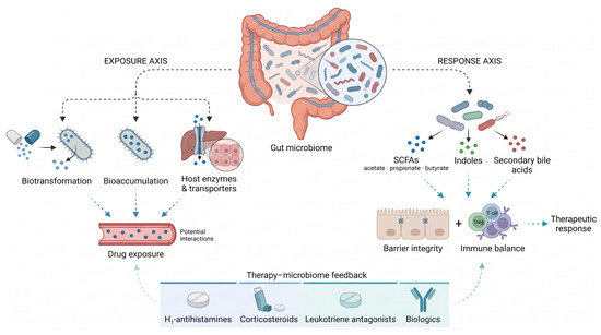
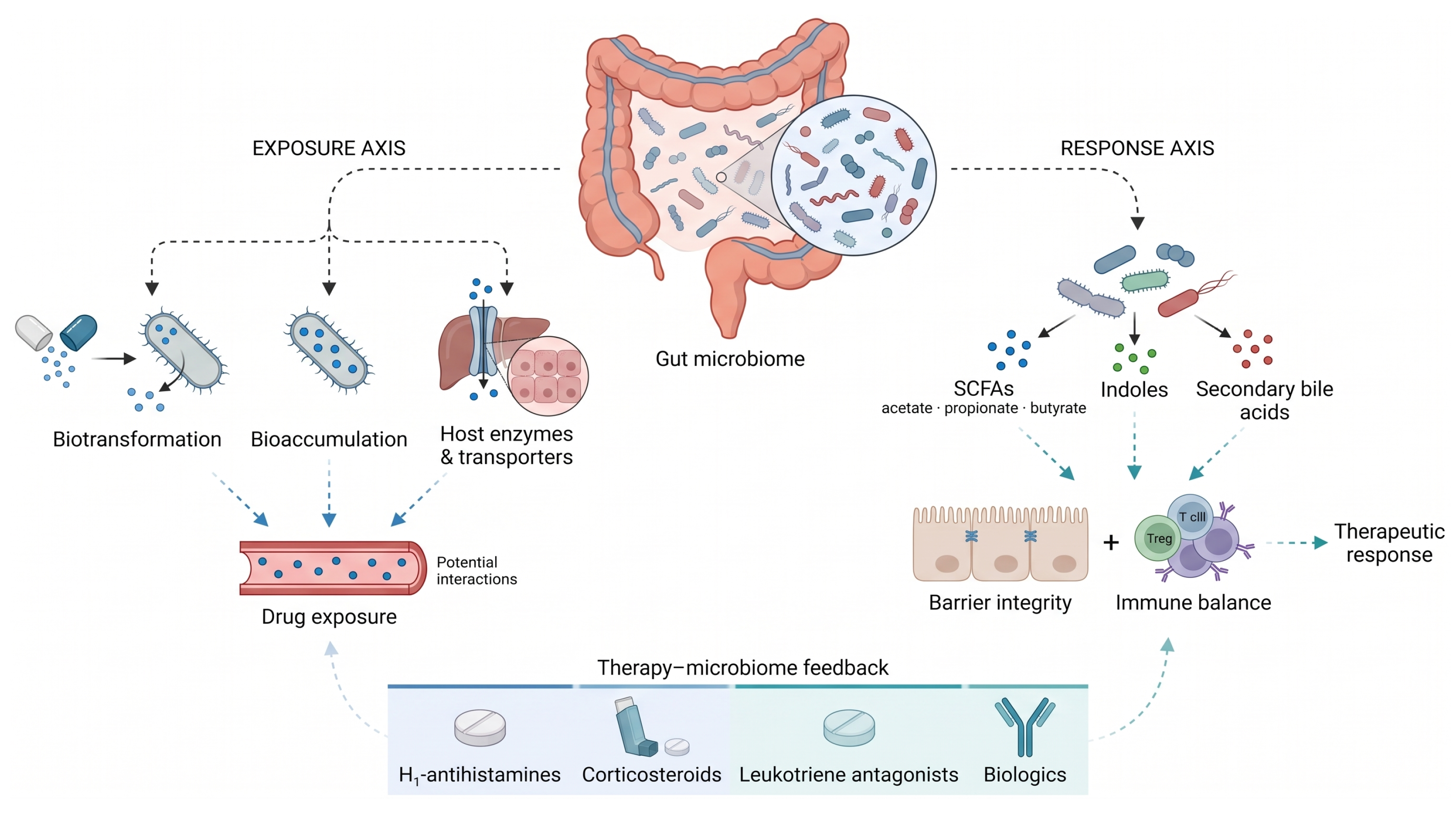
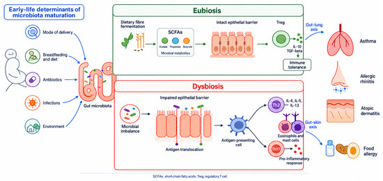
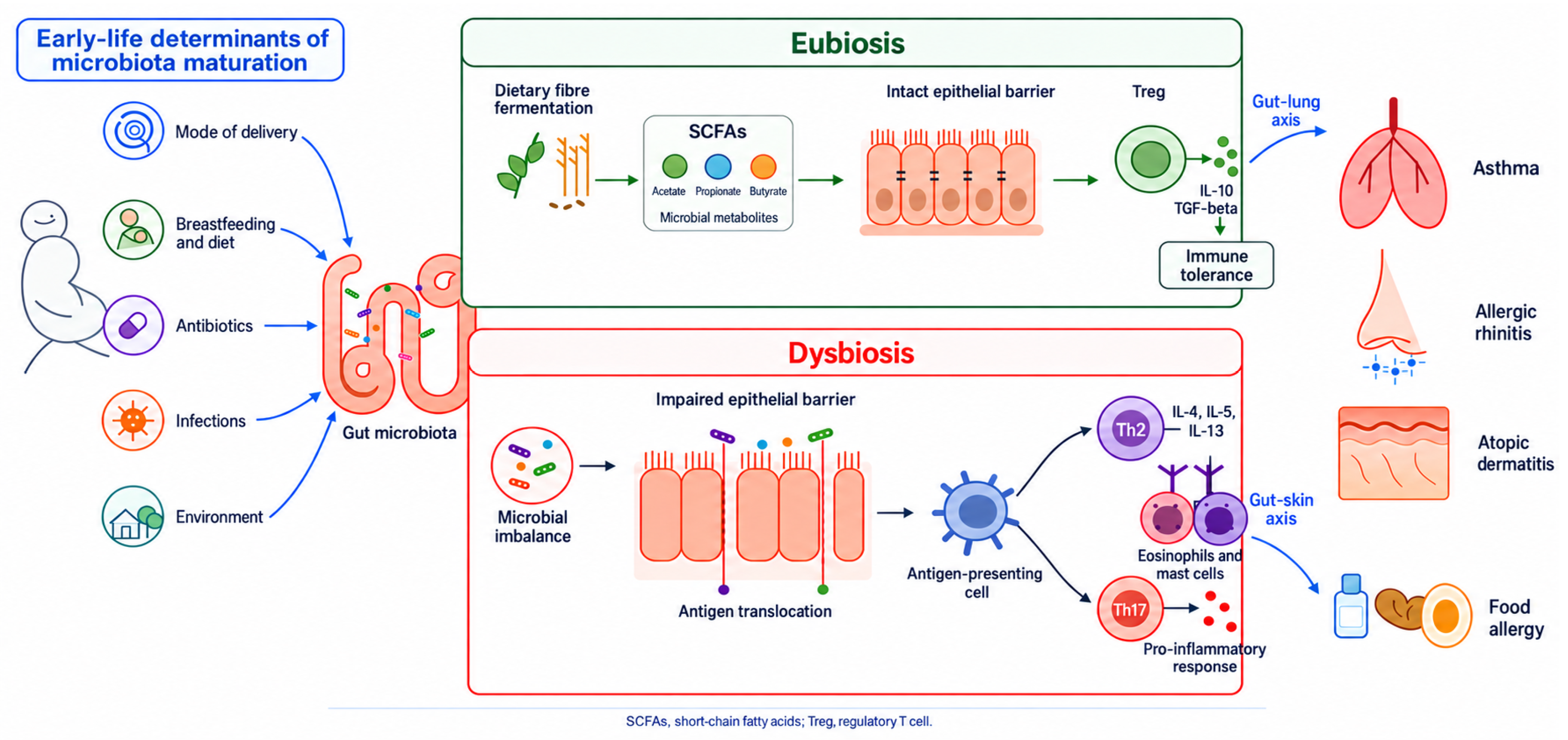

Disclaimer/Publisher’s Note: The statements, opinions and data contained in all publications are solely those of the individual author(s) and contributor(s) and not of MDPI and/or the editor(s). MDPI and/or the editor(s) disclaim responsibility for any injury to people or property resulting from any ideas, methods, instructions or products referred to in the content. |
© 2026 by the authors. Licensee MDPI, Basel, Switzerland. This article is an open access article distributed under the terms and conditions of the Creative Commons Attribution (CC BY) license.