


Abstract
Neonatal diarrhea remains a significant threat to piglet health, resulting in substantial economic losses worldwide. Among the viral pathogens associated with this condition, rotavirus (RV) has been extensively reported in Brazil; however, lineage-level classification of circulating strains remains limited. This study aimed to characterize G and P genotypes of porcine RV field strains associated with diarrhea in piglets in Southern Brazil. A total of 10 fecal samples were collected by field veterinarian from diarrheic suckling piglets aged 1 to 14 days and analyzed by RT-PCR for the detection of RV species A, B, C, and H. RV species A (RVA) was detected in 90% (9/10) of the samples, while no other RV species were identified. Genotyping based on the VP7 and VP4 genes revealed a single G3P[6] genotype combination in all RVA-positive samples. Nucleotide (nt) and deduced amino acid (aa) sequence analysis revealed high genetic similarity among strains, with values of up to 99.3% for nt and 98.0% for aa of the VP7 gene and 100% for the VP4 gene (nt and aa). Phylogenetic analysis indicated that the VP7 sequences clustered with Brazilian G3 strains, forming a distinct group consistent with a novel lineage (putative G3-XII), whereas VP4 sequences supported a new sublineage (putative P[6]-Ig). These findings demonstrate low genetic variability of RVA field strains in this neonatal diarrhea outbreak, suggesting the circulation of a single viral population. They also emphasize the importance of continuous molecular surveillance to gain a deeper understanding of viral evolution and transmission dynamics in swine populations.
1. Introduction
Neonatal diarrhea is recognized as a major cause of high morbidity and mortality in young animals, including piglets, resulting in significant economic losses for the pig industry [1]. This multifactorial and multietiological syndrome involves predisposing individual, management, and environmental factors, as well as determining factors, such as viruses, bacteria, and protozoa [2].
Porcine rotavirus A (PoRVA) is one of the primary pathogens responsible for diarrhea in piglets during the first weeks of age [3]. Belonging to the Sedoreoviridae family and the Rotavirus genus, these non-enveloped viruses are approximately 80 nm in diameter. Their icosahedral capsid consists of three concentric protein layers (inner, intermediate, and outer). This structure provides high environmental stability and resistance to adverse conditions [4].
The rotavirus (RV) genome consists of 11 segments of double-stranded RNA, which encode 12 proteins: six structural (VP1–VP4, VP6, and VP7) and six non-structural (NSP1-NSP6) [4,5]. Based on the VP6 protein, RV is classified into 11 species [4]. The outer layer of the RV capsid consists of two structural proteins. The VP7 gene encodes a glycoprotein (G genotype), and the VP4 gene encodes a protease-sensitive protein (P genotype); these proteins neutralize antibodies and are used for the binary classification system to characterize RV strains [4].
Rotavirus alphagastroenteritidis (previously named RVA) is the most common RV species associated with neonatal diarrhea in piglets worldwide [6,7], followed by Rotavirus betagastroenteritidis (previously named RVB), Rotavirus tritogastroenteritidis (previously named RVC), and Rotavirus aspergastroenteritidis (previously named RVH) [4,8]. PoRVA infection is often associated with coinfections with different genotype (VP7 and VP4) combinations [3] or with other enteropathogens, including other RV species (RVB, RVC, and RVH) [1,8,9,10,11,12,13] as well as bacterial agents such as Escherichia coli and protozoa like Cryptosporidium spp., which can worsen the clinical outcome [14,15].
Based on the molecular characteristics of the VP7 and VP4 genes, RVA strains are classified into G and P genotypes; to date, 12 G genotypes (G1–G6, G8–G12, and G26) and 20 P genotypes (P[1]–P[11], P[13], P[19], P[22], P[23], P[26], P[27], P[32], P[34], and P[49]) have been reported in PoRVA strains [1,16,17].
In Brazil, several associations of G and P genotypes of PoRVA have been described in piglets with neonatal diarrhea, particularly those associated with the G3 genotype (such as G3P[6], G3P[7], G3P[13], G3P[22], and G3P[23]) and P[6] (such as G4P[6], G5P[6], G9P[6], and G11P[6]), evidencing a high genetic variability of circulating PoRVA strains [3,18,19,20].
The classification of strains into lineages and sublineages was determined according to nt sequence similarity and their clustering patterns in the phylogenetic tree. A novel lineage within the G genotype was defined using a nt similarity threshold of >90% [21]. Similarly, P[6] lineages and sublineages were assigned based on >90% nt sequence similarity, supported by phylogenetic topology and bootstrap values [18,22]. Currently, 11 lineages are described for the G3 genotype (I–XI), highlighting the wide genetic diversity of this genotype in different hosts and geographic regions [23]. Similarly, the P[6] genotype also exhibits high genetic variability, with five (I–V) lineages already described, in addition to sublineages (Ia–If), which were defined based on phylogenetic analyses of the VP4 gene [18]. In Brazil, descriptions of lineages and sublineages are limited, particularly for swine. There has been no research on the PoRVA G3 genotype, and for the P[6] genotype, only Lorenzetti et al. [18] reported molecular characterization, classifying the P[6] strains into sublineages Ie and If.
PoRVA infection in pigs causes not only piglet mortality but also growth retardation, increased treatment costs, and productivity losses. This study aimed to molecularly characterize the G and P genotypes of PoRVA field strains involved in an outbreak of diarrhea in piglets aged ≤14 days, and evaluate the genetic variation among strains using phylogenetic analyses.
2. Materials and Methods
2.1. Diarrheic Fecal Samples
Diarrheic fecal samples were collected from 10 piglets (six piglets aged 1 to 7 days and four piglets aged 8 to 14 days) from a PoRVA unvaccinated pig herd with 8000 sows located in Santa Catarina State, Southern Brazil, in September 2024. The fecal samples were sent by the veterinarian responsible for the herd’s health program to the laboratory for diagnosis of RV infection during an outbreak of neonatal diarrhea.
2.2. Rotavirus Detection
The fecal samples were subjected to nucleic acid extraction using a combination of phenol/chloroform/isoamyl alcohol (25:24:1) and silica/guanidine isothiocyanate methods and were stored at −80 °C until processing [24,25].
Reverse transcription-polymerase chain reaction (RT-PCR) was performed, targeting the amplification of 876 bp of the VP4 gene and 1062 bp of the VP7 gene of RVA [26,27], 434 bp of the NSP2 gene of RVB [28], 1043 bp of the VP7 gene of RVC [29], and 817 bp of the VP7 gene of RVH [30]. Sequenced samples of RVA, RVB, RVC, and RVH from previous studies were used as positive controls [3,8], and sterile ultrapure water aliquots were used as negative controls in all the RT-PCR reactions.
Other relevant enteric viruses, including Porcine Epidemic Diarrhea Virus (PEDV), Transmissible Gastroenteritis Virus (TGEV), and Porcine Deltacoronavirus (PDCoV), were not tested because Brazil is considered free of these pathogens. In addition, bacterial and protozoan enteric agents were not assessed in this study.
2.3. Sequencing and Phylogenetic Analysis
The amplicons of the VP7 and VP4 genes of RVA obtained by RT-PCR assays were purified using the Wizard® SV Gel and PCR Clean-up System kit (Promega®, Madison, WI, USA). The PCR products were quantified with a Qubit® fluorometer using the Quant-iT dsDNA BR Assay Kit (InvitrogenTM, Eugene, OR, USA). Nucleotide (nt) sequencing was performed on the ABI 3500 Genetic Analyzer platform with the BigDye® Terminator v3.1 Cycle Sequencing Kit (Applied Biosystems®, Foster City, CA, USA) using the same primers as in the RT-PCR assays.
Nucleotide quality analysis and contig assembly of RVA sequences were performed using PHRED and CAP3 software, respectively (http://lbi.cenargen.embrapa.br/phph/ accessed on 19 May 2026). The resulting sequences were then compared to those in the GenBank database using BLAST (https://blast.ncbi.nlm.nih.gov/Blast.cgi, accessed on 19 May 2026). Additionally, the nt and deduced amino acid (aa) sequence identity matrix among sequences was constructed with BioEdit software, version 7.7.1 [31]. The phylogenetic trees were generated in MEGA 11 [32] using the neighbor-joining method with the Kimura 2-parameter model to construct trees based on nt sequences and the Poisson model to construct trees based on deduced aa sequences. Bootstrap probabilities were calculated using 1000 replicates.
3. Results
In nine (90%) of the 10 diarrheic fecal samples analyzed by RT-PCR, partial amplification of the RVA VP7 (G genotype) and VP4 (P genotype) genes was successfully achieved. All samples tested negative for RVB, RVC, and RVH. Based on nt and deduced aa sequence analyses of the G (VP7) and P (VP4) genotypes, all RVA field strains were classified as G3P[6] genotype combination.
The VP7 gene analysis revealed high sequence identity among the RVA field strains, with up to 99.3% nt and 98.0% deduced aa sequence identities. It was possible to classify the G genotypes identified in the PoRVA field strains into four distinct nt sequence patterns. According to the nt sequence percentage, in each pattern, the strains share 100% nt sequence identity. Specifically, pattern 1 comprised four strains, #FS602 to #FS605, all isolated from piglets at 1 to 7 days of age. In contrast, pattern 2 included three strains; of these, #FS606 and #FS607 were obtained from 1- to 7-day-old piglets, whereas #FS608 was isolated from an 8- to 14-day-old piglet. Moving to pattern 3, this category consisted of a single strain, #FS609, derived from an 8- to 14-day-old piglet. Finally, pattern 4 likewise comprised a single strain, #FS610, also isolated from a 14-day-old piglet. One representative strain from each pattern was selected to use in the phylogenetic analysis.
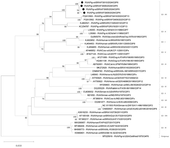
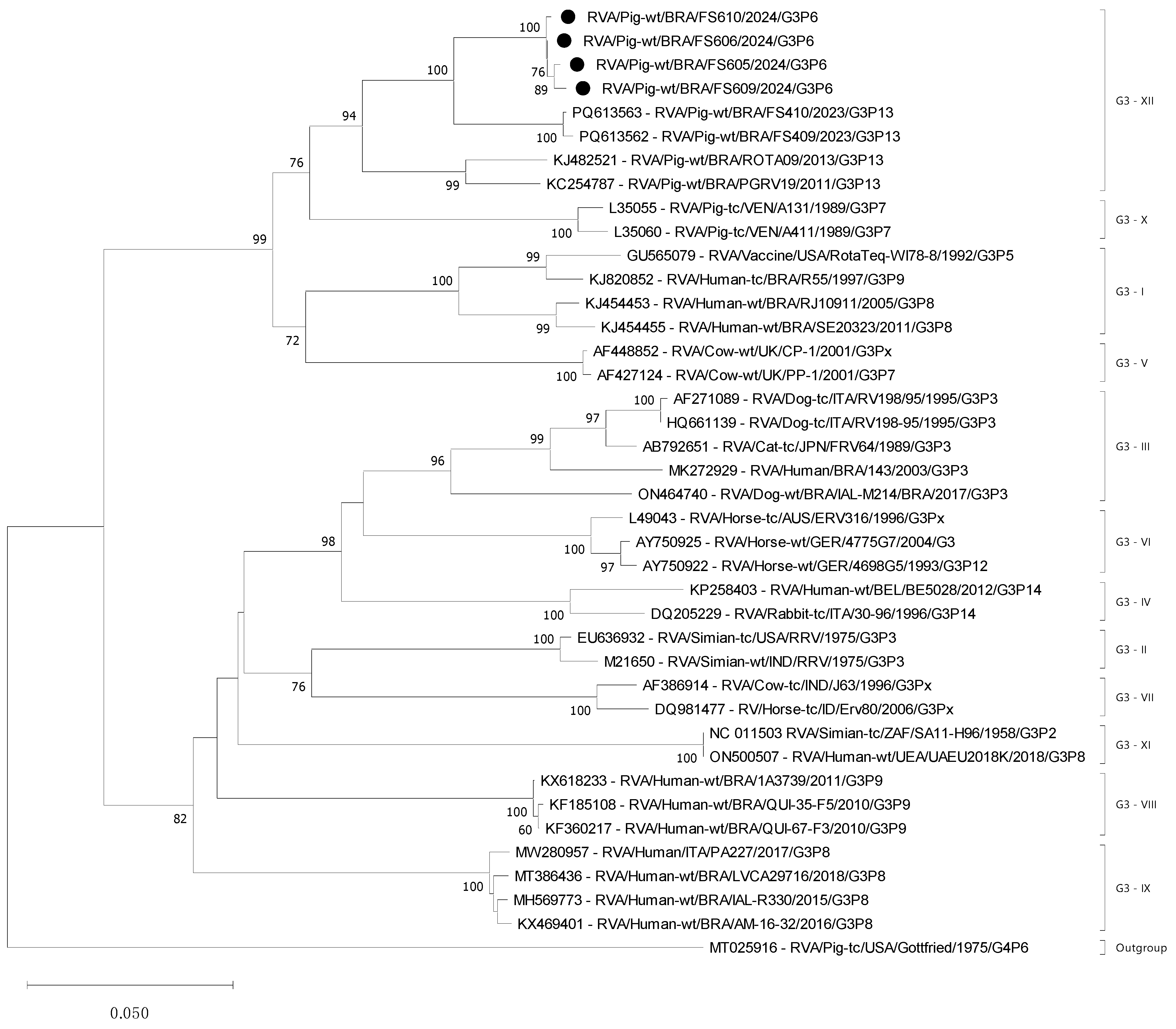
The Brazilian PoRVA field strains ranged from 91.0% to 95.3% nt sequence identity and 94.7% to 97.3% deduced aa sequence identity among them, grouping them together in the same cluster and forming a new branch in G3 genotype, supporting a novel putative G3 lineage XII (Figure 1 and Figure S1). Compared with other G3 lineages (I–XI), the PoRVA strains from this study exhibited 76.8% to 88.7% nt sequence identity and 89.4% to 96.0% deduced aa sequence identity (Table 1).
Figure 1.
A phylogenetic tree based on nucleotide sequences was constructed using 682 bp of the VP7 gene of the rotavirus A (RVA) G3 genotype. Representative reference strains corresponding to all recognized G3 lineages were included for comparative analysis. The tree was constructed using the neighbor-joining method with the Kimura two-parameter model, and the numbers adjacent to the nodes represent the percentage of bootstrap support for 1000 replicates for the clusters. Bootstrap values less than 50% are not shown. The black circles indicate the PoRVA G3 strains from this study. The PoRVA prototype Gottfried (G4P[6]) vaccine strain was used as an outgroup.
Table 1.
Comparative matrix of nucleotide (nt) and deduced amino acid (aa) sequence identities of the four patterns of nt sequences of G3 genotype RVA from fecal samples collected from 1- to 14-day-old suckling piglets compared with representative strains from established G3 lineages available in GenBank.
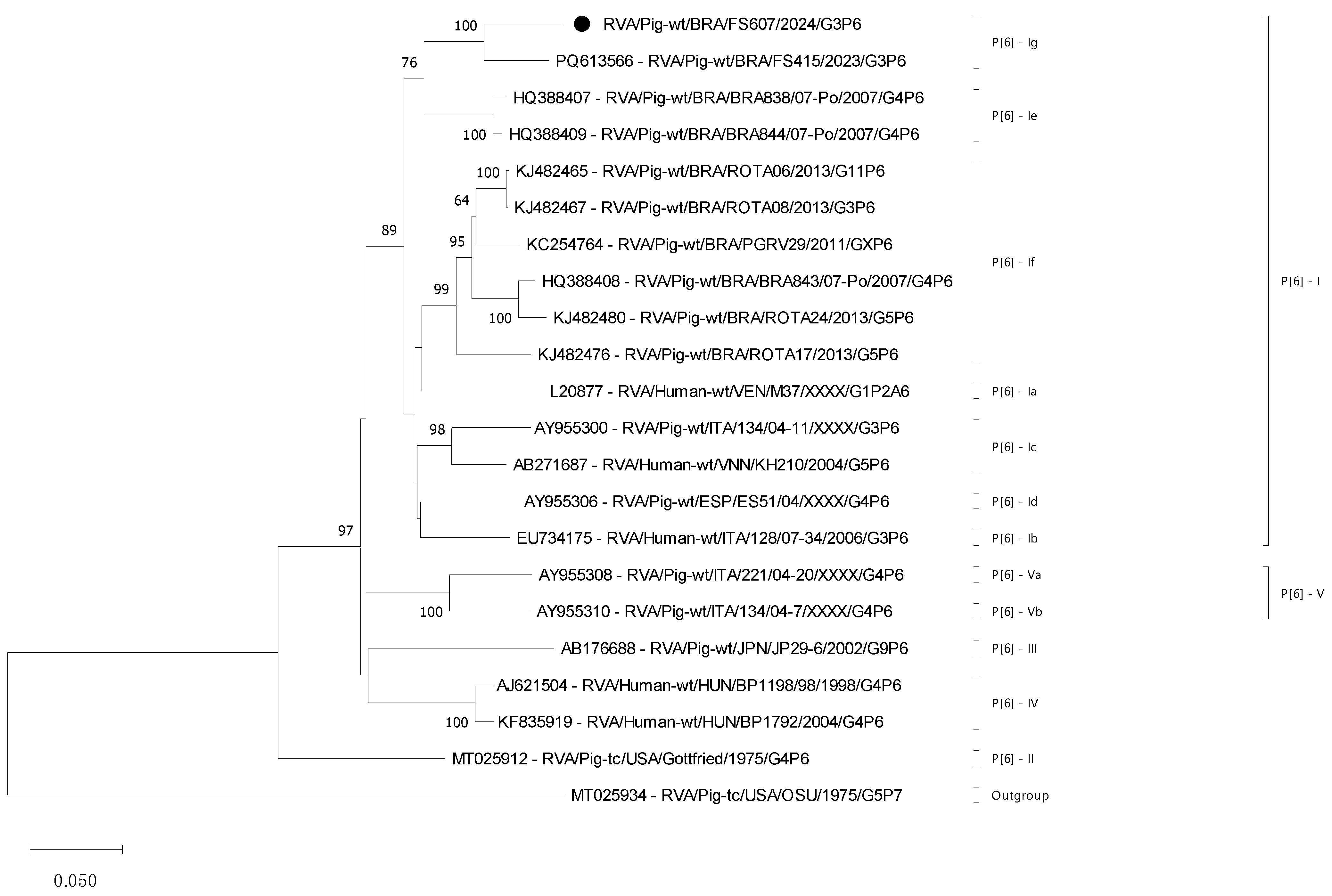
The VP4 gene was completely conserved across all nine PoRVA field strains, exhibiting 100% of nt and deduced aa identities; consequently, only one strain was selected for use in the phylogenetic analysis. Comparing the VP4 gene with the P[6] lineage I sublineages (Ia–If) revealed 84.7% to 89.3% nt and 89.3% to 94.2% of deduced aa sequence identities. The PoRVA field strain genotype P[6] from this study exhibited 92.7% of nt sequence identity and 95.1% of deduced aa sequence identity with the PoRVA FS415 strain from Brazil that was previously described [3], forming the P[6]-Ig sublineage. In relation to P[6] lineages (II–V), it shares 81.2% to 84.7% nt sequence identity and 85.3 to 90.2% deduced aa sequence identity (Table 2).
Table 2.
Comparative matrix of nucleotide (nt) and deduced amino acid (aa) sequence identities of the P[6] genotype RVA #FS607 strain from a fecal sample collected from a 7-day-old suckling piglet compared with representative strains from established P[6] lineages and sublineages available in GenBank.
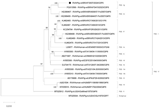
The proposed new P[6]-I sublineage was also based on the phylogenetic analysis of the VP4 gene, which revealed that the #FS607 strain clustered within a distinct branch of P[6] lineage I, together with the PoRVA strain FS415 that was previously described [3], with consistent nt and aa sequence identity values (Table 2). The high bootstrap support in P6-Ig (100%) and in P[6]-I (89%) for the nt-based tree further reinforces the reliability of this phylogenetic placement (Figure 2 and Figure S2).
Figure 2.
A phylogenetic tree based on nucleotide sequences was constructed using 681 bp of the VP4 gene of the rotavirus A (RVA) P[6] genotype. Representative reference strains corresponding to all recognized P[6] lineages and sublineages were included for comparative analysis. The tree was constructed using the neighbor-joining method with the Kimura two-parameter model, and the numbers adjacent to the nodes represent the percentage of bootstrap support for 1000 replicates for the clusters. The black circle indicates the PoRVA P[6] strain that represents all the positive strains from this study, which exhibited 100% nt identity (PZ381128). Bootstrap values less than 50% are not shown. The PoRVA prototype OSU (G5P[7]) strain was used as an outgroup.
The nt sequences for the FS602 to FS610 PoRVA strains obtained in this study were deposited in the GenBank database under accession numbers PZ381123 to PZ381131 (VP4) and PZ381132 to PZ381140 (VP7).
4. Discussion
Notably, this report describes a high incidence of diarrhea in suckling piglets (1–14 days old) caused by a single PoRVA G3P[6] genotype. The low diversity of G and P genotypes, and RVA being the only viral pathogen detected are uncommon in pig herds [33], although they are often observed in infections by bovine RVA in beef and dairy cattle herds in Brazil [34,35].
Analysis of the VP7 gene revealed a higher nt identity among the Brazilian PoRVA G3 strains detected in 2011 [19], 2013 [20], and 2023 [3], reaching up to 91.0%, although these studies did not perform lineage classification. The other RVA G3 strains, which were distributed in other lineages, presented nt identities ranging between 76.9% and 88.5% compared with the PoRVA G3 strains from this study. The nt identity percentages support the proposed new G3 lineage XII.
The PoRVA G3 sequences available in GenBank were distributed across three (V, IX, and X) lineages. The Thai CMP213 PoRVA strain (accession number: DQ786576) was assigned to lineage V, clustering with bovine RVA G3 strains from the United Kingdom (CP-1 and PP-1). The Brazilian BP64-clone-1 PoRVA strain (accession number: MN201922) grouped within lineage IX, alongside three Brazilian human RVA (HuRVA) G3 strains (LVCA29716, AL-3330, and AM-16-32) and an Italian HuRVA G3 strain (PA227). Two Venezuelan PoRVA strains (accession numbers: L35055 and L35060) formed lineage X. The Thai CMP213 and Brazilian BP64-clone-1 PoRVA strains were excluded from the phylogenetic tree due to their shorter nt sequences compared to those analyzed in this study.
The G3 genotype has a broad host range and is commonly found in species such as cats, dogs, monkeys, pigs, rabbits, horses, and humans [36,37]. Reassortment events and interspecies transmissions have been documented, including porcine–human RVA strains in circulation in Slovenia [38]. Furthermore, the G3 genotype of strain SO1199, which was detected in a pediatric case in Japan, exhibited the highest nt identity compared with PoRVA strains, providing evidence of zoonotic transmission [39]. In the present study, PoRVA G3 strains from Brazil shared up to 88.2% nt identity with the HuRVA strain RJ10911 from Brazil, which were classified within G3 lineage I, representing the highest similarity observed outside the newly proposed G3 lineage XII. The RVA G3 genotype strains available in the database can be classified into eleven (I–XI) lineages using 90% nt sequence identity as the demarcation criterion [21,40].
Phylogenetic analysis of the VP4 gene of the P[6] strains indicates the stability of this genotype within the studied pig herd, as well as high nt and deduced aa sequence identities compared to the RVA/Pig-wt/BRA/FS415/2023/G3P[6] strain [3], which was identified during a neonatal diarrhea outbreak in 2023 on an unvaccinated pig farm also located in the state of Santa Catarina.
In comparison with the PoRVA vaccine strain Gottfried (G4P[6]-II, accession number: MT025912), the P[6] strains identified in this study exhibited the lowest nt sequence identity (81.4%) among all P[6] lineages. A similar pattern was observed with circulating P[6] strains, which are more closely related to human P[6] strains than to the prototype PoRVA P[6] strain Gottfried [22].
The Brazilian PoRVA P[6] #FS607 field strain exhibited nt identities ranging from 84.7% to 89.3% compared with other strains within the same lineage I. Eight of 10 PoRVA strains belonging to P[6] lineage I are Brazilian strains (BRA838/07-Po, BRA844/07-Po, BRA843/07-Po, ROTA06, ROTA08, ROTA17, ROTA24, and PGRV29) [18,19,20], one is a Spanish strain (ES51/04) [22], and one is an Italian strain (134/04-11) [22]. Other PoRVA strains are distributed among three P[6] lineages: the PoRVA Gottfried strain (vaccine) in lineage II, the Japan JP29-6 strain [41] in lineage III, and two Italian strains [22] in lineage V.
The nine samples analyzed present four patterns of the G3 genotype (91.0% to 95.3% nt identity) and were identical in the P[6] genotype (100% nt identity), with the VP7 gene less conserved than the VP4 gene. The variability in the genotypes described in RCWG [42] determines more P genotypes (n = 58) than G genotypes (n = 42). Although, at the lineage level, the variability of G genotype lineages is higher (i.e., G3 lineages I–XI) than in P genotype lineages (i.e., P[6] lineages I–V).
Previous reports from Brazil reported that the G3P[6] genotype is often detected in co-infections with other RV species and/or with different genotype combinations of PoRVA, which are typically associated with diarrhea in piglets [3,20].
Silva et al. [20] identified the G3P[6] genotype over two years through the analysis of 126 fecal samples collected in the states of Mato Grosso do Sul and São Paulo, although the samples were not tested for other RV species. G3P[6] was detected in association with other PoRVA genotypes (G3P[13], G5P[6], G5P[13], G9P[23], and G11P[6]) in suckling and nursery piglets with diarrhea across four distinct herds.
Similarly, Yoshitani et al. [3], in a study also carried out in Santa Catarina, reported the occurrence of G3P[6] in 2- and 3-week-old piglets during a neonatal diarrhea outbreak in a single unvaccinated pig herd. The similarity of the G3P[6] samples in terms of VP4 and VP7 genes could be explained by the persistence of the strain in the nearby pig herds with herd-to-herd transmission. However, the genotype was detected with the other three genotype combinations (G3P[13], G4P[6], and G5P[23]) in the same age interval and was associated with co-infection with RVC in 2-week-old piglets. Considering the previous studies that described the PoRVA G3P[6] genotype, the present study presents low genetic variability.
Differences in production systems and pig herd characteristics may contribute to these constraints. The pig herd analyzed by Yoshitani et al. [3] was composed of 3400 sows, which were not vaccinated and were raised in a complete-cycle system. In the present study, the pig herd comprised 8000 non-vaccinated sows. This pattern differs from the epidemiological scenario commonly described in swine and suggests the circulation of a more homogeneous viral population within the herd.
The G3P[6] genotype combination has also been reported by Martella et al. [22], who identified PoRVA P[6] strains harboring human-like VP4 P[6] gene alleles. The authors analyzed the genetic relationships between porcine and human P[6] rotaviruses by examining 12 PoRVA strains from nursing and weaning piglets in outbreaks of diarrhea, including three from Spain and nine from Italy. These findings highlight the considerable genetic diversity of RVA and underscore the potential for interspecies transmission between porcine and human hosts.
PoRVA represents an important genetic reservoir for the emergence of novel HuRVA strains owing to its extensive genetic diversity and capacity for interspecies transmission [17,37]. The G3P[6] genotype combination appears to be associated with human-to-porcine transmission events, as suggested by the close evolutionary relationship between the VP7 and VP4 genes of HuRVA and PoRVA G3P[6] strains [39]. In this study, PoRVA G3P[6] strains showed high nt identity compared to other PoRVA strains from Brazil and clustered into a distinct VP7 lineage (G3-XII) and VP4 sublineage (P[6]-Ig).
5. Conclusions
This study reports the detection of a unique PoRVA G3P[6] genotype combination in diarrheic fecal samples from piglets aged 1–14 days from an unvaccinated pig herd located in Southern Brazil, in the absence of co-infection with other RVA genotype combinations (G and P) or other RV species (RVB, RVC, or RVH). The high nt identity among strains indicates low genetic variability. Phylogenetic analysis revealed distinct clustering of VP7 and VP4 sequences, consistent with a suggested novel lineage (G3-XII) and a potential new sublineage (P[6]-lg), respectively. These findings highlight the importance of continuous epidemiological and molecular surveillance to characterize RV genetic diversity, identify emerging or reemerging genotypes, and monitor changes in circulation patterns, thereby supporting the development of more effective control strategies.
Supplementary Materials
The following supporting information can be downloaded at: https://www.mdpi.com/article/10.3390/microbiolres17070122/s1, Figure S1: A phylogenetic tree based on amino acid (aa) sequences was constructed using 227 deduced sequences of amino acids of the VP7 gene of the RVA G3 genotype. The tree was constructed using the neighbor-joining method with the Poisson model. The black circles indicate the PoRVA G3 strains from this study. Representative reference strains corresponding to all recognized G3 lineages were included for comparative analysis. The numbers adjacent to the nodes represent the percentage of bootstrap support for 1000 replicates for the clusters. Bootstrap values less than 50% are not shown. The PoRVA prototype Gottfried (G4P[6]) strain was used as an outgroup; Figure S2: A phylogenetic tree based on amino acid (aa) sequences was constructed using 226 deduced sequences of amino acids of the VP4 gene of the RVA P[6] genotype. The tree was constructed using the neighbor-joining method with the Poisson model. The black circle indicates the PoRVA P[6] strain from this study. Representative reference strains corresponding to all recognized P[6] lineages were included for comparative analysis. The numbers adjacent to the nodes represent the percentage of bootstrap support for 1000 replicates for the clusters. Bootstrap values less than 50% are not shown. The PoRVA prototype OSU (G5P[7]) strain was used as an outgroup.
Author Contributions
Conceptualization, M.d.S.M., J.T.T.F. and A.A.A.; methodology, M.d.S.M., B.M.M., G.D.Y., J.T.T.F. and E.L.; investigation, M.d.S.M. and J.T.T.F.; resources, A.A.A.; writing—original draft preparation, M.d.S.M., B.M.M. and J.T.T.F.; writing—review and editing, E.L. and A.A.A.; funding acquisition, A.F.A. and A.A.A. All authors have read and agreed to the published version of the manuscript.
Funding
This research was funded by Araucaria Foundation (FAP-PR), National Council for Scientific and Technological Development (CNPq), and Coordination of Superior Level Staff Improvement (CAPES).
Institutional Review Board Statement
Ethical review and approval were waived for this study due to the samples used in this study did not originate from an experimental study but were received by the laboratory for diagnostic purposes.
Informed Consent Statement
Not applicable.
Data Availability Statement
The nucleotide sequences were deposited in the GenBank database under accession numbers PZ381123 to PZ381140.
Acknowledgments
The authors acknowledge the National Council for Scientific and Technological Development (CNPq), the Brazilian Federal Agency for Support and Evaluation of Graduate Education (CAPES), and the Araucária Foundation (FAP/PR). Alfieri, A.F., and Alfieri, A.A., are recipients of CNPq fellowships.
Conflicts of Interest
The authors declare no conflicts of interest.
References
- Vlasova, A.N.; Amimo, J.O.; Saif, L.J. Porcine Rotaviruses: Epidemiology, Immune Responses and Control Strategies. Viruses 2017, 9, 48. [Google Scholar] [CrossRef] [Scilit] [PubMed]
- Do, D.T.; Ngo, T.T.N.; Vu, H.D.; Duong, N.M.; Traiyarach, S.; Lai, D.C.; Escuder, J.M. Presence of toxin-producing bacterial pathogens and associated risk factors in neonatal diarrhea of piglets on commercial sow farms in Vietnam. Vet. World 2025, 18, 1288–1296. [Google Scholar] [CrossRef] [Scilit] [PubMed]
- Yoshitani, G.D.; Fritzen, J.T.T.; Yasumitsu, C.Y.; Machado, B.M.; Alfieri, A.F.; Alfieri, A.A. Diversity of species and genotypes of rotavirus field strains in a diarrhea outbreak in suckling piglets from an unvaccinated pig herd. Braz. J. Microbiol. 2025, 56, 2257–2262. [Google Scholar] [CrossRef] [Scilit] [PubMed]
- ICTV. Genus: Rotavirus. Available online: https://ictv.global/report/chapter/sedoreoviridae/sedoreoviridae/rotavirus (accessed on 9 September 2025).
- Uprety, T.; Wang, D.; Li, F. Recent advances in rotavirus reverse genetics and its utilization in basic research and vaccine development. Arch. Virol. 2021, 166, 2369–2386. [Google Scholar] [CrossRef] [Scilit] [PubMed]
- Linares, R.d.C.; Barry, A.F.; Alfieri, A.F.; Médici, K.C.; Feronato, C.; Grieder, W.; Alfieri, A.A. Frequency of group a rotavirus in piglet stool samples from non-vaccinated Brazilian pig herds. Braz. Arch. Biol. Technol. 2009, 52, 63–68. [Google Scholar] [CrossRef] [Scilit]
- Saif, L.J. Rotavirus. In Diseases of Swine; Zimmerman, J.J., Karriker, L.A., Ramirez, A., Schwartz, K.J., Stevenson, G.W., Eds.; Wiley-Blackwell: Oxford, UK, 2012. [Google Scholar]
- Molinari, B.L.D.; Possatti, F.; Lorenzetti, E.; Alfieri, A.F.; Alfieri, A.A. Unusual outbreak of post-weaning porcine diarrhea caused by single and mixed infections of rotavirus groups A., B., C., and H. Vet. Microbiol. 2016, 193, 125–132. [Google Scholar] [CrossRef] [Scilit] [PubMed]
- Médici, K.C.; Barry, A.F.; Alfieri, A.F. Porcine rotavirus groups A, B, and C identified by polymerase chain reaction in a fecal sample collection with inconclusive results by polyacrylamide gel electrophoresis. J. Swine Health Prod. 2011, 19, 146–150. [Google Scholar] [CrossRef] [Scilit] [PubMed]
- Lorenzetti, E.; Stipp, D.T.; Possatti, F.; Campanha, J.E.T.; Alfieri, A.F.; Alfieri, A.A. Diarrhea outbreaks in suckling piglets due to rotavirus group C single and mixed (rotavirus groups A and B) infections. Pesq. Vet. Bras. 2014, 34, 391–397. [Google Scholar] [CrossRef] [Scilit]
- Almeida, P.R.; Lorenzetti, E.; Cruz, R.S.; Watanabe, T.T.; Zlotowski, P.; Alfieri, A.A.; Driemeier, D. Diarrhea caused by rotavirus A, B, and C in suckling piglets from southern Brazil: Molecular detection and histologic and immunohistochemical characterization. J. Vet. Diagn. Investig. 2018, 30, 370–376. [Google Scholar] [CrossRef] [Scilit] [PubMed]
- Miyabe, F.M.; Dall Agnol, A.M.; Leme, R.A.; Oliveira, T.E.S.; Headley, S.A.; Fernandes, T.; de Oliveira, A.G.; Alfieri, A.F.; Alfieri, A.A. Porcine rotavirus B as primary causative agent of diarrhea outbreaks in newborn piglets. Sci. Rep. 2020, 10, 22002. [Google Scholar] [CrossRef] [Scilit] [PubMed]
- Ferrari, E.; Salogni, C.; Martella, V.; Alborali, G.L.; Scaburri, A.; Boniotti, M.B. Assessing the Epidemiology of Rotavirus A, B, C and H in Diarrheic Pigs of Different Ages in Northern Italy. Pathogens 2022, 11, 467. [Google Scholar] [CrossRef] [Scilit] [PubMed]
- Doerksen, T.; Christensen, T.; Lu, A.; Noll, L.; Bai, J.; Henningson, J.; Palinski, R. Assessment of porcine Rotavirus-associated virome variations in pigs with enteric disease. Vet. Microbiol. 2022, 270, 109447. [Google Scholar] [CrossRef] [Scilit] [PubMed]
- Ruiz, V.L.; Bersano, J.G.; Carvalho, A.F.; Catroxo, M.H.; Chiebao, D.P.; Gregori, F.; Miyashiro, S.; Nassar, A.F.; Oliveira, T.M.; Ogata, R.A.; et al. Case-control study of pathogens involved in piglet diarrhea. BMC Res. Notes 2016, 9, 22. [Google Scholar] [CrossRef] [Scilit] [PubMed]
- Díaz Alarcón, R.G.; Liotta, D.J.; Miño, S. Zoonotic RVA: State of the Art and Distribution in the Animal World. Viruses 2022, 14, 2554. [Google Scholar] [CrossRef] [Scilit] [PubMed]
- Papp, H.; László, B.; Jakab, F.; Ganesh, B.; De Grazia, S.; Matthijnssens, J.; Ciarlet, M.; Martella, V.; Bányai, K. Review of group A rotavirus strains reported in swine and cattle. Vet. Microbiol. 2013, 165, 190–199. [Google Scholar] [CrossRef] [Scilit] [PubMed]
- Lorenzetti, E.; da Silva Medeiros, T.N.; Alfieri, A.F.; Alfieri, A.A. Genetic heterogeneity of wild-type G4P[6] porcine rotavirus strains detected in a diarrhea outbreak in a regularly vaccinated pig herd. Vet. Microbiol. 2011, 154, 191–196. [Google Scholar] [CrossRef] [Scilit] [PubMed]
- Tonietti, P.O.; Hora, A.S.; Silva, F.D.; Ruiz, V.L.; Gregori, F. Phylogenetic analyses of the VP4 and VP7 genes of porcine group A rotaviruses in Sao Paulo State, Brazil: First identification of G5P[23] in piglets. J. Clin. Microbiol. 2013, 51, 2750–2753. [Google Scholar] [CrossRef] [Scilit] [PubMed]
- Silva, F.D.; Espinoza, L.R.; Tonietti, P.O.; Barbosa, B.R.; Gregori, F. Whole-genomic analysis of 12 porcine group A rotaviruses isolated from symptomatic piglets in Brazil during the years of 2012-2013. Infect. Genet Evol. 2015, 32, 239–254. [Google Scholar] [CrossRef] [Scilit] [PubMed]
- Fritzen, J.T.T.; Oliveira, M.V.; Lorenzetti, E.; Alfieri, A.F.; Alfieri, A.A. Genotype constellation of a rotavirus A field strain with an uncommon G8P[11] genotype combination in a rotavirus-vaccinated dairy cattle herd. Arch. Virol. 2020, 165, 1855–1861. [Google Scholar] [CrossRef] [Scilit] [PubMed]
- Martella, V.; Bányai, K.; Ciarlet, M.; Iturriza-Gómara, M.; Lorusso, E.; De Grazia, S.; Arista, S.; Decaro, N.; Elia, G.; Cavalli, A.; et al. Relationships among porcine and human P[6] rotaviruses: Evidence that the different human P[6] lineages have originated from multiple interspecies transmission events. Virology 2006, 344, 509–519. [Google Scholar] [CrossRef] [Scilit] [PubMed]
- Vizzi, E.; Rosales, R.E.; Piñeros, O.; Fernández, R.; Inaty, D.; López, K.; Peña, L.; De Freitas-Linares, A.; Navarro, D.; Neri, S.; et al. Emergence of Equine-like G3P[8] Rotavirus Strains Infecting Children in Venezuela. Viruses 2025, 17, 410. [Google Scholar] [CrossRef] [Scilit] [PubMed]
- Alfieri, A.A.; Parazzi, M.E.; Takiuchi, E.; Médici, K.C.; Alfieri, A.F. Frequency of group A rotavirus in diarrhoeic calves in Brazilian cattle herds, 1998-2002. Trop. Anim. Health Prod. 2006, 38, 521–526. [Google Scholar] [CrossRef] [Scilit] [PubMed]
- Boom, R.; Sol, C.J.; Salimans, M.M.; Jansen, C.L.; Wertheim-van Dillen, P.M.; van der Noordaa, J. Rapid and simple method for purification of nucleic acids. J. Clin. Microbiol. 1990, 28, 495–503. [Google Scholar] [CrossRef] [Scilit] [PubMed]
- Gentsch, J.R.; Glass, R.I.; Woods, P.; Gouvea, V.; Gorziglia, M.; Flores, J.; Das, B.K.; Bhan, M.K. Identification of group A rotavirus gene 4 types by polymerase chain reaction. J. Clin. Microbiol. 1992, 30, 1365–1373. [Google Scholar] [CrossRef] [Scilit] [PubMed]
- Gouvea, V.; Glass, R.I.; Woods, P.; Taniguchi, K.; Clark, H.F.; Forrester, B.; Fang, Z.Y. Polymerase chain reaction amplification and typing of rotavirus nucleic acid from stool specimens. J. Clin. Microbiol. 1990, 28, 276–282. [Google Scholar] [CrossRef] [Scilit] [PubMed]
- Gouvea, V.; Allen, J.R.; Glass, R.I.; Fang, Z.Y.; Bremont, M.; Cohen, J.; McCrae, M.A.; Saif, L.J.; Sinarachatanant, P.; Caul, E.O. Detection of group B and C rotaviruses by polymerase chain reaction. J. Clin. Microbiol. 1991, 29, 519–523. [Google Scholar] [CrossRef] [Scilit] [PubMed]
- Lee, S.G.; Youn, S.H.; Oh, M.H.; Rhee, O.J.; Oh, S.; Paik, S.Y. Molecular characterization of two strains of porcine group C rotavirus. J. Microbiol. 2011, 49, 1058–1062. [Google Scholar] [CrossRef] [Scilit] [PubMed]
- Molinari, B.L.; Alfieri, A.F.; Alfieri, A.A. Genetic variability of VP6, VP7, VP4, and NSP4 genes of porcine rotavirus group H detected in Brazil. Virus Res. 2015, 197, 48–53. [Google Scholar] [CrossRef] [Scilit] [PubMed]
- Hall, T.A. BioEdit: A user-friendly biological sequence alignment editor and analysis program for Windows 95/98/NT. Nucleic Acids Symp. Ser. 1999, 41, 95–98. [Google Scholar]
- Tamura, K.; Stecher, G.; Kumar, S. MEGA11: Molecular Evolutionary Genetics Analysis Version 11. Mol. Biol. Evol. 2021, 38, 3022–3027. [Google Scholar] [CrossRef] [Scilit] [PubMed]
- Dall Agnol, A.M.; Guimarães, N.S.; Leme, R.A.; da Costa, A.R.; Alfieri, A.F.; Alfieri, A.A. The vaccination changed the profile of rotavirus infection with the increase of non-rotavirus A species diagnosis in one-week-old diarrheic piglets. Braz. J. Microbiol. 2024, 55, 991–996. [Google Scholar] [CrossRef] [Scilit] [PubMed]
- Medeiros, T.N.S.; Lorenzetti, E.; Alfieri, A.F.; Alfieri, A.A. Phylogenetic analysis of a G6P[5] bovine rotavirus strain isolated in a neonatal diarrhea outbreak in a beef cattle herd vaccinated with G6P[1] and G10P[11] genotypes. Arch. Virol. 2015, 160, 447–451. [Google Scholar] [CrossRef] [Scilit] [PubMed]
- Fritzen, J.T.T.; Oliveira, M.V.; Lorenzetti, E.; Miyabe, F.M.; Viziack, M.P.; Rodrigues, C.A.; Ayres, H.; Alfieri, A.F.; Alfieri, A.A. Longitudinal surveillance of rotavirus A genotypes circulating in a high milk yield dairy cattle herd after the introduction of a rotavirus vaccine. Vet. Microbiol. 2019, 230, 260–264. [Google Scholar] [CrossRef] [Scilit] [PubMed]
- Ghosh, S.; Kobayashi, N. Exotic rotaviruses in animals and rotaviruses in exotic animals. Virusdisease 2014, 25, 158–172. [Google Scholar] [CrossRef] [Scilit] [PubMed]
- Martella, V.; Bányai, K.; Matthijnssens, J.; Buonavoglia, C.; Ciarlet, M. Zoonotic aspects of rotaviruses. Vet. Microbiol. 2010, 140, 246–255. [Google Scholar] [CrossRef] [Scilit] [PubMed]
- Kozyra, I.; Kocki, J.; Rzeżutka, A. Detection of Porcine-Human Reassortant and Zoonotic Group A Rotaviruses in Humans in Poland. Transbound. Emerg. Dis. 2024, 2024, 4232389. [Google Scholar] [CrossRef] [Scilit] [PubMed]
- Akari, Y.; Hatazawa, R.; Kuroki, H.; Ito, H.; Negoro, M.; Tanaka, T.; Miwa, H.; Sugiura, K.; Umemoto, M.; Tanaka, S.; et al. Full genome-based characterization of an Asian G3P[6] human rotavirus strain found in a diarrheic child in Japan: Evidence for porcine-to-human zoonotic transmission. Infect. Genet Evol. 2023, 115, 105507. [Google Scholar] [CrossRef] [Scilit] [PubMed]
- Komoto, S.; Pongsuwanna, Y.; Tacharoenmuang, R.; Guntapong, R.; Ide, T.; Higo-Moriguchi, K.; Tsuji, T.; Yoshikawa, T.; Taniguchi, K. Whole genomic analysis of bovine group A rotavirus strains A5-10 and A5-13 provides evidence for close evolutionary relationship with human rotaviruses. Vet. Microbiol. 2016, 195, 37–57. [Google Scholar] [CrossRef] [Scilit] [PubMed]
- Teodoroff, T.A.; Tsunemitsu, H.; Okamoto, K.; Katsuda, K.; Kohmoto, M.; Kawashima, K.; Nakagomi, T.; Nakagomi, O. Predominance of porcine rotavirus G9 in Japanese piglets with diarrhea: Close relationship of their VP7 genes with those of recent human G9 strains. J. Clin. Microbiol. 2005, 43, 1377–1384. [Google Scholar] [CrossRef] [Scilit] [PubMed]
- RCWG. Rotavirus Classification Working Group. Newly Assigned Genotypes—April 3rd 2023. Available online: https://rega.kuleuven.be/cev/viralmetagenomics/virus-classification/rcwg (accessed on 19 January 2025).

Disclaimer/Publisher’s Note: The statements, opinions and data contained in all publications are solely those of the individual author(s) and contributor(s) and not of MDPI and/or the editor(s). MDPI and/or the editor(s) disclaim responsibility for any injury to people or property resulting from any ideas, methods, instructions or products referred to in the content. |
© 2026 by the authors. Licensee MDPI, Basel, Switzerland. This article is an open access article distributed under the terms and conditions of the Creative Commons Attribution (CC BY) license.