


Abstract
Antimicrobial resistance (AMR) represents one of the most urgent global health threats, significantly impacting patient outcomes, healthcare systems, and economic sustainability. Rapid and accurate antimicrobial susceptibility testing (AST) are essential to guide targeted therapy, reduce inappropriate antimicrobial use, and support antimicrobial stewardship programs. However, conventional phenotypic AST methods, including broth microdilution, disk diffusion, agar dilution, and gradient strip tests, remain labor-intensive and require prolonged turnaround times, often delaying optimal therapeutic decisions. Although automated commercial platforms such as VITEK 2, BD Phoenix, MicroScan WalkAway, and Sensititre ARIS have improved laboratory workflow and standardization, they still rely on culture-based approaches and typically require 16–36 h to generate minimum inhibitory concentration (MIC) results. In recent years, several innovative rapid phenotypic AST technologies have emerged, aiming to significantly shorten the time to susceptibility results while maintaining high accuracy. This review provides an overview of currently available rapid automated phenotypic platforms for MIC determination, including VITEK® Reveal™, ASTar, FASTinov®AST, QuickMIC®, and the Accelerate Pheno® system. These systems employ advanced technologies such as volatile organic compound detection, flow cytometry, microfluidics, real-time imaging, and morphokinetic cellular analysis to deliver susceptibility results within a few hours directly from positive blood cultures. We summarize their technical principles, antibiotics and pathogens included, performances, and current limitations. Overall, the implementation of rapid phenotypic AST tools has the potential to substantially improve clinical decisions, optimize antimicrobial therapy, and contribute to fight AMR.
1. Introduction
Antimicrobial resistance (AMR) is an increasing emergency issue for the global health, representing the world’s most urgent public health concern. As reported by Murray C. J. L. et al. in the systematic review of 2022, Escherichia coli, Staphylococcus aureus, Klebsiella pneumoniae, Streptococcus pneumoniae, Acinetobacter baumannii, and Pseudomonas aeruginosa are the six-leading mortality-causing pathogens, responsible for 3.57 million associated- and 929,000 attributable-deaths in 2019 [1]. According to the World Health Organization (WHO), this number is estimated to rise to 10 million deaths annually by 2050 [2].
Resistance mechanisms are the results of a natural bacterial evolutionary process, but their emergence and spread can be accelerated by several factors, including, above all, an excessive and inadequate prescription of antimicrobials in humans, animals and farming [3,4].
Importantly, about the 50% of the antibiotic treatments are started empirically, without a proper diagnosis of the pathogen and with wrong or broad-spectrum antimicrobials [5]. This highly impacts patients’ management and outcome, with negative consequences on morbidity and mortality [6]; moreover, AMR has severe adverse effects on treatment expenses, healthcare systems, and consequently, on national economy [6,7].
This global epidemic of AMR necessitates an interdisciplinary approach focused on slowing the emergence of resistance with a smarter use of antimicrobials and accelerating the development of new antibiotics and therapeutic strategies. Simultaneously, in the actual epidemiological context of increasing AMR, it is fundamental to improve diagnostic tools to faster identify, isolate and monitor resistant pathogens, in order to better assist clinicians in infection control procedures and initiating an appropriate and timely treatment [2]. It has been proven that rapid AST, with a well-established antimicrobial stewardship program, significantly decrease time of hospitalization, broad-spectrum antibiotic use (with passage to target therapy), antimicrobial treatment duration, costs to the healthcare system and patients’ mortality [8,9].
Conventional manual antimicrobial susceptibility tests (ASTs) methods are highly time-consuming and labor-intensive and there are possibilities for human errors during manual steps such as the antimicrobial solution preparation [10].
Trying to reduce the labor and time required for the examination of AST of pathogens for multiple antimicrobials simultaneously, automated AST systems have been extensively employed [10,11]. However, they still have several drawbacks, such as limited time gain, the low sensibility, the need for pre-treatment steps, instrument maintenance, and the lack of portability. Moreover, standard culture-dependent tests for AST typically have a turn-around-time (TAT) of 16–36 h from the colony isolation and identification for the minimum inhibitory concentration (MIC) determination, causing a delay in the correct patient treatment.
In recent years, considering the current diagnostic landscape, the number of commercially available fast phenotypic tests, able to provide MIC data, continues to expand.
The timely determination of MIC represents a critical component of antimicrobial therapy. Conventional culture-dependent AST methods, requiring colony isolation and identification to provide MIC results, lead to delays in appropriate therapeutic decisions and potentially prolong exposure to ineffective or unnecessarily broad-spectrum agents. Such delays are especially critical in severe infections or in infections caused by multidrug-resistant organisms, or immunocompromised and critically ill patients where early optimization of therapy is strongly associated with improved outcomes and reduced mortality. By providing quantitative susceptibility results more rapidly, rapid MIC determination supports clinicians in addressing pharmacokinetic/pharmacodynamic targets, selecting optimal dosing strategies, and reducing the window of inappropriate therapy, enhancing antimicrobial stewardship strategies.
Thus, recently developed rapid diagnostic tests (RDTs) have demonstrated to reduce mortality, hospital stay and healthcare costs. Furthermore, a timely diagnosis allows us to adopt beforehand infection control policies, and the introduction of advanced monitoring systems and surveillance programs are also essential to track and control antimicrobial consumption and resistant pathogen diffusion [12,13].
For these reasons, it is fundamental to introduce RDTs in the clinical routine workflow; the recently developed commercial phenotypic rapid tools, with possibility of having MIC results in few hours, would make it possible. From a medical an ethical point of view, it is necessary to invest in this field for the benefits of patients and for limiting the AMR emergency.
The purpose of this review article is to outline the latest and rapid automated phenotypic tools for MIC determination that are commercially available. Our work could guide clinical microbiologists in the introduction and choice of the most suitable rapid technology to be used in the diagnostic workflows according to the local organization, logistics and availability of resources.
2. Routinary Phenotypic Methods: The Gold Standard
Even if new time-saving phenotypic technologies, able to rapidly obtain MIC data, have been introduced, the classic and conventional methods are still largely used in Clinical microbiology laboratories, representing the gold standard for AST up until now.
To better comprehend the advantages of the fast phenotypic technologies, we will go first to present an overview of the conventional phenotypic resistance detection methods, mainly culture-based and evaluating the bacterial growth in presence of antibiotics. Phenotypic methods can be divided into two main categories: Manual and automated tools (Table 1).
Table 1.
Conventional phenotypic methods: (A) Manual tests and (B) automated commercial platforms.
2.1. Manual Tests
Manual tests are still widely used in the routine laboratory practices, for example in case of ambiguous results or not available antibiotics in automated platforms, even if they are labor-intensive, time-consuming and requiring specialized staff.
Several manual methodologies exist, each with different specific applicability, pros and cons.
The agar dilution method involves the incorporation of different concentrations of antimicrobial substances directly to a non-selective medium before its solidification. Afterwards, a standardized bacterial inoculum is spotted on the agar surface. Following overnight incubation, plates are evaluated visually, determining whether growth has occurred at the inoculated sites. This method allows simultaneous testing of different bacterial strains [14]. Even if agar dilution is a laborious and time-consuming method, it remains the reference standard for fosfomycin and mecillinam ASTs [15,16].
Broth microdilution (BMD) is the reference method for antimicrobial susceptibility testing of rapidly growing bacteria, except for mecillinam and fosfomycin, where agar dilution is considered the reference method [15]. The BMD method is standardized, accurate and inexpensive but labor-intensive and time-consuming. It is performed in 96-well microtiter plates, allowing the testing of several antimicrobial substances simultaneously. After the dilutions are made, each well is inoculated with standardized bacterial inoculum and incubated for at least 16–24 h [17]. Results are recorded as the lowest concentration of antimicrobial agent that inhibits visible growth of a microorganism, the MIC. They are valid only when the following criteria are met: pure culture, correct inoculum (mainly 0.5 McFarland), and sufficient growth [15]. Results should be read manually. If an automated reader or camera system is used, it must be calibrated to manual reading.
Disk diffusion (DD), also known as Kirby–Bauer method, remains one of the most widely used AST methods in routine clinical laboratories. It is suitable for testing the majority of bacterial pathogens, including the more common fastidious bacteria; it is versatile in the range of antimicrobial agents that can be tested and requires no special equipment, but it provides categorical information only. If an antibiotic stops bacteria from growing or kills them, there will be an area around the disk called zone of inhibition. Even if the main gold standard for manual phenotypic ASTs remains the broth microdilution method, disk diffusion represents a valid alternative in some specific cases. For example, for testing germs susceptibility to cefiderocol in the case of absence of iron-depleted broth or for predicting resistance mechanisms of some species (e.g., Methicillin Resistant Staphylococcus aureus—MRSA) [18,19].
The gradient strip test could be considered a combination of DD and BMD, having advantageous properties of both methods. It allows the MIC to be determined while keeping it simple and easy to use. The method is based on a strip soaked in antibiotic with increasing concentrations laid on agar permitting to determine the MIC for the tested agent [17].
A substantial agreement in terms of susceptibility categories and MIC values obtained by gradient test with BMD method, the “gold standard” recommended by the European Committee on Antimicrobial Susceptibility Testing (EUCAST) and Clinical and Laboratory Standards Institute (CLSI), was observed [19,20]. However, for some antibiotics, such as colistin, cefiderocol and tigecycline, controversial results have been reported making these tests not always reliable and suitable [21,22]. In fact, some agar-related factors, i.e., the content of divalent cations, can affect the diffusion of colistin, resulting in false susceptibility. Moreover, vancomycin, teicoplanin, linezolid and daptomycin MICs for MRSA isolates should be determined by BMD according to the CLSI recommendations [23]. Consequently, BMD remains the only appropriate method for MIC determination for these antibiotics [24].
Both DD, BMD and gradient strips are also able to detect various AMR phenotypes (e.g., Extended-Spectrum-β-Lactamase, ESBL, and carbapenemases), using antibiotic combined with β-lactamase inhibitor such as clavulanate, boronic acids, EDTA or dipicolinic acid in disks, two-sided strips or different wells [17,25,26].
2.2. Automated Commercial Platforms
Automated commercial platforms have successively been developed to reduce the labor and time required for the simultaneous examination of several microorganisms and multiple antimicrobials for pathogens. This is thanks to sensitive optical monitoring systems allowing detection of subtle changes in bacterial growth and the possibility to load more panels at the same time. Automated systems, approved by the Food and Drug Administration (FDA), possess different antimicrobial panels for Gram-positive (GP) and Gram-negative (GN) pathogens, playing a crucial role in clinical laboratories [10]. The main commercialized automated systems are summarized in Table 2.
Table 2.
Summary table of automated commercial methods in comparison.
BD PhoenixTM Automated Microbiology System (Becton, Dickinson and Company, Franklin Lakes, NJ, USA) is an automated identification and susceptibility testing system which provides rapid, accurate and reliable detection of known and emerging bacterial pathogens and related antimicrobial resistance pattern [27]. It can analyze simultaneously 99 test panels containing from 85 to 136 wells for identification and/or antimicrobial dilutions in which the growth is monitored every 20 min using both turbidimetric and colorimetric growth detection [28]. The panels are set up after the preparation of the McFarland (McF) suspension, carried out manually or automatically by the Phoenix AP instrument. Test panels for GN, GP, S. pneumoniae, β-hemolytic, and viridans group streptococci are available and MIC values are generated in 4–16 h [27]. Also in this case, ID, AST or combo panels are available. Identification allows typing of a large number of microorganisms (over 140 GP species and over 160 GN species) [29]. The Phoenix AST is based both on the bacterial growth (turbidimetric approach) and on its metabolism (optical-colorimetric detection mechanism based on the presence of a reduction oxide indicator), making the results more accurate and less sensitive to the possibility of errors. Detection of resistance mechanisms/phenotypes (e.g., MRSA, Glicopeptide-Resistant Enterococci (GRE), ESBL, Carbapenemase-Producing Organisms (CPO)) are also available [27].
MicroScan WalkAway plus Microbiology System (Beckman Coulter, Brea, CA, USA) is a completely automated system that allows the identification and/or the AST of the majority of bacterial species, including GN bacteria (both fermenting and not), and GP. It is a large self-contained incubator/reader device that can incubate and analyze 40–96 microdilution trays [10,30]. The WalkAway system utilizes standard size microdilution trays containing freeze-dried biochemical and fluorogenic substrates for the identification and dehydrated antibiotics for the antibiogram, differently combined with each other. Trays could be manually or automatically hydrated and inoculated and then placed, by the operator, in one of the incubator slots in the instrument [10]. The device incubates the trays for the appropriate period, examining them to determine growth development. Several panels are available, each with specific features. Only ID in 2–4 h with colorimetric and/or fluorometric technology is available, as well as only antibiogram using colorimetric technique with turbidity measurement in 16–24 h. Combo panels (ID + AST) are also possible both from GP and GN pathogens in 16–24 h [31]. Moreover, there is the possibility to detect Multi Drug Resistance (MDR) strains (e.g., MRSA, GRE, ESBL) with antibiotics’ association when necessary. Panels performing AST only possess a wider range of antibiotics’ configurations, more MIC points for higher accuracy and the possibility to detect and deeply investigate MDR strains sensitivities. Finally, specific panels for fastidious microorganisms (e.g., Haemophilus influenzae) are available [31].
Thermo Scientific™ Sensititre™ ARIS™ 2X ID/AST System (Thermo Fisher Scientific, Waltham, MA, USA) is an ID/AST fluorescence-based automated system that requires 18–24 h of incubation to measure the growth for true MIC results [10] that can be read automatically and visually [32]. The Sensititre System utilizes 96-well microtiter plates, available in both standard and custom formats. With over 240 antimicrobials available in extended dilution ranges on a wide variety of formats, the Sensititre System allows to eliminate supplementary in-depth tests while meeting FDA, CLSI and EUCAST breakpoint requirements. Moreover, Sensititre MIC plates are accurate and able to detect emerging resistance (e.g., MRSA, ESBL, CPO, VRE) [33]. As well as the other platforms, the Sensititre ARIS 2X has an autoinoculator that further reduces human errors [32]. The Sensititre ARIS 2X can be utilized for GN, GP, S. pneumoniae, Haemophilus species, and non-fermentative GN bacilli [10].
VITEK® 2 Automated Microbiology System (bioMérieux, Marcy-l’Étoile, France) is a highly automated method for identification and/or AST and uses very compact plastic reagent cards that contain microliter quantities of antibiotics (including new generation molecules, e.g., meropenem/vaborbactam) and test media in a 64-well format. The instrument can be configured to accommodate 30–240 simultaneous tests. The cells of the cards contain freeze-dried elements (substrates, indicators, antimicrobials in scalar quantities) and are ready to be hydrated through an internal path. The homogenous and turbidity standardized bacterial suspension is manually prepared from the pure culture [10,34]. The Vitek 2 employs continuous turbidimetric monitoring of bacterial growth every hour to obtain the antibiogram, during an abbreviated incubation period (5–24 h). The MIC is determined by comparing the growth of the patient’s isolate with the growth of known and standardized MIC of thousands of isolates stored in the software instrument. Thus, using multiple parameters to examine growth, a single well instead of several can provide information for more than one MIC value [35]. The VITEK 2 AST cards can be used for a wide variety of organisms (more than 200 GN and more than 130 GP): Enterobacteriaceae, non-fermenting microorganisms, staphylococci, enterococci, streptococci, providing results starting from 5 h [36]. For biochemical identification instead, a colorimetric analysis is performed and results are available from 2 h [37]. Resistance detection is also available (e.g., MRSA, ESBL, aminoglycosides resistance, VRE, CPO) [10,38].
Thus, automated commercial platforms have for sure slightly speeded up the clinical diagnostic routine in microbiology, as well as reducing the manual work of technicians.
Moreover, in various reports, these automated AST systems were also shown to be applied directly for positive blood cultures (PBC) [39,40,41,42].
However, regarding antibiotic testing, they have some limitations. The systems show poor concordance for fosfomycin with the agar dilution, which remains the reference method and the most suitable technique for routine laboratory antimicrobial susceptibility testing [43,44].
Furthermore, cefiderocol is not included in panels (requiring an additional separate test). Finally, colistin shows poor sensibility because of limited MIC dilution values; reference testing of Enterobacterales, P. aeruginosa and Acinetobacter spp. is, by the ISO-standard broth microdilution method [45,46], in order to obtain a higher sensibility with more MIC values.
In conclusion, these platforms are not yet sufficiently performing in terms of timing, since they are mainly culture-based and needing of pre-treatment steps, necessitating of 16–36 h from the colony isolation and identification for the MIC determination, causing a delay in the correct and targeted treatment of patients.
New rapid phenotypic and automated tools, able to obtain MIC data, are increasingly essential in the context of enhancing AMR emergency, in order to better assist physicians in infection control procedures and initiating an appropriate and timely treatment.
3. New Rapid Phenotypic ASTs
In recent years, the availability of rapid phenotypic AST methods is exponentially increasing on the market, especially of automated platforms.
The concrete possibility of having MIC results in few hours, compared to days for routinary laboratory methods, should be warmly considered by clinical microbiologists in the context of global AMR emergency. From an ethical point of view, investing in this field could have a positive impact on mortality and morbidity of patients with difficult-to-treat infections. Moreover, target antibiotic therapies could benefit the patient’s health and hospitalization time, as well as helping to reduce the spread of antibiotic resistance mechanisms.
This is highly true for blood invasive infections, in which a timely response is fundamental and can make the difference for patients’ outcome.
Regarding automated rapid AST platforms, it is possible to outline a common workflow: flagged PBCs were subjected to Gram staining and subculture on appropriate solid medium. PBCs showing monomicrobial infections (for some systems only GN rods panels are available) at microscope observation were analyzed with rapid ASTs.
Successively, pathogen identification through MALDI-TOF technology is performed and ID is introduced in the AST instrument in order to obtain susceptibility interpretation from MIC values for each specific bacterium (Figure 1).
Figure 1.
Laboratory workflow from PBC to AST results. AST: Antimicrobial susceptibility tests; ID: Identification.
3.1. Article Selection Criteria
This work is designed as a narrative review rather than a systematic one. Literature was retrieved from the PubMed database using specific keywords, including ‘Rapid AST’ and/or the names of the individual systems under evaluation. The search was further supplemented by cross-referencing the bibliographies of the identified papers. Studies were excluded if they were not pertinent to the scope, consisted of conference posters, or focused on clinical specimens other than blood. Moreover, only available commercial platforms providing MIC values within 8 h, were included in the review. The search timeframe spanned from 2019 to February 2025. For the Accelerate Pheno® system (APS), since a comprehensive review from 2020 was already included, the subsequent literature search focused on primary research articles published from 2021 onwards. Rather than focusing on the theoretical emerging technologies, the main contribution of this review is to provide a practical and performance evaluation of the systems currently available for clinical implementation. At the time this manuscript was drafted, no other reviews had been published on this specific topic.
3.2. VITEK® REVEAL™
VITEK® REVEAL™ (bioMérieux, France) AST system, previously branded SPECIFIC REVEAL™, is a novel rapid phenotypic method based on sensor technology to detect the growth of bacterial populations through their emission of Volatile Organic Compounds (VOCs), using standard commercially available 96-well dried antibiotic plates (Microscan, Beckman Coulter) [47].
It offers extensive antimicrobial coverage for GN bloodstream infections delivering phenotypic AST results in an average of 5.5 h directly from PBCs [48,49]. Each susceptibility result is provided in real-time with the possibility to shorten the single antimicrobial reporting.
The instrument offers validated real-time MIC results for 23 antimicrobials for the most common Gram-negative sepsis-associated pathogens, with 176 bug-drug combinations [50].
The Reveal system requires organism ID to be furnished to enable determination of breakpoints, S/I/R interpretation and selection of therapy. ID can be entered by the user at any time during or after the AST run [47].
The workflow for the Reveal AST is a simple process of 3–5 min, involving inoculation of a single dilution of the PBC into the AST panel, followed by sealing the sensor sheet [47]. The PBC should be processed within 16 h from the instrumental blood culture positivity. Notably, with a maximum of 8 multiple stackable modules, up to 32 samples could be processed in parallel.
In a preliminary evaluation study in 2021 by the manufacturer, 867 clinical strains have been tested across 7 hospitals, showing promising results with 97.9% Essential Agreements (EA) and 96.7% CA with 19.1% resistant organisms; only 1.0% VME and 1.2% Major Error (ME) [51].
A successive analysis by Tibbetts et al. in 2022 evaluated the Reveal rapid AST system against randomly selected blood cultures with GN organisms obtained for routine clinical care at a large urban hospital in the Midwest of USA. Reveal versus Sensititre and Vitek 2, used as reference, registered EA, CA, and VME within the acceptable criteria [47]. The average time-to-result was 4.6 h ranging from 3.0 h to 6.8 h. Even if the most discordant antibiotic against Sensititre resulted to be ampicillin–sulbactam (66.3% CA, 98.8% EA), the majority of errors was mE. On the contrary, the highest number of VME was registered on piperacillin/tazobactam. Both results were confirmed on PBC spiked with well characterized MDR bacteria with also single VME registered for meropenem and tetracycline [47].
In 2024 the system performances were evaluated by different research groups interpreting MIC values according to EUCAST/CLSI (Table 3).
Table 3.
Summary table of overall results for VITEK® Reveal™.
Bianco et al. tested 128 samples for 2220 strain-antibiotic pair combinations, the CA and the EA were both over 97%. CA ranged from 97.5% in Enterobacterales to 97.9% in both P. aeruginosa and A. baumannii complex, while EA ranged from 95.2% in P. aeruginosa to 98.1% in Enterobacterales. The overall number of errors was acceptable with the exception of VME (1.6%) [52].
The study of Antonelli et al. focused on PBC, with a rate over 40% of isolates producing carbapenemases and/or ESBL. The reported EA and CA exceeded the 90% in most cases, with some exceptions: meropenem for Enterobacterales showed lower CA (85.5%), EA for piperacillin/tazobactam in P. aeruginosa was reduced to 83.3%, and both EA and CA reached only 80% for trimethoprim/sulfamethoxazole in A. baumannii. In addition, a bias trend was observed, with an underestimation for ceftazidime/avibactam and ceftazidime when tested against Enterobacterales and P. aeruginosa [53].
Menchinelli et al. tested a total of 100 simulated and clinical samples, obtaining EA respectively of 96.1% and 98.8% and CA respectively of 97.7% and 99.2%. Cefepime resulted to have the lowest EA rates when tested against Enterobacterales. The overall bias values were −3.1 for simulated samples and −11.0 for the clinical ones. Notably, in this study the only reference method used was the BMD according to the ISO 20776-1 [54].
In another study published in 2024 on 44 PBCs, Bonetti et al. reported an overall agreement with the reference method (Vitek 2) of more than 90%, except for piperacillin/tazobactam and cefepime (90.4%) [55].
In 2024, Yuceel-Timur et al. performed a retrospective study in a French tertiary hospital on less than 100 clinical samples focusing on the impact on TAT of VITEK® REVEAL™ system compared to Vitek 2. Results showed a significant reduction in time (7 h versus to 9.6 h), permitting an adaptation of antibiotic treatment strategies, with better outcome for patients [56].
Ostermann et al. tested about 100 PBCs from February to April 2021 from hospitalized patients with bacteremia caused by Gram-negative bacteria. In addition, 10 spiked challenging strains from CDC and FDA Antimicrobial resistance (AR) Isolate Bank with well characterized susceptibility and MDR profile were tested. The results were compared with MicroScan WalkAway, and gradient strip test in case of discrepancies. EA and CA were both over 97% with an average gain in terms of TAT of 10.6 h in comparison with reference methodology [57]. The VITEK® Reveal™ESBL screening test was not performed because it was not included in the panel at the time of the study.
Calvo et al. evaluated VITEK® Reveal™methodology during a four-month laboratory experience, reporting a comparison to their standard culture-based methods (BD Phoenix). They reported a perfect EA and a CA of 97.5% due to one VME for meropenem/vaborbactam in K. pneumoniae [58].
3.3. ASTar
ASTar (Q-linea AB, Uppsala, Sweden) is another commercially available system for rapid phenotypic AST directly from PBCs. It is a fully automated platform with a standardized inoculum preparation according to EUCAST guidelines [59]. It works with a compact disk (CD)-sized format, which has over 330 chambers dedicated to antimicrobials [59]. Up to 12 samples could be processed in parallel and with continuous random access. The disk should be loaded within 16 h from BC positivity, and BMD and microscopy are used to generate true MIC values in ~6 h. An image analysis algorithm continuously evaluates the collected images to quantify the bacterial biomass in the culturing chambers. When the incubation is completed, time-dependent bacterial biomass curves are recorded for each antimicrobial type in order to obtain MICs [60,61]. The instrumentation does not produce real-time growth curves for single antimicrobials: only end-point results are available.
ASTar is not designed to identify microbial species, which need to be provided to the system before results can be reported [60].
The available ASTar kit is dedicated to GN bacteria covering a broad range of dilutions of 23 different antimicrobials, both for non-fastidious and fastidious organisms. In total, it currently tests 14 species and supports 222 different bug/drug combinations [62]. ASTar workflow is operator friendly, with less than 2 min needed for the disk preparation [60]. Several analyses have been conducted in order to evaluate performances of ASTar (Table 4).
Table 4.
Summary table of overall results for ASTar.
J. Göransson et al. reported a study in 2023 on more than 400 isolates (both retrospective and prospective); the results were compared with costume-made Sensititre plates as reference method. The instrument showed an overall EA and CA both over 95% with VME rate > 1.5%. The authors reported that this study was not designed to assess clinical outcomes, and further evaluations regarding ASTar performance in hospital environments are planned for future analysis [59].
Another study by Esse et al. conducted in 2023 tested ASTar under real-life conditions and, additionally, stocked multidrug-resistant GN (MDRGN) bacteria from their archive were included; in total, 890 MICs were analyzed in comparison with Vitek 2 in short-term culture, Sensititre and Micronaut. The study reported acceptable EA rate and good CA. The rates of ME and EA were within the CLSI and FDA limits, while the rate of VMEs was within the CLSI limits, but not within the FDA ones (<1.5%). Interestingly, a common ME occurred with amoxicillin/clavulanic acid; problems with this antimicrobial also was highlighted in the study conducted by Göransson et al. together with the manufacturer. Of great concern, a VME of 9.1% was observed for piperacillin/tazobactam [61].
A recent short communication, published in May 2024 by Banchini et al., showed consistent results with the previous papers. Using Microscan as reference method, the results were good, with EA and CA both over 95%, but with VME values out of the range of acceptability. Piperacillin–tazobactam had the lowest CA, while for carbapenems an excellent CA (100%) was found. The highest rate of VME was reported for gentamicin and ceftolozane–tazobactam (16.7% and 20.0%, respectively) [63].
3.4. FASTinov®AST
FASTinov®AST (FASTinov S.A., Porto, Portugal) is the fastest phenotypic AST for both GP and GN bacteria, directly from PBCs. FASTinov®AST uses flow cytometry technology to detect early lesions of microbial cells or metabolic changes occurring in the first minutes of contact between bacteria and antibiotics [64,65]. Because this does not depend on bacteria growth, the results are available in less than 2 h, with 5–10 min of manual preparation [64].
The software generates the AST report after the processing of the flow cytometry data, providing interpretations for each antibiotic according to EUCAST and CLSI breakpoints [64].
The FASTinov®AST kits consist of panels with a 96-well microplate format with lyophilized antimicrobials and a fluorescent dye, permitting to upload up to 8 samples in parallel [66].
The FASTgramneg AST kit covers 12 antibiotics for Enterobacterales, Pseudomonas spp. and Acinetobacter spp., including the detection for ESBL and screening for the presence of AmpC and carbapenemases. FASTcolistin MIC kit contains several dilutions of colistin to provide complete MIC results on GN bacteria. On the other hand, the FASTgrampos AST panel covers 7 antibiotics for Staphylococcus and Enterococcus spp., including also MIC for vancomycin for S. aureus [64].
The company reports their own data on more than 2500 samples, stating the reliability of the instrument and underlining that it was validated with high CA compared to the reference method, with both the EUCAST and CLSI protocols [64].
Other studies were also conducted by external laboratories (Table 5). Costa-de-Oliveira et al. in 2017 performed a study in a Portuguese University Hospital using, in addition to clinical samples, spiked blood cultures with well characterized strains. Vitek 2 was used as reference method, as well as DD and gradient test in case of uncertainties; microdilution assay was also performed according to ISO recommendations. In comparison with BMD, they reported CA, ME, VME and mE, within the range of acceptability. The higher VME was found for amoxicillin-clavulanic acid (2.0%), gentamicin (2.8%) and meropenem (6.3%), while the higher ME for amoxicillin-clavulanic acid (3.8%) and ciprofloxacin (3.6%). Interestingly, the instrument reported an optimal detection of ESBL-producing strains (CA of 100%) [65].
Table 5.
Summary table of overall results for FASTinov®AST.
Silva-Dias et al. (2021) tested FASTinov®AST on a total of 447 PBC, using DD as reference method; BMD was used for vancomycin and colistin for Staphylococcus and for meropenem for carbapenemases screening. CA values for the FASTgramneg panel were 96.8% based on EUCAST criteria and 96.4% based on CLSI ones. When EUCAST criteria were used, the percentages of errors were 2.1% (mE), 1.3% (ME) and 0.6% (VME). With CLSI criteria, 2.9% mE, 0.9% ME and 0.4% VME were found. VME were mainly observed with amoxicillin-clavulanate, cefotaxime, ceftazidime and gentamicin. For the FASTgrampos panel, CA was 98.6% when using both criteria. Errors were 0.3% (mE), 1.4% (ME) and 0.4% (VME) when EUCAST criteria were used and 0.4% mE, 1.4% ME and no VME with CLSI ones [66].
Another study was conducted by Martins-Oliveira et al. in 2022 on a total of 100 well characterized strains. The aim of the analysis was to detect with a 2 h-assay based on flow cytometry the presence of AmpC enzymes in Enterobacterales, which are rarely screened in clinical microbiology laboratories. As reference method, ‘AmpC Confirm Kit: AmpC Enterobacteriaceae’ (Rosco Diagnostica, Taastrup, Denmark) was used; in case of discrepancies, Polymerase Chain Reaction (PCR) was also performed. Data showed an agreement of 93.0%, a sensitivity of 95.5% and a specificity of 91.1% of FASTinov®AST in comparison to DD [67].
Pérez-Viso et al. evaluated PBC for GN bacteria, deriving from the clinical collection of three different centers, characterized by carbapenemases production (including KPC, MBL and OXA-48) and/or meropenem MIC value > 0.125 mg/L as inclusion criterium. Moreover, 15 isolates with susceptibility to all carbapenems and negative meropenem screening were also included. The study innovatively aimed to assess the ability of FASTinov®AST to detect carbapenemases enzymes in Enterobacterales and classify them in classes, using them as reagent meropenem associated with diverse carbapenemases inhibitors. Results were compared with those obtained molecularly, by PCR, and phenotypically by BMD. Carbapenemases production was also assessed using the commercial ‘KPC, MBL, OXA-48 Confirm Kit’ (Rosco Diagnostica, Taastrup, Denmark). Data showed 97.7% agreement with the reference methods and discrepancies were obtained in four isolates compared with both reference results. The sensitivity and specificity were 95.3% and 98.5%, respectively, for KPCs, 97.6% and 99.3% for MBLs, and 96.9% and 98% for OXA-48 detection. However, even though the instrument was able to detect the presence of carbapenemases, it was unable to distinguish between MBL and KPC [68].
Pina-Vaz et al. tested a total of 670 PBC, divided in 367 g-negative and 303 g-positive, deriving from three different hospitals. Sensitivity and specificity were >95% for all antimicrobials. Reproducibility was 96.8% and 95.0% for FASTgramneg and 95.1% and 95.1% for FASTgrampos regarding EUCAST and CLSI criteria, respectively. CA was >97% for both Gram-negative and Gram-positive kits, with higher errors for the latter [69].
Regarding colistin, which remains a challenging antibiotic to be tested, some analysis has been conducted to evaluate the flow cytometry technology for colistin MIC determination, showing optimal results [70,71].
Thus, although flow cytometry is mainly used for other purposes, its principle can be applied in microbiology with impacting results, eventually also investigating customized items [72]. However, real MIC data are not available for all the antimicrobials present in the panels, but only for colistin for GN and vancomycin for S. aureus, representing a limit for the instrument.
3.5. QuickMIC®
QuickMIC® (Gradientech AB, Uppsala, Sweden) ultra-rapid AST system provides reliable and precise MIC results in 2–4 h, directly from PBCs and within 20 h from blood positivization. A maximum of 12 independent modules, one for each sample, could be stacked and used in parallel.
MIC values are around clinical breakpoints, with continuous linear antibiotic gradient, making AST more accurate. Susceptibility results are provided in real-time evaluating bacteria responses to linear antimicrobials concentration. The continuous bacterial growth monitoring of live cells is performed by imaging combined with microfluidics. The QuickMIC®Analyst software (version 1.2.0.26) analyses the growth patterns of each microcolony along the antibiotic gradient to determine precise MIC values.
Bacterial ID must be provided by the operator, in order to interpret the MIC value for microbial susceptibility or resistance.
The instrument has antibiotic panels for GN bacteria, and it tests one PBC sample per run. Cassettes are filled with 12 antibiotics tailored for common sepsis-causing bacteria. Manual preparation time requires about 30 min, including waiting time for heating and solidification of the agar [73].
Several studies have been performed (Table 6). Malmberg et al. performed an evaluation by speed, accuracy and precision of QuickMIC®AST with GN bacteria in clinical setting and on spiked samples. They demonstrated to reduce the TAT at least of 40% compared to routine diagnostics. For the antibiotic tested, for spiked and clinical samples, the EA was 83.4% and 91.0% respectively, in comparison with BMD as reference method; the CA was 87.4% and 96.7% respectively. The rates of VME against BMD were below 3% for all tested antibiotics except for amikacin, colistin, cefotaxime, meropenem and tigecycline [74].
Table 6.
Summary table of overall results for QuickMIC®.
Recently in 2024 García-Rivera et al. and Berinson et al. reported a concordance between QuickMIC® and reference methods over 98% in clinical and spiked samples, respectively [75,76].
3.6. Accelerate Pheno® System
Among the presented diagnostic tool, APS (Accelerate Diagnostics, Tucson, AZ, USA) is the only one able to identify bloodstream pathogens from PBCs in approximately 2 h. A complete report with MIC-based phenotypic susceptibility results is provided in about 7 h [77].
It provides rapid identification of GP, GN bacteria and yeasts (Candida albicans, Candida glabrata), and antimicrobial susceptibility testing of common bacterial pathogens responsible for bloodstream infections and sepsis [78]. The instrument includes 6 antibiotics for GP panels and 20 for GN ones, comprising AmpC and methicillin detection system [79].
The instrument is an automated tool, which permits to rapidly prepare and process one sample per module, with up to 8 modules, that follows several processes. It starts with gel electrofiltration which separates impurities such as salt, extracellular debris and proteins from the bacterial cells, which are then immobilized by electrokinetic concentration. The identification of the organism is performed through the fluorescence in situ hybridization (FISH) method, by identifying specific nucleic acid sequences which are then compared with universal bacterial probe signals. The MIC susceptibility is done using morphokinetic cellular analysis, which track phenotypic features including the size, shape and division rate of individual live cells growing into micro-colonies while being challenged by antibiotics. The sample concentration is automatically diluted to a target range, avoiding the use of manual McFarland preparation. Individual cells and colonies are identified, and their division and growth patterns are analyzed and interpreted according with breakpoints for the respective organism and antimicrobial agent. Importantly, the test can be performed with 8 h from blood cultures positivization [77,80].
Cenci et al. performed in 2020 a review focused on evaluating the diagnostic accuracy of APS. The overall agreement for identification ranges from 87.9 to 100% in comparison with MALDI Biotyper (Bruker Daltonics, Bremen, Germany), VITEK MS (bioMérieux) or MicroScan WalkAway as reference methods, with the highest efficiency for Enterobacterales. Regarding AST, CA and EA are both higher than 91% when compared with the main standard tools (MicroScan WalkAway, Vitek 2, E-test, agar dilution or BMD). Overall, APS performances resulted in slightly superior for GP than GN bacteria [78].
As many studies have been conducted using APS, the focus will be on the more recent ones (Table 7).
Table 7.
Summary table of overall results for Accelerate Pheno® system.
Patel et al. performed a single center, retrospective and prospective study. For Enterobacterales, CA was ≥90% and VME was <5% (except for ciprofloxacin, having CA 87% and VME 8%). For P. aeruginosa, CA was ≥90% for amikacin and tobramycin, while VME was ≥5% for several antibiotics. Consequently, they evaluated APC reliable for Enterobacterales and for some but not all agents in the panel [77].
Another study performed by Sze et al. reported APS ID agreement of 97.9%. Regarding AST, for GN APS showed an overall CA and EA of 90.3% and 93.2%, while for GP they were 97.2% and 98.89%. As reference methods, both molecular and phenotypic tests were used. Moreover, APS TAT was around 8–10 h [81].
Zalas-Więcek et al. reported a 78.6% of agreement of identification of APS in comparison to MALDI Biotyper as a reference method. About AST, CA was 99.0% for GP and 98.3% for GN, while the overall CA for antimicrobials was 98.6% [82].
Ligero-López et al. selected carbapenemase-producing Gram-negative bacilli to determine whether the APS system can provide an accurate susceptibility profile in comparison with routine methods (MicroScan WalkAway System and DD). APS was able to identify the resistance of 10 isolates, with an EA and CA of 84.2% for meropenem and 88.2% and 64.7% for ertapenem, respectively. Considering the DD technique, the CA was 57.9% and 76.5% for meropenem and ertapenem. Thus, in the presence of carbapenemases, APS was not able to provide adequate MICs or to infer the resistance mechanisms of the isolates accurately. Further studies with a larger number of isolates, including the new antibiotics ceftolozane/tazobactam and ceftazidime/avibactam, are needed for a more comprehensive comparison [83].
An Italian study by Migliorisi et al. showed a success rate of 100% for ID (MALDI Biotyper as reference), an EA of 98.9% and 98.7% for GN and GP respectively, and the same values were found also for CA [84].
Knabl et al. evaluated PBC samples obtained from patients suffering from sepsis. Pathogen identification was successful in 91.3%. CA in AST was 96.6%. Time to result showed a significant shortening of reporting times at considerably high agreement rates [85].
Ultimately, better performances for GP than GN have been reported; for Enterobacterales not all antimicrobials have been considered reliable, in particular carbapenems in CPO strains. On the other hand, microorganisms’ identification was considered more consistent than AST.
In the following table (Table 8), the main technical features of each instrument are reported. Additional information is available in Supplementary Materials.
Table 8.
Summary table of rapid platforms for AST in comparison by technical features.
Table 9 represents which pathogens are available for each instrument, while in Table 10 there is a comparison by antibiotics.
Table 9.
Summary table of rapid platforms for AST in comparison by microorganisms.
Table 10.
Summary table of rapid platforms for AST in comparison by antibiotics.
4. Discussion
The emergence of AMR represents a critical challenge in global healthcare, necessitating advancements in diagnostic methodologies.
Traditional AST methods, such as DD, BMD, gradient test and agar dilution remain the gold standard for susceptibility testing, despite their lengthy TAT, labor-intensive nature and susceptibility to human error. Automated commercial platforms such as BD Phoenix, Vitek 2, MicroScan WalkAway and Sensititre ARIS 2X, have enhanced laboratory efficiency by reducing manual workload and improving reproducibility. Despite these improvements, they still require extended TATs, ranging from 16 to 36 h, primarily due to their reliance on culture-based approaches.
In urgent clinical scenarios, particularly in bloodstream infections, an early and accurate identification of resistant pathogens, coupled with rapid AST, is essential to improve patients’ outcome as well as to reduce global antimicrobial resistance [2,8,9].
Commonly, half of the antimicrobial treatments are empirical with the administration of broad-spectrum antibiotics [5]. This may negatively impact patients’ management affecting hospitalization time, morbidity and mortality and healthcare-system-enhancing treatment expenses, and promoting AMR spreading [6,7].
This review highlights the innovative phenotypic and automated tools available for rapid MIC determination, discussing the main results in scientific literature up to now, advantages and limitations, in order to facilitate the introduction of RDTs in the diagnostic workflow.
It is demonstrated that early diagnosis and appropriate treatment make a critical difference in patients’ outcomes, mainly in septicemia conditions, reducing hospital mortality [86].
The possibility of having resistance results from 2 h is real and concrete and it is fundamental to upgrade diagnostic workflow to RDTs, for patients’ benefits and for limiting the AMR diffusion [6,8].
Systems like VITEK® Reveal™, ASTar, FASTinov®AST, QuickMIC® and Accelerate Pheno® provide results in a few hours, facilitating timely clinical decisions. These technologies employ innovative approaches such as flow cytometry, VOC detection, microfluidics and real-time bacterial growth monitoring to expedite MIC determination.
Regarding TAT of these new tools, it ranges from 2 h for FASTinov®AST, resulting in be the more rapid instrument, to 7 h, with an average of less than 5 h, significantly below gold standard timing.
Concerning germs, FASTinov®AST and APS have the widest range of application with both Gram-positive and Gram-negative kits available, while the other tools are limited to GN panels, right now. On the other hand, polymicrobial infections remain a challenging issue for all the systems.
APS is the only instrument up to now able to provide microorganisms’ identification (bacteria and yeasts) in parallel to AST. On the contrary, the other systems require identification of the pathogen to determine susceptibility (S, I or R), using mass spectrometry (e.g., MALDI-TOF) or other molecular techniques, which may increase the overall diagnostic time and staff workload.
Precisely in view of the workload and manual time, all the systems guarantee a limited increase in personnel activity with the exception of QuickMIC® and FASTinov®AST.
In terms of sample-processing capacity, VITEK® Reveal™offers the highest throughput, enabling the simultaneous analysis of up to 32 samples, whereas APS and FASTinov®AST have the lowest capacities.
As regards the number of antimicrobials available for Gram-negatives, all the technologies resulted to be approximately equivalent with about 15–20 molecules, except for QuickMIC® that have 12 antibiotics. Importantly, many of these systems provide results about new beta-lactam/beta-lactamase inhibitor (BBLI) such as ceftolozane/tazobactam and ceftazidime/avibactam. In addition, VITEK® Reveal™, unlike the other systems, tests meropenem/vaborbactam, and ASTar offers AST results for fastidious microorganisms.
The role of new BBLI testing in the rapid phenotypic AST is of primary importance in the management of patients’ therapy especially in context of high prevalence of AMR. Through the BBLI results, it is possible to deduce the presence of ESBL or class A/D or class B carbapenemase producers. Many genotypic tests could be used to provide information on ESBL or carbapenemases production directly in PBC, with unquestioned advantages in terms of time to optimal therapy, length of stay, and mortality [87]. Their impact was deeply evaluated in numerous studies and metanalysis; however, the inference from molecular to phenotypic AST is still challenging. On the one hand, we could have false resistant results due to unexpressed genes; on the other hand, we could have false sensitive results due to the absence of detectable gene, in presence of a combined resistance mechanism, as often occurs in P. aeruginosa [88].
The application to PBC rapid qualitative tests (i.e., immunochromatographic assays) for the detection of ESBL or CPE could overcome the false positive results issue. However, even in this case, the beta-lactamases detection is not directly related to the sensitivity to new BBL that necessarily needs to be phenotypically tested due to the emergence of resistant strains in many national contexts [89,90].
Despite these promising advancements, several challenges remain. The accuracy of some rapid AST methods is still being refined. The direct comparison among them is challenging due to the broad range of available studies focused, for example, on different species and resistance profiles. Although these rapid tools demonstrate high CA with traditional methods, variations in EA and VME rates necessitate continued validation, also considering the continuous updates to EUCAST and CLSI breakpoints. In particular, discrepancies have been observed in MIC values for specific antibiotics, mainly meropenem, piperacillin/tazobactam and amoxicillin/clavulanate. Although these systems are FDA or CE-IVD approved, meeting the required performance standards (e.g., ISO 20776-2) [91], these discrepancies could often be linked to the technical challenge of standardizing the bacterial load directly from PBCs. Furthermore, the shortened incubation time may limit the full phenotypic expression of certain resistance enzymes, such as inducible beta-lactamases. From a clinical perspective, while these errors fall within the statistically acceptable range for regulatory approval, they may lead to inappropriate therapeutic de-escalation in a minority of cases. Therefore, in clinical practice, rapid results should be considered as presumptive and preliminary reports that allow for earlier stewardship interventions, provided that clinicians remain aware of the potential errors for specific high-risk drug–pathogen combinations, until gold standard confirmation is available.
On the other hand, the implementation of rapid AST methodologies should offer substantial benefits in guiding targeted antibiotic therapy, reducing the use of broad-spectrum antimicrobials, and decreasing hospital stays and associated healthcare costs. The ability to switch from empirical to targeted therapy within hours can significantly impact patients’ morbidity and mortality, particularly in critical infections such as sepsis.
The choice of a system instead of another heavily depends on hospital-specific epidemiological, size and operational context. For large tertiary care centers or university hospitals characterized by the high prevalence of MDR organisms, high-throughput systems like ASTar or VITEK® Reveal™ are most suitable. These systems benefit from the broad antibiotic ranges and the ability to process multiple samples simultaneously. In contrast, smaller hospitals with lower sample volumes and more limited specialized personnel may find the Accelerate Pheno® system more advantageous due to its integrated ID + AST all-in-one approach. Technologies like QuickMIC® or FASTinov®AST are particularly valuable in settings where extreme speed is prioritized, as they offer the shortest time-to-result (under 4 h), allowing for same-day therapeutic adjustments.
In conclusion, the introduction of rapid phenotypic and automated AST tools marks a significant step forward in the fight against AMR. While conventional methods remain indispensable, the integration of these innovative technologies into routine clinical workflows holds great promise in improving patient management and enhancing the effectiveness of infection control strategies together with a consistent antimicrobial stewardship program [8,9].
The availability of these technologies nonetheless raises ethical dilemmas regarding their use in the clinical setting. At present, access to rapid testing is quantitatively limited when compared with routine diagnostic workflows, thereby necessitating the selection of eligible patients. Such selection should be regulated by clear, universally applicable criteria that prioritize critically ill patients. However, in the future, as these technologies become more readily accessible and more widely implemented, a new ethical issue will emerge concerning what should be considered the diagnostic standard of care.
Supplementary Materials
The following supporting information can be downloaded at https://www.mdpi.com/article/10.3390/microbiolres17050087/s1, Table S1: Vitek Reveal; Table S2: Q-linea ASTar; Table S3: FASTinov AST; Table S4: QuickMIC; Table S5: Accelerate Pheno.
Author Contributions
Conceptualization, writing—original draft, G.P. and A.C.; methodology literature search/investigation, G.P. and P.V.; screening/study selection, S.C., M.G. and C.P.; data curation/data extraction, formal analysis/synthesis, F.L., A.B., A.C. and G.P.; writing—review and editing, G.P., A.C., S.C., C.C. and F.G.D.R.; funding acquisition, G.P., A.C. and A.B.; supervision, C.C. and F.G.D.R. All authors have read and agreed to the published version of the manuscript.
Funding
This research was supported by EU funding within the NextGenerationEU-MUR PNRR Extended Partnership initiative on Emerging Infectious Diseases (Project no. PE00000007, INF-ACT).
Data Availability Statement
Not applicable.
Conflicts of Interest
A.C. and S.C. invited speaker by BioMérieux. The other authors declare no conflict of interest.
Abbreviations
| Abbreviation | Explanation |
| AMR | Antimicrobial resistance |
| AmpC | Ampicillinase C |
| APS | Accelerate Pheno® |
| AST | Antimicrobial susceptibility testing/tests |
| BBLI | Beta-lactam/beta-lactamase inhibitor |
| BC | Blood culture |
| BMD | Broth microdilution |
| CA | Categorical agreement |
| CDC | Centers for Disease Control and Prevention |
| CE-IVD | Conformité Européenne—In Vitro Diagnostic |
| CoNS | Coagulase-negative Staphylococci |
| CLSI | Clinical and Laboratory Standards Institute |
| CPO | Carbapenemase-producing organisms |
| DD | Disk diffusion |
| EA | Essential agreement |
| EDTA | Ethylenediaminetetraacetic acid |
| ESBL | Extended spectrum beta-lactamases |
| EUCAST | European Committee on Antimicrobial Susceptibility Testing |
| FDA | Food and Drug Administration |
| FISH | Fluorescence in situ hybridization |
| GN | Gram-negative |
| GP | Gram-positive |
| GRE | Glicopeptide-Resistant Enterococci |
| I | Increased exposure |
| ID | Identification |
| ISO | International Organization for Standardization |
| KPC | Klebsiella pneumoniae carbapenemase |
| MALDI-TOF | Matrix-Assisted Laser Desorption/Ionization—Time of Flight |
| MBL | Metallo-beta-lactamase |
| McF | McFarland |
| MDR | Multi-drug resistant |
| MDRGN | Multidrug-resistant Gram-negative |
| ME | Major error |
| mE | Minor error |
| MIC | Minimum inhibitory concentration |
| MRSA | Methicillin Resistant Staphylococcus aureus |
| OXA-48 | Oxacyllinase 48 |
| PBC | Positive blood cultures |
| PCR | Polymerase chain reaction |
| R | Resistant |
| RAST | Rapid Antimicrobial susceptibility test |
| RDTs | Rapid diagnostic tests |
| S | Susceptible |
| TAT | Turnaround time |
| VME | Very major error |
| VOCs | Volatile Organic Compounds |
| VRE | Vancomycin-resistant Enterococci |
| WHO | World Health Organization |
References
- Antimicrobial Resistance Collaborators. Global burden of bacterial antimicrobial resistance in 2019: A systematic analysis. Lancet 2022, 399, 629–655. [Google Scholar] [CrossRef] [Scilit] [PubMed]
- Shanmugakani, R.K.; Srinivasan, B.; Glesby, M.J.; Westblade, L.F.; Cárdenas, W.B.; Raj, T.; Erickson, D.; Mehta, S. Current state of the art in rapid diagnostics for antimicrobial resistance. Lab Chip 2020, 20, 2607–2625. [Google Scholar] [CrossRef] [Scilit]
- Collignon, P.J.; Conly, J.M.; Andremont, A.; McEwen, S.A.; Aidara-Kane, A.; World Health Organization Advisory Group Bogotá Meeting on Integrated Surveillance of Antimicrobial Resistance (WHO-AGISAR); Agerso, Y.; Andremont, A.; Collignon, P.; Conly, J.; et al. World Health Organization Ranking of Antimicrobials According to Their Importance in Human Medicine: A Critical Step for Developing Risk Management Strategies to Control Antimicrobial Resistance From Food Animal Production. Clin. Infect. Dis. 2016, 63, 1087–1093, Erratum in Clin. Infect. Dis. 2016, 63, 1536. https://doi.org/10.1093/cid/ciw689. [Google Scholar] [CrossRef] [Scilit] [PubMed]
- McAdams, D.; Wollein Waldetoft, K.; Tedijanto, C.; Lipsitch, M.; Brown, S.P. Resistance diagnostics as a public health tool to combat antibiotic resistance: A model-based evaluation. PLoS Biol. 2019, 17, e3000250. [Google Scholar] [CrossRef] [Scilit]
- Vasala, A.; Hytönen, V.P.; Laitinen, O.H. Modern Tools for Rapid Diagnostics of Antimicrobial Resistance. Front. Cell. Infect. Microbiol. 2020, 10, 308. [Google Scholar] [CrossRef] [Scilit] [PubMed]
- Roope, L.S.J.; Smith, R.D.; Pouwels, K.B.; Buchanan, J.; Abel, L.; Eibich, P.; Butler, C.C.; Tan, P.S.; Walker, A.S.; Robotham, J.V.; et al. The challenge of antimicrobial resistance: What economics can contribute. Science 2019, 364, eaau4679. [Google Scholar] [CrossRef] [Scilit]
- Friedman, N.D.; Temkin, E.; Carmeli, Y. The negative impact of antibiotic resistance. Clin. Microbiol. Infect. 2016, 22, 416–422. [Google Scholar] [CrossRef] [Scilit]
- Roth, F.; Leedahl, N.D.; Leedahl, D.D.; Guerrero, D.M. Clinical and Financial Impact of Rapid Antimicrobial Susceptibility Testing in Blood Cultures. Antibiotics 2022, 11, 122. [Google Scholar] [CrossRef] [Scilit] [PubMed]
- Brosh-Nissimov, T.; Tzur, A.; Grupel, D.; Cahan, A.; Ma’aravi, N.; Heled-Akiva, M.; Jawamis, H.; Leskes, H.; Barenboim, E.; Sorek, N. Clinical impact of the accelerate PhenoTest® BC system on patients with gram-negative bacteremia and high risk of antimicrobial resistance: A prospective before-after implementation study. Ann. Clin. Microbiol. Antimicrob. 2023, 22, 62. [Google Scholar] [CrossRef] [Scilit]
- Jorgensen, J.H.; Ferraro, M.J. Antimicrobial susceptibility testing: A review of general principles and contemporary practices. Clin. Infect. Dis. Off. Publ. Infect. Dis. Soc. Am. 2009, 49, 1749–1755. [Google Scholar] [CrossRef] [Scilit]
- Zhou, M.; Wang, Y.; Liu, C.; Kudinha, T.; Liu, X.; Luo, Y.; Yang, Q.; Sun, H.; Hu, J.; Xu, Y.-C. Comparison of five commonly used automated susceptibility testing methods for accuracy in the China Antimicrobial Resistance Surveillance System (CARSS) hospitals. Infect. Drug Resist. 2018, 11, 1347–1358. [Google Scholar] [CrossRef] [Scilit]
- Kaprou, G.D.; Bergšpica, I.; Alexa, E.A.; Alvarez-Ordóñez, A.; Prieto, M. Rapid Methods for Antimicrobial Resistance Diagnostics. Antibiotics 2021, 10, 209. [Google Scholar] [CrossRef] [Scilit]
- Curtoni, A.; Ghibaudo, D.; Veglio, C.; Imperatore, L.; Bianco, G.; Castiglione, A.; Ciccone, G.; Scaglione, L.; Scabini, S.; Corcione, S.; et al. Light Scattering Technology and MALDI-TOF MS in the microbiological fast-track of bloodstream infections: Potential impact on antimicrobial treatment choices in a real-life setting. J. Med. Microbiol. 2023, 72, 001638. [Google Scholar] [CrossRef] [Scilit]
- Wiegand, I.; Hilpert, K.; Hancock, R.E.W. Agar and broth dilution methods to determine the minimal inhibitory concentration (MIC) of antimicrobial substances. Nat. Protoc. 2008, 3, 163–175. [Google Scholar] [CrossRef] [Scilit]
- EUCAST. EUCAST Reading Guide for Broth Microdilution; Version 5.0; EUCAST: Växjö, Sweden, 2024. [Google Scholar]
- Croughs, P.D.; Konijnendijk-de Regt, M.; Yusuf, E. Fosfomycin Susceptibility Testing Using Commercial Agar Dilution Test. Microbiol. Spectr. 2022, 10, e0250421. [Google Scholar] [CrossRef] [Scilit] [PubMed]
- Gajic, I.; Kabic, J.; Kekic, D.; Jovicevic, M.; Milenkovic, M.; Mitic Culafic, D.; Trudic, A.; Ranin, L.; Opavski, N. Antimicrobial Susceptibility Testing: A Comprehensive Review of Currently Used Methods. Antibiotics 2022, 11, 427. [Google Scholar] [CrossRef] [Scilit]
- The European Committee on Antimicrobial Susceptibility Testing. Breakpoint Tables for Interpretation of MICs and Zone Diameters. Version 14.0. 2024. Available online: http://www.eucast.org (accessed on 15 April 2026).
- EUCAST. EUCAST Disk Diffusion Method for Antimicrobial Susceptibility Testing, Version 12.0; EUCAST: Växjö, Sweden, 2024.
- Weinstein, M.P.; Patel, J.B. Methods for Dilution Antimicrobial Susceptibility Tests for Bacteria That Grow Aerobically: M07-A11, 11th ed.; Committee for Clinical Laboratory Standards: Wayne, PA, USA, 2018. [Google Scholar]
- Gajic, I.; Ranin, L.; Kekic, D.; Opavski, N.; Smitran, A.; Mijac, V.; Jovanovic, S.; Hadnadjev, M.; Travar, M.; Mijovic, G. Tigecycline susceptibility of multidrug-resistant Acinetobacter baumannii from intensive care units in the western Balkans. Acta Microbiol. Immunol. Hung. 2020, 67, 176–181. [Google Scholar] [CrossRef] [Scilit] [PubMed]
- Available online: https://www.eucast.org/ast-of-bacteria/warnings (accessed on 15 April 2026).
- Xu, Y.; Wang, B.; Zhao, H.; Wang, X.; Rao, L.; Ai, W.; Yu, J.; Guo, Y.; Wu, X.; Yu, F.; et al. In Vitro Activity of Vancomycin, Teicoplanin, Linezolid and Daptomycin Against Methicillin-Resistant Staphylococcus aureus Isolates Collected from Chinese Hospitals in 2018–2020. Infect. Drug Resist. 2021, 14, 5449–5456. [Google Scholar] [CrossRef] [Scilit]
- Satlin, M.J. The Search for a Practical Method for Colistin Susceptibility Testing: Have We Found It by Going Back to the Future? J. Clin. Microbiol. 2019, 57, e01608-18. [Google Scholar] [CrossRef] [Scilit]
- EUCAST. EUCAST Guidelines for Detection of Resistance Mechanisms and Specific Resistances of Clinical and/or Epidemiological Importance, Version 2.01; EUCAST: Växjö, Sweden, 2017.
- Girlich, D.; Halimi, D.; Zambardi, G.; Nordmann, P. Evaluation of Etest® strips for detection of KPC and metallo-carbapenemases in Enterobacteriaceae. Diagn. Microbiol. Infect. Dis. 2013, 77, 200–201. [Google Scholar] [CrossRef] [Scilit] [PubMed]
- Available online: https://www.bd.com/en-ca/products-and-solutions/solutions/capabilities/bd-phoenix-automated-identification-and-susceptibility-testing-system (accessed on 15 April 2026).
- Available online: https://www.bd.com/it-it/products-and-solutions/products/product-families/bd-phoenix-panels (accessed on 15 April 2026).
- Available online: https://www.setunari.com/files/editor/source/Brochure%20Phoenix%20M50 (accessed on 15 April 2026).
- Available online: https://www.beckmancoulter.com/it/products/microbiology/microscan-walkaway-plus-system#/descrizione-generale (accessed on 15 April 2026).
- Beckman Coulter, Inc. 2021 Microscan WalkAway User Manual; Beckman Coulter, Inc.: Brea, CA, USA, 2026. [Google Scholar]
- Available online: https://tools.qa.thermofisher.com/content/sfs/brochures/Sensititre%20Brochure_EN.pdf (accessed on 15 April 2026).
- Available online: https://assets.thermofisher.cn/TFS-Assets/MBD/brochures/Sensititre-PlateGuide-US.pdf (accessed on 15 April 2026).
- Sanders, C.C.; Peyret, M.; Moland, E.S.; Cavalieri, S.J.; Shubert, C.; Thomson, K.S.; Boeufgras, J.-M.; Sanders, W.E. Potential Impact of the VITEK 2 System and the Advanced Expert System on the Clinical Laboratory of a University-Based Hospital. J. Clin. Microbiol. 2001, 39, 2379–2385. [Google Scholar] [CrossRef] [Scilit]
- Available online: https://www.biomerieux.com/corp/en/our-offer/clinical-products/vitek-reveal.html (accessed on 15 April 2026).
- Available online: https://www.biomerieux.it/prodotto/card-vitekr-2-ast (accessed on 15 April 2026).
- Available online: https://www.biomerieux.com/corp/en/our-offer/clinical-products/vitek-2-ast-cards.html (accessed on 15 April 2026).
- Available online: https://www.biomerieux.it/prodotto/vitekr-2 (accessed on 15 April 2026).
- Waites, K.B.; Brookings, E.S.; Moser, S.A.; Zimmer, B.L. Direct susceptibility testing with positive BacT/Alert blood cultures by using MicroScan overnight and rapid panels. J. Clin. Microbiol. 1998, 36, 2052–2056. [Google Scholar] [CrossRef] [Scilit]
- Ling, T.K.; Liu, Z.K.; Cheng, A.F. Evaluation of the VITEK 2 system for rapid direct identification and susceptibility testing of gram-negative bacilli from positive blood cultures. J. Clin. Microbiol. 2003, 41, 4705–4707. [Google Scholar] [CrossRef] [Scilit] [PubMed] [PubMed Central]
- Chapin, K.C.; Musgnug, M.C. Direct susceptibility testing of positive blood cultures by using Sensititre broth microdilution plates. J. Clin. Microbiol. 2003, 41, 4751–4754. [Google Scholar] [CrossRef] [Scilit] [PubMed]
- de Cueto, M.; Ceballos, E.; Martinez-Martinez, L.; Perea, E.J.; Pascual, A. Use of positive blood cultures for direct identification and susceptibility testing with the vitek 2 system. J. Clin. Microbiol. 2004, 42, 3734–3738. [Google Scholar] [CrossRef] [Scilit]
- Bondi, A.; Curtoni, A.; Peradotto, M.; Zanotto, E.; Boattini, M.; Bianco, G.; Iannaccone, M.; Barbui, A.M.; Cavallo, R.; Costa, C. Performance Evaluation of BD Phoenix and MicroScan WalkAway Plus for Determination of Fosfomycin Susceptibility in Enterobacterales. Antibiotics 2023, 12, 1106. [Google Scholar] [CrossRef] [Scilit] [PubMed]
- Bondi, A.; Peradotto, M.; Bianco, G.; Ghibaudo, D.; Barbui, A.M.; Costa, C.; Cavallo, R. Evaluation of BD Phoenix and Microscan WalkAway for determination of fosfomycin susceptibility in Staphylococcus aureus. Diagn. Microbiol. Infect. Dis. 2021, 99, 115240. [Google Scholar] [CrossRef] [Scilit]
- Borrego-Jiménez, J.; Soria-Segarra, C.; Moldovan, T.D.; Navarro-Marí, J.M.; Gutiérrez-Fernández, J. Use of WalkAway MicroScan system colistin well when determining the susceptibility of Pseudomonas aeruginosa and Acinetobacter baumannii recent clinical isolates. Rev. Esp. Quimioter. 2020, 33, 83–84. [Google Scholar] [CrossRef] [Scilit]
- Available online: https://www.eucast.org/fileadmin/src/media/PDFs/EUCAST_files/General_documents/Recommendations_for_MIC_determination_of_colistin_March_2016.pdf (accessed on 21 April 2026).
- Tibbetts, R.; George, S.; Burwell, R.; Rajeev, L.; Rhodes, P.A.; Singh, P.; Samuel, L. Performance of the Reveal Rapid Antibiotic Susceptibility Testing System on Gram-Negative Blood Cultures at a Large Urban Hospital. J. Clin. Microbiol. 2022, 60, e0009822. [Google Scholar] [CrossRef] [Scilit]
- Rottman, M.; Rhodes, P.A.; Singh, P.; Herrmann, J.L.; Jeannot, K.; Cattoir, V.; Carbonnelle, E.; Plesiat, P.; Williams, A.; Dortet, L. Clinical evaluation of the SPECIFIC REVEAL™ Rapid AST System with Gram-negative bacteremia samples in 6 hospitals in France and England. Presented at the 32nd European Congress of Clinical Microbiology & Infectious Diseases (ECCMID), Lisbon, Portugal, 23–26 April 2022. [Google Scholar]
- Available online: https://www.biomerieux.com/content/dam/biomerieux-com/03----our-offer/clinical/in-hospital--in-lab/products/vitek-reveal/documents/vitek-reveal-system-brochure.pdf (accessed on 21 April 2026).
- VITEK REVEAL GRAM-NEGATIVE (GN01-AST) PANEL Flyer. Available online: www.biomerieux.com (accessed on 15 April 2026).
- Available online: https://www.biomerieux.com/content/dam/biomerieux-com/03----our-offer/clinical/in-hospital--in-lab/products/vitek-reveal/documents/VITEK%20REVEAL%20GRAM-NEGATIVE%20BC%20PANEL%20GN01-AST.pdf (accessed on 15 April 2026).
- Bianco, G.; Boattini, M.; Comini, S.; Bondi, A.; Curtoni, A.; Piccinini, G.; Musso, T.; Broccolo, F.; Cavallo, R.; Nordmann, P.; et al. Detection of volatile organic compounds as new paradigm to accelerate antimicrobial susceptibility testing: Performance evaluation of VITEK® REVEALTM. J. Antimicrob. Chemother. 2024, 79, 2237–2245. [Google Scholar] [CrossRef] [Scilit]
- Antonelli, A.; Cuffari, S.; Casciato, B.; Giani, T.; Rossolini, G.M. Evaluation of the Vitek® RevealTM system for rapid antimicrobial susceptibility testing of Gram-negative pathogens, including ESBL, CRE and CRAB, from positive blood cultures. Diagn. Microbiol. Infect. Dis. 2024, 110, 116503. [Google Scholar] [CrossRef] [Scilit] [PubMed]
- Menchinelli, G.; Squitieri, D.; Magrì, C.; De Maio, F.; D’Inzeo, T.; Cacaci, M.; De Angelis, G.; Sanguinetti, M.; Posteraro, B. Verification of the Vitek Reveal System for Direct Antimicrobial Susceptibility Testing in Gram-Negative Positive Blood Cultures. Antibiotics 2024, 13, 1058. [Google Scholar] [CrossRef] [Scilit]
- Bonetti, C.; Radaelli, G.M.; De Francesco, M.A. Susceptibility of Gram-negative bacteria from blood cultures assessed by a rapid antimicrobial susceptibility assay, the VITEK® RevealTM: A preliminary study. J. Med. Microbiol. 2024, 73, 001941. [Google Scholar] [CrossRef] [Scilit] [PubMed]
- Yuceel-Timur, I.; Thierry, E.; Chainier, D.; Ndao, I.; Labrousse, M.; Grélaud, C.; Bala, Y.; Barraud, O. Retrospective evaluation of rapid genotypic ID and phenotypic AST systems on positive blood culture turnaround time and simulated potential impacts on bloodstream infection management. J. Antimicrob. Chemother. 2024, 79, i26–i31. [Google Scholar] [CrossRef] [Scilit]
- Ostermann, G.; Körber-Irrgang, B.; Krüger, A.; Singh, P.; Vo, K.; Gielen, J.; Aurbach, U.; Wisplinghoff, H.; Jazmati, N. Performance evaluation of the Specific Reveal system for rapid antibiotic susceptibility testing from positive blood cultures containing Gram-negative pathogens. J. Clin. Microbiol. 2024, 62, e0069224. [Google Scholar] [CrossRef] [Scilit]
- Calvo, M.; Maugeri, G.; Migliorisi, G.; Scalia, G.; Stefani, S. The volatile organic compounds detection in MDR Gram-negatives antimicrobial susceptibility testing: Results from a four-month laboratory experience. Diagn. Microbiol. Infect. Dis. 2024, 110, 116533. [Google Scholar] [CrossRef] [Scilit]
- Göransson, J.; Sundqvist, M.; Ghaderi, E.; Lisby, J.G.; Molin, Y.; Eriksson, E.; Carlsson, S.; Cederlöf, A.; Ellis, L.; Melin, J. Performance of a System for Rapid Phenotypic Antimicrobial Susceptibility Testing of Gram-Negative Bacteria Directly from Positive Blood Culture Bottles. J. Clin. Microbiol. 2023, 61, e0152522. [Google Scholar] [CrossRef] [Scilit]
- Available online: https://qlinea.com/why-astar/astar/astar-instrument/ (accessed on 15 April 2026).
- Esse, J.; Träger, J.; Valenza, G.; Bogdan, C.; Held, J. Rapid phenotypic antimicrobial susceptibility testing of Gram-negative rods directly from positive blood cultures using the novel Q-linea ASTar system. J. Clin. Microbiol. 2023, 61, e0054923. [Google Scholar] [CrossRef] [Scilit]
- Available online: https://qlinea.com/0986741_wp-uploads/2023/11/Appnote-Advantages-of-ASTar-10-2023-FINAL.pdf (accessed on 15 April 2026).
- Banchini, I.; Borgatti, E.C.; Foschi, C.; Lazzarotto, T.; Ambretti, S. Evaluation of an automated rapid phenotypic antimicrobial susceptibility testing (ASTar, Q-linea AB) applied directly on blood cultures bottles positive for Gram-negative pathogens. New Microbiol. 2024, 47, 107–110. [Google Scholar] [PubMed]
- Available online: https://www.fastinov.com (accessed on 15 April 2026).
- Costa-de-Oliveira, S.; Teixeira-Santos, R.; Silva, A.P.; Pinho, E.; Mergulhão, P.; Silva-Dias, A.; Marques, N.; Martins-Oliveira, I.; Rodrigues, A.G.; Paiva, J.A.; et al. Potential Impact of Flow Cytometry Antimicrobial Susceptibility Testing on the Clinical Management of Gram-Negative Bacteremia Using the FASTinov® Kit. Front. Microbiol. 2017, 8, 2455. [Google Scholar] [CrossRef] [Scilit]
- Silva-Dias, A.; Pérez-Viso, B.; Martins-Oliveira, I.; Gomes, R.; Rodrigues, A.G.; Cantón, R.; Pina-Vaz, C. Evaluation of FASTinov Ultrarapid Flow Cytometry Antimicrobial Susceptibility Testing Directly from Positive Blood Cultures. J. Clin. Microbiol. 2021, 59, e0054421. [Google Scholar] [CrossRef] [Scilit] [PubMed]
- Martins-Oliveira, I.; Pérez-Viso, B.; Silva-Dias, A.; Gomes, R.; Peixe, L.; Novais, Â.; Cantón, R.; Pina-Vaz, C. Rapid Detection of Plasmid AmpC Beta-Lactamases by a Flow Cytometry Assay. Antibiotics 2022, 11, 1130. [Google Scholar] [CrossRef] [Scilit] [PubMed]
- Pérez-Viso, B.; Martins-Oliveira, I.; Gomes, R.; Silva-Dias, A.; Peixe, L.; Novais, Â.; Pina-Vaz, C.; Cantón, R. Performance of Flow Cytometry-Based Rapid Assay in Detection of Carbapenemase-Producing Enterobacterales. Int. J. Mol. Sci. 2024, 25, 7888. [Google Scholar] [CrossRef] [Scilit]
- Pina-Vaz, C.; Silva-Dias, A.; Martins-Oliveira, I.; Gomes, R.; Perez-Viso, B.; Cruz, S.; Rodrigues, A.G.; Sarmento, A.; Cantón, R. A multisite validation of a two hours antibiotic susceptibility flow cytometry assay directly from positive blood cultures. BMC Microbiol. 2024, 24, 187. [Google Scholar] [CrossRef] [Scilit]
- e Silva, D.F.; Andrade, F.F.; Gomes, R.; Silva-Dias, A.; Martins-Oliveira, I.; Pérez-Viso, B.; Ramos, M.H.; Rodrigues, A.G.; Cantón, R.; Pina-Vaz, C. Ultra-rapid flow cytometry assay for colistin MIC determination in Enterobacterales, Pseudomonas aeruginosa and Acinetobacter baumannii. Clin. Microbiol. Infect. 2020, 26, 1559.e1–1559.e4. [Google Scholar] [CrossRef] [Scilit]
- e Silva, D.F.; Silva-Dias, A.; Gomes, R.; Martins-Oliveira, I.; Ramos, M.H.; Rodrigues, A.G.; Cantón, R.; Pina-Vaz, C. Evaluation of rapid colistin susceptibility directly from positive blood cultures using a flow cytometry assay. Int. J. Antimicrob. Agents 2019, 54, 820–823. [Google Scholar] [CrossRef] [Scilit] [PubMed]
- Lochmanová, A.; Chmelař, D.; Beran, V.; Hájek, M. [Flow cytometry in microbiology]. Epidemiol. Mikrobiol. Imunol. 2017, 66, 182–188. [Google Scholar]
- Available online: https://gradientech.se/quickmic/ (accessed on 15 April 2026).
- Malmberg, C.; Torpner, J.; Fernberg, J.; Öhrn, H.; Ångström, J.; Johansson, C.; Tängdén, T.; Kreuger, J. Evaluation of the Speed, Accuracy and Precision of the QuickMIC Rapid Antibiotic Susceptibility Testing Assay with Gram-Negative Bacteria in a Clinical Setting. Front. Cell. Infect. Microbiol. 2022, 12, 758262. [Google Scholar] [CrossRef] [Scilit] [PubMed]
- García-Rivera, C.; Ricart-Silvestre, A.; Parra Grande, M.; Ventero, M.P.; Tyshkovska-Germak, I.; Sánchez-Bautista, A.; Merino, E.; Rodríguez, J.C. Evaluation of the quickmic system in the rapid diagnosis of Gram-negative bacilli bacteremia. Microbiol. Spectr. 2024, 12, e0401123. [Google Scholar] [CrossRef] [Scilit]
- Berinson, B.; Davies, E.; Torpner, J.; Flinkfeldt, L.; Fernberg, J.; Åman, A.; Bergqvist, J.; Öhrn, H.; Ångström, J.; Johansson, C.; et al. A multicenter evaluation of a novel microfluidic rapid AST assay for Gram-negative bloodstream infections. J. Clin. Microbiol. 2024, 62, e0045824. [Google Scholar] [CrossRef] [Scilit]
- Patel, Y.A.; Kirn, T.J.; Weinstein, M.P.; Uprety, P. Systematic Evaluation of the Accelerate Pheno System for Susceptibility Testing of Gram-Negative Bacteria Isolated from Blood Cultures. Microbiol. Spectr. 2021, 9, e01836-21. [Google Scholar] [CrossRef] [Scilit]
- Cenci, E.; Paggi, R.; Socio, G.V.D.; Bozza, S.; Camilloni, B.; Pietrella, D.; Mencacci, A. Accelerate PhenoTM blood culture detection system: A literature review. Future Microbiol. 2020, 15, 1595–1605. [Google Scholar] [CrossRef] [Scilit]
- Available online: https://bdacademy.bd.com/wp-content/uploads/2023/04/Brochure-Accelerate-Pheno-System.pdf (accessed on 15 April 2026).
- Available online: https://acceleratediagnostics.com/technology/mca/?ref=product (accessed on 15 April 2026).
- Sze, D.T.T.; Lau, C.C.Y.; Chan, T.-M.; Ma, E.S.K.; Tang, B.S.F. Comparison of novel rapid diagnostic of blood culture identification and antimicrobial susceptibility testing by Accelerate Pheno system and BioFire FilmArray Blood Culture Identification and BioFire FilmArray Blood Culture Identification 2 panels. BMC Microbiol. 2021, 21, 350. [Google Scholar] [CrossRef] [Scilit] [PubMed]
- Zalas-Więcek, P.; Bogiel, T.; Gospodarek-Komkowska, E. The Accelerate PhenoTM System—A New Tool in Microbiological Diagnostics of Bloodstream Infections: A Pilot Study from Poland. Pathogens 2022, 11, 1415. [Google Scholar] [CrossRef] [Scilit]
- Ligero-López, J.; Falces-Romero, I.; Aranda-Díaz, A.; García-Ballesteros, D.; García-Rodríguez, J.; Cendejas-Bueno, E. Rapid AST: Possibility of inferring resistance mechanisms with complex phenotypes. Rev. Esp. Quimioter. 2024, 37, 88–92. [Google Scholar] [CrossRef] [Scilit]
- Migliorisi, G.; Calvo, M.; Collura, A.; Di Bernardo, F.; Perez, M.; Scalia, G.; Stefani, S. The Rapid Phenotypic Susceptibility Testing in Real-Life Experience: How the MIC Values Impact on Sepsis Fast Diagnostic Workflow. Diagnostics 2023, 14, 56. [Google Scholar] [CrossRef] [Scilit]
- Knabl, L.; Huber, S.; Lass-Flörl, C.; Fuchs, S. Comparison of novel approaches for expedited pathogen identification and antimicrobial susceptibility testing against routine blood culture diagnostics. Lett. Appl. Microbiol. 2021, 73, 2–8. [Google Scholar] [CrossRef] [Scilit] [PubMed]
- Messiaen, A.-S.; Vandendriessche, S.; De Muynck, E.; Strubbe, G.; De Bus, L.; Schelstraete, P.; Decommer, K.; De Smet, S.; Soetens, A.; Naesens, L.; et al. Impact of reporting rapid susceptibility results in Gram negative bloodstream infections: A real world prospective study. Eur. J. Clin. Microbiol. Infect. Dis. 2025, 44, 847–853. [Google Scholar] [CrossRef] [Scilit]
- Peri, A.M.; Chatfield, M.D.; Ling, W.; Furuya-Kanamori, L.; Harris, P.N.A.; Paterson, D.L. Rapid Diagnostic Tests and Antimicrobial Stewardship Programs for the Management of Bloodstream Infection: What Is Their Relative Contribution to Improving Clinical Outcomes? A Systematic Review and Network Meta-analysis. Clin. Infect. Dis. 2024, 79, 502–515. [Google Scholar] [CrossRef] [Scilit] [PubMed]
- Fournier, D.; Carrière, R.; Bour, M.; Grisot, E.; Triponney, P.; Muller, C.; Lemoine, J.; Jeannot, K.; Plésiat, P.; The GERPA Study Group. Mechanisms of Resistance to Ceftolozane/Tazobactam in Pseudomonas aeruginosa: Results of the GERPA Multicenter Study. Antimicrob. Agents Chemother. 2021, 65, e01117-20. [Google Scholar] [CrossRef] [Scilit]
- Zhanel, G.G.; Chung, P.; Adam, H.; Zelenitsky, S.; Denisuik, A.; Schweizer, F.; Lagacé-Wiens, P.R.S.; Rubinstein, E.; Gin, A.S.; Walkty, A.; et al. Ceftolozane/Tazobactam: A Novel Cephalosporin/β-Lactamase Inhibitor Combination with Activity Against Multidrug-Resistant Gram-Negative Bacilli. Drugs 2014, 74, 31–51. [Google Scholar] [CrossRef] [Scilit] [PubMed]
- Hobson, C.A.; Pierrat, G.; Tenaillon, O.; Bonacorsi, S.; Bercot, B.; Jaouen, E.; Jacquier, H.; Birgy, A. Klebsiella pneumoniae Carbapenemase Variants Resistant to Ceftazidime-Avibactam: An Evolutionary Overview. Antimicrob. Agents Chemother. 2022, 66, e0044722. [Google Scholar] [CrossRef] [Scilit] [PubMed]
- ISO 20776-2:2021; Clinical Laboratory Testing and In Vitro Diagnostic Test Systems—Susceptibility Testing of Infectious Agents and Evaluation of Performance of Antimicrobial Susceptibility Test Devices—Part 2: Evaluation of Performance of Antimicrobial Susceptibility Test Devices Against Reference Broth Micro-Dilution. ISO: Geneva, Switzerland, 2021.
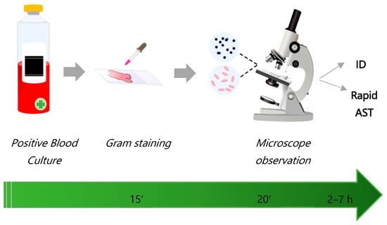
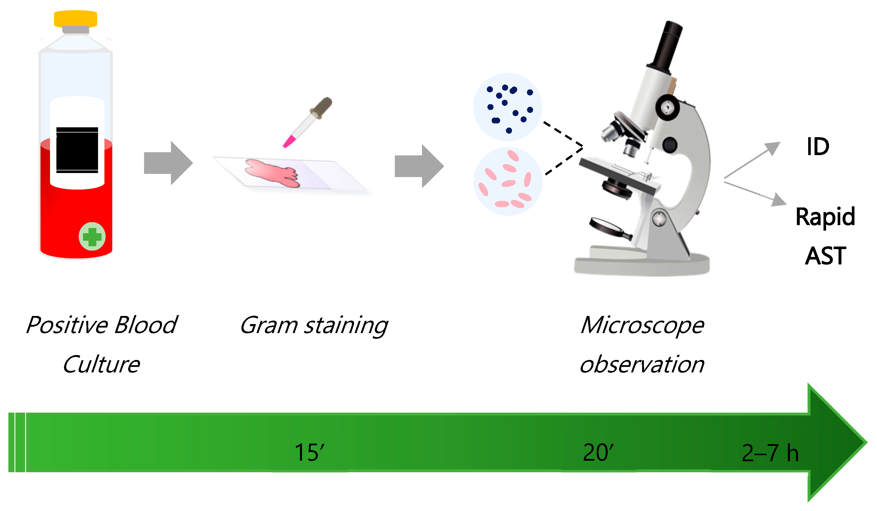

Disclaimer/Publisher’s Note: The statements, opinions and data contained in all publications are solely those of the individual author(s) and contributor(s) and not of MDPI and/or the editor(s). MDPI and/or the editor(s) disclaim responsibility for any injury to people or property resulting from any ideas, methods, instructions or products referred to in the content. |
© 2026 by the authors. Licensee MDPI, Basel, Switzerland. This article is an open access article distributed under the terms and conditions of the Creative Commons Attribution (CC BY) license.