


Abstract
Background: Many of the pathogenic bacteria and fungi found in hospital environments form biofilms, which allow them to persist in the environment for long periods, posing a risk of hospital-acquired infections. Although the pathogens within biofilms often have reduced levels of drug susceptibility, the efficacy of disinfectants routinely applied against planktonic pathogens must be evaluated against biofilms as well. Our objective in this study was to determine the efficacy of treatment using slightly acidic hypochlorous acid water and to compare the results with sodium hypochlorite when both were used to disinfect Candida parapsilosis biofilms. Methods: C. parapsilosis in the planktonic or biofilm state was treated with each disinfectant. The number of viable cells that remained was determined, and scanning electron microscopy (SEM) of the disinfectant-treated biofilms was performed. Results: Compared with sodium hypochlorite, in a shorter period of time, hypochlorous acid water completely killed not only planktonic C. parapsilosis but also C. parapsilosis in a biofilm that had been formed for 72 h. SEM showed that both disinfectants were effective in removing the C. parapsilosis biofilm to some extent. Conclusions: Slightly acidic hypochlorous acid water appears to be an effective disinfectant against C. parapsilosis both in suspension and in biofilms.
1. Introduction
Candida is a genus of fungi present in the environment and on the skin, mucous membranes, and intestinal tracts of humans and animals. These sources can cause an opportunistic infection referred to as candidiasis [1]. In an international study on bloodstream infections, C. parapsilosis was found to be the second most common Candida species in Europe, Canada, and Latin America and the third most common in the United States [2]. In recent years in Japan, C. parapsilosis has been the second most frequently isolated Candida species from bloodstream infections after C. albicans [3,4]. C. parapsilosis also has the ability to form biofilms [5,6]. Biofilm formation in indwelling catheters used in opportunistic patients increases the risk of bloodstream infection [7,8]. C. parapsilosis becomes more virulent and less susceptible to certain antifungal agents depending on the level of biofilm production. Cleaning and disinfecting the hospital environment is very important for infection control because many pathogenic microorganisms can persist on surfaces for long periods of time in biofilms, posing a great risk of hospital-acquired infection through horizontal transmission via contact with healthcare workers and medical equipment. Therefore, measures such as appropriate antifungal therapy and prevention of biofilm contamination in the hospital environment are required [9]. In 2020, three patients in the neonatal intensive care unit at Kurume University Hospital developed invasive C. parapsilosis infections, and C. parapsilosis strains were isolated from all of them. During this time, an environmental survey isolated one C. parapsilosis strain from the incubator of one patient and one from the humidifier of the incubator of a nonfungemia infant. The five C. parapsilosis strains were identical, and all had formed biofilms [10].
Sodium hypochlorite (NaOCl) is an intermediate-level disinfectant exhibiting a broad antimicrobial spectrum against general bacteria, mycobacteria, filamentous fungi, yeast-like fungi, and viruses. NaOCl is much more effective against bacterial spores and non-enveloped viruses than rubbing alcohol, which is considered a low-level disinfectant. For this reason, NaOCl is commonly used to disinfect medical devices and hospital environments. NaOCl is a fast-acting disinfectant; its effectiveness is due to hypochlorous acid (HOCl) [11]. The acid dissociation constant (pKa) of an aqueous HOCl solution at 25 °C is 7.5 [12], and HOCl is most stable as an undissociated molecule at a pH ranging from 4 to 6 [11]. In Japan, HOCl water was designated a food additive disinfectant by the Ministry of Health, Labor and Welfare in 2002 and is currently approved at concentrations ranging from 10 to 80 ppm. Slightly acidic HOCl water with a pH ranging from 5.0 to 6.5 is widely used because the molecule’s stability makes it safe. NaOCl is approved as both a food additive and pharmaceutical disinfectant and is used in concentrations ranging from 200 to 500 ppm to disinfect medical equipment and instruments. Commercially available high-concentration HOCl water is used for unapproved medical disinfection purposes. Various mechanisms of action for the antibacterial effect of HOCl have been reported: it can penetrate bacterial cell membranes, leading to results such as ATP production inhibition [13], protein denaturation [14], DNA damage [15], and DNA synthesis inhibition [16].
Although HOCl has been studied for certain pathogens, its efficacy against mature C. parapsilosis biofilms, an under-researched area in hospital infection control, has not been rigorously compared with that of NaOCl. Therefore, it is clinically important to investigate the antimicrobial spectrum of HOCl water, which, like NaOCl, uses HOCl as a disinfectant molecule. In the present study, our objective was to evaluate the efficacy of slightly acidic HOCl water when used as a disinfectant against C. parapsilosis in suspension and in biofilms and to compare the results with those when using NaOCl. After the disinfectant treatment, viable cell counts were measured, and the biofilms were observed using scanning electron microscopy (SEM).
2. Materials and Methods
2.1. Fungal Strain, Medium, and Disinfectants
C. parapsilosis ATCC 22019 and the E2 strains were used; the ATCC 22019 strain was purchased from Microbiologics (St. Cloud, MN, USA) and the E2 strain was isolated from the environment of a hospital-acquired case [10]. Fungal cells were grown on potato dextrose agar plates (8-MR31; Eiken Chemical Co., Tokyo, Japan) at 37 °C for 3 days under humidified atmospheric conditions and collected by scraping the agar surface using sterile cotton swabs with phosphate-buffered saline (PBS) containing 0.05% Tween 20. The fungal suspension was vortexed for 1 min and passed through a Falcon 40 µm cell strainer (352340; Corning Inc., New York, NY, USA). Fungal cells were pelleted, resuspended in PBS, and serially diluted 10-fold with PBS. Colony counts were performed on potato dextrose agar plates to prepare a final concentration of 1 × 10⁸ colony-forming units (CFU)/mL as a stock fungal solution.
Slightly acidic HOCl water with an effective chlorine concentration of 500 ppm was provided periodically by the Nipro Corporation (Osaka, Japan), generated by electrolysis of a sodium chloride solution using a diaphragm-type three-chamber electrolytic water generator. The resulting solution was subsequently subjected to an evaporative purification process to remove residual ions. The pH of the HOCl water was measured before use and confirmed to be 5.0. NaOCl with an effective chlorine concentration of 500 ppm was prepared by diluting a Yakulax disinfectant solution (509508; Nichi-Iko Pharmaceutical Co., Toyama, Japan) with sterile water, and its pH was measured and confirmed to be 10.4. These disinfectants were stored in light-shielded bottles, refrigerated, and diluted to the required concentration at the time of use. Under these storage conditions, 500 ppm HOCl remains stable for at least six months, and the decrease in effective chlorine concentration is limited to 20 ppm or less. The effective chlorine concentration was measured using AQUAB AQ-202 (Sibata Scientific Technology, Tokyo, Japan).
2.2. Disinfectant Susceptibility Testing
To test the disinfectant susceptibility of planktonic fungal cells, 1 × 10⁶ CFU (10 µL of the stock fungal solution) of each strain was mixed with 490 µL of either HOCl or NaOCl at 5–500 ppm with incubation at room temperature for 5 min. The reaction was terminated by adding 500 µL of PBS containing 1% sodium thiosulfate, and the number of viable cells was determined via counting colonies, as described above. To test the disinfectant susceptibility of fungal biofilms, 1 × 10⁶ CFU of each strain was added to 1 mL of potato dextrose medium (Beckton, Dickinson and Company, Sparks, MD, USA) in 24-well plates with incubation at 37 °C for 72 h in a static state under humidified atmospheric conditions. The medium was changed every 24 h. The biofilms were treated either with HOCl or NaOCl for 10, 30 or 60 min, and the reaction was stopped using 1% sodium thiosulfate. Then, 100 µL of 10-fold serial dilutions of the samples was spread on potato dextrose agar plates, and the colonies were counted.
2.3. Quantitative Biofilm Assay
In our previous study [10], we attempted to use crystal violet staining. However, because the dye did not effectively penetrate the C. parapsilosis biofilm, quantification was impossible. As a result, we used the dry weight method instead. To 1 mL of potato dextrose medium in 24-well plates, 1 × 10⁶ CFU of each strain was added and the plates were then incubated as described above and the dry weights of the biofilms were measured every 24 h. After rinsing with PBS, the biofilms were collected in 1 mL of PBS using cell scrapers (9000-220; AGC Techno Glass, Shizuoka, Japan). The samples were transferred to microtubes and centrifuged at 15,000 rpm for 5 min at 4 °C. The supernatant was removed from each sample, and each precipitate was treated with ice-cold 100% ethanol at 4 °C for 5 min, followed by centrifugation at 15,000 rpm for 5 min. Ethanol was removed from the samples and the precipitates were dried in a heat block at 70 °C for 5 h, with the tube caps open in a safety cabinet and a fan running. The dried materials were weighed using a semi-micro balance.
2.4. SEM
Biofilms were developed on coverslips (C1100; Matsunami Glass Ind., Osaka, Japan) at 37 °C for 72 h as described above and then fixed with 2.5% (v/v) glutaraldehyde and 2% (w/v) paraformaldehyde in PBS overnight at 4 °C. Samples were rinsed with PBS, washed with 7.5% sucrose in PBS for 1 h, and post-fixed in a mixture of 1.5% potassium ferrocyanide and 1% osmium tetroxide in PBS on ice for 1 h. Samples were treated with 2% tannic acid in PBS on ice for 1 h and fixed again with 1% osmium tetroxide in PBS on ice for 1 h. After dehydration in a graded ethanol series, ethanol was replaced with t-butyl alcohol, and the biofilm samples were freeze-dried in t-butyl alcohol in a vacuum freeze-dryer (VFD-30; Vacuum Device Inc., Mito, Japan). Using an osmium plasma coater (Tennant 20; Meiwafosis Co., Tokyo, Japan), the dried samples were coated with osmium metal that was 5 nm thick and then observed via SEM (Quanta 3D FEG; FEI Co., Hillsboro, OR, USA). Four to five random fields of view were captured for each sample. SEM images were obtained at magnifications of 250× and 6000×.
3. Results
3.1. Fungicidal Effect of Chlorine-Based Disinfectants on Planktonic C. parapsilosis
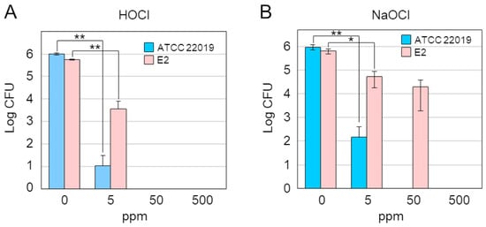
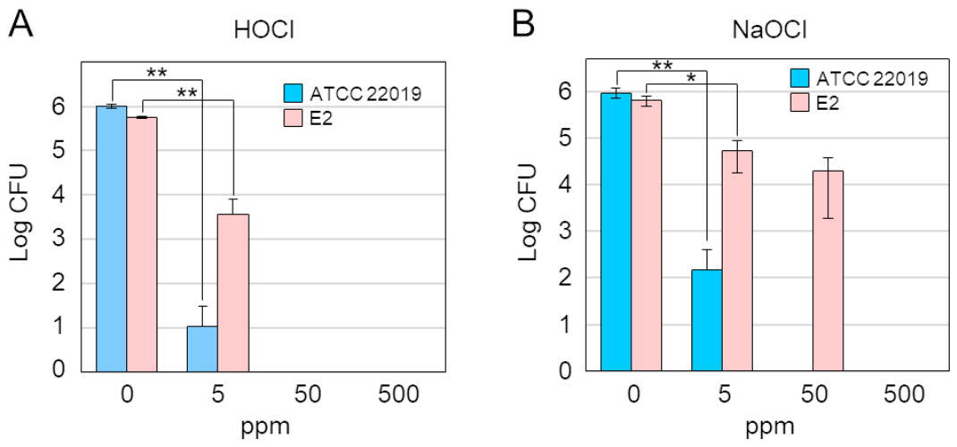
The fungicidal effect of slightly acidic hypochlorous acid (HOCl) water and sodium hypochlorite (NaOCl) on planktonic C. parapsilosis ATCC 22019 and the E2 strains was determined using 5, 50, or 500 ppm (as effective chlorine) of each disinfectant (Figure 1). For the ATCC 22019 strain, a 5-min treatment with either HOCl or NaOCl resulted in partial sterilization at 5 ppm, and sterilization was achieved at 50 ppm. For the E2 strain, a 5-min treatment with HOCl resulted in an average of more than 99% fungicidal activity at 5 ppm, and sterilization was achieved at 50 ppm. A 5-min treatment with NaOCl resulted in an average 92% fungicidal activity at 5 ppm and 97% at 50 ppm, whereas sterilization required 500 ppm.
Figure 1.
Fungicidal effect of chlorine-based disinfectants on planktonic C. parapsilosis. A total of 1 × 10⁶ CFU of each strain was treated with 5, 50, or 500 ppm of either HOCl (A) or NaOCl (B) at room temperature for 5 min. The number of viable cells was determined via colony counting. The results represent the means ± standard errors of three independent experiments, each of which was performed in triplicate (n = 3). Pairwise comparisons between adjacent groups were performed using the Bonferroni correction. ** p < 0.01, * p < 0.05.
3.2. Biofilm Formation and Quantification
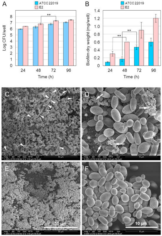
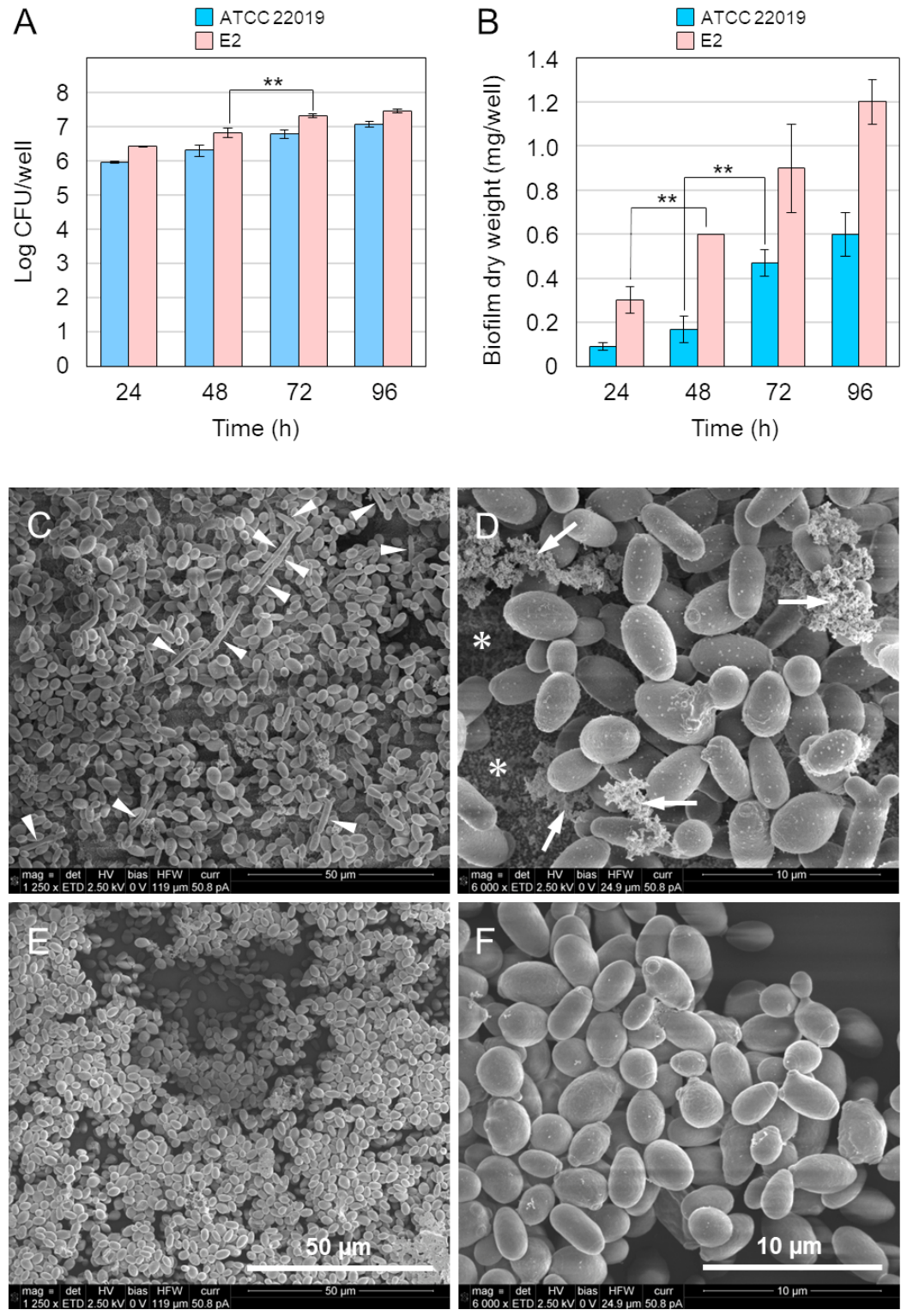
We examined the time courses for viable fungal cell counts and biofilm formation and performed SEM observation of the biofilm (Figure 2). Both the ATCC 22019 and E2 strains exhibited increases in both viable cell counts (Figure 2A) and biofilm weight (Figure 2B) for up to 96 h.
Figure 2.
Biofilm formation of C. parapsilosis. A total of 1 × 10⁶ CFU of each strain was cultured in potato dextrose medium at 37 °C for 72 h, and the medium was changed every 24 h. The numbers of viable fungal cells (A) and the biofilm dry weight (B) were determined every 24 h. The results represent the means ± standard errors of three independent experiments, each of which was performed in triplicate (n = 3). Pairwise comparisons between adjacent groups were performed using the Bonferroni correction. ** p < 0.01. SEM observations were performed on biofilms formed for 72 h by the ATCC 22019 strain (C,D) and the E2 strain (E,F). The arrowheads in panel (C) indicate pseudohyphal cells. The arrows and asterisks (*) in panel (D) denote the extracellular matrix located immediately adjacent to the cells and present at the basal surface of the biofilm, respectively. SEM images were obtained at magnifications of 1250× and 6000×.
C. parapsilosis exhibits oval or spherical yeast-like and pseudohyphal forms and does not form true hyphae [5]. SEM revealed that the ATCC 22019 biofilm formed for 72 h exhibited cells in both yeast-like and pseudohyphal forms, and an extracellular matrix was observed not only immediately adjacent to the cells but also at the bottom surface of the biofilm (Figure 2C,D). On the other hand, the E2 biofilm exhibited predominantly yeast-like cells, with no pseudohyphal morphology detected and only a sparse extracellular matrix observed (Figure 2E,F).
3.3. Fungicidal Effect of Chlorine-Based Disinfectants on C. parapsilosis Biofilms
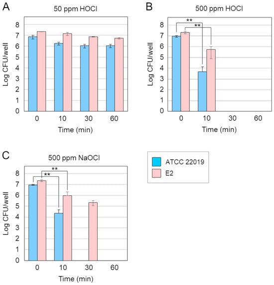
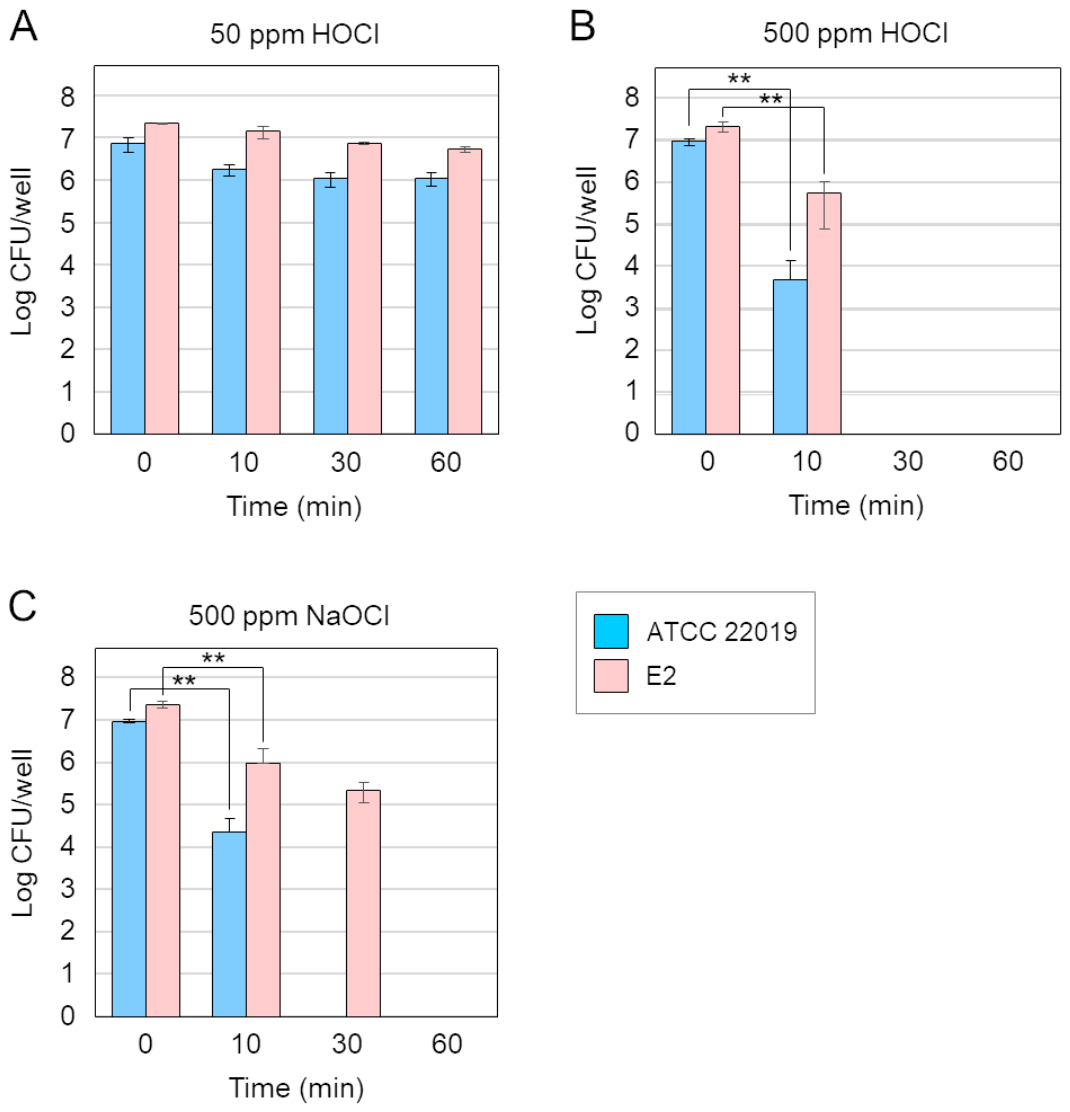
Next, we examined the effect of chlorine-based disinfectants on biofilms that had formed for 72 h (Figure 3). Even after treatment with 50 ppm HOCl for 60 min, an average of 85% of the ATCC 22019 strain and 75% of the E2 strain in the biofilm remained viable (Figure 3A), which indicated a significantly reduced level of susceptibility compared with that of the planktonic state (Figure 1A). These results could have been due to the extracellular matrix acting as a protective barrier for the cells within the biofilm or because the cells entered a metabolically dormant state [17], thereby reducing the effectiveness of HOCl. A 10-min treatment with 500 ppm HOCl achieved an average fungicidal activity of more than 99% against the ATCC 22019 biofilm and 97% against the E2 biofilm. Furthermore, treatment for 30 min completely sterilized the biofilms of both strains, which indicates a potent disinfection activity against C. parapsilosis biofilms (Figure 3B), while 500 ppm NaOCl demonstrated a level of efficacy against the ATCC 22019 biofilm that approximated that of 500 ppm HOCl; even after 30 min of treatment, an average of 2.4% of fungal cells survived in the E2 biofilm (Figure 3C). These results suggest that HOCl exhibits fungicidal activity against C. parapsilosis biofilms, with potency comparable to that of NaOCl. In a similar study, HOCl was found to be effective for cleaning implant surfaces contaminated with biofilms with an efficacy equivalent to that of NaOCl [18].
Figure 3.
Fungicidal effect of chlorine-based disinfectants on C. parapsilosis biofilms. A total of 1 × 10⁶ CFU of each strain was cultured in potato dextrose medium at 37 °C for 72 h and the medium was changed every 24 h. Biofilms were treated with 50 ppm (A) or 500 ppm (B) of HOCl or with 500 ppm of NaOCl (C) at room temperature for the indicated times. The results represent the means ± standard errors of three independent experiments, each of which was performed in triplicate (n = 3). Pairwise comparisons between adjacent groups were performed using the Bonferroni correction. ** p < 0.01.
Taken together, the minimum treatment time required for sterilization of C. parapsilosis biofilms is 30 min with 500 ppm HOCl and 60 min with 500 ppm NaOCl.
3.4. SEM of C. parapsilosis Biofilms Treated with Chlorine-Based Disinfectants
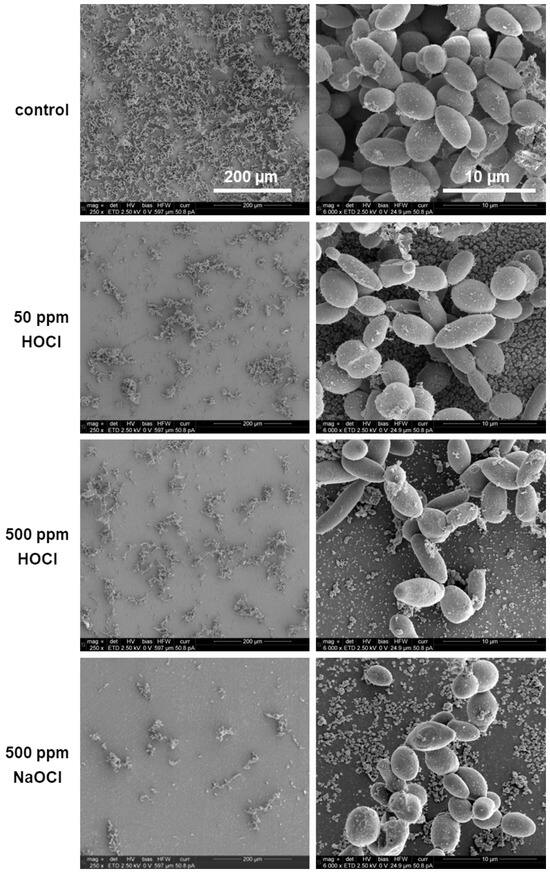
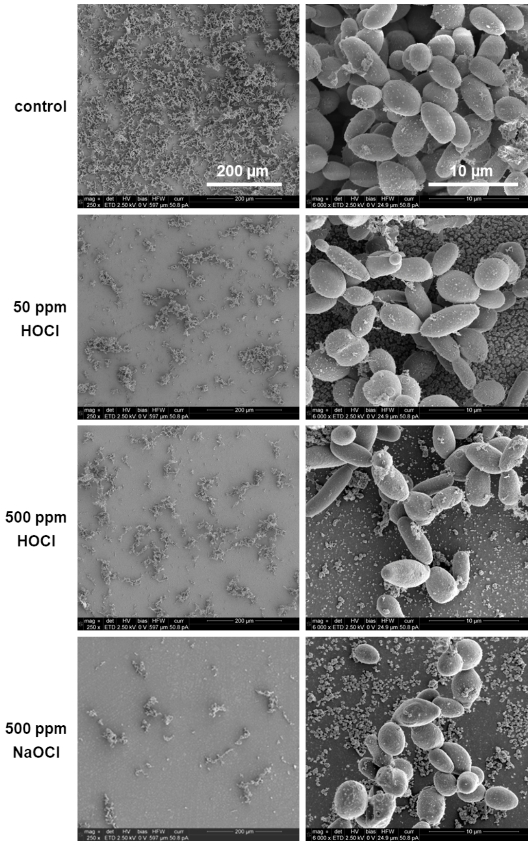
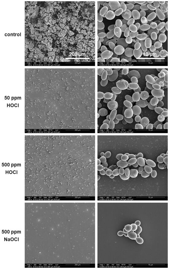
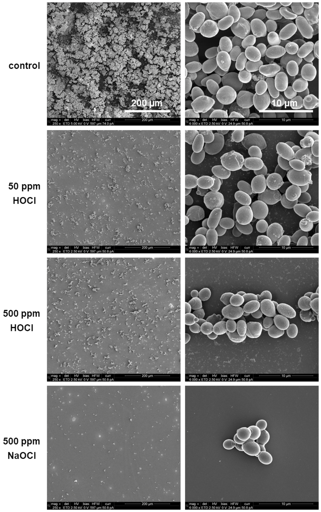
SEM observations were used to visualize the biofilms formed by the ATCC 22019 (Figure 4) strain and the E2 (Figure 5) strain following treatments with either HOCl or NaOCl. When the biofilms cultured for 72 h were treated with either reagent for 60 min, the biofilm quantities decreased significantly. However, neither reagent achieved complete biofilm removal, even when using concentrations of 500 ppm. SEM images of the biofilms after disinfectant treatment provide visual and qualitative data, which complement the quantitative data obtained by measuring the number of viable cells after disinfectant treatment.
Figure 4.
SEM images of the ATCC 22019 biofilms treated with either 50 or 500 ppm of HOCl, or with 500 ppm of NaOCl for 60 min. Biofilm formation was performed for 72 h. SEM images were obtained at magnifications of 250× and 6000×.
Figure 5.
SEM images of the E2 biofilms treated with either 50 or 500 ppm of HOCl, or with 500 ppm of NaOCl for 60 min. Biofilm formation was performed for 72 h. SEM images were obtained at magnifications of 250× and 6000×.
4. Discussion
C. parapsilosis is ubiquitous in nature and is also present on human skin as an indigenous fungus, with a possibility that its presence on human hands could contribute to the horizontal transmission of this fungus in neonatal intensive care units [19,20,21,22]. In neonates, C. parapsilosis is the second most common Candida species associated with bloodstream infections [23]. In 2020, an outbreak of C. parapsilosis occurred in the neonatal care unit at Kurume University Hospital, with five strains isolated: one each from three patients, one from the incubator of a patient with fungemia, and one (the E2 strain) from the humidifier of the incubator of a patient without fungemia. These strains were identical. C. parapsilosis was not isolated from the hands of either the doctors or the nurses who oversaw the three patients. The humidifier from which the E2 strain was isolated still contained water due to a lack of regular cleaning because it was not in use at the time. It is presumed that the remaining water and the material of the internal surfaces of the humidifier provided favorable conditions for the adsorption of C. parapsilosis and subsequent biofilm formation. C. parapsilosis was not isolated from other water environments such as sinks, milk preparation rooms, or a milk warmer, which suggests that the humidifier could have been the transmission source [9]. Other reports have also indicated that C. parapsilosis can be transmitted from the hospital environment to hospitalized patients, possibly through indirect contact via the hands of healthcare workers [24,25]. Therefore, efficiently and effectively disinfecting the hospital environment and the hands of medical staff is important to prevent hospital-acquired infections through horizontal transmission.
The disinfectant ethanol, which is widely used to disinfect hospital environments, is ineffective against bacterial spores [26] and non-enveloped viruses [27,28]. Ethanol is also inapplicable to some materials used in medical equipment and cannot be used over a wide area because it is flammable. NaOCl has a broad-spectrum disinfectant effect against bacteria, including spores, fungi, and viruses [11,27]. Because of its strong alkalinity, however, NaOCl is highly irritating to skin and mucous membranes and in severe cases could cause skin necrosis and blindness; it should therefore obviously be handled with care [29]. The disinfectant activity of NaOCl solutions depends on the concentration of undissociated HOCl, rather than on the available chlorine concentration [30,31,32]. In aqueous HOCl solutions, most of the HOCl molecules exist in an undissociated form in a pH range of 4 to 6 [11]. It presents a very low level of human toxicity and is non-irritating to the eyes and skin [33]; because slightly acidic HOCl water has the same pH as the surface of healthy skin, it recently attracted attention for use as a disinfectant [34]. Saline containing 0.025% HOCl is known to have been used to sterilize Ralstonia pickettii in planktonic and mature biofilms on three types of silicone implants [35]. Sterilization of R. pickettii biofilms with 0.025% HOCl required a longer reaction time than sterilization of R. pickettii in the planktonic state, which was similar to the results we obtained for C. parapsilosis in this study. Furthermore, our results showed that both HOCl and NaOCl required a high concentration of 500 ppm to sterilize C. parapsilosis biofilms and that they exhibited comparable efficacy. Barbot and colleagues reported the efficacy of other disinfectants used for disinfecting dental unit waterlines against C. parapsilosis ATCC 22019 [36]. Hydrogen peroxide exhibited only very weak antimicrobial activity against both planktonic and sessile ATCC 22019. Furthermore, these researchers found that Oxygenal 6, a hydrogen peroxide solution fortified with silver ions, could not sterilize the ATCC 22019 biofilm even at a concentration of 20 mg/mL. That HOCl is extremely safe for the human body is considered an excellent advantage for its use at high concentrations, and it is very promising for use as a disinfectant for C. parapsilosis biofilms in hospital environments.
HOCl is effective for microbial control in enclosed spaces even in its gaseous or fogged form. Enterococcus faecalis, which survives for long periods on surfaces even under dry conditions, could be a potential source of infection in medical settings. However, when present on aluminum plates, this bacterium can be completely killed by treatment with fogged HOCl [37]. HOCl volatilized from a humidifier has been shown to reduce the number of viable airborne microorganisms in a room by nearly half [38]. In the form of dry mist at high concentrations (300–2000 ppm), HOCl is known to be effective not only against bacteria, including spores, but also against fungi and viruses [39,40]. Many inexpensive commercially available sprays containing slightly acidic HOCl water at concentrations of 100–200 ppm are used for unapproved medical disinfection purposes as well as for environmental disinfection in homes and offices.
Through SEM, we have shown that both HOCl and NaOCl at 500 ppm efficiently remove C. parapsilosis biofilms, but a small amount of biofilm remains when sterilization is achieved. Residual disinfected biofilm could serve as a scaffold for secondary biofilm formation [41,42]. Therefore, a combination of wiping procedures is necessary to completely remove biofilms in the hospital environment when using these chlorine-based disinfectants. SEM observations of biofilms formed by two C. parapsilosis strains have revealed differences between the two strains in terms of either the presence or absence of pseudohyphae as well as the degree of biofilm development and amount of extracellular matrix. Comparing only two strains was insufficient for us to determine whether the results were due to strain-specific characteristics or because the E2 strain was isolated from the environment.
The results obtained in the present study are based solely on static in vitro conditions, and when considering the extremely limited number of pathogen species and strains used, such results might not be directly applicable to clinical settings.
In conclusion, our results indicate that 500 ppm HOCl is an effective disinfectant against C. parapsilosis biofilms and is equivalent to the same concentration of NaOCl. Currently, slightly acidic HOCl water is not approved as a pharmaceutical disinfectant in Japan, but confirming the efficacy of HOCl as a disinfectant against various biofilm-forming pathogens that cause invasive and hospital-acquired infections should lead to its approval as a pharmaceutical product.
Future research topics include combining HOCl treatment and wiping procedures to document their effects on C. parapsilosis biofilms that develop on medical devices, as well as determining the efficacy of spraying HOCl.
Author Contributions
Conceptualization, J.I., K.G. and H.W.; methodology, J.I. and H.W.; investigation, J.I.; validation, A.S. and H.F.; resources, A.S., A.T., K.O., N.S. and N.K.; data curation, J.I.; visualization, J.I.; writing—original draft preparation, J.I.; writing—review and editing, J.I., A.S., A.T., K.G., K.O., H.F., N.S., N.K. and H.W.; funding acquisition, H.W.; supervision, N.S., N.K. and H.W. All authors have read and agreed to the published version of the manuscript.
Funding
This study was funded by the Nipro Corporation (grant number 06260006) and conducted as a joint research project.
Institutional Review Board Statement
This study was approved by the Ethical Committee of Kurume University on 20 February 2023 (approval number 22208).
Informed Consent Statement
Not applicable.
Data Availability Statement
The original contributions presented in this study are included in the article. Further inquiries can be directed to the corresponding author.
Conflicts of Interest
Authors Akiko Shimizu, Hiroshi Fuketa, Norihiro Shinkai, and Naohisa Kawamura are employees of the Nipro Corporation. The remaining authors declare that the research was conducted in the absence of any commercial or financial relationships that could be construed as a potential conflict of interest.
References
- Vázquez-González, D.; Perusquía-Ortiz, A.M.; Hundeiker, M.; Bonifaz, A. Opportunistic yeast infections: Candidiasis, cryptococcosis, trichosporonosis and geotrichosis. J. Dtsch. Dermatol. Ges. 2013, 11, 381–394. [Google Scholar] [CrossRef] [Scilit]
- Pfaller, M.A.; Jones, R.N.; Doern, G.V.; Fluit, A.C.; Verhoef, J.; Sader, H.S.; Messer, S.A.; Houston, A.; Coffman, S.; Hollis, R.J. International surveillance of blood stream infections due to Candida species in the European SENTRY Program: Species distribution and antifungal susceptibility including the investigational triazole and echinocandin agents. Diagn. Microbiol. Infect. Dis. 1999, 35, 19–25. [Google Scholar] [CrossRef] [Scilit] [PubMed]
- Hirano, R.; Sakamoto, Y.; Kitazawa, J.; Yamamoto, S.; Kayaba, H. Epidemiology, practice patterns, and prognostic factors for candidemia; and characteristics of fourteen patients with breakthrough Candida bloodstream infections: A single tertiary hospital experience in Japan. Infect. Drug Resist. 2018, 11, 821–833. [Google Scholar] [CrossRef] [Scilit]
- Kajihara, T.; Yahara, K.; Nagi, M.; Kitamura, N.; Hirabayashi, A.; Hosaka, Y.; Abe, M.; Miyazaki, Y.; Sugai, M. Distribution, trends, and antifungal susceptibility of Candida species causing candidemia in Japan, 2010–2019: A retrospective observational study based on national surveillance data. Med. Mycol. 2022, 60, myac071. [Google Scholar] [CrossRef] [Scilit]
- Laffey, S.F.; Butler, G. Phenotype switching affects biofilm formation by Candida parapsilosis. Microbiology 2005, 151, 1073–1081. [Google Scholar] [CrossRef] [Scilit]
- Gómez-Molero, E.; De-la-Pinta, I.; Fernández-Pereira, J.; Groß, U.; Weig, M.; Quindós, G.; de Groot, P.W.J.; Bader, O. Candida parapsilosis colony morphotype forecasts biofilm formation of clinical isolates. J. Fungi 2021, 7, 33. [Google Scholar] [CrossRef] [Scilit]
- Pammi, M.; Holland, L.; Butler, G.; Gacser, A.; Bliss, J.M. Candida parapsilosis is a significant neonatal pathogen: A systematic review and meta-analysis. Pediatr. Infect. Dis. J. 2013, 32, e206–216. [Google Scholar] [CrossRef] [Scilit]
- Yamin, D.H.; Husin, A.; Harun, A. Risk factors of Candida parapsilosis catheter-related bloodstream infection. Front. Public Health 2021, 12, 631865. [Google Scholar] [CrossRef] [Scilit]
- Soldini, S.; Posteraro, B.; Vella, A.; De Carolis, E.; Borghi, E.; Falleni, M.; Losito, A.R.; Maiuro, G.; Trecarichi, E.M.; Sanguinetti, M.; et al. Microbiologic and clinical characteristics of biofilm-forming Candida parapsilosis isolates associated with fungaemia and their impact on mortality. Clin. Microbiol. Infect. 2018, 24, 771–777. [Google Scholar] [CrossRef] [Scilit] [PubMed]
- Miyake, A.; Gotoh, K.; Iwahashi, J.; Togo, A.; Horita, R.; Miura, M.; Kinoshita, M.; Ohta, K.; Yamashita, Y.; Watanabe, H. Characteristics of biofilms formed by C. parapsilosis causing an outbreak in a neonatal intensive care unit. J. Fungi 2022, 8, 700. [Google Scholar] [CrossRef] [Scilit] [PubMed]
- Fukuzaki, S. Mechanisms of actions of sodium hypochlorite in cleaning and disinfection processes. Biocontrol Sci. 2006, 11, 147–157. [Google Scholar] [CrossRef] [Scilit]
- Morris, J.C. The acid ionization constant of HOCl from 5 to 35°. J. Phys. Chem. 1966, 70, 3798–3805. [Google Scholar] [CrossRef] [Scilit]
- Barrette, W.C., Jr.; Hannum, D.M.; Wheeler, W.D.; Hurst, J.K. General mechanism for the bacterial toxicity of hypochlorous acid: Abolition of ATP production. Biochemistry 1989, 28, 9172–9178. [Google Scholar] [CrossRef] [Scilit]
- Winter, J.; Ilbert, M.; Graf, P.C.F.; Özcelik, D.; Jakob, U. Bleach activates a redox-regulated chaperone by oxidative protein unfolding. Cell 2008, 135, 691–701. [Google Scholar] [CrossRef] [Scilit]
- Dukan, S.; Touati, D. Hypochlorous acid stress in Escherichia coli: Resistance, DNA damage, and comparison with hydrogen peroxide stress. J. Bacteriol. 1996, 178, 6145–6150. [Google Scholar] [CrossRef] [Scilit]
- McKenna, S.M.; Davies, K.J.A. The inhibition of bacterial growth by hypochlorous acid. Possible role in the bactericidal activity of phagocytes. Biochem. J. 1988, 254, 685–692. [Google Scholar] [CrossRef] [Scilit]
- Hall, C.W.; Mah, T.-F. Molecular mechanisms of biofilm-based antibiotic resistance and tolerance in pathogenic bacteria. FEMS Microbiol. Rev. 2017, 41, 276–301. [Google Scholar] [CrossRef] [Scilit] [PubMed]
- Chen, C.J.; Chen, C.C.; Ding, S.J. Effectiveness of hypochlorous acid to reduce the biofilms on titanium alloy surfaces in vitro. Int. J. Mol. Sci. 2016, 17, 1161. [Google Scholar] [CrossRef] [Scilit] [PubMed]
- Lupetti, A.; Tavanti, A.; Davini, P.; Ghelardi, E.; Corsini, V.; Merusi, I.; Boldrini, A.; Campa, M.; Senesi, S. Horizontal transmission of Candida parapsilosis candidemia in a neonatal intensive care unit. J. Clin. Microbiol. 2002, 40, 2363–2369. [Google Scholar] [CrossRef] [Scilit]
- Bonassoli, L.A.; Bertoli, M.; Svidzinski, T.I.E. High frequency of Candida parapsilosis on the hands of healthy hosts. J. Hosp. Infect. 2005, 59, 159–162. [Google Scholar] [CrossRef] [Scilit] [PubMed]
- van Asbeck, E.C.; Huang, Y.C.; Markham, A.N.; Clemons, K.V.; Stevens, D.A. Candida parapsilosis fungemia in neonates: Genotyping results suggest healthcare workers hands as source, and review of published studies. Mycopathologia 2007, 164, 287–293. [Google Scholar] [CrossRef] [Scilit]
- Hernández-Castro, R.; Arroyo-Escalante, S.; Carrillo-Casas, E.M.; Moncada-Barrón, D.; Álvarez-Verona, E.; Hernández-Delgado, L.; Torres-Narváez, P.; Lavalle-Villalobos, A. Outbreak of Candida parapsilosis in a neonatal intensive care unit: A health care workers source. Eur. J. Pediatr. 2010, 169, 783–787. [Google Scholar] [CrossRef] [Scilit]
- Roilides, E.; Farmaki, E.; Evdoridou, J.; Dotis, J.; Hatziioannidis, E.; Tsivitanidou, M.; Bibashi, E.; Filioti, I.; Sofianou, D.; Gil-Lamaignere, C.; et al. Neonatal candidiasis: Analysis of epidemiology, drug susceptibility, and molecular typing of causative isolates. Eur. J. Clin. Microbiol. Infect. Dis. 2004, 23, 745–750. [Google Scholar] [CrossRef] [Scilit]
- Sanchez, V.; Vazquez, J.A.; Barth-Jones, D.; Dembry, L.; Sobel, J.D.; Zervos, M.J. Nosocomial acquisition of Candida parapsilosis: An epidemiologic study. Am. J. Med. 1993, 94, 577–582. [Google Scholar] [CrossRef] [Scilit]
- Sabino, R.; Sampaio, P.; Rosado, L.; Videira, Z.; Grenouillet, F.; Pais, C. Analysis of clinical and environmental Candida parapsilosis isolates by microsatellite genotyping—A tool for hospital infection surveillance. Clin. Microbiol. Infect. 2015, 21, 954.e1–954.e8. [Google Scholar] [CrossRef] [Scilit] [PubMed]
- Edwards, A.N.; Karim, S.T.; Pascual, R.A.; Jowhar, L.M.; Anderson, S.E.; McBride, S.M. Chemical and stress resistances of Clostridium difficile spores and vegetative cells. Front. Microbiol. 2016, 7, 1698. [Google Scholar] [CrossRef] [Scilit]
- Eterpi, M.; McDonnell, G.; Thomas, V. Disinfection efficacy against parvoviruses compared with reference viruses. J. Hosp. Infect. 2009, 73, 64–70. [Google Scholar] [CrossRef] [Scilit] [PubMed]
- Behrendt, P.; Friesland, M.; Wißmann, J.E.; Kinast, V.; Stahl, Y.; Praditya, D.; Hueffner, L.; Nörenberg, P.M.; Bremer, B.; Maasoumy, B.; et al. Hepatitis E virus is highly resistant to alcohol-based disinfectants. J. Hepatol. 2022, 76, 1062–1069. [Google Scholar] [CrossRef] [Scilit] [PubMed]
- Rutala, W.A.; Weber, D.J. Disinfection and sterilization in health care facilities: An overview and current issues. Infect. Dis. Clin. N. Am. 2021, 35, 575–607. [Google Scholar] [CrossRef] [Scilit]
- Charlton, D.B.; Levine, M. Some observations on the germicidal efficiency of chloramines-T and calcium hypochlorite. J. Bacteriol. 1935, 30, 163–171. [Google Scholar] [CrossRef] [Scilit]
- Sagripanti, J.L.; Bonifacino, A. Comparative sporicidal effects of liquid chemical agents. Appl. Environ. Microbiol. 1996, 62, 545–551. [Google Scholar] [CrossRef] [Scilit]
- Dantec, C.L.; Duguet, J.-P.; Montiel, A.; Dumoutier, N.; Dubrou, S.; Vincent, V. Chlorine disinfection of atypical mycobacteria isolated from a water distribution system. Appl. Environ. Microbiol. 2002, 68, 1025–1032. [Google Scholar] [CrossRef] [Scilit] [PubMed]
- Wang, L.; Bassiri, M.; Najafim, R.; Najafi, K.; Yang, J.; Khosrovi, B.; Hwong, W.; Barati, E.; Belisle, B.; Celeri, C.; et al. Hypochlorous acid as a potential wound care agent: Part I. Stabilized hypochlorous acid: A component of the inorganic armamentarium of innate immunity. J. Burns Wounds 2007, 6, e5. [Google Scholar]
- Haralović, V.; Mokos, M.; Špoljar, S.; Dolački, L.; Šitum, M.; Lugović-Mihić, L. Hypochlorous acid: Clinical insights and experience in dermatology, surgery, dentistry, ophthalmology, rhinology, and other specialties. Biomedicines 2025, 13, 2921. [Google Scholar] [CrossRef] [Scilit]
- Brindle, C.T.; Porter, S.; Bijlani, K.; Arumugam, S.; Matias, R.; Najafi, R.; Fisher, J. Preliminary results of the use of a stabilized hypochlorous acid solution in the management of Ralstonia pickettii biofilm on silicone breast implants. Aesthet. Surg. J. 2018, 38, S52–S61. [Google Scholar] [CrossRef] [Scilit]
- Barbot, V.; Costa, D.; Deborde, M.; Imbert, C. Efficacy of dental unit disinfectants against Candida spp. and Hartmannella vermiformis. Pathog. Dis. 2014, 70, 289–296. [Google Scholar] [CrossRef] [Scilit]
- Feng, K.-C.; Ghai, A.; Liu, H.; Salerno, A.; Miller, C.; Liu, J.; Manesh, S.; Cymerman, J.; Simon, M.; Walker, S.G.; et al. Efficacy of hypochlorous acid (HOCl) fog in sanitizing surfaces against Enterococcus faecalis. Am. J. Infect. Control 2022, 50, 1311–1315. [Google Scholar] [CrossRef] [Scilit]
- Nojima, S.; Omura, S.; Fukuzaki, S. Volatilization and disinfection efficacy of gaseous hypochlorous acid from an air washer-type humidifier in a large space. J. Microorg. Control 2024, 29, 105–111. [Google Scholar] [CrossRef] [Scilit] [PubMed]
- Park, G.W.; Boston, D.M.; Kase, J.A.; Sampson, M.N.; Sobsey, M.D. Evaluation of liquid- and fog-based application of Sterilox hypochlorous acid solution for surface inactivation of human norovirus. Appl. Environ. Microbiol. 2007, 73, 4463–4468. [Google Scholar] [CrossRef] [Scilit] [PubMed]
- Nasiłowska, B.; Włodarski, M.; Kaliszewski, M.; Bogdanowicz, Z.; Krzowski, Ł.; Kopczyński, K.; Witkowski, G.; Czeczott-Urban, A.; Bombalska, A.; Urbańska, M.; et al. Decontamination effect of hypochlorous acid dry mist on selected bacteria, viruses, spores, and fungi as well as on components of electronic systems. Int. J. Mol. Sci. 2024, 25, 7198. [Google Scholar] [CrossRef] [Scilit] [PubMed]
- Yamaguchi, M.; Noiri, Y.; Kuboniwa, M.; Yamamoto, R.; Asahi, Y.; Maezono, H.; Hayashi, M.; Ebisu, S. Porphyromonas gingivalis biofilms persist after chlorhexidine treatment. Eur. J. Oral Sci. 2013, 121, 162–168. [Google Scholar] [CrossRef] [Scilit] [PubMed]
- Ohsumi, T.; Takenaka, S.; Wakamatsu, R.; Sakaue, Y.; Narisawa, N.; Senpuku, H.; Ohshima, H.; Terao, Y.; Okiji, T. Residual structure of Streptococcus mutans biofilm following complete disinfection favors secondary bacterial adhesion and biofilm re-development. PLoS ONE 2015, 10, e0116647. [Google Scholar] [CrossRef] [Scilit] [PubMed]
Disclaimer/Publisher’s Note: The statements, opinions and data contained in all publications are solely those of the individual author(s) and contributor(s) and not of MDPI and/or the editor(s). MDPI and/or the editor(s) disclaim responsibility for any injury to people or property resulting from any ideas, methods, instructions or products referred to in the content. |
© 2026 by the authors. Licensee MDPI, Basel, Switzerland. This article is an open access article distributed under the terms and conditions of the Creative Commons Attribution (CC BY) license.