


Abstract
The rise in multidrug resistance in Klebsiella pneumoniae is an alarming issue, especially in invasive infections among patients with co-morbidities. With the gain of hypervirulence traits, multidrug-resistant K. pneumoniae has led to a significant increase in chronic infections and associated mortality. This study aims to explore the distribution of multidrug-resistant and hypervirulent (hv) K. pneumoniae in invasive infections in a tertiary care hospital. A total of 231 K. pneumoniae isolates were collected over a period of six months from invasive infections. These isolates were tested phenotypically and genotypically for the presence of antimicrobial resistance, along with molecular detection of hypervirulence determinants (iucA, rmpA, rmpA2, peg344, iroB). High levels of resistance to β-lactams, fluoroquinolones, and aminoglycosides were observed. Carbapenemase-encoding genes were widely distributed, and 22% showed the presence of at least one hypervirulence gene, most commonly iucA and rmpA. Co-carriage of resistance and hypervirulence determinants in K. pneumoniae was observed in nearly 20% of the isolates, indicating the emergence of MDR-hvKP phenotypes in the hospital setting. Mortality was significantly higher among patients infected with MDR isolates, whereas hypervirulence markers were not independently associated with mortality. The presence of MDR–hypervirulent strains remains clinically concerning and underscores the need for continued genomic surveillance.
1. Introduction
Klebsiella pneumoniae is an opportunistic, Gram-negative pathogen that is responsible for a broad spectrum of clinical infections, including bloodstream infections, pneumonia (including ventilator-associated pneumonia), urinary tract infections, and liver abscesses, particularly in immunocompromised or hospitalized patients [1]. Over the last decade, the pathogen has acquired both extensive antimicrobial resistance (AMR) and hypervirulence (hv) traits, resulting in convergent strains that are difficult to treat and capable of causing invasive infections, even in healthy hosts [2].
The globally disseminated multidrug-resistant (MDR), particularly carbapenem-resistant, K. pneumoniae (CRKP) is a significant member of the ESKAPE group of pathogens and an emerging health threat [3]. It is associated with severely limited therapeutic options, prolonged hospital stays, increased healthcare costs and high mortality. Multidrug resistance can be mediated by the production of antibiotic hydrolyzing enzymes such as carbapenemase (encoded by blaNDM, blaOXA-48-like, blaKPC, blaVIM, and blaIMP), β-lactamases (encoded by blaTEM, blaSHV, blaOXA-1), and the loss of porins or the overexpression of efflux pumps [4,5]. In parallel, K. pneumoniae has evolved hypervirulent phenotypes characterized by the presence of siderophore systems (iucA, iroB) [6,7], regulator genes (rmpA, rmpA2) [8] and a virulence-associated marker (peg344) [9], which contribute to enhanced iron acquisition, immune evasion, and increased tissue invasiveness [10,11]. The emergence of hypervirulent Klebsiella pneumoniae (hvKP) as a major clinical pathogen has raised significant concern, due to its ability to cause severe, invasive and often life-threatening infections. Unlike classical K. pneumoniae (cKP), hvKP strains possess distinct virulence determinants that enhance the bacterial capacity to invade host tissue, leading to survival and adverse clinical outcomes [12].
Although hvKP is traditionally associated with community-acquired infections, it is increasingly being reported in nosocomial settings, where it may acquire resistance or evolve from MDR cKP strains via horizontal gene transfer. This concern is particularly acute in the intensive care units (ICUs), which serve as critical reservoirs for MDR K. pneumoniae strains due to frequent exposure to antibiotics [13]. These dynamics highlight the potential for hvKP to disseminate within healthcare environments, requiring the urgent need to implement rigorous infection control measures to reduce its transmission rates.
Recently, co-occurrence of carbapenem resistance and hypervirulence determinants has been reported globally and poses a significant threat to public health [14,15]. In a genomic survey for more than two thousand global hvKP, 33.3% were found to harbor carbapenem-resistant determinants [16]. Similarly, studies from China [17], Malaysia [18], Portugal [19], United States [20], and France [21] have reported prevalence of carbapenem-resistant hvKP ranging from 1% to 15% and more than 30% in hospital-based cohorts [22,23,24]. In India, studies have also reported a rise in multidrug-resistant K. pneumoniae invasive infections in both neonatal and adult populations [25,26,27,28]. This convergence represents a particularly alarming development, as strains combining high-level resistance with enhanced virulence have the potential to cause severe, rapidly progressive infections for which effective treatment options are extremely limited. Thus, management of MDR-hvKP often relies on last-resort antimicrobials such as polymyxins (colistin), tigecycline, fosfomycin and newer β-lactam/β-lactamase inhibitor combinations (e.g., ceftazidime–avibactam), although their efficacy depends upon the underlying resistance mechanism [2,29,30].
Despite the growing threat, important gaps remain in our understanding of the epidemiology and clinical relevance of MDR-hypervirulent K. pneumoniae. Though many studies rely on whole genome sequencing, which is highly informative, it is still not available routinely in most diagnostic laboratories, especially in resource-limited settings. Thus, there is a need for systematic surveillance using accessible approaches such as phenotypic antimicrobial susceptibility testing combined with targeted PCR-based detection of key resistance and hypervirulence determinants. Therefore, the present study aims to determine the phenotypic and molecular resistance patterns and the distribution of hypervirulence markers among K. pneumoniae isolates from invasive clinical infections at a tertiary care center. In addition, the study evaluates the association of multidrug resistance and the presence of hypervirulence determinants with clinical outcome, defined at the time of discharge of the patient. By integrating microbiological and clinical data, this study seeks to provide insight into the emerging threat posed by MDR and hypervirulent K. pneumoniae in hospital settings.
2. Materials and Methods
2.1. Study Design and Isolate Collection
To determine the prevalence of hvKP, especially among isolates obtained from invasive infections, a total of 231 non-repetitive K. pneumoniae clinical isolates were collected over a period of six months from clinical samples such as blood, cerebrospinal fluid, and other sterile body fluids. These samples were received in the routine laboratory of the Department of Medical Microbiology, PGIMER, Chandigarh. The isolates were cultured on blood agar and MacConkey agar (HiMedia Pvt. Ltd, Mumbai, India) and incubated at 37 °C under aerobic conditions. The lactose-fermenting colonies identified as Klebsiella pneumoniae by matrix-assisted laser desorption ionization-time of flight mass spectrometry (MALDI-TOF) using Vitek MS system (bioMérieux, Marcy l’Etoile, France) were further subjected to the genotypic tests (PCR) to classify them as classical K. pneumoniae (cKP) or hvKP.
The clinical data (general demographics such as age, sex, admission status and discharge status) for each sample were collected retrospectively from the available routine clinical records. The clinical outcome for each patient was defined based on the documented status as death or discharged. Patients documented as discharges were considered alive at the time of discharge. Post-discharge follow-up data were not available, as longitudinal outcome assessment was beyond the scope of the study.
2.2. Identification and Antimicrobial Susceptibility Testing
Antimicrobial susceptibility testing (AST) was conducted by the Vitek 2 automated system (bioMérieux) with AST-N405 cards for Enterobacterales and the disk diffusion method. Disk diffusion was performed using the Kirby–Bauer method. Briefly, two to three pure colonies of the fresh overnight culture were suspended in sterile 0.85% normal saline and adjusted to a turbidity of 0.5 McFarland standard. The suspension was uniformly swabbed on fresh and dried Mueller Hinton agar plates (HiMedia). The antibiotic disks were placed on the inoculated agar surface using a sterile forceps, and the plates were incubated aerobically at 37 °C for 16–18 h. The zone of inhibition for each antibiotic was measured using an antibiotic zone scale (HiMedia) and interpreted according to the Clinical and Laboratory Standards Institute (CLSI) guidelines (M100, 34th Edition) [31].
Colistin susceptibility was determined using the microbroth dilution (MBD) method in Mueller Hinton Broth No. 2 Control Cations (HiMedia) with colistin sulfate salt (Thermo Scientific™, Waltham, MA, USA). Two-fold serial dilutions of colistin ranging from 0.25 µg/mL to 16 µg/mL were tested, and the minimum inhibitory concentrations (MICs) were interpreted according to the CLSI guidelines, with MICs ≤ 2 µg/mL considered intermediate susceptible and MICs ≥ 4 µg/mL as a full stop after resistant Pseudomonas aeruginosa ATCC 27853 used as the quality control for MIC determination.
2.3. DNA Extraction and PCR Amplification
Genomic DNA was extracted by the boiling lysis method [32]. Briefly, loopful bacterial growth was suspended in 100 µL of sterile nuclease free molecular grade water (HiMedia), heated at 95 °C for 10 min and immediately chilled on ice. The suspension was centrifuged at 10,000 rpm for 5 min. The collected supernatant was used as the template DNA for PCR amplification of carbapenemase genes (blaNDM, blaOXA-48-like, blaKPC, blaVIM, blaIMP), ESBL genes (blaTEM, blaSHV, blaOXA-1), and hypervirulence genes (iucA, rmpA, peg344, iroB) [33]. For each gene amplification, 12 µL PCR reaction mix was prepared, containing 100 ng of template DNA, 1× PCR buffer (supplemented with 1.5 mM MgCl2), 1U of Taq polymerase (DreamTaq, Thermo Scientific™), 0.2 mM of each dNTPs (dNTP Mix, Thermo Scientific™), and 0.5 µM each of the forward and reverse primers (Table 1), and was amplified using a thermocycler (SimpliAmp Thermal Cycler, Applied Biosystems, Foster City, CA, USA). The cycling conditions consisted of an initial denaturation at 95 °C for 5 min, followed by 30 cycles of denaturation at 95 °C for 30 s, annealing at gene-specific temperatures for 30 s, extension at 72 °C for 1 min, and a final extension at 72 °C for 7 min. PCR products were resolved on 1.5% agarose gels stained with ethidium bromide and visualized by a gel documentation system (Azure400, Azure Biosystems, Dublin, CA, USA).
Table 1.
Antimicrobial genes and PCR conditions.
2.4. Statistical Analysis
Phenotypic antimicrobial susceptibility and PCR-based detection of antimicrobial resistance and hypervirulence genes were compiled using Microsoft Excel. Descriptive statistics were used to summarize resistance patterns and gene distributions, expressed as frequencies and percentages. Associations between categorical variables were assessed using Chi-square or Fisher’s exact test, as appropriate, with odds ratios and 95% confidence intervals calculated. Penalized logistic regression using Firth’s correction was performed to evaluate the independent and combined effects of multidrug resistance and hypervirulence on mortality. The clinical outcome was categorized as death or alive at the last documented hospital contact. Patients discharged alive and outpatient cases were all classified as alive for the purpose of regression analysis. Statistical analyses were conducted using GraphPad Prism version 8 and R software (version 4.4.1) with the logistf package (version 1.26.1). A two-tailed p-value < 0.05 was considered statistically significant.
3. Results
3.1. Sample Distribution/Demographic Description of Patients
During the six-month study period, a total of 231 Klebsiella pneumoniae isolates were obtained from invasive clinical samples. Of these, 170 isolates were recovered from blood, 52 from sterile body fluids (including pleural fluid and ascitic fluid), and 9 from cerebrospinal fluid. A majority of the isolates were obtained from patients admitted to various intensive care units (n = 62) or inpatient wards (n = 156), while 13 isolates were recovered from outpatient department (OPD) cases.
The patient population comprised 155 males and 76 females. The cases were categorized according to the age groups defined by the WHO, and the distribution of the cases is presented in Table 2. Of these, 72 isolates (31.2%) were obtained from the pediatric population, spanning neonates to adolescents, while 159 isolates (68.8%) were from adults (>18 years). Among the pediatric cases, the highest representation was from young infants aged 1–3 months (n = 27), followed by toddlers aged 1–3 years (n = 13). Overall, in-hospital mortality at the time of discharge was recorded in 37% of the admitted patients, including 57 adults and 24 pediatric patients.
Table 2.
Distribution of patients in various age groups.
3.2. Antimicrobial Resistance Among Isolates
3.2.1. Phenotypic Antibiotic Susceptibility Pattern
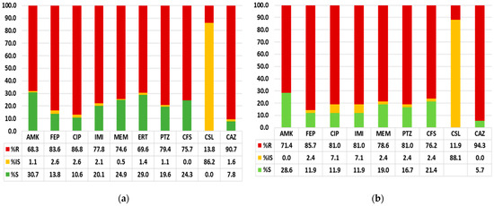
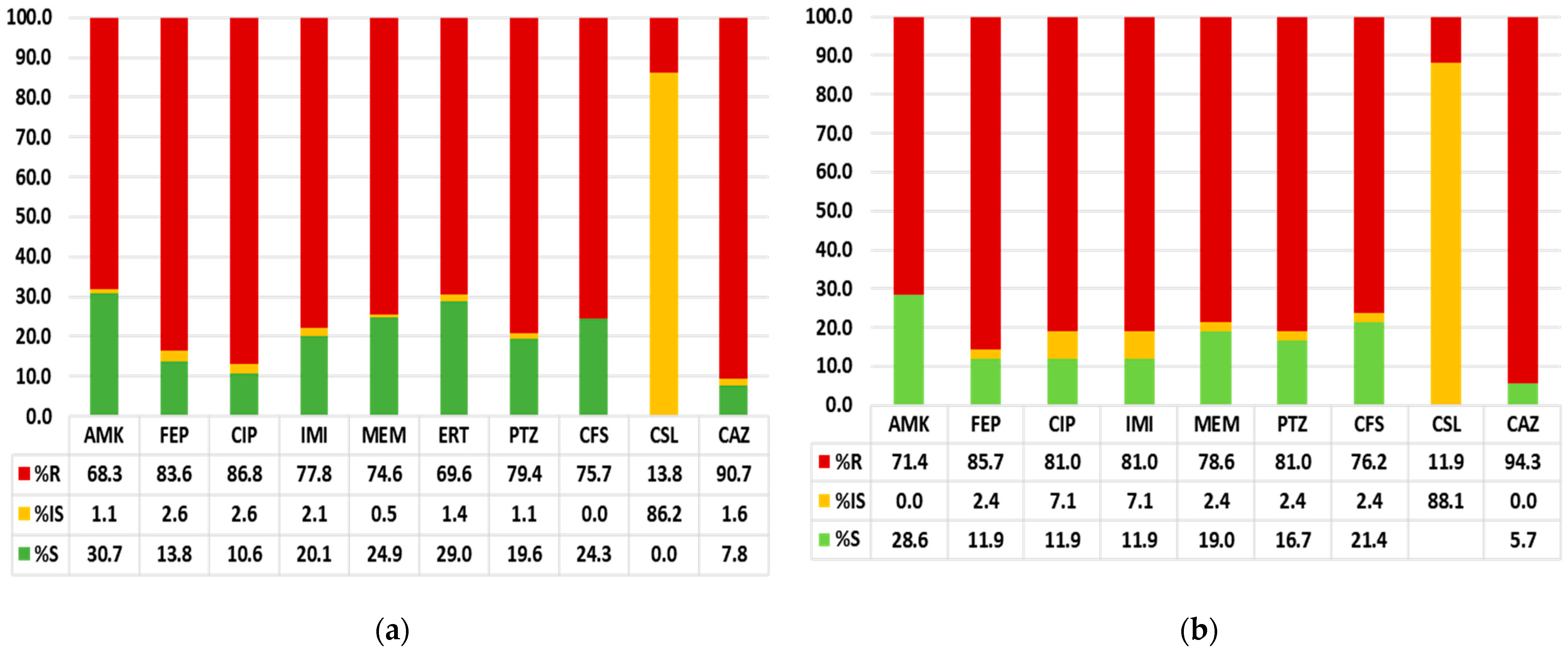
The phenotypic antibiotic susceptibility pattern revealed that more than 75% of the isolates were multidrug-resistant (MDR), as per the definitions proposed by Magiorakos et al. [34], suggesting non-susceptibility to at least one agent in three or more antimicrobial classes. Among aminoglycosides, only 30.3% of isolates were susceptible to amikacin, while 68.8% showed resistance. Resistance to cephalosporins was markedly high, with 84.0% of isolates being resistant to cefepime and 91.5% resistant to ceftazidime. Fluoroquinolone susceptibility was similarly poor, with 85.7% of isolates exhibiting resistance to ciprofloxacin. Carbapenem resistance was widespread, with 78.4% of isolates being resistant to imipenem, 75.3% to meropenem, and 70.0% to ertapenem. Only a minority of isolates demonstrated intermediate susceptibility across these agents. Among β-lactam/β-lactamase inhibitor combinations, resistance was also substantial, with 79.7% resistance to piperacillin–tazobactam and 75.8% resistance to cefoperazone–sulbactam. In contrast, colistin susceptibility remained high, with 86.6% of isolates showing intermediate susceptibility (as per CLSI MIC interpretive breakpoints) and only 13.4% demonstrating resistance.
Among the 42 isolates obtained from infants (<1 year), antimicrobial susceptibility patterns closely mirrored those of the overall cohort, with high rates of resistance across most drug classes. Resistance to amikacin (71.4%); cefepime (85.7%); ceftazidime (94.3%); ciprofloxacin (81.0%); and the carbapenems, imipenem (81.0%) and meropenem (78.6%), was prominent. β-lactam/β-lactamase inhibitor combinations also showed reduced activity, with 81.0% resistance to piperacillin–tazobactam and 76.2% resistance to cefoperazone–sulbactam. Colistin remained the most active agent, with 88.1% intermediate susceptibility and 11.9% resistance (Figure 1).
Figure 1.
Bar graph representing the antimicrobial susceptibility pattern of the K. pneumoniae isolates from (a) infants (<1 year) and (b) older age groups. The comparison highlights similarities in resistance profiles across age groups, suggesting shared circulating strains and antimicrobial selection pressure within the hospital setting. Antibiotic abbreviations: AMK, amikacin; FEP, cefepime; CIP, ciprofloxacin; IMI, imipenem; MEM, meropenem; ERT, ertapenem; PTZ, piperacillin–tazobactam; CFS, cefoperazone–sulbactam; CSL, colistin; and CAZ, ceftazidime.
3.2.2. Genotypic Antibiotic Susceptibility Pattern
Genotypic screening of the 231 isolates revealed a substantial burden of β-lactamase and carbapenemase genes. Among the ESBL determinants, blaSHV (69.3%, n = 160) and blaTEM (53.2%, n = 123) were most frequently detected, while blaOXA-1 was present in 16.0% (n = 37) of isolates. Carbapenemase genes were also widely distributed, with blaNDM identified in 47.2% (n = 109) and blaOXA-48-like in 39.4% (n = 91) of isolates, whereas blaVIM (4.8%, n = 11), blaKPC (4.8%, n = 11), and blaIMP (2.2%, n = 5) were detected at lower frequencies. A similar pattern was observed among isolates obtained from infants, which demonstrated high carriage of ESBL genes, blaSHV (78.6%, n = 33) and blaTEM (64.3%, n = 27) and the carbapenemase blaNDM (50%, n = 21).
Overall, 137 isolates (59%) harbored both ESBL and carbapenemase genes, while 26% (n = 59) carried only ESBLs and 8% (n = 19) carried only carbapenemase genes. Notably, 16 isolates (7%) lacked all tested resistance genes; however, a phenotype–genotype comparison revealed that only four of these were fully susceptible to all antibiotics, whereas the remaining 12 isolates were multidrug-resistant despite the absence of detectable resistance determinants. Conversely, seven isolates were phenotypically susceptible to all tested agents yet carried one or more AMR genes (Table 3).
Table 3.
Multidrug resistance, based on phenotypic antimicrobial susceptibility and presence of AMR determinants by conventional PCR.
3.3. Hypervirulence Determinants Among Isolates
Screening for hypervirulence-associated genes revealed that the majority of isolates (181/231, 78.4%) did not carry any of the tested hv markers. Among positive isolates, iucA was the most frequently detected gene, present in 35 isolates (15.2%), followed by rmpA, detected in 22 isolates (9.5%). A subset of isolates carried combinations of hypervirulence genes, including rmpA + iucA (n = 2), rmpA + iucA + iroB (n = 2), and rmpA + iucA + peg344 (n = 2). Less frequent patterns included iucA + rmpA2 (n = 1) and peg344 + iucA (n = 1). Only a single isolate (0.4%) harbored the complete set of tested hypervirulence markers (rmpA, peg344, iucA, iroB, and rmpA2). Overall, 50 isolates (21.6%) carried at least one hv gene, indicating a modest but notable presence of hypervirulence-associated genetic determinants within the K. pneumoniae isolates obtained from invasive samples.
3.4. Co-Occurrence of AMR and Hypervirulence Genes
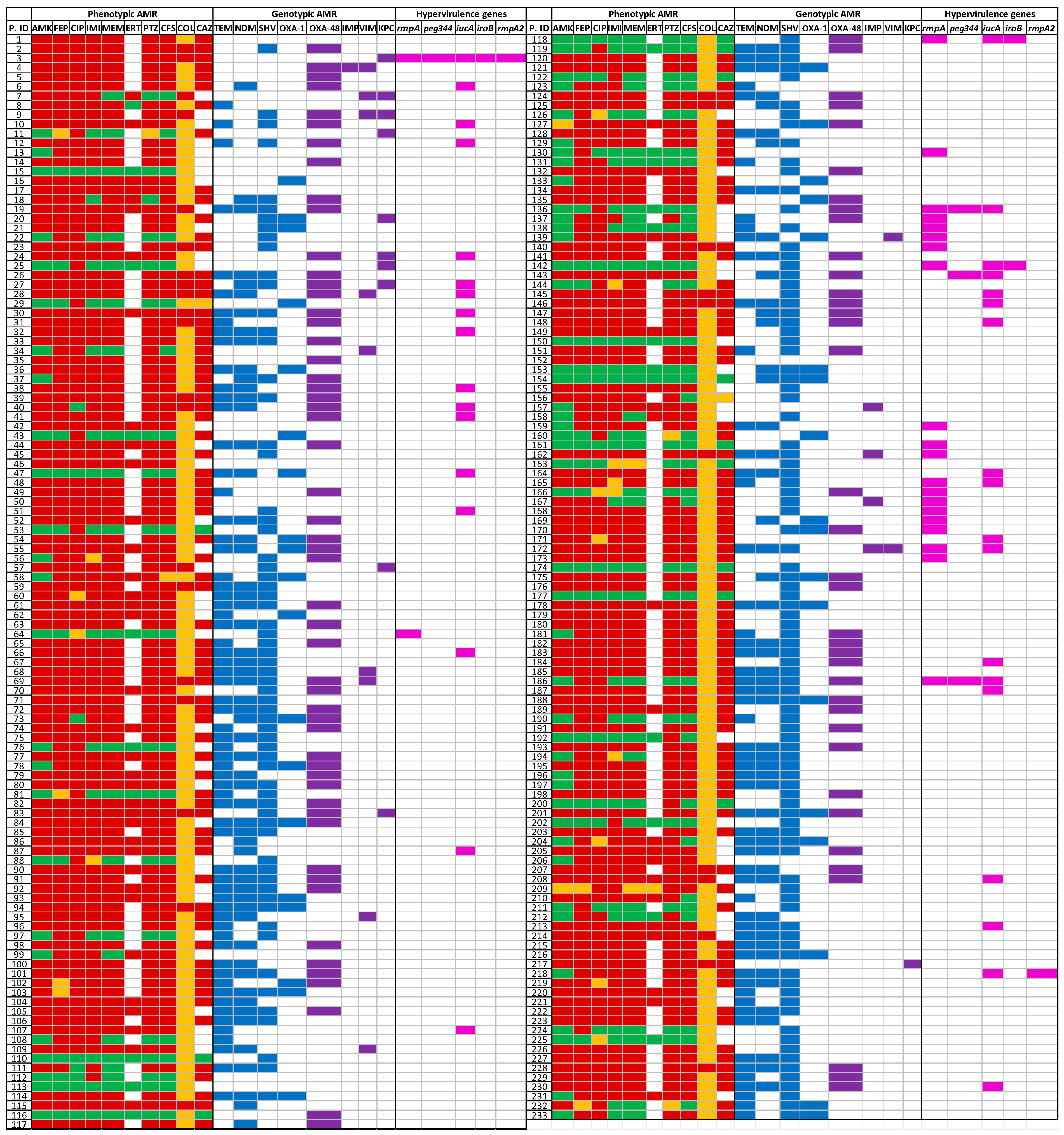
Assessment of the combined distribution of antimicrobial resistance and hypervirulence markers showed that convergent MDR–hvKP phenotypes were present but relatively uncommon in the cohort. Among the 50 isolates (21.6%) carrying at least one hypervirulence gene, the majority also possessed resistance determinants. Specifically, 35 isolates (15.2%) harbored both ESBL and hypervirulence genes, while 12 isolates (5.2%) carried a combination of hypervirulence genes and carbapenemase determinants. A smaller subset, three isolates (1.3%), carried hypervirulence genes in the absence of any tested AMR gene. Conversely, a substantial proportion of carbapenemase-producing isolates were also hypervirulence-positive: blaNDM was detected in 50% of infant hvKP isolates, and blaOXA-48-like in several multi-gene hvKP profiles, indicating overlap between high-level resistance and hypervirulent traits (Figure 2).
Figure 2.
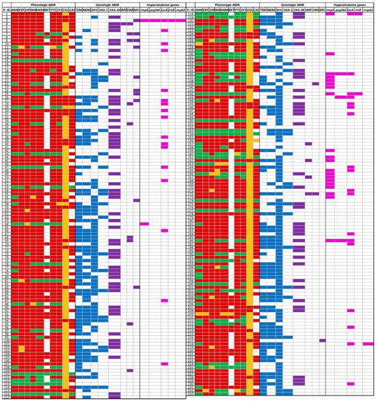
Heatmap showing the distribution and overlap of antimicrobial resistance phenotypes, corresponding genetic determinants, and hypervirulence markers in each isolate. Each row represents a single isolate (P.ID). Phenotypic susceptibility results for antimicrobial agents (AMK, FEP, CIP, IMI, MEM, ERT, PTZ, CFS, COL, CAZ) displayed using color-coded categories (green = susceptible, yellow = intermediate, red = resistant, and white/colorless = not tested). Presence of AMR determinants, including ESBL genes (blaTEM, blaSHV, blaOXA-1) and carbapenemase genes (blaNDM, blaOXA-48-like, blaKPC, blaVIM, blaIMP). Hypervirulence-associated genes (rmpA, rmpA2, iucA, iroB, peg344) shown as colored cell (blue, purple and pink respectively) and absence as white.
3.5. Association of Multidrug Resistance and Hypervirulence with Clinical Outcome
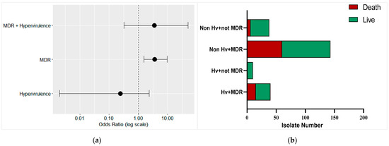
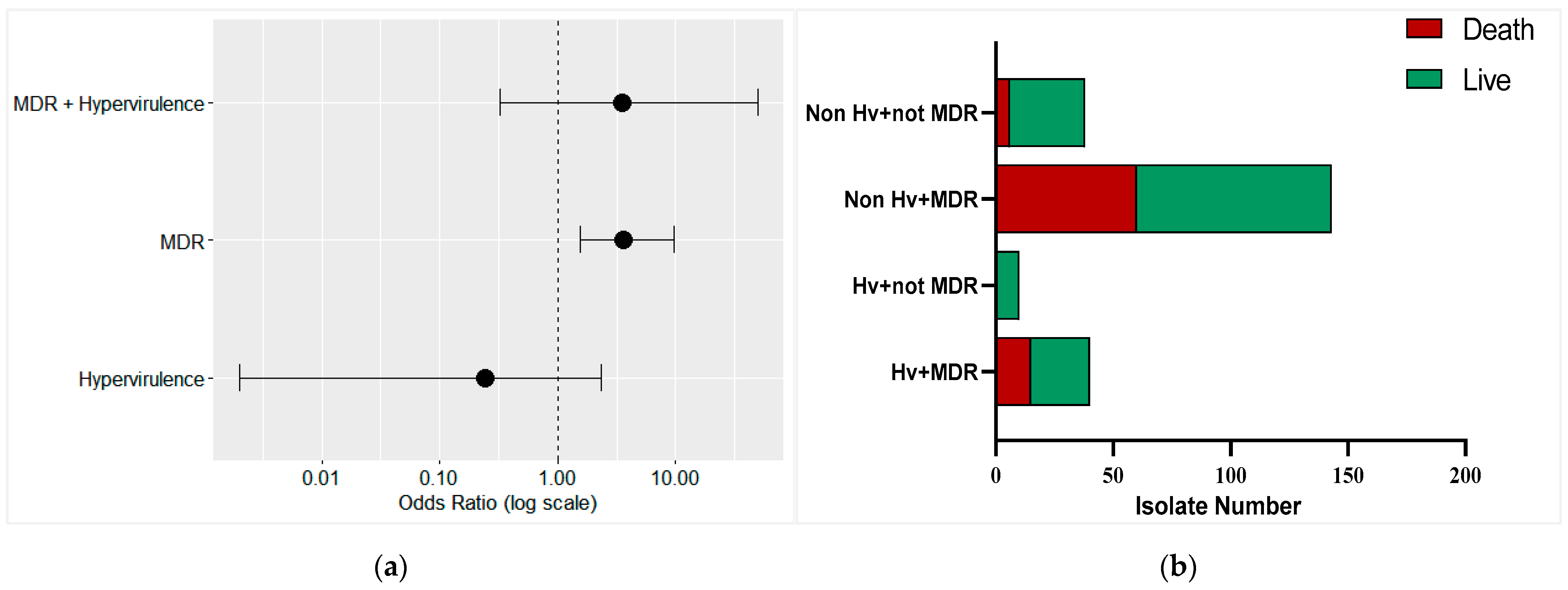
Clinical outcome was defined based on documented status at hospital discharge and categorized as death or discharge. Patients recorded as discharged were considered alive at the time of discharge. Of the 231 cases included in the study, in-hospital mortality was recorded in 81 cases (35.1%), while 150 patients (64.9%) were classified as alive at the last documented hospital contact. Alive outcomes included patients discharged from inpatient care and outpatient cases. Detailed stratification of clinical outcomes by patient care location is provided in Supplementary Table S1. Mortality was significantly higher among patients infected with multidrug-resistant (MDR) isolates compared to those infected with non-MDR isolates (75/149 vs. 6/38; p < 0.001). Though MDR infections were associated with a markedly increased risk of death (odds ratio: 3.62, 95% CI 1.55–9.73), hypervirulence alone was not independently associated with mortality (odds ratio: 0.24, 95% CI 0.002–2.32). To further evaluate the combined effect of MDR and hypervirulence on mortality, penalized logistic regression using Firth’s correction was performed to account for sparse data and complete separation. The interaction between MDR and hypervirulence was not statistically significant (odds ratio: 3.52, 95% CI 0.32–489.68), indicating no statistically demonstrable synergistic effect on mortality in this cohort. Nevertheless, a substantial number of deaths occurred among isolates co-harboring MDR and hypervirulence determinants, underscoring the clinical relevance of these convergent strains despite the absence of a statistically significant interaction (Figure 3).
Figure 3.
Clinical impact of multidrug resistance and hypervirulence on in-hospital mortality. (a) Forest plot showing odds ratios (ORs) and 95% confidence intervals derived from Firth penalized logistic regression evaluating the independent and combined effects of multidrug resistance (MDR), hypervirulence, and their interaction on mortality. Odds ratios are plotted on a logarithmic scale, with the dashed vertical line indicating no effect (OR = 1). (b) Stacked bar chart depicting the distribution of in-hospital clinical outcomes (death and survival) due to multidrug resistance or hypervirulence.
4. Discussion
The 231 invasive K. pneumoniae isolates collected from a tertiary care hospital characterized in the study demonstrated a high prevalence of multidrug resistance, as well as a substantial presence of key resistance and hypervirulence determinants. The majority of the isolates showed phenotypic resistance to fluoroquinolones and extended-spectrum cephalosporins, with more than 80% of the isolates being non-susceptible to ciprofloxacin, cefepime, and ceftazidime. Alarmingly, carbapenem resistance was also widespread (imipenem 78.4%, meropenem 75.3%), which was consistent with recent regional reports highlighting the spread of carbapenem-resistant K. pneumoniae in South Asia and reflecting a concerning reduction in effective empirical options [35,36,37]. Genotypically, the majority of the isolates harbored the ESBL and carbapenemase-encoding genes tested in the study. The high prevalence of NDM and OXA-48-like carbapenemase aligns with reports from India and other Asian regions, reflecting the plasmid-mediated dissemination of these genes [38,39]. The detection of ESBL genes (blaSHV, blaTEM, blaOXA-1) among carbapenemase-positive isolates underscores the accumulation of multiple resistance mechanisms.
While there was overall concordance between phenotypic and genotypic resistance profiles, notable discordances were also observed. Some isolates demonstrated phenotypic resistance despite the absence of the tested ESBL or carbapenemase genes, while others harbored resistance genes but remained phenotypically susceptible. Such discordance is well documented [40,41] and likely reflects the multifactorial nature of antimicrobial resistance. Although not evaluated in the present study, published evidence indicates that alterations in outer membrane porins (OmpK35/OmpK36) combined with ESBL or AmpC activity can elevate carbapenem minimum inhibitory concentrations (MICs) without a detectable carbapenemase gene [42]. Likewise, regulatory and expression differences, low gene copy numbers, or variants with attenuated activity can result in genotype-positive but phenotypically susceptible isolates. Additional resistance mechanisms not assessed here, including efflux pump upregulation and target site mutations (e.g., gyrA/parC for fluoroquinolones, 16S rRNA methyltransferases for aminoglycosides), have also been reported to contribute to phenotypic resistance in the absence of the primary resistance genes tested.
Clinically, the infections caused by multidrug-resistant K. pneumoniae presented substantially increased odds of death due to treatment failure, similarly to previous reports globally [43], though the effect of clinical confounders (e.g., age, severity of illness, co-morbidities, length of ICU stays, etc.) cannot be disregarded. The infant subset (<1 year; n = 42) also exhibited a concerning resistance profile, with high resistance to carbapenems, cephalosporins, aminoglycosides, and fluoroquinolones, along with elevated carriage of both ESBL and carbapenemase genes. These findings mirror those from other centers in India that have reported an increasing burden of carbapenem-resistant K. pneumoniae in neonatal intensive care units, where limited therapeutic options and high mortality rates remain significant concerns [39,44]. In infants, early acquisition or colonization by MDR strains supports the need for targeted infection control, enhanced surveillance, and judicious antimicrobial use in NICU settings.
While classical hvKP historically tended to be antibiotic-susceptible community strains, recent years have seen increasing convergence of hypervirulence and multidrug resistance, producing CR-hvKP strains that are both highly virulent and difficult to treat [45]. The WHO and several recent genomic studies highlight this worrying trend and its public health implications [46]. In our cohort, 21.6% (50/231) of isolates carried more than one hypervirulence determinant, most commonly iucA and rmpA. Only a single isolate carried all the tested hypervirulence marker genes (iucA, iroB, peg344, and rmpA/rmpA2). Though the presence of AMR genes and hypervirulence did not demonstrate a statistically significant synergistic effect on mortality, their co-occurrence suggests plasmid-mediated co-transfer of resistance and virulence determinants [47,48], which requires further confirmation and detailed analysis using whole-genome sequencing.
The study used conventional PCR to detect the presence of selected antimicrobial resistance and hypervirulence-associated genes, as PCR is a rapid and cost-effective method for routine surveillance, especially in resource-limiting settings with a high patient load and antimicrobial resistance burden. While the findings provide an important insight into the associations between multidrug resistance, hypervirulence and clinical outcomes, they should be interpreted in the context of certain limitations. The study does not involve follow-up of the patients and hence, long term outcomes could not be ascertained. The survival status was defined based on the last documented hospital contact, including for the patients who were initially managed in the outpatient department. This approach may have resulted in misclassification of the mortality status. Thus, the observed association between multidrug resistance and in-hospital mortality should be regarded as exploratory, rather than causal, as the key clinical confounders such as ICU admission, age, or severity of illness also affected the clinical outcome and were not adjusted in the study.
Despite these limitations, the study provides valuable insights into the burden of multidrug resistance and hypervirulence determinants among K. pneumoniae isolates. The identification of isolates with both MDR and hypervirulent genes is of critical concern, as it may lead to severe, rapidly progressive infections for which effective antibiotics are limited. These findings underscore the importance of continued surveillance, even through conventional PCR, to monitor the emergence and spread of convergent K. pneumoniae and to inform infection control and antimicrobial stewardship efforts.
5. Conclusions
This study documents a high prevalence of multidrug resistance among Klebsiella pneumoniae isolates obtained from invasive clinical samples, with MDR being associated with increased in-hospital mortality. This highlights the need for robust antimicrobial stewardship and tailored empirical therapy guided by susceptibility patterns. The detection of hv-associated markers in 21.6% of isolates underscores the value of incorporating virulence surveillance into routine diagnostics. Additionally, the detection of isolates carrying both resistance and hypervirulence determinants highlights an urgent need for integrated microbiological surveillance, genomic investigations, and strengthened clinical and public health measures to mitigate the threat posed by convergent K. pneumoniae strains. Thus, targeted infection control measures, particularly in high-risk units such as ICUs and NICUs, are essential to limit the dissemination of MDR and hypervirulent K. pneumoniae.
Supplementary Materials
The following supporting information can be downloaded at: https://www.mdpi.com/article/10.3390/microbiolres17040078/s1, Figure S1: Agarose gel electrophoresis showing PCR amplification of blaIMP, blaVIM, and blaKPC among representative K. pneumoniae isolates. Figure S2: Agarose gel electrophoresis showing PCR amplification of blaTEM, blaNDM, blaSHV, and blaOXA-1 genes among representative K. pneumoniae isolates. Figure S3: Agarose gel electrophoresis showing PCR amplification of blaOXA-48-like among representative K. pneumoniae isolates. Figure S4: Agarose gel electrophoresis showing PCR amplification of rmpA, iucA and peg344 among representative K. pneumoniae isolates. Figure S5: Agarose gel electrophoresis showing PCR amplification of (a) iroB and (b) rmpA2 among representative K. pneumoniae isolates. Table S1: Distribution of clinical outcomes at hospital discharge and respective patient care location of the study cohort.
Author Contributions
Conceptualization, S.K. and S.K.M.; methodology, K.G.; validation, N.T. and A.A.; formal analysis, S.K.; investigation, S.K. and K.G.; resources, A.A.; data curation, S.K. and K.G.; writing—original draft preparation, S.K.; writing—review and editing, S.K. and A.A.; visualization, S.K.; supervision, N.T.; project administration, A.A.; funding acquisition, A.A. All authors have read and agreed to the published version of the manuscript.
Funding
This research was funded by the Indian Council of Medical Research (ICMR), New Delhi, India, grant number EM/Dev/SG/87/5288/2023, and the APC was funded by the same grant.
Institutional Review Board Statement
The Institutional Ethics Committee approved the use of bacterial strains isolated from the clinical samples for the study (reference number IEC-04/2023-2746). The clinical samples were originally collected for routine diagnostic purposes and no additional intervention was done on the patients.
Informed Consent Statement
The informed consent was waived, as the study does not involve any intervention or collection of clinical history from the patient. The clinical outcome of each patient was recorded only during the course of study and no patient was tracked after being discharged from the hospital.
Data Availability Statement
The microbiological data supporting the findings of the study are included within the article and Supplementary Material. Patient-level clinical data are not publicly available due to institutional and ethical restrictions related to patient privacy. De-identified data may be available from the corresponding author upon reasonable request and subject to institutional approval.
Conflicts of Interest
The authors declare no conflicts of interest. The funders had no role in the design of the study; in the collection, analyses, or interpretation of data; in the writing of the manuscript; or in the decision to publish the results.
Abbreviations
The following abbreviations are used in this manuscript:
| AMR | Antimicrobial Resistance |
| AST | Antimicrobial Susceptibility Testing |
| cKP | Classical Klebsiella pneumoniae |
| CLSI | Clinical and Laboratory Standards Institute |
| CR | Carbapenem resistant |
| CR-KP | Carbapenem-resistant Klebsiella pneumoniae |
| ESBL | Extended spectrum Beta lactamases |
| hvKP | Hypervirulent Klebsiella pneumoniae |
| ICU | Intensive Care Unit |
| MDR | Multidrug Resistant |
| MIC | Minimum Inhibitory Concentration |
| NICU | Neonatal Intensive Care Unit |
| PCR | Polymerase Chain Reaction |
References
- Brescini, L.; Morroni, G.; Valeriani, C.; Castelletti, S.; Mingoia, M.; Simoni, S.; Masucci, A.; Montalti, R.; Vivarelli, M.; Giacometti, A.; et al. Clinical and Epidemiological Characteristics of KPC-Producing Klebsiella pneumoniae from Bloodstream Infections in a Tertiary Referral Center in Italy. BMC Infect. Dis. 2019, 19, 611. [Google Scholar] [CrossRef] [Scilit]
- Chen, T.-A.; Chuang, Y.-T.; Lin, C.-H.; Chen, T.-A.; Chuang, Y.-T.; Lin, C.-H. A Decade-Long Review of the Virulence, Resistance, and Epidemiological Risks of Klebsiella pneumoniae in ICUs. Microorganisms 2024, 12, 2548. [Google Scholar] [CrossRef] [Scilit]
- Dangor, Z.; Benson, N.; Berkley, J.A.; Bielicki, J.; Bijsma, M.W.; Broad, J.; Buurman, E.T.; Cross, A.; Duffy, E.M.; Holt, K.E.; et al. Vaccine Value Profile for Klebsiella pneumoniae. Vaccine 2024, 42, S125–S141. [Google Scholar] [CrossRef] [Scilit]
- Elshamy, A.A.; Aboshanab, K.M. A Review on Bacterial Resistance to Carbapenems: Epidemiology, Detection and Treatment Options. Future Sci. OA 2020, 6, FSO438. [Google Scholar] [CrossRef] [Scilit]
- Zheng, S.; Li, S.; Zhang, D.; Zhang, X.; Zhou, D.; Hou, Q.; Li, G.; Han, H. The Mechanisms of Antibiotic Resistance and Drug Resistance Transmission of Klebsiella pneumoniae. J. Antibiot. 2025, 78, 704–716. [Google Scholar] [CrossRef] [Scilit]
- Russo, T.A.; Olson, R.; Fang, C.-T.; Stoesser, N.; Miller, M.; MacDonald, U.; Hutson, A.; Barker, J.H.; La Hoz, R.M.; Johnson, J.R.; et al. Identification of Biomarkers for Differentiation of Hypervirulent Klebsiella pneumoniae from Classical K. pneumoniae. J. Clin. Microbiol. 2018, 56, e00776-18. [Google Scholar] [CrossRef] [Scilit]
- Russo, T.A.; Olson, R.; MacDonald, U.; Metzger, D.; Maltese, L.M.; Drake, E.J.; Gulick, A.M. Aerobactin Mediates Virulence and Accounts for Increased Siderophore Production under Iron-Limiting Conditions by Hypervirulent (Hypermucoviscous) Klebsiella pneumoniae. Infect. Immun. 2014, 82, 2356–2367. [Google Scholar] [CrossRef] [Scilit] [PubMed]
- Cheng, H.Y.; Chen, Y.S.; Wu, C.Y.; Chang, H.Y.; Lai, Y.C.; Peng, H.L. RmpA Regulation of Capsular Polysaccharide Biosynthesis in Klebsiella pneumoniae CG43. J. Bacteriol. 2010, 192, 3144–3158. [Google Scholar] [CrossRef] [Scilit] [PubMed]
- Bulger, J.; MacDonald, U.; Olson, R.; Beanan, J.; Russo, T.A. Metabolite Transporter PEG344 Is Required for Full Virulence of Hypervirulent Klebsiella pneumoniae Strain hvKP1 after Pulmonary but Not Subcutaneous Challenge. Infect. Immun. 2017, 85, e00093-17. [Google Scholar] [CrossRef] [Scilit] [PubMed]
- Russo, T.A.; Marr, C.M. Hypervirulent Klebsiella pneumoniae. Clin. Microbiol. Rev. 2019, 32, e00001-19. [Google Scholar] [CrossRef] [Scilit]
- Struve, C.; Roe, C.C.; Stegger, M.; Stahlhut, S.G.; Hansen, D.S.; Engelthaler, D.M.; Andersen, P.S.; Driebe, E.M.; Keim, P.; Krogfelt, K.A. Mapping the Evolution of Hypervirulent Klebsiella pneumoniae. mBio 2015, 6, e00630. [Google Scholar] [CrossRef] [Scilit]
- Hetta, H.F.; Alanazi, F.E.; Ali, M.A.S.; Alatawi, A.D.; Aljohani, H.M.; Ahmed, R.; Alansari, N.A.; Alkhathami, F.M.; Albogmi, A.; Alharbi, B.M.; et al. Hypervirulent Klebsiella pneumoniae: Insights into Virulence, Antibiotic Resistance, and Fight Strategies Against a Superbug. Pharmaceuticals 2025, 18, 724. [Google Scholar] [CrossRef] [Scilit] [PubMed]
- Osman, A.-H.; Darkwah, S.; Kotey, F.C.N.; Odoom, A.; Hotor, P.; Dayie, N.T.K.D.; Donkor, E.S. Reservoirs of Nosocomial Pathogens in Intensive Care Units: A Systematic Review. Environ. Health Insights 2024, 18, 11786302241243239. [Google Scholar] [CrossRef] [Scilit]
- Gan, L.; Mao, P.; Tian, Z.; Li, X.; Yu, Z.; Du, B.; Xue, G.; Yan, C.; Cui, J.; Zhao, H.; et al. Higher Prevalence of Hypervirulent Klebsiella pneumoniae Isolates with High-Risk Multidrug Resistance in Asia. J. Infect. Public Health 2025, 18, 102834. [Google Scholar] [CrossRef] [Scilit]
- Arcari, G.; Carattoli, A. Global Spread and Evolutionary Convergence of Multidrug-Resistant and Hypervirulent Klebsiella pneumoniae High-Risk Clones. Pathog. Glob. Health 2023, 117, 328–341. [Google Scholar] [CrossRef] [Scilit] [PubMed]
- Jiang, X.; Li, S.; Li, C.; Yin, Z.; Chen, F.; Hu, L.; Lu, T.; Liu, X.; Wang, Y.; Ma, G.; et al. The Global Genomic Landscape of Hypervirulent Klebsiella pneumoniae from 1932 to 2021. mLife 2025, 4, 378–396. [Google Scholar] [CrossRef] [Scilit]
- Zhou, W.; Yang, T.; Zhou, J.; Huang, K.; Zheng, Y.; Jiang, C.; Ji, Y.; Lei, D.; Liu, C. Emergence and Hospital Dissemination of Hypervirulent Carbapenem-Resistant Klebsiella pneumoniae Lineages in Eastern China. BMC Microbiol. 2025, 26, 73. [Google Scholar] [CrossRef] [Scilit]
- Kong, Z.X.; Karunakaran, R.; Abdul Jabar, K.; Ponnampalavanar, S.; Chong, C.W.; Teh, C.S.J. The Detection of Hypermucoviscous Carbapenem-Resistant Klebsiella pneumoniae from a Tertiary Teaching Hospital in Malaysia and Assessment of Hypermucoviscous as Marker of Hypervirulence. Microb. Drug Resist. 2021, 27, 1319–1327. [Google Scholar] [CrossRef] [Scilit]
- Loiodice, M.; Ribeiro, T.; Silva, L.M.; Castro, A.P.; Peixe, L.; Novais, Â. Molecular Epidemiology of Klebsiella pneumoniae Causing Bloodstream Infections in a District Hospital in Northern Portugal (2016–2018): Clonal Diversity and Detection of Hypervirulent Strains. Eur. J. Clin. Microbiol. Infect. Dis. 2025, 44, 3087–3095. [Google Scholar] [CrossRef] [Scilit] [PubMed]
- Jiang, J.; Long, T.; Porter, A.R.; Lovey, A.; Lee, A.; Jacob, J.T.; Arias, C.A.; Bonomo, R.; Kalayjian, R.; Zhao, Y.; et al. Carbapenem-Resistant, Virulence Plasmid-Harboring Klebsiella pneumoniae, United States. Emerg. Infect. Dis. 2025, 31, 761–771. [Google Scholar] [CrossRef] [Scilit]
- Beyrouthy, R.; Dalmasso, G.; Birer, A.; Robin, F.; Bonnet, R. Carbapenem Resistance Conferred by OXA-48 in K2-ST86 Hypervirulent Klebsiella pneumoniae, France. Emerg. Infect. Dis. 2020, 26, 1529–1533. [Google Scholar] [CrossRef] [Scilit]
- Yu, J.; Li, Y.; Zhang, Q.; Wang, Y. Hypervirulent Carbapenem-Resistant Klebsiella pneumoniae Infection: Epidemiology, Virulence, Resistance, and Treatment. Infect. Drug Resist. 2025, 18, 4689–4697. [Google Scholar] [CrossRef] [Scilit]
- Qala Nou, M.S.; Amirian, Z.; Dehghani, F.; Vejdan, A.-K.; Rooin, R.; Dehghanmehr, S. Systematic Review and Meta-Analysis on the Carbapenem-Resistant Hypervirulent Klebsiella pneumoniae Isolates. BMC Pharmacol. Toxicol. 2025, 26, 25. [Google Scholar] [CrossRef] [Scilit]
- Davoudabadi, S.; Goudarzi, H.; Goudarzi, M.; Ardebili, A.; Faghihloo, E.; Sharahi, J.Y.; Hashemi, A. Detection of Extensively Drug-Resistant and Hypervirulent Klebsiella pneumoniae ST15, ST147, ST377 and ST442 in Iran. Acta Microbiol. Immunol. Hung. 2022, 69, 77–86. [Google Scholar] [CrossRef] [Scilit]
- The Growing Burden of Klebsiella pneumoniae Infections in Indian Healthcare: A Comprehensive Review of Case Reports|South Eastern European Journal of Public Health. Available online: https://seejph.com/index.php/seejph/article/view/5943 (accessed on 2 January 2026).
- Mishra, S.; Kavitha, K.; Latha, R.; Sasi, A.C.; Devandra, B.I.; Gayathri, R.; Shree, K.H. Prevalence and Antimicrobial Resistance of Hypervirulent Klebsiella pneumoniae in Puducherry, India: A Cross-Sectional Analysis. J. Pure Appl. Microbiol. 2025, 19, 1993. [Google Scholar] [CrossRef] [Scilit]
- Banerjee, T.; Wangkheimayum, J.; Sharma, S.; Kumar, A.; Bhattacharjee, A. Extensively Drug-Resistant Hypervirulent Klebsiella pneumoniae From a Series of Neonatal Sepsis in a Tertiary Care Hospital, India. Front. Med. 2021, 8, 645955. [Google Scholar] [CrossRef] [Scilit]
- Yadav, B.; Mohanty, S.; Behera, B. Occurrence and Genomic Characteristics of Hypervirulent Klebsiella pneumoniae in a Tertiary Care Hospital, Eastern India. Infect. Drug Resist. 2023, 16, 2191–2201. [Google Scholar] [CrossRef] [Scilit] [PubMed]
- Oueslati, S.; Iorga, B.I.; Tlili, L.; Exilie, C.; Zavala, A.; Dortet, L.; Jousset, A.B.; Bernabeu, S.; Bonnin, R.A.; Naas, T. Unravelling Ceftazidime/Avibactam Resistance of KPC-28, a KPC-2 Variant Lacking Carbapenemase Activity. J. Antimicrob. Chemother. 2019, 74, 2239–2246. [Google Scholar] [CrossRef] [Scilit] [PubMed]
- Falcone, M.; Daikos, G.L.; Tiseo, G.; Bassoulis, D.; Giordano, C.; Galfo, V.; Leonildi, A.; Tagliaferri, E.; Barnini, S.; Sani, S.; et al. Efficacy of Ceftazidime-Avibactam Plus Aztreonam in Patients With Bloodstream Infections Caused by Metallo-β-Lactamase–Producing Enterobacterales. Clin. Infect. Dis. 2021, 72, 1871–1878. [Google Scholar] [CrossRef] [Scilit] [PubMed]
- Clinical and Laboratory Standards Institute. Performance Standard for Antimicrobial Susceptibility Testing, 34th ed.; CLSI Supplement M100; Clinical and Laboratory Standards Institute: Wayne, PA, USA, 2024. [Google Scholar]
- Dashti, A.A.; Jadaon, M.M.; Abdulsamad, A.M.; Dashti, H.M. Heat Treatment of Bacteria: A Simple Method of DNA Extraction for Molecular Techniques. Kuwait Med. J. 2009, 41, 117–122. [Google Scholar]
- Taha, M.S.; Elkolaly, R.M.; Elhendawy, M.; Elatrozy, H.; Amer, A.F.; Helal, R.A.E.F.; Salem, H.; El Feky, Y.G.; Harkan, A.; Mashaal, R.G.; et al. Phenotypic and Genotypic Detection of Hypervirulent Klebsiella pneumoniae Isolated from Hospital-Acquired Infections. Microorganisms 2024, 12, 2469. [Google Scholar] [CrossRef] [Scilit] [PubMed]
- Magiorakos, A.-P.; Srinivasan, A.; Carey, R.B.; Carmeli, Y.; Falagas, M.E.; Giske, C.G.; Harbarth, S.; Hindler, J.F.; Kahlmeter, G.; Olsson-Liljequist, B.; et al. Multidrug-Resistant, Extensively Drug-Resistant and Pandrug-Resistant Bacteria: An International Expert Proposal for Interim Standard Definitions for Acquired Resistance. Clin. Microbiol. Infect. 2012, 18, 268–281. [Google Scholar] [CrossRef] [Scilit]
- Ma, J.; Song, X.; Li, M.; Yu, Z.; Cheng, W.; Yu, Z.; Zhang, W.; Zhang, Y.; Shen, A.; Sun, H.; et al. Global Spread of Carbapenem-Resistant Enterobacteriaceae: Epidemiological Features, Resistance Mechanisms, Detection and Therapy. Microbiol. Res. 2023, 266, 127249. [Google Scholar] [CrossRef] [Scilit]
- Lathakumari, R.H.; Vajravelu, L.K.; Thulukanam, J.; Nair, D.M.; Vimala, P.B.; Panneerselvam, V. Prevalence and Molecular Insights into Carbapenem Resistance: A 2-Year Retrospective Analysis of Superbugs in South India. Front. Med. 2025, 12, 1571231. [Google Scholar] [CrossRef] [Scilit]
- DebBarma, P.; Kokkayil, P.; Sarfraz, A.; Pati, B.K.; Thakuria, B. Phenotypic Characterisation and Prevalence of Carbapenem-Resistant Enterobacterales in a Tertiary Care Centre in Bihar India. Sci. Rep. 2025, 15, 31477. [Google Scholar] [CrossRef] [Scilit]
- Bharadwaj, R.; Joshi, S.; Dohe, V.; Gaikwad, V.; Kulkarni, G.; Shouche, Y. Prevalence of New Delhi Metallo-β-Lactamase (NDM-1)-Positive Bacteria in a Tertiary Care Centre in Pune, India. Int. J. Antimicrob. Agents 2012, 39, 265–266. [Google Scholar] [CrossRef] [Scilit]
- Naha, S.; Sands, K.; Mukherjee, S.; Saha, B.; Dutta, S.; Basu, S. OXA-181-Like Carbapenemases in Klebsiella pneumoniae ST14, ST15, ST23, ST48, and ST231 from Septicemic Neonates: Coexistence with NDM-5, Resistome, Transmissibility, and Genome Diversity. mSphere 2021, 6, e01156-20. [Google Scholar] [CrossRef] [Scilit]
- Freitas, A.R.; Werner, G. Antibiotic Susceptibility Testing for Therapy and Antimicrobial Resistance Surveillance: Genotype Beats Phenotype? Future Microbiol. 2022, 17, 1093–1097. [Google Scholar] [CrossRef] [Scilit]
- Wyres, K.L.; Nguyen, T.N.T.; Lam, M.M.C.; Judd, L.M.; van Vinh Chau, N.; Dance, D.A.B.; Ip, M.; Karkey, A.; Ling, C.L.; Miliya, T.; et al. Genomic Surveillance for Hypervirulence and Multi-Drug Resistance in Invasive Klebsiella pneumoniae from South and Southeast Asia. Genome Med. 2020, 12, 11. [Google Scholar] [CrossRef] [Scilit] [PubMed]
- Tsai, Y.-K.; Fung, C.-P.; Lin, J.-C.; Chen, J.-H.; Chang, F.-Y.; Chen, T.-L.; Siu, L.K. Klebsiella pneumoniae Outer Membrane Porins OmpK35 and OmpK36 Play Roles in Both Antimicrobial Resistance and Virulence. Antimicrob. Agents Chemother. 2011, 55, 1485–1493. [Google Scholar] [CrossRef] [Scilit] [PubMed]
- Wp, S.E.; Norhidayah, M.; Ar, M.N.A. Factors Associated with Multidrug-Resistant Organism (MDRO) Mortality: An Analysis from the National Surveillance of Multidrug-Resistant Organism, 2018–2022. BMC Infect. Dis. 2025, 25, 60. [Google Scholar] [CrossRef] [Scilit] [PubMed]
- Panigrahi, P.; Chandel, D.S.; Hansen, N.I.; Sharma, N.; Kandefer, S.; Parida, S.; Satpathy, R.; Pradhan, L.; Mohapatra, A.; Mohapatra, S.S.; et al. Neonatal Sepsis in Rural India: Timing, Microbiology, and Antibiotic Resistance in a Population-Based Prospective Study in the Community Setting. J. Perinatol. 2017, 37, 911–921. [Google Scholar] [CrossRef] [Scilit] [PubMed]
- Nagendra, D.; Chaudhuri, S.; Gupta, N.; Shanbhag, V.; Eshwara, V.K.; Rao, S.; Varma, M.; Srinivas, T.; Todur, P.; Priya, P.S.; et al. Prevalence, Risk Factors, and Clinical Outcomes of Hypervirulent Klebsiella pneumoniae Strains among Klebsiella pneumoniae Infections: A Systematic Review and Meta-Analysis. Indian. J. Crit. Care Med. 2025, 29, 370–393. [Google Scholar] [CrossRef] [Scilit] [PubMed]
- Antimicrobial Resistance, Hypervirulent Klebsiella pneumoniae—Global Situation. Available online: https://www.who.int/emergencies/disease-outbreak-news/item/2024-DON527 (accessed on 2 January 2026).
- Wei, D.-W.; Song, Y.; Li, Y.; Zhang, G.; Chen, Q.; Wu, L.; Huang, J.; Tian, X.; Wang, C.; Feng, J. Insertion Sequences Accelerate Genomic Convergence of Multidrug Resistance and Hypervirulence in Klebsiella pneumoniae via Capsular Phase Variation. Genome Med. 2025, 17, 45. [Google Scholar] [CrossRef] [Scilit]
- Kulkarni, S.M.; Jacob, J.J.; Aravind, V.; Praveen, T.; Gunasekaran, K.; Lal, Y.B.; Walia, K.; Veeraraghavan, B. Evolutionary Transition of Hypervirulent Klebsiella pneumoniae to Multidrug-Resistant Hypervirulent Klebsiella pneumoniae: Indian Experience. Indian. J. Med. Microbiol. 2024, 50, 100619. [Google Scholar] [CrossRef] [Scilit]

Disclaimer/Publisher’s Note: The statements, opinions and data contained in all publications are solely those of the individual author(s) and contributor(s) and not of MDPI and/or the editor(s). MDPI and/or the editor(s) disclaim responsibility for any injury to people or property resulting from any ideas, methods, instructions or products referred to in the content. |
© 2026 by the authors. Licensee MDPI, Basel, Switzerland. This article is an open access article distributed under the terms and conditions of the Creative Commons Attribution (CC BY) license.