


Abstract
Intestinal infections caused by Salmonella enterica serovar Typhi (S. Typhi) remain a global health concern, making preventive strategies and diagnostic tools essential. This study aimed to identify mimotope peptides of the Vi antigen using phage display and assess their recognition by rabbit and 46 human sera, as well as their potential for diagnosis and immunogen design. Rabbits were immunized with the Vi antigen (AgVi) from S. Typhi ATCC 6539, and sera-derived IgG was used for phage biopanning. DNA sequences from selected phagotopes were synthesized as Salmonella mimotope peptides (SMPs), either linear or KLH-conjugated. Their reactivity was tested with ELISAs against AgVi and SMPs, using both rabbit sera and 46 human serum samples. Ten phagotopes were identified, with a consensus motif (D/G–A/V–x–P–x–x–G–x–x–x–x–x), suggesting α-helix structures. Immunization with KLH-conjugated peptides generated specific antibodies, particularly SMPVi/5 and SMPVi/10, which recognized AgVi and their respective peptides. Competitive inhibition assays confirmed that SMPVi/5 reduced the anti-AgVi binding in a dose-dependent manner. In human sera, AgVi recognition occurred in 52% of samples, while SMPVi/5 and SMPVi/10 were recognized in 45%. Overall, SMPVi/5 demonstrated immunogenicity and functional mimicry, supporting its use as a synthetic reagent for serological assays and as a candidate for immunogen design.
1. Introduction
Bacterial infections caused by Salmonella enterica serovar Typhi (S. Typhi) represent a worldwide health problem of clinical and epidemiological importance. S. Typhi contains lipopolysaccharide (LPS), a glycolipid composed of an outer region with repeating units of hydrophilic polysaccharides (somatic antigen “O”). LPS has a core section composed of polysaccharides and a hydrophobic region where lipid A (endotoxin) is located [1]. Another important component of S. Typhi is a capsular structure known as the virulence antigen (AgVi), also composed of polysaccharides [1,2,3,4,5]. Although both structures (LPS and AgVi) activate the host’s immune response, their use as immunogens for vaccine development is limited by the toxic effect of lipid A [6].
In the event of the recognition of pathogen-associated molecular patterns (PAMPs) present in macrophages, AgVi interferes with Toll-like receptors (TLRs), preventing phagocytosis and facilitating the systemic spread of Salmonella [7]. The polysaccharide composition of AgVi induces a short-lived thymus-independent response, failing to promote immunological memory [8].
Phage display is a procedure that uses the filamentous phage M13, in which genome a library of randomly expressed peptides has been inserted [9]. This procedure is used to identify structural sequences of peptides that show similarity (mimotopes) to epitopes of antigens from different origins. Thus, synthetic peptides can be synthesized, allowing us to design diagnostic methods and develop vaccines. In this regard, LPS and capsule mimotopes have been identified in bacteria, such as E. coli O157 and Vibrio cholerae O139, and in compounds such as toxins for their potential use as immunogens [10,11,12,13]. Trials on the immunogenicity of mimotope peptides reported a protective effect against the bacteria from which they were obtained [10,11].
Typhoid fever continues to be a public health problem in low- and middle-income countries, with high morbidity and mortality, particularly in regions of Asia, Africa, and Latin America, where the incidence remains considerable despite sanitation and control measures. Current vaccines are based on attenuated microorganisms or capsular polysaccharides such as the Vi antigen; however, the direct use of structures such as LPS is limited by the toxicity of lipid A and, in the case of AgVi, by the induction of a short-lived thymus-independent response and poor immunogenicity at the extremes of life. The use of phage display technology allows for the selection of mimotope peptides that mimic carbohydrate epitopes of LPS and AgVi but without the toxic effects associated with the whole bacterium. These synthetic peptides can be formulated as safe and stable immunogens capable of inducing antibodies that recognize native antigens; additionally, they can be used in ELISAs for a serological diagnosis. Within this context, the identification of mimotopes of AgVi from S. Typhi strain 190 (AgVi190) and their recognition using experimental sera and from open population individuals is the objective of the present work.
2. Materials and Methods
2.1. Antigens
S. Typhi strain ATCC 6539 (190) was used to obtain and purify AgVi190 from S. Typhi using the methods described by Westphal and Tacket [14,15]. The compound (AgVi190) was treated with DNase, RNase, and proteinase K; it was then dialyzed for 48 h with water using a 10 kDa cut-off membrane and then lyophilized (Labconco, Kansas City, MO, USA) and kept refrigerated (4 °C) until use. In addition, LPS was obtained from Salmonella serovar Urbana 459 (S. Urbana) for use as a negative control in ELISAs. Antigen concentration of carbohydrates was measured using the phenol–sulfuric acid method; the degree of purity was assessed by measuring the protein concentration using the Bradford method; and the antigens were separated by SDS-PAGE and stained with Coomassie Blue to confirm the absence of proteins and with silver to detect the presence of carbohydrates.
2.2. Anti-AgVi190 and Anti-SMPs Antibodies Obtained in Rabbits
The research protocol (IN216417) for the use of animals was approved by the Research Committee, CONBIETICA CEI066201402012 and CICUAL 004-CIC-2021, of the Faculty of Medicine, National Autonomous University of Mexico (UNAM). New Zealand (NZ) rabbits weighing 2.0 kg, provided by the Center for Teaching, Research, and Extension in Poultry Production, Faculty of Veterinary Medicine and Zootechnics, UNAM (CEIEPAv), were used to obtain antibodies. Prior to immunization, blood samples (5 mL) were obtained to use the serum antibodies as negative controls. The rabbits were challenged with S. Typhi AgVi190 using the protocol described by Ewing; briefly, the animals were immunized subcutaneously at 7-day intervals with five (100 µg/mL) doses per rabbit of previously obtained S. Typhi Ag-Vi190 diluted in PBS (pH 7.2). The first dose was administered with complete Freund’s adjuvant (1:1) and the remaining four doses with incomplete Freund’s adjuvant [16]. Anti-SMP antibodies were obtained, and KLH-conjugated SMPs were administered in four subcutaneous doses. In the first dose, 250 µg of the peptide dissolved in sterile pyrogen-free saline solution with Freund’s complete adjuvant was administered, and the following three doses were administered at 7-day intervals, inoculating 500 µg of the peptide on each occasion with incomplete Freund’s adjuvant [17]. The protocol for animal blood collection, anesthesia, and euthanasia was carried out in accordance with the recommendations of the Official Mexican Standard [18]. Immunoglobulins (IgGs) in the sera of AgVi-immunized rabbits were precipitated with 4.1 M ammonium sulfate (Sigma-Aldrich, St. Louis, MO, USA) [50% and 33% (v/v) saturation] as reported by Green [19]. Antibody concentration was determined using the Bradford method (Bio-Rad, Hercules, CA, USA), in accordance with the supplier’s specifications. The reactivity of anti-AgVi190 IgG and anti-SMPs was determined by ELISA, with its homologous antigen respectively, and specificity was evaluated against LPS of Salmonella Urbana.
2.3. Biopanning
The selection of phages (phage tags) by anti-AgVi190 IgG was performed using the phage display method described by Smith [9]. A library of 12-mer linear peptides (1012 pfu) fused to Protein III (pIII) of the filamentous phage M13 (New England Biolabs, Ipswich, MA, USA) was used. The specificity of the phage tropics was increased by performing three rounds of selection (biopanning), as previously reported [13].
2.4. Peptide Sequence
DNA was obtained from captured phage clones and used for automated sequencing (ABI PRISM® Model 3100 Genetic Analyzer 16-capillary sequencer, Thermo Fisher Scientific, Asheville, NC, USA) at the Biotechnology and Prototypes Unit from the Faculty of Higher Studies at Iztacala-UNAM, to determine the nucleotide sequence, using the −96 gIII primer (5′-CCCTCATAGTTAGCGTAACG-3′). The sequences were edited using the Chromas program to identify the signal sequence at the amino terminal end. The Translate tool program located on the ExPASy Proteomics Server (https://www.expasy.org/resources/uniprot-blast, accessed on 8 August 2025) was used to determine the nucleotide sequence of the DNA of the selected phage types. The sequences of the selected phagotopes were aligned to identify the presence of any consensus sequence using the Clustal Omega platform (https://www.ebi.ac.uk/jdispatcher/msa/clustalo/, accessed on 8 August 2025).
2.5. Synthesis of Mimotope Peptides
The consensus sequence was identified using the amino acid sequence of the peptides. Using these sequences, two 12-amino acid peptides were synthesized in linear sequences and conjugated with keyhole limpet hemocyanin (KLH) through a heterobifunctional linker SMCC (N-hydroxysuccinimid ester of 4-[N-maleimidomethyl] cyclohexane-1-carboxylic acid), with a purity greater than 90% (ProteoGenix, Schiltigheim, France). For the KLH-conjugated peptides, an extension of five amino acid residues was included in the carboxyl terminal group, with the sequence Gly-Gly-Gly-Ser-Cys (G-G-G-S-C).
2.6. Secondary Structure of Mimotope Peptides
The secondary structure of the obtained amino acid sequences from the selected mimotopes was defined by PEP-FOLD4 (free online version), considering the pH-dependent force field property for predicting the structure of peptides in an aqueous solution [20].
2.7. Reactivity of Anti-AgVi190 and Anti-SMP Sera
The reactivity of rabbit anti-AgVi190 and anti-SMP sera was analyzed by ELISA according to previous reports [13]. Briefly, 10 µg/mL of AgVi190 and SMP dissolved in pH 9.6 carbonate buffer was fixed in 96-well microplates (Nunca-imuno PlateMaxisorp F96, Thermo Fisher Scientific, Asheville, NC, USA). For the assays, the sera were titrated by making dilutions (1:2) from an initial 1/25 until 1/3200. The reaction was identified with an ELISA reader (Biotek ELx800, Agilent, Santa Clara, CA, USA) at 415 nm absorbance (OD415).
2.8. Inhibition Assay
To determine whether the peptide selected by anti-AgVi190, SMPVi/5, was a mimotope of AgVi190, the method described by Phalipon et al. was used with some modifications [17]. In brief, AgVi190 was immobilized in a 96-well ELISA microplate (Nunc-immuno Plate, Maxisorp F96, Thermo Fisher Scientific, Asheville, NC, USA) at a concentration of 10 µg/mL. Anti-AgVi190 serum was pre-incubated at 37 °C for 2 h with the SMPVi/5 peptide at different concentrations (0.1, 0.25, 0.5, 0.75, and 1 µg/µL). Anti-AgVi190 serum was used as a positive control, and pre-immune serum was used as a negative control. Goat anti-rabbit IgG antibody conjugated with alkaline phosphatase was used to visualize the reactions, and the ELISA reader (Biotek ELx800, Agilent, Santa Clara, CA, USA) was set to A415. To normalize the signal and express the magnitude of competition, the % inhibition (% competition) was calculated in each experiment and concentration using the formula:
OD control = The optical density of the positive binding control (reactivity of anti-AgVi190 serum vs. AgVi190). OD sample = The optical density of the sample containing the peptide that competes with or blocks the reaction.
2.9. Reactivity of Human Sera Against AgVi190 and SMPs
We used 46 human sera from individuals without clinical symptoms of typhoid fever, obtained from a previous study [21], to evaluate their reactivity to AgVi190 and the selected SMPs. To do this, 96-well microplates (Maxisorp F96, Thermo Fisher Scientific, Asheville, NC, USA) were independently coated with 10 µg/mL of AgVi190 or each SMP, dissolved in carbonate buffer at pH 9.6, and incubated overnight at 4 °C.
Subsequently, the wells were incubated with human sera at a 1:50 dilution in PBS for 2 h at 37 °C. After washing, the HRP-conjugated secondary antibody (1:1000) was added, and detection was performed. Absorbance was measured at 415 nm using an ELISA reader (Biotek ELx800, Agilent, Santa Clara, CA, USA). All assays were performed in duplicate and in two independent experiments. Pre-immune rabbit serum was included as a negative control.
2.10. Statistical Analysis
Statistical analysis was performed using GraphPad Prism version 10 software (GraphPad Software, San Diego, CA, USA). Competition assays were analyzed using Student’s t-test for comparison of the mean for different groups against a control. Fisher’s exact test was performed to evaluate reaction differences between phage mimotope peptides using human sera. In all analyses, an adjusted p-value < 0.05 was considered statistically significant.
3. Results
3.1. Selection of Phagotopes
A total of ten phagotopes were selected from the anti-AgVi190 serum (Table 1), where five phages had the peptide sequence DACPALGNRYRC/A, four had the GVDPWTLGAFMQA sequence, and only one had the GVDPFTGRPDAV sequence, with two amino acids in common xxxPxxGxxxxx. In addition, they were composed of aromatic amino acids (phenylalanine, tyrosine, and tryptophan), aliphatic amino acids (glycine, alanine, valine, leucine, and isoleucine), and proline. The sequence D/G–A/V–x–P–x–x–G–x–x–x–x–x was identified as the consensus sequence. For subsequent assays, peptides from the SMPVi/5 and SMPVi/10 phagotypes were selected, as they represented the two most prevalent amino acid sequences (Table 1).
Table 1.
Alignment and consensus sequences of mimotope peptides of the expressed Vi antigen of S. Typhi, selected by the phage display method.
3.2. Secondary Structure of Mimotope Peptides
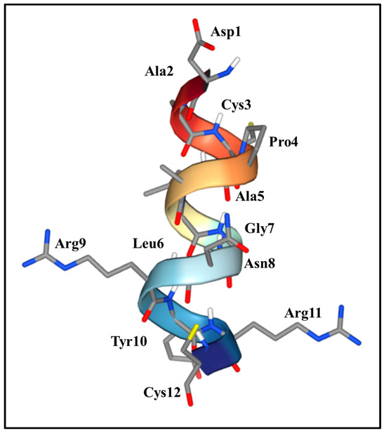
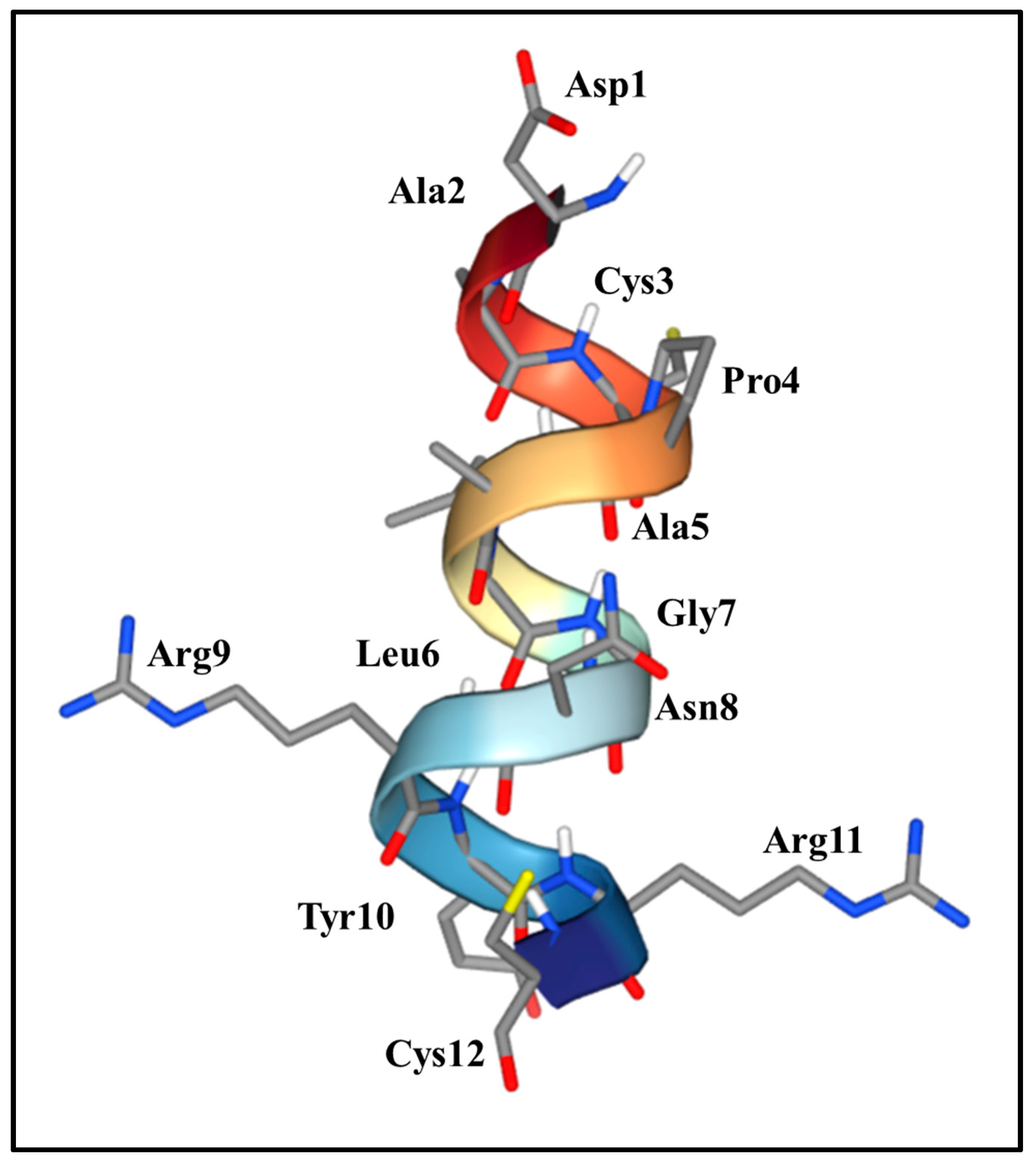
The bioinformatic analysis of the SMPVi/5 peptide showed that the secondary structure consists of α-helix and turn segments, with an amphipathic trait with the presence of hydrophobic amino acid residues on one side and charged/polar residues on the other (Figure 1). In addition, the structure’s composition showed hydrophobic amino acids (Ala, Leu, Pro, part of Tyr and Gly), uncharged polar amino acids (Asn, Cys and the –OH of Tyr) and charged amino acids (Arg with a positive charge and Asp with a negative charge). Overall, the composition of the amino acid residues of the peptide is amphipathic, allowing hydrophobic interactions, hydrogen bond formation, ionic bonds, and possible disulfide bridges (due to Cys). The presence of an aromatic residue (Tyr) is noteworthy, as it favors the formation of hydrogen bonds.
Figure 1.
The secondary structure of the SMPVi/5 peptide. The structure is predominantly composed of an alpha helix represented by the central ribbon, observing the outward projection of the side chains of the amino acids Ala, Arg, Leu, Tyr, Gly, Pro, Cys, Asp, and Cys, which are located toward the outside, minimizing steric interference between them and allowing them to interact with the environment or with other proteins. The numbering of the residues indicates the position of each amino acid along this secondary structure of the sequence: DACPALGNRYRC.
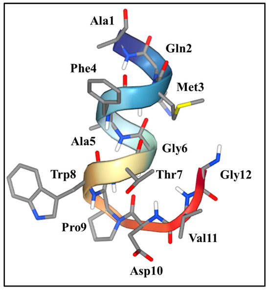
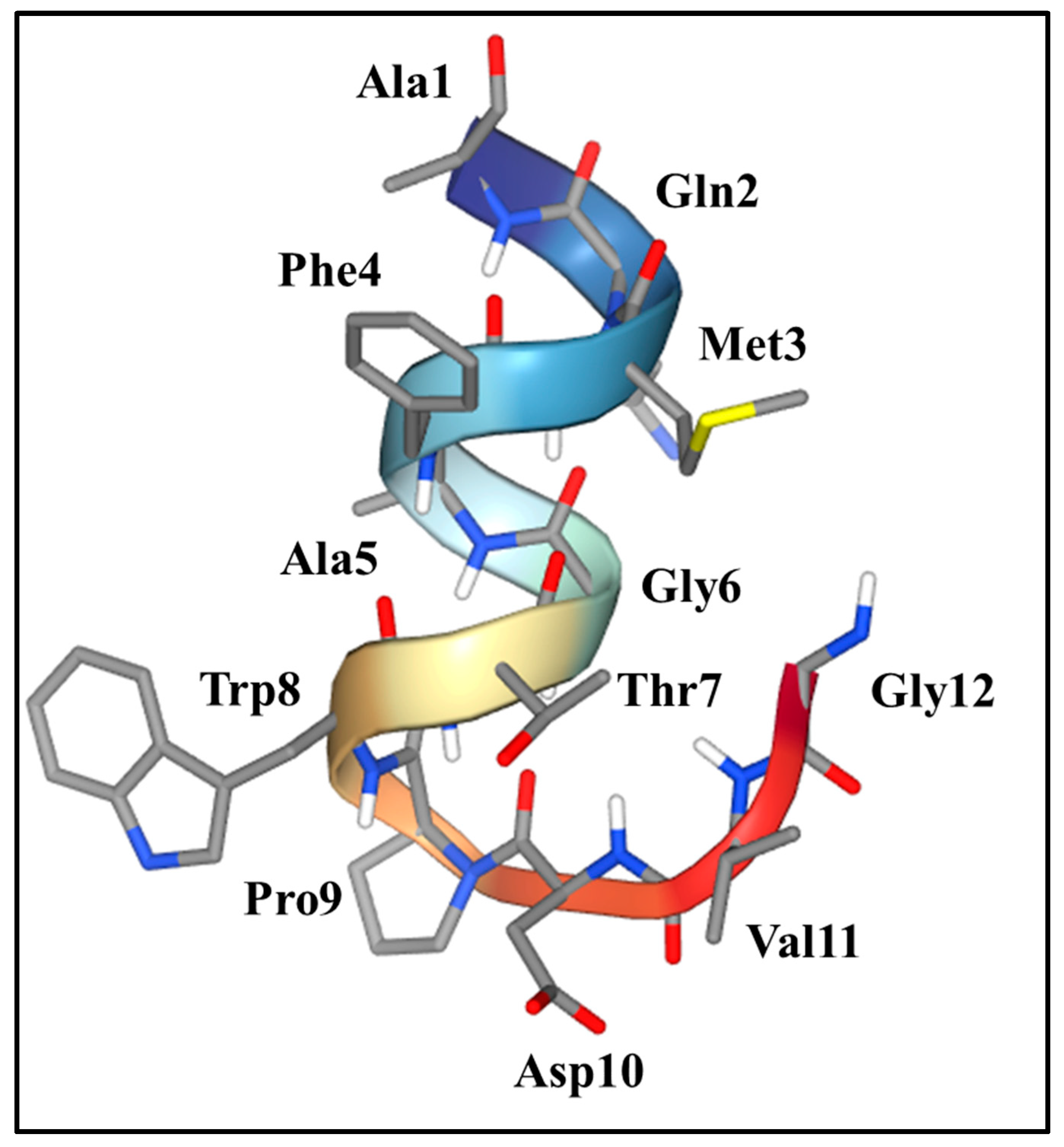
On the secondary structure of the SMPVi/10 peptide, the bioinformatic analysis showed a well-defined α-helix segment between Ala1 and Trp8, while the presence of a Pro9 residue acts as a breakpoint and termination of the α-helix, and the following residues (Asp10, Val11, and Gly12) form the final part of the peptide with a slight curvature (Figure 2). Therefore, unlike the SMPVi/5 peptide, this peptide behaves as a short, compact α-helix with a more flexible C-terminal end. In addition, the analysis showed the presence of aromatic residues (Phe4 and Trp8). Overall, the peptide is amphipathic, with several hydrophobic residues (Ala, Met, Phe, Trp, Val, and Pro) and some polar/charged residues (Gln, Thr, Asp, and Gly).
Figure 2.
The secondary structure of the SMPVi/10 peptide. The twelve amino acids are folded into an alpha helix represented by a central ribbon. The R groups of the amino acids Ala, Gln, Met, Phe, Gly, Thr, Trp, Pro, Asp, and Val are located on the outside, minimizing steric interference between them and allowing them to interact with the environment or with other proteins. The numbering of the residues indicates the position of each amino acid along this secondary structure of the sequence GVDPWTGAFMQA.
3.3. Reactivity of Anti-AgVi and Anti-SMP Sera
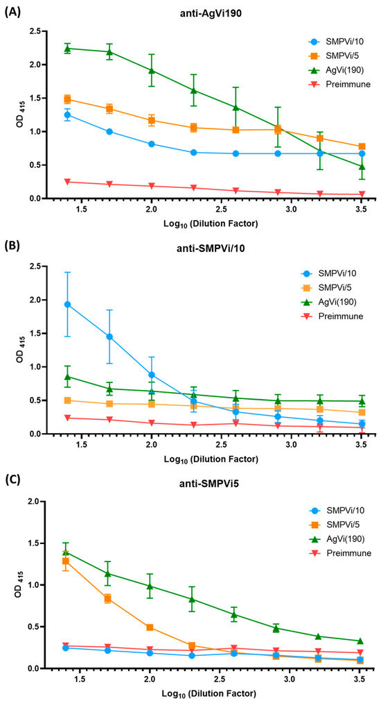
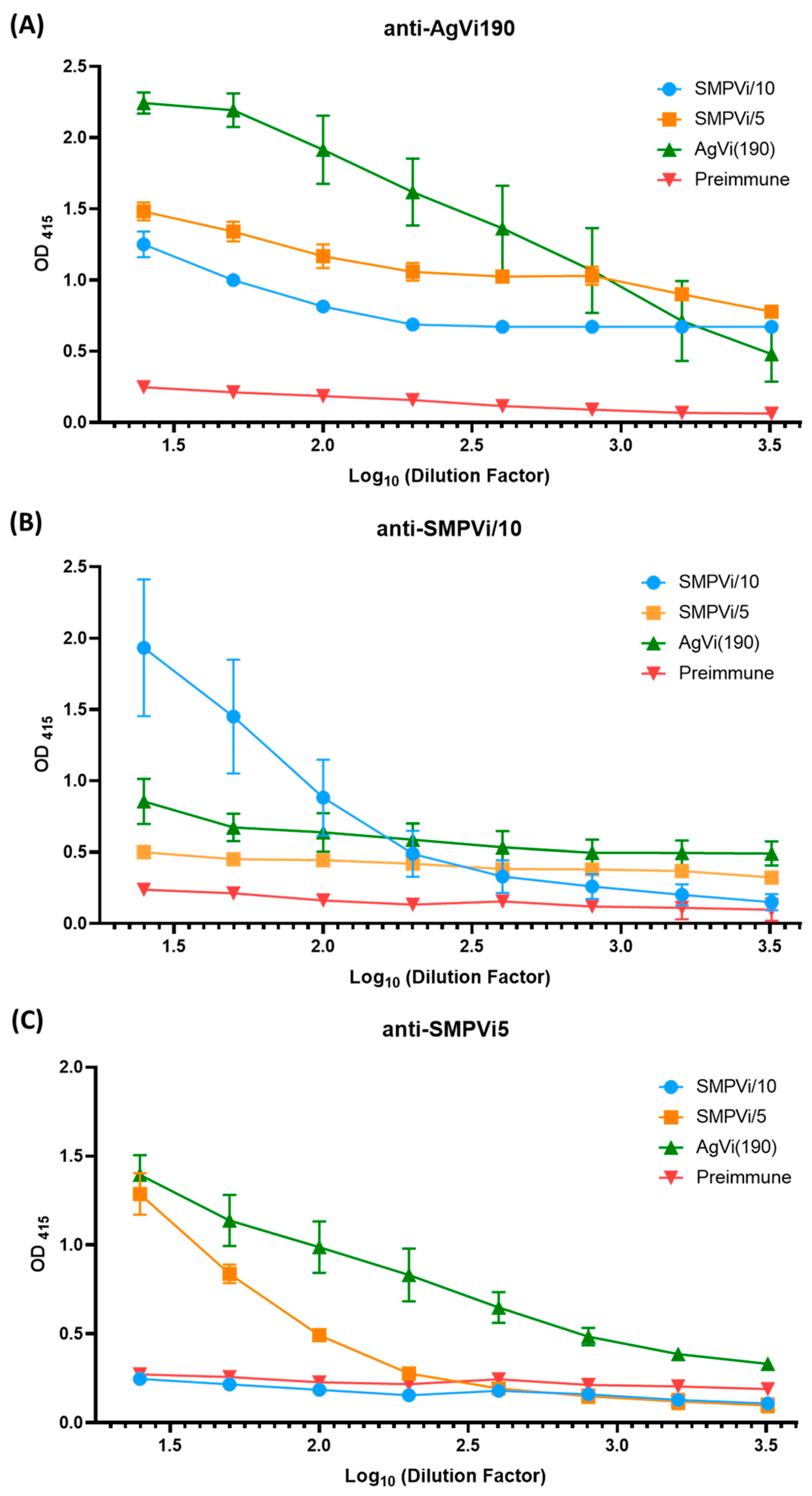
Anti-AgVi190, anti-SMPVi/5, and anti-SMPVi/10 sera were titrated against homologous and heterologous antigens using serial dilutions. The titration curves showed differential reactivity profiles (Figure 3). The anti-AgVi190 serum exhibited high reactivity against AgVi 190 and both mimotope peptides (Figure 3A), whereas the Anti-SMPVi/10 serum showed a greater reactivity against the homologous antigen; however, there is constant reactivity independent of the serum dilution against AgVi-190 and the SMVi/5 peptide (Figure 3B). Finally, the anti-SMPVi/5 serum showed a greater reactivity to AgVi190 than to its homologous peptide, and no reactivity was observed against the SMPVi/10 peptide (Figure 3C).
Figure 3.
ELISA titration curves (OD415) of anti-AgVi190 (A), anti-SMPVi/10 (PS14Vi) (B), and anti-SMPVi/5 (C) sera against AgVi190, SMPVi/5, and SMPVi/10. Data represents the mean of 3 independent experiments per triplicate ± SEM.
3.4. The Inhibition Assay of Anti-AgVi190 Serum in the Presence of the SMPVi/5 Peptide
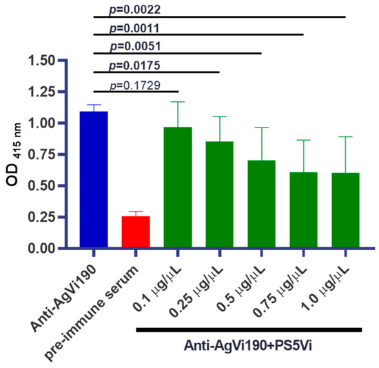
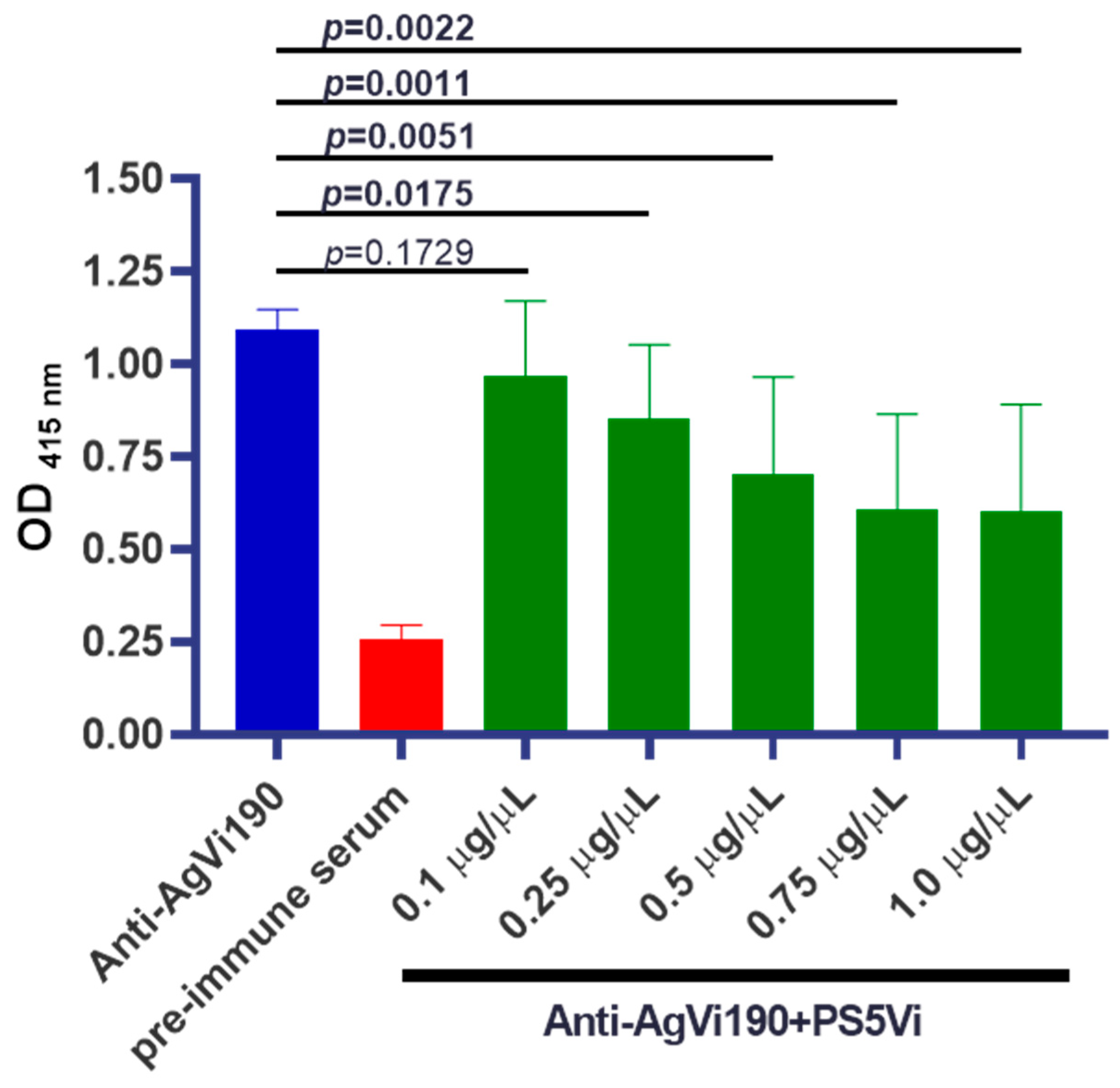
The competition assay showed a concentration-dependent decrease in the synthetic peptide SMPVi/5, with an inhibition percentage of 12.12, 15.51, 32.86, 47.10, and 47.54% at peptide concentrations of 0.1, 0.25, 0.5, 0.75, and 1.0 µg/µL, respectively. SMPVi/5 showed competition starting from 0.25 up to the highest concentration used—1.0 µg/µL (p < 0.05) (Figure 4).
Figure 4.
The inhibition assay for anti-AgVi190. The ELISA to determine the competition between AgVi190 and the SMPVi/5 peptide for binding to the anti-AgVi190 antibody. Data represents the mean of 3 independent experiments per duplicate ± SD.
3.5. Response of Sera from Open Population Against AgVi190, SMPVi/5 and SMPVi/10
The reactivity of 46 sera from individuals with no history of S. Typhi infection was compared against the SMPVi/5 and SMPVi/10 peptides and AgVi. A total of 22 sera (47.8%) failed to recognize any of the antigens, whereas 24 (52.2%) sera showed a positive reactivity grouped as follows: 7 sera to AgVi190, 1 serum to SMPVi/5, 1 serum to SMPVi/10, 5 sera to the combination of AgVi190 + SMPVi/5, 1 serum to SMPVi/5 + SMPVi/10, and 9 sera to all three antigens (AgVi190 + SMPVi/5 + SMPVi/10). In summary, 16 sera reacted to the SMPVi/5 peptide, whereas 15 of these sera also reacted to AgVi190. In an interesting manner, AgVi190 showed more cross-reactivity to SMPVi/5 than to SMPVi10 (p < 0.05).
4. Discussion
The main objective of this study was to identify S. Typhi Vi antigen mimotopes with immunogenic properties using the phage display technique. In total, 10 phage plates (phagotopes) were randomly collected, and a DNA analysis identified a common motif (D/G–A/V–x–P–x–x–G–x–x–x–x–x), suggesting that they are part of immunodominant mimotope epitopes of the Vi190 antigen of S. Typhi. This was corroborated after immunizing rabbits with the synthetic mimotope peptides SMPVi/5 and SMPVi/10, corresponding to the Vi antigen, and obtaining the respective antibodies. The administration of the peptides designated as SMPVi/5 and SMPVi/10 of AgVi190 failed to show any toxic effect in the immunized animals, such as that produced when administering the complete Vi antigen, which supports their potential use in humans if they prove useful as protective immunogens. The antibodies generated by rabbits challenged with the SMPVi/5 and SMPVi/10 peptides recognized the AgVi190 of S. Typhi; likewise, other studies have reported that phage-type mimotopes are immunogenic [22,23,24,25].
From a compositional standpoint, SMPVi/5 and SMPVi/10 contain aromatic residues (Y, F, and W), which have previously been associated with carbohydrate mimicry, likely because they promote hydrophobic contacts and a structure that is compatible with anti-carbohydrate paratopes [26,27]. Accordingly, the presence and possible exposure of these aromatic residues in the conformation of the peptides support their contribution to the structural mimicry of AgVi190 epitopes. In particular, the consensus motif D/G–A/V–x–P–x–x–G–x–x–x–x–x in the two mimotope peptides in this study is compatible with sequences obtained by phage display when the target was a capsular antigen, as this approach tends to enrich peptides with turns and aromatic residues that facilitate the mimicry of non-peptide epitopes [27]. In this context, proline (Pro) could contribute to the mimicry of AgVi190 by inducing turns that stabilize the presentation of the D/G–A/V–x–P–x–x–G–x–x–x–x–x motif. In libraries selected by phage display against carbohydrate antigens, Pro is recurrent because it imposes conformational restrictions compatible with anti-carbohydrate paratopes [28]. In fact, mimotopes with Pro that are capable of competing with carbohydrate epitopes have been described, supporting their role in functional “mimicry” [29]. In addition to proline, the presence of glycine (G) in peptides could contribute to conformational flexibility and frequently appear in turn regions where other residues would be sterically restricted. Likewise, it has been proposed that F, Y, and W contribute to carbohydrate mimicry, and YxY-type motifs have been described in mimotopes recognized by antibodies [30]. Finally, arginine (R) could contribute stability due to its positive charge, allowing for salt bridges and multiple hydrogen bonds [31]; in fact, the presence of R in dominant anti-Vi mimotopes suggests that basic residues may be part of the recognized motif [23].
The selected mimotopes contain the consensus motif D/G–A/V–x–P–x–x–G–x–x–x–x–x and present combinations of glycine and proline, a property that has been described in mimotopes obtained by the phage display of non-protein antigens, where selection favors surfaces and structures compatible with the antibody paratope rather than a literal “copy” of the polysaccharide [26,27,28]. Consistent with this, the structural alpha helix of SMPVi/5 and SMPVi/10 adopts a short helix. These structural differences provide a plausible basis for understanding how both peptides can share antigenic determinants; in particular, the presence of Pro/Gly may facilitate twists and conformational plasticity, while aromatic residues may contribute to hydrophobic contacts that stabilize antigen–antibody interactions [28,29,30].
According to the mimotope operational criterion, this is a peptide selected and validated as a mimetic peptide, which must be recognized by antibodies directed against the native antigen and, complementarily, show functional evidence of sharing the same paratope through the competitive inhibition of antigen binding [28,31]. In this regard, SMPVi/5 showed a dose-dependent increase in the percentage of inhibition, suggesting a more evident competitive effect above 50 µg and a tendency to stabilize at higher concentrations. However, the reactivity of the anti-AgVi190 serum was not completely eliminated, which is consistent with a scenario in which the mimotope reproduces only part of the antigenic determinant, for example immunodominant epitopes, while other epitopes of AgVi190 remain available for recognition by the polyclonal antibody mixture. This pattern is common in the field of mimotopes: cross-binding or partial competition may reflect incomplete conformational mimicry or the limited coverage of the antibody repertoire, and it has been reported that reactivity to a mimotope does not necessarily reflect a full equivalence with the native epitope or its cross-immunogenicity [32,33].
The comparative analysis using human sera with a positive reactivity to AgVi190 and the response to synthetic peptides showed no difference for the SMPVi/5 peptide (p > 0.05), confirming that the peptide sequence is a mimotope of AgVi190. A relevant fact in this regard is that these same sera also recognized the O157 LPS in a previous study [21]. The results obtained in the present study suggest that the immune response observed against AgVi in human serum samples could be related to exposure to common epitopes shared by S. Typhi and other enterobacteria such as S. Paratyphi A and B, E. coli, Klebsiella, and Citrobacter [34,35,36]. In Mexico, early-life contact with bacteria belonging to different E. coli serogroups and different Salmonella serovars could explain the antigenic reactivity observed in human serum samples. Recent studies on the prevalence of Salmonella infections in Mexico report that in the last 50 years, the serovars most frequently isolated from human samples have been S. Enteritidis, S. Typhi, and S. Paratyphi [37].
5. Conclusions
In this study, phage display was used to identify a mimotope peptide of S. Typhi AgVi190 (SMPVi/5) that reproduces determinants recognized by anti-AgVi antibodies. This peptide showed an immunogenic capacity by inducing antisera in rabbits that reacted with homologous peptides and with AgVi190. It was also observed that SMPVi/5 demonstrated functional competence by dose-dependently inhibiting the binding of the anti-AgVi190 serum to its homologous antigen. The reactivity observed in human sera suggests their potential as synthetic antigens for serological ELISAs and as components for safer immunogens. Nevertheless, both diagnostic and clinical applications require validation in cohorts with confirmed infection and specific evaluations of cross-reactivity. Overall, SMPVi/5 represents a promising AgVi mimotope with potential utility in the diagnosis and in the development of preventive strategies against typhoid fever.
Author Contributions
Conceptualization, A.N.-O. and U.H.-C.; data curation, A.N.-O., R.E.A.-C. and U.H.-C.; formal analysis, A.N.-O., A.N.-C.d.P., R.E.A.-C. and U.H.-C.; funding acquisition, A.N.-O.; investigation, A.N.-O., A.N.-C.d.P., R.E.A.-C. and U.H.-C.; methodology, A.N.-O., A.N.-C.d.P., R.E.A.-C. and U.H.-C.; supervision; A.N.-O., A.N.-C.d.P., R.E.A.-C. and U.H.-C.; writing—original draft, A.N.-O., R.E.A.-C. and U.H.-C.; writing—review and editing, A.N.-O., R.E.A.-C. and U.H.-C. All authors have read and agreed to the published version of the manuscript.
Funding
This work was funded by the Universidad Nacional Autónoma de México (UNAM) through Dirección General de Asuntos de Personal Académico (DGAPA) by the Programa de Apoyo a Proyectos de Investigación e Innovación Tecnológica (PAPIIT) with Project No. IN213422 “Detección de inumnógenos de Salmonella Typhi por Phage Dislpay” and Project No. IN220325 “Análisis del microbioma del pulque, aislamiento e identificación de microorganismos facultativos con propiedades probióticas”.
Institutional Review Board Statement
The animal study protocol was approved by the Institutional Review Board and Ethics Committee of the Faculty of Medicine-UNAM on 5 February 2021 and registered under No. CICUAL 004-CIC-2021.
Informed Consent Statement
Not applicable.
Data Availability Statement
The original contributions presented in this study are included in the article. Further inquiries can be directed to the corresponding author.
Acknowledgments
The authors would like to acknowledge Liliana Cortés, Gabriel Pérez and Delia Licona for their technical support in this work. R. E. A.-C. (CVU 556348) would like to thank CONAHCYT/SECIHTI for his post-doctoral scholarship grant.
Conflicts of Interest
The authors declare no conflicts of interest.
References
- Schultz, K.M.; Klug, C.S. Characterization of and Lipopolysaccharide Binding to the E. coli LptC Protein Dimer. Protein Sci. 2018, 27, 381–389. [Google Scholar] [CrossRef] [Scilit]
- Bertani, B.; Ruiz, N. Function and Biogenesis of Lipopolysaccharides. EcoSal Plus 2018, 8, 1–19. [Google Scholar] [CrossRef] [Scilit]
- Hu, X.; Chen, Z.; Xiong, K.; Wang, J.; Rao, X.; Cong, Y. Vi Capsular Polysaccharide: Synthesis, Virulence, and Application. Crit. Rev. Microbiol. 2017, 43, 440–452. [Google Scholar] [CrossRef] [Scilit]
- Vashishtha, V.M.; Kumar, P. Typhoid Conjugate Vaccines: Is a Single Dose Enough for Durable Protection? Expert. Rev. Vaccines 2025, 24, 194–205. [Google Scholar] [CrossRef] [Scilit]
- Zhang, L.F.; Lepenies, B.; Nakamae, S.; Young, B.M.; Santos, R.L.; Raffatellu, M.; Cobb, B.A.; Hiyoshi, H.; Bäumler, A.J. The Vi Capsular Polysaccharide of Salmonella Typhi Promotes Macrophage Phagocytosis by Binding the Human C-Type Lectin DC-SIGN. mBio 2022, 13, e0273322. [Google Scholar] [CrossRef] [Scilit]
- Ludwig, K.; Bitzan, M.; Bobrowski, C.; Müller-Wiefel, D.E. Escherichia coli O157 Fails to Induce a Long-Lasting Lipopolysaccharide-Specific, Measurable Humoral Immune Response in Children with Hemolytic-Uremic Syndrome. J. Infect. Dis. 2002, 186, 566–569. [Google Scholar] [CrossRef] [Scilit] [PubMed]
- Kaur, J.; Jain, S.K. Vi Antigen of Salmonella Enetrica Serovar Typhi—Biosynthesis, Regulation and Its Use as Vaccine Candidate. Cent. Eur. J. Biol. 2012, 7, 825–838. [Google Scholar] [CrossRef] [Scilit]
- Fraser, A.; Paul, M.; Goldberg, E.; Acosta, C.J.; Leibovici, L. Typhoid Fever Vaccines: Systematic Review and Meta-Analysis of Randomised Controlled Trials. Vaccine 2007, 25, 7848–7857. [Google Scholar] [CrossRef] [Scilit] [PubMed]
- Smith, G.P. Filamentous Fusion Phage: Novel Expression Vectors That Display Cloned Antigens on the Virion Surface. Science 1985, 228, 1315–1317. [Google Scholar] [CrossRef] [Scilit]
- Falklind-Jerkérus, S.; Felici, F.; Cavalieri, C.; Lo Passo, C.; Garufi, G.; Pernice, I.; Islam, M.M.; Qadri, F.; Weintraub, A. Peptides Mimicking Vibrio Cholerae O139 Capsular Polysaccharide Elicit Protective Antibody Response. Microbes Infect. 2005, 7, 1453–1460. [Google Scholar] [CrossRef] [Scilit]
- Lo Passo, C.; Romeo, A.; Pernice, I.; Donato, P.; Midiri, A.; Mancuso, G.; Arigò, M.; Biondo, C.; Galbo, R.; Papasergi, S.; et al. Peptide Mimics of the Group B Meningococcal Capsule Induce Bactericidal and Protective Antibodies After Immunization. J. Immunol. 2007, 178, 4417–4423. [Google Scholar] [CrossRef] [Scilit]
- Ulises, H.-C.; Tatiana, G.; Karlen, G.; Guillermo, M.-H.; Juan, X.-C.; Carlos, E. Peptide Sequences Identified by Phage Display Are Immunodominant Functional Motifs of Pet and Pic Serine Proteases Secreted by Escherichia coli and Shigella Flexneri. Peptides 2009, 30, 2127–2135. [Google Scholar] [CrossRef] [Scilit] [PubMed]
- Navarro, A.; Hernández-Chiñas, U.; Licona-Moreno, D.; Zenteno, E.; Cravioto, A.; Eslava-Campos, C.A. Immunogenic Peptide Mimotopes from an Epitope of Escherichia coli O157 LPS. Biochem. J. 2016, 473, 3791–3804. [Google Scholar] [CrossRef] [Scilit] [PubMed]
- Westphal, O.; Jann, K. Bacterial lipopolysaccharides. Extraction with phenol-water and further applications of the procedure. In Methods in Carbohydrate Chemistry; Whistler, R.L., Wolfan, M.L., Eds.; Academic Press: New York, NY, USA, 1965; pp. 83–91. [Google Scholar]
- Tacket, C.O.; Ferreccio, C.; Robbins, J.B.; Tsai, C.-M.; Schulz, D.; Cadoz, M.; Goudeau, A.; Levine, M.M. Safety and Immunogenicity of Two Salmonella Typhi Vi Capsular Polysaccharide Vaccines. J. Infect. Dis. 1986, 154, 342–345. [Google Scholar] [CrossRef] [Scilit]
- Ewing, W. Edwards and Ewing’s Identification of Enterobacteriaceae, 4th ed.; Elsevier: Amsterdam, The Netherlands, 1986. [Google Scholar]
- Phalipon, A.; Folgori, A.; Arondel, J.; Sgaramella, G.; Fortugno, P.; Cortese, R.; Sansonetti, P.J.; Felici, F. Induction of Anti-Carbohydrate Antibodies by Phage Library-Selected Peptide Mimics. Eur. J. Immunol. 1997, 27, 2620–2625. [Google Scholar] [CrossRef] [Scilit]
- NOM-062-ZOO-1999; Technical Specifications for the Production, Care, and Use of Laboratory Animals. Secretaría de Agricultura, Ganadería, Desarrollo Rural, Pesca y Alimentación (SAGARPA): Mexico City, Mexico, 2001.
- Green, A.A.; Hughes, W.L. Protein Fractionation on the Basis of Solubility in Aqueous Solutions of Salts and Organic Solvents. Methods Enzymol. 1955, 1, 67–90. [Google Scholar] [CrossRef] [Scilit]
- Rey, J.; Murail, S.; de Vries, S.; Derreumaux, P.; Tuffery, P. PEP-FOLD4: A pH-Dependent Force Field for Peptide Structure Prediction in Aqueous Solution. Nucleic Acids Res. 2023, 51, W432–W437. [Google Scholar] [CrossRef] [Scilit]
- Navarro, A.; Eslava, C.; Hernandez, U.; Navarro-Henze, J.L.; Aviles, M.; Garcia-de la Torre, G.; Cravioto, A. Antibody Responses to Escherichia coli O157 and Other Lipopolysaccharides in Healthy Children and Adults. Clin. Diagn. Lab. Immunol. 2003, 10, 797–801. [Google Scholar] [CrossRef] [Scilit]
- Smith, G.P.; Scott, J.K. Libraries of Peptides and Proteins Displayed on Filamentous Phage. Methods Enzymol. 1993, 217, 228–257. [Google Scholar] [CrossRef] [Scilit]
- Tang, S.-S.; Tan, W.-S.; Devi, S.; Wang, L.-F.; Pang, T.; Thong, K.-L. Mimotopes of the Vi Antigen of Salmonella enterica Serovar Typhi Identified from Phage Display Peptide Library. Clin. Diagn. Lab. Immunol. 2003, 10, 1078–1084. [Google Scholar] [CrossRef] [Scilit]
- Li, Y.; Ning, Y.; Wang, Y.; Peng, D.; Jiang, Y.; Zhang, L.; Long, M.; Luo, J.; Li, M. Mimotopes Selected with a Neutralizing Antibody against Urease B from Helicobacter Pylori Induce Enzyme Inhibitory Antibodies in Mice upon Vaccination. BMC Biotechnol. 2010, 10, 84. [Google Scholar] [CrossRef] [Scilit]
- Shi, H.; Dong, S.; Zhang, X.; Chen, X.; Gao, X.; Wang, L. Phage Vaccines Displaying YGKDVKDLFDYAQE Epitope Induce Protection Against Systemic Candidiasis in Mouse Model. Vaccine 2018, 36, 5717–5724. [Google Scholar] [CrossRef] [Scilit]
- Young, A.C.; Valadon, P.; Casadevall, A.; Scharff, M.D.; Sacchettini, J.C. The Three-Dimensional Structures of a Polysaccharide Binding Antibody to Cryptococcus Neoformans and Its Complex with a Peptide from a Phage Display Library: Implications for the Identification of Peptide Mimotopes. J. Mol. Biol. 1997, 274, 622–634. [Google Scholar] [CrossRef] [Scilit]
- Matsubara, T. Peptide Mimotopes to Emulate Carbohydrates. Chem. Soc. Rev. 2022, 51, 8160–8173. [Google Scholar] [CrossRef] [Scilit]
- Fukuda, M.N. Peptide-Displaying Phage Technology in Glycobiology. Glycobiology 2012, 22, 318–325. [Google Scholar] [CrossRef] [Scilit]
- Hoess, R.; Brinkmann, U.; Handel, T.; Pastan, I. Identification of a Peptide Which Binds to the Carbohydrate-Specific Monoclonal Antibody B3. Gene 1993, 128, 43–49. [Google Scholar] [CrossRef] [Scilit]
- Kieber-Emmons, T.; Saha, S.; Pashov, A.; Monzavi-Karbassi, B.; Murali, R. Carbohydrate-Mimetic Peptides for Pan Anti-Tumor Responses. Front. Immunol. 2014, 5, 308. [Google Scholar] [CrossRef] [Scilit]
- Moreau, V.; Granier, C.; Villard, S.; Laune, D.; Molina, F. Discontinuous Epitope Prediction Based on Mimotope Analysis. Bioinformatics 2006, 22, 1088–1095. [Google Scholar] [CrossRef] [Scilit]
- Saphire, E.O.; Montero, M.; Menendez, A.; van Houten, N.E.; Irving, M.B.; Pantophlet, R.; Zwick, M.B.; Parren, P.W.H.I.; Burton, D.R.; Scott, J.K.; et al. Structure of a High-Affinity “Mimotope” Peptide Bound to HIV-1-Neutralizing Antibody B12 Explains Its Inability to Elicit Gp120 Cross-Reactive Antibodies. J. Mol. Biol. 2007, 369, 696–709. [Google Scholar] [CrossRef] [Scilit]
- El Kasmi, K.C.; Deroo, S.; Theisen, D.M.; Brons, N.H.; Muller, C.P. Crossreactivity of Mimotopes and Peptide Homologues of a Sequential Epitope with a Monoclonal Antibody Does Not Predict Crossreactive Immunogenicity. Vaccine 1999, 18, 284–290. [Google Scholar] [CrossRef] [Scilit]
- Espersen, F.; Høiby, N.; Hertz, J.B. Cross-Reactions between Salmonella Typhi and 24 Other Bacterial Species. Acta Pathol. Microbiol. Scand. B 1980, 88, 243–248. [Google Scholar] [CrossRef] [Scilit]
- Neupane, D.P.; Dulal, H.P.; Song, J. Enteric Fever Diagnosis: Current Challenges and Future Directions. Pathogens 2021, 10, 410. [Google Scholar] [CrossRef] [Scilit]
- Frirdich, E.; Whitfield, C. Review: Lipopolysaccharide Inner Core Oligosaccharide Structure and Outer Membrane Stability in Human Pathogens Belonging to the Enterobacteriaceae. J. Endotoxin Res. 2005, 11, 133–144. [Google Scholar] [CrossRef] [Scilit]
- Contreras-Soto, M.B.; Medrano-Félix, J.A.; Ibarra-Rodríguez, J.R.; Martínez-Urtaza, J.; Chaidez, Q.C.; Castro-del Campo, N. The Last 50 Years of Salmonella in Mexico: Sources of Isolation and Factors That Influence Its Prevalence and Diversity. Rev. Bio Cienc. 2019, 6, 1–26. [Google Scholar] [CrossRef] [Scilit]

Disclaimer/Publisher’s Note: The statements, opinions and data contained in all publications are solely those of the individual author(s) and contributor(s) and not of MDPI and/or the editor(s). MDPI and/or the editor(s) disclaim responsibility for any injury to people or property resulting from any ideas, methods, instructions or products referred to in the content. |
© 2026 by the authors. Licensee MDPI, Basel, Switzerland. This article is an open access article distributed under the terms and conditions of the Creative Commons Attribution (CC BY) license.