


Abstract
Whole-genome sequencing followed by comprehensive genomic analyses was used to characterize 16 Salmonella isolates from water-overlying sediments in Conococheague Creek (PA), an agricultural irrigation water source. Our goal was to characterize the genomic profiles and diversity of these Salmonella isolates. We identified eight distinct serotypes, including Newport, the most prevalent (43.8%), providing environmental context relevant to agricultural water systems. Genomic surveys showed various Salmonella Pathogenicity Island (SPI) profiles. Although widespread antimicrobial resistance (AMR) genes were not detected, the consistent presence of the aac(6’)-Iaa gene across all isolates and a parC (T57S) mutation in 14 isolates were identified as inherent genotypic markers. Six distinct plasmid replicon types were observed in over 60% of isolates. Replicons for IncF and IncI2 plasmids, frequently associated with β-lactamase genes, were found, documenting the presence of mobile genetic elements despite a lack of acquired AMR genes. Restriction-Modification (RM) systems and CRISPR/Cas loci were also detected, suggesting Salmonella genomic plasticity. Our study showed that sediment-associated Salmonella, notably serotype Newport, harbored diverse virulence-associated genomic features. These findings contributed to the genomic baseline for irrigation water quality and food safety.
1. Introduction
Salmonella enterica is a major bacterial pathogen responsible for foodborne illness in the U.S. [1,2], with fresh produce increasingly recognized as a significant vehicle for outbreaks [3]. The ubiquitous nature of Salmonella in agricultural environments presents numerous opportunities for produce contamination across the entire production chain, from cultivation to consumption [4]. This concern is reinforced by recent reports of colistin-resistant Salmonella enterica Typhimurium in poultry environments and Salmonella contamination in smoked fish [5,6]. A primary concern within this continuum is the introduction of Salmonella during the pre-harvest phase, particularly through contaminated irrigation water [7,8]. Surface waters, a primary source of agricultural irrigation in the U.S., are susceptible to contamination from runoff containing animal waste and sewage [8,9] and have been shown to harbor Salmonella [10]. Notably, water-overlying sediments can act as significant reservoirs for bacterial pathogens, often harboring them at higher concentrations than the overlying water. This is attributed to favorable environmental conditions, including UV protection and rich nutrient availability, and the potential for resuspension during hydrodynamic events and anthropogenic activities [11,12,13,14]. Furthermore, environmental sediments have been shown to harbor antimicrobial-resistant (AMR) Salmonella [15,16], and its detection in sediments near leafy green farms indicates a significant risk for produce contamination [17]. This evidence underscores the potential importance of considering sediment-associated Salmonella within monitoring strategies for agricultural watershed regions.
The Salmonella genus features two species: Salmonella bongori and Salmonella enterica [18]. The latter, Salmonella enterica, is further divided into six subspecies, encompassing over 2600 serotypes that exhibit varied host ranges and infection capabilities [19]. This remarkable diversity in Salmonella enterica is classified by its somatic (O) and flagellar (H) antigens using the Kauffmann–White scheme [20]. The extensive serotype diversity within Salmonella enterica is often linked to genetic differences that lead to important phenotypic traits like AMR, host specificity, and pathogenicity, as well as their distribution across various environments, including agricultural water sources (e.g., surface water and sediment) [21,22] and even wildlife reservoirs such as free-living turtles [23]. Serovars like Salmonella Newport, Salmonella Typhimurium, and Salmonella I 4,[5],12: i:- are commonly associated with human illness [24]. While Newport and Typhimurium are also well-documented in a range of environmental matrices, the complete environmental distribution, and ecological niches of I 4,[5],12:i:- are not fully understood [25,26]. However, recent investigations have highlighted the role of diverse sources, including poultry, in the dissemination of Salmonella resistance determinants [27]. Other serovars, such as Salmonella Braenderup and Salmonella Hartford, have been extensively detected in various environments, including river water in the Southeastern United States during 2005 to 2011 [28]. Salmonella Anatum is notably reported in river water and sediments from Northwest Mexican rivers during summer and fall [15]. Similarly, Salmonella Give is consistently abundant in aquatic environments, appearing in rivers on Maryland’s Eastern Shore and Georgia’s coastal region, and is also present in fresh produce areas of central California [28,29,30]. Furthermore, Salmonella Holcomb has been identified within a Pennsylvania river basin [31].
The escalating prevalence of antibiotic-resistant Salmonella poses a significant threat to global health by complicating infection treatment [32,33,34]. Of similar concern is bacterial virulence potential, which is determined by various virulence factors, including biofilm formation [35], often organized within Salmonella Pathogenicity Islands (SPIs), and dictates the pathogen’s ability to cause severe infection [1]. Both antibiotic resistance and virulence potential can be disseminated to other bacterial strains in surrounding habitats via mechanisms associated with horizontal gene transfer and mobile genetic elements, such as plasmids [36,37]. Given this context, environmental reservoirs may facilitate the maintenance and evolution of AMR genes and virulence factors within Salmonella populations [38,39]. This highlights the importance of understanding the genetic and phenotypic characteristics of isolates from these environmental sources, particularly creek sediments.
Whole-genome sequencing (WGS) has emerged as a powerful tool for comprehensive genomic characterization of bacterial isolates [40]. It provides high-resolution data for accurate serotyping, phylogenetic analysis, and the identification of genes associated with AMR, virulence, and mobile genetic elements [41,42,43]. Additionally, WGS allows for the investigation of genomic features like SPIs, which are crucial for bacterial interaction with and adaptation to the host environment, along with restriction-modification systems and Clustered Regularly Interspaced Short Palindromic Repeats (CRISPR) regions, which contribute to bacterial defense [44]. Notably, a typical CRISPR locus comprises arrays of short, conserved direct repeat sequences separated by unique variable spacers, which are acquired from invading foreign genetic elements (e.g., phages or plasmids). These arrays are usually flanked by CRISPR-associated (Cas) genes encoding the enzymatic machinery responsible for spacer acquisition and targeted degradation of foreign DNA [45].
While studies have examined fecal indicator bacteria in the sediments of Conococheague Creek, Pennsylvania [46,47], the comprehensive analysis of pathogenic bacteria like Salmonella in these sediments remains limited. Notably, our recent findings have confirmed the prevalence of Salmonella in the sediments of this creek, with its occurrence correlating with seasonal temperature fluctuations and proximity to agricultural areas [48].
This study focused on characterizing Salmonella enterica isolates recovered from Conococheague Creek sediments in Pennsylvania. While our previous work established the prevalence of Salmonella in these sediments [48], their genomic features had not been explored. To our knowledge, this study provided the first WGS-based characterization of Salmonella enterica from these sediments. This work was framed within a One Health perspective, recognizing that Salmonella persistence in irrigation-associated sediments represented a critical link between environmental reservoirs, agricultural systems, and human food safety. WGS was performed on sixteen Salmonella enterica isolates, which were previously obtained through culture-based isolation and confirmed by PCR analysis in our laboratory [48]. Utilizing these WGS data, this study aimed to comprehensively characterize the genomic features of these environmental Salmonella enterica isolates, including their serotype distribution, phylogenetic relationships, the presence and distribution of AMR genes, key SPIs associated with virulence, plasmid replicons, restriction-modification enzyme sites, and CRISPR regions. The current study presented a detailed genomic analysis of these sixteen Salmonella enterica isolates from this specific environmental niche, highlighting their serotype diversity and exploring the distribution of genomic elements within their environmental context. This work contributed to a better understanding of the genomic diversity of environmental Salmonella enterica populations in the Mid-Atlantic United States.
2. Materials and Methods
2.1. Salmonella Isolates
Sixteen Salmonella isolates were obtained from sediment samples retrieved from Conococheague Creek, in Franklin County, Pennsylvania. These sediment samples were collected from three distinct research locations chosen for their differing topographical features: the Interstate 81 bridge (I81), an area with agricultural use for crops; a Treatment Plant in proximity (TP), associated with deciduous forest; and the Scotland School (SS), characterized by less intensive development, during the three-year period (July 2019 to June 2022). At each event, three independent physical replicates were collected within a one-square-meter area at each site, totaling 1088 samples over the three-year study. Briefly, 10 g of sediment was enriched in 40 mL of Buffered Peptone Water (Neogen, Lansing, MI, USA) for 24 h at 37 °C. This was followed by selective enrichment in 9 mL of Tetrathionate broth (Neogen) for 24 h at 37 °C and streaking onto Xylose Lysine Tergitol-4 agar (Neogen, Lansing, MI, USA). From 24 presumptive positive samples, 16 distinct isolates—one confirmed colony per positive replicate—were identified as Salmonella spp. via RT-qPCR targeting the invA gene [48], ensuring that each isolate originated from a different physical replicate. The isolate identifiers used in this study corresponded to the original sampling codes; the non-sequential numbering reflected the 16 confirmed isolates recovered from the broader pool of 24 presumptive samples. This selection ensured a non-redundant genomic dataset where each isolate represents a distinct spatio-temporal recovery event. Environmental covariates (air temperature and precipitation) were retrieved from the NOAA National Centers for Environmental Information (NCEI) [49] to provide hydrological context for the isolation events and were summarized in Supplementary Table S1. This approach was supported by localized high-frequency monitoring in this watershed, which established that precipitation events were the dominant drivers of stream discharge and the subsequent re-suspension of microorganisms from sandy creek sediments [46,47]. The 16 Salmonella isolates were cultured in 10 mL of Tryptic Soy Broth (TSB; Thermo Fisher Scientific, Waltham, MA, USA) at 37 °C for 24 h. One milliliter of the TSB culture (approx. 8 log CFU) was centrifuged at 1700× g for 3 min. The bacterial cell pellet was washed with 0.5 mL of low EDTA 1X TE buffer (Thermo Fisher Scientific, Waltham, MA, USA), and the washed cell pellet was resuspended in 0.2 mL of low EDTA 1X TE buffer (Thermo Fisher Scientific, Waltham, MA, USA). The final resulting culture solution from 16 isolates was frozen and shipped on dry ice to CosmosID Inc. (Germantown, MD, USA) for WGS.
2.2. DNA Extraction and WGS
DNA extraction and WGS were performed by CosmosID Inc. using their standard validated workflow. Briefly, DNA from the bacterial culture was isolated using the QIAGEN DNeasy PowerSoil Pro Kit (QIAGEN, Germantown, MD, USA), following the manufacturer’s protocol. DNA samples were quantified using a Qubit 4 fluorometer and the Qubit dsDNA HS Assay Kit (Thermo Fisher Scientific, Waltham, MA, USA). Libraries were prepared with IDT xGen UDI Primers and IDT Stubby Adapters (Integrated DNA Technologies, Coralville, IA, USA) using the Watchmaker DNA Library Preparation Kit (Watchmaker Genomics, Boulder, CO, USA) and purified using AMPure magnetic beads (Beckman Coulter, Brea, CA, USA). Libraries were then sequenced on the Element AVITI platform (Element Biosciences, San Diego, CA, USA) using a 2 × 150 bp configuration.
2.3. Genome Assembly and Quality Assessment of Isolates
CosmosID Inc. conducted de novo genome assembly and assessed the quality of the resulting genome. Raw paired-end reads from isolates were trimmed and processed using BBDuk [50] with a read quality trimming threshold of 22. The trimmed FASTQ files were then assembled using SPAdes with the careful parameter [51]. Assembly quality control (QC) metrics were generated using assembly-stats [52], a tool designed for comprehensive genome assembly evaluation. The completeness and contamination of the 16 reconstructed isolate assemblies were evaluated using CheckM’s lineage_wf function [53], based on lineage-specific single-copy marker genes. Taxonomic identification of the assembled contigs was performed using GTDB-Tk, employing FastANI to estimate the average nucleotide identity (ANI) between each isolate’s genome assembly and the Salmonella enterica subsp. enterica serovar Typhimurium strain LT2 reference genome (ASM694v2) [54]. Per-isolate metadata, including NCBI accession numbers, assembly statistics, and sequence types were provided in Supplementary Table S2.
2.4. Serotyping, Multi-Locus Sequence Typing, and Phylogenetic Analysis
The serovars of 16 Salmonella isolates were predicted based on the presence of the rfb gene cluster (for O-antigen) and fliC and fljB alleles (for H1- and H2-antigens, respectively) using SeqSero2S [55]. To increase confidence in the serovar predictions obtained with SeqSero2S, the results were corroborated using two additional tools: SeqSero 1.2 [56] and Salmonella Species Serotyping (SSP) [57]. Sequence types (STs) were identified using the web-based Multi-Locus Sequence Typing (MLST) 2.0 server (https://cge.food.dtu.dk/services/MLST-2.0/, accessed on 9 February 2026), as described previously [58]. Phylogenetic analysis was performed with NDtree 1.2 [59] in fast mode using draft genome assemblies as input. NDtree compared genomes by calculating pairwise nucleotide differences from shared genomic positions relative to a reference genome (Salmonella enterica subsp. enterica serovar 4,[5],12:i:- str. 08-1736, NC_021820), rather than by performing a conventional whole-genome multiple alignment. Because draft assemblies might contain fragmented regions, this fragmentation could reduce the number of shared informative positions, potentially affecting phylogenetic resolution or branch length estimation. Accordingly, the analysis was interpreted as an overview of genomic relatedness rather than a high-resolution phylogenetic reconstruction. The newick file based on the UPGMA algorithm was exported to Interactive Tree of Life (iTOL) web server (https://itol.embl.de/) for visualization [60].
2.5. Prediction of Pathogenicity Islands (SPIs), Resistance Genes, Plasmid Regions, Restriction-Modification Systems, and Clustered Regularly Interspaced Short Palindromic Repeats (CRISPR) Regions
Genomes were scanned for Salmonella Pathogenicity Islands (SPIs) using an in-house reference database of 24 SPIs, along with Salmonella Genomic Islands (SGIs: SGI-1 to SGI-4) and additional virulence factors retrieved from NCBI Nucleotide database. Island coordinates were extracted from the source genomes using samtools faidx (v1.17). A local database was constructed containing 16 genome assemblies to facilitate genomic screening. A BLASTN analysis similar to that described previously [61] was conducted with an 80% identity threshold for broad island detection, supplemented with a 50% identity filter for specific diagnostic markers of SPI-7 and SPI-23 to account for the mosaic nature of these islands and decrease the likelihood of false positives. To minimize false positives, an island was classified as present only if the cumulative query coverage across hits exceeds 70% with the additional requirement of validated marker presence for complex islands. The resulting presence/absence matrix was processed in R (v.4.3.0) using the pheatmap library to generate a binary heat map. To predict AMR genes and chromosomal mutations contributing to AMR, the ResFinder 4.1 tool was employed with thresholds of 90% for identity and 60% for minimum length [62]. Plasmid replicons were predicted using PlasmidFinder (v. 2.1) with a minimum identity of 95% and a minimum coverage of 60% [63]. Genes encoding Type I through Type IV RM enzymes were identified using Restriction-ModificationFinder v. 1.1 (https://cge.food.dtu.dk/services/Restriction-ModificationFinder/, accessed on 9 April 2025) with identity and minimum length thresholds of 95% and 60%, respectively [64]. The presence and absence of resistance genes, plasmid amplicons, and restriction enzyme genes were visualized using Morpheus, a web application developed for matrix visualization and analysis [65]. To determine the presence of CRISPR/Cas systems and their associated spacers within the Salmonella isolates, we analyzed CRISPR regions using CRISPRCasFinder with its default parameters [66].
Detailed bioinformatics software versions, primary literature citations, and specific command-line parameters used for genome assembly and characterization were provided in Supplementary Table S3.
3. Results
3.1. Genome Assembly and Quality Assessment
Genome assembly of the isolates generated a total of 758 contigs, with individual genomes containing between 34 and 71 contigs. The mean contig length ranged from 69,505 to 137,768 base pairs (bp), and the N50 values ranged from 178,702 to 405,062 bp. The estimated genome sizes (total length) of the isolates ranged from 4.68 to 4.93 megabases (Mb) (Table 1). CheckM analysis, utilizing the Enterobacteriaceae lineage-specific marker sets (n = 340), indicated no contamination in the genome assemblies. Multiple non-identical copies of expected single-copy markers were not detected, and all 16 assemblies showed 0% estimated contamination, indicating that each assembly represented a discrete genome without detectable cross-contamination. Subsequent species classification using GTDB-Tk showed >98.1% FastANI ANI, confirming that all 16 isolates belonged to Salmonella enterica.
Table 1.
Sample Description and Genome Assembly Quality Assessment.
3.2. Serotyping and Phylogenetic Analysis of Salmonella Isolates
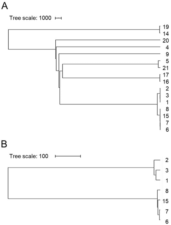
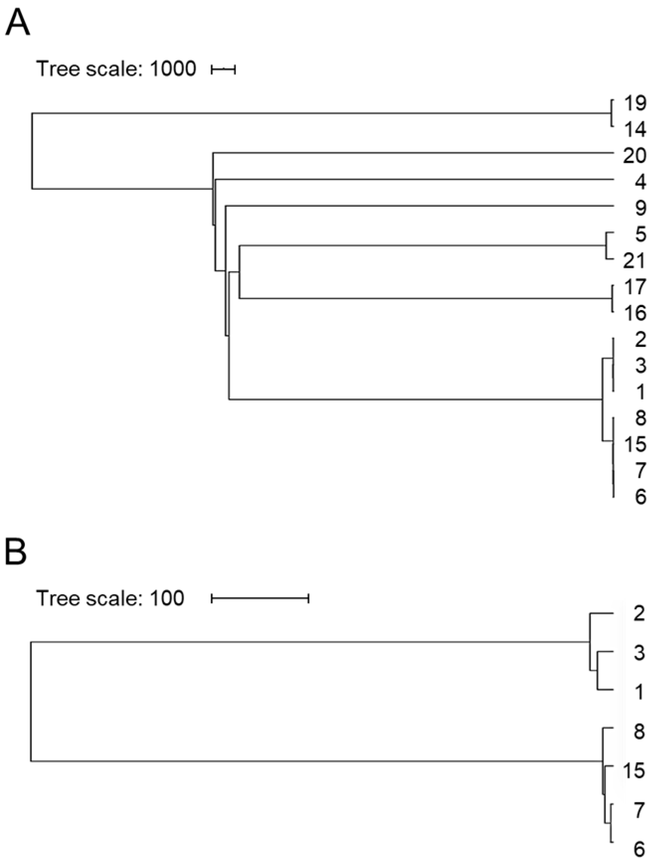
Serotyping of the 16 Salmonella isolates using SeqSero2, which predicted serotypes based on the isolates’ O, H1, and H2 antigenic profiles, identified eight distinct serotypes (Table 2). Newport was the most frequently observed serotype (n = 7, 43.8%), followed by Give and Braenderup, each represented by two isolates (n = 2, 12.5%). The remaining five serotypes, Holcomb, Typhimurium, Hartford, Anatum, and I 4,[5],12:i:- (a variant of Typhimurium), were each identified in a single isolate (n = 1, 6.3%). These findings were further corroborated by two additional serotyping tools, SeqSero (v1.2) and Salmonella Species Serotyping (SSP). Phylogenetic tree analysis revealed distinct clusters (Figure 1). One cluster comprised isolates 1, 2, and 3, along with a separate cluster containing isolates 6, 7, 8, and 15; both clusters were predicted as serotype Newport. Notably, isolates 1, 2, and 3 were recovered from three independent physical replicates at the TP site on the same date. Additional clusters were identified, including isolates 14 and 19 (predicted as serotype Give), 5 and 21 (predicted as serotype Typhimurium), and isolates 16 and 17 (predicted as serotype Braenderup). The remaining isolates were unique (Figure 1A). Further analysis of subclusters within the seven isolates predicted as serotype Newport indicated a closer genetic relationship between isolates 1 and 3, as well as between isolates 7 and 8, compared to the other isolates in this group, suggesting some level of genetic diversity within the predicted Newport serotype (Figure 1B). Notably, Salmonella recovery was predominantly concentrated in the warm season, with several isolation events occurring during or immediately following recorded precipitation (Supplementary Table S1). This was particularly evident for the seven Newport isolates, which were all recovered during May, August, or September 2021. Three of these Newport isolates originated from the Treatment Plant (TP) area, which was associated with a deciduous forest, while the remaining four were found near the Interstate 81 bridge (I81), an area characterized by agricultural use for crops (Table 2).
Table 2.
Serotype prediction by SeqSero2 and Sequence Type identification using MLST 2.0.
Figure 1.
Phylogenetic tree based on draft genome assemblies of Salmonella isolates. The UPGMA phylogenetic tree was generated using the online webtool NDtree 1.2 and visualized with iTOL. Phylogenetic analyses were performed using either all 16 Salmonella isolates (A) or a subgroup of seven isolates predicted as serotype Newport (B) separately. NDtree calculated pairwise nucleotide differences across shared genomic positions, and branch lengths were proportional to these SNP distances. The scale bars represented the number of SNP differences.
3.3. Sequence Types (ST) Identified Among the Isolates
Genomic characterization of the isolates identified seven distinct sequence types (STs) associated with diverse Salmonella serotypes (Table 2). The most prevalent were ST164 and ST118, both corresponding to serotype Newport, which constituted the majority of the sample set. Other identified STs included ST19, associated with both serotype Typhimurium and its monophasic variant (I 4,[5],12:i:-), and ST654, which was linked to serotype Give. Single occurrences of ST2507 (Holcomb), ST405 (Hartford), ST22 (Braenderup), and ST64 (Anatum) were also detected. The diversity in sequence types across the isolates highlighted the broad genetic range within the collection, with specific STs like ST19 and ST164 often being associated with clinically significant outbreaks and the genomic potential for multidrug resistance in global surveillance data.
3.4. Distribution of Salmonella Pathogenicity Islands (SPIs) in Isolates
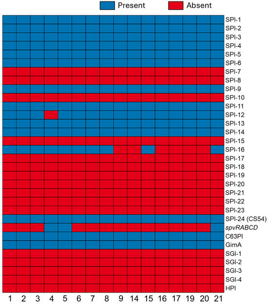
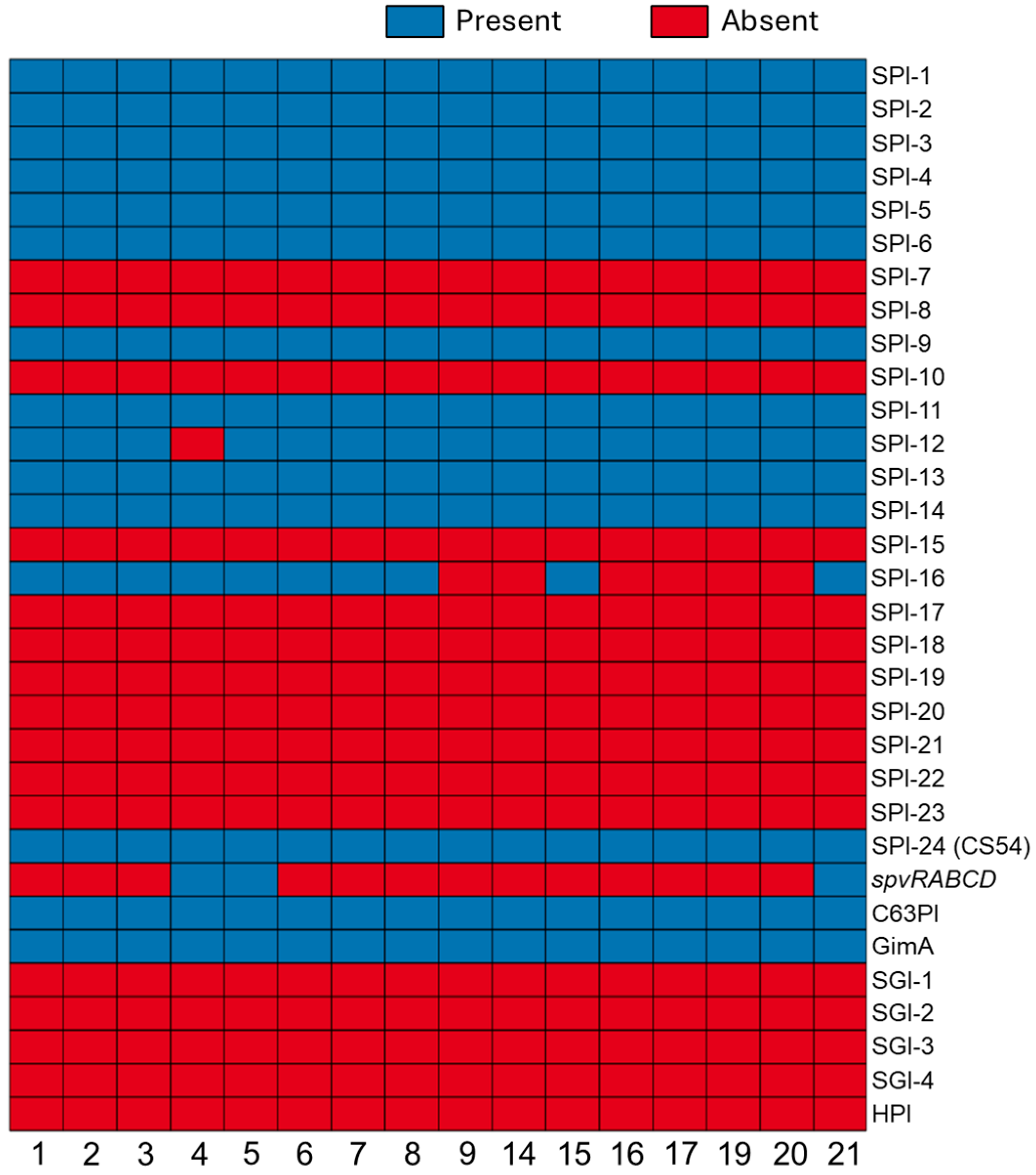
Genomic analysis of 16 Salmonella isolates identified a diverse and serotype-specific array of pathogenicity islands (SPIs) with individual genomes containing between 12 and 13 elements (Figure 2). A highly conserved core consisting of SPI-1, SPI-2, SPI-3, SPI-4, SPI-5, SPI-6, SPI-9, SPI-11, SPI-13, SPI-14, SPI-24, C63PI, and GimA was present in all isolates. Beyond this core, several islands showed variable distribution; SPI-12 was found in nearly all genomes but was notably absent in isolate 4 (serotype Holcomb). SPI-16 exhibited significant variation, appearing in all Newport isolates (1, 2, 3, 6, 7, 8, 15), as well as Holcomb (4), Typhimurium (5), and the monophasic variant (21), but not detected in serotypes Hartford (9), Give (14, 19), Braenderup (16, 17), and Anatum (20). The spvRABCD operon, an extra-chromosomal virulence factor, was limited to isolates 4, 5, and 21. Conversely, several elements, including SPI-7, SPI-8, SPI-10, SPI-15, SPI-17 through SPI-23, and the High-Pathogenicity Island (HPI), were completely absent across the dataset. Additionally, no SGIs (SGI-1, SGI-2, SGI-3, SGI-4) were detected, indicating a lack of these specific multidrug-resistance markers in this cohort.
Figure 2.
Salmonella Pathogenicity Islands (SPIs) in 16 Salmonella isolates. SPI profiles detected in the study genomes. Each column represented an isolate, and each row represented a specific SPI. Blue = SPI was detected. Red = SPI was not detected.
3.5. WGS-Based Identification of AMR Genes
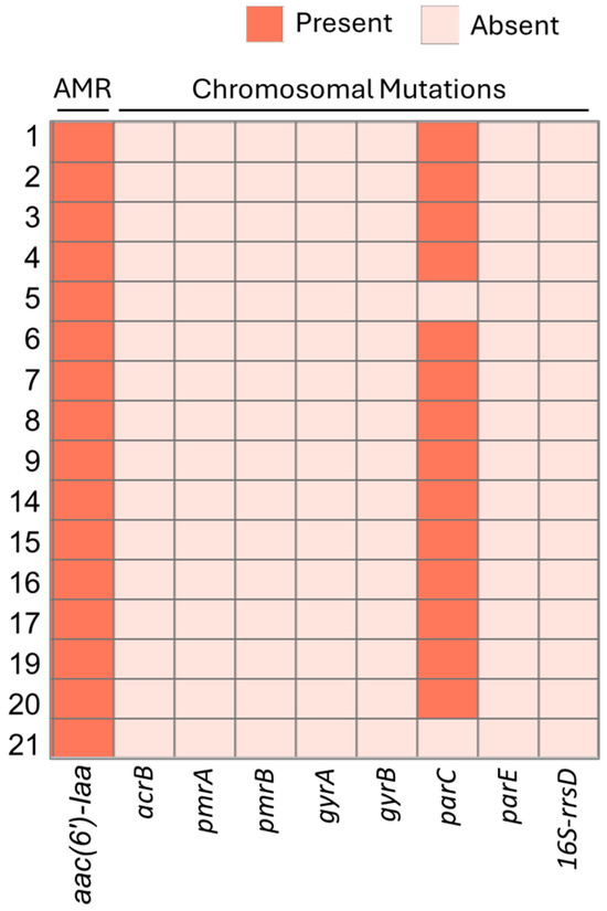
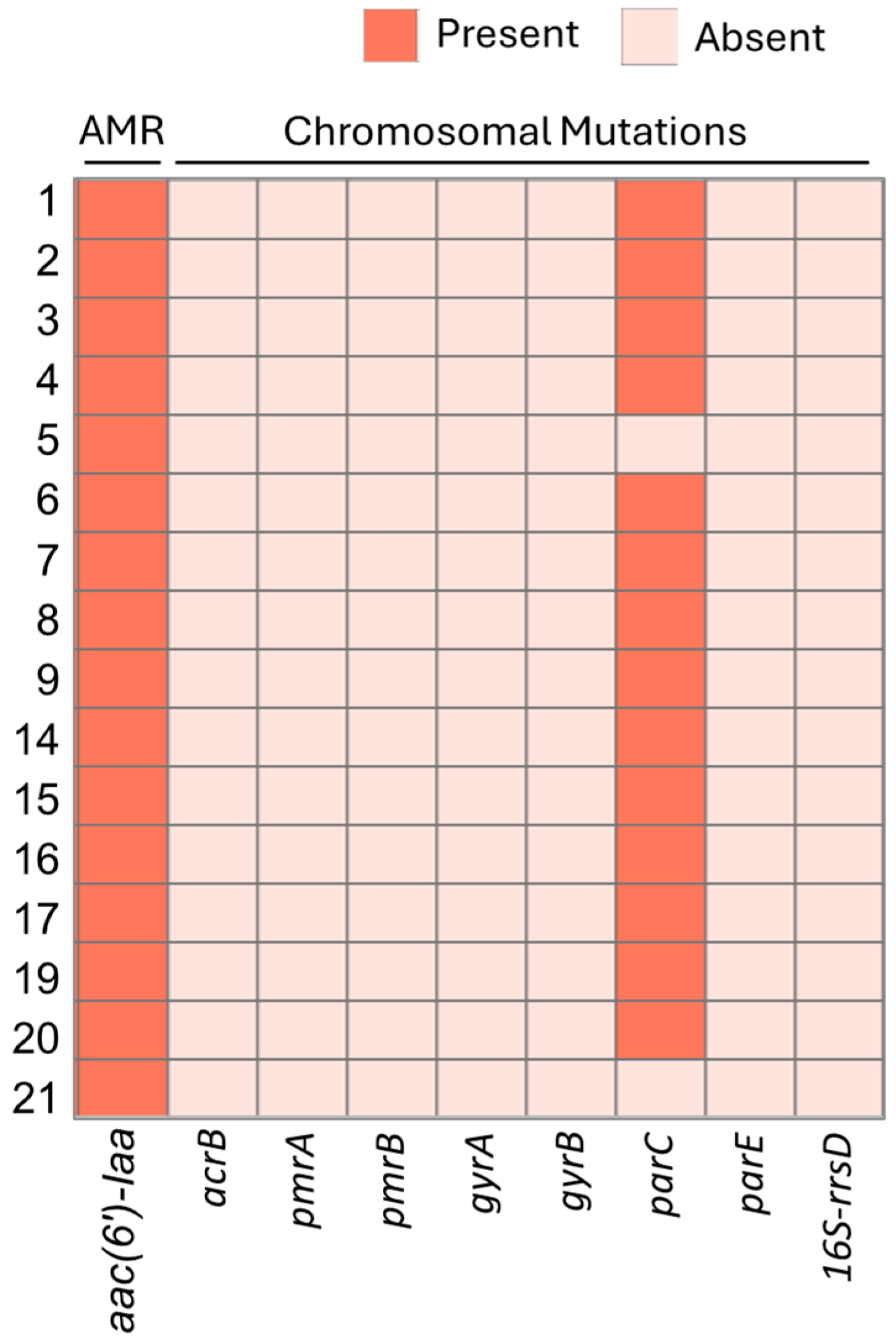
Based on ResFinder analysis, the aminoglycoside resistance gene aac(6’)-Iaa was detected in all isolates. Notably, apart from aac(6’)-Iaa, no other acquired AMR genes, including those related to β-lactam resistance, were detected by ResFinder. In a subset of 14 Salmonella isolates, a single chromosomal point mutation was found within the parC gene, a gene often linked to fluoroquinolone resistance (Figure 3). Specifically, this mutation in parC was identified as T57S, resulting from a nucleotide substitution of ACC to AGC, leading to an amino acid change from threonine (T) to serine (S) at position 57. Notably, isolates 5 and 21, both predicted as Salmonella serotype Typhimurium, did not exhibit this parC point mutation. Furthermore, no other chromosomal mutations in resistance-related genes, including acrB, pmrA, pmrB, gyrA, gyrB, parE, and 16S-rrsD, were detected in the 16 isolates tested.
Figure 3.
WGS-based detection of genomic AMR marker genes. The presence of antibiotic resistance genes and specific chromosomal mutations was determined using ResFinder. A Morpheus-generated heatmap illustrated the presence or absence of detected AMR marker genes and chromosomal mutations. These findings represented the genotypic profile of the isolates rather than confirmed phenotypic resistance.
3.6. Identification of Plasmid Replicons in Salmonella Isolates
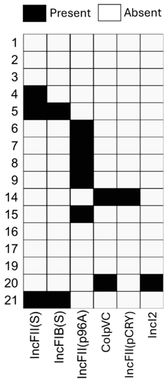
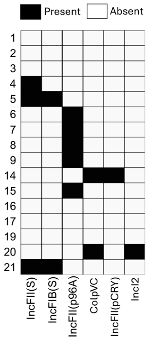
Analysis revealed a total of six different plasmid replicons in 62.5% (10 of 16) Salmonella isolates (Figure 4). While draft assemblies limited the resolution of complete circularized structures, these specific replicons were identified across various serotypes. The IncFII(p96A) replicon (accession number JQ418521) showed the highest frequency, present in five isolates, including serotypes Newport and Hartford. Conversely, the IncFII(pCRY) (NC005814) replicon was identified in a single isolate (serotype Give), and the IncI2 (KP347127) replicon was found in one other isolate (serotype Anatum), representing the lowest frequencies. Two replicons, IncFII(S) (CP000858) and IncFIB(S) (FN432031), were identified within the draft assemblies of isolates 5 and 21, which belonged to the Typhimurium serotype. The ColpVC replicon (JX133088) was uniquely identified within the draft assemblies of isolates 14 (serotype Give) and 20 (serotype Anatum).
Figure 4.
Detection of plasmids in Salmonella isolates through WGS analysis. PlasmidFinder was employed to identify six plasmid replicons in Salmonella isolates. A Morpheus-generated heatmap visualized the presence and absence patterns of each identified plasmid.
3.7. Identification of Restriction-Modification (RM) Systems
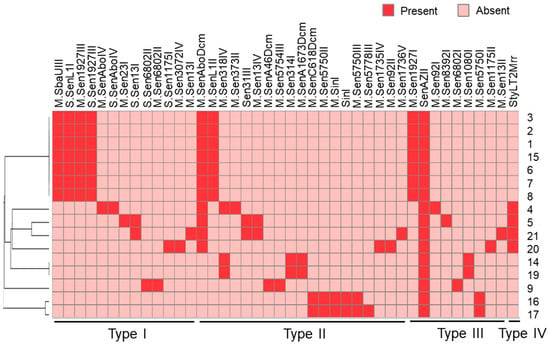
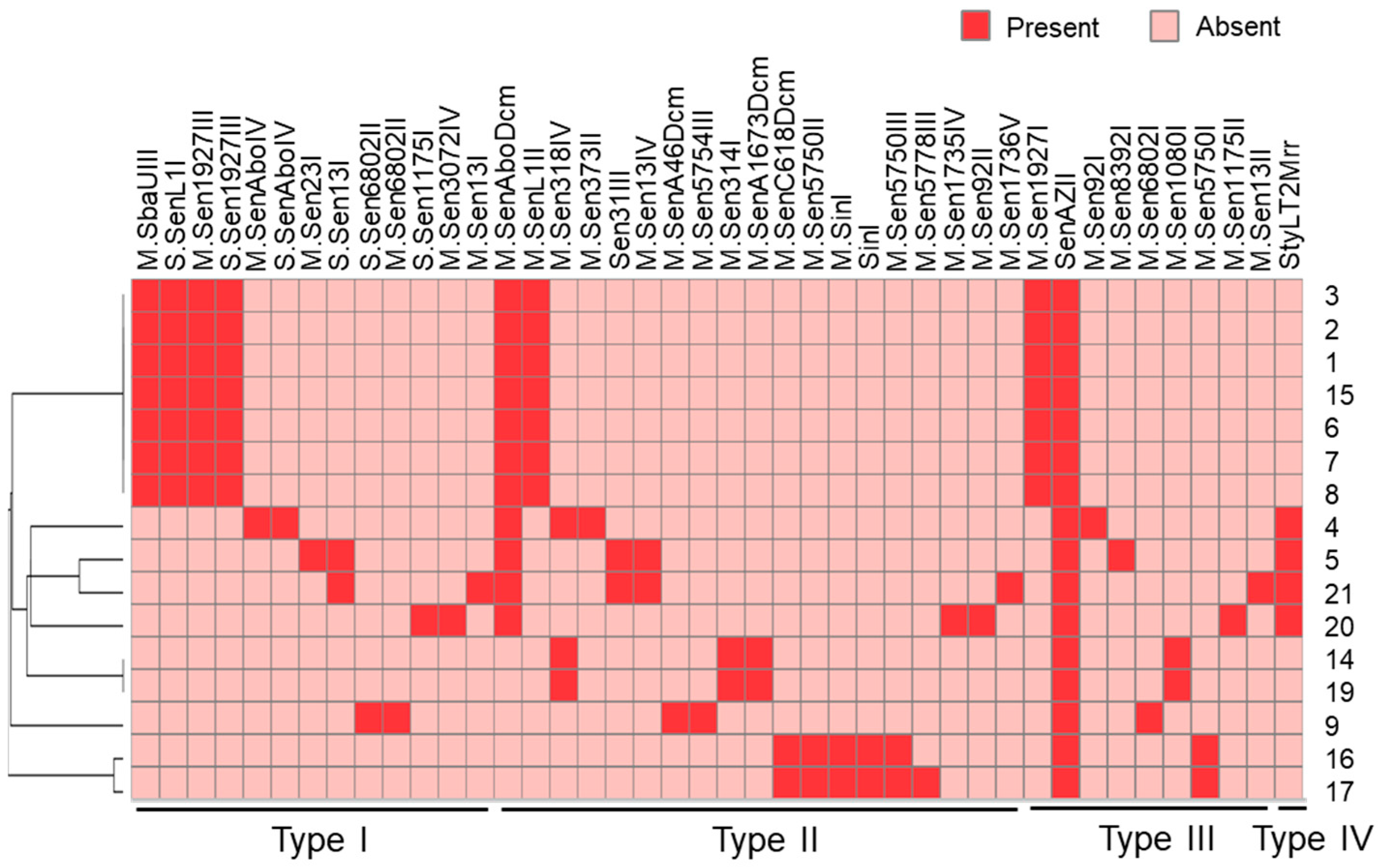
The Restriction-ModificationFinder tool (v1.1) identified genes for 42 RM systems across 16 Salmonella isolates, encompassing Types I through IV restriction enzymes, methyltransferases, site-specificity subunits, and methyl-directed restriction enzymes (Figure 5). These isolates harbored 13, 19, 9, and 1 RM systems belonging to Type I, Type II, Type III, and Type IV enzymes, respectively. Notably, the SenAZII enzyme, a Type III RM system, was present in all 16 isolates. Conversely, the other 19 RMs from Types I to III were each detected in only a single isolate. Isolates 1, 2, 3, 6, 7, 8, and 15, predicted to be serotype Newport, exhibited an identical RM profile for Type I enzymes (including M.SbaUIII, S.SenL1I, M.Sen1927III, and S.Sen1927III), Type II enzymes (including M.SenAboDcm and M.SenL1II), and Type III enzymes (including M.Sen1927I and SenAZII). Isolates 14 and 19, predicted as serotype Give, shared an identical RM profile for Type II enzymes (including M.Sen318IV, M.Sen314I, and M.SenA1673Dcm) and Type III enzymes (including SenAZII and M.Sen1080I).
Figure 5.
Identification of Restriction-Modification (RM) system profile in Salmonella isolates. Restriction-ModificationFinder 1.1 was used to determine the presence of RM-related genes in 16 Salmonella isolates. A Morpheus-generated heatmap visualized the variations in the presence or absence of these RM-related genes, as well as the hierarchical clustering of the isolates based on their RM-related gene profiles. The heatmap indicated 42 distinct RM-related genes and their respective restriction enzyme types.
3.8. Prediction of CRISPR/Cas Loci
CRISPR/Cas loci were predicted in all 16 Salmonella isolates analyzed (Table S4). While a core Type I-E Cas cluster (cas1, cas2, cas3, cas5, cas6, cas7, cse1, and cse2) was present in 14 isolates, the cas1 gene was absent in serotype Newport (isolates 1 and 3). Significant diversity was observed in spacer counts (1 to 34) across isolates. While 13 isolates (81%) presented a typical Salmonella CRISPR/Cas locus organization of two arrays, other configurations were evident. For example, serotype Typhimurium (isolates 5 and 21) featured more than three arrays associated with the Cas cluster. Isolate 9 (serotype Hartford) uniquely presented a single array and a high-confidence orphan array lacking nearby Cas proteins. A common finding across several isolates (e.g., 1, 3, 4, 5, 15, 19) was the presence of orphan CRISPR arrays, typically with single spacers and markers identified in this cohort.
4. Discussion
Building on our prior research [48] where 16 Salmonella strains were recovered from water-overlying sediment in Conococheague Creek, Mid-Atlantic United States, this study used WGS and comprehensive bioinformatics to characterize the genomic profiles and diversity of these isolates. Our goal was to characterize these isolates by analyzing their serotypes, phylogenetic placement, SPIs prevalence, WGS-derived AMR profiles, associated plasmid sequences, RM systems, and CRISPR/Cas loci. Although collected between 2019 and 2022, these isolates provided a critical genomic baseline for 2026 surveillance. Because Salmonella could persist in sediments for years, these historical lineages remained relevant to current irrigation risks and outbreak traceback. Establishing this baseline allowed for the identification of long-term environmental reservoirs and the tracking of micro-evolutionary trends in AMR and virulence today.
The identification of eight distinct Salmonella serotypes among a relatively small collection of 16 isolates from the creek sediment demonstrated considerable genetic diversity within this environmental reservoir. A subset of isolates (n = 8) was recovered from sites located in proximity to agricultural areas (I81) where there was potential for the creek’s water to be used for irrigation (Table 2). Salmonella Newport, found to be the most prevalent serotype in our study (43.8%), was consistently among the top serotypes causing human salmonellosis in the United States and globally, often linked to contaminated produce and other food sources [67,68]. As these Newport isolates were primarily recovered during the warmer months of 2021, this distribution likely reflected seasonal clustering or a transient event rather than a stable, long-term ecology. Thus, longitudinal sampling across multiple years was necessary to confirm whether Newport remained the dominant serotype in this environment. The detection of serotypes such as Give, Braenderup, and Typhimurium documented the occurrence of serotypes previously associated with human illness in this environmental reservoir. The recovery of these diverse Salmonella serotypes from water-overlying sediment was valuable, as sediment provided a protective environment that could allow Salmonella to persist for extended periods, shielding them from environmental stressors like UV radiation and nutrient limitation [69,70]. This could make creek sediment a long-term reservoir for potential foodborne pathogens. The presence of these isolates suggested the potential for sediment-associated Salmonella to be mobilized during irrigation events, representing a possible pathway for transferring pathogens to harvestable portions of crops. Monitoring these genomic profiles in irrigation sources supported compliance with the FDA’s FSMA Produce Safety Rule [71], which established standards for microbial water quality. Our detection of diverse serotypes such as Salmonella Newport in creek sediments emphasized the importance of tracking these environmental reservoirs to mitigate pathogen transfer from agricultural water to fresh produce [71].
While some clustering might have suggested common sources of contamination into the creek, the observed genetic diversity within a single predominant serotype like Newport indicated either multiple introductions over time or the persistence within the creek environment. Understanding these genetic relationships was important for characterizing Salmonella populations in agricultural water systems. The application of WGS-based tools for serotype prediction and phylogenetic analysis provided an accurate and rapid method for agricultural water quality monitoring programs, as it enabled rapid identification of circulating Salmonella serotypes and their genetic relatedness in irrigation sources. This approach facilitated improved characterization of isolates within irrigation sources and provided baseline data for future surveillance. Clustering patterns within serotypes, such as Newport (Figure 1B), were evident in our NDtree-based analysis. However, because NDtree in fast mode with UPGMA only generated a broad overview of relatedness and lacked the resolution of SNP-based pipelines with recombination filtering, these phylogenetic patterns needed to be interpreted cautiously and were not suitable for outbreak traceback or high-confidence source attribution. To place environmental isolates like these into a broader genomic context, future work would benefit from integration with established surveillance ecosystems such as NCBI Pathogen Detection (GenomeTrakr) and EnteroBase [72,73], which used harmonized approaches such as cgMLST and SNP-based workflows to enable broader comparative analyses.
Beyond serotype diversity, our analysis of SPI distribution revealed a conserved yet variable architectural pattern of these virulence factors. We identified a core set of islands (SPI-1 through SPI-6, SPI-9, SPI-11, SPI-13, SPI-14, SPI-24) across the isolates. However, the presence of variable islands underscored the potential for genomic plasticity existing between different serotypes and strains in the environment.
In contrast, fewer islands were detected in Salmonella Braenderup and Salmonella Anatum, with 12 elements detected. Notably, SPI-16 was not detected in Salmonella Braenderup and Hartford, and SPI-10 and SPI-23 were not detected in Salmonella Anatum, suggesting that variable SPIs were difficult to detect in draft genomes compared to closed chromosomes [74,75]. This variation in the virulence repertoire might reflect shared ecological niches or divergent evolutionary strategies for host colonization. The distribution of SPI-12 and SPI-16 provided further evidence of intra-serotype variation; SPI-12 was detected in 15 of 16 isolates but was notably not detected in isolate 4 (Holcomb), while SPI-16 was exclusively detected in the Newport, Typhimurium, Holcomb, and monophasic variants. The detection of the spvRABCD operon in only three isolates (Holcomb, Typhimurium, and I 4,[5],12:i:-) was particularly significant. These genes were crucial for intracellular survival and replication within host cells, a key step in systemic Salmonella infection [76]. The presence of these genes in the Typhimurium and monophasic isolates likely conferred a considerable selective advantage in host colonization and immune evasion [21]. Conversely, the absence of all SGIs (SGI-1 through SGI-4) indicated that while these isolates possessed a robust array of pathogenicity islands, they lacked these specific mobile elements often associated with global multidrug-resistant epidemic strains.
Regarding AMR, the consistent identification of the intrinsic chromosomal aac(6’)-Iaa gene across all tested isolates was a significant observation. However, its presence did not necessarily translate to a high-level aminoglycoside resistance phenotype, as this gene could often be transcriptionally silent unless activated by specific genetic events or environmental conditions [77]. While our WGS data revealed the presence of the aac(6’)-Iaa gene (Figure 3), further phenotypic susceptibility testing was required to confirm the functional expression of this gene and the actual resistance level in these isolates. Based on our ResFinder database analysis, no other AMR genes were detected, suggesting a relatively limited spectrum of resistance in these specific environmental isolates. Nonetheless, the widespread presence of aac(6’)-Iaa established the genotypic baseline for aminoglycoside resistance markers in these isolates. The identification of a single chromosomal point mutation (T57S) within the parC gene in a subset of our Salmonella isolates (Figure 3) provided insights into their genomic background. While not universally present, as parC mutations were commonly associated with reduced susceptibility to fluoroquinolones, this finding was a significant factor for consideration given the criticality of these antibiotics for treating severe Salmonella infections in humans [78]. Although the T57S mutation has been observed in Salmonella Heidelberg isolates linked to salmonellosis outbreaks, where these isolates also exhibited resistance to other antibiotics [79], this specific mutation was widely regarded as a common polymorphism that was unlikely to significantly alter phenotypic susceptibility [78]. Furthermore, research from Hopkins et al. [80] and Khalifa et al. [81] suggested that the T57S mutation may not substantially impact fluoroquinolone susceptibility. Given this complexity and potential discordance, performing phenotypic antimicrobial susceptibility testing for fluoroquinolones on our Salmonella isolates would be highly beneficial, providing a direct assessment of functional resistance to complement these descriptive genomic characterizations for food safety risk assessment and control strategies. These findings aligned with the CDC’s National Antimicrobial Resistance Monitoring System (NARMS) baseline data. The presence of aac(6’)-Iaa and specific parC mutations in our isolates mirrored common genotypic backgrounds reported in national surveillance [82]. While this study relied on genomic screening, we acknowledged that the presence of AMR gene and virulence determinants did not always guarantee phenotypic expression. In this study, AMR and virulence profiles were determined based on genomic presence only; no phenotypic susceptibility testing or in vivo virulence assays were performed. Consequently, these results represented the genotypic potential of the isolates rather than confirmed phenotypic expression. However, genomic AMR profiling was a well-established proxy for public health surveillance, providing a high-resolution map of the genetic potential for resistance. In Salmonella, genotype-to-phenotype correlations were consistently reported to be high across a wide range of antimicrobials [83]. However, these findings characterized the genomic reservoir and did not imply immediate phenotypic resistance in the absence of selective pressure or enhanced gene expression.
The detection of six distinct plasmid replicons in 62.5% of the Salmonella isolates highlighted significant plasmid replicon diversity within this environmental population (Figure 4). Incompatibility group F (IncF) plasmids, characterized by their large size, low copy number, and host restriction to Enterobacteriaceae, have been recognized as significant for their prevalence among Salmonella’s virulence-associated plasmids and frequent genomic association with various AMR genes, including those conferring resistance to fluoroquinolones and β-lactamases [36,84], though no such acquired genes were detected in these specific replicons. In contrast, virulence-associated elements were present. For example, the IncFII(p96A) replicon was genomically associated with genes encoding VirB, a Type IV secretion system component associated with persistent infections [85]. The high prevalence of IncFII(p96A) in five of our isolates suggested its potential importance in the genetic landscape of Salmonella in water-overlying sediment, possibly indicating stable association or successful dissemination within this ecological niche. Consistent with previous research, the co-detection of IncFII(S) and IncFIB(S) plasmids in Typhimurium isolates 5 and 21 was notable due to their frequent co-occurrence with various AMR determinants in clinically relevant serovars [86]. An IncFII(pCRY) plasmid, genomically associated with type IV secretion system-related genes that were previously characterized from Yersinia pestis [87], was found solely in isolate 14 (Give). The IncI2 plasmid in isolate 20 (Anatum) was a known vehicle for spreading β-lactamase genes [88]; while it lacked acquired AMR genes in this isolate, its presence indicated the potential for this environment to act as a reservoir for such clinically important resistance elements, characterizing the distribution of mobile genetic elements in the specific niche. Furthermore, the presence of the cryptic small ColpVC plasmid, lacking AMR genes, in isolates 14 and 20 was notable. While widely observed across Enterobacteriaceae and frequently associated with poultry-related Salmonella serovars [89], its specific occurrence in these isolates might suggest localized ecological adaptations or specific host associations within our study environment.
The collective analysis of RM system profiles and CRISPR/Cas loci in our Salmonella isolates highlighted their critical roles as prokaryotic defense mechanisms against invading foreign DNA like bacteriophages and plasmids. Both systems fundamentally operated by recognizing and interfering with foreign nucleic acids, thereby potentially influencing the capacity for horizontal gene transfer. Our findings revealed 42 diverse RM systems (Figure 5), reflecting robust defense, with the universal presence of the SenAZII RM system suggesting a core, conserved mechanism for genomic integrity. Conversely, high variability among other RM systems (19 unique RMs in Types I-III) indicated ongoing adaptive evolution. Conserved RM profiles within specific serotypes (Newport, Give) could further influence foreign DNA acquisition and the genomic potential for horizontal gene transfer. Complementarily, CRISPR/Cas analysis showed prevalence of adaptive defense systems in 81% of isolates, characterized by a typical CRISPR/Cas organization. This finding suggested their potential active role in genomic defense, consistent with typical Salmonella genomic architecture. Variations in CRISPR/Cas organization and a wide range in spacer numbers (6 to 34) reflected diverse past exposures, including bacteriophages, plasmids, or transposons. The presence of additional Cas clusters lacking cas3 in some isolates suggested alternative interference mechanisms. Together, this diversity in RM and CRISPR/Cas profiles underscored their dynamic interplay in shaping Salmonella evolution, influencing genomic plasticity, and the acquisition or loss of genes relevant to their survival and pathogenicity in environmental niches. Given that these Salmonella isolates originated from creek sediment near agricultural land, a significant pathway for the contamination of fresh produce, understanding these intrinsic defense mechanisms was valuable for characterizing the environmental persistence of this foodborne pathogen. The potential for Salmonella to acquire new genes through mechanisms associated with horizontal gene transfer, regulated by these defense systems, might impact its virulence, stress tolerance, and AMR, posing challenges in terms of food safety.
Several limitations were identified in our study. The small sample size (n = 16) and non-random isolate selection might not fully capture the complete Salmonella diversity in this watershed. Additionally, since our findings were based on short-read replicon detection, future long-read sequencing was required to characterize full plasmid sequences, resolve physical linkage between these elements and specific AMR or virulence genes, and confirm the inherent mobility of these genomic features. Furthermore, without phenotypic antimicrobial susceptibility testing (e.g., minimum inhibitory concentration testing) or in vivo virulence assays, the functional expression of these genomic markers remained to be confirmed. This limitation was consistent with recent studies that emphasized the necessity of laboratory confirmation for a full assessment of resistance profiles [90,91]. Finally, a larger dataset and higher-resolution analysis would be required for a more comprehensive genetic characterization of the watershed.
Our WGS of Salmonella isolates from creek sediment near agricultural land provided valuable insights into their genetic makeup and public health relevance. We found diverse Salmonella serotypes, including prevalent foodborne types like Newport, underscoring the potential for contamination to fresh produce via irrigation. The presence of virulence-associated SPIs and various plasmid replicons (representing the genotypic potential for AMR acquisition) suggested the pathogenic potential and the presence of mobile genetic elements associated with horizontal gene transfer within these environmental strains. Furthermore, the characterization of RM systems and CRISPR/Cas loci revealed the defense mechanisms that shaped Salmonella genomic plasticity. Collectively, these genomic insights were important for understanding Salmonella persistence and spread in agricultural environments, contributing to the genomic baseline data needed for future agricultural water quality monitoring.
Supplementary Materials
The following supporting information can be downloaded at: https://www.mdpi.com/article/10.3390/microbiolres17040072/s1, Table S1. Environmental covariates (daily temperature and precipitation) associated with the collection dates of Salmonella enterica environmental isolates (2019–2022); Table S2. Metadata, assembly statistics, and NCBI accession numbers for the 16 Salmonella enterica isolates recovered from Conococheague Creek sediments (BioProject PRJNA1403484); Table S3. Bioinformatics software, versions, and parameters used for genome analysis; Table S4. The CRISPR/Cas loci of 16 Salmonella isolates detected using CRISPRCasFinder (v4.2.2) program.
Author Contributions
Conceptualization, J.P.; Methodology, S.O. and B.J.H.; Writing—original draft preparation, S.O.; Writing—review and editing, J.P., B.J.H. and S.O.; Resources, J.P. All authors have read and agreed to the published version of the manuscript.
Funding
This research was funded by USDA research project No. 1265-32420-009-00D.
Institutional Review Board Statement
Not applicable.
Informed Consent Statement
Not applicable.
Data Availability Statement
This Whole-Genome project has been deposited at DDBJ/ENA/GenBank under the accession numbers JBTVXP000000000—JBTVXZ000000000, JBTVYA000000000—JBTVYE000000000, and BioProject number PRJNA1403484. The version described in this paper is the first version, JBTVXP010000000—JBTVXZ010000000, JBTVYA010000000—JBTVYE010000000. The raw sequence data are available in the Sequence Read Archive under the accession number SRR36868707—SRR36868722.
Acknowledgments
Authors thank Lauren M. McAllister for technical assistance. Mention of commercial products and companies in this publication is solely for the purpose of providing specific information and does not imply recommendation or endorsement by the US Department of Agriculture. USDA is an equal opportunity provider and employer.
Conflicts of Interest
The authors declare no conflicts of interest.
References
- Han, J.; Aljahdali, N.; Zhao, S.; Tang, H.; Harbottle, H.; Hoffmann, M.; Frye, J.G.; Foley, S.L. Infection Biology of Salmonella enterica. EcoSal Plus 2024, 12, eesp-0001-2023. [Google Scholar] [CrossRef] [Scilit] [PubMed]
- Scallan, E.; Hoekstra, R.M.; Angulo, F.J.; Tauxe, R.V.; Widdowson, M.-A.; Roy, S.L.; Jones, J.L.; Griffin, P.M. Foodborne Illness Acquired in the United States—Major Pathogens. Emerg. Infect. Dis. 2011, 17, 7–15. [Google Scholar] [CrossRef]
- Alves, J.M.; Silva, F.A.; Silveira, D.R.; Massaut, K.B.; Fiorentini, Â.M.; Lopes, G.V.; Magnani, M. Understanding the Potential of Fresh Produce as Vehicles of Salmonella enterica. In Advances in Food and Nutrition Research; Sant’ana, A.S., Ed.; Food-Borne and Microorganisms: Spoilage and Pathogens and their Control; Academic Press: Amsterdam, The Netherlands, 2025; Volume 113, pp. 133–180. [Google Scholar]
- Reddy, S.P.; Wang, H.; Adams, J.K.; Feng, P.C.H. Prevalence and Characteristics of Salmonella Serotypes Isolated from Fresh Produce Marketed in the United States. J. Food Prot. 2016, 79, 6–16. [Google Scholar] [CrossRef] [Scilit] [PubMed]
- Mahmood, S.; Rasool, M.H.; Khurshid, M.; Aslam, B. Genetic Outlook of Colistin-Resistant Salmonella enterica Serovar Typhimurium Recovered from Poultry-Environment Interface: A One Health Standpoint. Pak. Vet. J. 2025, 45, 246–256. [Google Scholar] [CrossRef] [Scilit]
- Tina, L.; Sudarnika, E.; Ridwan, Y.; Sudarwanto, M.B.; Pisestyani, H. Microbiological Safety of Smoked Fish: A Review of Escherichia coli and Salmonella spp. Contamination in Traditional Markets of Kendari City, Indonesia. Int. J. Vet. Sci. 2025, 14, 771–776. [Google Scholar] [CrossRef] [Scilit]
- Maffei, D.F.; Batalha, E.Y.; Landgraf, M.; Schaffner, D.W.; Franco, B.D.G.M. Microbiology of Organic and Conventionally Grown Fresh Produce. Braz. J. Microbiol. 2016, 47, 99–105. [Google Scholar] [CrossRef] [Scilit]
- Uyttendaele, M.; Jaykus, L.-A.; Amoah, P.; Chiodini, A.; Cunliffe, D.; Jacxsens, L.; Holvoet, K.; Korsten, L.; Lau, M.; McClure, P.; et al. Microbial Hazards in Irrigation Water: Standards, Norms, and Testing to Manage Use of Water in Fresh Produce Primary Production. Compr. Rev. Food Sci. Food Saf. 2015, 14, 336–356. [Google Scholar] [CrossRef] [Scilit]
- Thomas, G.A.; Paradell Gil, T.; Müller, C.T.; Rogers, H.J.; Berger, C.N. From Field to Plate: How Do Bacterial Enteric Pathogens Interact with Ready-to-Eat Fruit and Vegetables, Causing Disease Outbreaks? Food Microbiol. 2024, 117, 104389. [Google Scholar] [CrossRef] [Scilit]
- Sharma, M.; Handy, E.T.; East, C.L.; Kim, S.; Jiang, C.; Callahan, M.T.; Allard, S.M.; Micallef, S.; Craighead, S.; Anderson-Coughlin, B.; et al. Prevalence of Salmonella and Listeria monocytogenes in Non-Traditional Irrigation Waters in the Mid-Atlantic United States Is Affected by Water Type, Season, and Recovery Method. PLoS ONE 2020, 15, e0229365. [Google Scholar] [CrossRef] [Scilit]
- Bai, S.; Lung, W.-S. Modeling Sediment Impact on the Transport of Fecal Bacteria. Water Res. 2005, 39, 5232–5240. [Google Scholar] [CrossRef] [Scilit] [PubMed]
- Fries, J.S.; Characklis, G.W.; Noble, R.T. Sediment–Water Exchange of Vibrio Sp. and Fecal Indicator Bacteria: Implications for Persistence and Transport in the Neuse River Estuary, North Carolina, USA. Water Res. 2008, 42, 941–950. [Google Scholar] [CrossRef] [Scilit] [PubMed]
- Lang-Halter, E.; Schober, S.; Scherer, S. Permanent Colonization of Creek Sediments, Creek Water and Limnic Water Plants by Four Listeria Species in Low Population Densities. Z. Naturforschung C 2016, 71, 335–345. [Google Scholar] [CrossRef] [Scilit]
- Perkins, T.L.; Clements, K.; Baas, J.H.; Jago, C.F.; Jones, D.L.; Malham, S.K.; McDonald, J.E. Sediment Composition Influences Spatial Variation in the Abundance of Human Pathogen Indicator Bacteria within an Estuarine Environment. PLoS ONE 2014, 9, e112951. [Google Scholar] [CrossRef] [Scilit] [PubMed]
- Medrano-Félix, J.A.; Guerra-Rodríguez, J.C.; Verdugo Arredondo, V.A.; González-Torres, B.; Garrido-Palazuelos, L.I.; González-López, I.; Castro-Del Campo, N.; López-Cuevas, O.; Chaidez Quiroz, C. Prevalence, Diversity, and Antimicrobial Resistance of Salmonella Spp. Isolated from River Sediments in Northwest Mexico. J. Infect. Dev. Ctries. 2024, 18, 1875–1882. [Google Scholar] [CrossRef] [Scilit]
- Savariz, A.; Degenhardt, R.; Rebellato, R.; Duarte, S.C.; D’Agostini, F.M.; Millezi, A. Diversity and Antimicrobial Resistance of Salmonella enterica Serovars in Surface River Water and Sediment. Rev. Ciências Agroveterinárias 2021, 20, 231–240. [Google Scholar] [CrossRef] [Scilit]
- Marine, S.C.; Pagadala, S.; Wang, F.; Pahl, D.M.; Melendez, M.V.; Kline, W.L.; Oni, R.A.; Walsh, C.S.; Everts, K.L.; Buchanan, R.L.; et al. The Growing Season, but Not the Farming System, Is a Food Safety Risk Determinant for Leafy Greens in the Mid-Atlantic Region of the United States. Appl. Environ. Microbiol. 2015, 81, 2395–2407. [Google Scholar] [CrossRef] [Scilit] [PubMed]
- Cobo-Simón, M.; Hart, R.; Ochman, H. Gene Flow and Species Boundaries of the Genus Salmonella. mSystems 2023, 8, e00292-23. [Google Scholar] [CrossRef] [Scilit]
- Ferrari, R.G.; Rosario, D.K.A.; Cunha-Neto, A.; Mano, S.B.; Figueiredo, E.E.S.; Conte-Junior, C.A. Worldwide Epidemiology of Salmonella Serovars in Animal-Based Foods: A Meta-Analysis. Appl. Environ. Microbiol. 2019, 85, e00591-19. [Google Scholar] [CrossRef] [Scilit]
- Issenhuth-Jeanjean, S.; Roggentin, P.; Mikoleit, M.; Guibourdenche, M.; de Pinna, E.; Nair, S.; Fields, P.I.; Weill, F.-X. Supplement 2008–2010 (No. 48) to the White–Kauffmann–Le Minor Scheme. Res. Microbiol. 2014, 165, 526–530. [Google Scholar] [CrossRef] [Scilit]
- Fierer, J.; Guiney, D.G. Diverse Virulence Traits Underlying Different Clinical Outcomes of Salmonella Infection. J. Clin. Investig. 2001, 107, 775–780. [Google Scholar] [CrossRef] [Scilit]
- Jajere, S.M. A Review of Salmonella enterica with Particular Focus on the Pathogenicity and Virulence Factors, Host Specificity and Antimicrobial Resistance Including Multidrug Resistance. Vet. World 2019, 12, 504–521. [Google Scholar] [CrossRef] [Scilit] [PubMed]
- Phuket, N.R.N.; Buahom, J.; Senaphan, K.; Sukon, P.; Sringam, S.; Angkititrakul, S.; Sringam, P. Serovars and antimicrobial resistance of Salmonella isolated from free—Living turtles in the Turtle Village, Northeastern Thailand: One Health perspective. Int. J. Vet. Sci. 2025, 14, 212–218. [Google Scholar] [CrossRef] [Scilit]
- Dewey-Mattia, D.; Manikonda, K.; Hall, A.J.; Wise, M.E.; Crowe, S.J. Surveillance for Foodborne Disease Outbreaks—United States, 2009–2015. MMWR Surveill. Summ. 2018, 67, 1–11. [Google Scholar] [CrossRef] [Scilit] [PubMed]
- Gu, G.; Strawn, L.K.; Zheng, J.; Reed, E.A.; Rideout, S.L. Diversity and Dynamics of Salmonella enterica in Water Sources, Poultry Litters, and Field Soils Amended with Poultry Litter in a Major Agricultural Area of Virginia. Front. Microbiol. 2019, 10, 2868. [Google Scholar] [CrossRef] [Scilit]
- Trachsel, J.M.; Bearson, B.L.; Brunelle, B.W.; Bearson, S.M.D. Relationship and Distribution of Salmonella enterica Serovar I 4,[5],12:i:- Strain Sequences in the NCBI Pathogen Detection Database. BMC Genom. 2022, 23, 268. [Google Scholar] [CrossRef] [Scilit]
- Etoom, E.M.; Gharaibeh, M.H. Investigation and Molecular Assessment of Integron—Mediated Resistance Determinants in Salmonella Species from Broiler Chickens in Jordan. Pak. Vet. J. 2025, 45, 2036–2043. [Google Scholar] [CrossRef] [Scilit]
- Maurer, J.J.; Martin, G.; Hernandez, S.; Cheng, Y.; Gerner-Smidt, P.; Hise, K.B.; D’Angelo, M.T.; Cole, D.; Sanchez, S.; Madden, M.; et al. Diversity and Persistence of Salmonella enterica Strains in Rural Landscapes in the Southeastern United States. PLoS ONE 2015, 10, e0128937. [Google Scholar] [CrossRef] [Scilit]
- Callahan, M.T.; Van Kessel, J.A.; Micallef, S.A. Salmonella enterica Recovery from River Waters of the Maryland Eastern Shore Reveals High Serotype Diversity and Some Multidrug Resistance. Environ. Res. 2019, 168, 7–13. [Google Scholar] [CrossRef] [Scilit]
- Gorski, L.; Parker, C.T.; Liang, A.; Cooley, M.B.; Jay-Russell, M.T.; Gordus, A.G.; Atwill, E.R.; Mandrell, R.E. Prevalence, Distribution, and Diversity of Salmonella enterica in a Major Produce Region of California. Appl. Environ. Microbiol. 2011, 77, 2734–2748. [Google Scholar] [CrossRef] [Scilit]
- Deaven, A.M.; Ferreira, C.M.; Reed, E.A.; Chen See, J.R.; Lee, N.A.; Almaraz, E.; Rios, P.C.; Marogi, J.G.; Lamendella, R.; Zheng, J.; et al. Salmonella Genomics and Population Analyses Reveal High Inter- and Intraserovar Diversity in Freshwater. Appl. Environ. Microbiol. 2021, 87, e02594-20, Erratum in Appl. Environ. Microbiol. 2025, 91, e0122725. https://doi.org/10.1128/aem.01227-25. [Google Scholar] [CrossRef] [Scilit] [PubMed]
- Nazir, J.; Manzoor, T.; Saleem, A.; Gani, U.; Bhat, S.S.; Khan, S.; Haq, Z.; Jha, P.; Ahmad, S.M. Combatting Salmonella: A Focus on Antimicrobial Resistance and the Need for Effective Vaccination. BMC Infect. Dis. 2025, 25, 84. [Google Scholar] [CrossRef] [Scilit]
- Narinthorn, R.; Saengsawang, P.; Boripun, R.; Wongtawan, T.; Phongkidakarn, P.; Nissapatorn, V.; Pereira, M.D.L.; Paul, A.K.; Mitsuwan, W. Occurrence and Antimicrobial Resistance in Salmonella enterica from Sport Animals and Livestock in Southern Thailand. Pak. Vet. J. 2025, 45, 887–892. [Google Scholar] [CrossRef] [Scilit]
- Borovikov, S.; Kuibagarov, M.; Shirobokova, D.; Akanova, Z.; Muranets, A.; Rychshanova, R. Prevalence of Salmonella spp. on Poultry Farms in Kazakhstan and Analysis of Antimicrobial Resistance of the Isolates. Int. J. Vet. Sci. 2025, 14, 1084–1092. [Google Scholar] [CrossRef] [Scilit]
- Laban, S.E.; Arafa, A.A.; Ibrahim, E.S.; Eman, F.E.; Khalefa, H.S. Dry Biofilm Formation, Mono- and Dual-Attachment, on Plastic and Galvanized Surfaces by Salmonella Typhimurium and Staphylococcus aureus Isolated from Poultry House. Int. J. Vet. Sci. 2025, 14, 25–31. [Google Scholar] [CrossRef] [Scilit]
- McMillan, E.A.; Jackson, C.R.; Frye, J.G. Transferable Plasmids of Salmonella enterica Associated with Antibiotic Resistance Genes. Front. Microbiol. 2020, 11, 562181. [Google Scholar] [CrossRef] [Scilit]
- Lee, I.P.A.; Eldakar, O.T.; Gogarten, J.P.; Andam, C.P. Bacterial Cooperation through Horizontal Gene Transfer. Trends Ecol. Evol. 2022, 37, 223–232. [Google Scholar] [CrossRef] [Scilit]
- Samreen; Ahmad, I.; Malak, H.A.; Abulreesh, H.H. Environmental Antimicrobial Resistance and Its Drivers: A Potential Threat to Public Health. J. Glob. Antimicrob. Resist. 2021, 27, 101–111. [Google Scholar] [CrossRef] [Scilit] [PubMed]
- Larsson, D.G.J.; Flach, C.-F. Antibiotic Resistance in the Environment. Nat. Rev. Microbiol. 2022, 20, 257–269. [Google Scholar] [CrossRef] [Scilit]
- Gomes, E.; Araújo, D.; Nogueira, T.; Oliveira, R.; Silva, S.; Oliveira, L.V.N.; Azevedo, N.F.; Almeida, C.; Castro, J. Advances in Whole Genome Sequencing for Foodborne Pathogens: Implications for Clinical Infectious Disease Surveillance and Public Health. Front. Cell. Infect. Microbiol. 2025, 15, 1593219. [Google Scholar] [CrossRef] [Scilit] [PubMed]
- Cho, S.; Hiott, L.M.; House, S.L.; Woodley, T.A.; McMillan, E.A.; Sharma, P.; Barrett, J.B.; Adams, E.S.; Brandenburg, J.M.; Hise, K.B.; et al. Analysis of Salmonella enterica Isolated from a Mixed-Use Watershed in Georgia, USA: Antimicrobial Resistance, Serotype Diversity, and Genetic Relatedness to Human Isolates. Appl. Environ. Microbiol. 2022, 88, e00393-22. [Google Scholar] [CrossRef] [Scilit] [PubMed]
- Ben Hassena, A.; Haendiges, J.; Zormati, S.; Guermazi, S.; Gdoura, R.; Gonzalez-Escalona, N.; Siala, M. Virulence and Resistance Genes Profiles and Clonal Relationships of Non-Typhoidal Food-Borne Salmonella Strains Isolated in Tunisia by Whole Genome Sequencing. Int. J. Food Microbiol. 2021, 337, 108941. [Google Scholar] [CrossRef] [Scilit] [PubMed]
- Ibrahim, G.M.; Morin, P.M. Salmonella Serotyping Using Whole Genome Sequencing. Front. Microbiol. 2018, 9, 2993. [Google Scholar] [CrossRef] [Scilit] [PubMed]
- Carneiro, D.G.; Vidigal, P.M.P.; Morgan, T.; Vanetti, M.C.D. Genome Sequencing and Analysis of Salmonella enterica Subsp. enterica Serotype Enteritidis PT4 578: Insights into Pathogenicity and Virulence. Access Microbiol. 2024, 6, 000828.v3. [Google Scholar] [CrossRef] [Scilit]
- Elliott, J.F.K.; McLeod, D.V.; Taylor, T.B.; Westra, E.R.; Gandon, S.; Watson, B.N.J. Conditions for the Spread of CRISPR-Cas Immune Systems into Bacterial Populations. ISME J. 2024, 18, wrae108. [Google Scholar] [CrossRef] [Scilit] [PubMed]
- Pachepsky, Y.; Harriger, M.D.; Panko Graff, C.; Stocker, M.D.; Smith, J.E.; Slifka, J.M. Coupled Dynamics of Fecal Indicator Bacteria in Sandy Sediments and the Water Column: A 3-Year High-Frequency Study at a Pennsylvania Creek. Water Air Soil Pollut. 2023, 234, 398. [Google Scholar] [CrossRef] [Scilit]
- Pachepsky, Y.; Harriger, M.D.; Panko Graff, C.; Wyatt-Brown, L.; Stocker, M.D.; Smith, J.E. Multiscale Spatiotemporal Variability of Fecal Indicator Bacteria and Associated Particle Size Distributions in the Sandy Bottom Sediments of a Pennsylvania Creek. J. Environ. Qual. 2024, 53, 101–111. [Google Scholar] [CrossRef] [Scilit] [PubMed]
- Oh, S.; Patel, J.; Yin, H.-B.; Harriger, D. Spatial and Temporal Prevalence of Foodborne Bacterial Pathogens in Water-Overlying Sediments in the Mid-Atlantic United States. Water Air Soil Pollut. 2025, 236, 725. [Google Scholar] [CrossRef] [Scilit]
- NOAA National Centers for Environmental Information (NCEI). Climate Data Online. Available online: https://www.ncei.noaa.gov/cdo-web/ (accessed on 7 March 2026).
- Bushnell: BBMap: A Fast, Accurate, Splice-Aware Aligner–Google Scholar. Available online: https://scholar.google.com/scholar_lookup?title=BBMap:%20A%20Fast,%20Accurate,%20Splice-Aware%20Aligner.%20No.%20LBNL-7065E&author=B.%20Bushnell&publication_year=2014& (accessed on 22 July 2025).
- Bankevich, A.; Nurk, S.; Antipov, D.; Gurevich, A.A.; Dvorkin, M.; Kulikov, A.S.; Lesin, V.M.; Nikolenko, S.I.; Pham, S.; Prjibelski, A.D.; et al. SPAdes: A New Genome Assembly Algorithm and Its Applications to Single-Cell Sequencing. J. Comput. Biol. 2012, 19, 455–477. [Google Scholar] [CrossRef] [Scilit]
- Challis, R. Rjchallis/Assembly-Stats 17.02. 2017. Available online: https://github.com/rjchallis/Assembly-Stats (accessed on 8 March 2025).
- Parks, D.H.; Imelfort, M.; Skennerton, C.T.; Hugenholtz, P.; Tyson, G.W. CheckM: Assessing the Quality of Microbial Genomes Recovered from Isolates, Single Cells, and Metagenomes. Genome Res. 2015, 25, 1043–1055. [Google Scholar] [CrossRef] [Scilit]
- Chaumeil, P.-A.; Mussig, A.J.; Hugenholtz, P.; Parks, D.H. GTDB-Tk: A Toolkit to Classify Genomes with the Genome Taxonomy Database. Bioinformatics 2020, 36, 1925–1927. [Google Scholar] [CrossRef] [Scilit]
- Zhang, S.; den Bakker, H.C.; Li, S.; Chen, J.; Dinsmore, B.A.; Lane, C.; Lauer, A.C.; Fields, P.I.; Deng, X. SeqSero2: Rapid and Improved Salmonella Serotype Determination Using Whole-Genome Sequencing Data. Appl. Environ. Microbiol. 2019, 85, e01746-19. [Google Scholar] [CrossRef] [Scilit]
- Zhang, S.; Yin, Y.; Jones, M.B.; Zhang, Z.; Deatherage Kaiser, B.L.; Dinsmore, B.A.; Fitzgerald, C.; Fields, P.I.; Deng, X. Salmonella Serotype Determination Utilizing High-Throughput Genome Sequencing Data. J. Clin. Microbiol. 2015, 53, 1685–1692. [Google Scholar] [CrossRef] [Scilit] [PubMed]
- Sundaresan, S.; Rathinavelan, T. SSP: An In Silico Tool for Salmonella Species Serotyping Using the Sequences of O-Antigen Biosynthesis Proteins and H-Antigen Filament Proteins. J. Mol. Biol. 2023, 435, 168046. [Google Scholar] [CrossRef] [Scilit] [PubMed]
- Larsen, M.V.; Cosentino, S.; Rasmussen, S.; Friis, C.; Hasman, H.; Marvig, R.L.; Jelsbak, L.; Sicheritz-Pontén, T.; Ussery, D.W.; Aarestrup, F.M.; et al. Multilocus Sequence Typing of Total-Genome-Sequenced Bacteria. J. Clin. Microbiol. 2012, 50, 1355–1361. [Google Scholar] [CrossRef] [Scilit]
- Leekitcharoenphon, P.; Nielsen, E.M.; Kaas, R.S.; Lund, O.; Aarestrup, F.M. Evaluation of Whole Genome Sequencing for Outbreak Detection of Salmonella enterica. PLoS ONE 2014, 9, e87991. [Google Scholar] [CrossRef] [Scilit]
- Letunic, I.; Bork, P. Interactive Tree of Life (iTOL) v5: An Online Tool for Phylogenetic Tree Display and Annotation. Nucleic Acids Res. 2021, 49, W293–W296. [Google Scholar] [CrossRef] [Scilit] [PubMed]
- Hsu, C.H.; Li, C.; Hoffmann, M.; McDermott, P.; Abbott, J.; Ayers, S.; Tyson, G.H.; Tate, H.; Yao, K.; Allard, M.; et al. Comparative Genomic Analysis of Virulence, Antimicrobial Resistance, and Plasmid Profiles of Salmonella Dublin Isolated from Sick Cattle, Retail Beef, and Humans in the United States. Microb. Drug Resist. 2019, 25, 1238–1249. [Google Scholar] [CrossRef] [Scilit]
- Bortolaia, V.; Kaas, R.S.; Ruppe, E.; Roberts, M.C.; Schwarz, S.; Cattoir, V.; Philippon, A.; Allesoe, R.L.; Rebelo, A.R.; Florensa, A.F.; et al. ResFinder 4.0 for Predictions of Phenotypes from Genotypes. J. Antimicrob. Chemother. 2020, 75, 3491–3500. [Google Scholar] [CrossRef] [Scilit]
- Carattoli, A.; Zankari, E.; García-Fernández, A.; Voldby Larsen, M.; Lund, O.; Villa, L.; Møller Aarestrup, F.; Hasman, H. In Silico Detection and Typing of Plasmids Using PlasmidFinder and Plasmid Multilocus Sequence Typing. Antimicrob. Agents Chemother. 2014, 58, 3895–3903. [Google Scholar] [CrossRef] [Scilit]
- Roer, L.; Hendriksen, R.S.; Leekitcharoenphon, P.; Lukjancenko, O.; Kaas, R.S.; Hasman, H.; Aarestrup, F.M. Is the Evolution of Salmonella enterica subsp. enterica Linked to Restriction-Modification Systems? mSystems 2016, 1, e00009-16. [Google Scholar] [CrossRef] [Scilit]
- Gould, J. Morpheus: Versatile matrix Visualization and Analysis Software. 0.1Broad Institute. 2016. Available online: https://software.broadinstitute.org/morpheus/index.html (accessed on 22 July 2025).
- Couvin, D.; Bernheim, A.; Toffano-Nioche, C.; Touchon, M.; Michalik, J.; Néron, B.; Rocha, E.P.C.; Vergnaud, G.; Gautheret, D.; Pourcel, C. CRISPRCasFinder, an Update of CRISRFinder, Includes a Portable Version, Enhanced Performance and Integrates Search for Cas Proteins. Nucleic Acids Res. 2018, 46, W246–W251. [Google Scholar] [CrossRef] [Scilit]
- CDC. BEAM Dashboard. 2025. Available online: https://www.cdc.gov/ncezid/dfwed/BEAM-dashboard.html (accessed on 23 July 2025).
- Lamichhane, B.; Mawad, A.M.M.; Saleh, M.; Kelley, W.G.; Harrington, P.J.; Lovestad, C.W.; Amezcua, J.; Sarhan, M.M.; El Zowalaty, M.E.; Ramadan, H.; et al. Salmonellosis: An Overview of Epidemiology, Pathogenesis, and Innovative Approaches to Mitigate the Antimicrobial Resistant Infections. Antibiotics 2024, 13, 76. [Google Scholar] [CrossRef] [Scilit]
- Malham, S.K.; Rajko-Nenow, P.; Howlett, E.; Tuson, K.E.; Perkins, T.L.; Pallett, D.W.; Wang, H.; Jago, C.F.; Jones, D.L.; McDonald, J.E. The Interaction of Human Microbial Pathogens, Particulate Material and Nutrients in Estuarine Environments and Their Impacts on Recreational and Shellfish Waters. Environ. Sci. Process. Impacts 2014, 16, 2145–2155. [Google Scholar] [CrossRef] [Scilit]
- Liu, H.; Whitehouse, C.A.; Li, B. Presence and Persistence of Salmonella in Water: The Impact on Microbial Quality of Water and Food Safety. Front. Public Health 2018, 6, 159. [Google Scholar] [CrossRef] [Scilit] [PubMed]
- FDA. Standards for the Growing, Harvesting, Packing, and Holding of Produce for Human Consumption; Final Rule. Fed. Regist. 2015, 80, 74353–74568. Available online: https://www.govinfo.gov/app/details/FR-2015-11-27/2015-28159 (accessed on 10 March 2026).
- Allard, M.W.; Strain, E.; Melka, D.; Bunning, K.; Musser, S.M.; Brown, E.W.; Tim-me, R. GenomeTrakr Network: A Global Surveillance Network Formed to Accelerate Foodborne Pathogen Identification and Outbreak Investigations. Appl. Environ. Microbiol. 2016, 82, 6250–6255. [Google Scholar] [CrossRef] [Scilit]
- Zhou, Z.; Alikhan, N.-F.; Mohamed, K.; Fan, Y.; Agama, J.S. The EnteroBase User’s Guide, with Case Studies on Salmonella ansmission, Yersinia pestis Phylogeny, and Escherichia coli Mutation Rates. Genome Res. 2020, 30, 138–152. [Google Scholar] [CrossRef] [Scilit]
- Zhao, S.; Li, C.; Hsu, C.H.; Tyson, G.H.; Strain, E.; Tate, H.; Tran, T.T.; Abbott, J.; McDermott, P.F. Comparative Genomic Analysis of 450 Strains of Salmonella enterica Isolated from Diseased Animals. Genes. 2020, 11, 1025. [Google Scholar] [CrossRef] [Scilit] [PubMed]
- Li, C.; Tyson, G.H.; Hsu, C.H.; Harrison, L.; Strain, E.; Tran, T.T.; Tillman, G.E.; Dessai, U.; McDermott, P.F.; Zhao, S. Long-Read Sequencing Reveals Evolution and Acquisition of Antimicrobial Resistance and Virulence Genes in Salmonella enterica. Front. Microbiol. 2021, 12, 777817. [Google Scholar] [CrossRef] [Scilit]
- Waterman, S.R.; Holden, D.W. Functions and Effectors of the Salmonella Pathogenicity Island 2 Type III Secretion System. Cell. Microbiol. 2003, 5, 501–511. [Google Scholar] [CrossRef] [Scilit]
- Tang, B.; Elbediwi, M.; Nambiar, R.B.; Yang, H.; Lin, J.; Yue, M. Genomic Characterization of Antimicrobial-Resistant Salmonella enterica in Duck, Chicken, and Pig Farms and Retail Markets in Eastern China. Microbiol. Spectr. 2022, 10, e01257-22. [Google Scholar] [CrossRef] [Scilit]
- Cuypers, W.L.; Jacobs, J.; Wong, V.; Klemm, E.J.; Deborggraeve, S.; Van Puyvelde, S. Fluoroquinolone Resistance in Salmonella: Insights by Whole-Genome Sequencing. Microb. Genom. 2018, 4, e000195. [Google Scholar] [CrossRef] [Scilit] [PubMed]
- Etter, A.J.; West, A.M.; Burnett, J.L.; Wu, S.T.; Veenhuizen, D.R.; Ogas, R.A.; Oliver, H.F. Salmonella enterica Subsp. enterica Serovar Heidelberg Food Isolates Associated with a Salmonellosis Outbreak Have Enhanced Stress Tolerance Capabilities. Appl. Environ. Microbiol. 2019, 85, e01065-19. [Google Scholar] [CrossRef] [Scilit]
- Hopkins, K.L.; Arnold, C.; Threlfall, E.J. Rapid Detection of gyrA and parC Mutations in Quinolone-Resistant Salmonella enterica Using Pyrosequencing® Technology. J. Microbiol. Methods 2007, 68, 163–171. [Google Scholar] [CrossRef] [Scilit] [PubMed]
- Khalifa, H.O.; Kayama, S.; Ramadan, H.; Yu, L.; Hayashi, W.; Sugawara, Y.; Kondo, S.U.; Marzooq, F.A.; Matsumoto, T.; Sugai, M. Comprehensive Analysis of β-Lactam Resistant Non-Typhoidal Salmonella Isolates: Phenotypic and Genotypic Insights from Clinical Samples in Japan. J. Glob. Antimicrob. Resist. 2025, 43, 98–110. [Google Scholar] [CrossRef] [Scilit]
- CDC. National Antimicrobial Resistance Monitoring System for Enteric Bacteria (NARMS): Integrated Summary 2021. CDC/FDA/USDA. Available online: https://www.cdc.gov/narms/reports/index.html (accessed on 7 March 2026).
- McDermott, P.F.; Tyson, G.H.; Kabera, C.; Chen, Y.; Li, C.; Folster, J.P.; Ayers, S.L.; Lam, C.; Tate, H.P.; Zhao, S. Whole-Genome Sequencing for Detecting Antimicrobial Resistance in Nontyphoidal Salmonella. Antimicrob. Agents Chemother. 2016, 60, 5515–5520. [Google Scholar] [CrossRef] [Scilit]
- Munir, A.; Li, C.; Kolenda, R.; Gul, S.; Wang, Z.; Li, R. Exploring the Diversity and Dissemination Dynamics of Antimicrobial Resistance Genes in Enterobacteriaceae Plasmids across Varied Ecological Niches. Pak. Vet. J. 2025, 45, 662–672. [Google Scholar] [CrossRef] [Scilit]
- Voth, D.E.; Broederdorf, L.J.; Graham, J.G. Bacterial Type IV Secretion Systems: Versatile Virulence Machines. Future Microbiol. 2012, 7, 241–257. [Google Scholar] [CrossRef] [Scilit]
- Kudirkiene, E.; Andoh, L.A.; Ahmed, S.; Herrero-Fresno, A.; Dalsgaard, A.; Obiri-Danso, K.; Olsen, J.E. The Use of a Combined Bioinformatics Approach to Locate Antibiotic Resistance Genes on Plasmids from Whole Genome Sequences of Salmonella enterica Serovars from Humans in Ghana. Front. Microbiol. 2018, 9, 1010. [Google Scholar] [CrossRef] [Scilit] [PubMed]
- Song, Y.; Tong, Z.; Wang, J.; Wang, L.; Guo, Z.; Han, Y.; Zhang, J.; Pei, D.; Zhou, D.; Qin, H.; et al. Complete Genome Sequence of Yersinia pestis Strain 91001, an Isolate Avirulent to Humans. DNA Res. 2004, 11, 179–197. [Google Scholar] [CrossRef] [Scilit]
- Wong, M.H.; Liu, L.; Yan, M.; Chan, E.W.; Chen, S. Dissemination of IncI2 Plasmids That Harbor the blaCTX-M Element among Clinical Salmonella Isolates. Antimicrob. Agents Chemother. 2015, 59, 5026–5028. [Google Scholar] [CrossRef] [Scilit] [PubMed]
- Commichaux, S.; Rand, H.; Javkar, K.; Molloy, E.K.; Pettengill, J.B.; Pightling, A.; Hoffmann, M.; Pop, M.; Jayeola, V.; Foley, S.; et al. Assessment of Plasmids for Relating the 2020 Salmonella enterica Serovar Newport Onion Outbreak to Farms Implicated by the Outbreak Investigation. BMC Genom. 2023, 24, 165. [Google Scholar] [CrossRef] [Scilit]
- Durrani, R.H.; Sheikh, A.A.; Humza, M.; Ashraf, S.; Kokab, A.; Mahmood, T.; Khan, M.U.Z. Evaluation of Antibiotic Resistance Profile and Multiple Antibiotic Resistance Index in Avian Adapted Salmonella enterica serovar Gallinarum Isolates. Pak. Vet. J. 2024, 44, 1349–1352. [Google Scholar]
- Kipré, R.C.; Ainyakou-Sanga, M.A.; Goualie, B.G.; Sylla, A.; Kra, D.K.; Kakou-NGazoa, S.; Karou, G.A. Antimicrobial Resistance in Salmonella spp. Isolated from Poultry Waste in Abidjan District, Côte d’Ivoire. Int. J. Agri. Biosci. 2025, 14, 643–647. [Google Scholar] [CrossRef] [Scilit]

Disclaimer/Publisher’s Note: The statements, opinions and data contained in all publications are solely those of the individual author(s) and contributor(s) and not of MDPI and/or the editor(s). MDPI and/or the editor(s) disclaim responsibility for any injury to people or property resulting from any ideas, methods, instructions or products referred to in the content. |
© 2026 by the authors. Licensee MDPI, Basel, Switzerland. This article is an open access article distributed under the terms and conditions of the Creative Commons Attribution (CC BY) license.