


Abstract
While the gut microbiota of pollen-carrying bees (e.g., honeybees and bumblebees) has been well studied, the gut microbiota of vespine wasps remains poorly understood. Unlike pollen-carrying bees, which primarily consume pollen and nectar, vespine wasp larvae mainly feed on insects, suggesting that their gut bacterial communities may be different. Therefore, this study aimed to reveal the differences in larval gut bacterial communities between pollen-carrying bees and vespine wasps. Using datasets obtained from our own samplings and public resources released by other researchers, we compared the diversity and composition of larval gut bacterial communities between vespine wasps and pollen-carrying bees. Alpha diversity and beta diversity of bacterial communities were measured. Results showed that vespine wasp larvae harbored distinct gut bacterial communities from those of pollen-carrying bees, dominated by Leuconostoc, Hafnia-Obesumbacterium and Lactobacillus. Significant differences in bacterial composition were observed at both the community level and the dominant taxa level between pollen-carrying bee larvae and vespine wasp larvae. Moreover, significant differences were also found among larval gut bacteria of vespine wasps. These findings provide insights into the bacterial composition of aculeate wasps with different dietary habits.
1. Introduction
Gut bacterial communities in insect guts play key roles in host health by facilitating digestion [1], enhancing pathogen defense [2], and improving pesticide resistance [3]. In ecosystems, honeybees and bumblebees serve as key pollinators, whereas wasps serve as important predators. Both taxonomic groups are economically important insects that are commonly managed in Southern China. To date, high-throughput sequencing technology has significantly advanced our understanding of the gut bacteria of pollen-carrying bees [4]. However, the composition and contribution of gut bacteria of vespine wasps remain poorly understood.
Honeybees (Apis spp.) are typical herbivorous insects, and their core gut bacterial compositions are relatively stable. Studies find that honeybee gut bacteria are mainly composed of Bifidobacterium, Lactobacillus, Gilliamella, Snodgrassella and Frischella [5,6]. These bacteria play important roles in nutrient metabolism, immune regulation, and disease prevention [7,8]. For example, antibiotic-induced gut dysbiosis increases honeybee susceptibility to opportunistic pathogens and reduces survivorship [9]. In addition, core gut bacteria in honeybees have been shown to perform specific functions [10]. The bacteria Snodgrassella alvi and Gilliamella apicola are involved in maintaining honeybee health through specialized metabolic activities [10].
Bumblebees (Bombus spp.) harbor distinct core gut bacterial compositions. The gut core bacteria of bumblebee workers are typically dominated by a set of symbiotic bacteria, such as Gilliamella apicola, Snodgrassella alvi and Lactobacillus, which actively contribute to host health by metabolizing complex pollen polysaccharides, modulating gut pH and inhibiting pathogen colonization [11,12,13]. Their gut microbiota comprises two enterotypes. One enterotype is dominated by Gilliamella and Snodgrassella, while the other is mainly composed of bacteria opportunistically acquired from the environment, including Hafnia and Serratia [14]. Furthermore, gut bacterial composition differs markedly across castes and develop-mental stages. At the phylum level, the relative abundance of Cyanobacteria, Proteo-bacteria, and Firmicutes differs significantly between workers and queens in late-instar larvae (9-day-old) and adults. The gut microbiota diversity of workers and queens is similar during the early and mid-larval stages (1-day-old and 5-day-old) but diverges significantly in the late larval stage and adulthood. Additionally, age signifi-cantly affects microbial alpha diversity across the four developmental stages [15]. These findings highlight the context-dependent nature of bumblebee gut microbiota.
However, knowledge regarding the larval gut microbiota of vespine wasps remains limited. Previous studies have shown that Lactobacillus and Sphingomonas are dominant bacterial genera in the midgut communities of hornet workers, and the gut microbiota of Vespa species from different geographic populations exhibits significant differences at the genus level. Furthermore, research has confirmed that Lactobacillus plays a crucial role in regulating host physiological health [16].
Given that larvae of vespine wasps and pollen-carrying bees feed on different foods, we hypothesized that their gut bacterial composition would be highly divergent. However, no study has yet been conducted to compare their differences. To address this knowledge gap, we performed a comprehensive comparison of larval gut bacterial communities between vespine wasps and pollen-carrying bees, by integrating our sampled datasets and public datasets released by other researchers. Two key questions were addressed: (1) Are there differences in larval gut bacterial communities between vespine wasps and pollen-carrying bees? (2) Are there differences in larval gut bacterial communities among different vespine wasp species?
2. Materials and Methods
Two dataset sources were used to characterize larval gut bacteria: (1) high-throughput sequencing (HTS) of larval gut bacteria of vespine wasps collected in Yunnan Province, China, and (2) public data generated by other researchers who sequenced larval gut bacteria of either pollen-carrying bees or vespine wasps (Table 1). Vespine wasps primarily predate on insects, whereas pollen-carrying bees mainly consume pollen and nectar. These differences in dietary composition suggest a potential divergence in gut bacterial communities in aculeate wasps.
2.1. Gut Bacteria of Vespine Wasps
The larvae of vespine wasps, Provespa barthelemyi du Buysson (hereafter “bar”, cell number n = 5), Vespa binghami du Buysson (hereafter “bin”, cell number n = 6), Vespa velutina Lepeletier (hereafter “vel”, cell number n = 5), Vespa vivax Smith (hereafter “viv”, cell number n = 6) and Vespa bicolor Fabricius (hereafter “bic”, cell number n = 4), were collected from the Daxueshan Scenic Area (24.02° N, 99.73° E) in Lincang, Yunnan Province, China from July to August 2023. Due to the difficulty of finding or accessing vespine wasps’ nests under nature conditions, only one colony was collected for each vespine wasp species in Lincang. Larvae were dissected under sterile conditions on a clean bench. Guts were excised, placed into sterile tubes, and stored at −80 °C for DNA extraction. Meanwhile, combs of two hornet species Vespa crabro (hereafter “cra”, sample replication n = 2) and V. velutina (sample replication n = 3) were collected in Ventimiglia, Liguria, Italy (approximately 43.85° N, 7.83° E) and their larval gut bacteria were sequenced via HTS (Table 1). We also included the two hornet species in our study. However, we were not able to determine their detailed sampling methodologies based on their report; therefore, we arbitrarily suggested each raw datum derived from a single gut. Accordingly, 8 V. crabro gut bacteria (assigned to two replications with 2 guts and 6 guts respectively) and 8 V. velutina gut bacteria (assigned to three replications with 2 guts, 3 guts and 3 guts respectively) were sequenced. For each vespine species, we combined original data of replications together, achieving a new sample using in downstream comparison; nevertheless, since V. velutina was sampled at two countries (China and Italy), we treated them as two separated samples. As a result, a total of six vespine wasp species were examined for their larval gut bacteria in our study.
The full-length 16S rRNA gene was targeted for sequencing the gut bacteria of the five vespine wasp species in Lincang. However, amplification of the full-length 16S rRNA gene failed for P. barthelemyi and V. bicolor, likely due to extremely low bacterial DNA concentrations. Alternatively, the V3–V4 hypervariable region was sequenced for these two species. Total genomic DNA of the gut microbiota was extracted following the manufacturer’s instructions for the ALFA-SEQ Stool DNA Kit. For full-length amplification, the primers 27F (5′-AGRGTTYGATYMTGGCTCAG-3′) and 1492R (5′-RGYTACCTTGTTACGACTT-3′) were used. The PCR thermal cycling conditions were as follows: initial denaturation at 98 °C for 30 s; 30 cycles of denaturation at 98 °C for 10 s, annealing at 62 °C for 30 s, and extension at 72 °C for 60 s; followed by a final extension at 72 °C for 5 min and a hold at 4 °C. PCR products were verified by 1% agarose gel electrophoresis. Libraries for HTS were constructed, and quality control was performed using a Qubit 4.0 fluorometer and a Qsep400 bioanalyzer. Finally, sequencing was performed on the PacBio Sequel II platform by Guangdong Magigene Biotechnology Co., Ltd., Guangzhou, China.
The V3–V4 region of the 16S rRNA gene was amplified for both V. bicolor and P. barthelemyi in Lincang and V. crabro and V. velutina in Ventimiglia. For V. bicolor and P. barthelemyi, the forward and reverse primers were 5′-ACTCCTACGGGAGGCAGCA-3′ and 5′-GGACTACHVGGGTWTCTAAT-3′, respectively. The PCR thermal cycling conditions were as follows: initial denaturation at 94 °C for 5 min; followed by 30 cycles of denaturation at 94 °C for 30 s, annealing at 52 °C for 30 s, and extension at 72 °C for 30 s; followed by a final extension at 72 °C for 10 min and a hold at 4 °C. After rigorous quality control during library preparation, the libraries from different samples were pooled in equimolar ratios based on their effective concentrations and target data yield, and then loaded onto the flow cell. Clustering was performed using the cBOT system, followed by sequencing on an Illumina NovaSeq 6000 or MiSeq platform (2 × 250 bp). For V. crabro and V. velutina in Ventimiglia, the authors described in detail the DNA extractions and HTS procedures.
2.2. Gut Bacteria of Pollen-Carrying Bees
We conducted literature searches on Google Scholar and Web of Science using the keywords “bee larvae microbiota”, “honey bee gut microbiome”, and “bumblebee gut microbes” to identify publications on the gut microbiota of bees, particularly honey bees and bumblebees. Upon identifying relevant articles, we downloaded their raw data from the repositories specified in their Data Availability Statements. Additionally, we directly performed searches in the GenBank and European Nucleotide Archive (ENA) databases using the same keywords and downloaded their raw datasets. To date, most studies have sequenced adult gut bacteria, while only a few studies have examined larval gut bacteria. We excluded raw data derived from adult bees. Notably, although we did not exhaustively cover all available public resources, we considered that the selected datasets adequately represented the gut bacterial compositions of pollen-carrying bees. The selected resources are listed below (Table 1):
Table 1.
Studies and aculeata wasp species selected from public datasets to compare larval bacterial compositions between pollen-carrying bees and vespine wasps.
2.3. Bioinformatic Analysis
The raw datasets targeting the V3–V4 region first underwent quality control using fastp (v0.23.4), during which barcodes and primer sequences were trimmed, and paired-end reads were merged. Subsequently, low-quality sequences with expected errors greater than 0.01 were discarded using VSEARCH (v2.30.0). Sequences with lengths less than 350 bp were filtered out, ensuring sufficient length for taxonomic classification. Additionally, sequences with an abundance of less than 8 were discarded to reduce potential sequencing errors. The remaining high-quality sequences (clean reads) were clustered into operational taxonomic units (OTUs) at a 97% similarity threshold [23,24]. An OTU table was next generated by mapping the clean reads back to the OTU representative sequences. Taxonomic annotation was performed using the SILVA database (v138.2).
The full-length 16S rRNA subread BAM files were processed using SMRT Link (v3.4.1) to generate circular consensus sequence (CCS) reads. Samples were demultiplexed based on their barcodes, and the resulting sequences were exported to FASTQ format. Quality filtering and chimera removal were applied to exclude low-quality and chimeric reads, yielding high-fidelity, non-chimeric sequences (effective sequences). High-quality sequences from all samples were clustered at 100% sequence similarity to generate amplicon sequence variants (ASVs). ASV sequences were aligned against the SILVA database (v138.2). Taxonomic annotation of the ASVs was performed using the same classification pipeline as described above.
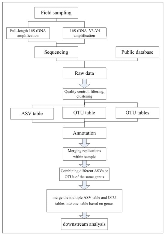
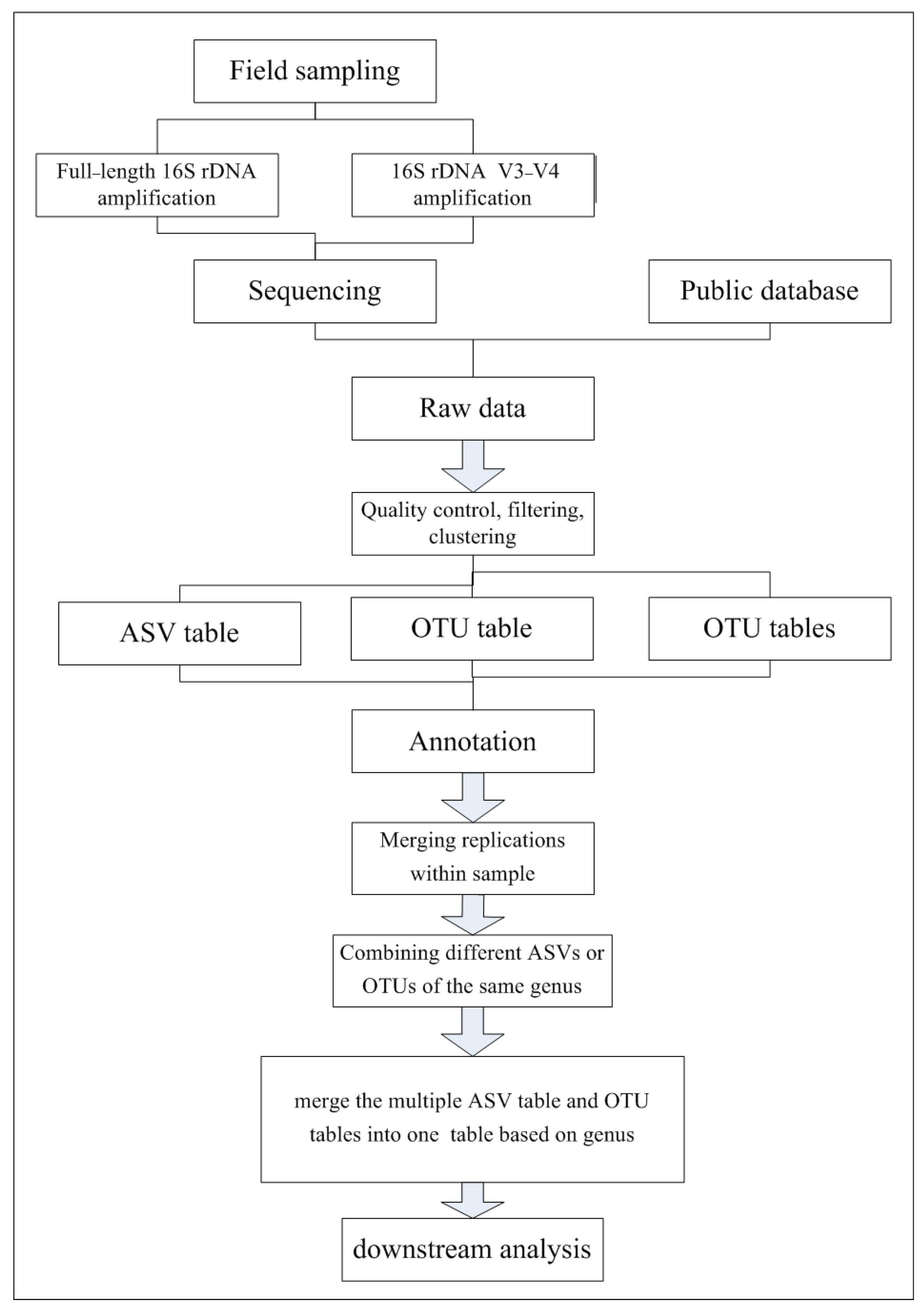
The bacterial communities analyzed in this study targeted different hypervariable regions (e.g., V1–V2, V3–V4, or V5–V7) and varied in length (partial vs. full-length 16S rRNA gene), posing a challenge for direct comparison. Therefore, we first annotated all sequences to the genus level and then merged the data into a single, genus-level taxonomic table according to taxonomic consistency. For example, the genus Leuconostoc was detected in seven samples, its original read counts were retained; for samples where this genus was not detected, the read count was assigned as zero. In this way, we integrated sequence counts from all datasets into a single table, regardless of variations in sequencing methods and depths (Figure 1).
Figure 1.
Dataset processing flowchart.
We analyzed bacterial communities at the genus level rather than at lower taxonomic levels (e.g., species level) to minimize potential biases derived from inaccurate annotation. Notably, despite differences in the amplified regions of the 16S rRNA gene among studies, all datasets were processed using identical bioinformatic pipelines for quality control, OTU clustering, and taxonomic annotation. Therefore, we assumed that the impact of misannotation was minimal. Moreover, since most bacteria within a genus share similar functions, our analysis of bacterial diversity at the genus level should not obscure their functional profiles. The pipeline used to analyze the datasets is outlined below (Figure 1):
2.4. Statistical Analysis
All statistical analyses were conducted using R (version 4.4.2) (R Core Team, 2025). Rarefaction curves indicated sufficient sequencing depth for each sample. To reduce bias arising from variations in sequencing depth, read counts were first rarefied to the same depth (i.e., the minimum library size) using the rrarefy() function in vegan package (v2.6.10). All downstream analyses were conducted on the rarefied dataset. Dominant bacteria were defined as taxa accounting for over 1% of the total relative abundance [25]. Alpha diversity was assessed for both the entire bacterial community and the dominant bacterial subset using observed richness (Sobs) and the Shannon diversity index. Overlaps in bacterial composition between the entire and dominant communities were visualized using VennDiagram package (v1.7.3), ggVennDiagram package (v1.5.2) and UpSetR packages (v1.4.0). To show the taxonomic composition, we constructed stacked bar plots to show the relative abundance of bacterial genera. Furthermore, to assess beta diversity, nonmetric multidimensional scaling (NMDS) was conducted based on Bray–Curtis dissimilarity matrices calculated using the vegdist() function in vegan package. Analysis of Similarity (ANOSIM) was employed to test for statistical differences in bacterial community by comparing the ranks of dissimilarity.
3. Results
3.1. Gut Bacterial Communities of Six Vespine Wasp Species
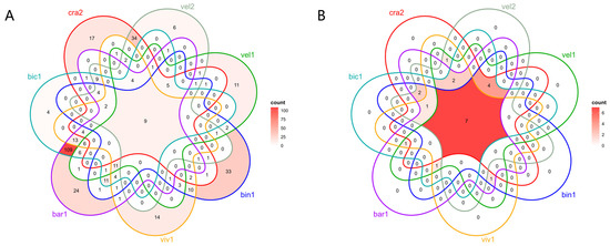
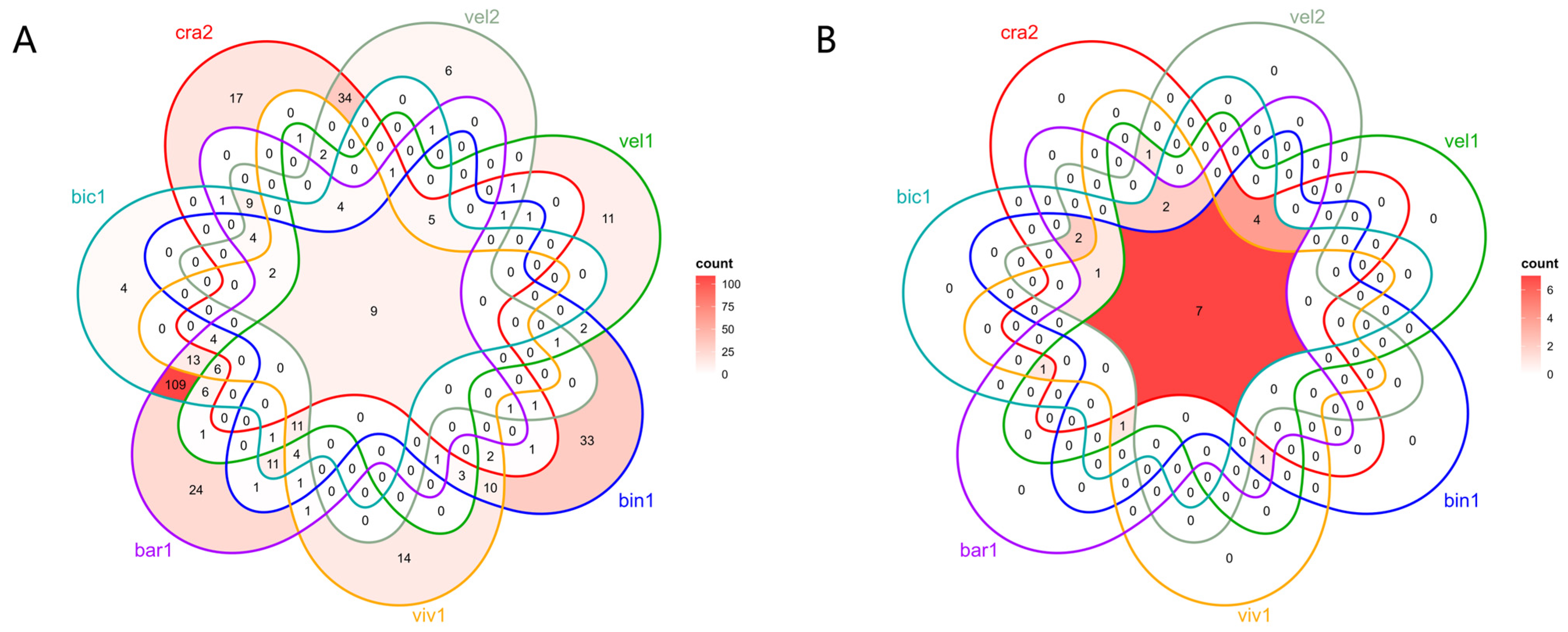
We annotated 376 genera across the six vespine wasp species. P. barthelemyi harbored the highest number of genera (231), followed by V. bicolor (205), V. binghami (106), V. crabro (101), V. vivax (88), V. velutina (IT) (82) and V. velutina (CN) (68). Leuconostoc was the dominant genus across the six vespine wasp species, followed by Hafnia-Obesumbacterium, Lactobacillus and Bifidobacterium. Notably, V. binghami harbored the highest number of unique genera (33), followed by P. barthelemyi (24) (Figure 2A).
Figure 2.
Venn diagrams illustrating shared bacteria of total gut bacterial community (A) and dominant gut bacterial community (B) of the six vespine wasps. Abbreviations: bar1: P. barthelemyi; bic1: V. bicolor; bin1: V. binghami; cra2: V. crabro; vel1: V. velutina (CN); vel2: V. velutina (IT); viv1: V. vivax. IT: Italy; CN: China. The same applies to the figures below.
A total of 20 dominant bacterial genera were selected from the six vespine wasp species. Leuconostoc and Hafnia-Obesumbacterium were the top dominant taxa. Specifically, the dominant genera included Hafnia-Obesumbacterium and Rickettsiella for V. binghami, Rickettsia and Lactobacillus for V. vivax, Leuconostoc and Ralstonia for P. barthelemyi, Lonsdalea and Leuconostoc for V. bicolor, Lactobacillus, Bifidobacterium and Leuconostoc for V. velutina (CN), Hafnia-Obesumbacterium and Lactiplantibacillus for V. velutina (IT), and Lactiplantibacillus and Lactococcus for V.crabro. The dominant Leuconostoc, Hafnia-Obesumbacterium and Lactobacillus were present in the seven specimens. Interestingly, no vespine wasp harbored a unique dominant genus, suggesting high conservation of the dominant gut bacteria (Figure 2B).
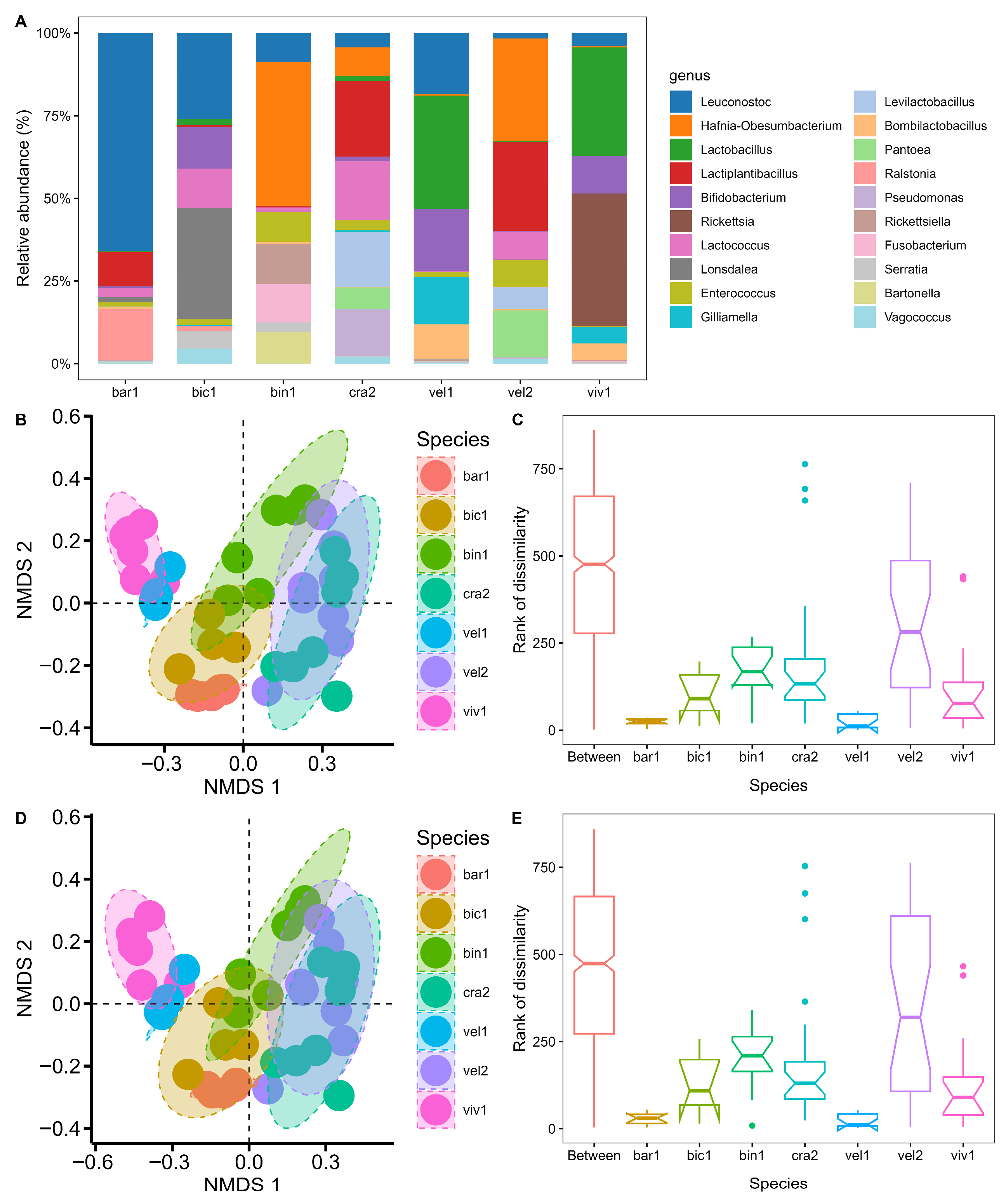
The relative read abundance of gut bacteria revealed substantial variations in bacterial communities among the six vespine wasps (Figure 3A). Significant differences in bacterial communities were observed among the six vespine wasps (ANOSIM, p < 0.05). Nonmetric multidimensional scaling (NMDS) revealed distinct separation of the gut bacterial communities among the six vespine wasp specimens (Figure 3B). ANOSIM confirmed that between-group dissimilarity was significantly higher than within-group dissimilarity (R = 0.68, p = 0.001) (Figure 3C), further confirming the differences in bacterial communities among the six vespine wasps. A similar pattern was observed for the dominant bacterial communities. NMDS ordination again showed distinct clustering of dominant bacterial communities (Figure 3D). ANOSIM analysis further confirmed significant differences in dominant bacterial communities (R = 0.655, p = 0.001), indicating that between-group dissimilarity was significantly greater than within-group dissimilarity (Figure 3E).
Figure 3.
Beta diversity of larval gut bacterial communities of the six vespine wasps. (A) Stacked bar chart showing relative read abundance of dominant gut bacteria measured at the genus level. (B) NMDS ordination of the total gut bacterial community. (C) Comparison of ranks of dissimilarities between groups and within groups for the total gut bacterial community. (D) NMDS ordination of the dominant gut bacterial community. (E) Comparison of ranks of dissimilarities between groups and within groups for the dominant gut bacterial community.
3.2. Larval Gut Bacterial Community Differences Between Pollen-Carrying Bees and Vespine Wasps
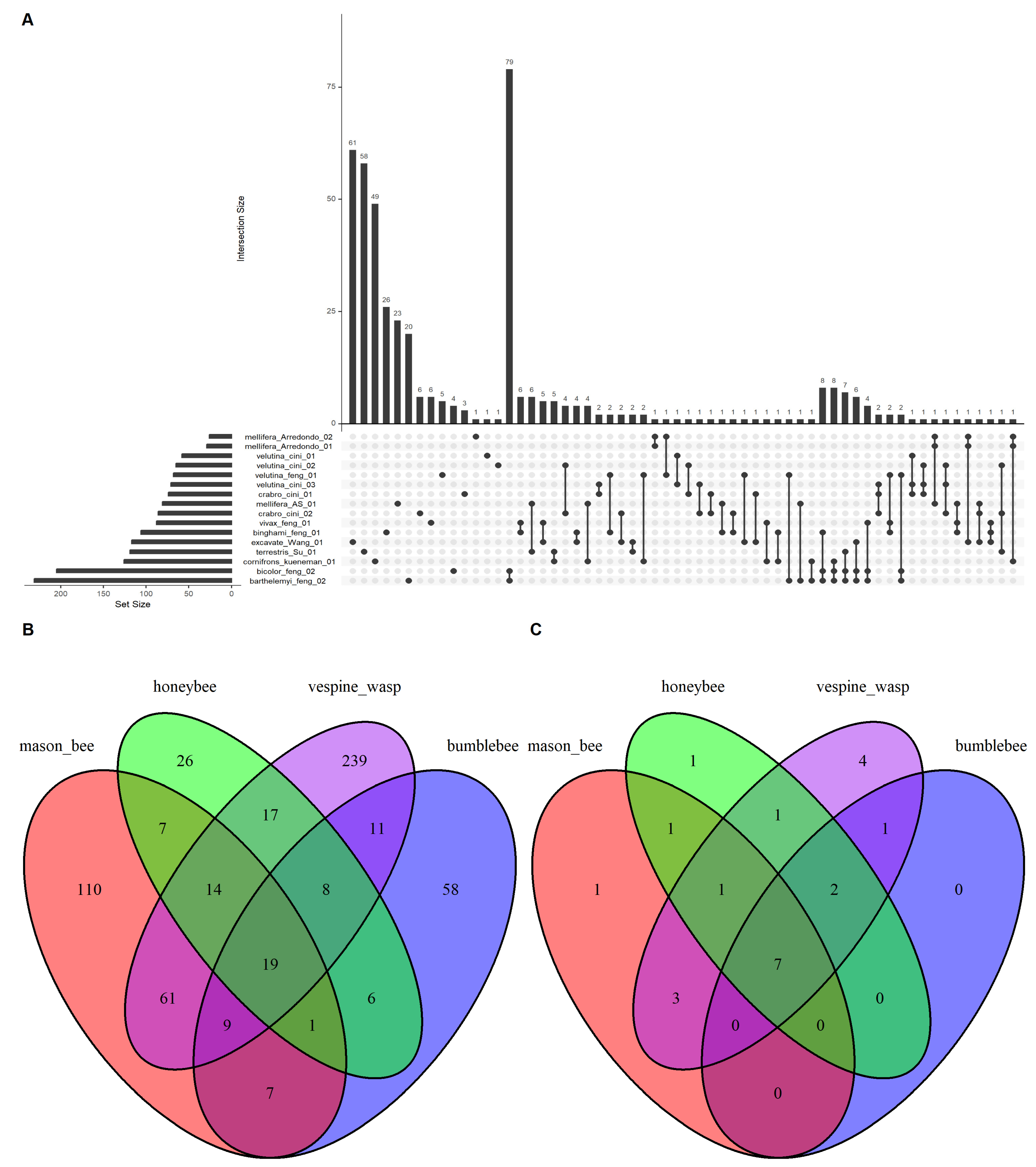
The gut bacterial communities of the four taxonomic groups of aculeate wasps comprised 593 bacterial genera. Specifically, mason bees harbored 228 genera, bumblebees 119 genera, honey bees 98 genera and vespine wasps 378 genera. Lactobacillus was the predominant taxon across the four groups, followed by Hafnia-Obesumbacterium, Leuconostoc and Lactiplantibacillus. Notably, Leuconostoc was detected exclusively in vespine wasps. Each aculeate group exhibited distinct top bacterial compositions. The gut bacterial communities of mason bees were primarily composed of Ralstonia, Sodalis and Lachnospiraceae UCG-010. The gut bacterial communities of bumblebees were dominated by Lactobacillus, Arsenophonus and Snodgrassella, with Lactobacillus being the most abundant genus. The gut bacterial communities of honey bees were characterized by Lactobacillus, Apilactobacillus and Gilliamella. Meanwhile, the gut bacterial communities of vespine wasps were characterized by high abundances of Hafnia-Obesumbacterium, Leuconostoc and Lactiplantibacillus. Notably, Lactobacillus was detected in all 16 samples, while Bombilactobacillus was present in 15 samples, absent in only one mason bee sample. At the individual level, the sample exavate_Wang_01 (a mason bee) harbored the highest number of unique genera (61), followed by terrestris_Su_01 (a bumblebee) (58) (Figure 4A).
Figure 4.
Venn diagrams showing shared bacterial genera of the four taxonomic groups of aculeate wasps (mason bees, honey bees, bumblebees and vespine wasps). (A) Shared bacterial genera measured at the individual sample level (16), with the top 60 comparisons presented. (B) Shared bacterial genera of the full bacterial community of the four taxonomic groups. (C) Shared dominant bacterial genera identified from the pooled data in (B).
When all samples were pooled together, 110, 26, 239 and 58 unique genera were detected in mason bees, bumblebees, honey bees and vespine wasps, respectively, while only 19 genera were shared across the four aculeate groups (Figure 4B). This suggested distinct gut bacterial compositions among the four taxa.
A total of 22 dominant bacteria were identified across the four taxa. Among them, Lactobacillus, Gilliamella, Lactococcus, Bifidobacterium, Bombilactobacillus, Pseudomonas and Snodgrassella were found in all four groups. Mason bees and honey bees each possessed one unique dominant bacterial genus, whereas bumblebees lacked any unique dominant genus. In contrast, vespine wasps harbored four unique dominant bacterial genera (Figure 4C).
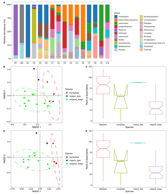
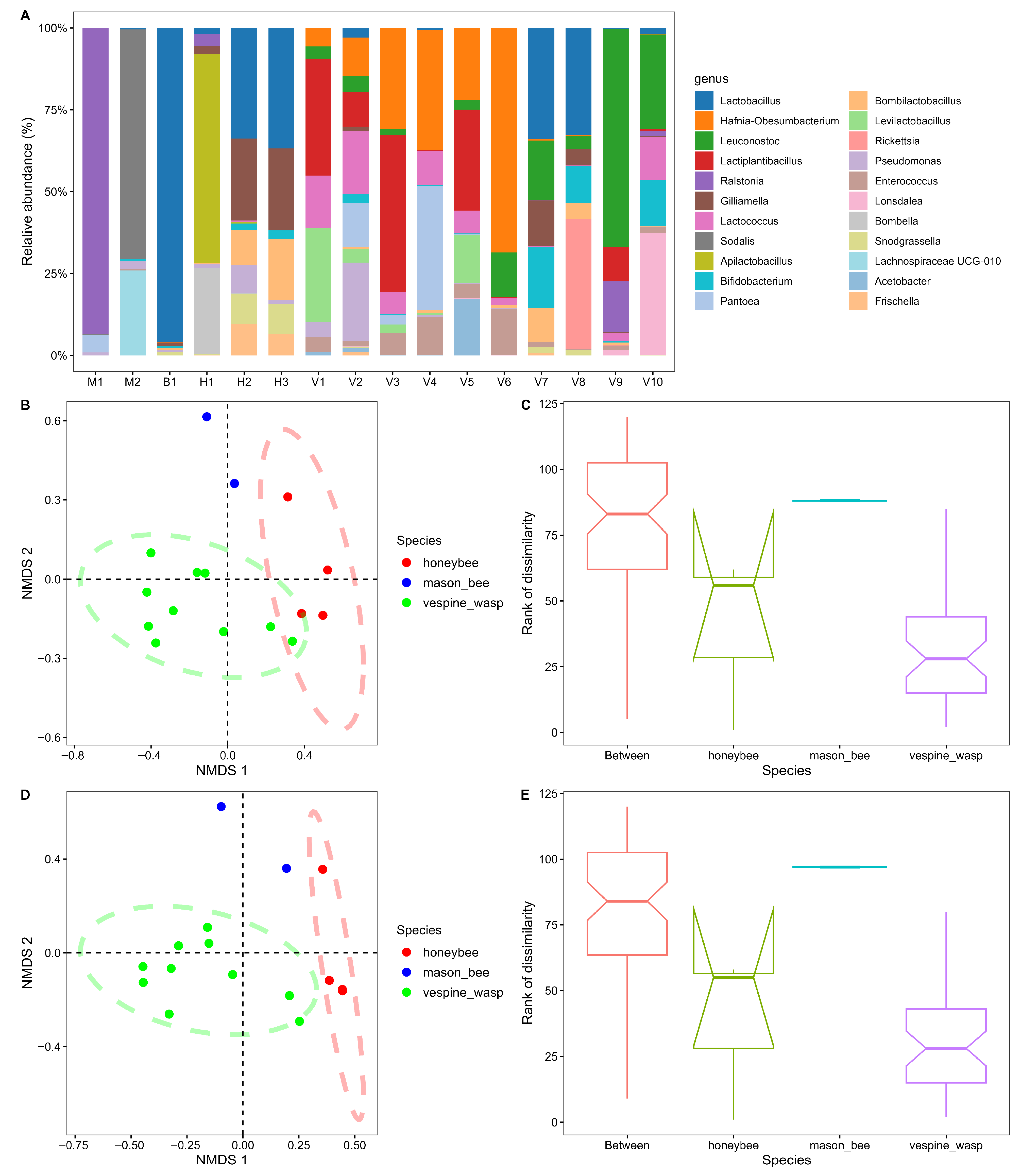
The beta diversity of the larval bacterial community of the four taxonomic groups was significantly different. The relative abundance of the top 22 bacterial genera varied among these groups (Figure 5A). Since only a single bumblebee sample was selected, an ellipse could not be drawn for this group on the first two axes of the NMDS plot. Therefore, bumblebees and honey bees were combined into a single group to facilitate ellipse calculation. The NMDS ordination revealed distinct compositional separations among the three resulting groups, although a narrow overlap was observed between the combined honey bee and bumblebee group and vespine wasps (Figure 5B). ANOSIM confirmed significant differences in bacterial composition among the four groups (R = 0.700, p = 0.001), characterized by greater dissimilarity between groups than within groups (Figure 5C). The dominant gut bacterial communities exhibited a similar pattern, showing taxon-specific clustering (Figure 5D). Furthermore, ANOSIM of the dominant gut bacteria supported these compositional differences (R = 0.708, p = 0.001) (Figure 5E).
Figure 5.
Beta diversity of larval gut bacterial communities in four taxonomic groups of aculeate wasps. (A) Stacked bar chart showing the relative read abundance of dominant bacterial genera across the four groups. Samples are labeled as follows: M1–M2, mason bees; B1, bumblebee; H1–H3, honey bees; V1–V10, vespine wasps. (B) Non-metric multidimensional scaling (NMDS) ordination of the full bacterial communities for the four groups. Note: The bumblebee group contained only a single sample; therefore, it was combined with the honey bee group to allow for calculating ellipses in the NMDS ordination. (C) Boxplots showing rank dissimilarities of the full bacterial community. The bumblebee group is not shown due to its small sample size (n = 1). (D) NMDS ordination of the dominant bacterial communities for the four groups. Note: The bumblebee group and the honey bee group were combined as a group as well. (E) Boxplots showing rank dissimilarities of the dominant bacterial communities. The bumblebee group is excluded here as well.
4. Discussion
This study revealed significant differences in larval gut bacterial communities among the four groups of aculeate wasps. We attribute these variations to several key factors. First, evolutionary consequences drive the divergences and differences in gut bacterial assemblages. Numerous studies have shown that host evolutionary history (i.e., phylogenetic relatedness and coalescence time) significantly shapes the composition of gut bacterial communities [26,27,28]. Second, gut bacterial composition is strongly influenced by dietary composition [29,30]. Pollen-carrying bees and vespine wasps exhibit distinct dietary differences. Pollen-carrying bees primarily feed on pollen and nectar, whereas vespine wasps are omnivorous predators, feeding mainly on protein-rich insects and plant-derived carbohydrates. These divergent diets likely exert strong selective pressures on gut microbial composition. Finally, host physiology, behavior, living habitats, and life-history traits may also contribute to the observed differences. For instance, factors such as susceptibility to viral infections or varying immune responses can alter the structure of gut bacterial communities [31].
The differences in gut bacterial communities among these seven vespine wasp species may be explained by their distinct dietary habits. Our previous investigations demonstrated that four vespine wasps (P. barthelemyi, V. binghami, V. velutina, and V. vivax) consumed different diets [32], which likely drove the divergence of their larval gut bacteria. Interestingly, however, the dominant bacterial taxa of these seven vespine wasp specimens were highly conserved. This suggests that the core bacteria is primarily shaped by evolutionary constraints, given that these species are closely related on the phylogenetic tree.
Leuconostoc, a group of heterofermentative lactic acid bacteria, was the dominant bacterial genus across the seven vespine wasp specimens, but was absent from the bacterial community of pollen-carrying bees. Leuconostoc metabolizes carbohydrates to produce lactic acid, acetic acid, ethanol, and mannitol. These metabolites create an acidic environment that inhibits pathogen colonization and promotes gut homeostasis. Commonly used in fermented foods such as sauerkraut and cheese, the occurrence of Leuconostoc in vespine wasps implies significant evolutionary and adaptive value. Alongside Leuconostoc, we identified other fermentation-associated bacteria, such as Lactobacillus [33]. These bacterial components suggest specialized metabolic functions. Notably, vespine larvae consume large amounts of prey, such as insects, but do not defecate. All ingested food is excreted only once at the end of the larval stage. This trait requires that their food be thoroughly decomposed and retained within the gut for a long time. These bacteria, such as Leuconostoc and other fermentative bacteria, likely help to metabolize their prey under such conditions.
In summary, our study revealed that the larval gut bacterial compositions of vespine wasps differed from those of pollen-carrying bees, likely shaped by both evolutionary history and dietary composition. Given that shifts in dominant bacterial communities in insect guts can lead to dysbiosis or disease, such replacements could negatively impact host health and survival. Therefore, it is particularly important to manage the larval gut bacterial compositions of reared hornets, since hornet keeping has become a regular practice in southern China.
Author Contributions
Conceptualization, Z.X.; methodology, Z.X. and X.F.; software, X.F.; validation, X.F. and Z.X.; formal analysis, X.F.; investigation, Z.X. and X.F.; resources, Z.X.; data curation, X.F.; writing—original draft preparation, X.F. and Z.X.; writing—review and editing, X.F., Z.X., J.W. and X.Y.; visualization, X.F.; supervision, Z.X.; project administration, Z.X.; funding acquisition, Z.X. All authors have read and agreed to the published version of the manuscript.
Funding
This research was funded by Yunnan Fundamental Research Projects, grant number 202401AS070015,202301AT070432 and Yunnan Provincial Program for Cultivating Middle-aged and Young Academic and Technical Leaders, grant number 202405AC350077. The APC was funded by 202401AS070015.
Institutional Review Board Statement
Not applicable. This study involved invertebrate insects (vespine wasps, etc.), which are not classified as experimental animals under Chinese regulations (Regulations on the Administration of Experimental Animals, 2017). No ethical approval is required for invertebrate research in China. All specimens were collected from non-protected areas with no specific permits required for these non-protected species.
Informed Consent Statement
Not applicable.
Data Availability Statement
The data that support the findings of this study are available from the corresponding authors upon reasonable request.
Acknowledgments
We thank the hornet farmers, Xuerong Yang, Lihui Ma, Yinhua Zhou and Shuchao Li, for providing reared yellow-legged hornets. We also thank Hongbo Yan and Xinnian Liu for collecting wild yellow-legged hornets. Luquan YiFeng hornet rearing farm provided supports for sampling hornets.
Conflicts of Interest
The authors declare no conflicts of interest.
Abbreviations
The following abbreviations are used in this manuscript:
| NMDS | non-metric multidimensional scaling |
| HTS | high throughput sequencing |
| ENA | European Nucleotide Archive |
| OTU | operational taxonomic unit |
| CCS | circular consensus sequence |
| ASV | amplicon sequence variant |
| ANOSIM | Analysis of similarity |
References
- Warnecke, F.; Luginbühl, P.; Ivanova, N.; Ghassemian, M.; Richardson, T.H.; Stege, J.T.; Cayouette, M.; McHardy, A.C.; Djordjevic, G.; Aboushadi, N.; et al. Metagenomic and functional analysis of hindgut microbiota of a wood-feeding higher termite. Nature 2007, 450, 560–565. [Google Scholar] [CrossRef] [Scilit]
- Dillon, R.J.; Vennard, C.T.; Buckling, A.; Charnley, A.K. Diversity of locust gut bacteria protects against pathogen invasion. Ecol. Lett. 2005, 8, 1291–1298. [Google Scholar] [CrossRef] [Scilit]
- Yoshitomo, K.; Masahito, H.; Takahiro, H.; Atsushi, N.; Kanako, T.; Takema, F. Symbiont-mediated insecticide resistance. Proc. Natl. Acad. Sci. USA 2012, 109, 8618–8622. [Google Scholar] [CrossRef] [Scilit]
- Zhang, X.; Li, X.A.; Su, Q.Z.; Cao, Q.N.; Li, C.Y.; Niu, Q.S.; Zheng, H. A curated 16S rRNA reference database for the classification of honeybee and bumblebee gut microbiota. Biodivers. Sci. 2019, 27, 557–566. [Google Scholar] [CrossRef] [Scilit]
- Kwong, W.K.; Moran, N.A. Gut microbial communities of social bees. Nat. Rev. Microbiol. 2016, 14, 374–384. [Google Scholar] [CrossRef] [Scilit]
- Engel, P.; Moran, N.A. Functional and evolutionary insights into the simple yet specific gut microbiota of the honey bee from metagenomic analysis. Gut Microbes 2013, 4, 60–65. [Google Scholar] [CrossRef] [Scilit]
- Li, H.Y.; Dong, Z.X.; Chen, Y.F.; Guo, J. Advances in research of colonization of gut microbiota in the honeybee. Microbiol. China 2019, 46, 3091–3101. [Google Scholar] [CrossRef]
- Dai, S.Y.; Liu, B.; Zhang, L.Z. Research progress on the composition and function of honeybee gut microbiota. Chin. Beekeep. 2024, 44, 1–7. [Google Scholar]
- Raymann, K.; Shaffer, Z.; Moran, N.A. Antibiotic exposure perturbs the gut microbiota and elevates mortality in honeybees. PLoS Biol. 2017, 15, e2001861. [Google Scholar] [CrossRef] [Scilit]
- Li, C.Y.; Zhou, X.; Zheng, H. Gut microbiota of social honey bees. Acta Microbiol. Sin. 2018, 58, 1016–1024. [Google Scholar] [CrossRef]
- Koch, H.; Schmid-Hempel, P. Socially transmitted gut microbiota protect bumble bees against an intestinal parasite. Proc. Natl. Acad. Sci. USA 2011, 108, 19288–19292. [Google Scholar] [CrossRef] [Scilit]
- Koch, H.; Welcome, V.; KendalSmith, A.; Thursfield, L.; Farrell, I.W.; Langat, M.K.; Brown, M.J.F.; Stevenson, P.C. Host and gut microbiome modulate the antiparasitic activity of nectar metabolites in a bumblebee pollinator. Philos. Trans. R. Soc. B 2022, 377, 20210162. [Google Scholar] [CrossRef] [Scilit]
- Landa, G.F.D.; Alberoni, D.; Braglia, C.; Baffoni, L.; Landa, M.F.D.; Revainera, P.D.; Quintana, S.; Zumpano, F.; Maggi, M.D.; Gioia, D.D. The gut microbiome of two wild bumble bee species native of South America: Bombus pauloensis and Bombus bellicosus. Microb. Ecol. 2024, 87, 121. [Google Scholar] [CrossRef] [Scilit]
- Li, J.; Powell, J.E.; Guo, J.; Evans, J.D.; Wu, J.; Williams, P.; Lin, Q.; Moran, N.A.; Zhang, Z. Two gut community enterotypes recur in diverse bumblebee species. Curr. Biol. 2015, 25, R652–R653. [Google Scholar] [CrossRef] [Scilit]
- Guo, B.; Tang, J.; Ding, G.; Mashilingi, S.K.; Huang, J.; An, J. Gut microbiota is a potential factor in shaping phenotypic variation in larvae and adults of female bumble bees. Front. Microbiol. 2023, 14, 1117077. [Google Scholar] [CrossRef] [Scilit]
- Zhang, L.; Liu, F.; Wang, X.L.; Wang, P.H.; Ma, S.L.; Yang, Y.; Ye, W.G.; Diao, Q.Y.; Dai, P.L. Midgut bacterial communities of Vespa velutina Lepeletier (Hymenoptera: Vespidae). Front. Ecol. Evol. 2022, 10, 934054. [Google Scholar] [CrossRef] [Scilit]
- Kueneman, J.G.; Gillung, J.; Van Dyke, M.T.; Fordyce, R.F.; Danforth, B.N. Solitary bee larvae modify bacterial diversity of pollen provisions in the stem-nesting bee, Osmia cornifrons (Megachilidae). Front. Microbiol. 2023, 13, 1057626. [Google Scholar] [CrossRef] [Scilit] [PubMed]
- Wang, G.; Wang, G.; Ma, Y.; Lv, Z.; You, Y.; Ma, P.; Yu, Y. Composition and diversity of gut bacterial community in different life stages of Osmia excavata Alfken (Hymenoptera: Megachilidae). Microorganisms 2024, 12, 1709. [Google Scholar] [CrossRef] [Scilit] [PubMed]
- Su, Q.; Wang, Q.; Mu, X.; Chen, H.; Meng, Y.; Zhang, X.; Zheng, L.; Hu, X.; Zhai, Y.; Zheng, H. Strain-level analysis reveals the vertical microbial transmission during the life cycle of bumblebee. Microbiome 2021, 9, 216. [Google Scholar] [CrossRef] [Scilit] [PubMed]
- Floyd, A.S.; Mott, B.M.; Maes, P.; Copeland, D.C.; McFrederick, Q.S.; Anderson, K.E. Microbial ecology of European foul brood disease in the honey bee (Apis mellifera): Towards a microbiome understanding of disease susceptibility. Insects 2020, 11, 555. [Google Scholar] [CrossRef] [Scilit]
- Arredondo, D.; Añón, G.; Campá, J.; Harriet, J.; Castelli, L.; Zunino, P.; Antúnez, K. Supplementation of honey bee production colonies with a native beneficial microbe mixture. Benef. Microbes 2023, 14, 385–400. [Google Scholar] [CrossRef] [Scilit]
- Cini, A.; Meriggi, N.; Bacci, G.; Cappa, F.; Vitali, F.; Cavalieri, D.; Cervo, R. Gut microbial composition in different castes and developmental stages of the invasive hornet Vespa velutina nigrithorax. Sci. Total Environ. 2020, 745, 140873. [Google Scholar] [CrossRef] [Scilit] [PubMed]
- Schloss, P.D.; Westcott, S.L.; Ryabin, T.; Hall, J.R.; Hartmann, M.; Hollister, E.B.; Lesniewski, R.A.; Oakley, B.B.; Parks, D.H.; Robinson, C.J.; et al. Introducing mothur: Open-source, platform-independent, community-supported software for describing and comparing microbial communities. Appl. Environ. Microbiol. 2009, 75, 7537–7541. [Google Scholar] [CrossRef] [Scilit] [PubMed]
- Caporaso, J.G.; Kuczynski, J.; Stombaugh, J.; Bittinger, K.; Bushman, F.D.; Costello, E.K.; Fierer, N.; Gonzalez, A.G.; Goodrich, J.K.; Gordon, J.I.; et al. QIIME allows analysis of high-throughput community sequencing data. Nat. Methods 2010, 7, 335–336. [Google Scholar] [CrossRef] [Scilit]
- Huang, Q.W.; Long, L.Y.; Ai, J.W.; Huang, X.X.; Liu, G.D.; Su, Y.L.; Xie, C.M.; Chen, G.L. Dynamic change in the bacterial community and dominant flora analysis of papaya pickles during low-temperature fermentation. China Brew. 2025, 44, 116–123. [Google Scholar]
- An, Y.; Braga, M.P.; Garcia, S.L.; Sterno, M.G.; Hambäck, P.A. Host phylogeny structures the gut bacterial community within Galerucella leaf beetles. Microb. Ecol. 2023, 86, 2477–2487. [Google Scholar] [CrossRef] [Scilit]
- Schwob, G.; Cabrol, L.; Saucède, T.; Gérard, K.; Poulin, E.; Orlando, J. Unveiling the co-phylogeny signal between plunderfish Harpagifer spp. and their gut microbiomes across the Southern Ocean. Microbiol. Spectr. 2024, 12, e0383023. [Google Scholar] [CrossRef] [Scilit]
- Sun, G.; Xia, T.; Wei, Q.; Wang, X.; Dong, Y.; Yang, X.; Zhang, L.; Sha, W.; Zhang, H. Host phylogeny shapes gut microbiota and predicted functions in captive Artiodactyls. Microorganisms 2025, 13, 2250. [Google Scholar] [CrossRef] [Scilit]
- Huang, J.; Chen, J.; Ying, S.L.; Ren, Z.Y.; Li, X.H. Dietary regulation of gut microbiota: Mechanisms and implications. Food Ferment. Ind. 2025, 51, 390–398. [Google Scholar] [CrossRef]
- Chen, J.Y.; Niu, X.H.; Zhao, Z.Q.; Wu, K.F.; Yuan, S.; Cheng, Y.; Hasi, G.W.; Fu, H.P. The spring diet of Myospalax aspalax in pinus sylvestris plantations and its relationship with intestinal microbiota. Pratacult. Sci. 2016, 1–22. Available online: https://link.cnki.net/urlid/62.1069.S.20260121.1406.002 (accessed on 26 March 2026).
- Deng, Y.; Zhao, H.; Zhang, L.; Yang, S.; Zou, D.; Ma, M.; Hou, C. Symbiotic Enterococcus faecalis potentiates viral pathogenesis via fructose-1,6-bisphosphate-mediated insect gut epithelial damage. Npj Biofilms Microbiomes 2025, 11, 215. [Google Scholar] [CrossRef] [Scilit] [PubMed]
- Feng, X.X.; Xie, Z.H.; Wang, J.M.; Liu, D.Y.; Jiang, X.J.; Zhao, P.F.; Jia, Y.K. Study on diets of four hornets in Yunnan Province: A case study of Daxueshan Scenic Area in Lincang, Yunnan Province. J. Environ. Entomol. 2026, 48, 549–558. [Google Scholar]
- Moles, G.B.; Chailyan, A.; Dolejs, I.; Forster, J.; Mikš, M.H. Uncovering carbohydrate metabolism through a genotype-phenotype association study of 56 lactic acid bacteria genomes. Appl. Microbiol. Biotechnol. 2019, 103, 3135–3152. [Google Scholar] [CrossRef] [Scilit] [PubMed]
Disclaimer/Publisher’s Note: The statements, opinions and data contained in all publications are solely those of the individual author(s) and contributor(s) and not of MDPI and/or the editor(s). MDPI and/or the editor(s) disclaim responsibility for any injury to people or property resulting from any ideas, methods, instructions or products referred to in the content. |
© 2026 by the authors. Licensee MDPI, Basel, Switzerland. This article is an open access article distributed under the terms and conditions of the Creative Commons Attribution (CC BY) license.