


Abstract
Emerging infectious diseases (EIDs) are an increasing global health concern, and central nervous system (CNS) involvement is particularly critical owing to its high morbidity and mortality. Despite advances in diagnostic technologies, timely and accurate microbiological diagnosis remains challenging. This scoping review maps the available evidence on microbiological diagnostic methods for emerging and re-emerging CNS infections and identifies key knowledge gaps. Articles published between January 2020 and December 2024 were reviewed, and 142 articles were included. Diagnostic approaches comprised polymerase chain reaction (PCR)-based molecular testing, metagenomic next-generation sequencing (mNGS), nanopore-targeted sequencing, cytokine profiling, and unbiased serology, among others. Viral pathogens were more frequently represented than bacterial, fungal and parasitic ones, for which evidence derived mainly from case reports, small case series and retrospective studies and requires validation in larger prospective studies. Although culture remains the reference standard for the diagnosis of many emerging and re-emerging CNS pathogens, molecular assays, particularly PCR-based methods, are currently the cornerstone of diagnosis. Overall, the findings highlight the central role of molecular diagnostics in the detection of emerging and re-emerging CNS infections, while underscoring the need for further validation of novel diagnostic approaches and more robust evidence across pathogen groups, particularly those underrepresented in the literature.
1. Introduction
Emerging infectious diseases (EIDs) are defined by the World Health Organization (WHO) as diseases that have appeared in a population for the first time, or that existed previously but are rapidly increasing in incidence or geographical distribution [1]. The emergence and re-emergence of infectious diseases have increased in recent years, with approximately 75% being of zoonotic origin [2]. These diseases are driven by numerous factors, including the destruction of natural habitats, climate change, the replacement of reservoir hosts, and microbial genetic mutations [3]. Surveillance plays a critical role in monitoring trends and signalling the emergence or re-emergence of pathogens [4].
Among the diverse clinical manifestations of EIDs, central nervous system (CNS) infections are of particular concern owing to their high morbidity and mortality and their potential to cause long-term neurological sequelae [5].
A timely lumbar puncture is essential for the diagnosis of CNS infections. Cerebrospinal fluid (CSF) analysis, including biochemistry, cell count, and microscopic examination (Gram stain, acid-fast bacilli stain, and India ink), can provide valuable clues to differentiate bacterial from viral meningitis but lacks specificity and cannot confirm CNS infection on its own. CSF culture and antimicrobial susceptibility testing remain the gold standard for the identification of bacterial and fungal pathogens. Blood cultures are useful when CSF cannot be obtained. It can also increase diagnostic yield when combined with CSF analysis [4]. Matrix-assisted laser desorption/ionisation time-of-flight mass spectrometry (MALDI-TOF MS) is a rapid analytical technique used for microbial identification based on the detection of characteristic protein profiles. It is a useful method for the identification of pathogenic microorganisms, including bacteria, fungi, and parasites, as it can provide more accurate identification than conventional phenotypic methods [6,7,8,9,10]. Antigen detection tests, including latex agglutination tests and lateral flow assays, provide rapid results; however, a positive result should always be confirmed by culture or a molecular method. Serology may be helpful, particularly when molecular or culture-based methods are unavailable or inconclusive, although interpretation can be complicated by cross-reactivity and prior vaccination. Viral neutralisation tests, such as plaque reduction neutralisation test (PRNT), can distinguish cross-reactive antibodies; however, their labour-intensive nature, long processing time, and requirement for high-level biosafety facilities limit their widespread use. More recently, polymerase chain reaction (PCR)-based molecular testing has become an important tool owing to its high sensitivity, even after the onset of antimicrobial treatment, and it is particularly useful for detecting viral infections and fastidious organisms. Multiplex PCR enables the simultaneous detection of multiple pathogens from a single sample with a rapid turnaround time [4].
Despite these advances, the timely and accurate microbiological diagnosis of CNS infections remains difficult, particularly in resource-limited settings [4,5]. These diagnostic limitations are especially concerning for emerging and re-emerging pathogens, for which delays in identification can have clinical and public health implications.
To our knowledge, no comprehensive synthesis of microbiological diagnostic methods for emerging and re-emerging CNS infections is currently available. Given the wide spectrum of pathogens included within the WHO definition, together with the recent evolution of diagnostic technologies, the available literature on this topic is highly heterogeneous. A scoping review was therefore considered the most appropriate methodological approach, as it enables the systematic identification, mapping, and characterisation of the existing evidence across diverse study designs and diagnostic strategies. Accordingly, this review aims to comprehensively map the available evidence on microbiological diagnostic methods used to identify emerging and re-emerging CNS infections across clinical and laboratory settings worldwide, identify current knowledge gaps, and inform future research priorities and clinical practice.
2. Materials and Methods
A scoping review was conducted, and the Preferred Reporting Items for Systematic Reviews and Meta-Analyses extension for Scoping Reviews–guided (PRISMA-ScR) checklist is provided in Supplementary Material File S1 [11]. This scoping review was not registered. A comprehensive literature search was performed, including publications from 1 January 2020 to 31 December 2024. Studies published before 2020 were not included because the COVID-19 pandemic marked a turning point in global mobility and the epidemiology of emerging and re-emerging infectious diseases, while simultaneously driving rapid advances in molecular diagnostic methods and laboratory medicine. Restricting the review to studies published from 2020 onwards ensured that these major epidemiological and technological developments were captured. Only articles published in English were considered, based on the language proficiency of the reviewers.
The search strategy was based on the two core concepts of the review, emerging infectious diseases and the central nervous system, which directly reflected the review question. A combination of free-text keywords and Medical Subject Headings (MeSH) terms was used, incorporating the terms (“emerging infection”) AND (“central nervous system”), combined using Boolean operators to identify studies on emerging infectious diseases affecting the central nervous system. Broader search terms, such as emerg’ were piloted but retrieved a substantial number of irrelevant records, considerably reducing the specificity of the search. Given the exploratory aim of this scoping review, which was to map the range of available diagnostic methods rather than achieve exhaustive retrieval, the selected search strategy was considered to provide an appropriate balance between sensitivity and specificity, yielding a feasible and relevant evidence base. PubMed was selected as the sole database because it provides comprehensive coverage of the biomedical literature and was considered appropriate for the mapping objective of this scoping review.
All references retrieved from PubMed were imported into EndNote (Version 2025) reference management software, and duplicate records were removed. To ensure consistent application of the eligibility criteria, 10% of the retrieved abstracts were independently screened by two reviewers as a calibration exercise. Inter-reviewer agreement was 92%. Following calibration, the remaining abstracts were screened by a single reviewer using the same predefined eligibility criteria. Although this approach was considered appropriate for the exploratory objectives of the scoping review, screening of the remaining abstracts by a single reviewer represents a potential source of selection bias and was therefore considered a methodological limitation.
Abstracts were included if they provided original data or analysis on microbiological diagnostic methods for emerging and re-emerging CNS infections. For the purposes of this review, emerging and re-emerging infections were defined according to the WHO as infections that have newly appeared in a population or have previously existed but are rapidly increasing in incidence or expanding into new geographical areas. For the purposes of screening, a pathogen was classified as emerging or re-emerging when the retrieved publication provided epidemiological or geographical context supporting recent emergence, re-emergence, increased incidence, or geographical expansion, consistent with the WHO definition. Classification was therefore assessed in the context of the individual publication rather than solely according to the pathogen name. When eligibility remained uncertain, the case was discussed within the review team and resolved by consensus.
Abstracts focusing exclusively on SARS-CoV-2 were excluded because the unprecedented volume of research on SARS-CoV-2 diagnostics represents a distinct body of evidence that merits a separate review. Additional exclusions comprised studies addressing diagnostic imaging alone, long-term complications rather than diagnostic methods, epidemiological surveillance, animal models, or diagnostic methods exclusively in people living with human immunodeficiency virus (HIV), owing to the distinct diagnostic considerations associated with this population. Any uncertainties regarding study eligibility were resolved by consensus following discussion among the review team.
Following full-text assessment, articles were evaluated against the predefined eligibility criteria to determine their final inclusion. Consistent with the objectives of a scoping review, no formal critical appraisal of the included studies was undertaken, as the review aimed to map the extent and characteristics of the available evidence rather than assess study quality.
Data were extracted from the included studies on the laboratory diagnostic methods evaluated, including specimen type, reported diagnostic performance (e.g., sensitivity and specificity, where available), and key limitations. The findings were synthesised narratively in two stages. First, an overview of the principal laboratory diagnostic methods identified across the included studies was provided. Subsequently, the evidence was organised and discussed according to microorganism type and individual pathogens. The results are summarised in Table 1. Sensitivity and specificity were reported inconsistently across the included studies, with some studies providing numerical estimates and others describing diagnostic performance qualitatively. Where numerical estimates of sensitivity and specificity were available, these are presented in Supplementary Material File S2. No quantitative meta-analysis was performed, consistent with the mapping objective of a scoping review.
Table 1.
Summary of the diagnostic methods for emerging and re-emerging CNS infections by pathogen group.
3. Results
The search retrieved 2051 abstracts, of which 239 were deemed eligible based on the predefined inclusion and exclusion criteria. Following full-text screening, 94 articles were excluded because they lacked relevant information on microbiological diagnostic methods, focused on non-emergent pathogens, or addressed epidemiological surveillance without reporting relevant laboratory-based diagnostic approaches. Details of the excluded articles are provided in Supplementary Material File S3.
A total of 142 articles were included in this scoping review and discussed in detail. A PRISMA-ScR flow diagram is shown in Figure 1.
Figure 1.
PRISMA-ScR flowchart of literature search, inclusion and exclusion criteria, and data collection.
3.1. Novel and Advanced Diagnostic Techniques
Several studies have reported local cases of CNS infections diagnosed using conventional methods; however, in many instances, the causative pathogen remained unidentified [5,12,13,14,15,16,17]. These findings underscore the critical need for more sensitive, comprehensive and innovative diagnostic approaches.
Cytokine multiplex assays can be a useful adjunct in differential diagnosis, complementing standard molecular or serological techniques. In tick-borne encephalitis (TBE), for example, PCR is informative only during the early prodromal phase, so laboratory diagnosis depends primarily on the detection of specific immunoglobulin M (IgM) in blood or CSF [18]. In a retrospective case–control study, Ygberg et al. showed that the interleukin (IL)-6/IL-10 ratio can help differentiate paediatric TBE from neuroborreliosis, with a ratio greater than 40 yielding a 97% positive predictive value for TBE. Nevertheless, cytokine profiles must be interpreted with caution, as they are not pathogen-specific and vary with patient age and disease stage [19]. Cytokines may also complement conventional diagnostics in parasitic CNS infections. In a retrospective study, John et al. found significantly elevated CSF levels of IL-6 and IL-8 in patients with granulomatous amoebic encephalitis (GAE), neurocysticercosis (NCC), and cerebral toxoplasmosis. Tumour necrosis factor-alpha was elevated in both GAE and NCC, whereas IL-17A elevation was unique to GAE [20].
Unbiased serological approaches, such as phage immunoprecipitation sequencing (PhIP-Seq), enable the detection of antibody responses against a broad repertoire of viral antigens using bacteriophage display libraries. Although this approach has the potential to improve diagnostic sensitivity, its clinical implementation remains limited by prolonged turnaround times, as sample processing and data analysis may require several days. Furthermore, in some cases, serial samples are necessary to demonstrate a detectable antibody response, limiting its utility for rapid diagnosis [21].
Metagenomic next-generation sequencing (mNGS) has become a preferred method for rapid and unbiased pathogen detection over the past two decades [22]. This approach allows the comprehensive analysis of all deoxyribonucleic acid (DNA) or ribonucleic acid (RNA) present in a sample, enabling the identification of known and novel pathogens without the need for specific primers or probes [22]. mNGS can detect pathogens that are unculturable or previously unrecognised and may also provide information on antimicrobial resistance genes and virulence factors [23]. A prospective study by Zhang et al. demonstrated that mNGS effectively identified pathogens in culture-positive patients, including those pre-treated with antibiotics, and detected additional bacterial and fungal pathogens in culture-negative cases [24]. A retrospective study by Lin et al. showed that mNGS use was associated with reduced antimicrobial use and shorter hospitalisation duration [25]. Fu et al. further demonstrated that internal control materials could enable quantitative pathogen detection using mNGS [26]. Notwithstanding these advantages, the sensitivity of mNGS is influenced by leukocyte content, human cell concentration, and environmental contaminants that may interfere with sequencing library composition [26,27]. Initially, the widespread implementation of mNGS was limited by high instrumentation costs and the need for complex bioinformatic workflows [28]. However, accumulating evidence supporting its clinical utility, together with continuing reductions in sequencing costs and improvements in analytical pipelines, is expected to facilitate its integration into routine clinical microbiology laboratories [22]. Moreover, even in resource-limited settings, mNGS has the potential to function as a “leapfrog” technology by circumventing the need for multiple pathogen-specific PCR assays and expanding access to comprehensive pathogen detection [28].
Virus Discovery cDNA-Amplified Fragment Length Polymorphism Next-Generation Sequencing (VIDISCA-NGS) is a targeted metagenomic technique that uses restriction enzymes to detect viral nucleic acids. In a retrospective study by Zeggeren et al., the diagnostic performance of VIDISCA-NGS was evaluated against quantitative polymerase chain reaction (qPCR) using CSF samples. The method demonstrated a sensitivity of 52% among patients with qPCR-confirmed viral infections and 32% among those with clinically diagnosed viral CNS infections. Despite this moderate sensitivity, no false-positive results were reported. Notably, VIDISCA-NGS detected an enterovirus missed by qPCR and identified uncommon pathogens such as anelloviruses. Owing to its low sequencing-depth requirements, the technique allows higher sample throughput, reduced costs, and shorter processing times [29].
Nanopore-targeted sequencing (NTS) enables the rapid detection of predefined pathogens, particularly in low-burden clinical samples, with a turnaround time of approximately 10 h. In a retrospective study, Yang et al. evaluated the performance of NTS for the diagnosis of intracranial tuberculosis using CSF samples Conventional diagnostic methods showed poor sensitivity, with culture and PCR yielding detection rates of only 2% and 5%, respectively, likely reflecting the paucibacillary nature of intracranial tuberculosis and the limited volume of CSF available for analysis. In contrast, NTS achieved a sensitivity of 60% and a specificity of 95.5% when compared with the final clinical diagnostic tool for CNS tuberculosis [30].
Comparatively, each molecular diagnostic platform occupies a distinct niche in the diagnostic pathway for CNS infections. Multiplex PCR remains the most practical first-line molecular test in routine clinical practice because of its rapid turnaround, relatively low cost, straightforward workflow, and high sensitivity for predefined pathogens. However, its targeted design limits detection to organisms included in the assay and precludes identification of novel or unexpected pathogens. In contrast, mNGS offers the broadest and most unbiased pathogen detection, including bacteria, viruses, fungi, and parasites, making it particularly valuable for diagnostically challenging or culture-negative cases. These advantages are offset by higher costs, specialised sequencing infrastructure, and the need for advanced bioinformatics expertise. NTS provides an intermediate approach, combining rapid turnaround with high analytical sensitivity for selected pathogens while also generating sequence data that enable pathogen genotyping and variant detection within targeted regions. However, because it relies on targeted primer design, its diagnostic scope remains narrower than that of unbiased mNGS and is limited to organisms included in the assay. In contrast to the broad pathogen detection offered by mNGS and the rapid targeted sequencing of NTS, VIDISCA-NGS provides a lower-cost platform for viral discovery, requiring less sequencing depth and supporting higher sample throughput, although at the expense of a narrower diagnostic scope and lower sensitivity. Overall, the choice between these platforms involves a trade-off between turnaround time, analytical breadth, cost, infrastructure requirements, and the ability to detect predefined versus unexpected pathogens.
In high-income countries (HIC), mNGS is increasingly being integrated into reference laboratories and tertiary care centres for the investigation of complex or diagnostically challenging CNS infections, while multiplex PCR remains the cornerstone of routine molecular frontline assay. In low- and middle-income countries (LMICs), however, financial constraints, limited sequencing infrastructure, and insufficient bioinformatics capacity continue to favour the use of multiplex PCR and other targeted molecular assays. Nevertheless, ongoing reductions in sequencing costs, together with the development of centralised sequencing facilities and streamlined bioinformatics workflows, are expected to improve feasibility of mNGS in these settings. In particular, mNGS has the potential to serve as a “leapfrog” technology by replacing multiple pathogen-specific assays with a single comprehensive diagnostic test, especially in regional referral centres.
3.2. Pathogen-Specific Diagnostic Approaches
In addition to the general diagnostic strategies described above, several studies reported specific methods used to diagnose emerging and re-emerging CNS pathogens. This section reviews the aforementioned studies, organised by microorganism type. Table 1 summarises the diagnostic methods reported, together with their limitations and key observations.
3.2.1. Virus
Dengue virus (DENV), Zika virus (ZIKV), Chikungunya virus (CHIKV), West Nile virus (WNV), tick-borne encephalitis virus (TBEV), Powassan virus (POWV), Japanese encephalitis virus (JEV), Toscana virus (TOSV), orthobunyaviruses (such as La Crosse virus), enteroviruses (notably EV-A71 and EV-D68), human parechovirus (HPeV), Nipah virus (NiV) and mpox virus are all recognised as emerging or re-emerging neurotropic pathogens.
Other viruses were found in our search but were not included in the scope of this article. We found only one article mentioning Lymphocytic Choriomeningitis Virus [31]; however, it is considered underreported due to the lack of adequate diagnostic tools in most laboratories, rather than being truly emerging or re-emerging. Only one article described measles virus outbreaks among those who elect not to vaccinate [32]. As this finding was limited to a single publication, measles was not included in the present review. Hepatitis E virus appeared in two articles [33,34], one of which was a case report. Although CNS involvement is possible and it represents a public health concern due to its complications, the virus itself is not considered emerging or re-emerging.
Dengue, Zika and Chikungunya
Among arboviruses of major public health importance in endemic regions, DENV, ZIKV, and CHIKV are recognised causes of neuroinvasive disease, with nervous system involvement reported in approximately 1–21% of DENV infections, 16% of CHIKV infections, and 13.4–33.3% of ZIKV infections [35,36]. Diagnosis remains challenging owing to overlapping clinical presentations and the co-circulation of multiple arboviruses, underscoring the importance of accurate microbiological diagnosis [36,37]. Current diagnostic strategies rely on a combination of molecular and serological assays, primarily using serum and CSF samples [35,37,38,39].
For all three viruses, reverse transcription polymerase chain reaction (RT-PCR) is considered the gold standard during the acute phase, given its high sensitivity and specificity. RT-PCR for DENV and ZIKV is most effective within the first five days of illness. CHIKV RNA can also be detected in CSF during neuroinvasive disease [36,38]. After the acute phase, diagnosis is supported by the detection of IgM and immunoglobulin G (IgG) by enzyme immunoassay. Serological assays for DENV and ZIKV are, however, limited by cross-reactivity with other flaviviruses, often requiring confirmation by PRNT [36,40,41].
For DENV, during the first five days after symptom onset, diagnosis can also rely on non-structural protein 1 (NS1) antigen detection. Thereafter, serological testing is preferred, based on dengue-specific IgM detection or seroconversion/increased IgG titres in paired samples. IgM typically peaks at 10–14 days and may persist for approximately three months [36]. The detection of RNA, NS1, or IgM in CSF is indicative of neuroinvasion, and combined NS1 antigen and specific IgM detection in cerebrospinal fluid may increase diagnostic sensitivity to 92% [36,39,42,43,44].
For ZIKV, RNA can be detected not only in CSF but also in saliva, urine, amniotic fluid, and plasma, with urinary RNA remaining detectable for up to 20 days after infection. IgM can persist in CSF for up to 12 weeks [36,39,41,45,46].
For CHIKV, the presence of RNA or IgM in CSF confirms CNS involvement, and the lack of serological cross-reactivity with flaviviruses enhances diagnostic specificity [36,39,43].
mNGS is useful when RT-PCR results are inconclusive, enabling the detection of unexpected arboviruses, although its routine use remains limited by cost and infrastructure requirements [37]. Moreover, commercial multiplex panels may fail to detect emerging or regionally relevant arboviruses unless specifically targeted, emphasising the need for comprehensive diagnostic approaches in endemic settings [23].
Other Arboviruses
- West Nile Virus
The diagnosis of WNV neuroinvasive disease remains complex, reflecting the absence of disease-specific clinical features and the narrow window for effective molecular detection [47].
Molecular methods such as RT-PCR and viral culture are most effective when performed during the early viraemic phase, using serum, CSF, urine, or tissue specimens [47,48,49]. Viraemia and CSF viral presence are, however, typically brief and often resolve before the onset of neurological symptoms, which limits the sensitivity of these methods, particularly in immunocompetent individuals [45,47]. In contrast, RT-PCR may be more effective in immunocompromised patients, who can have prolonged viral presence in the CSF and delayed or absent antibody responses, making molecular detection more reliable in this population [45].
Consequently, serological testing remains the primary diagnostic approach, particularly the detection of WNV-specific IgM and IgG antibodies in serum or CSF using enzyme-linked immunosorbent assay (ELISA) or multiplex microsphere immunoassay (MIA) [47]. IgM antibodies are generally detectable within four to eight days after symptom onset and can persist for more than a year, whereas IgG antibodies appear shortly thereafter and may remain detectable for life [45,47,48]. Cross-reactivity with other flaviviruses, including those targeted by vaccination complicates the interpretation of results [47,48]. NS1-based ELISAs, especially in-house versions, are promising tools for accurate diagnosis with minimal cross-reactivity [47]. Viral neutralisation tests, particularly PRNTs, are the reference standard for confirming WNV infection and distinguishing it from other closely related flaviviruses, although their use remains limited by the aforementioned constraints [45,47].
- Tick-Borne Flaviviruses
Tick-borne flaviviruses, including TBEV and POWV, are primarily diagnosed by serological assays owing to their brief viraemic phase, which limits the utility of molecular detection. Notably, RT-PCR plays a minimal role, as patients often present after viraemia has resolved [50].
In TBEV infection, diagnosis is typically confirmed by identifying virus-specific IgM and IgG antibodies in serum or CSF using enzyme immunoassays. Intrathecal antibody production typically occurs later than the serum response. Cross-reactivity with other flaviviruses is a diagnostic challenge, requiring confirmatory neutralisation testing. Recent developments, such as reporter virus particle (RVP)-based seroneutralisation assays, offer a safe and rapid alternative for TBEV confirmation [45].
For POWV, PRNT remains essential for diagnostic confirmation due to frequent serological cross-reactions [51].
- Japanese encephalitis virus
JEV is a neurotropic flavivirus, and its diagnosis is challenging owing to the short duration and low level of viraemia in humans, which render RT-PCR and virus isolation insensitive in most clinical cases [50,52,53].
Consequently, the serological detection of anti-JEV IgM antibodies, particularly in CSF, is the recommended diagnostic approach [45,54,55]. JEV-specific IgM is generally detectable in the CSF within four days of symptom onset, often preceding the systemic serum response observed by days seven to eight. The detection of these antibodies in the CSF is considered confirmatory of JEV CNS infection [53,54]. A raised serum or CSF immunoglobulin G (IgG) (four-fold increase between acute and convalescent samples) may also aid diagnosis [56]. The WHO-recommended IgM antibody-capture ELISA (MAC-ELISA) is widely used; however, its sensitivity varies from 53% to 90%, and the specificity is limited by cross-reactivity with other flaviviruses and prior vaccination [52,56].
PRNT is considered the gold standard for confirmation, although practical limitations continue to restrict its broader application [52]. Recent metagenomic approaches have not substantially improved RNA detection due to the early clearance of viraemia before neurological symptoms emerge [52,57].
- Toscana virus
TOSV is a sandfly-borne phlebovirus whose diagnosis may be supported by the detection of viral RNA in cerebrospinal fluid, TOSV-specific IgM antibodies in serum, or evidence of seroconversion [58]. RT-PCR, particularly when performed on cerebrospinal fluid samples, is the most reliable diagnostic method during the acute phase [59]. This method provides high sensitivity and specificity for detecting TOSV RNA in CSF, serum, and blood [59]. Automated multiplex PCR panels that include TOSV and other neurotropic viruses are increasingly used in endemic regions to facilitate diagnosis [59].
Serological testing of serum samples also plays a crucial role. IgM antibodies typically appear during the first week of illness and can persist for weeks to months, whereas IgG antibodies appear later and may remain detectable for years. IgG avidity reaches a plateau one to three months after infection and declines substantially after two years. Various serological methods are available for TOSV diagnosis, including indirect immunofluorescence assay (IFA), ELISA, haemagglutination inhibition, immunoblot, and neutralisation tests. However, cross-reactivity among related phleboviruses, particularly within the same serocomplex, is a diagnostic challenge that requires neutralisation-based assays to confirm antibody specificity [60].
- Orthobunyaviruses
Orthobunyaviruses, including La Crosse, Jamestown Canyon, Cache Valley, and Oropouche viruses, are globally distributed arboviruses that cause CNS disease [61,62,63].
Diagnosis is challenging owing to the short duration of viraemia, which limits the utility of molecular detection and makes serological testing the mainstay in most cases. However, serological assays are limited by the inability of IgG to distinguish recent from past infection and by considerable cross-reactivity among viruses within the same serogroup, which can lead to misclassification [61].
Accurate diagnosis often requires testing for all related viruses within the serogroup, and only acute infections confirmed by appropriate methods, such as detection of virus-specific IgM, seroconversion, or a fourfold rise in antibody titres, are considered valid. Neutralisation tests, such as PRNTs, provide higher specificity and can be used to confirm IgG-based screening [64].
mNGS is a promising approach for genome-level confirmation, offering unbiased detection of orthobunyaviruses, although its clinical utility is currently limited to cases with ongoing viraemia or CNS replication [61].
- Enteroviruses
Current evidence indicates that enteroviruses, particularly Enterovirus D68 (EV-D68) and Enterovirus A71 (EV-A71), both re-emerging pathogens, and Echovirus 30, an emerging virus, are increasingly recognised as neurotropic agents associated with CNS manifestations, such as acute flaccid myelitis (AFM) and aseptic meningitis, respectively [65,66,67,68,69,70,71]. While EV-D68 primarily causes respiratory illness, its association with AFM is supported by both epidemiological and biological data [66,70,72,73,74,75].
Detection of EV-D68 is most effective in nasopharyngeal specimens collected during the early stages of illness, whereas stool and CSF samples are usually negative [70,73,76,77,78]. By contrast, the WHO recommends stool samples as the preferred specimen for generic enterovirus detection.
Molecular diagnostics, particularly RT-PCR performed on CSF, remain the mainstay for generic enterovirus detection when CNS infection is suspected, owing to their high sensitivity and rapid turnaround time [68,79,80]. Routine enterovirus testing, however, often does not differentiate between individual virus types, which highlights the need for type-specific molecular assays, particularly when EV-D68 infection is clinically suspected [65,73,76].
Human Parechovirus
HPeV, especially genotype A3, has emerged as an important cause of CNS infection in neonates and young infants, with outbreaks increasingly recognised in recent years [71,81,82,83,84]. Improved detection is attributed to the adoption of multiplex RT-PCR panels, as HPeV is not detected by standard enterovirus PCR and requires specific assays, which are not universally available [81,85].
CSF RT-PCR is the diagnostic method of choice, given its high sensitivity. High viral loads in stool samples allow prolonged detection, sometimes up to two months after infection [81].
Nipah Virus
Quantitative reverse transcription polymerase chain reaction (qRT-PCR) is widely regarded as the gold standard for diagnosing acute NiV infection. It is favoured for its high sensitivity and specificity, as well as its rapid turnaround time. This technique can be applied to various clinical specimens, including CSF, nasal swabs, urine, and blood, making it crucial for early detection and effective outbreak control [86].
Serological assays, including ELISA and virus neutralisation tests, are primarily used for retrospective diagnosis and surveillance. These tests also play a critical role in enhancing diagnostic capacity in low-resource settings and during outbreaks. NiV-specific IgM and IgG antibodies can be detected within the first week of symptom onset, although ELISA is less sensitive for early infection and may be susceptible to cross-reactivity with related henipaviruses. Neutralisation assays are used as confirmatory tests because of their high specificity; nevertheless, as noted above, they may not be readily available in all healthcare settings [86].
Virus isolation is not routinely used for primary diagnosis but remains important for diagnostic confirmation and genomic characterisation. Owing to the high pathogenicity of NiV, this procedure requires biosafety level 4 facilities [86].
Mpox
CNS involvement in mpox infection is being increasingly documented, with reports of encephalitis and other neuroinflammatory complications [87,88].While mpox DNA is typically detected by PCR in cutaneous lesions, the diagnosis of neuroinvasive disease remains challenging. This largely reflects the limited sensitivity of PCR assays performed on CSF, especially in the later stages of infection [87,88].
In such cases, the detection of mpox-specific IgM antibodies in serum or CSF, together with evidence of intrathecal antibody synthesis, may serve as a supportive diagnostic indicator in the absence of PCR confirmation [89].
3.2.2. Bacteria
A limited number of articles reported on emerging or re-emerging bacterial CNS infections. The pathogens identified included Orientia tsutsugamushi, Mycobacterium tuberculosis and Klebsiella pneumoniae. Although Treponema pallidum is considered a re-emerging pathogen, it was excluded from this scoping review owing to the availability of only two relevant studies [90,91]. Similarly, other bacteria such as Bartonella spp. [92], Borrelia miyamotoi [93], Brucella melitensis [94], Rickettsia typhi [95], Rickettsia japonica [96], Streptococcus suis [97], Acinetobacter johnsonii [98], Actinomyces odontolyticus [99], and non-typeable Haemophilus influenzae [100] will not be addressed since they were reported only in isolated case reports, without any broader studies or evaluations. One isolated report of Streptococcus anginosus occurring exclusively in immunocompromised individuals was also excluded [101].
Orientia tsutsugamushi
Scrub typhus, caused by Orientia tsutsugamushi, is classically diagnosed by detecting IgM antibodies in serum or by PCR testing [102,103,104]. Although IFA is the reference standard, ELISA is more commonly used in clinical settings owing to its lower cost and easier interpretation [102,103,105]; the Weil-Felix test is not recommended because of its low specificity [103]. Serology has limitations, including delayed IgM appearance (five to six days after symptom onset), persistence beyond the acute phase, and potential cross-reactivity [102]. IgM detection in CSF may indicate intrathecal production and stronger association with CNS infection. While ELISA kits are validated for serum, studies have shown good correlation between serum and CSF cut-off values for O. tsutsugamushi [102].
PCR can be performed on blood, eschar, or CSF [102]. Although it may overcome some limitations of serology, it is time-dependent (positive only during the bacteraemic phase). Yield may be maximised by obtaining clinical samples earlier in the course of illness and by using whole blood or buffy coats, which are more likely than serum to contain intracellular bacteria [102]. The sensitivity in CSF is unclear, and the combination of CSF PCR and CSF IgM ELISA, when feasible, may enhance the diagnostic certainty in CNS O. tsutsugamushi infection. One case report using mNGS on CSF identified O. tsutsugamushi, suggesting its potential utility when conventional tests are inconclusive [42,106].
Mycobacterium tuberculosis
Mycobacterium tuberculosis (MTB) is a well-known re-emerging pathogen that affects the CNS, and the rise in multidrug-resistant tuberculosis (MDR-TB) adds an additional emerging threat [107]. Despite extensive research, CNS tuberculosis remains diagnostically challenging owing to the low bacillary load in CSF, often requiring a combination of clinical presentation, laboratory results, and neuroimaging findings for the diagnosis [28,30,48,108]. This challenge is further compounded in immunocompromised patients where atypical presentations and lower diagnostic yield are common [109].
The Ziehl-Neelsen (ZN) AFB smear is a conventional method used but has limited sensitivity [30]. Modified ZN smear was not considered superior to conventional ZN and is therefore no longer recommended [107]. CSF culture has sensitivity that ranges from 18.8% to 45.3%, with time-to-positivity for bacillary growth ranging from 10 days (BACTEC MGIT 960 system) (Becton, Dickinson and Company, Franklin Lakes, NJ, USA) to 8 weeks (Löwenstein Jensen) [30,107]. Regarding molecular diagnosis, nucleic acid amplification tests (NAATs) have low sensitivity (56%), whereas qPCR has a sensitivity of 86% [48,107]. The Xpert Mycobacterium tuberculosis/rifampicin resistance assay (Xpert MTB/RIF) (Cepheid, Sunnyvale, CA, USA), a fully automated NAAT recommended by the WHO, has the capacity to detect rifampicin resistance, but has reported sensitivity in CSF between 48.5% and 63.0% [30,107,108] and high specificity, making this test more effective for confirming rather than excluding CNS tuberculosis. NTS and mNGS have shown promise as novel diagnostic tools for CNS tuberculosis [28,30].
Klebsiella pneumoniae
Hypervirulent Klebsiella pneumoniae (hvKp) causes disseminated infection that can present with pyogenic liver abscess, pneumonia, meningitis, and endophthalmitis in both immunocompetent and immunocompromised individuals [110]. Although hvKp is identified by standard microbiological methods such as culture and MALDI-TOF MS, these techniques cannot distinguish hvKp from classical K. pneumoniae (cKp) [110]. However, hvKp often exhibits a hypermucoviscous phenotype, which may be confirmed by the “string test”, in which colonies form a viscous string for at least five millimetres when stretched with a loop [110,111]. Although this test is suggestive, no definitive diagnosis can be made [110,111,112].
Accurate identification of hvKp remains challenging, as it relies heavily on clinical suspicion and phenotypic characteristics [111]. One case report demonstrated the utility of nanopore-based rapid sequencing (MinION device) (Oxford Nanopore Technologies, Oxford, UK) for the real-time identification of hvKp strains [110]. In addition, one study proposed potential molecular biomarkers, such as peg-344, iroB, iucA, and plasmid-encoded rmpA/rmpA2, together with siderophore production quantification, as tools for distinguishing hvKp from cKp [111].
3.2.3. Fungi
Fungal meningitis is relatively rare yet carries a high mortality rate and primarily affects immunosuppressed individuals [113,114]. Acquired immunodeficiencies such as HIV, and chronic diseases including tuberculosis and diabetes, are traditionally linked to invasive fungal infections [115]. More recently, COVID-19 has emerged as a contributing factor through immune dysregulation [113]. The increasing use of immunomodulatory therapies for cancer, autoimmune diseases, and solid organ or stem cell transplantation, together with the widespread use of invasive medical devices, has contributed to the growing incidence of fungal infection [116,117,118,119].
The WHO fungal priority pathogens list includes the Cryptococcus neoformans and Cryptococcus gattii species complexes, Candida spp., Aspergillus spp., Histoplasma spp., Coccidioides spp., and Paracoccidioides spp. [115], all of which were identified in our search. Additional pathogens of concern include Lomentospora prolificans, Rhinocladiella mackenziei, Scedosporium spp., Exophiala spp., and Malassezia spp., which are not addressed here due to the lack of studies identified [114,115,120,121,122,123,124].
Cryptococcus gattii Complex
The C. gattii species complex includes C. gattii sensu stricto, C. deuterogattii, C. bacillisporus, C. tetragattii, and C. decagattii [125]. Members of the C. gattii species complex can be distinguished from C. neoformans species complex using L-canavanine glycine bromothymol blue (CGB) medium, which turns blue in the presence of the C. gattii species complex [126]. Cryptococcal antigen testing is highly sensitive and useful in resource-limited settings; yet it does not differentiate C. neoformans and C. gattii species complexes, and the prozone effect may yield false-negative results [119,125,127]. CSF β-D-glucan testing can support detection and monitoring [128]. Serotyping and mating-type analysis are valuable for isolate characterisation [126]. qPCR targeting the 18S rRNA gene can detect Cryptococcus spp. [129]. The gold standard for identification is culture with multilocus genotyping, which requires amplification and sequencing of seven loci [126]. PCR fingerprinting using the microsatellite primer (GTG)5 is another viable approach.
Candida spp.
Candida spp. are globally distributed pathogenic yeasts that may cause invasive candidiasis, particularly in critically ill and immunocompromised patients [115]. C. auris is notable for its high outbreak potential and intrinsic resistance to many antifungal agents [115]. Conventional systems such as VITEK 2 (bioMérieux, Marcy-l’Étoile, France) may misidentify C. auris owing to its phenotypic similarity with other species [130], whereas MALDI-TOF MS, PCR, or sequencing provides more accurate identification [130]. Other clinically important species include Nakaseomyces glabrata (C. glabrata), C. parapsilosis, C. tropicalis, and C. albicans [115]. mNGS is especially useful in detecting infections in immunocompetent individuals where fungal infection is unexpected [6]. CSF cultures remain the gold standard for definitive diagnosis, though positivity in both CSF and blood cultures is often low [6,130].
Aspergillus spp.
Aspergillus spp. are ubiquitous environmental moulds increasingly linked to pulmonary disease, chronic or granulomatous invasive fungal rhinosinusitis, and CNS infection, primarily in immunocompromised individuals [7,131]. Azole resistance is a growing concern [115]. Definitive diagnosis requires tissue biopsy, histopathology, and culture, though positivity is reported in fewer than 30% of cases [7,131]. Most PCR assays are validated only for serum and bronchoalveolar lavage samples [132]. Serum β-D-glucan and galactomannan assays have well-established roles in diagnosis, with CSF galactomannan aiding in diagnosing CNS aspergillosis [7]. While mNGS is also an option, it has shown lower sensitivity than CSF galactomannan alone; combined use significantly increases sensitivity (61.9%) and specificity (82.6%) [7].
Histoplasma spp.
Histoplasma spp. are globally distributed dimorphic fungi taking on a yeast form at human body temperature [115,133]. Histoplasmosis spreads to the CNS in 5–10% of cases [134]. Definitive diagnosis is achieved by culture or tissue histology, though these approaches are usually time-consuming and often insensitive [134]. Serum antigen testing can be useful but is rarely available in non-endemic regions [134]. Diagnostic delay may occur, mainly because geographical bias can lead clinicians to overlook histoplasmosis in non-endemic regions [133]. Low-cost screening of at-risk populations (for example, before initiating immunosuppressive therapies) is recommended [133]. Antigen and antibody testing can aid in diagnosis, although access to antigen testing is not fully adequate in all endemic regions and antibody testing may be less sensitive in immunocompromised individuals [134].
Coccidioides spp.
Coccidioides spp. are dimorphic fungi capable of causing invasive infection [115]. Diagnosis requires a high index of suspicion in non-endemic areas. CSF cultures are rarely positive (~25%), and diagnosis typically relies on complement fixation, antigen, and serological testing [135,136]. A lateral flow assay for antibody detection has been developed [135,136]. Antigen testing is available for blood, urine, and CSF [135]. Other diagnostic tools include MALDI-TOF MS, 18S DNA sequencing, DNA probe hybridisation, PCR, and mNGS [136]. Definitive diagnosis is confirmed by culture and histopathology [135,136].
Paracoccidioides spp.
Paracoccidioides spp. are dimorphic fungi endemic to Central and South America, with an increasing number of case reports in non-endemic regions such as Europe [115,137]. Chronic disease develops insidiously and may mimic tuberculosis [137]. Microscopy may reveal characteristic “ship’s wheel” budding [137]. Culture requires 3–6 weeks and shows high sensitivity (80–97%); histopathology also provides a definitive diagnosis [137]. Antibody tests (e.g., ELISA, immunodiffusion, immunofluorescence) are available, though cross-reactivity with Histoplasma and Aspergillus spp. may reduce specificity. Glycoprotein gp43 is the primary antigen used in serology; yet P. lutzii expresses this antigen weakly, potentially leading to false-negative results [113,137]. β-D-glucan assays are supportive, although species-specific antigen tests are lacking. Molecular assays, including PCR targeting the gp43 gene, can detect Paracoccidioides spp. [137].
3.2.4. Parasites
Parasitic CNS infections are increasingly diagnosed in non-endemic areas because of global travel, climate change, and the greater use of immunosuppressive therapies [8]. A high index of clinical suspicion is critical, especially given the heterogeneous presentations and the limitations of conventional serological assays [8].
Protozoal Infections
Free-living amoebae—Naegleria fowleri, Balamuthia mandrillaris and Acanthamoeba spp.
Warming water temperatures and the presence of coliforms favours the growth of N. fowleri [138], the causative agent of primary amoebic meningoencephalitis, an acute rapidly fatal disease. Successful diagnosis relies on awareness [139,140]. Visualisation of motile amoebae in CSF neutrophils is possible; however, amoebae degenerate quickly [138]. It is possible to culture N. fowleri and it exhibits thermotolerance at 42 °C. Confirmation is by immunohistochemistry or indirect immunofluorescence [139,141]. PCR and mNGS are effective for identification, although their utility is often limited to post-mortem diagnosis rather than guiding timely clinical treatment [138,141]. MALDI-TOF MS can also be used to identify various strains and other, non-pathogenic species [129]. Serological testing is impractical because of the fulminant nature of the infection [129].
B. mandrillaris and Acanthamoeba spp. cause GAE, which presents with subacute to chronic neurological symptoms [8]. Diagnostic techniques are similar to those for N. fowleri [142,143]. Genotype T4 of B. mandrillaris has been identified most frequently in human infections [143]. Trophozoites are rarely seen in CSF wet mounts [9,142]; when present, the absence of flagella and the larger cell size help distinguish B. mandrillaris from N. fowleri and Acanthamoeba spp., respectively [142]. Culture of B. mandrillaris is challenging, as it requires specialised axenic or mammalian cell culture [9]. Common molecular targets include the B. mandrillaris-specific mitochondrial 16S rRNA gene, the nuclear 18S rRNA gene, and the RNAse P gene [142]. MALDI-TOF MS, PCR, and mNGS can also be used [142,144]. A multiplex qPCR assay can simultaneously detect Acanthamoeba spp., B. mandrillaris, and N. fowleri [142].
- Entamoeba histolytica
E. histolytica is endemic in Africa, Asia, and Latin America with a considerable number of asymptomatic carriers [8]. Definitive diagnosis of CNS infection is through detection of trophozoites in cerebral biopsy specimens by immunohistochemistry. PCR can also be used to detect 18S rRNA gene, while serology (usually ELISA) can aid diagnosis [145].
Helminth Infections
- Angiostrongylus cantonensis
With its expanding geographical range, infection caused by the rat lungworm has now been reported in many European countries, parts of North America and Australia. The presentation is heterogenous but most often manifests as eosinophilic meningitis, which may be the first clue to diagnosis [8]. Definitive diagnosis relies on molecular methods or serology (ELISA and immunoblot), which may not be available in routine laboratory settings [10,146]. Antigen tests using (recombinant) galectin-2 protein have been developed but are not yet in routine use [146].
- Taenia solium
NCC, caused by T. solium spread to the CNS, is characterised by chronic inflammation and the formation of cysts or granulomas [8]. Serological and molecular methods can be used, though they are rarely available in routine laboratory settings [8]. Imaging, especially magnetic resonance imaging (MRI), has an important role in the identification of cysts [147,148].
4. Discussion
- Summary of evidence
This scoping review sought to provide an overview of the microbiological diagnostic methods currently available for the detection of emerging and re-emerging CNS infections. Its principal strength lies in the systematic mapping of diagnostic approaches across a broad spectrum of viral, bacterial, fungal, and parasitic pathogens. In addition, it encompasses advanced diagnostic technologies that are seldom addressed in conventional reviews thereby reflecting the rapid evolution of the field over the past five years. This evolution was accelerated by the COVID-19 pandemic, which stimulated the development and implementation of novel diagnostic platforms and reshaped laboratory priorities and clinical microbiology practice. Although SARS-CoV-2 was outside the pathogen-specific scope of this review, the pandemic offered methodological lessons on the rapid development, validation, and deployment of molecular diagnostics, which are relevant to the diagnosis of emerging infections more broadly. These lessons are discussed for context only, and SARS-CoV-2 was not included in the evidence synthesis.
A marked disparity in the available literature was observed across pathogen groups. Viral pathogens were extensively represented, whereas bacterial, fungal, and parasitic CNS infections were predominantly described in isolated case reports or small case series. Consequently, most of the included evidence originated from single case reports, small retrospective cohorts, or local outbreaks, resulting in substantial heterogeneity and limited generalisability. These characteristics further support the selection of a scoping review methodology over a systematic review with meta-analysis.
Several overarching themes emerged from this review. First, timely and accurate diagnosis of CNS infections. This reflects a combination of clinical and microbiological factors, including delayed patient presentation leading to late specimen collection, the difficulty of obtaining CSF samples of sufficient quality and volume, the inherently low sensitivity of CSF-based diagnostic methods due to the typically low pathogen burden and variable diagnostic window, and the limited value of blood-based testing because neurotropic pathogens are absent from the bloodstream or exhibit only transient viraemia or bacteraemia. Consequently, the diagnosis of CNS infections continues to rely heavily on clinical suspicion, epidemiological context, neuroimaging, and the careful integration of multiple imperfect laboratory results. The findings can be considered across three complementary themes: diagnostic utility, clinical relevance, and implementation challenges. Diagnostic utility varied according to the pathogen, specimen type, disease stage, and diagnostic platform, with molecular and serological methods frequently used in combination to improve diagnostic certainty. From a clinical perspective, timely microbiological diagnosis is particularly important for guiding antimicrobial therapy and supporting appropriate clinical management, while emerging technologies such as mNGS may provide additional value when conventional or targeted assays are inconclusive. However, implementation remains constrained by factors including diagnostic availability, cost, laboratory infrastructure, technical expertise, and differences in access between high- and low-resource settings. These findings highlight the need for further prospective studies evaluating diagnostic performance and clinical impact, while the broader evidence mapped in this scoping review provides a basis for future systematic reviews addressing these questions.
Microbiological culture remains the gold standard for the definitive diagnosis of most pathogens, yet serological and molecular assays often demonstrate superior performance. Serological cross-reactivity is a persistent challenge, particularly among flaviviruses and phleboviruses, especially in regions where multiple viruses co-circulate or where vaccination induces seropositivity. There is an urgent need for improved serological tools with greater pathogen specificity and wider clinical availability. PRNTs can help resolve serological cross-reactivity, although their use is restricted to specialised centres due to biosafety and technical constraints. Molecular diagnostics have a dynamically expanding role, and PCR-based assays now form the backbone of diagnostic testing for most CNS pathogens. Type-specific assays remain critically important for several re-emerging viruses (e.g., EV-D68, HPeV), which are not detected by generic enterovirus PCRs. For many emerging infections, pathogen-specific molecular tests remain limited in availability, and numerous pathogens are still not incorporated into existing multiplex panels.
Unbiased genomic technologies, particularly mNGS, hold substantial promise. Unlike targeted molecular assays, mNGS enables the detection of both expected and unexpected pathogens, including previously unrecognised or unculturable microorganisms, facilitates the identification of co-infections, and retains diagnostic value even in samples with low pathogen burden, such as those collected after the initiation of antimicrobial therapy. Despite these advantages, several barriers continue to limit its widespread clinical adoption, including restricted throughput, uncertain cost-effectiveness, complex laboratory and bioinformatics workflows, and the need for specialised expertise. Further progress will require high-quality prospective clinical studies together with the standardisation of pre-analytic and analytic procedures, including sample processing, assessment of host DNA and leukocyte content, sequencing platforms, bioinformatic pipelines, interpretation criteria, and reporting standards. Although mNGS is likely to play an increasingly promising role in routine clinical microbiology, its application currently remains largely confined to well-resourced reference and tertiary-care centres. Despite its broad diagnostic potential, the clinical utility of mNGS must be considered in the context of its technical and practical limitations. High costs, specialised infrastructure and expertise, complex bioinformatic workflows, and limited access to sequencing platforms remain important barriers, particularly in LMICs, where implementation may require substantial investment in laboratory capacity and workforce training.
The applicability of diagnostic strategies also differs substantially between HICs and LMICs. In HICs, expanding access to multiplex PCR panels, advanced neuroimaging, and reference laboratory sequencing has improved diagnostic capacity, although challenges remain for rare and emerging pathogens. In contrast, LMICs continue to face significant barriers, including limited laboratory infrastructure, shortages of trained personnel, restricted access to molecular diagnostics, and financial constraints, resulting in a greater reliance on clinical assessment and conventional microbiological methods. While targeted PCR assays remain the most feasible molecular approach in many LMICs, continued reductions in sequencing costs and the development of centralised sequencing facilities may enable mNGS to emerge as a cost-effective “leapfrog” technology, providing comprehensive pathogen detection where maintaining large panels of pathogen-specific assays is impractical. Reducing these disparities will require investment in laboratory capacity, workforce training, and equitable access to advanced diagnostic technologies.
- Limitations
This scoping review provides a broad overview of the available literature while acknowledging several important limitations. First, there is no universally accepted definition of which infections should be classified as emerging or re-emerging. Although the WHO framework was adopted to guide study selection, the classification of certain pathogens remained challenging, requiring subjective decisions in borderline cases, such as the inclusion of dengue and tuberculosis and the exclusion of measles. Similarly, pathogens associated with only occasional CNS involvement, such as Bartonella spp., Rickettsia spp., and Brucella spp., were not included. Second, because many publications did not explicitly describe the investigated pathogens as emerging or re-emerging, some relevant studies may have been missed. Furthermore, the search strategy was intentionally based on the overarching concepts of emerging infection diseases and CNS involvement rather than on individual pathogens. Although this approach may not have identified every relevant publication, it was aligned with the exploratory objectives of a scoping review, enabling a broad mapping of the available evidence and the identification of key advances in microbiological molecular diagnostics across a wide range of pathogens.
The title and abstract screening were performed by a single reviewer following a calibration exercise in which two reviewers independently screened 10% of the records and achieved an inter-reviewer agreement exceeding 90%, supporting the consistent application of the predefined eligibility criteria. Nevertheless, some relevant studies may have been missed. The review was also limited to a single biomedical database (PubMed) which may have excluded some relevant studies. Restricting the review to English-language publications may also have introduced language bias. We re-examined the 36 non-English records identified among the 2051 records retrieved by the PubMed search against the predefined eligibility criteria. Only one study (2.8% of the non-English records) was considered potentially eligible, indicating a limited impact of the language restriction on the identified evidence base. Finally, the search was limited to studies published between January 2020 and December 2024 in order to capture the substantial advances in molecular diagnostics and the evolving epidemiology of emerging and re-emerging infections following the COVID-19 pandemic. Consequently, relevant studies published outside this period were not included.
Despite these limitations, this review provides a comprehensive overview of the current landscape of molecular diagnostic methods for emerging and re-emerging central nervous system infections. By synthesising recent evidence across multiple pathogen groups, it identifies current diagnostic capabilities, highlights persistent challenges, and outlines areas where further methodological development and clinical validation are needed.
5. Conclusions
As climate change, global mobility, environmental change, and expanding human, animal, and vector interfaces continue to drive the emergence of novel pathogens and the re-emergence of previously controlled infections, strengthening surveillance systems, improving global data sharing, and establishing a clearer definition of emerging and re-emerging infections is increasingly important.
Early and accurate microbiological diagnosis of CNS infections remains essential to optimise patient outcomes, guide antimicrobial therapy, and support public health responses. However, diagnosis continues to rely on a high index of clinical suspicion and the integration of epidemiological, clinical, and laboratory findings, as no single diagnostic method is sufficient across all pathogens. PCR, particularly multiplex PCR, is currently the most established molecular diagnostic approach and forms the backbone of routine laboratory testing owing to its high sensitivity, specificity, and rapid turnaround time. Nevertheless, its inherent limitation is that it can detect only predefined target organisms, leaving unexpected, novel, or uncommon pathogens undiagnosed unless specifically included in the assay.
Among emerging technologies, mNGS offers the greatest potential to transform the diagnosis of CNS infections by enabling unbiased detection of virtually any microorganism present in a clinical specimen, including novel pathogens and mixed infections. Before widespread clinical implementation, however, standardisation of laboratory workflows, bioinformatic pipelines, quality control measures, and reporting criteria is required, together with investment in specialised infrastructure and trained multidisciplinary teams. Continued reductions in sequencing costs may also improve its accessibility, particularly in settings where maintaining extensive pathogen-specific molecular panels is impractical.
For bacterial, fungal, and parasitic CNS infections, the evidence was predominantly derived from case reports, small case series, and retrospective studies. Therefore, the diagnostic approaches identified in these pathogen groups require validation in larger prospective studies before definitive conclusions regarding their diagnostic performance and clinical utility can be drawn.
Looking ahead, advances in rapid molecular diagnostics, including expanded multiplex panels, improved pathogen-specific assays, point-of-care molecular testing, and mNGS, are likely to reshape the diagnostic landscape. As these technologies become more affordable, standardised, and widely available, they have the potential to substantially improve the timely identification of emerging and re-emerging CNS pathogens and reduce global disparities in diagnostic capacity.
Supplementary Materials
The following supporting information can be downloaded at: https://www.mdpi.com/article/10.3390/idr18060129/s1, The PRISMA-ScR checklist used for this review is available as Supplementary Material File S1. Estimates of sensitivity and specificity reported in the included studies, stratified by etiological agent and diagnostic method, are provided in Supplementary Material File S2. The articles excluded after full-text reading are available as Supplementary Material File S3.
Author Contributions
M.E., M.A., E.M., J.C.-T. and S.R. conceived and designed the study. Titles and abstracts were reviewed by E.M. and M.E. Full-text screening and data extraction was conducted by M.E., M.A. and E.M. The manuscript was drafted by M.E., M.A., E.M. and S.R. All authors have read and agreed to the published version of the manuscript.
Funding
This research received no external funding.
Institutional Review Board Statement
Not applicable.
Informed Consent Statement
Not applicable.
Data Availability Statement
No new data were created or analysed in this study. All data were derived from publicly available published literature, which is cited throughout the manuscript.
Conflicts of Interest
The authors declare no conflicts of interest.
Abbreviations
The following abbreviations are used in this manuscript:
| AFB | Acid-Fast bacillus |
| AFM | Acute flaccid myelitis |
| BAL | Bronchoalveolar lavage |
| CGB | L-Canavanine glycine bromothymol blue |
| CHIKV | Chikungunya virus |
| cKp | Classical Klebsiella pneumoniae |
| CNS | Central nervous system |
| CSF | Cerebrospinal fluid |
| DENV | Dengue virus |
| DNA | Deoxyribonucleic acid |
| EIDs | Emerging infectious diseases |
| ELISA | Enzyme-linked immunosorbent assay |
| GAE | Granulomatous amoebic encephalitis |
| HIC | High-income countries |
| HIV | Human immunodeficiency virus |
| HPeV | Human parechovirus |
| hvKp | Hypervirulent Klebsiella pneumoniae |
| IFA | Indirect immunofluorescence assay |
| IgG | Immunoglobulin G |
| IgM | Immunoglobulin M |
| IL | Interleukin |
| JEV | Japanese encephalitis virus |
| LMIC | Low- and medium-income countries |
| MAC-ELISA | IgM antibody capture ELISA |
| MALDI-TOF MS | Matrix-assisted laser desorption/ionisation time-of-flight mass spectrometry |
| MDR-TB | Multidrug-resistant tuberculosis |
| MeSH | Medical Subject Headings |
| MIA | Multiplex microsphere immunoassay |
| mNGS | Metagenomic next-generation sequencing |
| MRI | Magnetic resonance imaging |
| MTB | Mycobacterium tuberculosis |
| NAAT | Nucleic acid amplification test |
| NCC | Neurocysticercosis |
| NiV | Nipah virus |
| NS1 | Non-structural protein 1 |
| NTS | Nanopore-targeted sequencing |
| PCR | Polymerase chain reaction |
| PhIP-Seq | Phage immunoprecipitation sequencing |
| POWV | Powassan virus |
| PRISMA-ScR | Preferred Reporting Items for Systematic Reviews and Meta-Analyses extension for Scoping Reviews |
| PRNT | Plaque reduction neutralisation test |
| qPCR | Quantitative polymerase chain reaction |
| qRT-PCR | Quantitative reverse transcription polymerase chain reaction |
| RNA | Ribonucleic acid |
| rRNA | Ribosomal RNA |
| RT-PCR | Reverse transcription polymerase chain reaction |
| RVP | Reporter virus particle |
| TB | Tuberculosis |
| TBE | Tick-borne encephalitis |
| TBEV | Tick-borne encephalitis virus |
| TOSV | Toscana virus |
| VIDISCA-NGS | Virus discovery cDNA-amplified fragment length polymorphism next-generation sequencing |
| WHO | World Health Organisation |
| WNV | West Nile virus |
| ZN | Ziehl-Neelsen |
| ZIKV | Zika virus |
References
- World Health Organization. Global Framework to Define and Guide Studies into the Origins of Emerging and Re-Emerging Pathogens with Epidemic and Pandemic Potential; World Health Organization: Geneva, Switzerland, 2024. [Google Scholar]
- Mostafavi, E.; Ghasemian, A.; Abdinasir, A.; Mahani, S.A.N.; Rawaf, S.; Vaziri, M.S.; Gouya, M.M.; Nguyen, T.M.N.; Al Awaidy, S.; Al Ariqi, L.; et al. Emerging and Re-Emerging Infectious Diseases in the WHO Eastern Mediterranean Region, 2001–2018. Int. J. Health Policy Manag. 2022, 11, 1286–1300. [Google Scholar] [CrossRef] [Scilit] [PubMed]
- Topluoglu, S.; Taylan-Ozkan, A.; Alp, E. Impact of wars and natural disasters on emerging and re-emerging infectious diseases. Front. Public Health 2023, 11, 1215929. [Google Scholar] [CrossRef] [Scilit] [PubMed]
- World Health Organization. WHO Guidelines on Meningitis Diagnosis, Treatment and Care; World Health Organization: Geneva, Switzerland, 2025. [Google Scholar]
- Zellweger, R.M.; Yacoub, S.; Chan, Y.F.Z.; Soon, D.; Shafi, H.; Ooi, S.T.; Chan, M.; Jacobson, L.; Sessions, O.M.; Vincent, A.; et al. Disentangling etiologies of CNS infections in Singapore using multiple correspondence analysis and random forest. Sci. Rep. 2020, 10, 18219. [Google Scholar] [CrossRef] [Scilit] [PubMed]
- Cao, X.G.; Yu, C.W.; Zhou, S.S.; Huang, Y.; Wang, C.Y. Case Report: A Candida Meningitis in an Immunocompetent Patient Detected Through the Next-Generation Sequencing. Front. Med. 2021, 8, 656066. [Google Scholar] [CrossRef] [Scilit] [PubMed]
- Zhou, L.H.; Zhu, R.S.; Gong, Y.P.; Chen, Z.Q.; Luo, Y.; Cheng, J.H.; Jiang, Y.K.; Zhao, H.Z.; Wang, X.; Chen, W.J.; et al. Diagnostic performance of noncultural methods for central nervous system aspergillosis. Mycoses 2023, 66, 308–316. [Google Scholar] [CrossRef] [Scilit] [PubMed]
- Tunali, V.; Korkmaz, M. Emerging and Re-Emerging Parasitic Infections of the Central Nervous System (CNS) in Europe. Infect. Dis. Rep. 2023, 15, 679–699. [Google Scholar] [CrossRef] [Scilit] [PubMed]
- Tootla, H.D.; Eley, B.S.; Enslin, J.M.N.; Frean, J.A.; Hlela, C.; Kilborn, T.N.; Moodley, B.; Peer, S.; Singh, S.; Nuttall, J.J.C. Balamuthia mandrillaris Granulomatous Amoebic Encephalitis: The First African Experience. J. Pediatr. Infect. Dis. Soc. 2022, 11, 578–581. [Google Scholar] [CrossRef] [Scilit] [PubMed]
- Federspiel, F.; Skovmand, S.; Skarphedinsson, S. Eosinophilic meningitis due to Angiostrongylus cantonensis in Europe. Int. J. Infect. Dis. 2020, 93, 28–39. [Google Scholar] [CrossRef] [Scilit] [PubMed]
- Tricco, A.C.; Lillie, E.; Zarin, W.; O’Brien, K.K.; Colquhoun, H.; Levac, D.; Moher, D.; Peters, M.D.J.; Horsley, T.; Weeks, L.; et al. PRISMA Extension for Scoping Reviews (PRISMA-ScR): Checklist and Explanation. Ann. Intern. Med. 2018, 169, 467–473. [Google Scholar] [CrossRef] [Scilit] [PubMed]
- Kumar, D.; Pannu, A.K.; Dhibar, D.P.; Singh, R.; Kumari, S. The epidemiology and clinical spectrum of infections of the central nervous system in adults in north India. Trop. Dr. 2021, 51, 48–57. [Google Scholar] [CrossRef] [Scilit] [PubMed]
- Saba Villarroel, P.M.; Castro Soto, M.D.R.; Melendres Flores, O.; Peralta Landívar, A.; Calderón, M.E.; Loayza, R.; Boucraut, J.; Thirion, L.; Dubot-Pérès, A.; Ninove, L.; et al. A clinical, aetiological, and public health perspective on central nervous system infections in Bolivia, 2017–2018. Sci. Rep. 2021, 11, 23235. [Google Scholar] [CrossRef] [Scilit] [PubMed]
- Nam, E.J.; Ham, J.Y.; Song, K.E.; Kim, Y.K.; Lee, N.Y. Incidence and Distribution of the Pathogens Causing Central Nervous System Infections at the University Hospital of Korea. Clin. Lab. 2021, 67, 1409–1420. [Google Scholar] [CrossRef] [Scilit]
- Brindle, H.E.; Nadjm, B.; Choisy, M.; Christley, R.; Griffiths, M.; Baker, S.; Bryant, J.E.; Campbell, J.I.; Nguyen, V.V.C.; Nguyen, T.N.D.; et al. Aetiology and Potential Animal Exposure in Central Nervous System Infections in Vietnam. Ecohealth 2022, 19, 463–474. [Google Scholar] [CrossRef] [Scilit] [PubMed]
- Alisjahbana, D.H.; Nurmawati, S.; Milanti, M.; Djauhari, H.; Ledermann, J.P.; Antonjaya, U.; Dewi, Y.P.; Johar, E.; Wiyatno, A.; Sriyani, I.Y.; et al. Central nervous system infection in a pediatric population in West Java. PLoS Negl. Trop. Dis. 2023, 17, e0011769. [Google Scholar] [CrossRef] [Scilit] [PubMed]
- Völk, S.; Dobler, F.; Koedel, U.; Pfister, H.W.; Bruegel, M.; Schubert, S.; Klein, M. Cerebrospinal fluid analysis in emergency patients with suspected infection of the central nervous system. Eur. J. Neurol. 2023, 30, 702–709. [Google Scholar] [CrossRef] [Scilit] [PubMed]
- Schley, K.; Friedrich, J.; Pilz, A.; Huang, L.; Balkaran, B.L.; Maculaitis, M.C.; Malerczyk, C. Evaluation of under-testing and under-diagnosis of tick-borne encephalitis in Germany. BMC Infect. Dis. 2023, 23, 139. [Google Scholar] [CrossRef] [Scilit] [PubMed]
- Ygberg, S.; Fowler, Å.; Bogdanovic, G.; Wickström, R. The Cerebrospinal Fluid Interleukin-6/Interleukin-10 Ratio Differentiates Pediatric Tick-borne Infections. Pediatr. Infect. Dis. J. 2020, 39, 239–243. [Google Scholar] [CrossRef] [Scilit] [PubMed]
- John, D.V.; Sreenivas, N.; Deora, H.; Purushottam, M.; Debnath, M.; Mahadevan, A.; Patil, S.A. Cerebrospinal fluid inflammatory cytokine profiles of patients with neurotropic parasitic infections. Trop. Biomed. 2023, 40, 406–415. [Google Scholar] [CrossRef] [Scilit] [PubMed]
- Olie, S.E.; Staal, S.L.; van de Beek, D.; Brouwer, M.C. Diagnosing infectious encephalitis: A narrative review. Clin. Microbiol. Infect. 2025, 31, 522–528. [Google Scholar] [CrossRef] [Scilit] [PubMed]
- Guo, Y.; Yang, Y.; Xu, M.; Shi, G.; Zhou, J.; Zhang, J.; Li, H. Trends and Developments in the Detection of Pathogens in Central Nervous System Infections: A Bibliometric Study. Front. Cell. Infect. Microbiol. 2022, 12, 856845. [Google Scholar] [CrossRef] [Scilit] [PubMed]
- Vila, J.; Bosch, J.; Muñoz-Almagro, C. Molecular diagnosis of the central nervous system (CNS) infections. Enferm. Infecc. Microbiol. Clin. (Engl. Ed.) 2021, 39, 403–410. [Google Scholar] [CrossRef]
- Zhang, Y.; Cui, P.; Zhang, H.C.; Wu, H.L.; Ye, M.Z.; Zhu, Y.M.; Ai, J.W.; Zhang, W.H. Clinical application and evaluation of metagenomic next-generation sequencing in suspected adult central nervous system infection. J. Transl. Med. 2020, 18, 199. [Google Scholar] [CrossRef] [Scilit] [PubMed]
- Lin, K.; Zhang, H.C.; Zhang, Y.; Zhou, Y.; Fu, Z.F.; Wang, H.Y.; Zhao, Y.H.; Qiu, C.; Fan, M.X.; Song, J.Y.; et al. Clinical application and drug-use-guidance value of metagenomic next-generation sequencing in central nervous system infection. Am. J. Transl. Res. 2023, 15, 47–62. [Google Scholar] [PubMed]
- Fu, Z.; Ai, J.; Zhang, H.; Cui, P.; Xu, T.; Zhang, Y.; Zhang, Y.; Wu, H.; Shen, A.; Lin, K.; et al. Pathogen quantitative efficacy of different spike-in internal controls and clinical application in central nervous system infection with metagenomic sequencing. Microbiol. Spectr. 2023, 11, e0113923. [Google Scholar] [CrossRef] [Scilit] [PubMed]
- Olausson, J.; Brunet, S.; Vracar, D.; Tian, Y.; Abrahamsson, S.; Meghadri, S.H.; Sikora, P.; Lind Karlberg, M.; Jakobsson, H.E.; Tang, K.W. Optimization of cerebrospinal fluid microbial DNA metagenomic sequencing diagnostics. Sci. Rep. 2022, 12, 3378. [Google Scholar] [CrossRef] [Scilit] [PubMed]
- Ramachandran, P.S.; Ramesh, A.; Creswell, F.V.; Wapniarski, A.; Narendra, R.; Quinn, C.M.; Tran, E.B.; Rutakingirwa, M.K.; Bangdiwala, A.S.; Kagimu, E.; et al. Integrating central nervous system metagenomics and host response for diagnosis of tuberculosis meningitis and its mimics. Nat. Commun. 2022, 13, 1675. [Google Scholar] [CrossRef] [Scilit] [PubMed]
- van Zeggeren, I.E.; Edridge, A.W.D.; van de Beek, D.; Deijs, M.; Koekkoek, S.M.; Wolthers, K.C.; van der Hoek, L.; Brouwer, M.C. Diagnostic accuracy of VIDISCA-NGS in patients with suspected central nervous system infections. Clin. Microbiol. Infect. 2021, 27, 631.e7–631.e12. [Google Scholar] [CrossRef] [Scilit] [PubMed]
- Yang, C.; Wang, T.; Guo, Y.; Zeng, Y.; Gao, W. Nanopore-targeted sequencing (NTS) for intracranial tuberculosis: A promising and reliable approach. Ann. Clin. Microbiol. Antimicrob. 2024, 23, 89. [Google Scholar] [CrossRef] [Scilit] [PubMed]
- Vilibic-Cavlek, T.; Savic, V.; Ferenc, T.; Mrzljak, A.; Barbic, L.; Bogdanic, M.; Stevanovic, V.; Tabain, I.; Ferencak, I.; Zidovec-Lepej, S. Lymphocytic Choriomeningitis-Emerging Trends of a Neglected Virus: A Narrative Review. Trop. Med. Infect. Dis. 2021, 6, 88. [Google Scholar] [CrossRef] [Scilit] [PubMed]
- Mehta, M.; Reynolds, M.; Yee, J. Primary Measles Encephalitis. J. Educ. Teach. Emerg. Med. 2020, 5, S26–S54. [Google Scholar] [CrossRef] [Scilit] [PubMed]
- Thakur, V.; Ratho, R.K.; Kumar, S.; Saxena, S.K.; Bora, I.; Thakur, P. Viral Hepatitis E and Chronicity: A Growing Public Health Concern. Front. Microbiol. 2020, 11, 577339. [Google Scholar] [CrossRef] [Scilit] [PubMed]
- Rahmig, J.; Grey, A.; Berning, M.; Schaefer, J.; Lesser, M.; Reichmann, H.; Puetz, V.; Barlinn, K.; Siepmann, T. Disseminated inflammation of the central nervous system associated with acute hepatitis E: A case report. BMC Neurol. 2020, 20, 391. [Google Scholar] [CrossRef] [Scilit] [PubMed]
- Sousa, I.P., Jr.; Dos Santos, F.B.; de Paula, V.S.; Vieira, T.; Dias, H.G.; Barros, C.A.; da Silva, E.E. Viral and Prion Infections Associated with Central Nervous System Syndromes in Brazil. Viruses 2021, 13, 1370. [Google Scholar] [CrossRef] [Scilit] [PubMed]
- Puccioni-Sohler, M.; Soares, C.N.; Christo, P.P.; Almeida, S.M. Review of dengue, zika and chikungunya infections in nervous system in endemic areas. Arq. Neuropsiquiatr. 2023, 81, 1112–1124. [Google Scholar] [CrossRef] [Scilit] [PubMed]
- Milhim, B.; da Rocha, L.C.; Terzian, A.C.B.; Mazaro, C.C.P.; Augusto, M.T.; Luchs, A.; Zini, N.; Sacchetto, L.; Dos Santos, B.F.; Garcia, P.H.C.; et al. Arboviral Infections in Neurological Disorders in Hospitalized Patients in São José do Rio Preto, São Paulo, Brazil. Viruses 2022, 14, 1488. [Google Scholar] [CrossRef] [Scilit] [PubMed]
- Klein, R.S. Encephalitic Arboviruses of Africa: Emergence, Clinical Presentation and Neuropathogenesis. Front. Immunol. 2021, 12, 769942. [Google Scholar] [CrossRef] [Scilit] [PubMed]
- Schultz, J.S.; Sparks, H.; Beckham, J.D. Arboviral central nervous system infections. Curr. Opin. Infect. Dis. 2021, 34, 264–271. [Google Scholar] [CrossRef] [Scilit] [PubMed]
- Guzman, M.G.; Martinez, E. Central and Peripheral Nervous System Manifestations Associated with Dengue Illness. Viruses 2024, 16, 1367. [Google Scholar] [CrossRef] [Scilit] [PubMed]
- Marbán-Castro, E.; Goncé, A.; Fumadó, V.; Romero-Acevedo, L.; Bardají, A. Zika virus infection in pregnant women and their children: A review. Eur. J. Obstet. Gynecol. Reprod. Biol. 2021, 265, 162–168. [Google Scholar] [CrossRef] [Scilit] [PubMed]
- Angel Miraclin, T.; Singh, B.; Rupali, P. Central nervous system infections in the tropics. Curr. Opin. Infect. Dis. 2024, 37, 201–210. [Google Scholar] [CrossRef] [Scilit] [PubMed]
- da Costa, B.K.; Sato, D.K. Viral encephalitis: A practical review on diagnostic approach and treatment. J. Pediatr. 2020, 96, 12–19. [Google Scholar] [CrossRef] [Scilit] [PubMed]
- Alisjahbana, D.H.; Nurmawati, S.; Hakim, D.D.; Milanti, M.; Dewi, Y.P.; Johar, E.; Myint, K.S.A.; Lederman, J.P.; Powers, A.M.; Alisjahbana, B.; et al. Detection of dengue virus serotype 1 in central nervous system of a child in Bandung, West Java: A case report. SAGE Open Med. Case Rep. 2021, 9, 2050313x211034393. [Google Scholar] [CrossRef] [Scilit] [PubMed]
- Caldwell, M.; Boruah, A.P.; Thakur, K.T. Acute neurologic emerging flaviviruses. Ther. Adv. Infect. Dis. 2022, 9, 20499361221102664. [Google Scholar] [CrossRef] [Scilit] [PubMed]
- Mercado, M.; Ailes, E.C.; Daza, M.; Tong, V.T.; Osorio, J.; Valencia, D.; Rico, A.; Galang, R.R.; González, M.; Ricaldi, J.N.; et al. Zika virus detection in amniotic fluid and Zika-associated birth defects. Am. J. Obstet. Gynecol. 2020, 222, 610.e11–610.e13. [Google Scholar] [CrossRef] [Scilit] [PubMed]
- Girl, P.; Euringer, K.; Coroian, M.; Mihalca, A.D.; Borde, J.P.; Dobler, G. Comparison of Five Serological Methods for the Detection of West Nile Virus Antibodies. Viruses 2024, 16, 788. [Google Scholar] [CrossRef] [Scilit] [PubMed]
- Cárdenas, G.; Salgado, P.; Laura-Foronda, E.; Popoca-Rodriguez, I.; Delgado-Hernández, R.D.; Rojas, R.; Palacios, E. Neglected and (re-)emergent infections of the CNS in low-/middle-income countries. Infez. Med. 2021, 29, 513–525. [Google Scholar] [CrossRef] [Scilit] [PubMed]
- Wiley, C.A. Emergent Viral Infections of the CNS. J. Neuropathol. Exp. Neurol. 2020, 79, 823–842. [Google Scholar] [CrossRef] [Scilit] [PubMed]
- Kubinski, M.; Beicht, J.; Gerlach, T.; Volz, A.; Sutter, G.; Rimmelzwaan, G.F. Tick-Borne Encephalitis Virus: A Quest for Better Vaccines Against a Virus on the Rise. Vaccines 2020, 8, 451. [Google Scholar] [CrossRef] [Scilit] [PubMed]
- Conde, J.N.; Sanchez-Vicente, S.; Saladino, N.; Gorbunova, E.E.; Schutt, W.R.; Mladinich, M.C.; Himmler, G.E.; Benach, J.; Kim, H.K.; Mackow, E.R. Powassan Viruses Spread Cell to Cell During Direct Isolation from Ixodes Ticks and Persistently Infect Human Brain Endothelial Cells and Pericytes. J. Virol. 2022, 96, e0168221. [Google Scholar] [CrossRef] [Scilit] [PubMed]
- Bharucha, T.; Gangadharan, B.; Kumar, A.; Myall, A.C.; Ayhan, N.; Pastorino, B.; Chanthongthip, A.; Vongsouvath, M.; Mayxay, M.; Sengvilaipaseuth, O.; et al. Deep Proteomics Network and Machine Learning Analysis of Human Cerebrospinal Fluid in Japanese Encephalitis Virus Infection. J. Proteome Res. 2023, 22, 1614–1629. [Google Scholar] [CrossRef] [Scilit] [PubMed]
- Su, Q.; Xie, Z.X.; He, F.; Liu, Z.C.; Song, X.J.; Zhao, F.C.; Li, D.; Che, F.Y. Adults with severe Japanese encephalitis: A retrospective analysis of 9 cases in Linyi, China. Neurol. Sci. 2021, 42, 2811–2817. [Google Scholar] [CrossRef] [Scilit] [PubMed]
- Zhao, J.; Chen, F.; Lu, L.; Li, C.; Du, Y. Japanese encephalitis (JE) mimicking acute ischemic stroke: A case report. Medicine 2020, 99, e23071. [Google Scholar] [CrossRef] [Scilit] [PubMed]
- Phi, D.L.; Tran, X.D.; To, M.M.; Dang, H.Y.; Pham, T.D.; Vu, T.T.T.; Tran, T.K.; Do, M.D.; Vu, T.T.; Ranque, S.; et al. Outbreak of central nervous system infections among children in Thai Binh, Viet Nam. Emerg. Microbes Infect. 2022, 11, 1683–1692. [Google Scholar] [CrossRef] [Scilit] [PubMed]
- Wong, K.T.; Hooi, Y.T.; Tan, S.H.; Ong, K.C. Emerging and re-emerging viral infections of the central nervous system in Australasia and beyond. Pathology 2025, 57, 230–240. [Google Scholar] [CrossRef] [Scilit] [PubMed]
- Maamary, J.; Maddocks, S.; Barnett, Y.; Wong, S.; Rodriguez, M.; Hueston, L.; Jeoffreys, N.; Eden, J.S.; Dwyer, D.E.; Floyd, T.; et al. New Detection of Locally Acquired Japanese Encephalitis Virus Using Clinical Metagenomics, New South Wales, Australia. Emerg. Infect. Dis. 2023, 29, 627–630. [Google Scholar] [CrossRef] [Scilit] [PubMed]
- Antonello, R.M.; Formica, G.; Attala, L.; Mannini, D.; Zammarchi, L.; Bartoloni, A.; Di Pietro, M.A. Toscana virus (TOSV) meningitis with atypical characteristics: Report of two cases. IDCases 2024, 37, e02034. [Google Scholar] [CrossRef] [Scilit] [PubMed]
- Thirion, L.; Pezzi, L.; Pedrosa-Corral, I.; Sanbonmatsu-Gamez, S.; de Lamballerie, X.; Falchi, A.; Perez-Ruiz, M.; Charrel, R.N. Evaluation of a Trio Toscana Virus Real-Time RT-PCR Assay Targeting Three Genomic Regions within Nucleoprotein Gene. Pathogens 2021, 10, 254. [Google Scholar] [CrossRef] [Scilit] [PubMed]
- Polat, C.; Ayhan, N.; Saygan, M.B.; Karahan, S.; Charrel, R.; Ergünay, K. Comprehensive Cross-Sectional Evaluation of Human Sandfly-Borne Phlebovirus Exposure in an Endemic Region. Viruses 2023, 15, 1902. [Google Scholar] [CrossRef] [Scilit] [PubMed]
- Edridge, A.W.D.; van der Hoek, L. Emerging orthobunyaviruses associated with CNS disease. PLoS Negl. Trop. Dis. 2020, 14, e0008856. [Google Scholar] [CrossRef] [Scilit] [PubMed]
- Cesaroni, C.A.; Frattini, D.; Lecis, M.; Bonvicini, F.; Bartolomeo, D.; Rizzi, S.; Spagnoli, C.; Napoli, M.; Pascarella, R.; De Fanti, A.; et al. Tick-Borne Encephalitis in a 6-Year-Old Patient: A Case Report. Neurohospitalist 2024, 14, 64–68. [Google Scholar] [CrossRef] [Scilit] [PubMed]
- Almeida, G.M.; Souza, J.P.; Mendes, N.D.; Pontelli, M.C.; Pinheiro, N.R.; Nogueira, G.O.; Cardoso, R.S.; Paiva, I.M.; Ferrari, G.D.; Veras, F.P.; et al. Neural Infection by Oropouche Virus in Adult Human Brain Slices Induces an Inflammatory and Toxic Response. Front. Neurosci. 2021, 15, 674576. [Google Scholar] [CrossRef] [Scilit] [PubMed]
- Kato, H.; Satoh, M.; Kawahara, M.; Kitaura, S.; Yoshikawa, T.; Fukushi, S.; Dimitrova, K.; Wood, H.; Saijo, M.; Takayama-Ito, M. Seroprevalence of Jamestown Canyon virus in the Japanese general population. BMC Infect. Dis. 2020, 20, 790. [Google Scholar] [CrossRef] [Scilit] [PubMed]
- Simoes, M.P.; Hodcroft, E.B.; Simmonds, P.; Albert, J.; Alidjinou, E.K.; Ambert-Balay, K.; Andrés, C.; Antón, A.; Auvray, C.; Bailly, J.L.; et al. Epidemiological and Clinical Insights into the Enterovirus D68 Upsurge in Europe 2021–2022 and Emergence of Novel B3-Derived Lineages, ENPEN Multicentre Study. J. Infect. Dis. 2024, 230, e917–e928. [Google Scholar] [CrossRef] [Scilit] [PubMed]
- Rattanavong, S.; Dubot-Pérès, A.; Mayxay, M.; Vongsouvath, M.; Lee, S.J.; Cappelle, J.; Newton, P.N.; Parker, D.M. Spatial epidemiology of Japanese encephalitis virus and other infections of the central nervous system infections in Lao PDR (2003–2011): A retrospective analysis. PLoS Negl. Trop. Dis. 2020, 14, e0008333. [Google Scholar] [CrossRef] [Scilit] [PubMed]
- Mauri, E.; Mastrangelo, A.; Testa, S.; Pellegrinelli, L.; Pariani, E.; Binda, S.; Triulzi, F.; Barbieri, S.; Bana, C.; Montini, G.; et al. Acute flaccid paralysis due to Echovirus 30 in an immunosuppressed transplant recipient. J. Neurovirol. 2020, 26, 284–288. [Google Scholar] [CrossRef] [Scilit] [PubMed]
- Del Cuerpo, M.; de Audicana, J.G.; Fernandez-Garcia, M.D.; Marín, P.; Esteban, M.; Español, M.; Cabrerizo, M.; Rabella, N. Epidemiology of Echovirus 30 Infections Detected in a University Hospital in Catalonia, Spain, in 1995–2020. Microorganisms 2022, 10, 592. [Google Scholar] [CrossRef] [Scilit] [PubMed]
- Ide, W.; Melicosta, M.; Trovato, M.K. Acute Flaccid Myelitis. Phys. Med. Rehabil. Clin. N. Am. 2021, 32, 477–491. [Google Scholar] [CrossRef] [Scilit] [PubMed]
- Uprety, P.; Graf, E.H. Enterovirus infection and acute flaccid myelitis. Curr. Opin. Virol. 2020, 40, 55–60. [Google Scholar] [CrossRef] [Scilit] [PubMed]
- Autore, G.; Bernardi, L.; Perrone, S.; Esposito, S. Update on Viral Infections Involving the Central Nervous System in Pediatric Patients. Children 2021, 8, 782. [Google Scholar] [CrossRef] [Scilit] [PubMed]
- Sooksawasdi Na Ayudhya, S.; Laksono, B.M.; van Riel, D. The pathogenesis and virulence of enterovirus-D68 infection. Virulence 2021, 12, 2060–2072. [Google Scholar] [CrossRef] [Scilit] [PubMed]
- Bigi, S.; Ramette, A.; Barbani, M.T.; Bieri, A.; Hoffmann, A.; Aebi, C. Acute flaccid myelitis in Switzerland—Association with enterovirus D68. Swiss Med. Wkly. 2023, 153, 40045. [Google Scholar] [CrossRef] [Scilit] [PubMed]
- Murphy, O.C.; Pardo, C.A. Acute Flaccid Myelitis: A Clinical Review. Semin. Neurol. 2020, 40, 211–218. [Google Scholar] [CrossRef] [Scilit] [PubMed]
- Hu, Y.L.; Chang, L.Y. Current status of enterovirus D68 worldwide and in Taiwan. Pediatr. Neonatol. 2020, 61, 9–15. [Google Scholar] [CrossRef] [Scilit] [PubMed]
- de Sousa, I.P., Jr.; Giamberardino, H.I.; Raboni, S.M.; Debur, M.C.; de Lourdes Aguiar Oliveira, M.; Burlandy, F.M.; da Silva, E.E. Simultaneous enterovirus EV-D68 and CVA6 infections causing acute respiratory distress syndrome and hand, foot and mouth disease. Virol. J. 2021, 18, 88. [Google Scholar] [CrossRef] [Scilit] [PubMed]
- Bao, J.; Thorley, B.; Elliott, E.J.; McIntyre, P.; Britton, P.N. Acute flaccid myelitis—Has it gone unrecognised in Australian children? Commun. Dis. Intell. 2020, 44. [Google Scholar] [CrossRef] [Scilit] [PubMed]
- Bustos, B.R.; Díaz, F.; Cores, C.; Castro, Z.F.; Cruces, P. Acute flaccid myelitis and enterovirus infection: A severe emerging disease. Andes Pediatr. 2022, 93, 552–560. [Google Scholar] [CrossRef] [Scilit] [PubMed]
- Liu, B.M.; Mulkey, S.B.; Campos, J.M.; DeBiasi, R.L. Laboratory diagnosis of CNS infections in children due to emerging and re-emerging neurotropic viruses. Pediatr. Res. 2024, 95, 543–550. [Google Scholar] [CrossRef] [Scilit] [PubMed]
- Yoon, Y.; Lee, Y.P.; Lee, D.Y.; Kim, H.J.; Lee, J.W.; Lee, S.; Kang, C.; Choi, W.; Bin, J.H.; Kim, Y.H.; et al. Non-Polio Enteroviruses from Acute Flaccid Paralysis Surveillance in Korea, 2012–2019. Viruses 2021, 13, 411. [Google Scholar] [CrossRef] [Scilit] [PubMed]
- Ancora, G.; Faldella, G.; Chiereghin, A.; Marsico, C.; Nigro, C.S.; Lazzarotto, T.; Sambri, V.; Brusa, G.; Capretti, M.G. Parechovirus infection causing sepsis-like illness in newborns: A NICU approach. New Microbiol. 2020, 43, 144–147. [Google Scholar] [PubMed]
- Sasidharan, A.; Banerjee, D.; Harrison, C.J.; Selvarangan, R. Emergence of Parechovirus A3 as the Leading Cause of Central Nervous System Infection, Surpassing Any Single Enterovirus Type, in Children in Kansas City, Missouri, USA, from 2007 to 2016. J. Clin. Microbiol. 2021, 59, e02935-20. [Google Scholar] [CrossRef] [Scilit] [PubMed]
- Klatte, J.M.; Harrison, C.J.; Pate, B.; Queen, M.A.; Neuhart, J.; Jackson, M.A.; Selvarangan, R. Maternal parechovirus A (PeV-A) shedding, serostatus, and the risk of central nervous system PeV-A infections in infants. J. Clin. Virol. 2021, 142, 104939. [Google Scholar] [CrossRef] [Scilit] [PubMed]
- Katragkou, A.; Sheth, A.; Gagliardo, C.; Aquino, J.; Shah, N.; Nwaobasi-Iwuh, E.; Melchionne, C.; Black, P.; Chiu, S.; Di Pentima, C. Human Parechovirus Central Nervous System Infection in a Young Infant Cohort. Pediatr. Infect. Dis. J. 2023, 42, e490–e492. [Google Scholar] [CrossRef] [Scilit] [PubMed]
- George, G.; Restivo, L.; Freire Jijon, D.; Robinson, J.L.; Fonseca, K.; Pabbaraju, K.; Dong, X.; Tellier, R.; Lynch, T.; Vayalumkal, J.V. Emergence of a New Pathogen: A Retrospective Study of HPeV-5 Central Nervous System Disease in Alberta, Canada. Viruses 2024, 16, 1684. [Google Scholar] [CrossRef] [Scilit] [PubMed]
- Mehnaz, S.; Anjum, R.; Mithila, F.R.; Dewan, S.M.R.; Islam, M.R. The Current Pathogenicity and Potential Risk Assessment of Nipah Virus as Potential Cause of “Disease X”: A Narrative Review. Health Sci. Rep. 2024, 7, e70241. [Google Scholar] [CrossRef] [Scilit] [PubMed]
- Money, K.M.; Barnett, T.A.; Rapaka, S.; Osborn, R.; Kitani, T.; Fuguet, D.; Amjad, F.; Clark, J.R.; Chakravarty, D.; Copeland, M.J.; et al. Monkeypox-Associated Central Nervous System Disease: A Case Series and Review. Ann. Neurol. 2023, 93, 893–905. [Google Scholar] [CrossRef] [Scilit] [PubMed]
- Sharma, R.; Nguyen-Luu, T.; Dhaubhadel, P.; Sharma, A.; Naik, R. A Rare Co-occurrence of Monkeypox Encephalitis and Neurosyphilis. Cureus 2023, 15, e35945. [Google Scholar] [CrossRef] [Scilit] [PubMed]
- Puccioni-Sohler, M.; de Oliveira, C.M.; Namen, M.; Damaso, C.R. Emerging Monkeypox virus and neuroinflammatory disorders. IJID Reg. 2022, 5, 51–53. [Google Scholar] [CrossRef] [Scilit] [PubMed]
- Lee, S.H.; Yang, H.; Kang, N.R.; Park, J.H. Re-Emerging Neurosyphilis in Korea as a Possible Etiology of Psychotic Disorders with Pleomorphic Symptoms and Cognitive Dysfunction: A Case Report and Literature Review. J. Korean Med. Sci. 2020, 35, e271. [Google Scholar] [CrossRef] [Scilit] [PubMed]
- Labudde, E.J.; Lee, J. A Review of Syphilis Infection in Pediatric Patients. Pediatr. Rev. 2024, 45, 373–380. [Google Scholar] [CrossRef] [Scilit] [PubMed]
- Lashnits, E.; Maggi, R.; Jarskog, F.; Bradley, J.; Breitschwerdt, E.; Frohlich, F. Schizophrenia and Bartonella spp. Infection: A Pilot Case-Control Study. Vector Borne Zoonotic Dis. 2021, 21, 413–421. [Google Scholar] [CrossRef] [Scilit] [PubMed]
- Kubiak, J.M.; Klevay, M.; Hilt, E.E.; Ferrieri, P. Acute Meningoencephalitis Associated with Borrelia miyamotoi, Minnesota, USA. Emerg. Infect. Dis. 2024, 30, 1472–1474. [Google Scholar] [CrossRef] [Scilit] [PubMed]
- Cao, J.; Cai, Q.; Su, W.; Ge, Z.; Zhao, H.; Zhou, X.; Ma, K.; Xia, Z. Case Report: Metagenomic Next-Generation Sequencing Confirmed a Case of Central Nervous System Infection with Brucella melitensis in Non-endemic Areas. Front. Med. 2021, 8, 723197. [Google Scholar] [CrossRef] [Scilit] [PubMed]
- Talhelm, C.F.; Helms, J.L.; Tran, L.T.; Contreras, B.X.; Stevens, M.L.; Sierra-Hoffman, M.; Castro-Lainez, M.T.; Deliz, R.J. Rickettsia typhi central nervous system infection. IDCases 2020, 21, e00852. [Google Scholar] [CrossRef] [Scilit] [PubMed]
- Zhou, Y.; Wang, Q.; Shen, Y.; Shen, B.; Zhang, Y.; Wang, W.; Li, X. A Case of Critical Japanese Spotted Fever in Zhejiang, China. Infect. Drug Resist. 2023, 16, 3425–3430. [Google Scholar] [CrossRef] [Scilit] [PubMed]
- Jiang, Z.J.; Hong, J.C.; Tang, Q.X.; Lin, B.W.; Zhang, W.Q.; Xia, H.; Yao, X.P. Streptococcus suis meningoencephalitis diagnosed with metagenomic next-generation sequencing: A case report with literature review. J. Infect. Chemother. 2024, 30, 544–547. [Google Scholar] [CrossRef] [Scilit] [PubMed]
- Gutiérrez-Gaitán, M.P.; Montoya-Moncada, A.D.; Suescún-Vargas, J.M.; Pinzón-Salamanca, J.Y.; Aguirre-Borrero, B.L. Emerging species in pediatrics: A case of Acinetobacter johnsonii meningitis. Bol. Med. Hosp. Infant. Mex. 2022, 79, 51–55. [Google Scholar] [CrossRef] [Scilit] [PubMed]
- Ali, M.; Razok, A.; Ziglam, H. A 5-year retrospective study of Actinomyces odontolyticus bacteremia in the state of Qatar, case series. Ann. Med. Surg. 2022, 76, 103583. [Google Scholar] [CrossRef] [Scilit] [PubMed]
- Thakur, A.A.; Abdalla, E.M.; Saeed, L.; Abouazab, Y.; Khalil, S.K. Non-typeable Haemophilus influenza ventriculitis, a case report and literature review. Ann. Med. Surg. 2022, 84, 104883. [Google Scholar] [CrossRef] [Scilit] [PubMed]
- Suzuki, T.; Morioka, S.; Nomoto, H.; Takaya, S.; Ishikane, M.; Yanagisawa, S.; Hayakawa, K.; Ohmagari, N. Disseminated Streptococcus anginosus invasive infection with intracranial subdural abscess formation. J. Infect. Chemother. 2022, 28, 696–698. [Google Scholar] [CrossRef] [Scilit] [PubMed]
- Damodar, T.; Singh, B.; Prabhu, N.; Marate, S.; Gowda, V.K.; Lalitha, A.V.; Dsouza, F.S.; Sajjan, S.V.; Kariyappa, M.; Kinhal, U.V.; et al. Association of Scrub Typhus in Children with Acute Encephalitis Syndrome and Meningoencephalitis, Southern India. Emerg. Infect. Dis. 2023, 29, 711–722. [Google Scholar] [CrossRef] [Scilit] [PubMed]
- Alam, A.M.; Gillespie, C.S.; Goodall, J.; Damodar, T.; Turtle, L.; Vasanthapuram, R.; Solomon, T.; Michael, B.D. Neurological manifestations of scrub typhus infection: A systematic review and meta-analysis of clinical features and case fatality. PLoS Negl. Trop. Dis. 2022, 16, e0010952. [Google Scholar] [CrossRef] [Scilit] [PubMed]
- Biswas, U.; Ghosh, R.; Chakraborty, A.; Mondal, S.R.; Roy, D.; Bhattacharjee, A.; Roy, D.; Benito-León, J. Cerebral Venous Sinus Thrombosis Following Scrub Typhus Infection: A Case Report and a Review of the Literature. Med. Res. Arch. 2022, 10. [Google Scholar] [CrossRef] [Scilit] [PubMed]
- Bhatta, O.P.; Chand, S.; Chand, H.; Bhetwal, P.; Awasthi, S.; Acharya, A.; Poudel, R.C. Scrub typhus with hemorrhagic stroke: A case report. J. Med. Case Rep. 2024, 18, 343. [Google Scholar] [CrossRef] [Scilit] [PubMed]
- Zhang, B.C.; Yang, Z.B.; Liao, R.L.; Ma, Z.Q.; Zhang, Q.J.; He, Q.K.; Duan, X.Y.; Liu, M.W. A case of scrub typhus with meningitis as the onset: Case report and literature review. Medicine 2024, 103, e38613. [Google Scholar] [CrossRef] [Scilit] [PubMed]
- Katrak, S.M. Central nervous system tuberculosis. J. Neurol. Sci. 2021, 421, 117278. [Google Scholar] [CrossRef] [Scilit] [PubMed]
- Abdulrabu, M.; Ebrahim, E.; Warki, A.; Alsotuhy, A.; Anjum, S. Uncommon presentation of craniospinal tuberculosis. Qatar Med. J. 2021, 2021, 37. [Google Scholar] [CrossRef] [Scilit] [PubMed]
- Martins, F.; Rodrigues, A.; Oliveira, J.F.; Malheiro, R.; Cerqueira, L. Cerebral Tuberculosis After Therapy with Adalimumab for Hidradenitis Suppurativa: A Rare Case. Cureus 2024, 16, e52267. [Google Scholar] [CrossRef] [Scilit] [PubMed]
- Macleod, C.K.; Khokhar, F.A.; Warne, B.; Wick, R.; Butcher, R.; Cassimon, B.; Hayden, P.; Holt, K.; Török, M.E. Rapid Whole Genome Sequencing of Serotype K1 Hypervirulent Klebsiella pneumoniae from an Undocumented Chinese Migrant. Case Rep. Infect. Dis. 2021, 2021, 6638780. [Google Scholar] [CrossRef] [Scilit] [PubMed]
- Choby, J.E.; Howard-Anderson, J.; Weiss, D.S. Hypervirulent Klebsiella pneumoniae—Clinical and molecular perspectives. J. Intern. Med. 2020, 287, 283–300. [Google Scholar] [CrossRef] [Scilit] [PubMed]
- Inoue, K.; Hikone, M.; Oishio, M.; Noji, S.; Sugiyama, K. Klebsiella pneumoniae sepsis complicated with central nervous system involvement: A case series. Clin. Case Rep. 2022, 10, e6793. [Google Scholar] [CrossRef] [Scilit] [PubMed]
- Corrêa-Moreira, D.; Castro, R.; da Costa, G.L.; Lima-Neto, R.G.; Oliveira, M.M.E. Cerebrospinal fluid: A target of some fungi and an overview. Mem. Inst. Oswaldo Cruz 2023, 118, e220251. [Google Scholar] [CrossRef] [Scilit] [PubMed]
- Ramadán, S.; Dalmaso, H.; Luque, A.; Sortino, M.; Cuestas, M.L.; Alava, K.H.; Bertola, D.; Bulacio, L. Scedosporium boydii finding in an immunocompromised patient and review of the literature. Rev. Iberoam. Micol. 2023, 40, 39–44. [Google Scholar] [CrossRef] [Scilit] [PubMed]
- World Health Organization. WHO Fungal Priority Pathogens List to Guide Research, Development and Public Health Action; World Health Organization: Geneva, Switzerland, 2022. [Google Scholar]
- Penumarthi, L.R.; La Hoz, R.M.; Wolfe, C.R.; Jackson, B.R.; Mehta, A.K.; Malinis, M.; Danziger-Isakov, L.; Strasfeld, L.; Florescu, D.F.; Vece, G.; et al. Cryptococcus transmission through solid organ transplantation in the United States: A report from the Ad Hoc Disease Transmission Advisory Committee. Am. J. Transplant. 2021, 21, 1911–1923. [Google Scholar] [CrossRef] [Scilit] [PubMed]
- Coussement, J.; Heath, C.H.; Roberts, M.B.; Lane, R.J.; Spelman, T.; Smibert, O.C.; Longhitano, A.; Morrissey, O.; Nield, B.; Tripathy, M.; et al. Current Epidemiology and Clinical Features of Cryptococcus Infection in Patients Without Human Immunodeficiency Virus: A Multicenter Study in 46 Hospitals in Australia and New Zealand. Clin. Infect. Dis. 2023, 77, 976–986. [Google Scholar] [CrossRef] [Scilit] [PubMed]
- Lahiri, S.; Maji, S.; Manjunath, N.; Bahubali, V.H.; Chandrashekar, N. Trends of CNS Cryptococcosis During Pre- and Post-HIV era: A 38 years’ retrospective cohort analysis from south India. J. Med. Mycol. 2023, 33, 101358. [Google Scholar] [CrossRef] [Scilit] [PubMed]
- Dao, A.; Kim, H.Y.; Garnham, K.; Kidd, S.; Sati, H.; Perfect, J.; Sorrell, T.C.; Harrison, T.; Rickerts, V.; Gigante, V.; et al. Cryptococcosis—A Systematic Review to Inform the World Health Organization Fungal Priority Pathogens List. Med. Mycol. 2024, 62, myae043. [Google Scholar] [CrossRef] [Scilit] [PubMed]
- Xiao, Y.; Li, X.; Hu, L.; Xu, Y.; Cao, X.; Zhong, Q. Clinical features and antifungal treatment of invasive Scedosporium boydii infection: Report of a case and literature overview. Ann. Clin. Microbiol. Antimicrob. 2024, 23, 94. [Google Scholar] [CrossRef] [Scilit] [PubMed]
- Kucukkaya, I.C.; Gulsever, C.I.; Dolas, I.; Genc, G.E.; Kuskucu, M.A.; Sabanci, P.A.; Erturan, Z. First case of Rhinocladiella mackenziei brain abscess in Turkey: Case report and review of the literature. Mycoses 2023, 66, 755–766. [Google Scholar] [CrossRef] [Scilit] [PubMed]
- Naik, B.; Sasikumar, J.; Das, S.P. From Skin and Gut to the Brain: The Infectious Journey of the Human Commensal Fungus Malassezia and Its Neurological Consequences. Mol. Neurobiol. 2025, 62, 533–556. [Google Scholar] [CrossRef] [Scilit] [PubMed]
- Kaur, H.; Gupta, P.; Ahmad, H.; Shankarnarayan, S.A.; Salunke, P.; Bansal, D.; Shah, A.; Tarai, B.; Patel, K.; Singla, N.; et al. Clinical spectrum, phenotypic and molecular characterization, and antifungal susceptibility of an emerging human pathogen, Acrophialophora, from India. Med. Mycol. 2024, 62, myae061. [Google Scholar] [CrossRef] [Scilit] [PubMed]
- Valerio, M.; Vásquez, V.; Álvarez-Uria, A.; Zatarain-Nicolás, E.; Pavone, P.; Martínez-Jiménez, M.D.C.; Barrio-Gutiérrez, J.M.; Cuerpo, G.; Guinea-Ortega, J.; Vena, A.; et al. Disseminated lomentosporiosis in a heart transplant recipient: Case report and review of the literature. Transpl. Infect. Dis. 2021, 23, e13574. [Google Scholar] [CrossRef] [Scilit] [PubMed]
- Diaz, J.H. The Disease Ecology, Epidemiology, Clinical Manifestations, and Management of Emerging Cryptococcus gattii Complex Infections. Wilderness Environ. Med. 2020, 31, 101–109. [Google Scholar] [CrossRef] [Scilit] [PubMed]
- Lahiri, S.; Manjunath, N.; Bhat, M.; Hagen, F.; Bahubali, V.H.; Palaniappan, M.; Maji, S.; Chandrashekar, N. Clinical insights and epidemiology of central nervous system infection due to Cryptococcus neoformans/gattii species complexes: A prospective study from South India. Med. Mycol. 2020, 58, 600–608. [Google Scholar] [CrossRef] [Scilit] [PubMed]
- Natarajan, P.; Lockhart, S.R.; Basavaraju, S.V.; Anjan, S.; Lindsley, M.D.; McGrath, M.M.; Oh, D.H.; Jackson, B.R. Donor-derived Cryptococcus gattii sensu stricto infection in two kidney transplant recipients, southeastern United States. Am. J. Transplant. 2021, 21, 3780–3784. [Google Scholar] [CrossRef] [Scilit] [PubMed]
- Walsh, T.J.; Zhang, S.X. Emerging Roles of (1→3)-β-D-Glucan in Cerebrospinal Fluid for Detection and Therapeutic Monitoring of Invasive Fungal Diseases of the Central Nervous System. Clin. Infect. Dis. 2024, 78, 11–14. [Google Scholar] [CrossRef] [Scilit] [PubMed]
- Dollo, I.; Menu, E.; Dudouet, P.; Aubry, C.; L’Ollivier, C.; Ranque, S. Cryptococcosis at the university hospital of Marseille: A case series. J. Med. Mycol. 2024, 34, 101500. [Google Scholar] [CrossRef] [Scilit] [PubMed]
- Halioti, A.; Vrettou, C.S.; Neromyliotis, E.; Gavrielatou, E.; Sarri, A.; Psaroudaki, Z.; Magira, E.E. Cerebrospinal Drain Infection by Candida auris: A Case Report and Review of the Literature. J. Fungi 2024, 10, 859. [Google Scholar] [CrossRef] [Scilit] [PubMed]
- Le, T.H.; Kumar, V.; Gondal, K.; Barnes, M.; Siddique, H.; Buttar, B.; Kaell, A. Isolated central nervous system Aspergillosis infection in a chronic lymphocytic leukemia patient on Ibrutinib: A case report. BMC Infect. Dis. 2020, 20, 175. [Google Scholar] [CrossRef] [Scilit] [PubMed]
- Grüter, B.E.; Reuss, A.M.; Rushing, E.J.; Pangalu, A.; Oertel, M.F. An unexpected intracerebral lesion—Case report of a superinfected aspergillosis mimicking a brain metastasis. BMC Infect. Dis. 2021, 21, 537. [Google Scholar] [CrossRef] [Scilit] [PubMed]
- Mazi, P.B.; Arnold, S.R.; Baddley, J.W.; Bahr, N.C.; Beekmann, S.E.; McCarty, T.P.; Polgreen, P.M.; Rauseo, A.M.; Spec, A. Management of Histoplasmosis by Infectious Disease Physicians. Open Forum Infect. Dis. 2022, 9, ofac313. [Google Scholar] [CrossRef] [PubMed]
- Dao, A.; Kim, H.Y.; Halliday, C.L.; Oladele, R.; Rickerts, V.; Govender, M.N.P.; Shin, J.H.; Heim, J.; Ford, N.P.; Nahrgang, S.A.; et al. Histoplasmosis: A systematic review to inform the World Health Organization of a fungal priority pathogens list. Med. Mycol. 2024, 62, myae039. [Google Scholar] [CrossRef] [Scilit] [PubMed]
- Crum, N.F. Coccidioidomycosis: A Contemporary Review. Infect. Dis. Ther. 2022, 11, 713–742. [Google Scholar] [CrossRef] [Scilit] [PubMed]
- Guo, X.; Ruan, Q.; Jin, J.; Zheng, J.; Shao, L.; Li, N.; Zhu, L.; Zhang, W.; Hu, Y.; Chen, M. Disseminated coccidioidomycosis in immunocompetent patients in non-endemic areas: A case series and literature review. Eur. J. Clin. Microbiol. Infect. Dis. 2022, 41, 925–939. [Google Scholar] [CrossRef] [Scilit] [PubMed]
- Peçanha, P.M.; Peçanha-Pietrobom, P.M.; Grão-Velloso, T.R.; Rosa Júnior, M.; Falqueto, A.; Gonçalves, S.S. Paracoccidioidomycosis: What We Know and What Is New in Epidemiology, Diagnosis, and Treatment. J. Fungi 2022, 8, 1098. [Google Scholar] [CrossRef] [Scilit] [PubMed]
- Maciver, S.K.; Piñero, J.E.; Lorenzo-Morales, J. Is Naegleria fowleri an Emerging Parasite? Trends Parasitol. 2020, 36, 19–28. [Google Scholar] [CrossRef] [Scilit] [PubMed]
- Rojo, J.U.; Rajendran, R.; Salazar, J.H. Laboratory Diagnosis of Primary Amoebic Meningoencephalitis. Lab. Med. 2023, 54, e124–e132. [Google Scholar] [CrossRef] [Scilit] [PubMed]
- Hall, A.D.; Kumar, J.E.; Golba, C.E.; Luckett, K.M.; Bryant, W.K. Primary amebic meningoencephalitis: A review of Naegleria fowleri and analysis of successfully treated cases. Parasitol. Res. 2024, 123, 84. [Google Scholar] [CrossRef] [Scilit] [PubMed]
- Gharpure, R.; Bliton, J.; Goodman, A.; Ali, I.K.M.; Yoder, J.; Cope, J.R. Epidemiology and Clinical Characteristics of Primary Amebic Meningoencephalitis Caused by Naegleria fowleri: A Global Review. Clin. Infect. Dis. 2021, 73, e19–e27. [Google Scholar] [CrossRef] [Scilit] [PubMed]
- Bhosale, N.K.; Parija, S.C. Balamuthia mandrillaris: An opportunistic, free-living ameba—An updated review. Trop. Parasitol. 2021, 11, 78–88. [Google Scholar] [CrossRef] [Scilit] [PubMed]
- Haston, J.C.; O’Laughlin, K.; Matteson, K.; Roy, S.; Qvarnstrom, Y.; Ali, I.K.M.; Cope, J.R. The Epidemiology and Clinical Features of Non-Keratitis Acanthamoeba Infections in the United States, 1956–2020. Open Forum Infect. Dis. 2023, 10, ofac682. [Google Scholar] [CrossRef] [Scilit] [PubMed]
- Qin, L.; Xiang, Y.; Wu, Z.; Zhang, H.; Wu, X.; Chen, Q. Metagenomic next-generation sequencing for diagnosis of fatal Balamuthia amoebic encephalitis. Infect. Genet. Evol. 2024, 119, 105570. [Google Scholar] [CrossRef] [Scilit] [PubMed]
- Victoria-Hernández, J.A.; Ventura-Saucedo, A.; López-Morones, A.; Martínez-Hernández, S.L.; Medina-Rosales, M.N.; Muñoz-Ortega, M.; Ávila-Blanco, M.E.; Cervantes-García, D.; Barba-Gallardo, L.F.; Ventura-Juárez, J. Case report: Multiple and atypical amoebic cerebral abscesses resistant to treatment. BMC Infect. Dis. 2020, 20, 669. [Google Scholar] [CrossRef] [Scilit] [PubMed]
- Somboonpatarakun, C.; Intapan, P.M.; Sadaow, L.; Rodpai, R.; Sanpool, O.; Maleewong, W. Development of an immunochromatographic device to detect antibodies for rapid diagnosis of human angiostrongyliasis. Parasitology 2020, 147, 194–198. [Google Scholar] [CrossRef] [Scilit] [PubMed]
- Baro, V.; Anglani, M.; Martinolli, F.; Landi, A.; d’Avella, D.; Denaro, L. The rolling cyst: Migrating intraventricular neurocysticercosis—A case-based update. Child’s Nerv. Syst. 2020, 36, 669–677. [Google Scholar] [CrossRef] [Scilit] [PubMed]
- Yerneni, K.; Karras, C.; Weiss, H.K.; Horbinski, C.M.; Bloch, O. Intraventricular adult Taenia solium causing hydrocephalus: A case report. Surg. Neurol. Int. 2020, 11, 202. [Google Scholar] [CrossRef] [Scilit] [PubMed]
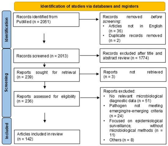

Disclaimer/Publisher’s Note: The statements, opinions and data contained in all publications are solely those of the individual author(s) and contributor(s) and not of MDPI and/or the editor(s). MDPI and/or the editor(s) disclaim responsibility for any injury to people or property resulting from any ideas, methods, instructions or products referred to in the content. |
© 2026 by the authors. Licensee MDPI, Basel, Switzerland. This article is an open access article distributed under the terms and conditions of the Creative Commons Attribution (CC BY) license.