


Abstract
Background. Hafnia alvei is considered an uncommon pathogen in immunocompetent individuals. It has been isolated in several cases of nosocomial pneumonia, but it has rarely been associated with community-acquired pneumonia (CAP) in immunocompetent adults. In 2010 a genotypically distinct species of Hafnia was identified, for which the name Hafnia paralvei was proposed. Since then, very few cases of human infection by H. paralvei have been reported. We describe here a case of CAP sustained by H. paralvei in a man without major systemic comorbidities and the genomic characterization of antibiotic resistance, together with a review of the literature on this topic. Case presentation. A 54-year-old man was admitted to the Emergency Room (ER) with a 3-day history of fever, fatigue, nausea. In the ER a thoracic CT scan revealed an extensive area of consolidation in the left lower lobe. He was empirically treated with piperacillin/tazobactam + azithromycin. After an initial improvement, clinical conditions worsened and empirical therapy was changed to meropenem + linezolid. An aerosol-induced sputum for bacterial culture yielded Hafnia alvei 107 colony-forming-units/mL; the germ was resistant to piperacillin/tazobactam and was sensitive to ceftriaxone, cefepime, and meropenem. Therefore, linezolid was withdrawn and meropenem continued obtaining resolution of the pneumonia. During the revision process, in silico analysis of sequences using three independent genome-based methods was performed: all three approaches, each relying on a different taxonomic metric, consistently assigned the isolate to H. paralvei. Moreover, the study of resistance-associated genes showed that the isolate harbored blaACC-1a and blamcr-9.1. Conclusions. We have described a case of CAP due to H. paralvei in an immunocompetent man. Limitations in antibiotic choices and duration of treatment have been discussed, and genomic characterization of antibiotic resistance has been studied.
1. Introduction
Hafnia alvei is a Gram-negative, facultative anaerobic, rod-shaped bacterium belonging to the family Hafniaceae within the order Enterobacterales; it was formerly named Enterobacter hafniae [1]. Sources from which H. alvei can be recovered include mammals, birds, reptiles, soil, water, sewage, and food. Historically, it was occasionally isolated from the intestine of honeybees and from honey, whence the epithet alvei, derived from the Latin “alveus”, meaning beehive. The genus name Hafnia comes from the historical name (Havn) for the city of Copenhagen, Denmark [2].
H. alvei is an uncommon human pathogen. It has been reported as the cause of gastroenteritis, bacteremia, pneumonia, urinary tract infection, wound infection and, more rarely, meningitis, endophthalmitis, abscesses, and septic arthritis [2,3,4,5,6,7,8,9,10].
H. alvei is seldom considered pathogenic in immunocompetent individuals; however, it is known to be responsible for serious nosocomial and community-acquired infections, mainly in patients with underlying diseases or immunosuppression [1]. H. alvei has been isolated in several cases of nosocomial pneumonia [11,12,13,14,15,16], but it has rarely been associated with community-acquired pneumonia (CAP) in immunocompetent adults [17,18,19,20,21,22].
Until 2010 H. alvei was the only recognized species in the genus Hafnia, although DNA–DNA hybridization studies had previously revealed that H. alvei was genetically heterogeneous and consisted of at least two DNA hybridization groups (HGs), with HG 1 representing H. alvei sensu stricto, by convention. In 2010 Huys and coll [23] found that strains formerly classified as members of H. alvei HG 2 represented a novel species, for which the name Hafnia paralvei was proposed. Since then, very few cases of human infection by H. paralvei have been reported. Moreover, it is possible that strains previously identified as H. alvei could have actually been H. paralvei.
We describe here a case of CAP sustained by H. paralvei in an immunocompetent man and the genomic characterization of antibiotic resistance, together with a review of the literature on this topic.
2. Methods
Preliminary species identification and antimicrobial susceptibility was performed by MALDI-TOF Biotyper System (Bruker Daltonics, Bremen, Germany) and Phoenix system (Becton Dickinson Diagnostics, San Diego, CA, USA), respectively. Determination of minimum inhibitory concentrations (MICs) for colistin was obtained through broth microdilution (Liofilchem, Roseto degli Abruzzi, Italy). Results were interpreted according to the European Committee on Antimicrobial Susceptibility Testing (EUCAST) guidelines (http://www.eucast.org/clinical_breakpoints/) (accessed on 6 August 2026). Whole-genome sequencing (WGS) was achieved on Illumina Miseq platform (Illumina, San Diego, CA, USA). Quality control of raw reads was performed using FastQC v0.11.9, followed by read trimming with Trimmomatic v0.39 in paired-end mode. Trimmed reads were assembled de novo using Unicycler v0.5.1. Assembly quality was assessed using QUAST v5.3.0 and BUSCO v5.8.2. The resulting genome assembly was used to further confirm the taxonomic assignment of the isolate using three independent genome-based approaches. Pathogenwatch was used for preliminary species identification based on Mash distance analysis (https://pathogen.watch) (accessed on 6 August 2026). Species assignment was subsequently confirmed using the Type (Strain) Genome Server, which performs genome-wide taxonomic comparisons and digital DNA–DNA hybridization (dDDH, formula d4) analyses against reference type strains. Species delineation was assessed using the 70% dDDH threshold (https://tygs.dsmz.de/) (accessed on 6 August 2026). Finally, GTDB-Tk v2.7.2 with the GTDB reference database vr232 was used to independently verify species designation through genome-based taxonomic classification and average nucleotide identity (ANI) analysis according to the GTDB species circumscription framework (95% ANI threshold) (https://github.com/ecogenomics/gtdbtk) (accessed on 6 August 2026). Antimicrobial resistance genes were extracted from the WGS data and identified by in silico analysis using the ResFinder v3.0 web server (http://www.genomicepidemiology.org) (accessed on 6 August 2026). The minimum percentage of sequence identity was set at 100%, with an alignment length of >98%. Raw reads were submitted to the Sequence Read Archive (SRA) under the BioProject PRJNA1474296.
We made a search in Pubmed using as keywords “Hafnia alvei” AND “pneumonia” (last accessed 6 August 2026) and obtained 80 papers, among which 12 described cases with H. alvei pneumonia. Seven more reports were found by examining the references of the above-mentioned papers. Abstracts without full case information were included and no language restriction was used. We considered attributable to H. alvei the reported cases of pneumonia, in addition to the clinical judgment of the Authors, when H. alvei was reported as isolated from respiratory specimens. We considered a pneumonia as community-acquired if diagnosed on admission or <48 h from hospital admission, and hospital-acquired if developed in patients admitted to the hospital for >48 h. A pneumonia was considered ventilator-associated if developed in patients who had been mechanically ventilated for at least 48 h.
Moreover, during the revision process, when the Hafnia strain of our patient was found to be a H. paralvei, we made a new search in Pubmed using as keyword simply “Hafnia paralvei” (last accessed 12 October 2026) and obtained 45 papers; only 5 papers reported human cases. Abstracts without full case information were included and no language restriction was used.
3. Case Report
A 54-year-old man was admitted to the Emergency Room (ER) with a 3-day history of fever (maximum 40.2 °C), fatigue, nausea. He was free of major systemic comorbidities. In the ER a thoracic CT scan revealed an extensive area of consolidation with pleural effusion in the left lower lobe. Blood tests showed the following: white blood cells (WBC) 8200/mm3, hemoglobin 13 g/dL, platelets (PLT) 232,000/mm3, C-reactive protein (CRP) 26 mg/dL, procalcitonin 0.22 ng/mL. Urinary antigens of Legionella and S. pneumoniae were absent. A multiplex polymerase chain reaction (PCR) for respiratory pathogens was negative in naso-pharyngeal swab. A Quantiferon TB test was positive. Blood cultures were negative. A transthoracic echocardiography was negative for endocarditis. In the ER, the patient received antibiotic therapy with piperacillin/tazobactam 4.5 g three times/day (TID) for 2 days (on days −3 and −2) and subsequently clarithromycin 500 mg two times/day (BID) + meropenem 1 g TID + linezolid 600 mg BID for 2 more days (on days −1 and 0). The patient was then transferred to our Center.
On admission (day 0) the patient was alert, oriented and eupneic; body temperature was 37.5 °C, blood pressure 120/80, heart rate 81/m, respiratory rate 18/m and oxygen saturation 94% in room air. Physical examination revealed reduced vesicular murmur with hypophonesis in the lower third of the left hemithorax. A blood gas analysis in room air showed pO2 67 mmHg, P/F 319. Oxygen was administered through nasal cannulas 2 L/m. Blood tests results were as follows: WBC 9040/mm3, hemoglobin 12.8 g/dL, PLT 281,000/mm3, glucose 108 mg/dL, creatinine 1.2 mg/dL, aspartate aminotransferase 44 U/L, alanine aminotransferase 57 U/L, CRP 22.3 mg/dL. Antibiotic therapy was prescribed consisting of piperacillin/tazobactam 4.5 g four times/day + azithromycin 500 mg once a day (on days +1 to +4). On day 3 the patient was afebrile and eupneic, WBC 8210/mm3 and CRP 7.3 mg/dL. Due to the positive result of the Quantiferon TB test performed in the ER, we prescribed an aerosol-induced sputum for bacterial culture and research of Mycobacterium tuberculosis.
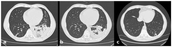
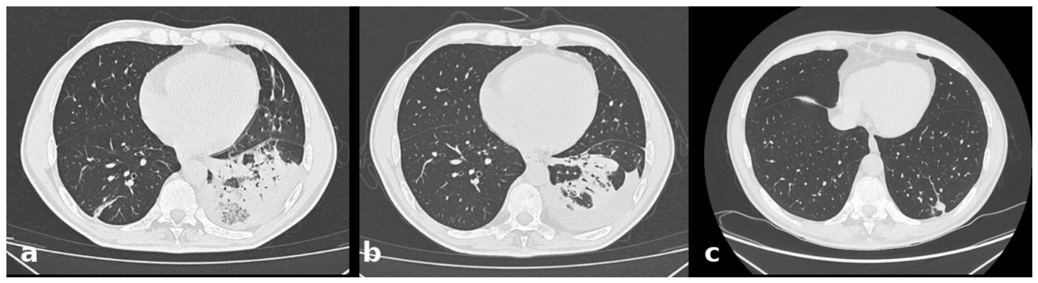
On day 5 the clinical conditions slightly worsened with mild fever (37.5 °C) and fatigue; the WBC count increased to 13,220/mm3 and the CRP was 12 mg/dL. The thoracic CT scan showed consolidation of the left lower lobe with ipsilateral pleural effusion and a linear opacity in the right lower lobe (Figure 1a). Empirical antibiotic therapy was changed to meropenem 1 g TID + linezolid 600 mg BID on days +5 and +6. On day +7 we obtained the results of the aerosol-induced sputum tests: the search for M. tuberculosis was negative on direct examination and by PCR, but the culture grew Hafnia alvei 107 colony-forming units/mL; the germ was resistant to piperacillin/tazobactam and amoxicillin/clavulanate and was sensitive to ceftriaxone, cefepime, and meropenem. Thereafter, linezolid was withdrawn and meropenem continued until day +19.
Figure 1.
Evolution of the pleuropulmonary lesions. Chest CT scans showing the evolution of the pleuropulmonary lesions in our patient with H. paralvei pneumonia on day +5 (a), +16 (b), and +78 (c) from hospital admission.
The patient’s clinical conditions gradually improved and oxygen therapy was withdrawn. On day +16 a new thoracic CT scan revealed a reduction in the consolidation in the left lower lobe with a slight increase in the ipsilateral pleural effusion (Figure 1b). The blood tests showed WBC 4680/mm3 and CRP 0.33 mg/dL. A repeated Quantiferon TB test was negative. On day +19 a dental CT scan showed a marked horizontal resorption of the alveolar bone, particularly affecting the lower jaw. The patient did not report oral pain or recent dental care. On the same day, the patient was discharged and advised to see his dentist.
On day +78 a follow-up thoracic CT scan showed a residual small nodular consolidation (mm 14 × 9) in the posterior-basal segment of the left lower lobe (Figure 1c).
During the revision process for publication of this paper, the Hafnia strain isolated in our patient was subsequently found to be H. paralvei.
4. Discussion
H. alvei is often considered a colonizing enterobacterium. It has been recovered from oropharyngeal specimens, from the gastrointestinal tract, less often from blood and from other anatomic sites including airways, urine, soft tissue, and catheters. However, it has rarely been reported as a pathogenic organism [2,22]. For decades, H. alvei has been presumably associated with diarrheal disease even in outbreak settings [24,25,26,27], but its causative role has been questioned. Probably the first report linking H. alvei to human illness is a 1967 case report of bacteremia with fever and hypotension occurring in a man operated for a duodenal ulcer; the patient was successfully treated with oxytetracycline [2,28]. Subsequently, several reports described asymptomatic colonization or infections due to H. alvei; transmission was thought to be mainly nosocomial in one study [29] and community-acquired in another [30]. Sites of infection included the biliary tree, psoas abscess, appendix, prosthetic endocarditis, the urinary and respiratory tract. In the largest series of cases, H. alvei was isolated from 80 samples collected from 61 patients: 38 from respiratory specimens, 16 and 12 from the gastrointestinal and urogenital tract respectively, 8 from blood, 3 from intravenous catheters and from the skin [1]. Only 25% of all H. alvei isolates grew in pure culture; in the remaining cases it was cultured with a variety of other bacteria and fungi. Of 61 patients, 57 had underlying conditions including malignancies, abdominal and heart surgery, pancreatitis, trauma, chronic obstructive pulmonary disease, and lung transplantation. Six patients were judged to have true H. alvei infection, three nosocomial and three community-acquired: they all recovered with antibiotic therapy chosen according to the results of susceptibility testing [1]. When H. alvei was isolated in pure culture or as predominant germ there was a better correlation with disease status [29].
To date, we retrieved from the medical literature 19 reports on H. alvei pneumonia (Table 1) in 30 patients. Nine out of nineteen papers described patients with CAP and in just three cases the patient was otherwise healthy. All of the patients with CAP (for whom outcome is reported) recovered from the pneumonia. The vast majority of patients with H. alvei pneumonia had underlying diseases:
Table 1.
Cases of H. alvei pneumonia.
Chronic obstructive pulmonary disease (the most common), Human Immunodeficiency Virus infection, chronic kidney disease, heart disease, diabetes, neoplasia. Other patients described in three reports had ventilator-associated pneumonia in the context of a severe COVID-19 disease [12,14,35]. All the patients who died presented hospital-acquired pneumonia and underlying diseases.
When, during the revision process for publication of this paper, the Hafnia strain isolated in our patient was found to be H. paralvei, we made a new search in Pubmed using as keywords simply “Hafnia paralvei”. We obtained 45 papers: the large majority concerned strains isolated from environment, food or animals. Among these studies, only five [36,37,38,39,40] reported human cases (Table 2). None of them described cases of pneumonia. Abbott and coll. [36], the largest case series, reported on microbiologic characteristics of H. paralvei strains isolated from humans, but did not give clinical information about patients.
Table 2.
Cases of H. paralvei isolation in humans.
Antimicrobial susceptibility profile showed (Table 3) that the isolate was resistant to amoxicillin/clavulanic acid (MIC > 32/2 mg/L), ampicillin (>8 mg/L), piperacillin/tazobactam (>16/4 mg/L), ceftolozane/tazobactam (4/4 mg/L) and colistin (8 mg/L) and remained susceptible to aminoglycosides (gentamicin, amikacin and tobramicin), carbapenems (meropenem and imipenem), fluoroquinolones (levofloxacin and ciprofloxacin), and to new combinations of cephalosporin and carbapenem with beta-lactamase inhibitors (ceftazidime/avibactam and meropenem/vaborbactam).
Table 3.
Antimicrobial susceptibility profile of H. paralvei in this case.
To determine whether the isolate should be assigned to Hafnia paralvei or Hafnia alvei, in silico analysis of sequences using three independent genome-based methods was performed. Pathogenwatch initially identified the isolate as H. paralvei, with a Mash distance of 0.0114865. This assignment was subsequently confirmed by TYGS, which yielded a dDDH (formula d4) value of 93.9% against the H. paralvei type strain ATCC 29927, whereas the corresponding value obtained against the H. alvei type strain ATCC 13337 was 25.4%. Consistent results were obtained with GTDB-Tk, which assigned the isolate to H. paralvei with an ANI value of 99.07% relative to reference genome GCF_001655005.1, while the ANI value against H. alvei reference genome GCF_034424155.1 was 84.44%. Overall, all three approaches, each relying on a different taxonomic metric (Mash distance, dDDH, and ANI), consistently assigned the isolate to H. paralvei, providing strong evidence that the isolate belongs to this species. Finally, resistance-associated genes, identified using Resfinder, showed that the isolate harbored blaACC-1a and blamcr-9.1. H. paralvei usually carries a chromosomally encoded, inducible class C β-lactamase ACC-1, which can confer resistance to a broad range of β-lactams [41]. Colistin-resistance of this bacterium has been well described. Hafnia genus (including H. alvei and H. paralvei) are intrinsically resistant to colistin and WGS has occasionally identified plasmid-borne or chromosomal mcr-9 genes [42,43]. The presence of blamcr-9, if located on plasmid, could represent a potential reservoir of colistin resistance. Moreover, Turbett and coll. [44] recently underscored the intrinsic resistance to colistin of the genus Hafnia: 117 out of their 119 Hafniaceae isolates resulted colistin-resistant; interestingly, just two isolates from travelers (both H. paralvei) harbored plasmid-borne blamcr-9. They report that the identification of colistin-resistance mechanisms was inconclusive due to the limited variability in the phenotypic susceptibility profile of their isolates, which prohibited the identification of variants or genes associated with resistant phenotypes. Another group of Authors described the complete genome sequence of a Hafnia paralvei harboring blamcr-9 on a plasmid [45]. Furthermore, plasmid-mediated colistin resistance blamcr-9 has been increasingly detected in Enterobacterales [42] and the finding of the blamcr-9.1 in our isolate highlights the potential risk of horizontal gene transmission. It is cause for concern because colistin is used as a final line of treatment for life threatening infections caused by multidrug resistant Gram-negative bacteria. Our results underline the importance of relying on molecular or screening tests in combination with MIC-based susceptibility testing and phenotypic tests.
In the ER our patient was initially treated with piperacillin/tazobactam and later with meropenem; no details were provided in the clinical notes for the reasons for this change in therapy, so on admission we prescribed empiric therapy with piperacillin/tazobactam and azithromycin. Due to worsening of clinical conditions, we empirically replaced piperacillin/tazobactam with meropenem. The patient gradually improved and when the antibiogram was available, we decided to continue meropenem. We acknowledge that this decision is probably a limitation: H. alvei harbors AmpC β-lactamases and, in a carbapenem-sparing vision, cefepime could have been chosen. Quite recently (8 August 2026), the updated IDSA 2026 Guidance [46], as new evidence emerged, included H. alvei among the Enterobacterales species at moderate risk for clinically significant inducible ampC and accordingly suggested cefepime for the treatment of infections caused by E. cloacae complex, K. aerogenes, C. freundii, H. alvei. Also, the duration of meropenem therapy (from day +5 to +19) could have been shortened, but the initially unfavorable clinical course induced us to extend effective antibiotic therapy for up to two weeks. The recent American Thoracic Society (ATS) Guideline suggests less than 5 days of antibiotics (minimum 3 days) for adult inpatients with non-severe CAP and 5 or more days in case of severe CAP [47]. Our patient had no ATS criteria for severe CAP.
Several elements support the etiological role of H. paralvei in the pneumonia of our patient. First, the Hafnia strain was the only microorganism isolated. Monomicrobial growth is rare for this organism, occurring in only 25% of isolates in the study with the largest available dataset [1] and is more strongly correlated with actual infection than isolation within a mixed microbial flora [29]. The isolation of a single Enterobacterales species also makes sample contamination less likely: indeed, the diagnostic pathway proposed by the Italian Association of Clinical Microbiologists (AMCLI) limits the identification of Enterobacterales in unprotected respiratory samples (such as induced sputum) to cultures showing no more than two colony types [48].
Second, the bacterial load (107 CFU/mL) exceeds the significance thresholds adopted in Italy. The AMCLI diagnostic pathway indicates that pneumonia-causing pathogens are generally present in respiratory secretions at concentrations of ≥105–106 CFU/mL [48]. An inter-society document, approved by Italian scientific societies specialized in infectious diseases, microbiology, and intensive care, adopts a threshold of 106 CFU/mL for quantitative cultures of potentially contaminated lower respiratory tract samples [49]. The bacterial load of the isolate in our clinical case exceeded these thresholds by one to two orders of magnitude.
Thirdly, despite extensive diagnostic efforts, no alternative cause for the pneumonia was identified: testing for L. pneumophila and S. pneumoniae urinary antigens, multiplex PCR for respiratory pathogens, and blood cultures all yielded negative results. Neither thoracentesis nor bronchoscopy was performed, as the pleural effusion was minimal and a microbiological diagnosis had already been established, with tuberculosis ruled out via negative microscopy and PCR on the same sample.
Finally, the clinical course was consistent with the isolate’s susceptibility profile. The patient experienced clinical deterioration on the fifth day of therapy with piperacillin/tazobactam, an antibiotic to which our isolate showed resistance, though it is generally active against the most common causative agents of CAP, with improvement occurring after switching to meropenem. Following the discontinuation of linezolid on day seven, the pneumonia resolved with meropenem monotherapy alone. A similar course has been described in a previously healthy adult with H. alvei CAP (confirmed via bronchoalveolar lavage): a lack of response to β-lactam/β-lactamase inhibitor combinations, followed by a clinical response to cefepime [22].
In our view, the isolation of a single microorganism—present in high bacterial load and resistant to the ongoing antibiotic regimen—followed by improvement after switching to effective therapy, is consistent with a causative role for that organism in the infection.
The patient had a poor dental condition, as described by dental scan. H. alvei has been associated with periodontal inflammation in several reports [50,51]. It is known that the periodontal biofilm may be a source of dissemination and development of systemic infection, including pneumonia. Even if not demonstrable, in this patient H. paralvei could have reached the lungs after aspiration from the oral cavity.
5. Conclusions
In conclusion, we have described a case of community-acquired pneumonia due to H. paralvei in an immunocompetent man, successfully treated by medical therapy. Limitations in antibiotic choices and duration of treatment have been discussed. Genomic characterization of antibiotic resistance has been studied. Future investigations, including examinations of additional genomic data and functional studies, may contribute to the understanding of the mechanisms of antibiotic resistance in Hafnia spp.
Author Contributions
Conceptualization, P.C. and C.V.; methodology, D.R.D. and M.d.P.; validation, P.C., F.T. and S.T.; writing—original draft preparation, P.C. and P.G.P.; writing—review and editing, P.C., S.T. and M.d.P.; investigation, C.V.; visualization, P.G.P.; supervision, F.T. and D.R.D. All authors have read and agreed to the published version of the manuscript.
Funding
This research received no external funding.
Institutional Review Board Statement
Ethical review and approval were waived for this study due to the Italian law for publication of case reports regarding patients cured in research institutes.
Informed Consent Statement
Written informed consent has been obtained from the patient to publish this paper.
Data Availability Statement
The original contributions presented in this study are included in the article. Further inquiries can be directed to the corresponding author.
Conflicts of Interest
The authors declare no conflicts of interest.
References
- Günthard, H.; Pennekamp, A. Clinical significance of extraintestinal Hafnia alvei isolates from 61 patients and review of the literature. Clin. Infect. Dis. 1996, 22, 1040–1045. [Google Scholar] [CrossRef] [Scilit] [PubMed]
- Janda, J.M.; Abbott, S.L. The genus Hafnia: From soup to nuts. Clin. Microbiol. Rev. 2006, 19, 12–18. [Google Scholar] [CrossRef] [Scilit] [PubMed] [PubMed Central]
- Reina, J.; Hervas, J.; Borrell, N. Acute gastroenteritis caused by Hafnia alvei in children. Clin. Infect. Dis. 1993, 16, 443. [Google Scholar] [CrossRef] [Scilit] [PubMed]
- Kim, J.; Choi, Y.; Sung, H.; Kim, S.H.; Lee, S.O.; Choi, S.H. Clinical characteristics and outcomes of adult patients with Hafnia alvei bacteremia in a tertiary care hospital. Diagn. Microbiol. Infect. Dis. 2026, 115, 117301. [Google Scholar] [CrossRef] [Scilit] [PubMed]
- Rahman, S.R.; Ahmed, M.F.; Begum, A. Occurrence of urinary tract infection in adolescent and adult women of shanty town in Dhaka City, Bangladesh. Ethiop. J. Health Sci. 2014, 24, 145–152. [Google Scholar] [CrossRef] [Scilit] [PubMed] [PubMed Central]
- Berger, S.A.; Edberg, S.C.; Klein, R.S. Enterobacter hafnaie infection: Report of two cases and review of the literature. Am. J. Med. Sci. 1977, 273, 101–104. [Google Scholar] [PubMed]
- Mojtabaee, A.; Siadati, A. Enterobacter Hafnia meningitis. J. Pediatr. 1978, 93, 1062–1063. [Google Scholar] [CrossRef] [Scilit] [PubMed]
- Caravalho, J., Jr.; McMillan, V.M.; Ellis, R.B.; Betancourt, A. Endogenous endophthalmitis due to Salmonella arizonae and Hafnia alvei. South. Med. J. 1990, 83, 325–327, Erratum in South. Med. J. 1990, 83, 496. [Google Scholar] [CrossRef] [Scilit] [PubMed]
- Agustin, E.T.; Cunha, B.A. Buttock abscess due to Hafnia alvei. Clin. Infect. Dis. 1995, 20, 1426. [Google Scholar] [CrossRef] [Scilit] [PubMed]
- Centers for Disease Control and Prevention (CDC). Septic arthritis following anterior cruciate ligament reconstruction using tendon allografts--Florida and Louisiana, 2000. MMWR Morb. Mortal. Wkly. Rep. 2001, 50, 1081–1083. [Google Scholar] [PubMed]
- Frick, T.; Kunz, M.; Vogt, M.; Turina, M. Typische Nosokomiale Infektion Mit Seltenem Erreger: Hafnia alvei. Bericht Über Zwei Fälle Und Literaturübersicht [Typical Nosocomial Infection with an Unusual Cause: Hafnia alvei. Report of 2 Cases and Literature Review]. Schweiz. Rundsch. Med. Prax. 1990, 79, 1092–1094. (In German). [Google Scholar] [PubMed]
- Cutuli, S.L.; De Maio, F.; De Pascale, G.; Grieco, D.L.; Monzo, F.R.; Carelli, S.; Tanzarella, E.S.; Pintaudi, G.; Piervincenzi, E.; Cascarano, L.; et al. COVID-19 influences lung microbiota dynamics and favors the emergence of rare infectious diseases: A case report of Hafnia alvei pneumonia. J. Crit. Care 2021, 64, 173–175. [Google Scholar] [CrossRef] [Scilit] [PubMed] [PubMed Central]
- Redondo, J.; Maseda, E.; Riquelme, A.; Alday, E.; Uña, R.; Criado, A. Hafnia alvei: Una causa infrecuente de neumonía grave en la unidad de reanimación [Hafnia alvei in a rare case of severe pneumonia in a postanesthesia recovery unit]. Rev. Esp. Anestesiol. Reanim. 2005, 52, 359–362. (In Spanish). [Google Scholar] [PubMed]
- Méndez, L.; Ferreira, J.; Caneiras, C. Hafnia alvei Pneumonia: A Rare Cause of Infection in a Patient with COVID-19. Microorganisms 2021, 9, 2369. [Google Scholar] [CrossRef] [Scilit] [PubMed] [PubMed Central]
- Millán Rodríguez, M.R.; Muñoz Pérez, M.A.; Meseguer Frutos, M.D.; Cano Sánchez, A.; Román López Andreu, F.; Soriano Palao, J. Neumonía nosocomial por Hafnia alvei [Nosocomial pneumonia caused by Hafnia alvei]. An. Med. Interna 2003, 20, 595–596. (In Spanish). [Google Scholar] [CrossRef] [Scilit] [PubMed]
- Klapholz, A.; Lessnau, K.D.; Huang, B.; Talavera, W.; Boyle, J.F. Hafnia alvei: Respiratory tract isolates in a community hospital over a three-year period and a literature review. Chest 1994, 105, 1098–1100. [Google Scholar] [CrossRef] [Scilit] [PubMed]
- Lim, J.; Bolger, K.; Canavan, B. Isolation of Hafnia alvei from bronchoalveolar lavage of an immunocompetent host presenting with cavitating pneumonia: Contaminant or Causative? Germs 2023, 13, 292–296. [Google Scholar] [CrossRef] [Scilit] [PubMed] [PubMed Central]
- Fazal, B.A.; Justman, J.E.; Turett, G.S.; Telzak, E.E. Community-acquired Hafnia alvei infection. Clin. Infect. Dis. 1997, 24, 527–528. [Google Scholar] [CrossRef] [Scilit] [PubMed]
- Begbey, A.; Guppy, J.H.; Mohan, C.; Webster, S. Hafnia alvei pneumonia: A rare cause of infection in the multimorbid or immunocompromised. BMJ Case Rep. 2020, 13, e237061. [Google Scholar] [CrossRef] [Scilit] [PubMed] [PubMed Central]
- Mo, M.; Peng, C.; Peng, G.; Yuan, H.; Zhang, Z.; Xie, J.; Cheng, W. Invasive Hafnia alvei pneumonia complicated with lung abscess and pneumothorax triad: Imaging features and therapeutic dilemmas in an extremely rare case of severe infection. Zhonghua Wei Zhong Bing Ji Jiu Yi Xue 2026, 38, 198–201. [Google Scholar] [CrossRef] [PubMed]
- Galeas, F.; de la Torre, F.J.; Prada, J.L.; del Arco, A. Neumonía por Hafnia alvei en paciente infectado por el virus de la inmunodeficiencia humana [Pneumonia caused by Hafnia alvei in a patient infected with human immunodeficiency virus]. Enferm. Infecc. Microbiol. Clin. 2001, 19, 41–42. (In Spanish). [Google Scholar] [PubMed]
- Severiche-Bueno, D.F.; Vargas-Cuervo, M.T.; Medina-Lee, L.; Oliver-Hernandez, G.; Buitrago-Toro, K.; Insignares, D.A.; Conde-Camacho, R. Hafnia alvei pneumonia: From bees to human beings. Germs 2021, 11, 306–309. [Google Scholar] [CrossRef] [Scilit] [PubMed] [PubMed Central]
- Huys, G.; Cnockaert, M.; Abbott, S.L.; Janda, J.M.; Vandamme, P. Hafnia paralvei sp. nov., formerly known as Hafnia alvei hybridization group 2. Int. J. Syst. Evol. Microbiol. 2010, 60, 1725–1728. [Google Scholar] [CrossRef] [Scilit] [PubMed]
- Stuart, C.A.; Rustigian, R. Further Studies on One Type of Paracolon Organism. Am. J. Public Health Nations Health 1943, 33, 1323–1325. [Google Scholar] [CrossRef] [Scilit] [PubMed] [PubMed Central]
- Harada, K.; Shimizu, K.; Matsuyama, T. Hafnia isolated from men. Gunma J. Med. Sci. 1957, 6, 109–112. [Google Scholar]
- Ratnam, S. Etiologic role of Hafnia alvei in human diarrheal illness. Infect. Immun. 1991, 59, 4744–4745. [Google Scholar] [CrossRef] [Scilit] [PubMed] [PubMed Central]
- Ratnam, S.; Butler, R.W.; March, S.; Parsons, S.; Clarke, P.; Bell, A.; Hogan, K. Enterobacter hafniae-associated gastroenteritis—Newfoundland. Can. Dis. Wkly. Rep. 1979, 5, 231–232. [Google Scholar]
- Jennis, F.; Mccarthy, S.W. Hafnia: An unusual cause of postoperative gram-negative bacteriae. Med. J. Aust. 1967, 1, 286–287. [Google Scholar] [CrossRef] [Scilit] [PubMed]
- Washington, J.A., 2nd; Birk, R.J.; Ritts, R.E., Jr. Bacteriologic and epidemiologic characteristics of Enterobacter hafniae and Enterobacter liquefaciens. J. Infect. Dis. 1971, 124, 379–386. [Google Scholar] [CrossRef] [Scilit] [PubMed]
- Ramos, A.; Dámaso, D. Extraintestinal infection due to Hafnia alvei. Eur. J. Clin. Microbiol. Infect. Dis. 2000, 19, 708–710. [Google Scholar] [CrossRef] [Scilit] [PubMed]
- Hidalgo Tenorio, C.; Pasquau Liaño, J. Tuberculosis pulmonar bilateral y sobreinfección respiratoria por Hafnia alvei [Bilateral lung tuberculosis and over-infection by Hafnia alvei]. An. Med. Interna 2002, 19, 547–548. (In Spanish). [Google Scholar] [CrossRef] [Scilit] [PubMed]
- Loya, M.K.; Walsh, J. A case of community acquired pneumonia caused by Hafnia alvei developing into an empyema. Chest 2015, 148, 101A. [Google Scholar] [CrossRef] [Scilit]
- Karamanli, H.; Ozer, T.T. A rare case of Hafnia alvei pneumonia presenting with chronic obstructive pulmonary disease. Turk. Gogus Kalp Dama 2017, 25, 308–311. [Google Scholar] [CrossRef] [Scilit]
- Baral, N.; Nayak, N.; Shrestha, R.; Parajuli, R.; Hamal, D.; Bhatta, D.R.; Gowda, S.; Gokhale, S. Hafnia alvei bacteremia following bronchopneumonia in an eleven month old child: A case report from a tertiary care hospital in Nepal. Int. J. Adv. Life Sci. Res. 2018, 1, 22–25. [Google Scholar] [CrossRef] [Scilit]
- Blonz, G.; Kouatchet, A.; Chudeau, N.; Pontis, E.; Lorber, J.; Lemeur, A.; Planche, L.; Lascarrou, J.B.; Colin, G. Epidemiology and microbiology of ventilator-associated pneumonia in COVID-19 patients: A multicenter retrospective study in 188 patients in an un-inundated French region. Crit. Care 2021, 25, 72. [Google Scholar] [CrossRef] [Scilit] [PubMed] [PubMed Central]
- Abbott, S.L.; Moler, S.; Green, N.; Tran, R.K.; Wainwright, K.; Janda, J.M. Clinical and laboratory diagnostic characteristics and cytotoxigenic potential of Hafnia alvei and Hafnia paralvei strains. J. Clin. Microbiol. 2011, 49, 3122–3126. [Google Scholar] [CrossRef] [Scilit] [PubMed] [PubMed Central]
- Osuka, H.; Hitomi, S.; Koganemaru, H.; Kaneko, T. A case of bacteremia caused by Hafnia paralvei. J. Infect. Chemother. 2011, 17, 855–857. [Google Scholar] [CrossRef] [PubMed]
- Savini, V.; Argentieri, A.V.; Marrollo, R.; Carretto, E.; Fazii, P.; D’Antonio, D.; Huys, G. The Italian Hafnia alvei strain LMG 27376 is Hafnia paralvei. Vet. Microbiol. 2013, 167, 742–743. [Google Scholar] [CrossRef] [Scilit] [PubMed]
- Appel, T.M.; Stein, C.; Brandt, C.; Rödel, J.; Frietsch, J.J.; Miethke, J.; Hochhaus, A.; Hilgendorf, I. Isolation of Hafnia paralvei co-harbouring blaNDM-1 and blaVIM-1 in a woman who underwent allogeneic hematopoietic stem cell transplantation. Infection 2023, 51, 1161–1164. [Google Scholar] [CrossRef] [Scilit] [PubMed] [PubMed Central]
- Cho, S.M.; Kang, M.S.; Hong, S.G. First Report of KPC-2-Producing Hafnia paralvei: Evidence of Horizontal Gene Transfer from Klebsiella pneumoniae. Ann. Lab. Med. 2026, 46, 351–353. [Google Scholar] [CrossRef] [Scilit] [PubMed] [PubMed Central]
- Bauernfeind, A.; Schneider, I.; Jungwirth, R.; Sahly, H.; Ullmann, U. A novel type of AmpC beta-lactamase, ACC-1, produced by a Klebsiella pneumoniae strain causing nosocomial pneumonia. Antimicrob. Agents Chemother. 1999, 43, 1924–1931. [Google Scholar] [CrossRef] [Scilit] [PubMed] [PubMed Central]
- Ikhimiukor, O.O.; Montoya-Giraldo, M.; Souza, S.S.R.; Akintayo, I.J.; Zac Soligno, N.I.; Marmion, M.; Eckhardt, E.M.; Carreiro, N.; Workman, A.A.; Martin, I.W.; et al. Dissemination dynamics of colistin resistance genes mcr-9 and mcr-10 across diverse Inc plasmid backbones. Commun. Med. 2025, 5, 399. [Google Scholar] [CrossRef] [Scilit] [PubMed] [PubMed Central]
- Łopatek, M.; Denis, E. Prevalence and antimicrobial resistance of Enterobacterales bacteria isolated from retail food in Poland. J. Vet. Res. 2025, 69, 371–379. [Google Scholar] [CrossRef] [Scilit] [PubMed] [PubMed Central]
- Turbett, S.E.; Bronson, R.A.; Worby, C.J.; McGrath, G.E.G.; Hodgkins, E.; Becker, M.; Belford, B.; Kogut, L.; Oliver, E.; Ryan, E.T.; et al. Intrinsic Resistance to Colistin in the Genus Hafnia. J. Clin. Microbiol. 2023, 61, e0132622. [Google Scholar] [CrossRef] [Scilit] [PubMed] [PubMed Central]
- Biggel, M.; Zurfluh, K.; Hoehn, S.; Schmitt, K.; Frei, A.; Jans, C.; Stephan, R. Complete Genome Sequence of Hafnia paralvei Isolate AVS0177, Harboring mcr-9 on a Plasmid. Microbiol. Resour. Announc. 2022, 11, e0096621. [Google Scholar] [CrossRef] [Scilit] [PubMed] [PubMed Central]
- Tamma, P.D.; Bonomo, R.A.; Heil, E.L.; Justo, J.A.; Satlin, M.J.; Mathers, A.J. Infectious Diseases Society of America 2026 Guidance on the Treatment of Antimicrobial-Resistant Gram-Negative Infections. Clin. Infect. Dis. 2026, ciag481. [Google Scholar] [CrossRef] [Scilit] [PubMed]
- Jones, B.E.; Ramirez, J.A.; Oren, E.; Soni, N.J.; Sullivan, L.R.; Restrepo, M.I.; Musher, D.M.; Erstad, B.L.; Pickens, C.; Vaughn, V.M.; et al. Diagnosis and Management of Community-acquired Pneumonia: An Official American Thoracic Society Clinical Practice Guideline. Am. J. Respir. Crit. Care Med. 2026, 212, 24–44. [Google Scholar] [CrossRef] [Scilit] [PubMed]
- AMCLI, E.T.S. Percorso Diagnostico—Polmoniti nel Paziente Ospedalizzato [Diagnostic Pathway—Pneumonia in the Hospitalized Patient], Rif. 2025–26. Available online: https://amcli.it/percorsi-diagnostici-218/ (accessed on 13 October 2026).
- Sartelli, M.; Antonelli, G.; Barchitta, M.; Bartoli, S.; Borghi, F.; Busani, S.; Buttarelli, C.; Carsetti, A.; Catena, F.; Cavaliere, A.; et al. Documento Multi-Societario per La Prevenzione e Controllo Delle Infezioni Correlate All’assistenza in Chirurgia [Multi-Society Document for the Prevention and Control of Healthcare-Associated Infections in Surgery]; SIMPIOS: Milan, Italy; ACOI: Rome, Italy; SIAARTI: Rome, Italy; SIMIT: Prato, Italy, 2023; Available online: https://www.simpios.eu/2023/05/29/documento-multi-societario-per-la-prevenzione-delle-infezioni-correlate-allassistenza-in-chirurgia/?utm_source=chatgpt.com (accessed on 13 October 2026).
- Vieira Colombo, A.P.; Magalhães, C.B.; Hartenbach, F.A.; Martins do Souto, R.; Maciel da Silva-Boghossian, C. Periodontal-disease-associated biofilm: A reservoir for pathogens of medical importance. Microb. Pathog. 2016, 94, 27–34. [Google Scholar] [CrossRef] [Scilit] [PubMed]
- Zhang, W.; Chen, Y.; Shi, Q.; Hou, B.; Yang, Q. Identification of bacteria associated with periapical abscesses of primary teeth by sequence analysis of 16S rDNA clone libraries. Microb. Pathog. 2020, 141, 103954. [Google Scholar] [CrossRef] [Scilit] [PubMed]

Disclaimer/Publisher’s Note: The statements, opinions and data contained in all publications are solely those of the individual author(s) and contributor(s) and not of MDPI and/or the editor(s). MDPI and/or the editor(s) disclaim responsibility for any injury to people or property resulting from any ideas, methods, instructions or products referred to in the content. |
© 2026 by the authors. Licensee MDPI, Basel, Switzerland. This article is an open access article distributed under the terms and conditions of the Creative Commons Attribution (CC BY) license.