


Abstract
Background: The movement of endemic diseases from tropical and disadvantaged areas is gradually spreading to developed countries as well. Amoebiasis, in particular, represents a significant threat to public health, amplified by globalization and increasing contact between humans and animals or vectors. In this way, atypical diseases may emerge in our country, also through an unusual mode of transmission—likely sexual, as “sexually transmitted enteric (STE) diseases”. Objective: We report and discuss the clinic approach to a rare case of dual human infestations by ecto- and endo-parasites with multiple liver abscesses presentation in a young truck driver residing in Northern Italy. Methods: The patient underwent a comprehensive screening with laboratory and microbiological tests, and CT images. Results: In July, the patient was admitted to our hepatology unit following the ultrasound identification of two hypoechoic lesions in the right lobe of the liver. In his medical history, in April, after a seemingly harmless infestation of body lice, effectively treated, he suffered from a prolonged and debilitating episode of acute diarrhea, lasting a month. This status was also accompanied by intestinal cramps, weight loss, and recurring fever episodes. Afterwards also appeared a Quincke’s-like angioedema involving the lips and mouth mucosa, with evening time exacerbation. Conclusions: The differential diagnosis of this complex and unusual clinical picture, together with the temporal evolution of the patient’s signs and symptoms, led us to hypothesize the presence of concomitant infections. These were most likely borreliosis and a STE disease, the latter ultimately diagnosed as amebiasis with hepatic invasion, allowing appropriate treatment and a favorable clinical outcome.
1. Introduction
Amoebiasis, caused by Entamoeba histolytica, is the second leading cause of parasitic death, especially in developing countries such as some countries in the middle east and eastern Asia, central and meridional America and occidental-meridional Africa, where water and food are easily contaminated with feces containing E. histolytica cysts, due to poor sanitation, [1,2]. However, a growing challenge in many developed countries is the increasing incidence of invasive amoebiasis, particularly as a STE infection, which may occur through direct sexual contact among individuals practicing oro-anal sex, either in heterosexual or homosexual relationships [3]. In this context, asymptomatic infected individuals from at-risk regions constitute the main reservoir of E. histolytica, acting as contagious carriers and also spreading the infection to unaffected areas around the world, where the diagnosis is frequently overlooked [4].
Furthermore, the often degraded environment in which sex workers live is never free from the risk of transmission of other pathogens. Among these, some sexually transmitted infections (e.g., chlamiosis, syphilis, and HIV) certainly play a preponderant role. However, we must also consider some parasitic infestations that may act as vectors and selectively transmitted insidious bacterial infections, which are now spreading everywhere, including in Italy [5]. Body lice, although considered harmless, can cause serious cases of borreliosis, especially in frail individuals already suffering from other comorbidities. Relapsing fever (RF), caused by Borrelia recurrentis and B. miyamotoi, is transmitted, respectively, by the bite of parasites of the species Pediculus humanus corporis, which live and lay eggs in clothing, moving to the skin to feed [6], and Ixodes ricinus, a tick present in mountainous and wooded areas of northern Italy [7]. These infected vectors are reaching every continent, even carrying new species, which are more difficult to diagnose due to the specific cross-reactivity of antigens [8], and even cause co-infections of parasites [9,10] among the Borrelia species (spp.).
We have described a complex clinical case, characterized by a diagnostic scenario that was challenging within our regional context due to the concurrent presence of a zoonosis caused by human ecto- and endo-parasites. This infestation—potentially sexually acquired—gave rise to an episode suggestive of borreliosis, followed by invasive amoebiasis with liver abscess formation. The chronological reconstruction of events and the search for risk factors guided us through a diagnostic process, supported by a precise anamnesis, specific diagnostic tests, and effective treatment.
2. Case Presentation
A 41-year-old man, recently diagnosed with Quincke-like angioedema, presented to our hospital in July to investigate the etiology of newly developed hepatic abscesses. The patient reported, about 2 months before, the onset of intermittent edema episodes of the lips and oral mucosa, sometimes accompanied by fever (within 38 °C). This swelling was associated with arthralgia of the small joints of the hands and feet, and diffuse myalgia, which had already led to a rheumatological consultation, after a failed 3-day trial of antibiotic therapy (ABT) prescribed by his GP. These symptoms tended to get worse in the evening and during the night, and were not associated with itching, but with chills, fever and headache. The patient received a second course of empirical ABT (azithromycin, 500 mg tablets, once daily) for 5 days, associated with steroids (methylprednisolone 16 mg tablets, one daily dose for 3 days, de-escalate to 8 and 4 mg for a total of another 6 days), with the disappearance of symptoms occurring by the end of May.
After hospital admission in our hepatologic unit, the patient underwent a complete laboratory profile and some specific microbiological diagnostic tests, as reported in Table 1. Moderate liver enzyme alterations with leukocytosis, mild anemia, and a modest increase in the C-reactive protein (CRP) appeared. The normal eosinophil count and level of C1 inhibitor were inconsistent with Quincke’s edema or a suspected helminth infestation, prompting us to investigate alternative causes for the patient’s clinical presentation.
Table 1.
Biochemical, hematological, microbiological and molecular test results.
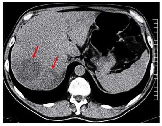
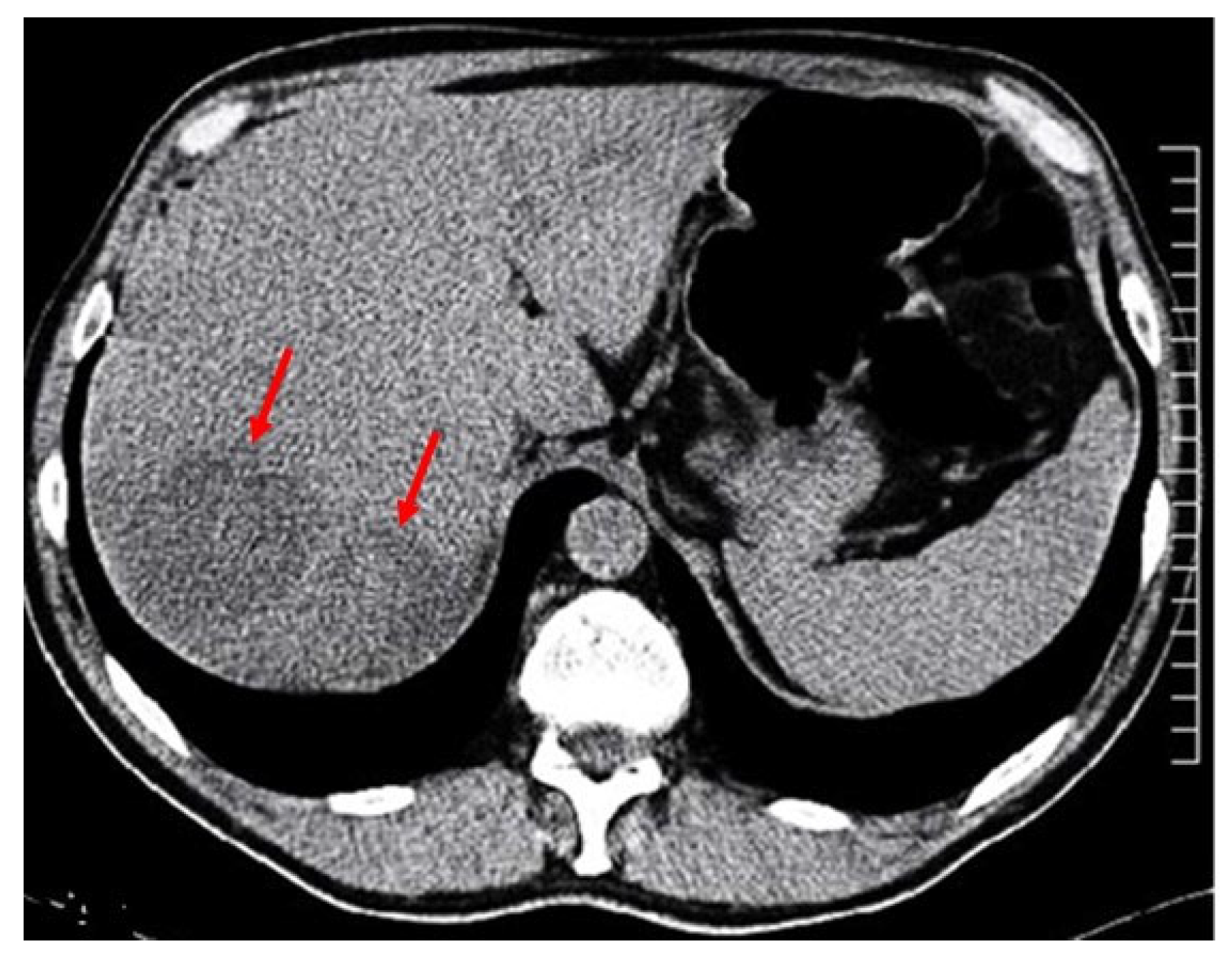
The two hypoechoic areas diagnosed by abdominal-US in the VII hepatic segment, measuring 8 and 6 cm in diameter, respectively, were confirmed with an abdominal CT scan (Figure 1).
Figure 1.
Two large hypodense liver lesions, each 8 and 6 cm in diameter, are visible in the seventh segment of the right lobe, highlighted by red arrows.
The roundish liver lesions were soft capsulated and hypointense, and had irregular contours, being rather inhomogeneous, like necrotic liquefactive lesions. The largest one was aspirated extemporaneously by needle prick and contained a slightly thick brown necrotic fluid that, upon examination under a light microscope, revealed inflammatory blood cells with pyocytes. Bacterial and fungal cultures were negative, and no cysts were isolated. No Plasmodium species were detected, and tuberculosis was also ruled out with rapid molecular tests (NAAT).
Upon further review of the medical history, the patient reported a body louse infestation in April, which was successfully treated with a pyrethrin- and piperonyl butoxide-containing shampoo, together with decontamination of clothing and bed linens by machine washing at 65 °C. Shortly thereafter, he developed an episode of acute diarrhea associated with severe abdominal pain, which resolved spontaneously within 2–3 weeks.
Upon further questioning, the patient also recalled that the first episode of oral edema and polyarthralgia had occurred during the same period and was accompanied by recurrent febrile episodes preceded by chills and worsening headache. Moreover, he reported that the most severe episode of edema developed immediately after the first ABT course.
The patient came from a province in Northern Italy and spent part of the year traveling throughout Europe for work as a truck driver. He did not smoke and consumed alcohol only occasionally; nor did he use drugs or other narcotic substances. He enjoyed the outdoors and lived in a wooded area, but did not recall having been bitten by a tick recently. He had never traveled to tropical countries, nor had he had direct contact with animals in the woods. His family history did not confirm cases of chronic liver disease, such as cirrhosis, or hepatocellular carcinoma (HCC) among close relatives. Physical examination revealed marked weight loss (from 75 to 63 kg) over the preceding four months, associated with mild, fluctuating febrile episodes, as well as profound asthenia, hyporexia and headache. No overt jaundice was observed.
Considering the possibility of multiple causes for the various patient symptoms, and paying particular attention to reported episodes of recurrent fever, we investigated the presence of louse- or tick-borne infectious diseases, finding significant antibody positivity for Borrelia spp. Microbiological investigations resulted in IgG and IgM positivity by indirect chemiluminescence immunoassay (CLIA) specific for B. burgdorferi (LIAISON Borrelia Quant IgM and IgG tests; DIASORIN, via Crescentino snc, 13040 Saluggia, Vercelli, Italy). B. burgdorferi infection was not subsequently confirmed by a two-step process using recombinant and highly specific IgG-IgM line immunoassay (LIA) (recomLine IgG and IgM Borrelia tests, Mikrogen diagnostic; Ref. 4272 and 4273; Anna-Sigmund-Str. 10, 82061 Neuried, Germany).
Therefore, the diagnosis of borreliosis would have been possible only through direct microscopic detection of spirochetes in a peripheral blood smear, obtained at the height of fever and stained specifically with Wright’s or Giemsa stain [11]. We were unable to complete this diagnostic investigation phase because the acute phase had already passed, and the patient was already cured after the ABT cycle. Furthermore, the patient reported neither recent tick bites nor erythematous skin lesions indicative of an active infection. We therefore hypothesized the possibility that, the B. recurrentis infection vector might have been the Pediculus corporis [12], also considering the well-recognized occurrence of cross-reactivity among Borrelia spp. antigens.
Although CT images were not characteristic of worm colonization, such as Strongyloides infestation that could explain both the edema and hepatic abscesses [13], we performed a biomolecular assay for S. stercoralis that gave a negative result. Furthermore, we were able to exclude echinococcosis because plain radiographs and an abdominal ultrasound showed hepatomegaly, but not the characteristic scattered areas of hyperechoic lesions bordered by calcified rings, which were also not visible on CT. Furthermore the patient did not present eosinophilia or positivity to specific serum antibody test for E. multilocularis [14].
Therefore, considering the sequence of signs and symptoms revealed by the patient’s medical history—ranging from pediculosis to episodes of dysentery and Quincke’s edema—we also investigated other potential infections capable of causing diarrhea followed by the appearance of abscess-like hepatic lesions, sometimes accompanied by allergic manifestations. Starting from the hypothesis of close contact with people belonging to degraded environments, usually the only carriers of pediculosis corporis, we re-evaluated the anamnesis of the patient and he admitted that about a month before the onset of diarrhea, around mid-March, he had had paid sex with a non-Caucasian man.
Thus, we tested him for the most common sexually transmitted infections, such as HIV and Treponema pallidum, for which he tested negative. Given the rare forms of transmission involving liver abscesses, we hypothesized that this might be a case of invasive amoebiasis, also considering that the risk of intestinal E. histolytica infection is well documented in association with homosexual practices [15].
From an epidemiological perspective, two main and distinct species of Entamoeba are recognized: E. histolytica, which is highly pathogenic to humans, and E. dispar, a commensal species not associated with disease [16,17]. More recently, rare cases of cerebral abscesses were related to E. moshkovskii infection [18]. Considering that E. histolytica is transmitted via the fecal–oral route and can cause liver abscesses, amebiasis should be suspected in patients with an appropriate exposure history (residence in or travel to an endemic area) and characteristic clinical features, including fever, right upper quadrant pain, and marked hepatic tenderness [19,20].
Therefore, although amebiasis is not endemic in our region, we tested the patient’s blood for the research of specific antibodies against E. histolytica. Two independent serum samples had positive results, with an IgG-specific 1:1024 titer by ELISA (Entamoeba histolytica IgG ELISA; Ref. EIA-3830; DRG Instruments GmbH, Frauenbergstrasse, 18, 35039 Marburg, Germany).
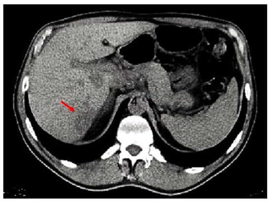
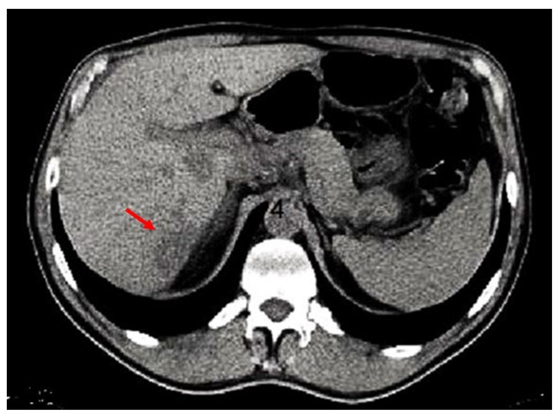
Thus, the patient was treated for E. histolytica with two cycles of metronidazole 750 mg tables and TID for 10 days, followed by paromomycin 30 mg/kg/day tablets in three doses daily, to be repeated after 20 days to eradicate luminal amoebae and prevent relapse. Patient evaluation after 6 months showed a decrease in E. histolytica-specific antibody titer (1:256) and CT images showed almost complete resolution of liver lesions (Figure 2).
Figure 2.
The larger liver lesion in segment 8 appears to have completely resolved, while the other lesion showed a significant reduction in size (red arrow) following effective antiparasitic treatment, as demonstrated by the CT performed 6 months later.
Clinical case was resolved without complications, but we want to report some final considerations and limitations: (a) The patient received steroids to treat perioral edema and arthralgias, which are contraindicated in amoebiasis, because they can suppress the host’s immune response and likely facilitate the spread of the amoeba, especially during the acute intestinal phase. It is better to use acetaminophen 650 mg orally, to control symptoms or toxemia resulting from the use of spirochaeticidal; (b) The abscess puncture should always be avoided, because it is difficult to obtain a sample that is useful for diagnosis, as in our case, and furthermore, there are highly specific serological tests for diagnosis and very effective therapies for the treatment of the disease; and (c) Cross-reactions between antigens from Borrelia spp. can be observed, which can make B. recurrentis infection diagnosis more difficult if a specific microscopy identification or genetic test cannot be executed, as happened in this case.
3. Discussion
Entamoeba histolytica is the causative agent of amoebiasis—amoebic dysentery, one of the most common intestinal parasitic infections worldwide, accounting for an estimated 40 million infections and 100,000 deaths annually [1]. However, only 5–15% of infected individuals develop clinically manifested disease [2].
3.1. Diagnostic Protocols for Suspected Cases of Amoebiasis
The diagnosis of amoebiasis relies on parasite or cyst identification in stool or tissue by microscopy, or with the detection of specific serum antibodies by ELISA or of antigen by PCR [21]. During the acute phase, the presence of E. histolytica forms have the highest diagnostic sensitivity, although microscopy research in stool is highly dependent on the examiner’s expertise and identification in tissue is enhanced by periodic acid–Schiff (PAS) staining [22]. Despite its high specificity, PCR may be costly and not widely available [23], whereas ELISA antibodies may persist long after clinical recovery. In contrast, asymptomatic infections often remain undiagnosed, particularly in non-endemic countries where routine screening is not performed [1].
Therefore, in this complex case, a systematic clinical approach integrating multiple diagnostic clues was essential to confirm amoebiasis. These included identification of a relevant exposure history, temporal correlation between exposure and symptom onset, recognition of characteristic liver lesions, detection of specific serological markers, and confirmation through the patient’s favorable response to antiparasitic therapy.
3.2. Signs and Symptoms of Systemic Amoebiasis
The hepatic abscess, as found in this case, must be considered the first extraintestinal form related to amoebiasis [1]. It is predominant in young males by about 10:1 [24], although pregnant women and children are more prone to developing the infection [25]. Amoebic liver abscesses, which are highly endemic in most developing tropical countries, are now more frequently encountered in other geographic non-endemic areas [3]. Therefore clinical manifestations of amebiasis can range from asymptomatic carriers without disease to severe invasive forms, such as tissue abscesses (hepatic, cerebral, pulmonary) or fulminant colitis with paralytic ileus and intestinal perforation [26,27,28].
Occasionally, abscess aspiration is required to exclude a pyogenic liver abscess, whereas aspiration of an amoebic abscess is not routinely performed for diagnostic purposes because its core consists of liquefactive necrotic tissue. Unlike pyogenic liver abscesses, which are primarily managed with percutaneous or surgical drainage, amoebic liver abscesses are generally treated with first-line medical therapy [29]. Antiparasitic therapy, most commonly with metronidazole [30], is usually effective in treating amoebic dysentery and liver abscesses. Although clinical resolution is generally rapid, the complete radiological resolution of the abscess may take months to years.
E. histolytica is classified as a Category B biodefense agent because of its environmental stability, resistance to chlorination, and ability to spread through contaminated food or water in settings with poor sanitation, as well as through sexual transmission.
Abdominal ultrasonography, CT, and MRI may help identify a hypodense hepatic lesion, although an amebic liver abscess can sometimes be difficult to distinguish from other types of hepatic abscesses [31]. The differential diagnosis of a hepatic mass should include pyogenic abscesses and/or necrotic hepatocellular carcinoma (HCC), as well as amebic liver abscesses (Table 2), as reported in the literature from tropical regions [32,33,34].
Table 2.
Differential diagnosis of various types of liver lesions which tend to form abscesses.
3.3. Clinical Characteristics and Diagnostic Difficulties of Borrelia spp. Infections
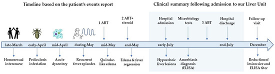
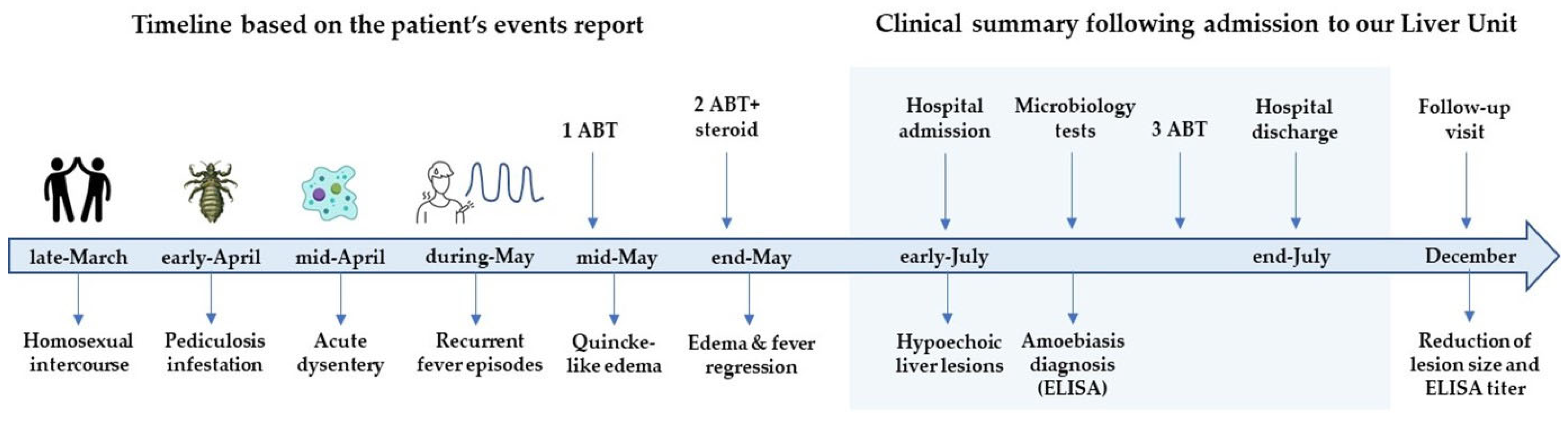
Returning to some considerations regarding the clinical case, we believe that the time-sequence of events were crucial to solving this diagnostic puzzle (Figure 3).
Figure 3.
Chronology of clinical events as reported before and after hospital admission.
Given that pediculosis developed approximately two weeks after the presumed exposure and an episode of hemorrhagic dysentery occurred two weeks later, we investigated the possibility of a common risk factor. The temporal sequence and concomitant occurrence of ecto- and endo-parasitic infections raised the possibility of shared exposure, potentially suggesting sexual transmission. Nevertheless, this hypothesis cannot be confirmed, as the route of transmission remains uncertain, but highly probable.
Subsequently, the recurrent febrile state, characterized by episodes lasting 3–4 days alternating with remission phases of approximately one week—likely mimicked a form of RF. B. recurrentis, transmitted by lice, and B. miyamotoi, carried by ticks of the Ixodes genus—might have been the most likely causative agents of RF in this case. Unfortunately, it was not possible to diagnose the infection using serological methods. However, the etiological diagnosis of RF—based on a Borrelia spp.-specific CLIA test—might have been influenced by either false-positive results (due to other spirochetal infections, including LB) or considering that the patient had already recovered from the infection following prior specific antibiotic therapy. We are confident in the future availability of specific and sensitive diagnostic tools for RF causative agents by novel immunoassays, which are based on the N-terminal CihC and GlpQ fragment antigens that are highly specific to B. recurrentis infection [35], to overcome the difficulties of performing specific “in vivo” methods to detect B. recurrentis [8].
Furthermore, B. burgdorferi infection—the most widespread tick-borne disease endemic to northern Italy [36,37]—has not yet been isolated in body lice. The clinical manifestations of Lyme disease do not allow for an easy diagnosis of the infection, as it is a condition characterized by low-level bacteremia and more insidious, prolonged symptoms. Conversely, it is typically marked by a “bull’s-eye” rash or erythema migrans (EM) [38]—a sign never reported in this patient. However, occasional cases of viral-like febrile illness without EM have been attributed to LB, especially after spontaneous resolution of an unrecognized asymptomatic EM [39].
In conclusion, the patient could have experienced a typical episode of RF, probably related to Borrelia spp. infection, as suggested only by clinical features, temporal sequence of events, and the subsequent onset of a typical Jarisch–Herxheimer (JH) reaction [40]. This type of angioedema has been associated with the release of spirochete toxins and cellular cytokines, resulting in swelling of the tissues and mucous membranes, typically without the pruritus characteristic of Quincke-type angioedema. In this case, the reaction occurred shortly after the initiation of antibiotic therapy, consistent with the bactericidal activity, which typically begins within approximately 8–12 h after the dose intake.
4. Conclusions
In conclusion, we described a rare case of concurrent borreliosis and invasive amebiasis, presumably sexually acquired, presenting with hepatic abscesses. Such infections may reach non-endemic regions through population migration from areas where these pathogens are prevalent. Clinicians and general practitioners should be aware that fecal–oral pathogens may also be transmitted during sexual practices, including heterosexual and/or homosexual encounters, and may therefore present as sexually associated infections. Targeted screening of individuals who are at increased risk may help to identify and treat asymptomatic carriers and prevent further transmission.
The diagnosis and etiological characterization of Borrelia spp.-associated RF may be challenging. Diagnostic difficulties arise from serological cross-reactivity among Borrelia species, which may complicate the identification of the causative pathogen, as well as from the ability of B. recurrentis to undergo antigenic variation in its surface proteins, allowing immune evasion. This phenomenon contributes to the characteristic relapsing clinical pattern observed in infected individuals.
5. Key Points
- ✓
- Effective antiparasitic therapy can rapidly eliminate E. histolytica trophozoites from infected tissues and eradicate cystic forms from the intestinal lumen.
- ✓
- Acute E. histolytica infection typically presents with bloody diarrhea 2–3 weeks after infection, lasting 2–3 weeks, often accompanied by abdominal pain, flatulence, tenesmus, mild-to-moderate fever, and night sweats.
- ✓
- Painful hepatomegaly, elevated ALT levels, and mild hyperbilirubinemia are typical findings of chronic, relapsing extraintestinal amebiasis.
- ✓
- Diagnosis relies on imaging evidence of a hypodense focal liver lesion on ultrasound, CT, or MRI, supported by detection of E. histolytica-specific antigen or DNA in stool and/or positive serum antibodies.
- ✓
- Body lice may transmit B. recurrentis, causing RF and, rarely, a Jarisch–Herxheimer reaction shortly after initiation of antibiotic therapy.
- ✓
- B. recurrentis is diagnosed by identification of the spirochete in thick blood smears and, when necessary, confirmed by species-specific molecular methods.
Author Contributions
Conceptualization, L.C. (Liliana Chemello); Methodology and resources, L.C. (Luisa Cavalletto), S.V., C.G., C.M., V.M.B. and L.C. (Liliana Chemello); Data curation, L.C. (Liliana Chemello); Writing—original draft preparation, L.C. (Luisa Cavalletto), S.V. and L.C. (Liliana Chemello); Writing—review and editing, L.C. (Liliana Chemello), L.C. (Luisa Cavalletto), S.V., C.G., C.M. and V.M.B.; Visualization, L.C. (Liliana Chemello); Supervision, L.C. (Luisa Cavalletto), S.V., C.G., C.M., V.M.B. and L.C. (Liliana Chemello). All authors have read and agreed to the published version of the manuscript.
Funding
This research received no external funding.
Institutional Review Board Statement
Ethical review and approval were waived for this study due to the retrospective nature of the publication.
Informed Consent Statement
At hospital admission, the patient provided informed consent for the anonymous presentation and publication of his clinical case for scientific and educational purposes. The case was fully de-identified in accordance with current privacy legislation.
Data Availability Statement
The data presented in this study are available from the corresponding author upon reasonable request, in accordance with the General Data Protection Regulation (GDPR).
Acknowledgments
The authors used ChatGPT (GPT-5, OpenAI, San Francisco, CA, USA) for linguistic editing.
Conflicts of Interest
The authors declare no conflicts of interest.
Abbreviations
The following abbreviations are used in this manuscript:
| ABT | Antibiotic therapy |
| AP | Alkaline phosphatase |
| ALT | Alanine amino-transferase |
| APHE | Arterial-phase hyperenhancement |
| AST | Aspartate amino-transferase |
| C1, C2, C4 | Complement C1, C2, C4 components |
| CLIA | Chemiluminescence assay |
| CPR | C protein reactive |
| CT | Computer tomography scan |
| DWI | Diffusion-weighted images |
| ELISA | Enzyme-linked immunosorbent assay |
| EM | Erythema migrans |
| GGT | Gamma-glutamil transferase |
| GP | General practitioner |
| HB | Hemoglobin |
| HBV | Human hepatitis B virus |
| HCC | Hepatocellular carcinoma |
| HCV | Human hepatitis c virus |
| HIV | Human immunodeficiency virus |
| IHC | Immunohistochemical |
| JH | Jarisch Herxheimer reaction |
| LB | Lyme borreliosis |
| LDH | Lattic dehydrogenase |
| LIA | Line-recombinant immunoassay |
| MR | Magnetic resonance |
| NAAT | Nucleic acid amplification test |
| PCR | Polymerase chain reaction |
| PLTS | Platelets count |
| RBC | Red blood count |
| RF | Relapsing fever |
| RT | Reverse transcriptasi |
| spp. | Species |
| STE | Sexually transmitted enteric disease |
| TID | Two intakes daily |
| US | Ultrasound examination |
| WBC | White blood count |
References
- Stanley, S.L., Jr. Amoebiasis (Seminar). Lancet 2003, 361, 1025–1034. [Google Scholar] [CrossRef] [Scilit] [PubMed]
- Petri, W.A., Jr.; Haque, R.; Lyerly, D.; Vines, R. Estimating the impact of amebiasis on health. Parasitol. Today 2000, 16, 320–321. [Google Scholar] [CrossRef] [Scilit] [PubMed]
- Kawashima, A.; Yanagawa, Y.; Shimogawara, R.; Yagita, K.; Gatanaga, H.; Watanabe, K. Amebiasis as a sexually transmitted infection: A re-emerging health problem in developed countries. Glob. Health Med. 2023, 5, 319–327. [Google Scholar] [CrossRef] [Scilit] [PubMed]
- Billet, A.C.; Salmon, R.A.; Piroth, L.; Martins, C. An underestimated sexually transmitted infection: Amoebiasis. BMJ Case Rep. 2019, 12, e228942. [Google Scholar] [CrossRef] [Scilit] [PubMed]
- Lapi, F.; Marconi, E.; Ferroglio, E.; Cricelli, I.; Rossi, A.; Cricelli, C. The burden of some of the most common zoonoses in primary care: A population-based study in Italy. Postgrad. Med. J. 2025, 101, 1280–1285. [Google Scholar] [CrossRef] [Scilit] [PubMed]
- Fu, T.-Y.; Yao, C.; Deng, Y.P.; Elsheikha, H.M.; Shao, R.; Zhu, X.Q.; Liu, G.H. Human pediculosis, a global public health problem. Infect. Dis. Poverty 2022, 11, 58–64. [Google Scholar] [CrossRef] [Scilit] [PubMed]
- Olivieri, E.; Rigamonti, S.; Vicari, N.; Grassi, A.; Karaman, I.; Bertoletti, I.; Bianchi, A.; Lodola, C.M.; Polonini, G.; Maioli, G.; et al. Borrelia miyamotoi in Ixodes ricinus Ticks Removed from Hosts in Northern Italy. Vector Borne Zoonotic Dis. 2026, 26, 173–175. [Google Scholar] [CrossRef] [Scilit] [PubMed]
- Takeuchi, T.; Yasuhiro Gotoh, Y.; Hayashi, T.; Kawabata, H.; Takano, A. Antigenic variation is caused by long plasmid segment conversion in a hard tick-borne relapsing fever Borrelia miyamotoi. PLoS Pathog. 2025, 21, e1013514. [Google Scholar] [CrossRef] [Scilit] [PubMed]
- Barbieri, R.; Drancourt, M.; Raoult, D. The role of louse-transmitted diseases in historical plague pandemics. Lancet Infect. Dis. 2021, 21, e17–e25. [Google Scholar] [CrossRef] [Scilit] [PubMed]
- Tran, T.-N.-N.; Signoli, M.; Fozzati, L.; Aboudharam, G.; Raoult, D.; Drancourt, M. High throughput, multiplexed pathogen detection authenticates plague waves in medieval Venice, Italy. PLoS ONE 2011, 6, e16735. [Google Scholar] [CrossRef] [Scilit] [PubMed]
- Kahlig, P.; Paris, D.H.; Neumayr, A. Louse-borne relapsing fever—A systematic review and analysis of the literature: Part 1—Epidemiology and diagnostic aspects. PLoS Negl. Trop. Dis. 2021, 15, e0008564. [Google Scholar] [CrossRef] [Scilit] [PubMed]
- Talagrand-Reboul, E.; Boyer, P.H.; Bergström, S.; Vial, L.; Boulanger, N. Relapsing Fevers: Neglected Tick-Borne Diseases. Front. Cell. Infect. Microbiol. 2018, 8, 98. [Google Scholar] [CrossRef] [Scilit] [PubMed]
- Gialanella, J.P.; Tyler Steidl, T.; Korpela, K.; Grandhi, M.S.; Langan, R.C.; Alexander, H.R.; Hudacko, R.M.; Ecker, B.L. Hepatic pseudotumor associated with Strongyloides infection: A case report. World J. Hepatol. 2023, 15, 1338–1343. [Google Scholar] [CrossRef] [Scilit] [PubMed]
- Tao, Y.; Wang, Y.F.; Wang, J.; Long, S.; Seyler, B.C.; Zhong, X.-F.; Lu, Q. Pictorial review of hepatic echinococcosis: Ultrasound imaging and differential diagnosis. World J. Gastroenterol. 2024, 30, 4115–4131. [Google Scholar] [CrossRef] [Scilit] [PubMed]
- Weinke, T.; Friedrich-Jaenicke, B.; Hopp, P.; Janitschke, K. Prevalence and clinical importance of Entamoeba histolytica in two high-risk groups: Travelers returning from the tropics and male homosexuals. J. Infect. Dis. 1990, 161, 1029–1031. [Google Scholar] [CrossRef] [Scilit] [PubMed]
- Guillén, N. Pathogenicity and virulence of Entamoeba histolytica, the agent of amoebiasis. Virulence 2023, 14, 2158656. [Google Scholar] [CrossRef] [Scilit] [PubMed]
- Pillai, D.R.; Keystone, J.S.; Sheppard, D.C.; MacLean, J.D.; MacPherson, D.W.; Kain, K.C. Entamoeba histolytica and Entamoeba dispar: Epidemiology and comparison of diagnostic methods in a setting of non-endemicity. Clin. Infect. Dis. 1999, 29, 1315–1318. [Google Scholar] [CrossRef] [Scilit] [PubMed]
- Ali, I.K.M.; Hossain, M.B.; Roy, S.; Ayeh-Kumi, P.F.; Petri, W.A.; Haque, R.; Clark, C.G. Entamoeba moshkovskii infections in children in Bangladesh. Emerg. Infect. Dis. 2003, 9, 580–584. [Google Scholar] [CrossRef] [Scilit] [PubMed]
- Blessmann, J.; Van Linh, P.; Nu, P.A.T.; Thi, H.D.; Muller-Myhsok, B.; Buss, H.; Tannich, E. Epidemiology of amebiasis in a region of high incidence of amebic liver abscess in central Vietnam. Am. J. Trop. Med. Hyg. 2002, 66, 578–583. [Google Scholar] [CrossRef] [Scilit] [PubMed]
- Jackson-Akers, J.Y.; Prakash, V.; Goosenberg, E.; Oliver, T.I. Amebic liver abscess. In StatPearls [Internet]; StatPearls Publishing: Treasure Island, FL, USA, 2025. [Google Scholar] [PubMed]
- Haque, R.; Mollah, N.U.; Ali, I.K.M.; Alam, K.; Eubanks, A.; Lyerly, D.; Petri, W.A. Diagnosis of amebic liver abscess and intestinal infection with the TechLab Entamoeba histolytica II antigen detection and antibody tests. J. Clin. Microbiol. 2000, 38, 3235–3239. [Google Scholar] [CrossRef] [Scilit] [PubMed]
- Pham, V.L.; Duong, M.H.; Pham, N.H. Amebic abscess of the liver: Ultrasound guided puncture. Ann. Chir. 1996, 50, 340–343. [Google Scholar] [PubMed]
- Ghosh, S.; Frisardi, M.; Ramirez-Avila, L.; Descoteaux, S.; Sturm-Ramirez, K.; Newton-Sanchez, O.A.; Santos-Preciado, J.I.; Ganguly, C.; Lohia, A.; Reed, S.; et al. Molecular epidemiology of Entamoeba spp.: Evidence of a bottleneck (demographic sweep) and transcontinental spread of diploid parasites. J. Clin. Microbiol. 2000, 38, 3815–3821. [Google Scholar] [CrossRef] [Scilit] [PubMed]
- Acuna, R.; Maguire, J.H.; Wirth, D.F. Gender distribution in asymptomatic and invasive amebiasis. Am. J. Gastroenterol. 2000, 95, 1277–1283. [Google Scholar] [CrossRef] [PubMed]
- Haque, R.; Duggal, P.; Ali, I.M.; Hossain, M.B.; Mondal, D.; Sack, R.B.; Farr, B.M.; Beaty, T.H.; Petri, J.W.A. Innate and acquired resistance to amebiasis in Bangladeshi children. J. Infect. Dis. 2002, 186, 547–552. [Google Scholar] [CrossRef] [Scilit] [PubMed]
- Santi-Rocca, J.; Rigothier, M.C. Host-Microbe Interactions and Defense Mechanisms in the Development of Amoebic Liver Abscesses. Clin. Microbiol. Rev. 2009, 22, 65–75. [Google Scholar] [CrossRef] [Scilit] [PubMed]
- World Health Organization (WHO). Entoamoeba Histolytica 2025. Available online: https://iris.who.int/server/api/core/bitstreams/8dbcbdf5-ef4a-481a-b712-41679524e70d/content (accessed on 12 July 2025).
- Tian, Y.D.; Hao, D.; Du, J.; Wang, H.; Wen, X. Summary of clinical features of 1800 cases of pyogenic liver abscess. Eur. J. Gastroenterol. Hepatol. 2024, 37, 483–487. [Google Scholar] [CrossRef] [Scilit] [PubMed]
- Rahmatullah, Z.F.; Arshad, H.; Chu, L.C.; Kawamoto, S.; Fishman, E.K. Hepatic abscess on CT: Pearls, pitfalls and diagnostic challenges. Emerg. Radiol. 2026, 33, 234–245. [Google Scholar] [CrossRef] [Scilit] [PubMed]
- Blessmann, J.; Tannich, E. Treatment of asymptomatic intestinal Entamoeba histolytica infection. N. Engl. J. Med. 2002, 347, 1384. [Google Scholar] [CrossRef] [Scilit] [PubMed]
- Visser, L.G.; Verweij, J.J.; Van Esbroeck, M.; Edeling, W.M.; Clerinx, J.; Polderman, A.M. Diagnostic methods for differentiation of Entamoeba histolytica and Entamoeba dispar in carriers: Performance and clinical implications in a non-endemic setting. Int. J. Med. Microbiol. 2006, 296, 397–403. [Google Scholar] [CrossRef] [Scilit] [PubMed]
- Acheson, M.E.; Garrity, R.E.; Bussey-Jones, J.; Garrity, R. Unusual case of hepatic amebiasis without recent endemic exposure: A diagnostic puzzle. Cureus 2025, 17, e94107. [Google Scholar] [CrossRef] [Scilit] [PubMed]
- Farraj, K.; Diaz-Marty, C.; Lannom, M.; Delshad, J.; Koumas, P.; Smielewski, M.; Schneider, Z.; Verley, J.; Rankov, L. A case of Entamoeba histolytica infection among men who have sex with men. J. Investig. Med. High Impact Case Rep. 2022, 10, 23247096221078711. [Google Scholar] [CrossRef] [Scilit] [PubMed]
- Sofia, R.; de Valdoleiros, S.R.; Carvalho, J.A.; Gonçalves, C.; Vasconcelos, O. Nontravel-related invasive Entamoeba histolytica infection with probable heterosexual transmission. IDCases 2019, 18, e00592. [Google Scholar] [CrossRef] [Scilit] [PubMed]
- Röttgerding, F.; Njeru, J.; Schlüfter, E.; Latz, A.; Mahdavi, R.; Steinhoff, U.; Cutler, S.J.; Besier, S.; Kempf, V.A.J.; Fingerle, V.; et al. Novel approaches for the serodiagnosis of louse-borne relapsing fever. Front. Cell. Infect. Microbiol. 2020, 12, 983770. [Google Scholar] [CrossRef] [Scilit] [PubMed]
- Ciceroni, L.; Ciarrocchi, S.; Ciervo, A.; Mondarini, V.; Guzzo, F.; Caruso, G.; Murgia, R.; Cinco, M. Isolation and characterization of Borrelia burgdorferi sensu lato strains in an area of Italy endemic for Lyme Borreliosis. J. Clin. Microbiol. 2001, 39, 2254–2260. [Google Scholar] [CrossRef] [Scilit] [PubMed]
- Colomba, C.; Scarlata, F.; Di Carlo, P.; Giammanco, A.; Fasciana, T.; Trizzino, M.; Cascio, A. Fourth case of louse-borne relapsing fever in young migrant, Sicily, Italy, December 2015. Mini Review Article. Public Health 2016, 139, 22–26. [Google Scholar] [CrossRef] [Scilit] [PubMed]
- Feder, H.M., Jr.; Abeles, M.; Bernstein, M.; Whitaker-Worth, D.; Grant-Kels, J.M. Diagnosis, treatment, and prognosis of erythema migrans and Lyme arthritis. Clin. Dermatol. 2006, 24, 509–520. [Google Scholar] [CrossRef] [Scilit] [PubMed]
- Vržogić, P.; Perica, A. Skin Changes in Suspected Lyme Disease. Acta Dermatovenerol. Croat. 2023, 31, 225–228. [Google Scholar] [PubMed]
- Kahlig, P.; Neumayr, A.; Paris, D.H. Louse-borne relapsing fever—A systematic review and analysis of the literature: Part 2—Mortality, jarisch-herxheimer reaction, impact on pregnancy. PLoS Negl. Trop. Dis. 2021, 15, e0008656. [Google Scholar] [CrossRef] [Scilit] [PubMed]

Disclaimer/Publisher’s Note: The statements, opinions and data contained in all publications are solely those of the individual author(s) and contributor(s) and not of MDPI and/or the editor(s). MDPI and/or the editor(s) disclaim responsibility for any injury to people or property resulting from any ideas, methods, instructions or products referred to in the content. |
© 2026 by the authors. Licensee MDPI, Basel, Switzerland. This article is an open access article distributed under the terms and conditions of the Creative Commons Attribution (CC BY) license.