


Abstract
Objective: Hepatitis B virus (HBV) is a globally distributed infectious disease affecting the liver. This literature review aims to summarize all available relevant information on the PubMed database about HBV’s connection to the microbiome and to consider possible treatment adjuncts. Materials and methods: Database used: PubMed. Keywords used: “HBV”, “Hepatitis B”, “microbiome”. In the PubMed database, 179 research publications were identified using these keywords; 69 studies were excluded as they were irrelevant or retracted. Of the remaining, 110 were analyzed in this literature review, and four additional literature sources were used to supply background information and context. Information was summarized. The analysed studies in total included 14,814 participants (excluding animal studies), of whom 8564 were HBV-infected individuals. Results: Results characterizing abundance or decrease in specific bacterial, viral, and fungal species are heterogeneous; multiple studies support that the HBV patient oral and fecal microbiome is different from that in healthy controls (HCs) and varies throughout disease progression. The HBV seems to transform the microbiome negatively, leading to dysbiosis and decreased microbial diversity in most studies. Evidence links HBV microbiome changes with influence on HbeAg seroconversion, HBV-DNA load, metabolic pathways, liver cirrhosis, and hepatocellular carcinoma. The research proposes that members of microbiota could potentially promote or protect against liver injury in HBV. Four studies proposed that the plasma virome in HBV patients was primarily composed of members of the Anelloviridae. One study researched a parasite (Entamoeba gingivalis) in HBV patients. Two studies analyzed HBV patients’ fungal profiles. Conclusions: Microbiota research, although promising, at the present moment is heterogeneous. HBV patients’ microbiota is distinguishable from HCs, and multiple studies have tried to identify the HBV characteristic microbiome; however, more precise information is needed to draw conclusions. Fecal microbiota transplantation and probiotics have the potential to be therapy adjuncts for HBV patients, but more research is needed.
1. Introduction
Hepatitis B virus (HBV) is a globally distributed infectious disease affecting the liver. The WHO estimates that more than 254 million people were living with chronic hepatitis B infection in 2022, with 1.2 million new infections each year [1]. In 2022, HBV resulted in an estimated 1.1 million deaths, mostly from cirrhosis and hepatocellular carcinoma [1]. This literature review aims to find available information about HBV’s connection to the microbiome and to consider possible treatment directions.
2. HBV Microbiome Function
The microbiome is a functionally active organic entity that has been linked to the formation of different diseases and conditions; however, it can also potentially have a protective role. Overall results characterizing abundance or decrease in specific bacterial, viral, and fungal species are quite heterogeneous; however, evidence supports that the HBV patient oral and fecal microbiome is different from that in the HC and varies throughout disease progression [2,3,4,5,6,7,8,9,10,11,12,13]. Most studies included support lower microbial diversity in HBV patients; however, two studies propose higher diversity or more pronounced microbial richness in HBV patients compared to HCs [12,14]. Two studies found no difference comparing the HBV microbiome to HC ([15] (oral microbiome); [16] fecal microbiome).
Growing evidence supports that in multiple HBV-associated liver conditions, microbiota affect different substance metabolic mechanisms and therefore metabolites [5,14,17,18,19,20,21]. Nevertheless, gut microbiota metabolites without a causal effect on chronic HBV were identified in one publication [22]. In HBV, the intestinal microbial community may act as an independent organ to regulate the body’s metabolic balance, which may affect the organism and further disease progression.
In chronic HBV (CHB) patients, when compared to healthy individuals, analysis of gut microbiota reveals that lipopolysaccharide, glycan biosynthesis, lipid, and butanoate metabolism are significantly elevated [5,23]. The host–microbiota–metabolite interplay, amino acid metabolism, nicotinate and nicotinamide metabolism, starch and sucrose metabolism, steroid biosynthesis, and vitamin metabolism were found to be significantly lower in CHB patients than in HCs [24]. The fecal microbiota of the HBV-liver cirrhosis (HBV-LC) patients show enrichment in the metabolism of glutathione, gluconeogenesis, branched-chain amino acids, nitrogen, and lipids, whereas there was a decrease in the level of aromatic amino acids, bile acids, and cell cycle-related metabolism [17]. In liver cirrhosis hepatocellular carcinoma (LC-HCC) patients, the butyrate-producing genera are found to be decreased, while the genera producing lipopolysaccharides (LPSs) increased [4]. In HBV-acute-on-chronic liver failure (HBV-ACLF) patients, alterations in circulating microbiota are also associated with multiple metabolic changes and systemic inflammation, which can affect clinical outcomes [10,20].
3. HBV Treatment Effect on Microbiota
Not all patients with HBV infection require or receive treatment; the effect of medication composition on the microbiota in the HBV treatment patient group has been explored. Multiple studies focused on HBV medication in relation to the microbiota. The available evidence suggests that long-term treatment (mentioned treatment options—tenofovir dipivoxil (TDF), tenofovir alafenamide (TAF), entecavir (ETV) combined with Biejiajian Pill (BJJP), ETV mono-therapy) could help restore a more balanced microbiota in HBV patients [21,25,26,27,28]. Clinacanthus nutans has also been used in HBV therapy, and in a murine model, C. nutans has been found to decrease the expression levels of HBsAg in the serum of mice [29].
4. HbeAg Seroconversion
Three fecal microbiota studies were identified regarding the HBeAg seroconversion-related microbial signature and prediction model for HBeAg seroconversion. HBeAg seroconversion (the loss of serum hepatitis B e antigen (HBeAg) and the development of anti-HBe antibodies) marks a transition from the immune-active (IA) phase of disease to the inactive carrier state. Results indicated that the those that reached seroconversion induced by oral antiviral therapy with tenofovir and entecavir tend to have a higher abundance of the Firmicutes phylum and a lower abundance of the Bacteroidetes phylum [30]. Interestingly, results also indicate that patients with a higher abundance of Prevotellaceae and genus Sutterella have fewer opportunities to obtain HBeAg seroconversion [30]. Akkermansia muciniphila has also been found to be enriched in immune-active phase patients and associated with HBeAg loss and early HBe-seroconversion [31].
However, oral microbiome compositions were not found to be significantly different in HBeAg-negative patients compared to the HCs who were vaccinated against HBV [15].
5. HBV DNA Load and Functional Cure
HBV DNA load is one of the factors that are taken into consideration when a decision of treatment is discussed. Research has compared high and low HBV DNA loads (>2000 IU/mL versus <2000 IU/mL) against HC. Alloprevotella and Eubacterium coprostanoligenes were found to be dominant in the low viral load group compared to the controls [14]. The gut microbiome in HBV-infected individuals with a low viral load was found to be highly diverse [14]. Serum HBV DNA has also been shown to be negatively correlated with fecal Lactobacillus counts [32].
In traditional Chinese medicine, tongue coating is important for clinical syndrome differentiation and drug choice; however, this method lacks clear scientific basis. An interesting relationship between tongue coating and HBV-DNA has been described in CHB patients, showing that the yellow tongue coating patients had higher HBV-DNA titers compared with the CHB white tongue coating patients (median 21,210 vs. 500, respectively), and a significantly lower level of Bacteroidetes, but a higher level of Proteobacteria at the phylum level [33]. It was also noted that the abundance of Neisseriaceae in the yellow tongue patients additionally positively correlated with the HBV-DNA level [33]. However, more reproducible research would be needed to draw precise conclusions about the connection between HBV DNA load and the tongue coating microbiome.
Functional cure (FC) has also been studied. Functional HBV cure is defined as undetectable HBsAg and unquantifiable serum HBV DNA for at least 24 weeks after a finite course of therapy. The gut microbiota of patients who achieved FC has been assessed and compared with that of patients with high-titer HBV DNA or low-titer HBV DNA. There was no difference found in the alpha or beta diversity of the gut microbiota between the FC group and the other groups [34]. However, the FC group presented a greater relative abundance of bacteria that produce short-chain fatty acids (SCFAs), especially butyrate, and the findings suggested that butyrate-producing bacteria contribute to FC, possibly through butyrate-mediated inhibition of HBV production [34].
6. ALT and AST Levels
Ten studies analyzed serum alanine aminotransferase (ALT), aspartate aminotransferase (AST) levels, and their possible connection to the microbiome; the results were heterogeneous [3,5,10,11,21,31,35,36,37,38]. Overall, the research supports a connection between different bacterial abundance and levels of ALT and AST.
Evidence suggests the following:
- (1)
- Decrease in Turicibacter in HBV-LC was associated with increased AST [21];
- (2)
- Significant decreases in Akkermaniacaee, Muribaculateae, (Eubacterium) Coprostanoligenes group, RF39 negatively correlate with indicators of AST, ALT [5];
- (3)
- Decreases in Lactobacillus, Clostridium, and Bifidobacterium were found in HBV patients with normal ALT when compared to [35] HCs; additionally, these patients were found to have more Anaerostipes in their gut [37];
- (4)
- Decrease in Lactobacillus has been associated with higher ALT in HBV patients [36];
- (5)
- Desulfovibrio and Megasphaera have shown positive correlations, while Acidaminococcus exhibits a negative correlation with high ALT levels in HBV patients [37];
- (6)
- The pro-inflammatory bacteria (Veillonella, Escherichia-Shigella) increased counts were found to be associated with ALT and AST elevations in HBV-LC patients [30];
- (7)
- Prevotella, Blautia, Ruminococcus, Eubacterium eligens group, Bacteroides uniformis, and Ruminococcus sp. 5_1_39BFAA have been associated with the critical biochemical indicators (ALT) and liver injury in CHB [38];
- (8)
- Akkermansia muciniphila was found to be predominantly enriched in IA patients and associated with ALT flares [31];
- (9)
- The ATL and AST levels in HBV-ACLF patients were found to be positively correlated with the abundance of Moryella and Fusobacterium, respectively [10,11].
7. Bile Acids in HBV
Bile acids have a function in maintaining the balance in the gut and regulating microbial growth. The specific role of bile acids (BAs) remains unclear in HBV patients; nevertheless, the virus causes hepatic inflammation and can alter bile acid metabolism. We found four studies regarding this aspect; nevertheless, they support a link with the microbiome [5,31,35,39]. Even without apparent liver injury, CHB patients have been linked to a significantly higher percentage of primary BAs and conjugated BAs [39]. Alteration of the gut microbiota and its function in bile acid homeostasis in CHB patients with different degrees of fibrosis has been explored. Analysis of gut microbiota exhibits a trend of decreased abundance in bacterial genera responsible for BA metabolism in patients with moderate/advanced fibrosis [39]. In HBV-IT patients, cholesterol-to-bile acid metabolism was found to be increased [31]. It has been proposed that R. gnavus facilitates HBV persistence and prolongs the IT phase, possibly through encoding bile salt hydrolase to deconjugate primary BAs, which further augments the total BAs pool [31]. In contrast, A. muciniphila counteracts this previously mentioned activity, possibly through the direct removal of cholesterol, which inhibits the growth and function of R. gnavus and promotes HBV elimination [31].
Analyzing HBV-HCC patients, the research has linked increased counts of Acinetobacter, Pantoea, Paenibacillus, and Pseudomonas with an increase in the total bile acids and triglycerides (TGs) [5].
8. HBV Clearance and Microbiota
In terms of microbiological factors regarding HBV clearance, research is proposing different mechanisms; for example, acute or chronic infection presentation and factors such as the age of infection, antibiotic use, cell receptors, and specific bacterial species, and nucleoside analogue therapy play a role. Research proposes probiotics as an adjunct to HBV therapy to promote HBV clearance, as probiotics have been found to accelerate serum HbsAg and HBV DNA decline in mice [40]. Evidence further points to age as being a strong factor for HBV clearance [41,42]. Research found that the sterilization of gut microbiota using antibiotics in younger mice prevented them from rapidly clearing HBV [41,43]. In murine experiments, antibiotic use can cause dysbiosis in mice and facilitate HBV persistence [27,43]. These discoveries indirectly support the connection between microbiota and HBV. Additionally, Toll-like receptors (TLRs) have been studied and could be the potential mechanism through which bacteria can influence liver inflammation. An alteration or a mutation in these receptors could lead to faster HBV clearance [41,44]. A murine study analyzed the role of Peyer’s patches (PPs) in gut microbiota-deficient mice and the HBV clearance context [45]. The authors concluded that depletion of gut microbiota impaired systemic adaptive immune responses, resulting in a delayed HBV antigen clearance [45]. In regard to specific bacterial involvement, Ruminococcus gnavus and Akkermansia muciniphila may play opposite roles in HBV infection [31]. A. muciniphila metabolites were found to benefit the elimination of HBV [31]. Evidence indicates that butyrate-producing bacteria as a group can contribute to FC in HBeAg-negative patients with CHB through butyrate-mediated inhibition of HBV production [34]. We additionally identified two experiments regarding nucleoside analogs (NAs) and HBV clearance [25,46]. When patients were characterized by their dominant genus, the Bacteroides dominant group was associated with virological undetectability; however, the Blautia dominant group was associated with fibrosis evolution during NAs therapy in chronic HBV [46]. Increased Escherichia coli relative abundance was found in on-treatment patients with advanced fibrosis despite HBV DNA undetectability [46]. However, when describing ETV’s effect on murine microbiota, it was observed that the abundance of Akkermansia was negatively correlated with HBV DNA load both in serum and the liver [25]. Antibiotics have been studied in regard to this topic in four studies [44,47,48,49]. Data presented that antibiotics could have a beneficial effect in certain cases where possibly translocated gut-derived microbial products promote liver injury [44]. However, further studies propose more negative effects of antibiotic use in HBV conditions. Antibiotic treatment has been demonstrated to be insufficient in altering levels of serological HBV antigens, intrahepatic HBV RNA transcripts, and HBc protein (mouse model), and has been found to contribute to HBsAg increase after breaking of IT [47]. Mice, when treated with broad-spectrum antibiotics, demonstrated gut microbiota depletion, which was further associated with impairment of colon epithelial integrity and live commensal gut microbiota translocation to the liver [48]. Furthermore, normal mice have been found to exhibit complete clearance of HBV within 6 weeks post-hydrodynamic injection (HDI) of HBV-containing plasmid, whereas multiple antibiotic treated mice have been documented to have lost this capacity, showing high serum levels of HBsAg without hepatitis B surface antibodies (anti-HBs) [49]. The authors of the research also concluded that each antibiotic alone could not significantly influence HBV clearance compared to the antibiotic combination, suggesting that global commensal microbial load is critical for promoting HBV clearance [49].
In regard to this topic, occult HBV infection (OBI) has also been studied. It is defined as the presence of HBV DNA in the liver (with detectable (usually very low HBV DNA < 200 IU/mL) or undetectable HBV DNA in the serum) of individuals testing HBsAg negative by currently available assays [50]. Interestingly, research found that the abundance of Faecalibacterium was significantly reduced in samples from OBI blood donors compared with those from healthy blood donors [51]. Compared with samples from HBV carriers, the samples from OBI blood donors had a significantly increased abundance of Subdoligranulum, which might stimulate immune activation, thus inhibiting HBV replication and contributing to the formation of occult infection [51].
9. HBV Patient Microbiome
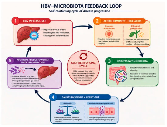
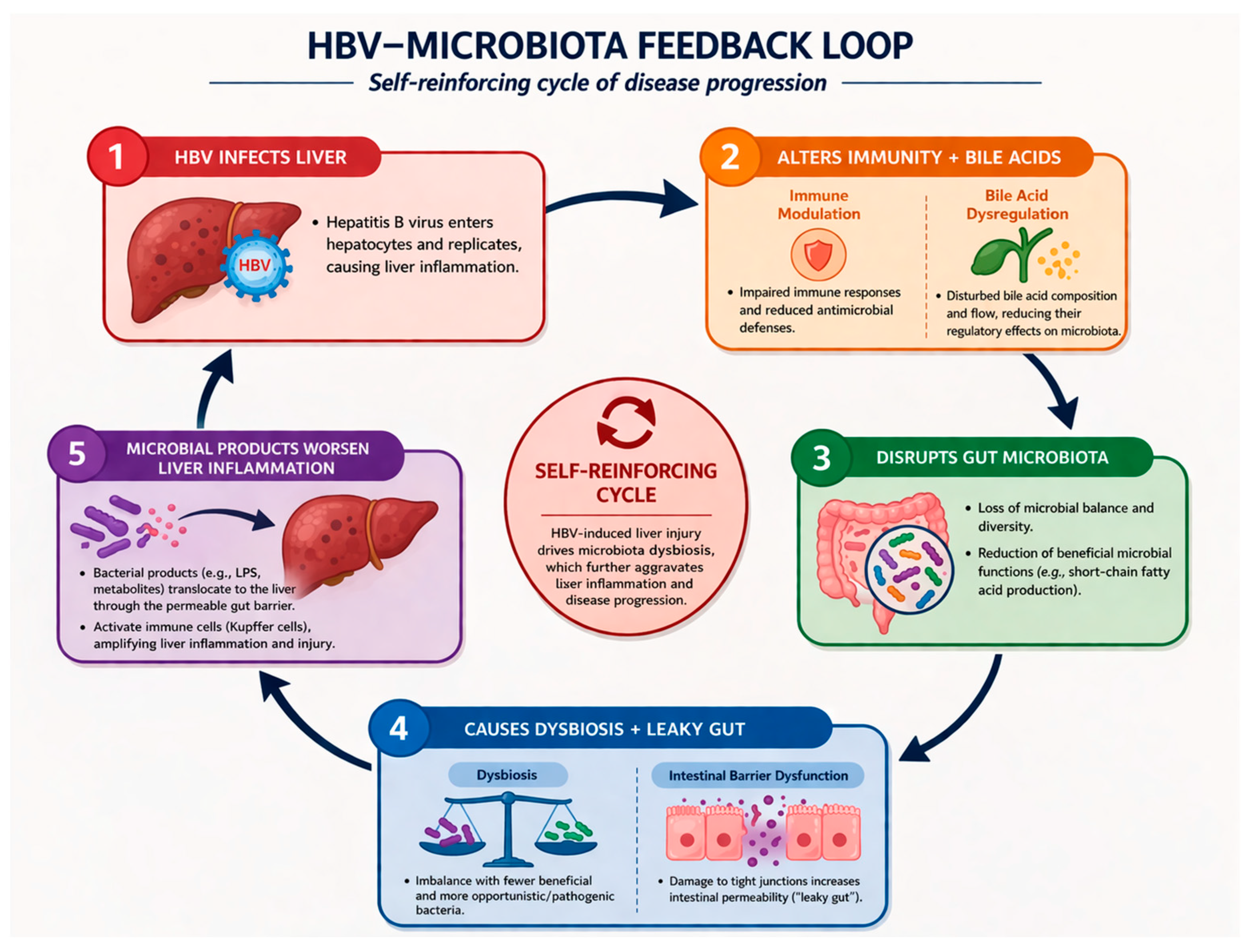
Multiple studies have analyzed, but have not yet reached a consensus in terms of a unified HBV characteristic microbiome signature. HBV potentially alters the microbiome and causes a self-reinforcing cycle of disease progression through bile acid metabolism, impairment of host immunity, chronic systemic and hepatic inflammation, and intestinal barrier disruption (Figure 1). The researchers conclude that community structures and diversity of the gut microbiota in CHB patients become more unbalanced and decrease [5,9,52,53]. The available literature suggests HBV as the driver behind the changes in microbiota [13,16,52]; however, microbiota could also potentially affect HBV clinical progression [54,55]. Interestingly, Bacteroidetes and Firmicutes were found to be decreased from healthy subjects, hepatitis B virus-infected, and CHB to HBV-LC patients, while Proteobacteria and Actinobacteria showed an increasing distribution [52]. Research additionally links some bacteria to an increased risk of developing CHB, such as an increase in Family XIII, genus Eggerthella group, genus Eubacterium ventriosum group, genus Holdemania, and Ruminococcus gauvreauii group [54]. Additionally, ethanol-producing Enterocloster bolteae was found to be enriched in CHB, which could also promote chronic inflammation [55]. Bacteria, however, believed to have a protective effect against CHB are noted—Family XIII AD3011 group, genus Prevotella 7, for instance [54].
Figure 1.
HBV-microbiota feedback loop.
In HBV patients overall, sources propose an abundance of the following:
- (1)
- Actinomycetales, Micromonosporaceae, Cryomorphaceae, and Prevotellaceae [6].
- (2)
- Fusobacteria, Veillonella, and Haemophilus [52].
- (3)
- Actinomyces, Clostridium sensu stricto, unclassified Lachnospiraceae, Megamonas [16].
- (4)
- Clostridia, such as Clostridium perfringens, Clostridium sporogenes, Enterocloster aldenensis, Enterocloster bolteae, Enterocloster clostridioformis, and Clostridium innocuum [55].
- (5)
- Bacteroidetes [23].
- (6)
- Lachnospiraceae [9]. (Haemophilus and Roseburia) [9].
- (7)
- At the genus level, Dialister, Eubacterium hallii group, Halomonas, Collinsella, Sphingomonas, Xanthomonadaceae unclassified, and Rhizobiaceae unclassified [26].
- (8)
- Fusobacterium, Filifactor, Eubacterium, Parvimonas, and Treponema, phylum level Firmicutes and Spirochaetes (saliva) [53].
- (9)
- Streptococcus, Blautia, Veillonella, Fusobacterium, and Akkermansia [12].
- (10)
- Rikenellaceae, Lachnospiraceae, Barnesiellaceae, Megamonas, Alistipes, Erysipelatoclostridium [24]. Alloprevotella, Paraprevotella, and Hungatella [14].
- (11)
- Fecalibacterium, Streptococcus, Sutterella, Lachnospiraceae ND-3007, Ruminiclostridium 9, Lachnospiraceae UCG-010 [21].
- (12)
- Lautropia, Abiotrophia, and Veillonella were enriched in HBV patients’ saliva [56].
After our analysis, the most dominant phyla of bacteria in the HBV patient microbiome overall seem to be Bacillota (Firmicutes), followed by Bacteroidota—this discovery also corresponds to “normal” gut microbiome structure; however, it is followed by Pseudomonadota (Proteobacteria), which is associated with inflammation, dysbiosis, and contains pathobionts.
In sources, the HBV-LC group had a higher abundance of the following:
- (1)
- Veillonella, Megasphaera, Dialister, Atopobium, and Prevotella [57].
- (2)
- Blautia_A (e.g., B. wexlerae, B. massiliensis, and B. obeum), Dorea (e.g., D. longicatena and D. formicigenerans), Streptococcus, Erysipelatoclostridium [58].
- (3)
- Bacteroidetes [23].
- (4)
- The pro-inflammatory bacteria (Veillonella, Escherichia-Shigella) [3].
- (5)
- Pasteurellaceae, Enterobacteriaceae, Campylobacteraceae, Streptococcaceae, and Leptotrichiaceae [52].
- (6)
- Lactobacillus [9].
- (7)
- Proteobacteria (patients with LC accompanied by ascites) [59].
- (8)
- Phyllobacterium, Sphingomonas, Enterococcus, Erysipelatoclostridium, and Romboutsia [4].
- (9)
- Escherichia-Shigella, Streptococcus [2].
- (10)
- Proteobacteria, Actinobacteria, and Fusobacteria. The family level of microbiota, such as Bacteroidaceae, Enterobacteriaceae, Sutterellaceae, Bifidobacteriaceae, Streptococcaee, and Pasteurellacaee [5].
- (11)
- Enterobacteriaceae (additionally, the abundance of Steptococcus and Ruminococcus was higher in patients with decompensated cirrhosis than in those with compensated cirrhosis) [60].
- (12)
- Bacteroidia, Streptococcaceae, Streptococcus, Veillonella, Bacteroidales, Lactobacillales, Pasteurellales, and Veillonella parvula [18].
- (13)
- Fusobacterium, Catonella, Eubacterium, Filifactor, and Yersinia (saliva) [53].
- (14)
- Veillonella [61].
- (15)
- Proteobacteria [17].
- (16)
- Treponema, Selenomonas, and Oribacterium (HBV-LC patients’ saliva) [56].
Interestingly, evidence also suggests that the HBV-related cirrhosis patient microbiome can be set apart from that of alcoholic cirrhosis [62].
The HBV-HCC group had a higher abundance of the following:
- (1)
- Bacteroidetes [23].
- (2)
- Holdemanell [9].
- (3)
- Escherichia-Shigella, Streptococcus [2].
- (4)
- Sarcina [4].
- (5)
- Proteobacteria, Actinobacteria, and Fusobacteria. The family level of microbiota, such as Bacteroidaceae, Enterobacteriaceae, Sutterellaceae, Bifidobacteriaceae, Streptococcaee, and Pasteurellacaee [5].
- (6)
- Blautia, Escherichia-Shigella, Bifidobacterium, Klebsiella, Parasutterella, E. hallii group, Collinsella, Erysipelotrichaceae UCG-003, Lactococcus [21].
- (7)
- Haemophilus, Porphyromonas, and Filifactor (in saliva) [56].
HBV-acute liver cirrhosis (HBV-ACLF) patients were found to be enriched in:
- (1)
- Moraxellaceae, Sulfurovum, Comamonas, and Burkholderiaceae. Additionally, the relative abundance of Prevotellaceae independently predicted 28-day mortality [20].
- (2)
- Veillonella, Streptococcus, Enterococcus, and Klebsiella (phylum level Firmicutes, Proteobacteria, Actinobacteria) [10].
- (3)
- Proteobacteria, Firmicutes, Bacteroidetes, Actinobacteria (genus level Bacteroides, Veillonella, and Streptococcus) [11].
- (4)
- Parabacteroides distasonis [20].
Interestingly, a high abundance of Enterococcus was associated with progression, while that of Faecalibacterium was associated with regression of HBV-ACLF [8].
10. Gut Microbiome and HBV-Related HCC
HBV infection is a major global cause of HBV-related hepatocellular carcinoma (HCC) [63]. There have been multiple studies proposing a connection between HBV-related HCC and the microbiome through different mechanisms.
The gut microbiome is a complex entity in close proximity to the liver, and the gut–liver axis has long been recognized. Gut microbiota produces excreted substances, such as SCFA, LPS, enzymes, vitamins, and other byproducts of bacteria, viruses, protozoa, fungi, and archaea, that travel to the liver through portal circulation and potentially can induce or suppress inflammation, carcinogenesis, which, combined with HBV presence in liver tissue, could help promote HCC development.
11. Gut Microbiota Signatures in Different Aspects and Disease Stages of HBV-HCC
Multiple studies in humans and murine models have looked at the microbiome and HBV connection to HCC risk, recurrence, and different HBV-HCC patient microbiome signatures according to age, stage of the disease, and other factors included.
We identified two studies that were conducted to identify a gut microbiota signature in differentiating between viral-related HCC (viral-HCC) and non-hepatitis B- and non-hepatitis C-related HCC (NBNC-HCC) [64,65]. The evidence supports that dysbiosis is connected to hepatocarcinogenesis and that the gut microbiota composition is significantly altered in different etiological factors of HCC. A microbiota-based signature has been found to distinguish between viral-HCC and NBNC-HCC [64,65]. Research has tried to predict the risk of HCC in HBV patients; the evidence proposes that gut microbes are important factors between HBV and HCC through potential mediating and moderating effects [66], and a similar link has been documented in mice [67]. Hepatectomies, for instance, were significantly associated with a gut microbial imbalance in HBV-HCC patients, and a significant elevation of Klebsiella abundance was observed in post-hepatectomy liver failure (PHLF) patients [68].
Multiple microbiota biomarkers are considered promising with the aim of providing clinicians useful tools against HCC—predicting occurrence [6,66,69] and recurrence of HCC [70], aid treatment strategy [71], predict liver failure after surgical intervention [71], moderate treatment response [72], serving as potential treatment targets [66,73], additional early diagnostic tools [69], and evidence links certain microbial genera to the size of HCC [4].
Evidence shows that the gut microbes Bacteroidia and Bacteroidales have been demonstrated to exert mediating effects between HBV and HCC; the moderating effects vary across Bacilli, Lactobacillales, Erysipelotrichaceae, Actinomyces, and Roseburia, and in general, HBV-gut microbe models could be established to predict HCC [66]. However, when noting HCC recurrence, Dialister, Veillonella, the Eubacterium coprostanoligenes group, Lactobacillus genus, the Streptococcus pneumoniae, and Bifidobacterium faecale species were associated with and may help predict early recurrence of HCC [70]. Gut microbial diversity has been found significantly different in studies in HC, patients with HBV and LC, and HCC compared to each other, and these microbiome characteristics could serve as potential diagnostic and treatment targets [4]. The evidence further indicates that the levels of inflammation, oxidative stress, and adiponectin are closely correlated with the progression of CHB to LC and HCC [6]. Biomarkers, such as Enterococcus, Limnobacter, and Phyllobacterium, could be used for precision diagnosis in HCC patients [4]. Research has also taken into consideration the changes in microbiota induced by HCC immunotherapy, specifically nivolumab [72]. Several taxa specific to therapeutic response have been designated as follows: Dialister pneumosintes, Escherichia coli, Lactobacillus reuteri, Streptococcus mutans, Enterococcus faecium, Streptococcus gordonii, Veillonella atypica, Granulicatella sp., and Trichuris trichiura for the non-responders to therapy; Citrobacter freundii, Azospirillum sp., and Enterococcus durans for the responders [72]. Interestingly, a skewed Firmicutes/Bacteroidetes ratio and a low Prevotella/Bacteroides ratio could serve as predictive markers of non-response, whereas the presence of Akkermansia species predicted a good response [72]. Additionally, early antibiotic exposure might have a negative effect on HCC disease outcome in patients treated with immune checkpoint inhibitors, but also in those treated with tyrosine kinase inhibitors and placebo [74]. However, more research is definitely needed in this field.
12. Bacteria Have Been Recognized as Potential Pathobionts or Protective Influences
Gut bacteria are capable of having positive (Table 2) or negative (Table 1) effects on health; this section highlights and presents in tables some of the identified specific bacteria and their potential proposed effects on liver health.
Table 1.
Bacteria That Are Considered to Be a Risk Factor for HBV-HCC.
We conclude that most identified sources that demonstrate a protective microbiological effect promote the bacteria that are part of the probiotic group as protective substances in HBV, HBV-LC and HCC groups (Table 2).
Table 2.
Bacteria That Are Considered to Have Potentially Protective Effects in HBV.
13. Bile Acids and Other Markers’ Role in HBV-HCC
We identified two studies that focused on bile acids in the gut–liver axis and HCC [83,84]. It was acknowledged that changes in the tumor immune microenvironment caused by the gut microbiota via serum bile acids may be important factors associated with tumor burden and adverse clinical outcomes [84]. There is evidence that higher concentrations of bile acids, specifically conjugated primary bile acids, are associated with increased HCC risk; however, the data do not support the hypothesis that higher levels of secondary bile acids increase liver cancer risk [83].
The role of other potential markers has additionally been explored, such as serum zonulin level (a gut protein that increases the permeability of tight junctions) [85]. It was found to be significantly increased both in HBV-LC and in HBV-HCC, and correlated with the advanced stage of LC and HCC [85]. Additionally, a combination of zonulin and AFP granted a significant benefit to diagnostic accuracy in differentiating LC from HCC [85]. Lastly, IL-2 was found to be highly associated with the whole gut bacterial communities of HCC and LC groups; it could be a potential marker for the future [3].
14. Intratumoral Microbiome
Emerging evidence supports a high prevalence of cancer type-specific microbiota residing within tumor tissues [2,86,87,88,89]. The intratumoral microbiome has been reported to regulate the development and progression of cancers [86]. The intratumoral microbiota may contribute to the promotion of the initiation and progression of cancers by DNA mutations, activating carcinogenic pathways, promoting chronic inflammation, the complement system, and initiating metastasis [90]. Studies have researched the intratumoral microbiome, its signature [87], possible prognosis prediction [87], early detection [2], and connection with immune cell infiltration in the tumor tissue. Research has aimed to characterize intratumoral microbial heterogeneity (IMH) and establish microbiome-based molecular subtyping of HBV-HCC to elucidate the correlation between IMH and HCC tumorigenesis [86]. Microbiome-based molecular subtyping demonstrated IMH of HBV-HCC that correlates with a disparity in clinical-pathologic features and tumor microenvironment (TME) [86]. In regard to microbial biomarkers in tumor and non-tumor samples, evidence has emerged that microbial biomarkers represented by Streptococcus displayed a constantly increasing trend during the disease transition [2]. However, Streptococcus is not the only bacterium associated with HBV-HCC, and the presence of several dominant microbiota species has been confirmed in hepatic tumor and non-tumor tissues, and this discovery could possibly aid in the early detection of HCC [2]. In viral hepatitis patients (HBV and hepatitis C), research has linked Bacteroidetes, Firmicutes, and Proteobacteria as the dominant phyla associated with HCC (genus level unclassified Bacteroides genus, Romboutsia, Lachnospiraceae, and Ruminococcus gnavus) [89]. Furthermore, in HBV-HCC, the intra-tumoral microbiota has been positively associated with increased tumor-infiltrating CD8+ T lymphocytes and two subtypes of myeloid-derived suppressor cells (MDSCs) [88]. This finding additionally indicates an inhibitory role of microbial species in antitumor immunity and the contribution to the liver tumor microenvironment [88].
15. Interesting Discoveries and Related Themes to HBV Microbiome
There are publications exploring beyond the previously discussed themes, which look at specific topics in relation to microbiota and HBV.
Two studies focus on the microbiota’s link to vaccination. Interestingly, Bifidobacterium abundance in early infancy was found to be linked to an increase in the protective efficacy of the HBV (and other) vaccine, possibly through enhanced immunologic memory [91]. However, prebiotics (specifically chicory long-chain inulin, a soluble dietary fiber extracted from chicory root (Cichorium intybus)) have been found to induce changes in microbiota composition, although this has not been proven to translate into an improved immune response to the HBV vaccine [92].
HBV patient microbiota in association with other coexisting conditions has been researched as well. Some examples are HBV and type 2 diabetes mellitus [93,94], sleep disturbances, hepatic encephalopathy [95], chemotherapy [96], liver transplantation [97], patients after transjugular intrahepatic portosystemic shunt (TIPS) placement [98], and patients with cirrhotic portal hypertension after undergoing a splenectomy and pericardial devascularization (SPD) [99].
In regard to HBV and steatotic liver disease, there has been identified a reduced risk of developing dyslipidemia and non-alcoholic fatty liver disease in HBV patients, which has been possibly linked to microbiota [14]. However, murine research, for instance, has found that apolipoprotein H-negative mice with persistent HBV replication display steatohepatitis and gut microbiota dysbiosis more often [100], a conclusion that underlines a potentially significant difference between human and animal research.
Researchers have also tried to analyze the predominant intestinal microbiome of asymptomatic adult carriers of HBV (as controls without any antibiotics) and liver transplantation recipients [97]. The results demonstrated that the predominant intestinal microbial diversity decreased during the perioperative period, and postoperative fecal profiles showed an increase in Bacteroides, Firmicutes, and antibiotic use could switch the balance towards opportunistic pathogens [97].
Evidence has also demonstrated a difference in HBV-LC patients who have diabetes [94]. Proteobacteria, Streptococcus, Escherichia-Shigella, and Lactobacillus were found to be increased, while the abundance of Bacteroidota, Bacteroides, Prevotella, Faecalibacterium, and Lachnospira decreased in the LC-diabetes mellitus (LCDM) patients compared with the HC [94].
Bacterial diversity has been researched and found to be decreased not only in HBV patients alone but also in patients with HBV coexisting with NAFLD and T2DM [93]. There was a significantly lower abundance of bacteria of Faecalibacterium, Gemmiger, and Clostridium XIVA genera, but a higher abundance of Megamonas and Phascolarctobacterium genera in the HBV+NAFD+T2DM group [93]. Therefore, the bacterial signatures could potentially be the “drivers” for the comorbidities; however, it is also possible that these and other comorbidities further shape the microbiome, since the distinct gut microbiome profile associated with HBV infection has a role in lipid metabolism and glucose metabolism in patients with coexistent NAFLD and T2DM [93].
Alterations in the gut microbiome and intestinal permeability in CHB patients with cirrhotic portal hypertension after undergoing a splenectomy and a pericardial devascularization (SPD) have also been investigated [99]. Patients had microbiome analysis before and after surgery (12 months later), and researchers documented that the microbiome diversity increased, and the bacterial composition came to a level similar to that of the HC [99]. This discovery promotes the theory that the microbiome could be successfully manipulated and reversed through surgical procedures that affect the gut–liver axis.
In HBV-LC patients, sleep disturbances and different degrees of hepatic encephalopathy (HE) are common issues. Interestingly, a study aimed to evaluate the association between sleep disturbances and altered gut microbiota in patients with minimal hepatic encephalopathy (MHE) caused by HBV-LC [95], and a similar study focused on HBV-LC HE patient microbiome differences compared to HBV-LC patients without HE [18]. The patient’s microbiome was found to have decreased diversity, and specific bacteria, both S. salivarius and Veillonella, were found to be associated with sleep disturbances in patients with MHE caused by HBV-LC [95], and in HBV-LC HE patients, Pasteurellales, Pasteurellaceae, Haemophilus, and Selenomonas were found to be significantly increased bacterial genera compared to other groups [18].
In the context of chemotherapy, a murine study aimed to identify the mechanisms of Paclitaxel (PTX)-induced HBV reactivation [96]. The study revealed that PTX directly causes immunosuppression and HBV replication stimulation, and PTX induced dysbiosis of gut microbiota, which could additionally promote HBV reactivation [96].
Lastly, gut microbiome alterations in patients with HBV-related portal hypertension (PH) after transjugular intrahepatic portosystemic shunt (TIPS) placement have been studied [98]. After TIPS placement, the following results were observed: (1) the abundance of Haemophilus and Eggerthella increased, whereas that of Anaerostipes, Dialister, Butyricicoccus, and Oscillospira declined in the HE group; (2) the richness of Eggerthella, Streptococcus, and Bilophila increased, whereas that of Roseburia and Ruminococcus decreased in the non-HE group; and (3) members from the pathogenic genus Morganella appeared in the HE group but not in the non-HE group [98]. Further, it has been concluded that intestinal microbiota-related synergism may predict the risk of HE following TIPS placement in patients with HBV-related PH [98].
16. Fungal Microbiota in HBV Patients
Fungal microbiota has also been studied; we identified two publications on this topic.
Evidence proposes that as the disease progresses, the fungal microbiome expands [101], possibly relating to antibacterial agent use and overall dysbiosis progression. Additionally, it was found that there was a higher richness of fungal species in patients with HBV-LC than in patients with CHB, and the latter was higher than that in HBV carriers and HC [101]. There has been little difference found in enteric fungal diversity between HBV carriers and HC [101].
A total of 16 different fungal species were identified in HBV-LF patients in a study, which, on the contrary, found that HC “exhibited a slightly higher level of gut fungal richness compared to HBV-LF patients” [58]. Fungi such as Malassezia spp. (e.g., M. japonica and M. sympodialis), Candida spp. (e.g., C. parapsilosis), and Mucor circinelloides were more abundant in HBV-LF patients, while Mucor irregularis, Phialophora verrucosa, Hortaea werneckii, and Aspergillus fumigatus were found to be decreased [58].
17. Parasitic Microbiota in HBV Patients
The parasitic microbiome in HBV has not been widely studied; however, one study has researched the parasitic oral microbiome in these patients. A study aimed to evaluate a potential link between the colonization of gingival crevices by Entamoeba gingivalis and HBV infection among gum disease [102]. A statistically highly significant correlation was found between the detection of E. gingivalis in saliva/gingival scrapings and gum disease in HBV patients compared to HC [102]. The presence of the amoeba was not related to the degree of gum disease, but to the HBV infection diagnosis [102].
18. Virome and HBV Patients
In terms of virome research in association with HBV, there were four studies that we identified [58,103,104,105]. The evidence proposed plasma virome in HBV patients was primarily composed of members of the Anelloviridae [103,104,105], followed by Flaviviridae, and Hepadnaviridae (HBV) families [104]. Research on whether the virome differs in HBV stages and patients is contradictory; there is evidence that the gut virome of HBV-LF patients is altered and enriched by Siphoviridae, Myoviridae, and Podoviridae viruses compared to HC [58]; however, there is research that proposes that the virome structure and dynamics do not correlate with the different stages of chronic HBV infection, nor with the administration of antiviral therapy [104].
Interestingly, in HBV liver transplant (LT) patients, the initiation of immunosuppression was found to induce a bloom of the Anelloviridae that can dominate the post-liver transplant plasma (post-LT) virome [105].
19. HBV and Fecal Microbiota Transplantation
Fecal microbiota transplantation (FMT) has shown promising results in severe alcoholic hepatitis [106] and other non-liver-associated diseases, such as recurrent Clostridium difficile colitis [107]. Data on FMT in HBV are limited; however, we found FMT studies in HBV infection susceptibility, outcome, HE, hepatic myelopathy, and a possible connection of microbiota, HBV, and dyslipidemia.
There was a small study done on the role of FMT in HBeAg-positive CHB in terms of its effect on HBeAg, HBsAg, and HBV DNA [108]. The results showed that in the FMT arm, 16.7% of patients had HBeAg clearance in comparison to none in the control-antiviral therapy arm; however, none of the patients in either arm had HBsAg loss, and FMT was tolerated well [108]. Research in mice has shown that reconstitution of the gut microbiota by FMT can alter the susceptibility to HBV infection and disease outcome in mice [109], and a study on dyslipidemia using HBV-infected/free human FMT on two groups of mice noticed that altered gut microbiota accompanied by HBV infection was associated with a robust increase in alpha diversity and butyrate producers, which resulted in a reduced level of triglycerides after FMT in the recipient mice group [110]. Research shows that liver function and clinical symptoms can be improved with FMT in HBV cirrhosis patients after transjugular intrahepatic portosystemic shunt (TIPS); additionally, the number of HE attacks can be decreased significantly [111]. In HBV-associated hepatic myelopathy, FMT can improve patient condition and muscle strength [112]. Interestingly, proalbuminemia has been found to significantly increase after FMT in CHB patients, combined with a significant decrease in the HBsAg [24]. Research also found that besides live bacteria, microRNAs (miRNAs) might also play a role in the development of HE, and transferring feces and fecal miRNAs from patients with HE to the recipient mice can aggravate thioacetamide-induced HE [113]. Even though evidence in available studies is leaning towards a modulatory, safe and positive effect on patient health in terms of FMT, more research is necessary to conclude a certain benefit and safety profile for FMT in HBV patients.
20. Discussion
Multiple studies have tried to identify the HBV characteristic microbiome; however, more precise information is needed. The issue could come from the fact that HBV patients have different disease forms; patients may have other factors influencing the microbiota, for example, excessive alcohol use, diet choices, steatohepatitis, drug-induced liver injury, comorbidities, surgical manipulations, and patients might be receiving HBV and other disease-targeted medications. Multiple human and murine studies have been included and reviewed in this PubMed literature review. As demonstrated, research directions are diverse, often conclusions include bacteria at different ranks of taxonomy, which makes robust, accurate data analysis far more challenging and almost impossible. In terms of HBV clearance, HBeAg seroconversion, metabolic changes, influence of HBV and other medications on microbiota, more research is needed to draw significant conclusions about the possible ways the microbiome could affect these subjects. Probiotics and FMT could potentially be positive influences on HBV patient health; however, they theoretically come with their individual risks and considerations, especially in immunosuppressed patients. In general, microbiome research is promising; however, more large-scale controlled studies are needed to conclude certain benefits.
Author Contributions
Conceptualization, I.Z., A.M.P., A.K., A.R. and O.V.; methodology, A.M.P.; software, A.M.P.; validation, I.Z., A.M.P., A.K., A.R. and O.V.; formal analysis, A.M.P., I.Z. and A.K.; investigation, A.M.P.; resources, A.K.; data curation, A.M.P.; writing—original draft preparation, A.M.P.; writing—review and editing, A.M.P.; visualization, A.M.P. (using AI); supervision, I.Z., A.K.; project administration, I.Z.; funding acquisition, A.K. All authors have read and agreed to the published version of the manuscript.
Funding
This research received no external funding.
Institutional Review Board Statement
Ethics committee approval was not obtained, since all the gathered information is publicly available, and no animals or human participants were included directly in any experiments by the authors of this work.
Informed Consent Statement
Patient consent was waived due to the fact that all included research data was publicly available and our research did not directly experiment with participants, however participant identity is concealed. For studies involving mice, not applicable.
Data Availability Statement
The datasets generated during and/or analyzed during the current study are available in the PubMed repository, https://pubmed.ncbi.nlm.nih.gov/ (accessed on 19 March 2026).
Acknowledgments
During the preparation of this manuscript, an author (A.M.P.) used ChatGPT, (GPT-5.3, OpenAI, San Francisco, CA, USA) for the purpose of generating a graphic visualization of information (Figure 1. “HBV-microbiota feedback loop”). The authors have reviewed and edited the output and take full responsibility for the content of this publication.
Conflicts of Interest
The authors declare no conflicts of interest.
References
- World Health Organization. Global Hepatitis Report 2024: Action for Access in Low- and Middle-Income Countries, 1st ed.; World Health Organization: Geneva, Switzerland, 2024; 10p. [Google Scholar]
- Yang, J.; He, Q.; Lu, F.; Chen, K.; Ni, Z.; Wang, H.; Zhou, C.; Zhang, Y.; Chen, B.; Bo, Z.; et al. A distinct microbiota signature precedes the clinical diagnosis of hepatocellular carcinoma. Gut Microbes 2023, 15, 2201159. [Google Scholar] [CrossRef] [Scilit]
- Tang, Y.; Zhou, H.; Xiang, Y.; Cui, F. The diagnostic potential of gut microbiome for early hepatitis B virus-related hepatocellular carcinoma. Eur. J. Gastroenterol. Hepatol. 2021, 33, e167–e175. [Google Scholar] [CrossRef] [Scilit]
- Zheng, R.; Wang, G.; Pang, Z.; Ran, N.; Gu, Y.; Guan, X.; Yuan, Y.; Zuo, X.; Pan, H.; Zheng, J.; et al. Liver cirrhosis contributes to the disorder of gut microbiota in patients with hepatocellular carcinoma. Cancer Med. 2020, 9, 4232–4250. [Google Scholar] [CrossRef] [Scilit]
- Yan, F.; Zhang, Q.; Shi, K.; Zhang, Y.; Zhu, B.; Bi, Y.; Wang, X. Gut microbiota dysbiosis with hepatitis B virus liver disease and association with immune response. Front. Cell. Infect. Microbiol. 2023, 13, 1152987. [Google Scholar] [CrossRef] [Scilit] [PubMed]
- Xu, J.; Liu, K.; Ma, Z.; Lin, Y.; Pang, L. Exploration of inflammation, oxidative stress, adiponectin and intestinal flora in patients with hepatitis B, liver cirrhosis and hepatocellular carcinoma. Minerva Gastroenterol. 2022, 68, 365–367. [Google Scholar] [CrossRef] [Scilit] [PubMed]
- Lu, H.; Wu, Z.; Xu, W.; Yang, J.; Chen, Y.; Li, L. Intestinal Microbiota Was Assessed in Cirrhotic Patients with Hepatitis B Virus Infection: Intestinal Microbiota of HBV Cirrhotic Patients. Microb. Ecol. 2011, 61, 693–703. [Google Scholar] [CrossRef] [Scilit]
- Wang, K.; Zhang, Z.; Mo, Z.S.; Yang, X.H.; Lin, B.L.; Peng, L.; Xu, Y.; Lei, C.Y.; Zhuang, X.D.; Lu, L.; et al. Gut microbiota as prognosis markers for patients with HBV-related acute-on-chronic liver failure. Gut Microbes 2021, 13, 1921925. [Google Scholar] [CrossRef] [Scilit]
- Li, R.; Yi, X.; Yang, J.; Zhu, Z.; Wang, Y.; Liu, X.; Huang, X.; Wan, Y.; Fu, X.; Shu, W.; et al. Gut Microbiome Signatures in the Progression of Hepatitis B Virus-Induced Liver Disease. Front. Microbiol. 2022, 13, 916061. [Google Scholar] [CrossRef] [Scilit] [PubMed]
- Yao, X.; Yu, H.; Fan, G.; Xiang, H.; Long, L.; Xu, H.; Wu, Z.; Chen, M.; Xi, W.; Gao, Z.; et al. Impact of the Gut Microbiome on the Progression of Hepatitis B Virus Related Acute-on-Chronic Liver Failure. Front. Cell. Infect. Microbiol. 2021, 11, 573923. [Google Scholar] [CrossRef] [Scilit]
- Song, Z.; Xie, Q.; Zhou, Y.; Song, S.; Gao, Z.; Lan, Y.; Wu, Z.; Cai, H.; Yu, D.; Liu, C.; et al. Effect of Artificial Liver Support Systems on Gut Microbiota in Patients with HBV-Related Acute-on-Chronic Liver Failure. Pathogens 2023, 12, 1094. [Google Scholar] [CrossRef] [Scilit]
- Lin, M.J.; Su, T.H.; Chen, C.C.; Wu, W.K.; Hsu, S.J.; Tseng, T.C.; Liao, S.H.; Hong, C.M.; Yang, H.C.; Liu, C.J.; et al. Diversity and composition of gut microbiota in healthy individuals and patients at different stages of hepatitis B virus-related liver disease. Gut Pathog. 2023, 15, 24. [Google Scholar] [CrossRef] [Scilit]
- Zhu, Q.; Xia, P.; Zhou, X.; Li, X.; Guo, W.; Zhu, B.; Zheng, X.; Wang, B.; Yang, D.; Wang, J. Hepatitis B Virus Infection Alters Gut Microbiota Composition in Mice. Front. Cell. Infect. Microbiol. 2019, 9, 377. [Google Scholar] [CrossRef] [Scilit] [PubMed]
- Joo, E.J.; Cheong, H.S.; Kwon, M.J.; Sohn, W.; Kim, H.N.; Cho, Y.K. Relationship between gut microbiome diversity and hepatitis B viral load in patients with chronic hepatitis B. Gut Pathog. 2021, 13, 65. [Google Scholar] [CrossRef] [Scilit] [PubMed]
- Lu, L.; Yan, L.; Sohrabi, A.; Lindahl, K.; Cederberg, S.; Wang, R.; Ye, W.; Aleman, S.; Sällberg Chen, M. The salivary microbiome and oral health status in HBeAg-negative chronic hepatitis B. J. Dent. Sci. 2024, 19, S17–S25. [Google Scholar] [CrossRef] [Scilit]
- Wang, J.; Wang, Y.; Zhang, X.; Liu, J.; Zhang, Q.; Zhao, Y.; Peng, J.; Feng, Q.; Dai, J.; Sun, S.; et al. Gut Microbial Dysbiosis Is Associated with Altered Hepatic Functions and Serum Metabolites in Chronic Hepatitis B Patients. Front. Microbiol. 2017, 8, 2222. [Google Scholar] [CrossRef] [Scilit]
- Wei, X.; Yan, X.; Zou, D.; Yang, Z.; Wang, X.; Liu, W.; Wang, S.; Li, X.; Han, J.; Huang, L.; et al. Abnormal fecal microbiota community and functions in patients with hepatitis B liver cirrhosis as revealed by a metagenomic approach. BMC Gastroenterol. 2013, 13, 175. [Google Scholar] [CrossRef] [Scilit] [PubMed]
- Hua, X.; Feng, H. Changes in intestinal microbiota of HBV-associated liver cirrhosis with/without hepatic encephalopathy. Medicine 2022, 101, e29935. [Google Scholar] [CrossRef] [Scilit]
- Li, Y.N.; Kang, N.L.; Jiang, J.J.; Zhu, Y.Y.; Liu, Y.R.; Zeng, D.W.; Wang, F. Gut microbiota of hepatitis B virus-infected patients in the immune-tolerant and immune-active phases and their implications in metabolite changes. World J. Gastroenterol. 2022, 28, 5188–5202. [Google Scholar] [CrossRef] [Scilit]
- Zhang, Y.; Zhao, R.; Shi, D.; Sun, S.; Ren, H.; Zhao, H.; Wu, W.; Jin, L.; Sheng, J.; Shi, Y. Characterization of the circulating microbiome in acute-on-chronic liver failure associated with hepatitis B. Liver Int. 2019, 39, 1207–1216. [Google Scholar] [CrossRef] [Scilit]
- Shen, Y.; Wu, S.D.; Chen, Y.; Li, X.Y.; Zhu, Q.; Nakayama, K.; Zhang, W.Q.; Weng, C.Z.; Zhang, J.; Wang, H.K.; et al. Alterations in gut microbiome and metabolomics in chronic hepatitis B infection-associated liver disease and their impact on peripheral immune response. Gut Microbes 2023, 15, 2155018. [Google Scholar] [CrossRef] [Scilit]
- Xing, T.; Wang, X.; He, S. A Mendelian randomization study investigating causal links between gut microbiota or metabolites and chronic hepatitis B. Front. Public Health 2024, 12, 1398254. [Google Scholar] [CrossRef] [Scilit]
- Zeng, Y.; Chen, S.; Fu, Y.; Wu, W.; Chen, T.; Chen, J.; Yang, B.; Ou, Q. Gut microbiota dysbiosis in patients with hepatitis B virus–induced chronic liver disease covering chronic hepatitis, liver cirrhosis and hepatocellular carcinoma. J. Viral Hepat. 2020, 27, 143–155. [Google Scholar] [CrossRef] [Scilit] [PubMed]
- Deng, L.; Guo, X.; Chen, J.; Li, B.; Liu, N.; Xia, J.; Ou, M.; Hong, Z. Effect of intestinal microbiota transplantation on chronic hepatitis B virus infection associated liver disease. Front. Microbiol. 2024, 15, 1458754. [Google Scholar] [CrossRef] [Scilit] [PubMed]
- Li, X.; Wu, S.; Du, Y.; Yang, L.; Li, Y.; Hong, B. Entecavir therapy reverses gut microbiota dysbiosis induced by hepatitis B virus infection in a mouse model. Int. J. Antimicrob. Agents 2020, 56, 106000. [Google Scholar] [CrossRef] [Scilit]
- Long, J.; Saw, M.; Zhang, P.; Wang, L.; Li, L.; Ren, H.; Liu, C.; Ma, Z.; Zhang, J.; Wang, B. Role of tenofovir dipivoxil in gut microbiota recovery from HBV-infection induced dysbiosis. BMC Microbiol. 2024, 24, 359. [Google Scholar] [CrossRef] [Scilit]
- Long, J.; Gong, J.; Zhu, H.; Liu, X.; Li, L.; Chen, B.; Ren, H.; Liu, C.; Lu, H.; Zhang, J.; et al. Difference of gut microbiota between patients with negative and positive HBeAg in chronic hepatitis B and the effect of tenofovir alafenamide on intestinal flora. Front. Microbiol. 2023, 14, 1232180. [Google Scholar] [CrossRef] [Scilit] [PubMed]
- Chi, X.; Cheng, D.; Sun, X.; Liu, S.; Wang, R.; Chen, Q.; Xing, H. Efficacy of Biejiajian Pill on Intestinal Microbiota in Patients with Hepatitis B Cirrhosis/Liver Fibrosis: A Randomized Double-Blind Controlled Trial. Chin. J. Integr. Med. 2023, 29, 771–781. [Google Scholar] [CrossRef] [Scilit]
- Zhang, R.; Lu, Z.; Xu, Y.; Peng, Q.; Xiao, M.; Sang, S. Protective Effects of Clinacanthus nutans (Burm.f.) Lindau Aqueous Extract on HBV Mouse Model by Modulating Gut Microbiota and Liver Metabolomics. Evid. Based Complement. Altern. Med. 2023, 2023, 5625222. [Google Scholar] [CrossRef] [Scilit]
- Zeng, Y.L.; Qin, L.; Wei, W.J.; Cai, H.; Yu, X.F.; Zhang, W.; Wu, X.L.; Liu, X.B.; Chen, W.M.; You, P.; et al. Meta-omics characteristics of intestinal microbiota associated to HBeAg seroconversion induced by oral antiviral therapy. Sci. Rep. 2021, 11, 3253. [Google Scholar] [CrossRef] [Scilit]
- Chua, H.H.; Chen, Y.H.; Wu, L.L.; Yang, H.C.; Lin, C.R.; Chen, H.L.; Wu, J.F.; Chang, M.H.; Chen, P.J.; Ni, Y.H. Antagonism Between Gut Ruminococcus gnavus and Akkermansia muciniphila Modulates the Progression of Chronic Hepatitis B. Cell. Mol. Gastroenterol. Hepatol. 2024, 17, 361–381. [Google Scholar] [CrossRef] [Scilit]
- Zheng, J.-S.; Chen, M.-M.; Yang, H.-F.; Zhou, X.-T.; Liu, Y.-Y.; Li, J.-B. Stool vs. Serum Hepatitis B Virus DNA in Patients with Chronic Hepatitis B. Med. Sci. Monit. 2015, 21, 3840–3846. [Google Scholar] [CrossRef] [Scilit]
- Zhao, Y.; Mao, Y.F.; Tang, Y.S.; Ni, M.Z.; Liu, Q.H.; Wang, Y.; Feng, Q.; Peng, J.H.; Hu, Y.Y. Altered oral microbiota in chronic hepatitis B patients with different tongue coatings. World J. Gastroenterol. 2018, 24, 3448–3461. [Google Scholar] [CrossRef] [Scilit]
- Honda, T.; Ishigami, M.; Ishizu, Y.; Imai, N.; Ito, T.; Yamamoto, K.; Yokoyama, S.; Muto, H.; Inukai, Y.; Kato, A.; et al. Gut microbes associated with functional cure of chronic hepatitis B. Hepatol. Int. 2025, 19, 519–528. [Google Scholar] [CrossRef] [Scilit]
- Sun, Z.; Huang, C.; Shi, Y.; Wang, R.; Fan, J.; Yu, Y.; Zhang, Z.; Zhu, K.; Li, M.; Ni, Q.; et al. Distinct Bile Acid Profiles in Patients with Chronic Hepatitis B Virus Infection Reveal Metabolic Interplay Between Host, Virus and Gut Microbiome. Front. Med. 2021, 8, 708495. [Google Scholar] [CrossRef] [Scilit]
- Liu, Y. Effect of intestinal microbiota imbalance associated with chronic hepatitis B virus infection on the expression of microRNA-192 and GLP-1. Mol. Med. Rep. 2021, 24, 662. [Google Scholar] [CrossRef] [Scilit]
- Yun, Y.; Chang, Y.; Kim, H.N.; Ryu, S.; Kwon, M.J.; Cho, Y.K.; Kim, H.L.; Cheong, H.S.; Joo, E.J. Alterations of the Gut Microbiome in Chronic Hepatitis B Virus Infection Associated with Alanine Aminotransferase Level. J. Clin. Med. 2019, 8, 173. [Google Scholar] [CrossRef] [Scilit] [PubMed]
- Zhao, Z.; Qin, S.; Wang, L.; Li, L.; Liu, Y.; Deng, L.; Zou, Z. Correlation between gut microbiota and liver biochemical indicators in patients with chronic hepatitis B. Chin. J. Biotechnol. 2021, 37, 301–311. [Google Scholar] [CrossRef] [PubMed]
- Wang, X.; Chen, L.; Wang, H.; Cai, W.; Xie, Q. Modulation of bile acid profile by gut microbiota in chronic hepatitis B. J. Cell. Mol. Med. 2020, 24, 2573–2581. [Google Scholar] [CrossRef] [Scilit] [PubMed]
- Wang, T.; Fan, Y.; Tan, S.; Wang, Z.; Li, M.; Guo, X.; Yu, X.; Lin, Q.; Song, X.; Xu, L.; et al. Probiotics and their metabolite spermidine enhance IFN-γ+CD4+ T cell immunity to inhibit hepatitis B virus. Cell Rep. Med. 2024, 5, 101822. [Google Scholar] [CrossRef] [Scilit]
- Chou, H.H.; Chien, W.H.; Wu, L.L.; Cheng, C.H.; Chung, C.H.; Horng, J.H.; Ni, Y.H.; Tseng, H.T.; Wu, D.; Lu, X.; et al. Age-related immune clearance of hepatitis B virus infection requires the establishment of gut microbiota. Proc. Natl. Acad. Sci. USA 2015, 112, 2175–2180. [Google Scholar] [CrossRef] [Scilit]
- Yi, X.; Yuan, Y.; Li, N.; Yi, L.; Wang, C.; Qi, Y.; Gong, L.; Liu, G.; Kong, X. A mouse model with age-dependent immune response and immune-tolerance for HBV infection. Vaccine 2018, 36, 794–801. [Google Scholar] [CrossRef] [Scilit]
- Zhou, W.; Luo, J.; Xie, X.; Yang, S.; Zhu, D.; Huang, H.; Yang, D.; Liu, J. Gut Microbiota Dysbiosis Strengthens Kupffer Cell-mediated Hepatitis B Virus Persistence through Inducing Endotoxemia in Mice. J. Clin. Transl. Hepatol. 2022, 10, 17–25. [Google Scholar] [CrossRef] [Scilit]
- Tan-Garcia, A.; Wai, L.E.; Zheng, D.; Ceccarello, E.; Jo, J.; Banu, N.; Khakpoor, A.; Chia, A.; Tham, C.Y.L.; Tan, A.T.; et al. Intrahepatic CD206+ macrophages contribute to inflammation in advanced viral-related liver disease. J. Hepatol. 2017, 67, 490–500. [Google Scholar] [CrossRef] [Scilit]
- Li, Y.; Zhong, S.; Jin, Z.; Ye, G.; Zhang, T.; Liu, Z.; Liu, Z.; Zeng, Z.; Li, Q.; Wang, Y.; et al. Peyer’s patch-involved gut microbiota facilitates anti-HBV immunity in mice. Virus Res. 2023, 331, 199129. [Google Scholar] [CrossRef] [Scilit] [PubMed]
- Zhang, S.; Chau, H.T.; Tun, H.M.; Huang, F.Y.; Wong, D.K.H.; Mak, L.Y.; Yuen, M.F.; Seto, W.K. Virological response to nucleos(t)ide analogues treatment in chronic hepatitis B patients is associated with Bacteroides-dominant gut microbiome. eBioMedicine 2024, 103, 105101. [Google Scholar] [CrossRef] [Scilit] [PubMed]
- Bu, Y.; Zhao, K.; Xu, Z.; Zheng, Y.; Hua, R.; Wu, C.; Zhu, C.; Xia, Y.; Cheng, X. Antibiotic-induced gut bacteria depletion has no effect on HBV replication in HBV immune tolerance mouse model. Virol. Sin. 2023, 38, 335–343. [Google Scholar] [CrossRef] [Scilit] [PubMed]
- Guo, W.; Zhou, X.; Li, X.; Zhu, Q.; Peng, J.; Zhu, B.; Zheng, X.; Lu, Y.; Yang, D.; Wang, B.; et al. Depletion of Gut Microbiota Impairs Gut Barrier Function and Antiviral Immune Defense in the Liver. Front. Immunol. 2021, 12, 636803. [Google Scholar] [CrossRef] [Scilit]
- Wu, T.; Li, F.; Chen, Y.; Wei, H.; Tian, Z.; Sun, C.; Sun, R. CD4+ T Cells Play a Critical Role in Microbiota-Maintained Anti-HBV Immunity in a Mouse Model. Front. Immunol. 2019, 10, 927. [Google Scholar] [CrossRef] [Scilit]
- Raimondo, G.; Allain, J.P.; Brunetto, M.R.; Buendia, M.A.; Chen, D.S.; Colombo, M.; Craxì, A.; Donato, F.; Ferrari, C.; Gaeta, G.B.; et al. Statements from the Taormina expert meeting on occult hepatitis B virus infection. J. Hepatol. 2008, 49, 652–657. [Google Scholar] [CrossRef] [Scilit]
- Liu, B.; Yang, H.; Liao, Q.; Wang, M.; Huang, J.; Xu, R.; Shan, Z.; Zhong, H.; Li, T.; Li, C.; et al. Altered gut microbiota is associated with the formation of occult hepatitis B virus infection. Microbiol Spectr. 2024, 12, e00239-24. [Google Scholar] [CrossRef] [Scilit]
- Chen, Z.; Xie, Y.; Zhou, F.; Zhang, B.; Wu, J.; Yang, L.; Xu, S.; Stedtfeld, R.; Chen, Q.; Liu, J.; et al. Featured Gut Microbiomes Associated with the Progression of Chronic Hepatitis B Disease. Front. Microbiol. 2020, 11, 383. [Google Scholar] [CrossRef] [Scilit]
- Ling, Z.; Liu, X.; Cheng, Y.; Jiang, X.; Jiang, H.; Wang, Y.; Li, L. Decreased Diversity of the Oral Microbiota of Patients with Hepatitis B Virus-Induced Chronic Liver Disease: A Pilot Project. Sci. Rep. 2015, 5, 17098. [Google Scholar] [CrossRef] [Scilit]
- Zhang, Q.; Zhou, J.; Zhang, X.; Mao, R.; Zhang, C. Mendelian randomization supports causality between gut microbiota and chronic hepatitis B. Front. Microbiol. 2023, 14, 1243811. [Google Scholar] [CrossRef] [Scilit] [PubMed]
- Magdy Wasfy, R.; Mbaye, B.; Borentain, P.; Tidjani Alou, M.; Murillo Ruiz, M.L.; Caputo, A.; Andrieu, C.; Armstrong, N.; Million, M.; Gerolami, R. Ethanol-Producing Enterocloster bolteae Is Enriched in Chronic Hepatitis B-Associated Gut Dysbiosis: A Case–Control Culturomics Study. Microorganisms 2023, 11, 2437. [Google Scholar] [CrossRef] [Scilit] [PubMed]
- Li, D.; Xi, W.; Zhang, Z.; Ren, L.; Deng, C.; Chen, J.; Sun, C.; Zhang, N.; Xu, J. Oral microbial community analysis of the patients in the progression of liver cancer. Microb Pathog. 2020, 149, 104479. [Google Scholar] [CrossRef] [Scilit] [PubMed]
- Chen, Y.; Ji, F.; Guo, J.; Shi, D.; Fang, D.; Li, L. Dysbiosis of small intestinal microbiota in liver cirrhosis and its association with etiology. Sci. Rep. 2016, 6, 34055. [Google Scholar] [CrossRef] [Scilit]
- Chen, W.; Liang, F.; Zhang, Y.; Zhang, Y.; Lv, J.; Jin, X.; Ran, Y.; Li, S.; Sun, W. Metagenome-based characterization of the gut bacteriome, mycobiome, and virome in patients with chronic hepatitis B-related liver fibrosis. Front. Microbiol. 2024, 15, 1449090. [Google Scholar] [CrossRef] [Scilit]
- Luan, Y.T.; Liu, C.H.; Jiang, S.L.; Gu, H.T.; Lyu, J.; Xing, F.; Zhao, C.Q.; Yuan, J.L.; Liu, P.; Mu, Y.P. Comparative analysis of intestinal microbiota distribution characteristics based on metagenomics in patients with hepatitis B cirrhosis with or without ascites. Chin. J. Hepatol. 2023, 31, 974–985. [Google Scholar] [CrossRef] [Scilit]
- Wu, S.; Chen, S.; Lin, J.; Ou, Q. Gut microbiota dysbiosis in patients with hepatitis B virus-related cirrhosis. Ann. Hepatol. 2022, 27, 100676. [Google Scholar] [CrossRef] [Scilit]
- Deng, Y.D.; Peng, X.B.; Zhao, R.R.; Ma, C.Q.; Li, J.N.; Yao, L.Q. The intestinal microbial community dissimilarity in hepatitis B virus-related liver cirrhosis patients with and without at alcohol consumption. Gut Pathog. 2019, 11, 58. [Google Scholar] [CrossRef] [Scilit]
- Chen, Y.; Qin, N.; Guo, J.; Qian, G.; Fang, D.; Shi, D.; Xu, M.; Yang, F.; He, Z.; Van Nostrand, J.D.; et al. Functional gene arrays-based analysis of fecal microbiomes in patients with liver cirrhosis. BMC Genom. 2014, 15, 753. [Google Scholar] [CrossRef] [Scilit]
- Arbuthnot, P.; Kew, M. Hepatitis B virus and hepatocellular carcinoma. Int. J. Exp. Pathol. 2001, 82, 77–100. [Google Scholar] [CrossRef] [PubMed] [PubMed Central]
- Jinato, T.; Anuntakarun, S.; Satthawiwat, N.; Chuaypen, N.; Tangkijvanich, P. Distinct alterations of gut microbiota between viral- and non-viral-related hepatocellular carcinoma. Appl. Microbiol. Biotechnol. 2024, 108, 34. [Google Scholar] [CrossRef] [Scilit] [PubMed]
- Liu, Q.; Li, F.; Zhuang, Y.; Xu, J.; Wang, J.; Mao, X.; Zhang, Y.; Liu, X. Alteration in gut microbiota associated with hepatitis B and non-hepatitis virus related hepatocellular carcinoma. Gut Pathog. 2019, 11, 1. [Google Scholar] [CrossRef] [Scilit] [PubMed]
- Hu, B.; Yang, Y.; Yao, J.; Lin, G.; He, Q.; Bo, Z.; Zhang, Z.; Li, A.; Wang, Y.; Chen, G.; et al. Gut Microbiota as Mediator and Moderator Between Hepatitis B Virus and Hepatocellular Carcinoma: A Prospective Study. Cancer Med. 2024, 13, e70454. [Google Scholar] [CrossRef] [Scilit] [PubMed]
- Fox, J.G.; Feng, Y.; Theve, E.J.; Raczynski, A.R.; Fiala, J.L.A.; Doernte, A.L.; Williams, M.; McFaline, J.L.; Essigmann, J.M.; Schauer, D.B.; et al. Gut microbes define liver cancer risk in mice exposed to chemical and viral transgenic hepatocarcinogens. Gut 2010, 59, 88–97. [Google Scholar] [CrossRef] [Scilit]
- Peng, Y.C.; Zhao, X.H.; Zeng, C.F.; Xu, J.X.; Qi, L.N.; Li, L.Q. Integrated omics analysis: The relationship between significantly increased Klebsiella post-hepatectomy and decreased hub-metabolite 3-methyl-2-oxobutanoic acid is associated with induced liver failure. J. Gastrointest. Oncol. 2022, 13, 326–343. [Google Scholar] [CrossRef] [Scilit]
- Zhang, H.; Wu, J.; Liu, Y.; Zeng, Y.; Jiang, Z.; Yan, H.; Lin, J.; Zhou, W.; Ou, Q.; Ao, L. Identification reproducible microbiota biomarkers for the diagnosis of cirrhosis and hepatocellular carcinoma. AMB Express 2023, 13, 35. [Google Scholar] [CrossRef] [Scilit]
- Zheng, C.; Lu, F.; Chen, B.; Yang, J.; Yu, H.; Wang, D.; Xie, H.; Chen, K.; Xie, Y.; Li, J.; et al. Gut microbiome as a biomarker for predicting early recurrence of HBV -related hepatocellular carcinoma. Cancer Sci. 2023, 114, 4717–4731. [Google Scholar] [CrossRef] [Scilit]
- Peng, Y.C.; Xu, J.X.; You, X.M.; Huang, Y.Y.; Ma, L.; Li, L.Q.; Qi, L.N. Specific gut microbiome signature predicts hepatitis B virus-related hepatocellular carcinoma patients with microvascular invasion. Ann. Med. 2023, 55, 2283160. [Google Scholar] [CrossRef] [Scilit]
- Chung, M.W.; Kim, M.J.; Won, E.J.; Lee, Y.J.; Yun, Y.W.; Cho, S.B.; Joo, Y.E.; Hwang, J.E.; Bae, W.K.; Chung, I.J.; et al. Gut microbiome composition can predict the response to nivolumab in advanced hepatocellular carcinoma patients. World J. Gastroenterol. 2021, 27, 7340–7349. [Google Scholar] [CrossRef] [Scilit]
- Peng, Y.C.; Xu, J.X.; Zeng, C.F.; Zhao, X.H.; You, X.M.; Xu, P.P.; Li, L.Q.; Qi, L.N. Operable hepatitis B virus-related hepatocellular carcinoma: Gut microbiota profile of patients at different ages. Ann. Transl. Med. 2022, 10, 477. [Google Scholar] [CrossRef] [Scilit]
- Pinato, D.J.; Li, X.; Mishra-Kalyani, P.; D’Alessio, A.; Fulgenzi, C.A.M.; Scheiner, B.; Pinter, M.; Wei, G.; Schneider, J.; Rivera, D.R.; et al. Association between antibiotics and adverse oncological outcomes in patients receiving targeted or immune-based therapy for hepatocellular carcinoma. JHEP Rep. 2023, 5, 100747. [Google Scholar] [CrossRef] [Scilit] [PubMed]
- Hernandez, B.Y.; Zhu, X.; Risch, H.A.; Lu, L.; Ma, X.; Irwin, M.L.; Lim, J.K.; Taddei, T.H.; Pawlish, K.S.; Stroup, A.M.; et al. Oral Cyanobacteria and Hepatocellular Carcinoma. Cancer Epidemiol. Biomark. Prev. 2022, 31, 221–229. [Google Scholar] [CrossRef] [Scilit] [PubMed]
- Han, X.; Huang, T.; Han, J. Cytokines derived from innate lymphoid cells assist Helicobacter hepaticus to aggravate hepatocellular tumorigenesis in viral transgenic mice. Gut Pathog. 2019, 11, 23. [Google Scholar] [CrossRef] [Scilit]
- Grąt, M.; Wronka, K.M.; Krasnodębski, M.; Masior, Ł.; Lewandowski, Z.; Kosińska, I.; Grąt, K.; Stypułkowski, J.; Rejowski, S.; Wasilewicz, M.; et al. Profile of Gut Microbiota Associated with the Presence of Hepatocellular Cancer in Patients with Liver Cirrhosis. Transplant. Proc. 2016, 48, 1687–1691. [Google Scholar] [CrossRef] [Scilit]
- Yang, J.; Gao, H.; Zhang, T.; Fan, Y.; Wu, Y.; Zhao, X.; Li, Y.; Wu, L.; Zhao, H.; Yang, L.; et al. In Vitro Lactic Acid Bacteria Anti-Hepatitis B Virus (HBV) Effect and Modulation of the Intestinal Microbiota in Fecal Cultures from HBV-Associated Hepatocellular Carcinoma Patients. Nutrients 2024, 16, 600. [Google Scholar] [CrossRef] [Scilit]
- Hu, Y.C.; Ding, X.C.; Liu, H.J.; Ma, W.L.; Feng, X.Y.; Ma, L.N. Effects of Lactobacillus paracasei N1115 on gut microbial imbalance and liver function in patients with hepatitis B-related cirrhosis. World J. Gastroenterol. 2024, 30, 1556–1571. [Google Scholar] [CrossRef] [Scilit] [PubMed]
- Rong, Y.; Dong, Z.; Hong, Z.; Jin, Y.; Zhang, W.; Zhang, B.; Mao, W.; Kong, H.; Wang, C.; Yang, B.; et al. Reactivity toward Bifidobacterium longum and Enterococcus hirae demonstrate robust CD8+ T cell response and better prognosis in HBV-related hepatocellular carcinoma. Exp. Cell Res. 2017, 358, 352–359. [Google Scholar] [CrossRef] [Scilit]
- Shi, K.; Zhang, Q.; Zhang, Y.; Bi, Y.; Zeng, X.; Wang, X. Association between probiotic therapy and the risk of hepatocellular carcinoma in patients with hepatitis B-related cirrhosis. Front. Cell. Infect. Microbiol. 2023, 12, 1104399. [Google Scholar] [CrossRef] [Scilit]
- Xia, X.; Chen, J.; Xia, J.; Wang, B.; Liu, H.; Yang, L.; Wang, Y.; Ling, Z. Role of probiotics in the treatment of minimal hepatic encephalopathy in patients with HBV-induced liver cirrhosis. J. Int. Med. Res. 2018, 46, 3596–3604. [Google Scholar] [CrossRef] [Scilit]
- Petrick, J.L.; Florio, A.A.; Koshiol, J.; Pfeiffer, R.M.; Yang, B.; Yu, K.; Chen, C.; Yang, H.; Lee, M.; McGlynn, K.A. Prediagnostic concentrations of circulating bile acids and hepatocellular carcinoma risk: REVEAL-HBV and HCV studies. Int. J. Cancer 2020, 147, 2743–2753. [Google Scholar] [CrossRef] [Scilit]
- Huang, H.; Ren, Z.; Gao, X.; Hu, X.; Zhou, Y.; Jiang, J.; Lu, H.; Yin, S.; Ji, J.; Zhou, L.; et al. Integrated analysis of microbiome and host transcriptome reveals correlations between gut microbiota and clinical outcomes in HBV-related hepatocellular carcinoma. Genome Med. 2020, 12, 102. [Google Scholar] [CrossRef] [Scilit] [PubMed]
- Wang, X.; Li, M.M.; Niu, Y.; Zhang, X.; Yin, J.B.; Zhao, C.J.; Wang, R.T. Serum Zonulin in HBV-Associated Chronic Hepatitis, Liver Cirrhosis, and Hepatocellular Carcinoma. Dis. Markers 2019, 2019, 5945721. [Google Scholar] [CrossRef] [Scilit]
- Li, S.; Xia, H.; Wang, Z.; Zhang, X.; Song, T.; Li, J.; Xu, L.; Zhang, N.; Fan, S.; Li, Q.; et al. Intratumoral microbial heterogeneity affected tumor immune microenvironment and determined clinical outcome of HBV-related HCC. Hepatology 2023, 78, 1079–1091. [Google Scholar] [CrossRef] [Scilit] [PubMed]
- Huang, J.H.; Wang, J.; Chai, X.Q.; Li, Z.C.; Jiang, Y.H.; Li, J.; Liu, X.; Fan, J.; Cai, J.B.; Liu, F. The Intratumoral Bacterial Metataxonomic Signature of Hepatocellular Carcinoma. Microbiol. Spectr. 2022, 10, e00983-22. [Google Scholar] [CrossRef] [Scilit]
- Liu, Y.; Kim, E.S.; Guo, H. Hepatitis B virus-related hepatocellular carcinoma exhibits distinct intratumoral microbiota and immune microenvironment signatures. J. Med. Virol. 2024, 96, e29485. [Google Scholar] [CrossRef] [Scilit]
- Komiyama, S.; Yamada, T.; Takemura, N.; Kokudo, N.; Hase, K.; Kawamura, Y.I. Profiling of tumour-associated microbiota in human hepatocellular carcinoma. Sci. Rep. 2021, 11, 10589. [Google Scholar] [CrossRef] [Scilit]
- Yang, L.; Li, A.; Wang, Y.; Zhang, Y. Intratumoral microbiota: Roles in cancer initiation, development and therapeutic efficacy. Signal Transduct. Target. Ther. 2023, 8, 35. [Google Scholar] [CrossRef] [Scilit]
- Huda, M.N.; Ahmad, S.M.; Alam, M.J.; Khanam, A.; Kalanetra, K.M.; Taft, D.H.; Raqib, R.; Underwood, M.A.; Mills, D.A.; Stephensen, C.B. Bifidobacterium Abundance in Early Infancy and Vaccine Response at 2 Years of Age. Pediatrics 2019, 143, e20181489. [Google Scholar] [CrossRef] [Scilit] [PubMed]
- Kiewiet, M.B.G.; Elderman, M.E.; El Aidy, S.; Burgerhof, J.G.M.; Visser, H.; Vaughan, E.E.; Faas, M.M.; De Vos, P. Flexibility of Gut Microbiota in Ageing Individuals during Dietary Fiber Long-Chain Inulin Intake. Mol. Nutr. Food Res. 2021, 65, 2000390. [Google Scholar] [CrossRef] [Scilit]
- Han, W.; Huang, C.; Ji, Y.; Zhou, L.; Chen, J.; Hou, J. Alterations in the Gut Microbiota and Hepatitis-B-Virus Infection in Southern Chinese Patients with Coexisting Non-Alcoholic Fatty Liver Disease and Type-2 Diabetes Mellitus. Front. Med. 2021, 8, 805029. [Google Scholar] [CrossRef] [Scilit]
- Sun, X.; Chi, X.; Zhao, Y.; Liu, S.; Xing, H. Characteristics and Clinical Significance of Intestinal Microbiota in Patients with Chronic Hepatitis B Cirrhosis and Type 2 Diabetes Mellitus. J. Diabetes Res. 2022, 2022, 1826181. [Google Scholar] [CrossRef] [Scilit] [PubMed]
- Luo, M.; Hu, F.R.; Xin, R.J.; Yao, L.; Hu, S.J.; Bai, F.H. Altered gut microbiota is associated with sleep disturbances in patients with minimal hepatic encephalopathy caused by hepatitis B-related liver cirrhosis. Expert Rev. Gastroenterol. Hepatol. 2022, 16, 797–807. [Google Scholar] [CrossRef] [Scilit]
- Chen, S.; Li, B.; Luo, W.; Rehman, A.U.; He, M.; Yang, Q.; Wang, S.; Guo, J.; Chen, L.; Li, X. Paclitaxel-induced Immune Dysfunction and Activation of Transcription Factor AP-1 Facilitate Hepatitis B Virus Replication. J. Clin. Transl. Hepatol. 2024, 12, 457–468. [Google Scholar] [CrossRef] [Scilit]
- Lu, H.; He, J.; Wu, Z.; Xu, W.; Zhang, H.; Ye, P.; Yang, J.; Zhen, S.; Li, L. Assessment of Microbiome Variation During the Perioperative Period in Liver Transplant Patients: A Retrospective Analysis. Microb. Ecol. 2013, 65, 781–791. [Google Scholar] [CrossRef] [Scilit] [PubMed]
- Zhao, H.W.; Zhang, J.L.; Liu, F.Q.; Yue, Z.D.; Wang, L.; Zhang, Y.; Dong, C.B.; Wang, Z.C. Alterations in the gut microbiome after transjugular intrahepatic portosystemic shunt in patients with hepatitis B virus-related portal hypertension. World J. Gastroenterol. 2024, 30, 3668–3679. [Google Scholar] [CrossRef] [Scilit] [PubMed]
- Zhao, Y.; Zhou, R.; Guo, Y.; Chen, X.; Zhang, A.; Wang, J.; Ji, F.; Qin, B.; Geng, J.; Kong, G.; et al. Improvement of gut microbiome and intestinal permeability following splenectomy plus pericardial devascularization in hepatitis B virus-related cirrhotic portal hypertension. Front. Immunol. 2022, 13, 941830. [Google Scholar] [CrossRef] [Scilit]
- Liu, Y.; Wu, Y.; Jiang, X.; Chen, B.; Lu, J.; Cai, Z.; Fu, B.; Zheng, W.; Wu, R.; Chen, G.; et al. Apolipoprotein H induces sex-specific steatohepatitis and gut dysbiosis during chronic hepatitis B infection. iScience 2023, 26, 106100. [Google Scholar] [CrossRef] [Scilit]
- Chen, Y.; Chen, Z.; Guo, R.; Chen, N.; Lu, H.; Huang, S.; Wang, J.; Li, L. Correlation between gastrointestinal fungi and varying degrees of chronic hepatitis B virus infection. Diagn. Microbiol. Infect. Dis. 2011, 70, 492–498. [Google Scholar] [CrossRef] [Scilit]
- Tamomh, A.G.; Suliman, M.A.; Yousif, S.R.; Liu, H. The Oral Parasitic Microbiome in Hepatitis B Virus Infected Su-danese Patients with Gum Disease. Iran. J. Parasitol. 2020, 15, 602–607. [Google Scholar] [CrossRef] [Scilit]
- Li, Y.; Cao, L.; Ye, M.; Xu, R.; Chen, X.; Ma, Y.; Tian, R.R.; Liu, F.L.; Zhang, P.; Kuang, Y.Q.; et al. Plasma Virome Reveals Blooms and Transmission of Anellovirus in Intravenous Drug Users with HIV-1, HCV, and/or HBV Infections. Microbiol. Spectr. 2022, 10, e01447-22. [Google Scholar] [CrossRef] [Scilit]
- Thijssen, M.; Tacke, F.; Van Espen, L.; Cassiman, D.; Naser Aldine, M.; Nevens, F.; Van Ranst, M.; Matthijnssens, J.; Pourkarim, M.R. Plasma virome dynamics in chronic hepatitis B virus infected patients. Front. Microbiol. 2023, 14, 1172574. [Google Scholar] [CrossRef] [Scilit]
- Thijssen, M.; Tacke, F.; Beller, L.; Deboutte, W.; Yinda, K.C.; Nevens, F.; Laleman, W.; Van Ranst, M.; Pourkarim, M.R. Clinical relevance of plasma virome dynamics in liver transplant recipients. eBioMedicine 2020, 60, 103009. [Google Scholar] [CrossRef] [Scilit]
- Philips, C.A.; Phadke, N.; Ganesan, K.; Ranade, S.; Augustine, P. Corticosteroids, nutrition, pentoxifylline, or fecal microbiota transplantation for severe alcoholic hepatitis. Indian J. Gastroenterol. 2018, 37, 215–225. [Google Scholar] [CrossRef] [Scilit] [PubMed]
- Minkoff, N.Z.; Aslam, S.; Medina, M.; Tanner-Smith, E.E.; Zackular, J.P.; Acra, S.; Nicholson, M.R.; Imdad, A. Fecal microbiota transplantation for the treatment of recurrent Clostridioides difficile (Clostridium difficile). Cochrane Database Syst. Rev. 2023, 2023, CD013871. [Google Scholar] [CrossRef] [Scilit]
- Chauhan, A.; Kumar, R.; Sharma, S.; Mahanta, M.; Vayuuru, S.K.; Nayak, B.; Kumar, S.; Shalimar. Fecal Microbiota Transplantation in Hepatitis B e Antigen-Positive Chronic Hepatitis B Patients: A Pilot Study. Dig Dis. Sci. 2021, 66, 873–880. [Google Scholar] [CrossRef] [Scilit] [PubMed]
- Wang, J.; Zhou, X.; Li, X.; Guo, W.; Zhu, Q.; Zhu, B.; Lu, Y.; Zheng, X.; Yang, D.; Wang, B. Fecal Microbiota Transplantation Alters the Outcome of Hepatitis B Virus Infection in Mice. Front. Cell. Infect. Microbiol. 2022, 12, 844132. [Google Scholar] [CrossRef] [Scilit]
- Kim, H.N.; Cheong, H.S.; Kim, B.; Sohn, W.; Cho, Y.K.; Kwon, M.J.; Kim, J.; Song, Y.; Joo, E.J. Human gut microbiota from hepatitis B virus-infected individuals is associated with reduced triglyceride level in mice: Faecal transplantation study. Microbes Infect. 2024, 26, 105281. [Google Scholar] [CrossRef] [Scilit]
- Li, J.; Wang, D.; Sun, J. Application of fecal microbial transplantation in hepatic encephalopathy after transjugular intrahepatic portosystemic shunt. Medicine 2022, 101, e28584. [Google Scholar] [CrossRef] [Scilit]
- Sun, L.; Li, J.; Lan, L.L.; Li, X.A. The effect of fecal microbiota transplantation on Hepatic myelopathy: A case report. Medicine 2019, 98, e16430. [Google Scholar] [CrossRef] [Scilit] [PubMed]
- Wang, Y.; Li, Y.; Lv, L.; Zhu, L.; Hong, L.; Wang, X.; Zhang, Y.; Wang, X.; Diao, H. Faecal hsa-miR-7704 inhibits the growth and adhesion of Bifidobacterium longum by suppressing ProB and aggravates hepatic encephalopathy. Npj Biofilms Microbiomes 2024, 10, 13. [Google Scholar] [CrossRef] [Scilit] [PubMed]

Disclaimer/Publisher’s Note: The statements, opinions and data contained in all publications are solely those of the individual author(s) and contributor(s) and not of MDPI and/or the editor(s). MDPI and/or the editor(s) disclaim responsibility for any injury to people or property resulting from any ideas, methods, instructions or products referred to in the content. |
© 2026 by the authors. Licensee MDPI, Basel, Switzerland. This article is an open access article distributed under the terms and conditions of the Creative Commons Attribution (CC BY) license.