


Abstract
Background/Objectives: Tuberculosis treatment remains challenging due to the limited stability and side effects of conventional rifampicin formulations. This study aimed to synthesize and optimize mucoadhesive chitosan–gellan gum nanoparticles for improved rifampicin delivery. The novelty of this work was the introduction of ethanol into the synthesis process, which improved the solubility of rifampicin and contributed to the formation of nanoparticles with the desired physicochemical characteristics. Methods: Rifampicin-loaded chitosan–gellan gum nanoparticles were produced using the polyelectrolyte complex coacervation method. The polymer ratios, drug-to-polymer ratio, temperature and ethanol volume were the main factors that were optimized using the Taguchi method. The physicochemical properties, such as TGA, DSC and FTIR spectroscopy, were investigated. In addition, drug release, mucoadhesive properties and mycobacterial activity against the H37Rv strain of Mycobacterium tuberculosis were examined. Results: Optimization using the Taguchi method produced nanoparticles with a narrow particle distribution (PDI: 0.212 ± 0.021), a satisfactory average size (153 ± 3 nm) and stability against aggregation (zeta potential: 22.94 ± 1.30 mV). A study of the degree of rifampicin release from nanoparticles showed that the drug release is influenced by pH and has a prolonged effect. Drug-loaded nanoparticles exhibited increased mucoadhesion compared with the pure drug. The minimum inhibitory concentration of rifampicin in chitosan–gellan gum nanoparticles for the suppression of the H37RV strain of Mycobacterium tuberculosis was determined. Spectroscopic and thermal analyses confirmed the incorporation of rifampicin in the polymer matrix. Conclusions: The developed chitosan–gellan gum nanoparticles represent a promising mucoadhesive delivery system for rifampicin. The incorporation of ethanol and the use of Taguchi optimization provide an effective strategy for controlling nanoparticle properties and improving drug delivery performance.
1. Introduction
Tuberculosis (TB) is a bacterial infection caused by Mycobacterium tuberculosis (MTB) and is one of the most common diseases in the world. The leading causes of death worldwide, based on the total number of deaths, are divided into two broad groups: cardiovascular diseases (such as coronary heart disease and stroke) and respiratory diseases (such as COVID-19, chronic obstructive pulmonary disease and lower respiratory tract infections), with tuberculosis ranking third among infectious diseases [1]. The total number of people who died from tuberculosis in 2024 was 1.23 million, of whom 150,000 were people living with human immunodeficiency virus [2]. Although the World Health Organization has developed a strategy to eradicate tuberculosis (aiming to reduce tuberculosis incidence by 50% and tuberculosis mortality by 75%), this has not yet been achieved, so more effective therapeutic measures need to be implemented [2]. Despite the availability of effective antituberculosis drugs, the treatment of this disease is a serious problem due to insufficient delivery of drugs to the sites of infection, low adherence to treatment, high dosages and treatment regimens, the emergence of drug resistance and serious side effects [3,4,5].
Traditional treatment for tuberculosis involves oral therapy, in which the patient takes the drug in the form of tablets, capsules or powder. First-line drugs such as isoniazid, rifampicin (RIF), pyrazinamide and ethambutol are used to treat tuberculosis in its early stages and in cases of primary infection [6]. However, with a high bacterial load, achieving and maintaining therapeutically significant concentrations in lung tissue and alveolar macrophages can be challenging, usually requiring higher doses. This, in turn, predisposes patients to hepatotoxicity and other undesirable side effects, impairs patients’ quality of life, and reduces their adherence to treatment [6]. In addition, suboptimal antibiotic concentrations contribute to the development of drug-resistant strains of Mycobacterium tuberculosis, which significantly complicates treatment [3,7,8].
In this regard, a relevant task for modern pharmaceuticals and nanomedicine is to develop new strategies for delivering antituberculosis drugs aimed at improving treatment efficacy and reducing systemic toxicity. In recent years, controlled delivery systems based on biodegradable polymers that are capable of providing prolonged and dose-controlled release of drugs have attracted particular interest [9,10,11]. Such systems make it possible to reduce the frequency of drug administration, decrease the total dose and, as a result, reduce the risk of side effects [12].
Natural polymers, particularly polysaccharides, are widely used for drug delivery due to their biocompatibility, biodegradability, low immunogenicity, and non-toxicity [13,14,15]. Chitosan (CS), the second most abundant polysaccharide in nature after cellulose, is a bioavailable and mucoadhesive polymer that is widely used for producing nanoparticles for the delivery of antituberculosis drugs [16,17,18]. Chitosan is often combined with other polysaccharides, such as alginate, gellan gum (GG) and guar gum, resulting in improved physicochemical and biological properties [19,20]. The inclusion of anionic polysaccharides increases the stability of nanoparticles, improves mucoadhesion, and ensures controlled drug release while maintaining the high biocompatibility of the delivery system [21,22,23].
Although there are some works on the development of chitosan–gellan gum nanoparticles, some important parameters, such as loading efficiency and nanoparticle yield, remain insufficiently investigated in previous works [21,22,23,24]. To date, no work on the development of chitosan–gellan gum nanoparticles loaded with rifampicin has been reported, making this study a novel contribution to the field of the delivery of antituberculosis drugs. Moreover, ethanol was added during the synthesis process, which improves the solubility of rifampicin and contributes to the formation of nanoparticles with desired physicochemical characteristics. Optimizing particle size further improves their distribution in the lower respiratory tract [25]. Various methods are used to optimize and minimize experiments, such as the Taguchi method, central composite design, and the Box–Behnken design [26,27,28]. In our study, we used the Taguchi method to optimize the synthesis of chitosan–gellan gum nanoparticles for loading rifampicin (CS-GG-RIF NPs). Parameters such as the GG/CS ratio, the RIF/polymer ratio, temperature and the volume of ethanol were studied. To improve the solubility of rifampicin, increase the drug-loading efficiency, and minimize the size of nanoparticles, we used ethanol for the first time to produce chitosan–gellan gum nanoparticles by the polyelectrolyte complex coacervation method. In addition, the important physicochemical properties of the produced nanoparticles, the degree of drug release from the polymer matrix, and the mucoadhesive and antimycobacterial properties of the nanoparticles were investigated.
2. Materials and Methods
2.1. Materials
Chitosan (low molecular weight; 50–190 kDa; ≥75% deacetylated), gellan gum (GelzanTM CM; M = 1000 kg/mol), rifampicin (≥97%; HPLC) and fluorescein isothiocyanate (FITC) isomer I (≥90%; HPLC) were purchased from Sigma–Aldrich (Taufkirchen, Germany). Ethanol (90%; produced by Dosfarm, Almaty, Kazakhstan) was purchased from a pharmacy.
2.2. Purification of Chitosan
The purification of chitosan followed the method reported by Qian et al., with modifications [29,30]. Pale-yellow chitosan powder (0.500 g) was dissolved in 0.1 M HCl (50 mL) by shaking the suspension overnight at 40 °C (Biobase Orbital Shaker-Incubator BK-CIS20, Jinan, China). Insoluble particles were removed from the solution by filtration with cotton wool and then centrifugation at 10,400 rpm for 1 h (Eppendorf 5420, Eppendorf SE, Hamburg, Germany). The supernatant was titrated by dropwise addition of 0.5 M NaOH with stirring until the pH reached 10. Purified chitosan was obtained by centrifugation (7300 rpm for 30 min) and washed with distilled water 3 times (7300 rpm for 10 min each time). Lyophilization (Martin Christ Alpha 1-2 LDplus, Osterode am Harz, Germany) of the pellet produced 0.455 g of dried white chitosan (yield of purification = 91%).
2.3. Purification of Gellan Gum
The purification of gellan gum followed the method reported by Lavikainen et al., with modifications [31]. Pale-yellow gellan gum powder (0.300 g) was dissolved in deionized water (30 mL) by shaking the suspension overnight at 60 °C. The solution was filtered through cotton wool and centrifuged at 5000 rpm for 30 min (CENLEE 4K, Changsha, China). The supernatant was precipitated in acetone (75 mL). Purified white gellan gum (0.245 g) was collected by vacuum filtration and dried by lyophilization (yield of purification = 81.7%).
2.4. Preparation of Chitosan and Gellan Gum Stock Solutions
The chitosan solution (1 mg/mL) was prepared by the addition of purified chitosan in 0.1 M hydrochloric acid and stirred overnight at 40 °C. The gellan gum solution (1 mg/mL) was prepared by stirring the purified gellan gum in deionized water at 80 °C for 1 h and then overnight at 60 °C. The gellan gum solution was filtered by cotton wool prior to use.
2.5. Preparation of Chitosan–Gellan Gum Nanoparticles with Rifampicin (CS-GG-RIF NPs)
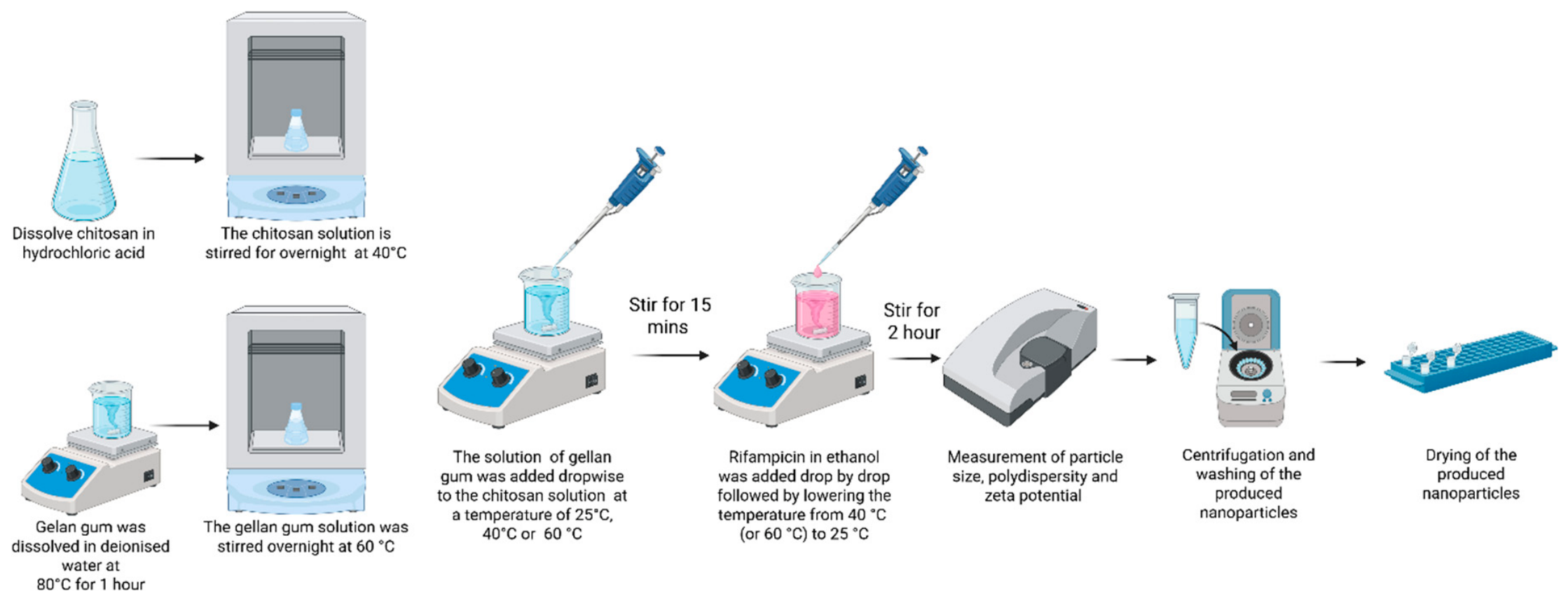
The preparation of nanoparticles followed the method of chitosan–gellan gum gel preparation, with modifications [32]. Different mass ratios of gellan gum to chitosan were mixed (1:9, 4:6 or 7:3). Both solutions were adjusted to pH 5 by adding 0.1 M HCl or 0.2 M NaOH in advance. The gellan gum solution was added to the chitosan solution dropwise (at 25 °C, 40 °C or 60 °C). After stirring for 15 min, rifampicin (rifampicin: polymers = 1:3, 1:1 or 3:1) in ethanol (6 mg/mL) was added dropwise, followed by adding 90% ethanol dropwise (total volume of ethanol = 5 mL, 10 mL or 15 mL). The reaction mixture was stirred for 2 h, and the temperature was decreased from 60 °C (or 40 °C) to 25 °C simultaneously. Then, the reaction mixture was taken for the measurements of particle size, polydispersity and zeta potential. The nanoparticles were then isolated by centrifugation at 15,060 rpm (Eppendorf 5420, Hamburg, Germany) for 30 min and rinsed with distilled water by three cycles of centrifugation for 5 min each. The pellet was collected and dried.
2.6. Design of Taguchi Experiment
The Taguchi design of experiments method was applied in this study to evaluate the impacts of various parameters on the optimization and reduction in the number of experiments. This method is suitable for studying multiple factors using the Design-Expert software (version 13, Stat-Ease, Minneapolis, MN, USA). We evaluated four factors, including the GG/CS ratio, the RIF/polymer ratio, temperature and the volume of ethanol by using the L9 orthogonal matrix (Table 1).
Table 1.
Taguchi L9 experimental scheme for the production of CS-GG-RIF NPs.
2.7. Preparation of FITC–Chitosan
The preparation procedure followed the methods reported by Cook et al. and Ge et al., with modifications [33,34]. To the chitosan solution (1% w/v in 0.1 M acetic acid; 1 mL), ethanol (1 mL) and FITC (2 mg/mL; 0.5 mL) were added, and the reaction mixture was stirred in the dark at 25 °C for 3 h, followed by precipitation in NaOH (0.1 M; 10 mL). FITC–chitosan was isolated by centrifugation at 15,060 rpm (Eppendorf 5420, Hamburg, Germany) for 30 min and washed with distilled water (15,060 rpm; 10 min each) until no fluorescence was detected in the supernatant by using a fluorescence microscope (AOSVI, Shenzhen, China). The pellet was re-dissolved in 3 mL of 0.1 M acetic acid and dialyzed (MwCO: 8000–14,000 D, Lombard, IL, USA) in 50 mL of distilled water for 3 days in the dark, with the water being replaced every day. The dialyzed product was dried by lyophilization.
2.8. Preparation of FITC–Chitosan–Gellan Gum Nanoparticles with Rifampicin (FITC-CS-GG-RIF NPs)
The preparation was carried out according to the method reported by Cook et al., with modifications [33]. Both chitosan and gellan gum solutions were adjusted to pH 5 by adding 0.1 M HCl or 0.2 M NaOH in advance. An amount of 0.52 mg of fluorescein isothiocyanate (FITC) (in 0.5 mL of ethanol) was added dropwise to 3.6 mL of the chitosan solution in the dark at 25 °C for 4 h. An amount of 2.4 mL of the gellan gum solution was added to the reaction mixture dropwise at 40 °C. After stirring for 15 min, 2 mg of rifampicin in ethanol (6 mg/mL) was added dropwise, followed by adding 90% ethanol dropwise (total volume of ethanol = 15 mL). The reaction mixture was stirred for 2 h in the dark, and the temperature was decreased from 40 °C to 25 °C simultaneously. Then, the nanoparticles were isolated by centrifugation at 15,060 rpm (Eppendorf 5420, Hamburg, Germany) for 30 min and rinsed with distilled water by four cycles of centrifugation for 10 min each. The absence of free FITC in the supernatant was confirmed by fluorescence microscopy. The pellet was collected and dried by lyophilization.
2.9. Measurement of Particle Size, Polydispersity, Zeta Potential and Transmission Electron Microscopy
The average diameter, polydispersity index (PDI) and zeta potential of the nanoparticles were measured by a Zetasizer Advance Pro (Malvern Panalytical Ltd., Worcestershire, UK). Ten drops of the nanoparticle suspension were added to 1.5–2 mL of distilled water in a quartz cuvette. The dynamic light scattering measurements were carried out at 25 °C with a 173° angle determination. The zeta potentials were determined by adding 20 drops of the nanoparticle suspension to 1 mL of distilled water in a folded capillary cell. Transmission electron microscopy (TEM) images were examined using a Hitachi S-4800 field-emission scanning electron microscope (Hitachi High-Tech, Hitachinaka, Japan) at an accelerating voltage of 30 kV. TEM grids were prepared by placing 10 µL of the diluted sample solutions on a carbon-coated copper grid and allowing the solution to evaporate completely at room temperature.
2.10. Determination of the Yield of Nanoparticles, Drug Encapsulation Efficiency and Drug-Loading Degree
The centrifugation of nanoparticle suspensions was carried out after the synthesis. The pellet was dried, and the yield of nanoparticles was determined:
In addition, the supernatant was taken to determine the amount of free drugs by UV–visible spectroscopy (Promecolab PE-5400UV, Shanghai Mapada Instruments Co., Ltd., Shanghai, China). The encapsulation efficiency (EE) was calculated according to the formula below.
2.11. Thermogravimetric Analysis (TGA) and Differential Scanning Calorimetry (DSC)
Thermogravimetric and differential scanning calorimetry analyses were performed with a LabSYS evo TGA/DTA/DSC analyzer (Setaram Instrumentation Caluire-et-Cuire, France). The temperature spectrum was varied from 30 to 700 °C, employing an aluminum oxide crucible. The sample was heated at 10 °C/min in a nitrogen atmosphere at a flow rate of 30.
2.12. Fourier-Transform Infrared Spectroscopy
The samples were analyzed by infrared spectroscopy with an FSM 1202 spectrometer (Infraspek Ltd., Saint Petersburg, Russia). Fourier-transform infrared (FTIR) spectra were obtained by using the KBr method. A pellet was prepared by blending around 3 mg of the sample with 100 mg of KBr. The scanning range was set from 4000 to 500 cm−1, with a resolution of 8 cm−1.
2.13. In Vitro Release of Drug from Nanoparticles
The release behavior of rifampicin from nanoparticles was examined at pH 7.4 using different rifampicin-to-polymer ratios (1:3, 1:1, and 3:1). The effect of pH (1.2, 6.8, and 7.4) was further evaluated for nanoparticles with a rifampicin-to-polymer ratio of 3:1. Samples of nanoparticles containing the same amount of rifampicin were put into dialysis membranes (MwCO: 8000–14,000 D, Lombard, IL, USA), followed by adding 2.5 mL of dilute HCl (pH 1.2), phosphate buffer solution (PBS) (pH 6.8) and PBS (pH 7.4), respectively. They were immersed in 100 mL of dilute HCl (pH 1.2), PBS (pH 6.8) and PBS (pH 7.4), separately, at 37 ± 0.5 °C, which was regulated by a circulating water bath (SIA “ELMI”, Riga, Latvia). The portions of the medium (2 mL) were taken at predetermined times. The extracts were analyzed by UV–visible spectroscopy at 470 nm.
2.14. Mucoadhesion Study of CS-GG-RIF Nanoparticles
Samples of FITC–chitosan, FITC-CS-GG-RIF and rifampicin were separately applied to the surfaces of three ovine lung tissue samples [35]. The ovine lung tissue was cut into pieces of 2 × 2 cm2 and was mounted on a glass slide. The samples were mounted on microscope slides, inclined at 45° and rinsed with phosphate buffer solution (pH 7.4) at 37 °C using a flow rate of 2 mL/min until a total volume of 500 mL was delivered. Each experiment was conducted for more than 4 h. After every 100 mL of buffer had been applied, the amounts of the samples remaining on the lung tissue were determined. FITC–chitosan and FITC-CS-GG-RIF were analyzed by fluorescence microscopy under blue light with the software ImageJ 1.54m, while rifampicin was quantified by measuring its amount in the rinsate using UV–visible spectroscopy. The results of the FITC–chitosan and FITC-CS-GG-RIF were both expressed as the normalized intensity, whereas rifampicin retention was reported as the percentage of mass retained.
2.15. Determination of Antimycobacterial Activity of CS-GG-RIF Nanoparticles
In vitro studies were performed in Airstream AC2-4E8 biosafety cabinets (Esco Micro Pte. Ltd., Singapore), which provided sterile conditions through negative pressure and air filtration through HEPA filters. The antimycobacterial activity of the nanoparticles was evaluated using an antituberculosis-sensitive wild strain of MTB H37Rv, obtained from the Asklepios pulmonology clinic (Munich-Hauting, Germany). Determination of mycobacterial activity was carried out on a dense Löwenstein–Jensen medium. CS-GG NPs were synthesized with rifampicin, and the drug concentrations were 0.2 mg/mL, 0.5 mg/mL and 1 mg/mL. Blank CS-GG NPs without the drug were used as a control. All tubes of each experimental row were seeded with 0.2 mL of a bacterial suspension. The tubes were sealed with silicone stoppers and incubated in the thermostat at 37 °C for 21 days. The results of suppressive activity were evaluated by counting the number of colony-forming units (CFUs) of MTB on the nutrient medium and comparing the control with the experimental cultures. For samples showing positive results, smears were made and stained according to the Ziehl–Neelsen method [36]. The smear was allowed to air-dry and examined microscopically using a Leica DMLS (Wetzlar, Germany) binocular microscope.
2.16. Statistical Processing of the Produced Data
All studies were carried out at least three times. The results are presented as mean values with standard deviations. In order to compare the independent groups, a one-way ANOVA was performed using the OriginPro 2019b software.
The effects of the proposed experiments on responses were analyzed using the Design-Expert 13 software to obtain the main effects of different factors independently, and then an analysis of variance (ANOVA) was performed to determine statistically significant factors. The optimal conditions were determined by selecting the function.
3. Results
3.1. Synthesis of CS-GG-RIF Nanoparticles and Optimization Using the Taguchi Method
Polysaccharide nanoparticles can be synthesized using various methods, including ionotropic gelation, complex coacervation, nanoprecipitation, and others [37,38,39,40,41,42]. Table 2 presents a summary of representative rifampicin-loaded polysaccharide nanoparticles.
Table 2.
Polysaccharide nanoparticles for rifampicin delivery.
In this study, the polyelectrolyte complex coacervation method was used (Figure 1), which does not require expensive equipment or the use of additional cross-linking or stabilizing agents. Chitosan–gellan gum-based nanoparticles are mostly used for encapsulating hydrophilic drugs [37,45], but there are also examples in the literature of such systems being used for hydrophobic drugs [37,46]. CS-GG-RIF nanoparticles were produced using the previously developed technique for producing CS-GG gel, with modifications [32]. Owing to the large size (2286 ± 27) and the broad PDI value (0.958 ± 0.059) of the prepared particles, the preparation method needs to be optimized for the production of nanoparticles. Schnell et al. reported that the size of xylan/chitosan nanoparticles decreased from 1055 nm to 651 nm as the ethanol concentration was increased from 0 wt% to 16 wt, which could be due to the decrease in the ionic charges in the system [47]. In our work, we used ethanol to load the hydrophobic drug rifampicin, which helped increase the encapsulation efficiency. In this study, for the first time, ethanol was used in the synthesis of CS-GG nanoparticles by the polyelectrolyte complex coacervation method. Ethanol not only improves the solubility of rifampicin but also contributes to the formation of nanoparticles with smaller sizes and a narrower size distribution.
Figure 1.
Schematic representation of CS-GG-RIF NP production by polyelectrolyte complex coacervation method. This image was created using BioRender.
The aim of this study was to optimize the production of nanoparticles based on chitosan and gellan gum loaded with rifampicin. Some factors affecting the nanoparticle preparation processes were analyzed: the GG/CS ratio, the RIF/polymer ratio, temperature and the volume of ethanol. The physicochemical properties of the produced NPs were studied, and the in vitro release of rifampicin was determined.
We used a Taguchi orthogonal array to determine the optimal conditions and identify the key parameters affecting the size, polydispersity index (PDI), zeta potential, encapsulation efficiency of the drug and yield of nanoparticles. CS-GG-RIF nanoparticles were synthesized using the polyelectrolyte complex coacervation method. We performed nine experiments to determine the best synthesis conditions for the nanoparticles. Table 3 shows the structure of the orthogonal matrix and the results of the physicochemical characteristics. The particle size of each nanoparticle was measured using dynamic light scattering, the zeta potential was measured using mixed-mode measurement phase analysis light scattering, and the drug encapsulation efficiency and nanoparticle yield were calculated.
Table 3.
Physicochemical characteristics of the produced CS-GG-RIF NPs.
According to the obtained results, depending on the synthesis conditions, chitosan–gellan gum particles with an average particle size ranging from 151 ± 3 nm to 836 ± 19 nm can be produced, and the polydispersity varies significantly from 0.221 ± 0.014 to 0.683 ± 0.113. The zeta potential of the produced particles was positive and ranged from 17.80 ± 0.72 mV to 32.48 ± 0.53 mV. In addition, the encapsulation efficiency of rifampicin and the nanoparticle yield ranged from 20% to 73% and from 10% to 65%, respectively.
The particle size, polydispersity, zeta potential, drug encapsulation efficiency and nanoparticle yield data of CS-GG-RIF NPs were analyzed using the Design-Expert software. An ANOVA table was used to determine the key process parameter affecting the properties of CS-GG-RIF NPs. The ANOVA tables for the Taguchi method are presented in Table 4. For all nanoparticle characteristics, the initial response values were converted using the following equation: y′= (y + k)λ, where constant (k) = 0, and lambda (λ) = −1.24 for size, −1.78 for the PDI, and 0.8 for the zeta potential. The best lambda was predicted using the Design-Expert program.
Table 4.
ANOVA results for size, PDI and zeta potential.
p-values of less than 0.05 indicate the significance of the model terms. According to the ANOVA results, the GG/CS ratio and temperature did not have a significant effect on the average particle size, and these parameters, as well as the ethanol volume, did not affect the polydispersity. According to the results of the ANOVA analysis, none of the factors had a significant effect on the zeta potential.
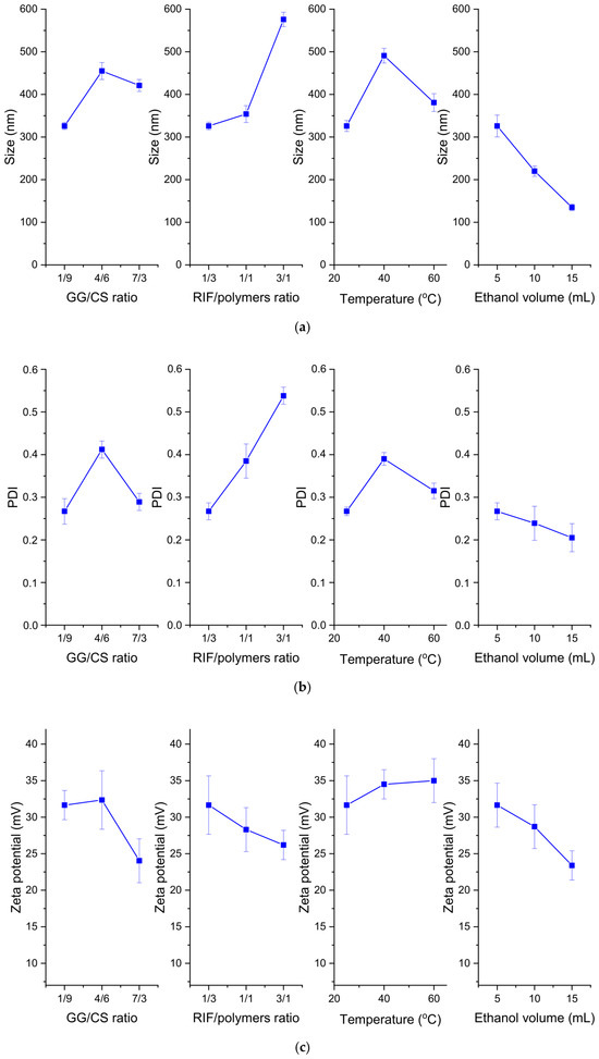
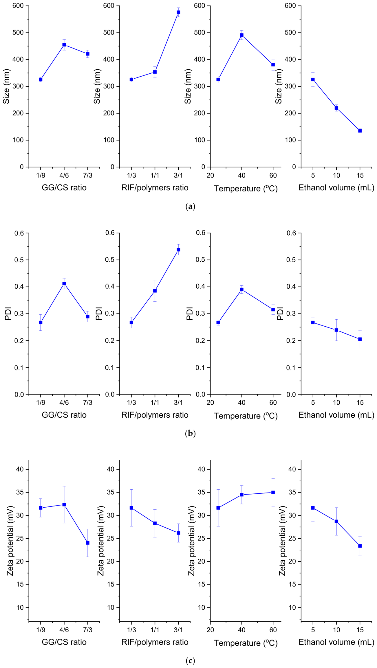
As we can see from the graphs presented in Figure 2, as the ratio of drug to polymer increased, the average size and PDI of the nanoparticles increased, which may be due to the inclusion of rifampicin in the space of the polymeric nanoparticles [10]. As the volume of ethanol increased, the average size and PDI of the nanoparticles decreased. This is explained by the fact that ionic charges decreased, thereby strengthening the interactions between polysaccharides [47,48]. As the temperature increased, the size of the nanoparticles increased, with the largest particle size and polydispersity occurring at 40 °C.
Figure 2.
Effects of parameters on (a) average particle size, (b) PDI and (c) zeta potential of CS-GG-RIF NPs. Data are presented as means ± standard deviations (n = 3).
When the gellan gum content in the GG/CS ratio increased (Figure 2), the zeta potential of the particles decreased generally, which can be explained by the fact that the negative charges of gellan gum neutralize part of the positive charges of chitosan [22]. In addition, as the amount of ethanol increased, the zeta potential decreased, which may be due to the fact that ethanol solution has a higher viscosity and lower dielectric permeability than water, affecting, therefore, the calculation of the zeta potential based on electrophoretic mobility [49,50].
The encapsulation efficiency and nanoparticles yield are also important parameters, so we performed an ANOVA analysis, the results of which are presented in Table 5. The formula y′= (y + k)λ was also used for the calculation, where constant (k) = 0, and lambda (λ) = −2.88 for EE and −0.55 for NPs’ yield.
Table 5.
ANOVA results for encapsulation efficiency and nanoparticle yield.
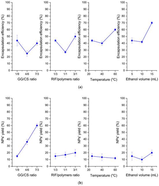
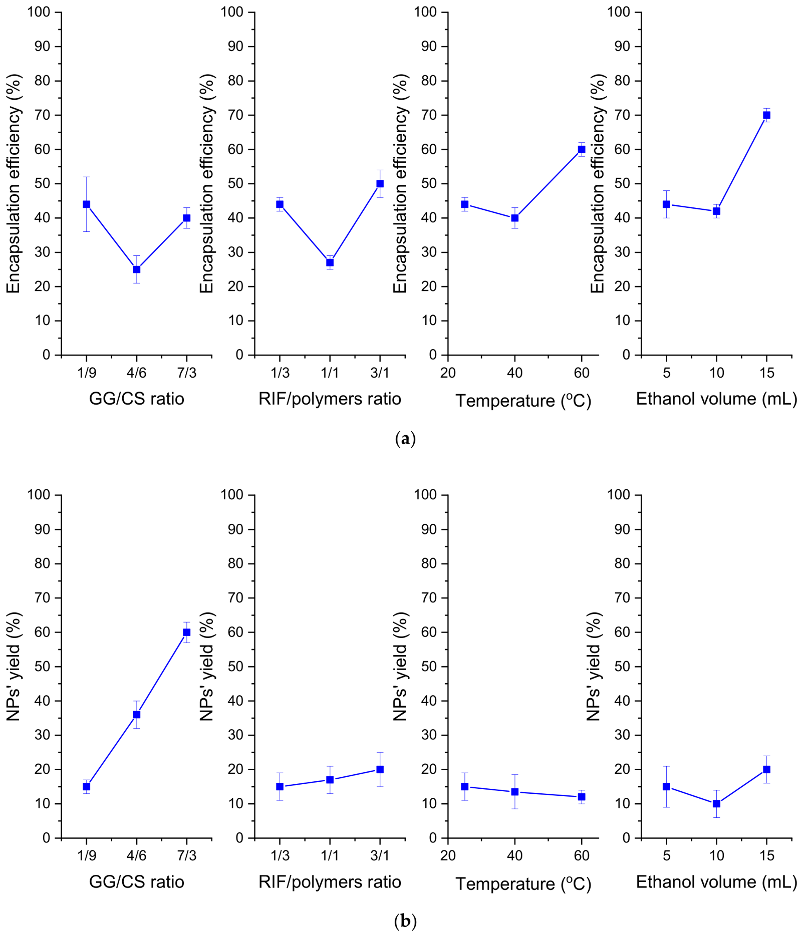
The encapsulation efficiency was influenced by the GG/CS ratio, RIF/polymer ratio, and temperature, and the model for nanoparticles yield was significant when the GG/CS ratio and volume of ethanol parameters were included. Thus, it can be concluded that both factors influence the properties of nanoparticles. The highest encapsulation efficiency was observed at a RIF/polymer ratio of 3/1 and an ethanol volume of 15 mL (Figure 3). This is due to the hydrophobicity of rifampicin in water and its good solubility when ethanol is used as a co-solvent [51]. Figure 3 shows that the highest nanoparticle yield was observed at a GG/CS ratio of 7/3 and an increase in the ethanol volume.
Figure 3.
Effects of parameters on (a) encapsulation efficiency and (b) nanoparticle yield of CS-GG-RIF NPs. Data are presented as mean ± standard deviations (n = 3).
Based on the ANOVA results, we performed optimization using the Design-Expert software for the synthesis of CS-GG-RIF NPs. We applied criteria (Table 6) to optimize the synthesis process for nanoparticles with the minimum size and polydispersity, as well as the maximum zeta potential, encapsulation efficiency and particle yield. According to the ANOVA results, the minimum size and PDI value were theoretically predicted at 25 °C (Figure 2). Therefore, the temperature for optimization was set at 25 °C.
Table 6.
Criteria for the synthesis of CS-GG-RIF NPs.
Among the options proposed by the program, the solution with the highest desirability of 62.5% was selected, which provides satisfactory nanoparticle characteristics. Next, we carried out the synthesis under the proposed optimal conditions: a GG/CS ratio of 4/6, an RIF/polymer ratio of 1/3, a temperature of 25 °C, and a volume of ethanol of 15 mL. The experimental results obtained were in reasonable agreement with the model (Table 7). There was a difference between the predicted and experimental values, but according to the model, it was within the desirability limit (the error was no more than 62.5%). It is crucial to note that nanoparticles with a higher encapsulation efficiency and nanoparticle yield can be produced under alternative conditions, but this leads to an increase in particle size and polydispersity. Therefore, the selected parameters enable the synthesis of nanoparticles with a smaller size, a narrower size distribution and better colloidal stability, as these characteristics are crucial for mucoadhesive drug delivery systems. Thus, it can be concluded that the Taguchi method is suitable for optimizing the synthesis conditions for producing CS-GG-RIF NPs.
Table 7.
Predicted and experimental results for CS-GG-RIF NPs.
3.2. Study of the Physicochemical Properties of the Produced CS-GG-RIF Nanoparticles
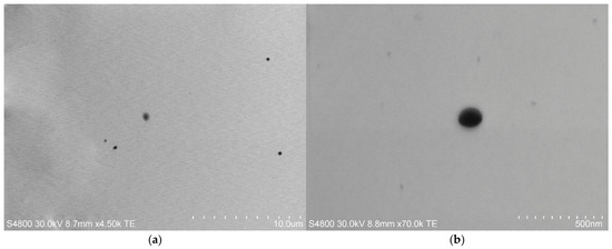
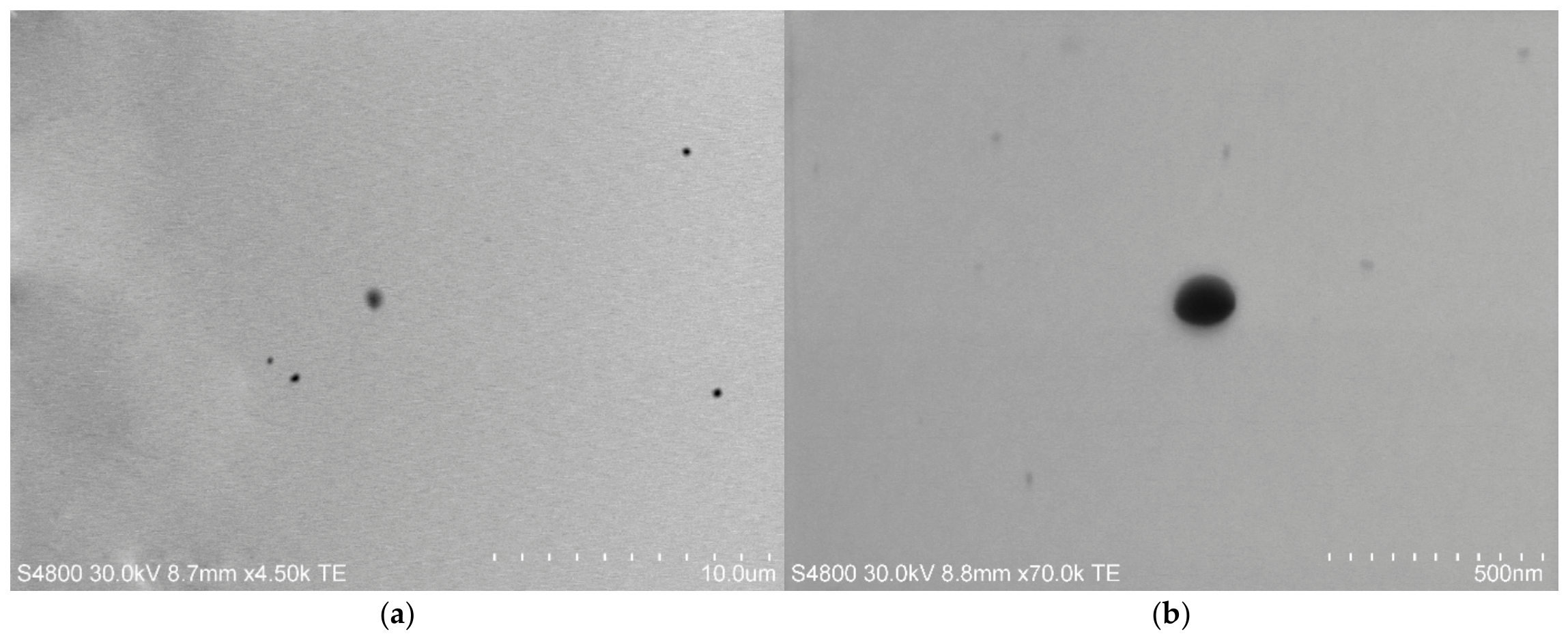
To determine the morphology of the produced CS-GG-RIF nanoparticles, transmission electron microscopy (TEM) was performed (Figure 4). Using the ImageJ software, the average size of the nanoparticles was calculated as 137 ± 11 nm. The produced CS-GG-RIF particles had a spherical shape.
Figure 4.
TEM images of CS-GG-RIF nanoparticles. Scale bars are (a) 10 µm and (b) 500 nm.
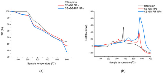
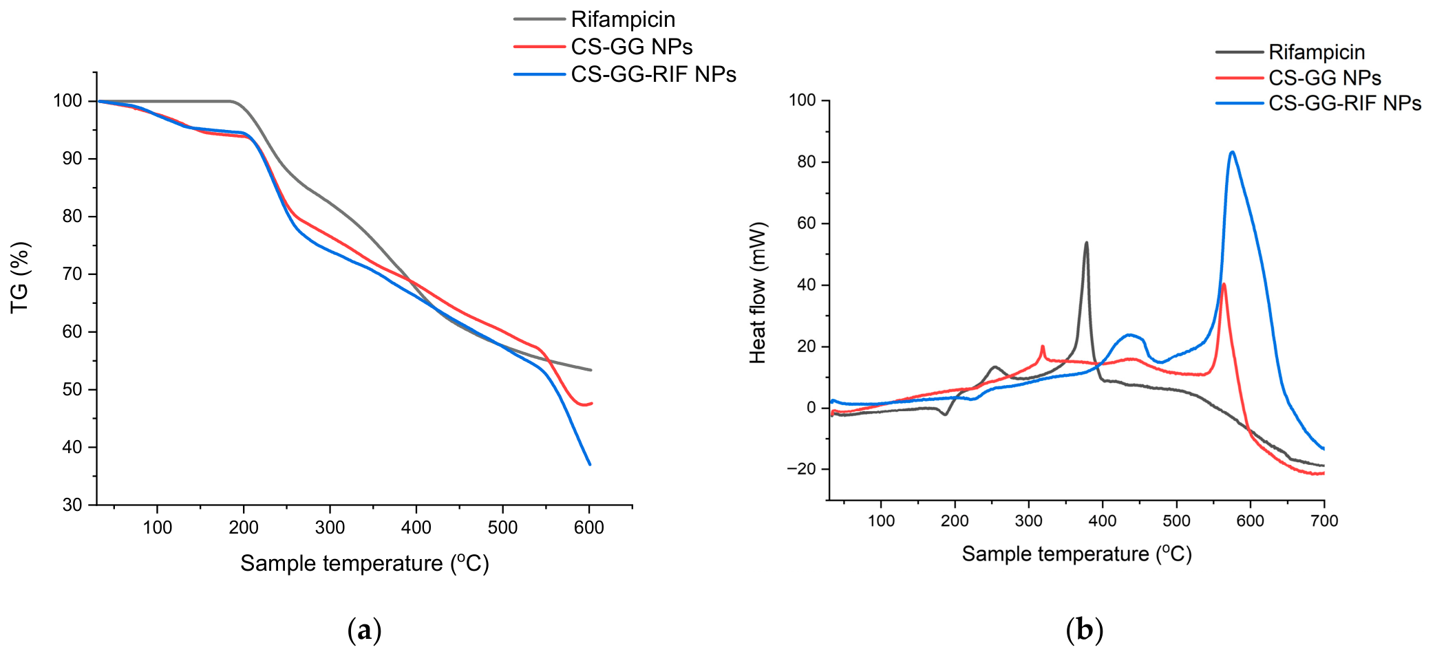
The TGA and DSC curves of rifampicin, CS-GG nanoparticles without the drug, and rifampicin-loaded CS-GG-RIF nanoparticles are shown in Figure 5. The results showed a slower rate of decomposition of rifampicin-loaded chitosan–gellan gum nanoparticles compared with empty chitosan–gellan gum nanoparticles, indicating improved stability. The results show that the particles begin to disintegrate at a temperature of 86 °C; after 200 °C, a sharp decrease in weight was observed, which may be associated with the degradation of polysaccharides [52,53]. The endothermic peak characteristic of rifampicin was marked by a change in enthalpy at a temperature of 186.6 °C (Figure 5b), which corresponded to its melting point, i.e., reflecting the crystalline nature of rifampicin. Next, an exothermic peak was observed at a temperature of 378.3 °C, in the range of 188–550 °C, with a weight loss of up to 45% (Figure 5a), which may correspond to the decomposition temperature of the drug. There was no endothermic peak for rifampicin in the thermogram of CS-GG-RIF nanoparticles, which indicates the formation of an amorphous state or molecular dispersion of the drug in the matrix.
Figure 5.
(a) TGA and (b) DSC analysis results for rifampicin, CS-GG NPs and CS-GG-RIF NPs.
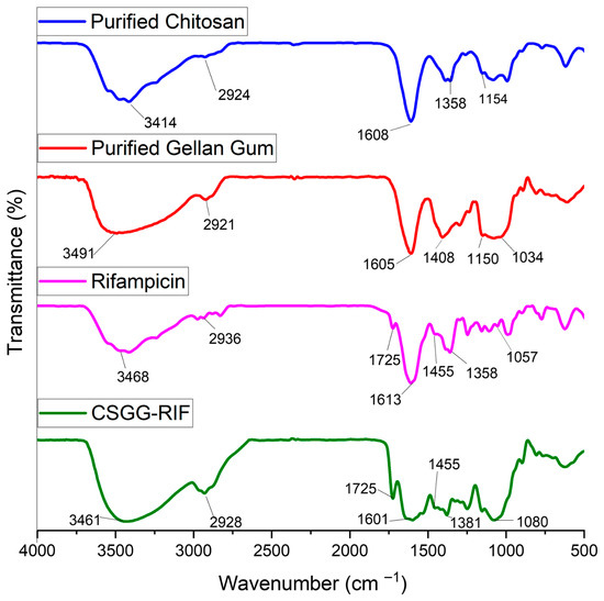
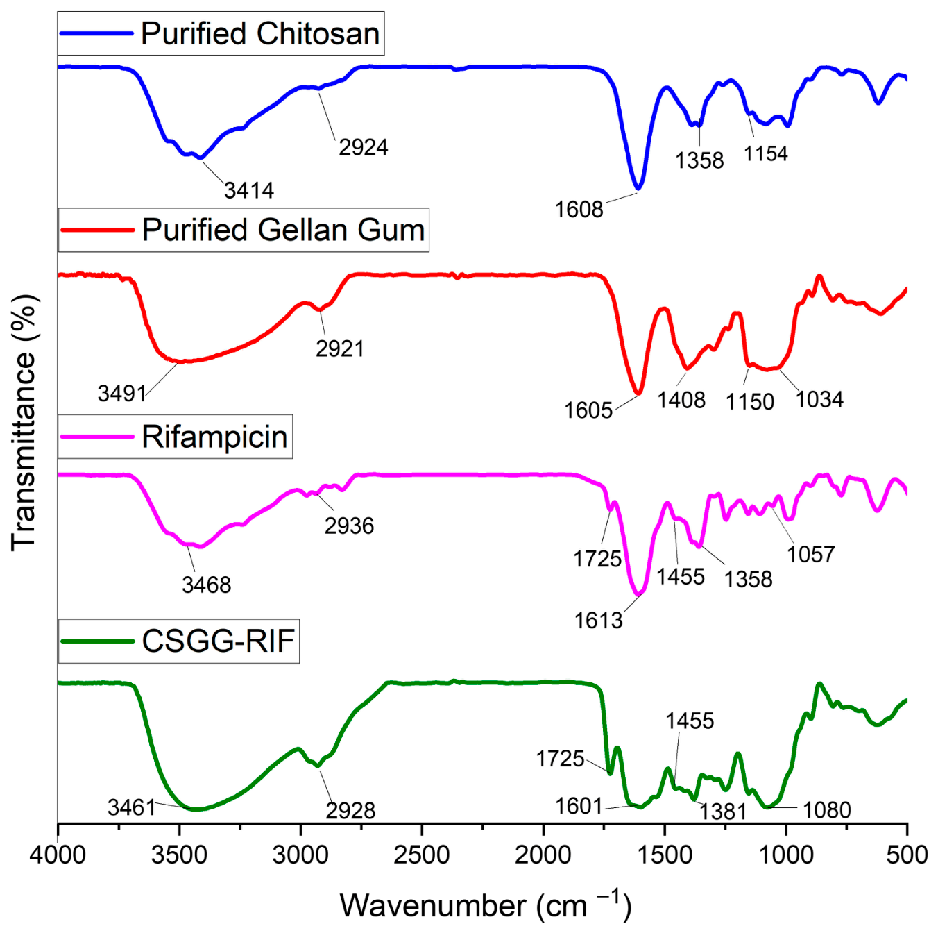
To determine the incorporation of rifampicin into the polymer matrix of chitosan and gellan gum, FTIR spectroscopy studies were conducted for the initial components of the system and the synthesized nanoparticles. The results are presented in Figure 6.
Figure 6.
FTIR spectra of purified chitosan, purified gellan gum, rifampicin and CS-GG-RIF nanoparticles.
For purified chitosan, the absorption band at 3414 cm−1 corresponds to the N–H group in primary amines and O–H in the pyranose ring, 2924 cm−1 for C–H stretching, 1608 cm−1 for C = O stretching and 1358 cm−1 for CH3 in NHCOCH3, and 1154 cm−1 for glycosidic linkage C–O–C [54,55]. The spectra of pure gellan gum are characterized by broad and intense peaks at 3491 cm−1, which are associated with O–H stretching, at 2921 cm−1 for C–H vibrations, at 1605 cm−1 and 1408 cm−1 for symmetric and asymmetric valence vibrations of COO−, and at 1150 cm−1 and 1034 cm−1 associated with C–O stretching [56,57]. The spectra of pure rifampicin show broad and intense peaks at 3468 cm−1, which are associated with N–H stretching, and C–H stretching vibrations are noticeable in the range of 2936 cm−1. Bands at 1725 cm−1 and 1613 cm−1 correspond to C = O and C = N stretching, respectively; 1455 cm−1 is due to C = C stretching, 1358 cm−1 is associated with CH2 and C = C, and 1059 cm−1 corresponds to –CH, CO, and C–H [10,58,59]. The graph for CS-GG-RIF nanoparticles shows peaks corresponding to both polymers and drugs, confirming the inclusion of rifampicin in the polymer matrix without chemical interaction.
3.3. Study of Rifampicin Release from the Polymer Matrix In Vitro
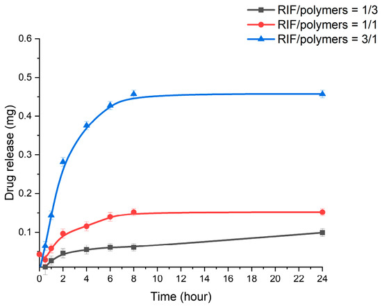
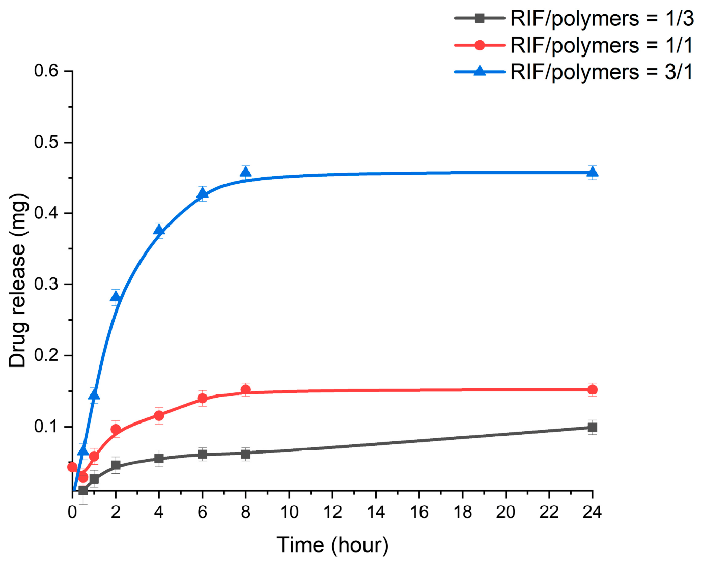
Rifampicin release from the polymer matrix was studied by dialysis at 37 °C and pH 7.4. Nanoparticles with different RIF/polymer ratios (1/3, 1/1, and 3/1) were studied. The amount of rifampicin loaded into the polymers was the same, and the results are presented in Figure 7. At the beginning of the process, there was a rapid release of the drug adsorbed on the surface of the nanoparticles. The polymer then swelled, causing the drug to be gradually released from the polymer matrix [60]. As the RIF/polymer ratio increased from 1/3 to 3/1, the amount of release tended to increase from 0.1 mg to 0.45 mg after 24 h. The higher the drug loading, the higher the concentration of rifampicin in the polymer matrix. This enhanced the driving force of diffusion, and the drug was released more rapidly from the chitosan–gellan gum nanoparticles [60,61].
Figure 7.
Drug release from CS-GG-RIF NPs with different rifampicin/polymers ratios. Data are presented as means ± standard deviations (n = 3).
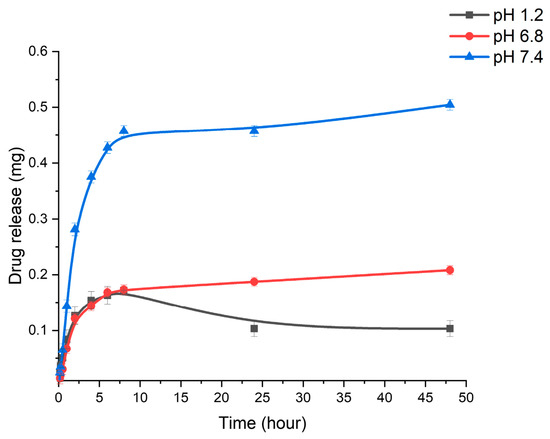
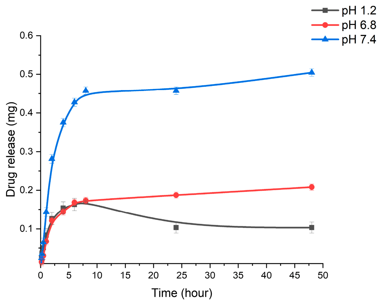
In addition, the degree of drug release depended on the medium’s pH. Thus, we studied the release of rifampicin in a RIF/polymer ratio of 3/1 at different medium pH values (pH 1.2, 6.8, and 7.4). The curves (Figure 8) show that the medium pH affected the release of rifampicin from chitosan–gellan gum nanoparticles. After 48 h of ± study, the content of rifampicin released from CS-GG nanoparticles at pH 7.4, 6.8, and 1.2 corresponded to 0.5 mg, 0.2, and 0.1 mg, respectively. Rifampicin was released faster in a neutral environment compared with an acidic environment, which may be due to electrostatic interactions between oppositely charged polymers. In an acidic environment, chitosan is more protonated and attracted to gellan gum, which leads to a decrease in swelling and a slowdown in drug release. At a neutral pH, weakened electrostatic interactions contribute to the relaxation of the matrix and accelerate the diffusion of the drug.
Figure 8.
Cumulative drug release from CS-GG-RIF NPs (RIF/polymers = 3/1) at different pH values. Data are presented as means ± standard deviations (n = 3).
It can be concluded that the in vitro release of rifampicin from chitosan–gellan gum nanoparticles was prolonged, which makes it possible to use the produced particles for the delivery of antituberculosis drugs.
The rifampicin release kinetics from chitosan–gellan gum nanoparticles were analyzed using mathematical models, including zero-order (Qt = Q0 + k0t), first-order (dC/dt = −kC), Higuchi (Mt/M∞ = k√t) and Korsmeyer–Peppas (Mt/M ∞ = ktn) [62,63,64] to determine the mechanism of drug release from the developed polymeric system (Table 8).
Table 8.
Coefficients of determination (R2) for rifampicin release models from CS-RIF nanoparticles.
The zero-order and Higuchi models demonstrated moderate applicability under various pH conditions (1.2, 6.8 and 7.4). The release of rifampicin from chitosan–gellan gum nanoparticles is not strictly governed by zero-order kinetics and is not controlled by simple Fickian diffusion through a homogeneous matrix. The Korsmeyer–Peppas model showed high correlation coefficients (R2 = 0.9149–0.9705) at all pH values, indicating that the mechanism of rifampicin release is a combined process involving diffusion and polymer degradation. At pH 6.8 and 7.4, the first-order model showed excellent correlation (R2 = 0.9802 and 0.9976, respectively). Thus, under neutral conditions, the nanoparticles behave more predictably. During rifampicin release, processes of diffusion and swelling of the polymer matrix occur. Overall, the kinetics of rifampicin release from chitosan–gellan gum nanoparticles depend on pH. This behavior is desirable for oral delivery systems, as it can help protect the drug from premature release in the stomach and ensure prolonged release in the intestine.
3.4. Investigation of the Mucoadhesive Properties of CS-GG-RIF NPs
Antituberculosis drugs are administered orally, which is often associated with low bioavailability and side effects [65,66,67], whereas inhalation delivery to the lungs is preferable but requires overcoming biological barriers [10]. A promising solution is the use of mucoadhesive polymeric nanoparticles based on chitosan, which are capable of interacting with mucin and improving drug retention [68,69].
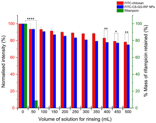
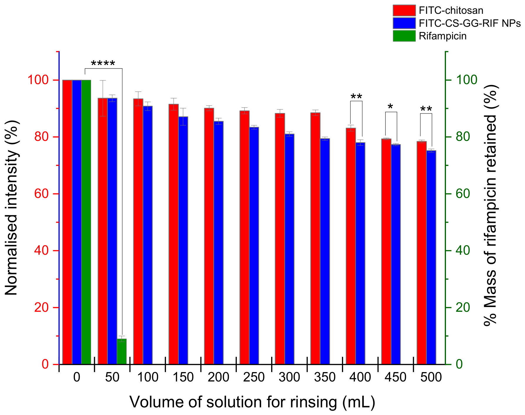
The mucoadhesive properties of rifampicin, chitosan and CS-GG-RIF nanoparticles containing fluorescein isothiocyanate were examined using a modified fluorescence flow-through retention test developed earlier by the Khutoryanskiy group [35]. The lungs were washed with 500 mL of phosphate buffer solution (pH 7.4; 37 °C) at a rate of 2 mL/min. For pure rifampicin (Figure 9), the percentage of mass after washing was determined using UV spectroscopy. As a result, rapid removal of rifampicin from the surface of the ovine lung tissue was observed during the first 25 min.
Figure 9.
Retention of rifampicin, chitosan and CS-GG-RIF nanoparticles in the ovine lung tissue after washing with phosphate buffer solution (pH 7.4) at various volumes. Data are presented as means ± standard deviations (n = 3). Statistically significant differences are indicated as follows: * p < 0.05; ** p < 0.01; and **** p < 0.0001. The red and blue columns share the same Y-axis scale.
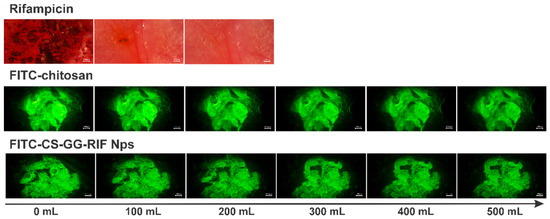
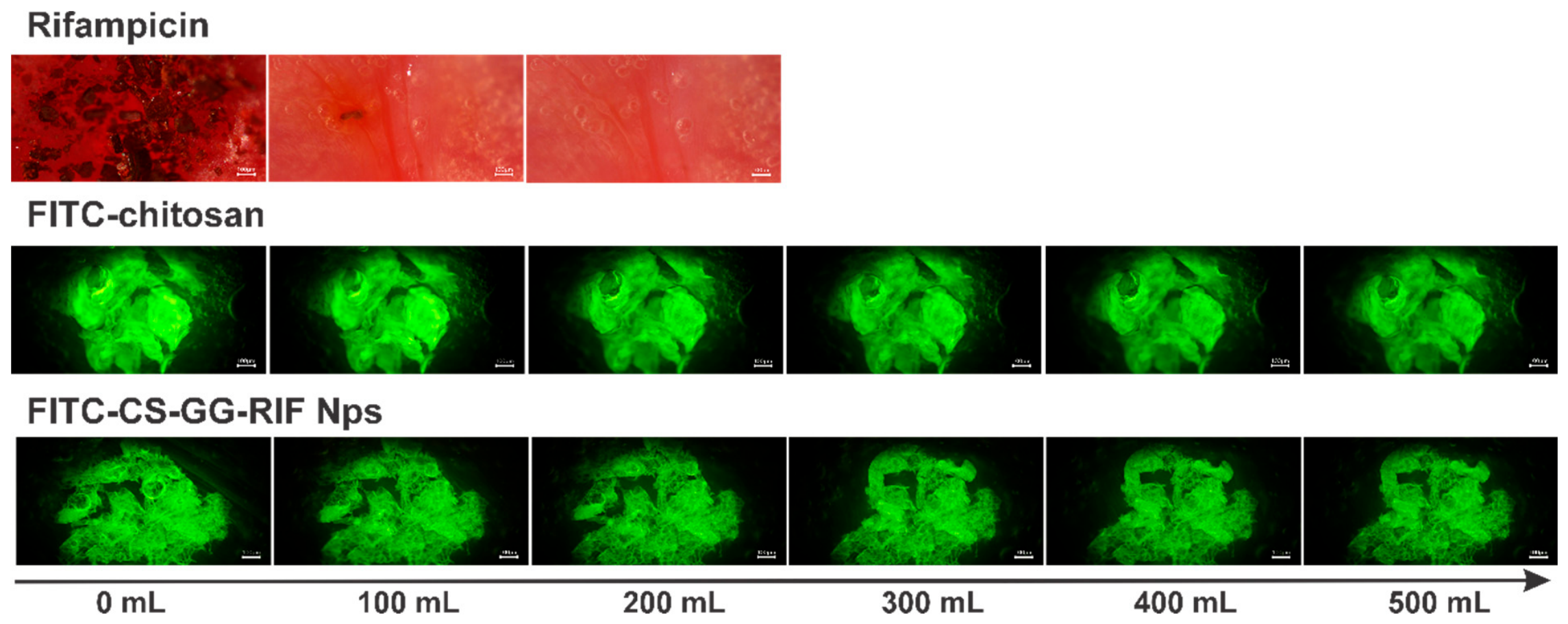
The retention profiles observed for chitosan and CS-GG-RIF nanoparticles in the ovine lung tissue were analyzed through a series of captured fluorescent photomicrographs, which are shown in Figure 10. Using the ImageJ software, the intensity was calculated, confirming that the nanoparticles could remain in the lung tissue for more than 4 h (Figure 9). Chitosan demonstrated a high retention capacity (78%), while the CS-GG-RIF nanoparticles exhibited a comparable level of mucoadhesion (75%). This suggests that the inclusion of rifampicin with gellan gum does not result in a significant loss of the system’s adhesive properties.
Figure 10.
Fluorescent images illustrating the adhesion of polymeric materials to the ovine lung tissue washed with phosphate buffer solution (pH 7.4) at various volumes. Scale bar is 100 μm.
Chitosan and gellan gum were selected for their ability to improve the mucoadhesiveness of rifampicin and to increase the duration of drug release [70,71]. This minimizes washout effects, leading to improved patient compliance and a reduced frequency of administration, which is beneficial in drug delivery systems [72,73].
3.5. Investigation of the Mycobacterial Activity of CS-GG-RIF NPs
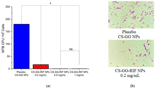
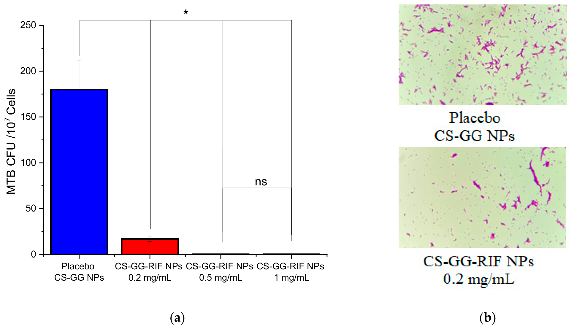
The antituberculosis activity of nanoparticles was tested on H37Rv strains. Mycobacterial growth was studied in Löwenstein–Jensen medium. The antimycobacterial activity of the nanoparticles was investigated at rifampicin concentrations of 0.2 mg/mL, 0.5 mg/mL, and 1 mg/mL. As a control, we studied placebo chitosan–gellan gum nanoparticles without the drug. Samples in which mycobacterial growth was observed were stained using the Ziehl–Neelsen method, and smears were examined under a microscope. The resulting microscopic images were analyzed using the ImageJ software, and the number of colony-forming units (CFUs/107 cells) was calculated. The results are shown in Figure 11.
Figure 11.
(a) Results of antimycobacterial activity test of the nanoparticles on the growth of H37Rv on Löwenstein–Jensen nutrient-dense medium. “*” indicates p < 0.05, ns—denotes no significant difference. Data are presented as means ± standard errors of the means (n = 3). (b) Microphotographs of MTB H37RV growth on the samples, stained using the Ziehl–Neelsen method.
As shown in the graph, the addition of nanoparticles with rifampicin reduced the growth of mycobacteria compared with the group without the drug (placebo CS-GG NPs). As the concentration of rifampicin increased, the suppression of mycobacterial growth also increased. At concentrations of 0.5 mg/mL and 1 mg/mL, the number of CFUs decreased significantly, confirming the high efficacy of rifampicin in the nanoparticles. Figure 11b shows micrographs of smears stained using the Ziehl–Neelsen method, which also indicated differences in MTB growth. Thus, the minimum inhibitory concentration of rifampicin in CS-GG-RIF nanoparticles for the suppression of Mycobacterium tuberculosis growth was determined to be 0.5 mg/mL. It is confirmed that rifampicin loaded in chitosan–gellan gum nanoparticles retained its antituberculosis activity and effectively inhibited the growth of mycobacteria.
4. Conclusions
Using the polyelectrolyte complex coacervation method, chitosan–gellan gum-based nanoparticles were produced and effectively loaded with the antituberculosis drug rifampicin. The Taguchi method allowed the synthesis parameters to be optimized and nanoparticles to be produced with specified dispersion characteristics (a minimum size of 153 ± 3 nm, a polydispersity of 0.212 ± 0.021 and a zeta potential of 22.94 ± 1.30 mV), while the chemical structure of the drug remained intact. The polymer matrix ensured the sustained release of rifampicin under in vitro conditions, with the rate of release depending on the pH of the medium. Chitosan and gellan gum nanoparticles demonstrated high mucoadhesive activity towards the lung tissue, confirming their potential as an effective mucoadhesive platform for drug delivery. Rifampicin loaded into the chitosan–gellan gum complex did not lose its ability to inhibit the growth of Mycobacterium tuberculosis, particularly with regard to the H37Rv strain. Thus, the chitosan–gellan gum nanoparticles represent a promising platform for creating nanoparticles with adjustable sizes, pronounced mucoadhesive properties, and the possibility of controlled drug release.
Author Contributions
Investigation, S.-Y.C., T.Z., Y.B. and V.A.; data curation, S.-Y.C. and A.G.; formal analysis, S.-Y.C.; methodology, A.G.; writing—review and editing, S.-Y.C., A.G. and Y.T.; funding acquisition, Y.T. All authors have read and agreed to the published version of the manuscript.
Funding
This research was funded by the Science Committee of the Ministry of Science and Higher Education of the Republic of Kazakhstan under grant no. AP23484897, titled “Creation of Nanocarriers of Antituberculosis Drugs Based on Polysaccharide Derivatives and Their Complexes”.
Institutional Review Board Statement
The animal study protocol was approved by the Local Bioethics Committee of Karaganda Medical University (protocol code: 15; 27 November 2023) for studies involving animals.
Informed Consent Statement
Not applicable.
Data Availability Statement
The original contributions presented in this study are included in this article. Further inquiries can be directed to the corresponding author.
Acknowledgments
Yuliia Bardadym acknowledges the Academy of Finland (no. 359456 for Ukrainian researchers) for its financial support. During the preparation of this work, the authors used ChatGPT based on GPT 5.3 in order to refine the language of this manuscript. The authors have reviewed and edited the output and take full responsibility for the content of this publication.
Conflicts of Interest
The authors declare no conflicts of interest.
Abbreviations
The following abbreviations are used in this manuscript:
| TB | Tuberculosis |
| MTB | Mycobacterium tuberculosis |
| CS | Chitosan |
| GG | Gellan gum |
| NPs | Nanoparticles |
| RIF | Rifampicin |
| PDI | Polydispersity index |
| EE | Encapsulation efficiency |
| TGA | Thermogravimetric analysis |
| DSC | Differential scanning calorimetry |
| FTIR | Fourier-transform infrared spectroscopy |
| ANOVA | Analysis of variance |
| FITC | Fluorescein isothiocyanate |
| TEM | Transmission electron microscopy |
References
- World Health Organization. The Top 10 Causes of Death. Available online: https://www.who.int/ru/news-room/fact-sheets/detail/the-top-10-causes-of-death (accessed on 7 August 2024).
- Global Tuberculosis Report 2025; World Health Organisation: Geneva, Switzerland, 2025.
- Nguyen, L. Antibiotic resistance mechanisms in M. tuberculosis: An update. Arch. Toxicol. 2016, 90, 1585–1604. [Google Scholar] [CrossRef] [Scilit]
- Galiyeva, A.; Tazhbayev, Y.; Zhumagaliyeva, T.; Karimova, B.; Tabriz, N.; Khutoryanskiy, V.V. Synthesis and Investigation of Albumin Nanoparticles Loaded with Anti-Tuberculosis Drug Isoniazid. Biopolymers 2025, 116, e70036. [Google Scholar] [CrossRef] [Scilit] [PubMed]
- Galiyeva, A.R.; Yessentayeva, N.A.; Marsel, D.T.; Daribay, A.T.; Sadyrbekov, D.T.; Moustafine, R.I.; Kutulutskaya, T.V. Polylactide Acid-Based Nanoparticles for Controlled Delivery of Isoniazid and Rifampicin: Synthesis, Characterization, and In Vitro Release Study. Eurasian J. Chem. 2025, 30, 95–108. [Google Scholar] [CrossRef] [Scilit]
- Mazlan, M.K.N.; Mohd Tazizi, M.H.D.; Ahmad, R.; Noh, M.A.A.; Bakhtiar, A.; Wahab, H.A.; Mohd Gazzali, A. Antituberculosis Targeted Drug Delivery as a Potential Future Treatment Approach. Antibiotics 2021, 10, 908. [Google Scholar] [CrossRef] [Scilit]
- Omoteso, O.A.; Fadaka, A.O.; Walker, R.B.; Khamanga, S.M. Innovative Strategies for Combating Multidrug-Resistant Tuberculosis: Advances in Drug Delivery Systems and Treatment. Microorganisms 2025, 13, 722. [Google Scholar] [CrossRef] [Scilit]
- Liebenberg, D.; Gordhan, B.G.; Kana, B.D. Drug resistant tuberculosis: Implications for transmission, diagnosis, and disease management. Front. Cell. Infect. Microbiol. 2022, 12, 943545. [Google Scholar] [CrossRef] [Scilit]
- Olamide Ishola, B.; Rahaman, K.A.; Razzak, S.A.; Rumon, M.M.H.; Shakil, M.S.; Uddin, S. Advanced mechanisms of polymer-based drug delivery systems for clinical applications. RSC Pharm. 2026, 3, 331–360. [Google Scholar] [CrossRef] [Scilit]
- Yessentayeva, N.A.; Galiyeva, A.R.; Daribay, A.T.; Sadyrbekov, D.T.; Moustafine, R.I.; Tazhbayev, Y.M. Optimization of Polylactide-Co-Glycolide-Rifampicin Nanoparticle Synthesis, In Vitro Study of Mucoadhesion and Drug Release. Polymers 2024, 16, 2466. [Google Scholar] [CrossRef] [Scilit]
- Jha, R.; Mayanovic, R.A. A Review of the Preparation, Characterization, and Applications of Chitosan Nanoparticles in Nanomedicine. Nanomaterials 2023, 13, 1302. [Google Scholar] [CrossRef] [Scilit] [PubMed]
- Visan, A.I.; Cristescu, R. Polysaccharide-Based Coatings as Drug Delivery Systems. Pharmaceutics 2023, 15, 2227. [Google Scholar] [CrossRef] [Scilit] [PubMed]
- Damiri, F.; Fatimi, A.; Santos, A.C.P.; Varma, R.S.; Berrada, M. Smart stimuli-responsive polysaccharide nanohydrogels for drug delivery: A review. J. Mater. Chem. B 2023, 11, 10538–10565. [Google Scholar] [CrossRef] [Scilit]
- Prasher, P.; Sharma, M.; Mehta, M.; Satija, S.; Aljabali, A.A.; Tambuwala, M.M.; Anand, K.; Sharma, N.; Dureja, H.; Jha, N.K.; et al. Current-status and applications of polysaccharides in drug delivery systems. Colloid Interface Sci. Commun. 2021, 42, 100418. [Google Scholar] [CrossRef] [Scilit]
- Huang, G.; Mei, X.; Xiao, F.; Chen, X.; Tang, Q.; Peng, D. Applications of Important Polysaccharides in Drug Delivery. Curr. Pharm. Des. 2015, 21, 3692–3696. [Google Scholar] [CrossRef] [Scilit]
- Rawal, T.; Patel, S.; Butani, S. Chitosan nanoparticles as a promising approach for pulmonary delivery of bedaquiline. Eur. J. Pharm. Sci. 2018, 124, 273–287. [Google Scholar] [CrossRef] [Scilit] [PubMed]
- Garg, T.; Rath, G.; Goyal, A.K. Inhalable chitosan nanoparticles as antitubercular drug carriers for an effective treatment of tuberculosis. Artif. Cells Nanomed. Biotechnol. 2015, 44, 997–1001. [Google Scholar] [CrossRef] [Scilit]
- Chogale, M.M.; Gaikwad, S.S.; Kulkarni, S.P.; Patravale, V.B. Quality-by-design Enabled Chitosan Nanoparticles for Antitubercular Therapy: Formulation, Statistical Optimization, and In Vitro Characterization. Curr. Drug Ther. 2021, 16, 64–82. [Google Scholar] [CrossRef] [Scilit]
- Mehnath, S.; Ayisha Sithika, M.A.; Arjama, M.; Rajan, M.; Amarnath Praphakar, R.; Jeyaraj, M. Sericin-chitosan doped maleate gellan gum nanocomposites for effective cell damage in Mycobacterium tuberculosis. Int. J. Biol. Macromol. 2019, 122, 174–184. [Google Scholar] [CrossRef] [Scilit] [PubMed]
- Goyal, A.K.; Garg, T.; Rath, G.; Gupta, U.D.; Gupta, P. Chemotherapeutic Evaluation of Guar Gum Coated Chitosan Nanoparticle Against Experimental Tuberculosis. J. Biomed. Nanotechnol. 2016, 12, 450–463. [Google Scholar] [CrossRef] [Scilit] [PubMed]
- Kumar, S.; Kaur, P.; Bernela, M.; Rani, R.; Thakur, R. Ketoconazole encapsulated in chitosan-gellan gum nanocomplexes exhibits prolonged antifungal activity. Int. J. Biol. Macromol. 2016, 93, 988–994. [Google Scholar] [CrossRef] [Scilit]
- Picone, C.S.F.; Cunha, R.L. Chitosan–gellan electrostatic complexes: Influence of preparation conditions and surfactant presence. Carbohydr. Polym. 2013, 94, 695–703. [Google Scholar] [CrossRef] [Scilit]
- Cardoso, V.M.d.O.; de Brito, N.A.P.; Ferreira, N.N.; Boni, F.I.; Ferreira, L.M.B.; Carvalho, S.G.; Gremião, M.P.D. Design of mucoadhesive gellan gum and chitosan nanoparticles intended for colon-specific delivery of peptide drugs. Colloids Surf. A Physicochem. Eng. Asp. 2021, 628, 127321. [Google Scholar] [CrossRef] [Scilit]
- Carvalho, S.G.; Haddad, F.F.; dos Santos, A.M.; Scarim, C.B.; Ferreira, L.M.B.; Meneguin, A.B.; Chorilli, M.; Gremião, M.P.D. Chitosan surface modification modulates the mucoadhesive, permeation and anti-angiogenic properties of gellan gum/bevacizumab nanoparticles. Int. J. Biol. Macromol. 2024, 263, 130272. [Google Scholar] [CrossRef] [Scilit] [PubMed]
- Costa, A.; Pinheiro, M.; Magalhães, J.; Ribeiro, R.; Seabra, V.; Reis, S.; Sarmento, B. The formulation of nanomedicines for treating tuberculosis. Adv. Drug Deliv. Rev. 2016, 102, 102–115. [Google Scholar] [CrossRef] [Scilit] [PubMed]
- Maddiboyina, B.; Roy, H.; Nakkala, R.K.; Gandhi, S.; Kavisri, M.; Moovendhan, M. Formulation, optimization and characterization of raloxifene hydrochloride loaded PLGA nanoparticles by using Taguchi design for breast cancer application. Chem. Biol. Drug Des. 2023, 102, 457–470. [Google Scholar] [CrossRef] [Scilit]
- El-Naggar, N.E.-A.; Shiha, A.M.; Mahrous, H.; Mohammed, A.B.A. Green synthesis of chitosan nanoparticles, optimization, characterization and antibacterial efficacy against multi drug resistant biofilm-forming Acinetobacter baumannii. Sci. Rep. 2022, 12, 19869. [Google Scholar] [CrossRef] [Scilit]
- Galiyeva, A.; Daribay, A.; Zhumagaliyeva, T.; Zhaparova, L.; Sadyrbekov, D.; Tazhbayev, Y. Human Serum Albumin Nanoparticles: Synthesis, Optimization and Immobilization with Antituberculosis Drugs. Polymers 2023, 15, 2774. [Google Scholar] [CrossRef] [Scilit]
- Qian, R.Q.; Glanville, R.W. Methods for Purifying Chitosan. U.S. Patent No. 6,896,809 B2. Available online: https://patents.google.com/patent/US6896809B2 (accessed on 24 May 2025).
- Tazhbayev, Y.M.; Galiyeva, A.R.; Syrymova, U.Y.; Zhaparova, L.Z.; Zhumagaliyeva, T.S. Rifampicin Loaded Chitosan-Based Nanoparticles: Optimization, Characterization, and Mucoadhesion. Eurasian J. Chem. 2026, 31, 45–60. [Google Scholar] [CrossRef] [Scilit]
- Lavikainen, J.; Dauletbekova, M.; Toleutay, G.; Kaliva, M.; Chatzinikolaidou, M.; Kudaibergenov, S.E.; Tenkovtsev, A.; Khutoryanskiy, V.V.; Vamvakaki, M.; Aseyev, V. Poly(2-ethyl-2-oxazoline) grafted gellan gum for potential application in transmucosal drug delivery. Polym. Adv. Technol. 2021, 32, 2770–2780. [Google Scholar] [CrossRef] [Scilit]
- de Oliveira, A.C.; Vilsinski, B.H.; Bonafé, E.G.; Monteiro, J.P.; Kipper, M.J.; Martins, A.F. Chitosan content modulates durability and structural homogeneity of chitosan-gellan gum assemblies. Int. J. Biol. Macromol. 2019, 128, 114–123. [Google Scholar] [CrossRef] [Scilit]
- Cook, M.T.; Tzortzis, G.; Charalampopoulos, D.; Khutoryanskiy, V.V. Production and Evaluation of Dry Alginate-Chitosan Microcapsules as an Enteric Delivery Vehicle for Probiotic Bacteria. Biomacromolecules 2011, 12, 2834–2840. [Google Scholar] [CrossRef] [Scilit]
- Ge, Y.; Zhang, Y.; He, S.; Nie, F.; Teng, G.; Gu, N. Fluorescence Modified Chitosan-Coated Magnetic Nanoparticles for High-Efficient Cellular Imaging. Nanoscale Res. Lett. 2009, 4, 287–295. [Google Scholar] [CrossRef] [Scilit] [PubMed]
- Phuong Ta, L.; Bujna, E.; Kun, S.; Charalampopoulos, D.; Khutoryanskiy, V.V. Electrosprayed mucoadhesive alginate-chitosan microcapsules for gastrointestinal delivery of probiotics. Int. J. Pharm. 2021, 597, 120342. [Google Scholar] [CrossRef] [Scilit]
- Dzodanu, E.G.; Afrifa, J.; Acheampong, D.O.; Dadzie, I. Diagnostic Yield of Fluorescence and Ziehl-Neelsen Staining Techniques in the Diagnosis of Pulmonary Tuberculosis: A Comparative Study in a District Health Facility. Tuberc. Res. Treat. 2019, 2019, 4091937. [Google Scholar] [CrossRef] [Scilit]
- Jafernik, K.; Ładniak, A.; Blicharska, E.; Czarnek, K.; Ekiert, H.; Wiącek, A.E.; Szopa, A. Chitosan-Based Nanoparticles as Effective Drug Delivery Systems—A review. Molecules 2023, 28, 1963. [Google Scholar] [CrossRef] [Scilit]
- Yudishter Shams, R.; Dash, K.K. Polysaccharide nanoparticles as building blocks for food processing applications: A comprehensive review. Food Sci. Biotechnol. 2024, 34, 527–546. [Google Scholar] [CrossRef] [Scilit]
- Gavory, C.; Durand, A.; Six, J.-L.; Nouvel, C.; Marie, E.; Leonard, M. Polysaccharide-covered nanoparticles prepared by nanoprecipitation. Carbohydr. Polym. 2011, 84, 133–140. [Google Scholar] [CrossRef] [Scilit]
- Elzayat, A.M.; Adam-Cervera, I.; Albus, M.; Cháfer, A.; Badia, J.D.; Pérez-Pla, F.F.; Muñoz-Espí, R. Polysaccharide/Silica Microcapsules Prepared via Ionic Gelation Combined with Spray Drying: Application in the Release of Hydrophilic Substances and Catalysis. Polymers 2023, 15, 4116. [Google Scholar] [CrossRef] [Scilit] [PubMed]
- Dong, Z.; Yu, S.; Zhai, K.; Bao, N.; Rashed, M.M.A.; Wu, X. Fabrication and Characterization of Complex Coacervation: The Integration of Sesame Protein Isolate-Polysaccharides. Foods 2023, 12, 3696. [Google Scholar] [CrossRef] [Scilit] [PubMed]
- Plucinski, A.; Lyu, Z.; Schmidt, B.V.K.J. Polysaccharide nanoparticles: From fabrication to applications. J. Mater. Chem. B 2021, 9, 7030–7062. [Google Scholar] [CrossRef] [Scilit]
- Thomas, D.; Latha, M.S.; Thomas, K.K. Alginate/Chitosan Nanoparticles for Improved Oral Delivery of Rifampicin: Optimization, Characterization and in vitro Evaluation. Asian J. Chem. 2018, 30, 736–740. [Google Scholar] [CrossRef] [Scilit]
- Ghosh, R.; Mondal, S.; Mukherjee, D.; Adhikari, A.; Ahmed, S.A.; Alsantali, R.I.; Khder, A.S.; Altass, H.M.; Moussa, Z.; Das, R.; et al. Oral drug delivery using a polymeric nanocarrier: Chitosan nanoparticles in the delivery of rifampicin. Mater. Adv. 2022, 3, 4622–4628. [Google Scholar] [CrossRef] [Scilit]
- Dahiya, S.; Rani, R.; Kumar, S.; Dhingra, D.; Dilbaghi, N. Chitosan-Gellan Gum Bipolymeric Nanohydrogels—A Potential Nanocarrier for the Delivery of Epigallocatechin Gallate. BioNanoScience 2017, 7, 508–520. [Google Scholar] [CrossRef] [Scilit]
- Zegarra-Urquia, C.L.; Santiago-Contreras, J.; Zaragoza-Contreras, E.A. Development of a pH-responsive gellan gum/sodium polyaspartate/chitosan hydrogel for delivery of coumarin-6 via chitosan-g-oleic acid micelles. Mater. Today Commun. 2026, 50, 114362. [Google Scholar] [CrossRef] [Scilit]
- Schnell, C.N.; Galván, M.V.; Solier, Y.N.; Inalbon, M.C.; Zanuttini, M.A.; Mocchiutti, P. High strength biobased films prepared from xylan/chitosan polyelectrolyte complexes in the presence of ethanol. Carbohydr. Polym. 2021, 273, 118602. [Google Scholar] [CrossRef] [Scilit]
- Shilova, S.V.; Tret’YAkova, A.Y.; Barabanov, V.P. Association of Chitosan in Aqueous-Alcohol Solutions. Polym. Sci. Ser. A 2018, 60, 184–189. [Google Scholar] [CrossRef] [Scilit]
- Kosmulski, M.; Mączka, E. The Effects of Ethanol Concentration and of Ionic Strength on the Zeta Potential of Titania in the Presence of Sodium Octadecyl Sulfate. Colloids Interfaces 2020, 4, 49. [Google Scholar] [CrossRef] [Scilit]
- Widegren, J.; Bergström, L. Electrostatic Stabilization of Ultrafine Titania in Ethanol. J. Am. Ceram. Soc. 2002, 85, 523–528. [Google Scholar] [CrossRef] [Scilit]
- Peyrovedin, H.; Sajadian, S.A.; Bahmanzade, S.; Zomorodian, K.; Khorram, M. Studying the rifampin solubility in supercritical CO2 with/without co-solvent: Experimental data, modeling and machine learning approach. J. Supercrit. Fluids 2025, 218, 106510. [Google Scholar] [CrossRef] [Scilit]
- Vrabič-Brodnjak, U.; Yavorov, N.; Lasheva, V.; Todorova, D. Chitosan-Coated Packaging Papers—Strength and Thermal Stability. Coatings 2023, 13, 828. [Google Scholar] [CrossRef] [Scilit]
- Rahmi, F.; Purnama Wati, F. Preparation of magnetic chitosan using local iron sand for mercury removal. Heliyon 2019, 5, e01731. [Google Scholar] [CrossRef] [Scilit]
- Reay, S.L.; Jackson, E.L.; Ferreira, A.M.; Hilkens, C.M.U.; Novakovic, K. In vitro evaluation of the biodegradability of chitosan–genipin hydrogels. Mater. Adv. 2022, 3, 7946–7959. [Google Scholar] [CrossRef] [Scilit]
- Cardenas, G.; Miranda, S.P. FTIR and TGA studies of chitosan composite films. J. Chil. Chem. Soc. 2004, 49, 291–295. [Google Scholar] [CrossRef] [Scilit]
- Silva-Correia, J.; Oliveira, J.M.; Caridade, S.G.; Oliveira, J.T.; Sousa, R.A.; Mano, J.F.; Reis, R.L. Gellan gum-based hydrogels for intervertebral disc tissue-engineering applications. J. Tissue Eng. Regen. Med. 2010, 5, e97–e107. [Google Scholar] [CrossRef] [Scilit] [PubMed]
- Modrogan, C.; Pandele, A.M.; Bobirică, C.; Dobrotǎ, D.; Dăncilă, A.M.; Gârleanu, G.; Orbuleţ, O.D.; Borda, C.; Gârleanu, D.; Orbeci, C. Synthesis, Characterization and Sorption Capacity Examination for a Novel Hydrogel Composite Based on Gellan Gum and Graphene Oxide (GG/GO). Polymers 2020, 12, 1182. [Google Scholar] [CrossRef] [Scilit]
- Sharma, A.; Puri, V.; Kumar, P.; Singh, I.; Huanbutta, K. Development and Evaluation of Rifampicin Loaded Alginate–Gelatin Biocomposite Microfibers. Polymers 2021, 13, 1514. [Google Scholar] [CrossRef] [Scilit]
- Prabhu, P.; Fernandes, T.; Chaubey, P.; Sawarkar, S.P. Design, development and characterization of carrier mediated drug delivery system for effective management of Osteoarticular Tuberculosis. Res. Sq. 2021. [Google Scholar] [CrossRef] [Scilit]
- Herdiana, Y.; Wathoni, N.; Shamsuddin, S.; Muchtaridi, M. Drug release study of the chitosan-based nanoparticles. Heliyon 2022, 8, e08674. [Google Scholar] [CrossRef] [Scilit] [PubMed]
- Rawal, S.U.; Patel, M.M. Lipid nanoparticulate systems. Lipid Nanocarriers Drug Target 2018, 49–138. [Google Scholar] [CrossRef] [Scilit]
- England, C.G.; Miller, M.C.; Kuttan, A.; Trent, J.O.; Frieboes, H.B. Release kinetics of paclitaxel and cisplatin from two and three layered gold nanoparticles. Eur. J. Pharm. Biopharm. 2015, 92, 120–129. [Google Scholar] [CrossRef] [Scilit] [PubMed]
- Pavaloiu, R.-D.; Sha’at, F.; Hlevca, C.; Sha’at, M.; Savoiu, G.; Osman, S. Evaluation of drug release kinetics from polymeric nanoparticles loaded with poorly water-soluble APIs. Ovidius Univ. Ann. Chem. 2021, 32, 132–136. [Google Scholar] [CrossRef] [Scilit]
- Jahangiri, S.; Amirkhani, L.; Akbarzadeh, A.; Hajimohammadi, R. Study of the Release Kinetic and the Diffusion Coefficient of Doxorubicin-Chrysin Coated with Fe3O4 and Polycaprolactone-Polyethylene glycol Copolymers. Iran. J. Chem. Chem. Eng. 2023, 42, 1436–1446. [Google Scholar]
- Galiyeva, A.; Daribay, A.; Tabriz, N.; Tazhbayev, Y. Production of Polylactide Nanoparticles Loaded With Isoniazid and Vitamin C: A Promising Candidate for the Treatment of Resistant Forms of Tuberculosis. J. Polym. Sci. 2024, 63, 5372–5382. [Google Scholar] [CrossRef] [Scilit]
- Mereškevičienė, R.; Danila, E. The Adverse Effects of Tuberculosis Treatment: A Comprehensive Literature Review. Medicina 2025, 61, 911. [Google Scholar] [CrossRef] [Scilit]
- Nair, A.; Greeny, A.; Nandan, A.; Sah, R.K.; Jose, A.; Dyawanapelly, S.; Junnuthula, V.; KV, A.; Sadanandan, P. Advanced drug delivery and therapeutic strategies for tuberculosis treatment. J. Nanobiotechnol. 2023, 21, 414. [Google Scholar] [CrossRef] [Scilit] [PubMed]
- Sogias, I.A.; Williams, A.C.; Khutoryanskiy, V.V. Why is Chitosan Mucoadhesive? Biomacromolecules 2008, 9, 1837–1842. [Google Scholar] [CrossRef] [Scilit]
- Kaldybekov, D.B.; Shatabayeva, E.O.; Polatkhan, A.A.; Tuleyeva, R.N.; Irmukhametova, G.S.; Khutoryanskiy, V.V. Development and Investigation of Mucoadhesive Polymers Based on Chitosan for Intravesical Therapy. Eurasian J. Chem. 2024, 29, 13–21. [Google Scholar] [CrossRef] [Scilit]
- Gonciarz, W.; Balcerczak, E.; Brzeziński, M.; Jeleń, A.; Pietrzyk-Brzezińska, A.J.; Narayanan, V.H.B.; Chmiela, M. Chitosan-based formulations for therapeutic applications. A recent overview. J. Biomed. Sci. 2025, 32, 62. [Google Scholar] [CrossRef] [Scilit]
- Biswas, R.; Mondal, S.; Ansari, M.A.; Sarkar, T.; Condiuc, I.P.; Trifas, G.; Atanase, L.I. Chitosan and Its Derivatives as Nanocarriers for Drug Delivery. Molecules 2025, 30, 1297. [Google Scholar] [CrossRef] [Scilit]
- Mikušová, V.; Mikuš, P. Advances in Chitosan-Based Nanoparticles for Drug Delivery. Int. J. Mol. Sci. 2021, 22, 9652. [Google Scholar] [CrossRef] [Scilit] [PubMed]
- Stefanache, A.; Lungu, I.I.; Anton, N.; Damir, D.; Gutu, C.; Olaru, I.; Plesea Condratovici, A.; Duceac, M.; Constantin, M.; Calin, G.; et al. Chitosan Nanoparticle-Based Drug Delivery Systems: Advances, Challenges, and Future Perspectives. Polymers 2025, 17, 1453. [Google Scholar] [CrossRef] [Scilit]

Disclaimer/Publisher’s Note: The statements, opinions and data contained in all publications are solely those of the individual author(s) and contributor(s) and not of MDPI and/or the editor(s). MDPI and/or the editor(s) disclaim responsibility for any injury to people or property resulting from any ideas, methods, instructions or products referred to in the content. |
© 2026 by the authors. Licensee MDPI, Basel, Switzerland. This article is an open access article distributed under the terms and conditions of the Creative Commons Attribution (CC BY) license.