


Abstract
Magnetic liposomes (MLs) integrate the drug-loading capacity of liposomes with the magnetic responsiveness of nanoparticles, providing a theranostic platform that combines magnetic targeting, MRI contrast, magnetically triggered release, and magnetic hyperthermia. Preclinical studies have demonstrated these functions, but clinical translation has lagged behind the volume of published work. This review focuses on translation-critical issues in MLs. Structural comparisons (core–shell, embedded, conjugated) are discussed with attention to whether buffer-based structure–property relationships hold in biological fluids. Synthesis methods are compared for size uniformity, encapsulation efficiency, and scalability. Surface modification is assessed with emphasis on ligand density and on the protein corona as a variable modulating ligand accessibility, biomimetic coating function, and magnetothermal heating. Applications are reviewed, including imaging, targeted delivery, controlled release, combination therapy, and gene editing, with attention to thermal dose heterogeneity and the temporal coupling between heating and drug release. Clinical barriers are framed as specific methodological gaps in magnetic field standardization, sterilization, and in vitro–in vivo correlation.
1. Introduction
Precision medicine aims to tailor therapy to individual patients, but conventional chemotherapeutic agents lack the biodistribution specificity required to achieve this [1,2,3]. In current practice, diagnosis and treatment are also typically performed as separate procedures: imaging identifies the lesion, and systemic drug administration follows [4]. This “diagnose-then-treat” sequence delays intervention and provides no real-time information on drug distribution or therapeutic response [4]. Theranostics—the integration of diagnostic and therapeutic functions into a single platform—has emerged as a framework to address this gap [4,5].
Magnetic liposomes (MLs) are among the platforms explored for this framework. They combine a lipid bilayer, which provides high drug-loading capacity and biocompatibility, with magnetic nanoparticles (MNPs) that confer magnetic targeting, MRI contrast, magnetically triggered drug release, and magnetic hyperthermia [6,7,8,9,10,11]. These four functions can, in principle, be orchestrated from a single external magnetic field input. However, the translation of MLs from laboratory demonstration to clinical application has been slower than the breadth of published preclinical work would suggest. The reasons are not primarily a lack of new materials. They lie in unresolved questions about how structural design determines performance under physiological conditions, how the biological interface—particularly the protein corona—modifies the functions that MLs are designed to perform, and how manufacturing and magnetic field parameters can be standardized across studies [12,13].
This review discusses translation-critical issues in MLs, covering structural design, synthesis, surface modification, applications, and clinical barriers. Emphasis is placed on how MNP arrangement, ligand density, and the protein corona affect stability, heating, targeting, and release. Methodological gaps in magnetic field parameters, sterilization, and in vitro–in vivo correlation are identified, and established findings are distinguished from those requiring further validation. Compared with prior reviews that cataloged compositions and applications, this review focuses more on protein corona effects and on relaxation mechanisms, thermal dose heterogeneity, and release thermodynamics [14,15].
This review prioritizes studies published between 2020 and 2025, while retaining earlier landmark reports where they established mechanistic principles that remain central to the field. We retrieved the literature from PubMed, Scopus, and Web of Science using combinations of terms for magnetic liposomes, theranostics, MRI, drug delivery, and magnetic hyperthermia. The selection focuses on studies that reported structural characterization, quantitative performance metrics, or in vivo validation relevant to clinical translation. By organizing the discussion around biological interface effects and translation barriers, the aim is to provide a critical assessment of what MLs can and cannot yet do, and to identify the specific gaps that must be addressed before clinical evaluation.
2. Composition and Structural Classification of MLs
2.1. Composition
MLs, as a class of smart responsive nanomaterials, can be analogized to an “intelligent autonomous vehicle” (Figure 1). Liposomes and MNPs constitute the core components of this material, wherein liposomes serve as biocompatible drug-loading containers, acting as the “vehicle cabin”; while the “navigation system and engine” refers to the MNPs, endowing the entire system with the unique capability of being “localizable, drivable, and guidable” under an external magnetic field [16].
Figure 1.
Schematic view and three main structural types of MLs. MLs resemble intelligent cargo vehicles driven by magnetic guidance, where MNPs act as the engine and navigation system under a magnetic field, and the lipid shell serves as the vehicle body. According to the spatial arrangement of MNPs relative to the lipid bilayer, MLs are categorized into core–shell, embedded, and conjugated types.
2.1.1. Liposomes
Liposomes are vesicular structures formed by self-assembly of a phospholipid bilayer, typically ranging in size from 100 to 200 nm [17]. Phospholipid molecules possess unique amphiphilicity—hydrophilic heads oriented toward the aqueous phase and hydrophobic tails avoiding water—which allows liposomes to spontaneously form closed vesicular structures in aqueous environments [18]. As the ideal drug containers, liposomes represent the first nanoparticle-based drug delivery system to achieve clinical application [19]. First, liposomes have diverse drug-loading capabilities. Their unique structure enables simultaneous loading of drugs with different properties—hydrophilic drugs can be encapsulated in the internal aqueous core, while hydrophobic drugs can be embedded in the lipid bilayer—making them suitable for the delivery of various therapeutic molecules [20,21,22]. Second, phospholipids are the main components of cell membranes; therefore, liposomes possess good biodegradability and low immunogenicity, minimizing adverse reactions upon entering the body [23]. Furthermore, liposomes have strong surface modifiability. By modifying the liposome surface with polyethylene glycol (PEG), their circulation time in the blood can be prolonged, allowing them to evade clearance by the mononuclear phagocyte system (MPS) [24,25]; by conjugating specific ligands (such as antibodies or folate), active targeting functionality can be achieved [26,27,28]. Therefore, tailoring the surface of these liposomal “carriers” with different modifications, their functionality can be adapted to diverse application scenarios, enabling on-demand customization.
2.1.2. Magnetic Nanoparticles
The MNPs loaded within the interior or the lipid bilayer of liposomes are typically superparamagnetic iron oxide nanoparticles (SPIONs), generally ranging in size from 5 to 20 nm [29]. These small magnetic particles provide three core functionalities for the overall system.
First, the magnetic resonance imaging capability serves as a “diagnostic tool”. SPIONs can significantly shorten the transverse relaxation time (T2) of water protons, acting as negative contrast agents for MRI to monitor drug distribution in the body in real time, enabling visualization during treatment [30,31]. Compared with free MNPs, MLs often exhibit superior T2 contrast enhancement due to the increased local concentration of MNPs achieved by liposomal encapsulation and the reduced non-specific interactions with plasma proteins [32]. For example, embedding 5 nm iron oxide nanoparticles (IONPs) in the lipid bilayer increased r2 values to 153–673 s−1·mM−1, compared with 16 s−1·mM−1 for free IONPs at 9.4 T [33].
Second, the magnetocaloric effect acts as a “therapeutic engine”. Under an AMF, MNPs convert magnetic energy into heat through Néel or Brownian relaxation. This heat can directly kill tumor cells via MHT and can also trigger drug release from thermosensitive liposomes, enabling on-demand drug delivery [34]. The heating efficiency is governed by the dominant relaxation mechanism: Néel relaxation involves magnetic moment reorientation within the crystal lattice (τN = τ0·exp(KV/kBT)), whereas Brownian relaxation arises from physical rotation of the entire particle (τB = 3ηVH/kBT) [35,36]. Néel relaxation dominates for smaller particles (typically <15–20 nm for Fe3O4), while Brownian relaxation is favored for larger particles in low-viscosity media [37]. Once MNPs are immobilized within the lipid bilayer or internalized by cells, Brownian relaxation is suppressed, and Néel relaxation becomes the sole contributor [38]. Surface coatings further modulate heating efficiency by altering the hydrodynamic size and thermal diffusivity. For example, stearic acid-coated Fe3O4 MNPs exhibited ~36.1% higher specific absorption rate (SAR) (223.9 ± 6.4 W/g Fe vs. 164.5 ± 5.4 W/g Fe) than palmitic acid-coated counterparts with identical magneto-structural properties, a difference attributed to the ~1.7-fold higher thermal diffusivity of the stearic acid coating [39]. The heating power is quantified by the SAR, defined as the absorbed power per unit mass of MNPs [40]. SAR values vary by several orders of magnitude across different MNP types and measurement conditions, highlighting the need for standardized measurement protocols [40,41]. It should be noted that these SAR values are typically obtained in aqueous buffer; the influence of the biological interface, particularly protein corona formation, on heating efficiency and relaxivity is discussed in Section 6.
Third, the magnetic targeting function plays a role in the “navigation system”. Under the guidance of an external magnetic field, MLs can move directionally in vivo and accumulate at the lesion site, thereby increasing local drug concentration and reducing systemic side effects [42]. This physical mechanism does not rely on receptor expression, offering particular advantages for heterogeneous tumors. However, its efficiency may be reduced in poorly perfused tumor regions, because heterogeneous tumor vascularization can lead to variable accumulation profiles during magnetic targeting [43,44]. Moreover, it can be combined with ligand–receptor-mediated active targeting to achieve dual targeting, further enhancing therapeutic selectivity [45,46].
This dual role of “navigation and engine” makes MNPs the core component that endows MLs with smart responsive properties.
2.2. Structural Classification
Based on the location of MNPs and their interaction with the lipid bilayer, the combination mode can be classified into several structural types (Figure 1). Different structural types of MLs exhibit significant differences in stability, magnetic responsiveness, and drug release behavior.
2.2.1. Core–Shell Type
The core–shell structure is the most classic configuration, characterized by multiple MNPs encapsulated within the interior cavity of the lipid bilayer, forming a “magnetic core encased by a lipid shell”, also known as “aqueous magnetoliposomes (AMLs)” [47]. This structure is analogous to having the entire “navigation engine” housed inside the “vehicle cabin”. MNPs are aggregated in the internal aqueous core of the liposome, with the lipid bilayer acting as a shell isolating them from the external environment. This structure exhibits high stability and low drug leakage during storage, ensuring long-term stability. This is because the MNPs are effectively isolated by the lipid membrane, reducing interactions with the external environment [48]. However, due to the encapsulation of MNPs in the interior core, which is distant from the lipid membrane, the heat generated by the AMF must first transfer to the lipid membrane to trigger drug release, resulting in relatively lower efficiency of magnetically controlled release [40,49].
2.2.2. Embedded Type
Solid magnetoliposomes (SMLs) represent the embedded type structure, where MNPs are embedded within the lipid bilayer, akin to an “engine” directly integrated into the “vehicle cabin wall” [50]. MNPs are encapsulated within the hydrophobic region of the lipid bilayer, interacting with the hydrophobic tails of phospholipid molecules and partially exposed to the interior of the lipid membrane. Typically, this is achieved by co-dispersing hydrophobically modified MNPs with phospholipids, allowing the nanoparticles to insert into the lipid bilayer during self-assembly [51]. This structure exhibits the most sensitive response to AMF—the heat generated by the MNPs directly acts on the surrounding lipid membrane, rapidly inducing phase transition or structural perturbation, thereby enabling fast drug release [52]. However, because the MNPs are embedded within the membrane structure, they may affect the compactness of the lipid bilayer, leading to reduced stability during storage and a higher rate of spontaneous drug leakage [53].
2.2.3. Conjugated Type
In the conjugated type structure, MNPs are neither encapsulated nor embedded but are conjugated to the external surface of the liposome, resembling a “navigation engine” attached to the outside of the “vehicle cabin” [54]. MNPs are attached to the liposome surface via covalent bonds or non-covalent interactions, being directly exposed to the external environment [55,56]. By chemically conjugating (e.g., via EDC/NHS activation of carboxyl and amino groups) surface-modified MNPs to the liposome surface, the most direct interaction with the external magnetic field is achieved, resulting in high magnetic targeting efficiency and fast response [54,57]. Concurrently, this structure facilitates further functional modifications, such as conjugating targeting ligands for dual-targeting strategies [46,58]. However, direct exposure of MNPs to the bloodstream may lead to recognition and clearance by the immune system [59,60]; furthermore, nanoparticles conjugated on the surface may detach during circulation, affecting targeting efficacy and safety [54,57].
These structural comparisons, however, are largely derived from buffer-based characterization [12]. In biological fluids, the three architectures are not equally exposed to serum proteins: conjugated MNPs are directly accessible to corona adsorption, embedded MNPs are partially shielded by the lipid bilayer, and core–shell MNPs are fully isolated within the aqueous core [59,61,62]. Whether the ranking of structural types in terms of stability and magnetic responsiveness is preserved under physiological protein concentrations remains an open question. The following sections therefore describe each architecture as characterized under standard conditions, while Section 6 addresses how the protein corona may reshape these structure–property relationships in vivo.
3. Synthesis Methods of MLs
The synthesis method not only determines the particle size, distribution, and stability of MLs but also directly influences the spatial relationship between MNPs and the lipid bilayer. Currently, the main synthesis methods include thin-film hydration, reverse-phase evaporation, and microfluidic technology (Figure 2).
Figure 2.
Synthesis methods of MLs. MLs are prepared via three primary approaches: thin-film hydration, reverse-phase evaporation, and microfluidic technology. Thin-film hydration involves lipid film formation followed by hydration with an MNP solution, yielding core–shell MLs with variable sizes. In reverse-phase evaporation, the lipid and aqueous phases are emulsified to form a water-in-oil emulsion; removal of the organic solvent yields core–shell MLs with larger dimensions. Microfluidic technology enables direct mixing of lipid and aqueous phases within a microchip, allowing precise control of core–shell or embedded configurations, lamellarity, and particle size uniformity.
3.1. Thin-Film Hydration
The thin-film hydration method is one of the most classic and widely used techniques for preparing MLs [46]. Its main process involves dissolving lipid materials such as phospholipids and cholesterol in an organic solvent, followed by removal of the solvent via rotary evaporation to form a uniform lipid film on the container wall [48,63]. Subsequently, an aqueous phase containing MNPs is added for hydration, during which the lipid film swells and self-assembles into multilamellar vesicles [64,65]. Finally, these vesicles are homogenized into small unilamellar vesicles through sonication or extrusion through membranes [66]. This method predominantly favors the formation of core–shell type structures [49]. This is because during this process, MNPs are dissolved in the aqueous phase; when the lipid film hydrates and self-assembles, the MNPs in the aqueous phase are randomly encapsulated within the internal aqueous cavity of the liposomes, forming a “magnetic core encapsulated by a lipid shell” configuration [48]. The main advantages lie in its simple operation, low equipment requirements, and good reproducibility. Additionally, this method exhibits strong drug compatibility, enabling the encapsulation of both hydrophilic drugs (dissolved in the aqueous phase) and hydrophobic drugs (pre-mixed into the lipid film), facilitating co-delivery [67]. However, liposomes prepared by this method often have a broad size distribution, requiring subsequent extrusion or sonication for homogenization; the encapsulation efficiency of MNPs is relatively low, and it is difficult to precisely control the number of MNPs per liposome [67].
3.2. Reverse-Phase Evaporation
The reverse-phase evaporation method is developed as an improvement to address the low encapsulation efficiency of the thin-film hydration method, and is particularly suitable for applications requiring efficient encapsulation of MNPs [68]. Its main process involves dissolving lipids in an organic solvent, followed by the addition of an aqueous phase containing MNPs [69]. Sonication is then used to form a water-in-oil emulsion [69]. Under reduced pressure rotary evaporation, the organic solvent is slowly removed, causing lipid molecules to gradually aggregate into a gel state [69]. Upon complete solvent removal, the lipid gel collapses to form vesicles that efficiently encapsulate the MNPs [70]. This method also primarily yields core–shell type structures. This is because during the reverse-phase evaporation, MNPs are enclosed within microscopic water droplets; as the organic solvent is removed, these droplets become encapsulated by the lipid bilayer, resulting in liposomes enriched with MNPs [71]. The main advantage of this method is its high encapsulation efficiency, especially suitable for encapsulating water-soluble drugs and MNPs [60]. Furthermore, liposomes prepared by this method are often large unilamellar vesicles with a large internal aqueous cavity capable of accommodating more MNPs [72]. However, its limitations include a relatively complex process involving multiple steps such as emulsification and solvent removal; residual organic solvents may affect biosafety; the resulting liposomes are relatively large and require further processing for in vivo delivery [73]. Additionally, the intensity of sonication during emulsification requires precise control—excessive intensity may damage the structure of MNPs, while insufficient intensity leads to inadequate emulsification [74].
3.3. Microfluidic Technology
Microfluidic technology is an emerging synthesis method for MLs developed in recent years [66]. Its process involves precisely controlling the hydrodynamic conditions of the lipid organic phase and the aqueous phase within micro-scale channels, utilizing flow-focusing or droplet generation principles to enable lipid molecules to self-assemble into uniformly sized liposomes during solvent exchange [75]. This method allows for flexible and controllable preparation of various structural types based on the design of fluidic conditions [76]. When MNPs are dissolved in the aqueous phase, adjusting the flow rate ratio and channel geometry can yield highly uniform “core–shell” type structures [77]; when hydrophobically modified MNPs are pre-mixed into the lipid organic phase, they can insert into the lipid bilayer during self-assembly to form “embedded” type structures [78]; through multi-stage fluidic design, the construction of multi-lamellar or multi-vesicular structures is also possible, enabling the formation of multi-layered MLs [79]. The advantages of this method include precise control over particle size with high uniformity, scalability for production, and the ability to perform online monitoring and real-time control [80]. The polydispersity index (PDI) achieved by microfluidic technology can be as low as below 0.5, enabling monodisperse preparation [81]. Uniform particle size renders the pharmacokinetics and tissue distribution of nanoparticles more predictable in vivo, which is of great significance for clinical application [82]. Microfluidic technology, based on continuous flow operation, can achieve linear scale-up from milligram-scale laboratory production to gram-scale industrial manufacturing through chip parallelization and process optimization, overcoming the scaling-up difficulties [83]. Currently, liposome production equipment based on microfluidic technology has entered the commercial application stage, paving the way for the clinical application of MLs [84]. Furthermore, by integrating optical sensors, real-time monitoring of the liposome formation process can be achieved, enabling precise control and quality assurance, aligning with Good Manufacturing Practice (GMP) requirements [85]. Certainly, microfluidic technology also has certain limitations: high equipment costs and high technical requirements for operators; relatively limited processing throughput, requiring chip parallelization for large-scale production; and potential risks of channel blockage when encapsulating high concentrations of MNPs [86].
4. Surface Modification of MLs
As a class of multifunctional nanoplatforms, the surface properties of MLs determine their in vivo circulation, targeting capability, and biocompatibility [87] (Figure 3). Functional modification of the MLs’ surface can endow them with active targeting ability, prolonged blood circulation time, reduced immunogenicity, and even the synergistic integration of multiple functions [88,89,90]. Various surface modification strategies have been developed. Based on the nature of the modifying agent, they can be categorized into small molecule ligands (e.g., lactose, folic acid), macromolecular biological ligands (e.g., antibodies, proteins), and polymers (e.g., polyethylene glycol) [24,91,92].
Figure 3.
Modification strategies of MLs. Surface functionalization of MLs with small molecules (e.g., lactose, folic acid), polymers (e.g., PEG, hyaluronic acid), macromolecular biological ligands (e.g., antibodies, OmpA protein), and biomimetic coatings or contents (e.g., cell membranes, MamC protein) endows MLs with diverse functionalities, including active targeting, prolonged circulation, and enhanced biocompatibility.
4.1. Small Molecule Ligand Modification
Small molecule ligand modification involves attaching functional molecules with low molecular weight to the surface of MLs to achieve specific targeting recognition [93]. For example, lactose modification is a classic strategy for hepatocyte targeting [89]. Lactose and its derivatives (e.g., galactose residues) specifically recognize the asialoglycoprotein receptor (ASGP-r) highly expressed on the surface of hepatocytes, mediating receptor-mediated endocytosis [94]. This strategy has been successfully applied in both MRI contrast agents and theranostic platforms for liver diseases, enabling hepatocyte-selective imaging and drug delivery [95,96].
Folic acid (FA) modification is the most widely employed small molecule ligand strategy for active tumor targeting. The folate receptor (FR) is specifically overexpressed on the surface of various tumor cells (such as ovarian, breast, lung, and kidney cancers), making it an ideal target [97]. FA, as a natural ligand for FR, offers advantages such as low molecular weight, high stability, lack of immunogenicity, and ease of chemical conjugation [98]. Folate-functionalized liposomes have been designed for dual-modal MRI/fluorescence tumor imaging, incorporating gadolinium and calcein as respective contrast agents [99]. Notably, folate-modified MLs have been integrated into oral colon cancer-targeting systems using dextran microgels for gastrointestinal protection, as well as applied in lung cancer theranostics with high drug loading efficiency [100,101].
Other small molecule ligands include RGD peptides (targeting integrin αvβ3), glucose (targeting glucose transporters), and T7 peptides (targeting transferrin receptors) [102,103,104]. Moreover, dual-targeting strategies combining FA with T7 peptide have been shown to further enhance delivery efficiency compared to single ligand modifications, highlighting the advantage of multi-target synergistic modification [105].
4.2. Macromolecular Biological Ligand Modification
Macromolecular biological ligand modification involves the conjugation of proteins, antibodies, or peptides to the ML surface to confer highly specific biological recognition [106]. Antibody modification is a classic strategy for achieving highly specific targeting [26]. Antibodies or their fragments can recognize specific antigens on the cell surface, mediating active targeting through antigen–antibody recognition and binding [107]. Compared with whole antibodies, fragments such as Fab’ and scFv offer advantages of smaller size, lower immunogenicity, and enhanced tissue penetration, making them more suitable for nanocarrier modification [108,109,110,111].
Antibody-modified thermosensitive MLs have been developed for active targeting and synergistic chemo-hyperthermia therapy of liver cancer stem cells, achieving enhanced tumor accumulation and superior anti-tumor efficacy compared to single-modality treatments [112].
Protein corona formation and regulation represent another important aspect of macromolecule modification [113]. When MLs enter biological fluids, serum proteins rapidly adsorb onto the lipid bilayer, forming a protein corona that alters the biological identity of the nanocarrier [114,115]. The corona composition depends on the lipid formulation, surface charge, and the physicochemical properties of the magnetic core [116]. Imperlini et al. demonstrated that different liposomal formulations generate distinct plasma protein corona profiles, which correlate with differential targeting capabilities [13]. This suggests that corona composition may serve as a predictive parameter for nanocarrier performance [13,115].
The susceptibility of targeting ligands to corona-mediated masking is not uniform and depends on the chemistry of ligand attachment and the architecture of the spacer [117]. A recent study developed galloylated liposomes in which targeting antibodies were adsorbed through non-covalent physical interactions rather than covalent coupling [118]. This design preserved ligand orientation and maintained binding-site exposure even in the presence of a protein corona, resulting in improved tumor inhibition in an SKOV3 model compared with covalently conjugated counterparts [118]. The underlying principle is that physically adsorbed ligands retain a degree of lateral mobility within the corona, allowing them to remain accessible, whereas covalently anchored ligands are fixed in place and become buried when corona proteins adsorb above them [118]. This does not imply that covalent conjugation is inherently inferior; rather, the optimal anchoring strategy depends on the expected corona composition and the kinetics of ligand–receptor engagement [119,120,121,122]. Spacer architecture further modulates this balance. The introduction of a PEG spacer in nanoparticle–peptide conjugates was explicitly designed to provide sufficient distance to avoid protein corona interference and to allow the peptide sufficient flexibility to bind its target [119]. Masking is therefore not solely a question of coverage but also one of spatial accessibility—whether a ligand can function above rather than beneath the corona depends on the length and flexibility of its spacer [119].
Ligand density adds a further dimension to this problem. Beyond its established effects on receptor avidity and pharmacokinetics, which are discussed in Section 5.2, high ligand density also creates additional hydrophobic or charged interfacial anchors for corona protein adsorption, potentially exacerbating masking [117,123,124]. Conversely, low ligand density reduces corona adsorption sites but also limits the number of functional ligands that remain accessible after corona formation [125,126,127,128]. Ligand density, spacer length, and anchoring chemistry must therefore be optimized jointly with the expected corona composition in mind, rather than independently maximized [117,129].
Strategies to regulate the protein corona have begun to emerge [12]. OmpA protein-functionalized MLs have been designed for Parkinson’s disease treatment, where OmpA facilitates transmembrane transport and blood–brain barrier penetration, as demonstrated by molecular dynamics simulations [130]. This system exhibited excellent biocompatibility and efficient cellular uptake [130]. More recently, Zhang et al. developed a remote magnetothermal stimulation approach to regulate the in vivo targeting capability of transferrin-functionalized magnetic nanoparticles [131]. Under in vivo AMF stimulation, the magnetothermal effect altered the protein corona composition on the nanoparticle surface, thereby enhancing tumor-targeting ability that would otherwise be compromised by corona-mediated ligand masking [131]. This work represents a conceptual advance: rather than attempting to prevent corona formation entirely, the magnetic functionality of the nanocarrier can be harnessed to actively modulate the corona toward a more favorable composition [131]. However, this strategy shares the same physical input as magnetically triggered drug release, and the field amplitudes used for corona remodeling must be kept below the therapeutic hyperthermia threshold to avoid non-specific thermal damage [131]. Whether a temporal window can be defined in which corona remodeling precedes and does not compromise subsequent drug release remains to be established. Bispecific architectures may offer a partial solution by enabling one arm to engage the target while the other recruits or reorganizes corona components, but the asymmetric masking of two ligand arms under a shared corona has not been systematically characterized for MLs [132]; the targeting applications of bispecific constructs are discussed in Section 5.2.
From an analytical perspective, Van der Sanden et al. introduced quartz crystal microbalance with dissipation (QCM-D) as an in vitro technique capable of incorporating dynamic flow conditions to study protein corona effects on nanoliposome binding to cell surfaces [133]. This approach revealed that low-PEGylated and high-PEGylated liposome formulations exhibit markedly different binding kinetics in the presence of a protein corona under flow, providing a more physiologically relevant assessment than static incubation methods [133].
Despite these advances, current research on protein corona–modified MLs remains limited [12]. Most existing studies employ serum concentrations and incubation conditions that do not fully recapitulate the dynamic protein adsorption processes occurring in vivo [59,114,131,134]. Few have systematically correlated corona composition with magnetic responsiveness or drug release kinetics [114,135,136,137]. Future efforts should focus on characterizing the protein corona formed on MLs under physiologically relevant flow and shear conditions, and on exploiting pre-formed corona proteins or biomimetic coatings to enhance cellular penetration and blood–brain barrier traversal [138,139].
4.3. Polymer Modification
Polymer modification is a core strategy for improving the in vivo circulation behavior and stability of MLs [140]. Polyethylene glycol (PEG) is the most widely used polymer for this purpose. As a hydrophilic, non-ionic polymer with high chain flexibility, PEG forms a dense conformational cloud on the liposome surface, creating a steric barrier that hinders opsonin binding and subsequent MPS clearance, thereby prolonging blood circulation [141,142]. PEGylation has been successfully applied to MLs. For example, PEGylated solid MLs (SML-PEG) incorporating 1,2-distearoyl-sn-glycero-3-phosphoethanolamine-N-[PEG-2000] (DSPE-PEG-2000) achieved sustained tumor accumulation for over 24 h post-injection in pancreatic cancer patient-derived xenograft models, with tumor deposition comparable to clinical liposomal formulations (Doxil® and Onivyde®) [42]. In addition, biomimetic polymer modification—such as hyaluronic acid (HA) functionalization—combines steric stabilization with CD44 receptor-mediated active targeting, achieving both prolonged circulation and enhanced tumor specificity [143].
4.4. Biomimetic Modification
Biomimetic modification represents a crucial strategy for enhancing the biocompatibility and functionality of MLs. Mimicking the structure and function of biological entities can reduce immune recognition, prolong circulation time, and enhance targeting capability [144,145]. Cell membrane coating is a representative approach in biomimetic modification. Coating MNPs with membranes derived from RBCs, platelets, or tumor cells can endow them with a “camouflage” capability, allowing them to evade recognition and clearance by the MPS [146]. For example, platelet membrane-coated MNPs can leverage the natural tropism of platelets to achieve active targeted delivery to hepatocellular carcinoma under magnetic guidance [147].
In mechanistic terms, however, cell membrane coating is better understood as a pre-formed biological interface that competes with subsequent corona adsorption rather than as a passive camouflage layer [115,148]. The outcome of this competition is not predetermined [149,150]. A recent study on biomimetic nanoparticles showed that the phenotype of the source macrophage used for membrane coating fundamentally influences the biointerfacing behavior of the resulting nanoparticles, and that the protein corona formed in biological fluids governs ligand accessibility and immune clearance in a phenotype-dependent manner [149]. This finding indicates that the functional proteins displayed on a biomimetic membrane are not protected indefinitely; their accessibility depends on whether corona proteins adsorb above them or are diverted by the pre-existing membrane surface [149,151]. For platelet membrane-coated particles in particular, co-purified plasma proteins and lipoproteins can form a corona that masks GPIbα and CD47, thereby compromising the extent to which the coating reproduces the natural platelet surface [152,153]. The functional vulnerability of the pre-formed interface is therefore membrane-source dependent: membranes whose primary function is immune evasion, such as RBC membranes, rely largely on CD47-mediated “don‘t eat me” signaling and are relatively tolerant of corona coverage, whereas membranes whose function depends on active ligand recognition, such as platelet or tumor cell membranes, are more sensitive to masking of their surface receptors [154,155,156].
This distinction has a practical consequence for the design of biomimetic MLs: the choice of membrane source should not be based solely on the native function of the source cell, but also on the accessibility of its functional proteins in the expected protein environment [149,157]. At present, most studies report that biomimetic modification reduces immune recognition or prolongs circulation, but few demonstrate directly that the membrane-associated targeting or stealth proteins remain accessible after serum incubation [149,157]. Without such evidence, it is difficult to distinguish whether a biomimetic coating preserves its intended function under corona coverage or merely extends circulation and thereby increases passive accumulation [158]. Biomimetic protein modification is an emerging research direction in this context [159,160]. The MamC protein, derived from magnetotactic bacteria, has garnered significant interest [161]. The MamC is a key protein involved in the synthesis of magnetosomes, contributing to the formation and stabilization of the magnetic vesicle [162,163]. The MamC-mediated biomimetic MLs acted as a targeted drug delivery system of oxaliplatin (Oxa) in colon cancer [164]. Furthermore, The MamC-mediated biomimetic MLs loaded with DO-24 monoclonal antibody could recognize the Met/HGF receptor overexpressed on tumor cells, with highly specific active targeting capability [165]. These constructs illustrate how a pre-formed protein interface can be engineered, but the same question applies: whether the engineered protein remains functional after exposure to biological fluids has not been systematically addressed. Biomimetic modification, by capitalizing on naturally derived cellular architectures, therefore offers a promising strategy to improve biocompatibility and therapeutic bioavailability, provided that the functional accessibility of the pre-formed interface is evaluated rather than assumed.
5. Biomedical Applications of MLs
Currently, the application of MLs is primarily focused on the cancer field, where they play roles in diagnosis, drug delivery, controlled drug release, combined physico-chemical and even immunotherapy, as well as gene therapy (Figure 4). Table 1 summarizes the representative biomedical studies of MLs in recent years.
Figure 4.
Biomedical applications of MLs. MLs serve as contrast-enhancing agents for T1- and T2-weighted MRI, enabling targeted imaging upon surface modification. They enable smart, controlled drug release in response to magnetic fields, temperature elevation, and acidic pH. MLs achieve targeted drug delivery through magnetic guidance and ligand-receptor recognition. They can deliver CRISPR/Cas9 machinery for gene knockout, and drug-loaded MLs generate localized hyperthermia under magnetic fields or NIR irradiation for synergistic chemo-physical therapy.
Table 1.
The recent studies of MLs.
5.1. Diagnosis
The use of MLs in medical imaging leverages the unique physical properties of their magnetic cores [181]. For instance, SPIONs modulate water proton relaxation to serve as MRI contrast agents, enabling early diagnosis and real-time monitoring [182]. T2-weighted imaging (negative contrast) represents the most classic imaging modality for MLs. SPIONs generate local magnetic field gradients that disrupt proton phase coherence, shortening the transverse relaxation time (T2) and appearing as hypointense regions on T2-weighted images (T2WI) [183,184]. Studies have shown that MLs often exhibit superior T2 contrast enhancement compared to free MNPs [33]. This is attributed to the increased local concentration of MNPs achieved by liposomal encapsulation, along with the lipid bilayer reducing non-specific interactions with plasma proteins and prolonging blood circulation time [185]. The RGD-modified theranostic MLs (FTY720@SPION/PFP/RGD-NBs), encapsulating SPIONs and fingolimod, showed high relaxivity and excellent T2WI sensitivity [183]. Under low-intensity focused ultrasound, RGD-mediated active targeting facilitated the internalization of MLs into hepatoma cells (HepG2 and Huh7), triggering apoptosis via Caspase-3/9 and p53 activation [183]. This serves as an excellent example that illustrates the potential of MLs in theranostic applications.
Beyond SPIONs, other metal-based materials can also serve as T2WI contrast agents. For example, the prostate-specific membrane antigen (PSMA) targeted liposomes encapsulated Mn0.6Zn0.4Fe2O4 (MZ) MNPs and Ying Yang 1 (YY1) siRNA for prostate cancer. Under an AMF, this system achieved spatiotemporally controlled release of siRNA, inducing cellular ferroptosis through Fe2+ overloaded. Simultaneously, the MZ nanoparticles served to improve T2-weighted MRI contrast of the tumors [167]. This study highlights the unique advantages of MLs in integrating targeted imaging with precision therapy.
T1-weighted imaging (positive contrast) represents a new direction [186]. Ultrasmall MNPs (<5 nm) possess a large specific surface area, and the unsaturated coordination of metal ions allows direct interaction with water protons, shortening the longitudinal relaxation time (T1) and appearing as hyperintense regions on T1-weighted images (T1WI) [187]. A kind of thermosensitive MLs loaded with ultrasmall iron oxide nanoparticles (LP-IONPs) as T2-T1 dual-mode MRI contrast agents. During cellular internalization, this system underwent a dynamic imaging transition from T2 to T1 contrast concomitant with liposome degradation, offering a novel imaging tool for real-time monitoring of nanocarrier degradation [168]. This system therefore enables real-time monitoring of both the accumulation of the nanocarrier in target organs and subsequent drug release, providing valuable feedback for therapy optimization.
Targeted imaging represents a further advancement in MLs imaging applications. By modifying the liposome surface with specific ligands (such as antibodies, peptides, and FA), selective accumulation of contrast agents at lesion sites can be achieved [188,189,190,191,192,193,194]. An et al. reported anti-CD90 monoclonal antibody-modified MLs. In liver cancer cells, the fluorescence intensity in the targeted group was approximately twice that of the non-targeted group [112]. In this case, antibody modification enables specific deposition of MLs in target organs, thereby achieving targeted imaging.
Furthermore, MLs can serve as multimodal imaging platforms, integrating MRI with fluorescence or photoacoustic imaging to achieve complementary functional information. Embedding fluorescent dyes or conjugating fluorophores onto MNPs combines the high spatial resolution of MRI with the sensitivity of fluorescence [195]. The gadolinium liposome dual-modal imaging system not only enabled MRI visualization of tumors but also allowed verification of liposome intracellular distribution and localization via fluorescence microscopy [195]. Photoacoustic imaging (PAI) is an emerging modality that combines the high contrast of optical imaging with the deep penetration of ultrasound. Some MNPs (e.g., Fe3O4) can generate photoacoustic signals under near-infrared irradiation, making them ideal probes for MRI/PAI dual-modal imaging [196,197]. MLs hold significant application value in the early and accurate diagnosis of diseases.
5.2. Targeted Drug Delivery
The non-specific distribution of drugs in vivo is a primary cause of chemotherapy-induced side effects [198]. To address this challenge, MLs have been engineered with three complementary targeting strategies: passive, active, and physical (magnetic) targeting [199]. Passive targeting exploits the enhanced permeability and retention (EPR) effect, whereby nanoparticles extravasate through leaky tumor vasculature and accumulate in the tumor interstitium [200,201]. However, the magnitude of the EPR effect varies considerably across tumor types and even within a single tumor, limiting its predictive value for therapeutic outcomes [202]. Active targeting via ligand–receptor recognition can partially overcome this heterogeneity by promoting selective binding and internalization at the cellular level, although its benefit at the tissue level is often less pronounced than anticipated [203]. Magnetic targeting represents a third, physical modality that operates independently of both EPR and receptor expression [199]. A comparative in vivo study using PLGA nanoparticles co-loaded with paclitaxel and SPIOs demonstrated that combining active targeting (RGD grafting) with magnetic targeting increased tumor accumulation 8-fold compared with passive targeting (1.12% vs. 0.135% of the injected dose), and extended median survival from 13 to 22 days [204].
Active targeting exploits specific ligand–receptor interactions to promote selective accumulation and cellular uptake [205]. However, ligand surface density is a critical but often overlooked parameter [206]. Increasing ligand density generally improves receptor-mediated uptake; however, exceeding an optimal density can cause steric hindrance, receptor saturation, or loss of stealth properties, diminishing targeting efficiency, particularly in vivo [207,208,209]. Ligand density also directly affects the pharmacokinetics of nanoparticles [206,210,211]. For example, increasing the folate density on ultrasmall gold nanoparticles nonlinearly reduced renal clearance efficiency and elevated accumulation in extrarenal organs, with a critical balance point identified at approximately 5.28 FA/nm2 [206]. Moreover, when targeting ligands and PEG chains are coupled to the same surface, their mutual interactions can alter arrangement, conformation, and orientation, further complicating optimization [212,213,214]. Ligand density, PEG spacer architecture, and nanoparticle size must therefore be co-optimized rather than independently maximized. As a case in point, RGD-labeled MLs co-stabilized with PEG showed lower cellular internalization than bare MLs, probably because PEG chains on the liposomal surface sterically hindered ligand–receptor interaction despite the localization of RGD at the PEG tip [215]. To avoid this drawback, targeting drug delivery systems can be prepared with two types of PEG: a short-chain PEG for biocompatibility and a longer-chain PEG to carry the ligand [215].
Bispecific and multispecific targeting strategies represent an emerging frontier for MLs-based therapeutics [216]. A recent study engineered a magnetic bispecific nano-antibody (M-BiNanoAb) co-displaying anti-CD3 and anti-PD-L1 antibodies on magnetic nanoparticles, enabling in situ generation of CAR-T-mimicking cells with magnetic guidance capability [217]. The anti-CD3 moiety specifically recognized and bound circulating T cells, mimicking the signal activation domain of CAR, while the anti-PD-L1 moiety targeted PD-L1-overexpressing tumor cells, mimicking the antigen recognition domain of CAR [217]. In multiple solid tumor models including melanoma (B16-F10) and triple-negative breast cancer (4T1), this magnetically guided M-BiNanoAb strategy achieved over 90% tumor inhibition and significantly enhanced intratumoral CD8+ T cell infiltration and activation [217]. For MLs, bispecific ligand display could similarly enable simultaneous targeting of tumor cells and modulation of the immunosuppressive microenvironment [218]. Alternatively, dual-ligand modification of pH-sensitive liposomes with folic acid and iRGD peptides has been shown to enhance tumor cell specificity and penetration, with superior antitumor efficacy and reduced systemic toxicity in a breast cancer rat model [219]. These advances suggest that multi-ligand functionalization of MLs may overcome the limitations of single-target approaches and enable more precise spatiotemporal control over drug delivery and immune modulation [220].
5.3. Smart Controlled Drug Release
MLs possess the intelligent responsiveness required for controlled drug release [221]. By responding to external stimuli or endogenous microenvironmental changes, MLs can precisely release their payloads at specific times and locations, achieving “on-demand” drug delivery [222]. Based on the triggering mechanism, controlled release can be primarily classified into physically triggered release and biochemically responsive release.
5.3.1. Physically Triggered Release
Magnetothermal-triggered release represents the most classic smart response mode of MLs [223,224]. Under an AMF, MNPs convert magnetic energy into heat via Néel or Brownian relaxation [222,225]. The permeability of the lipid bilayer is fundamentally governed by its phase state and lateral compressibility [223,225,226]. Near the lipid main phase transition temperature (Tm), acyl chains transition from an ordered all-trans (gel) conformation to a disordered gauche (liquid crystalline) conformation, reducing membrane thickness and increasing free volume, thereby facilitating drug diffusion through transient pores and defects formed at the boundaries between coexisting gel and fluid domains [227,228,229,230]. Coarse-grained molecular dynamics simulations have confirmed that the peak in bilayer permeability correlates with a jump in the area per lipid near the phase transition temperature, as well as increased fluctuations in the lipid bilayer free volume [227,231].
Notably, a non-thermal contribution also exists: MNPs embedded within or encapsulated by the bilayer undergo mechanical oscillation under a low-frequency AMF (LF-AMF, 0.1–6 kHz), exerting shear stress on surrounding lipid molecules and locally disrupting packing order [232,233]. Nappini et al. demonstrated that for MLs exposed to a LF-AMF, where the thermal contribution is minimized, carboxyfluorescein release was strongly promoted by nanoparticle motions in the liposome pool, confirming that mechanical perturbation alone can alter bilayer structure and promote release [234]. This study further revealed that in biological fluids (bovine serum), protein corona formation leads to destabilization and aggregation of liposomes under LF-AMF exposure, significantly reducing release efficiency [114]. This finding is particularly significant for MLs designed with high-Tm lipids, where bulk temperature elevation may be insufficient to reach the phase transition temperature [235].
Regarding release kinetics, drug efflux from MLs without magnetic exposure typically follows Fickian diffusion, where cumulative release is proportional to the square root of time [233,236]. In contrast, samples exposed to LF-AMF exhibit zero-order kinetics, indicating a shift from passive diffusion to actively regulated membrane permeability [222,233,237]. The Ritger–Peppas (Korsmeyer–Peppas) model has been applied to quantify these release profiles: untreated samples yielded a diffusion exponent n of 0.55–0.57, confirming the dominance of Fickian diffusion, whereas LF-AMF-exposed samples yielded n values approaching 1.0, unambiguously indicating zero-order release kinetics [50,238]. From a thermodynamic perspective, the free energy barrier for pore formation is dominated by an unfavorable entropy change, while the enthalpy of pore formation is favorable [229]. Near the phase-transition temperature, enhanced lateral compressibility and thermodynamic fluctuations in the lipid bilayer lower the free energy cost of forming transient pores, thereby facilitating drug release [239].
For example, Cintra et al. developed FR-targeted PEGylated MLs (DFPML) loaded with doxorubicin and MnFe2O4. Under an AMF, the system achieved approximately 50% drug release within 30 min, and FA modification enabled active targeting, resulting in an IC50 as low as 1 μg/mL against FR-overexpressing B16F10 cells [240].
5.3.2. Biochemically Responsive Release
Biochemical responsiveness exploits microenvironmental differences between diseased and normal tissues—such as the acidic environment of tumors—to achieve site-specific drug release without external equipment [241,242,243]. Tumor cells exhibit elevated glycolysis (Warburg effect) and ATP hydrolysis, leading to extracellular acidification (pH 5.7–7.8 versus normal blood pH 7.4) [244,245,246,247,248]. This acidic microenvironment provides favorable conditions for tumor growth, invasion, and even immune escape [248].
The molecular mechanism of pH-triggered release in DOPE/CHEMS-based MLs involves a protonation-induced phase transition [249,250]. DOPE (dioleoylphosphatidylethanolamine) possesses an inverted cone molecular shape and a tendency to form an inverted hexagonal phase (HII) above its phase transition temperature (10 °C) under slightly acidic conditions [251,252]. At physiological pH, the carboxyl group of CHEMS (cholesteryl hemisuccinate) is deprotonated and negatively charged, and the resulting electrostatic repulsion between CHEMS molecules stabilizes DOPE in a lamellar bilayer configuration [253,254]. Upon acidification (e.g., pH < 6.0), the carboxylate groups of CHEMS become protonated, eliminating the electrostatic stabilization of the lamellar phase [255]. Consequently, DOPE transitions to its HII phase, leading to destabilization of the liposomal membrane and release of the encapsulated cargo.
Thermodynamically, the lamellar-to-hexagonal transition is driven by the intrinsic curvature preference of DOPE [256]. The free energy of the system is minimized when DOPE adopts its preferred HII packing, but this transition is kinetically trapped at neutral pH by the electrostatic repulsion provided by ionized CHEMS [256]. Protonation of CHEMS removes this kinetic barrier, allowing the system to relax to the thermodynamically favored HII phase, which creates pore-like defects at the bilayer surface through which encapsulated drugs can rapidly diffuse [257]. Studies have shown that at a DOPE/CHEMS molar ratio of 9:1, approximately 90% doxorubicin release was achieved within 2 h at pH 5.5, compared to only 40% after 24 h at pH 7.4 [258].
Additionally, for drugs loaded by remote pH-gradient methods, a rise in intraliposomal pH upon membrane permeabilization can collapse the pH gradient that drives drug retention, inducing “unloading” of the neutralized drug species [259]. The Henderson–Hasselbalch relationship governs the fraction of protonated versus deprotonated drug at a given pH, which in turn determines the driving force for transmembrane diffusion [260].
Based on these principles, Pacheco et al. developed pH-sensitive MLs loaded with magnesium ferrite MNPs (Mg0.75Ca0.25Fe2O4) and doxorubicin, confirming significantly higher drug release and cytotoxicity at pH 5.0 compared to physiological pH [40].
Thus, through both physically triggered and biochemically responsive release modes, MLs offer spatiotemporally controllable drug delivery, which forms the basis for their application in combination therapy and precision medicine.
5.4. Combination Therapy
The core advantage of MLs lies in their integration of MNPs’ physical responsiveness with liposomal drug-loading capacity, making them ideal for combination therapy [19,30,31]. Under an AMF, MNPs generate heat (42–46 °C) to induce tumor cell death via protein denaturation, DNA repair impairment, and apoptosis activation (magnetic hyperthermia, MHT), while simultaneously triggering the release of encapsulated chemotherapeutics for synergistic chemo-MHT [34]. Incorporation of NIR-responsive components further enables photothermal-chemotherapy combination [168,261]. This multimodal strategy not only ablates tumors locally but also enhances vascular permeability, drug penetration, and reverses multidrug resistance.
Currently, MHT-chemotherapy synergy and photothermal therapy-chemotherapy combination represent the two most extensively investigated directions for MLs in tumor therapy. For instance, quercetin-loaded CoFe2O4 MLs under an AMF exhibited enhanced cytotoxicity against breast cancer cells, with increased apoptosis attributed to elevated membrane fluidity and intracellular drug concentration [57]. In photothermal therapy, near-infrared (NIR) light (650–900 nm) is the preferred light source due to its deeper tissue penetration capability. Some MNPs (e.g., iron oxides) possess inherent NIR light absorption, or can achieve enhanced photothermal conversion efficiency after surface modification with photothermal materials such as gold shells [54,262,263]. Magnesium ferrite MLs (Mg0.75Ca0.25Fe2O4) can generate both MHT effects under an AMF and photothermal effects under NIR irradiation, enabling “dual-mode hyperthermia” [40]. And NIR-II-responsive MLs (LP-IONPs) have achieved remotely triggered drug release via deep-penetrating photothermal effects [168].
These studies generally treat the tumor as a uniformly heated volume and report a single average or representative temperature [264,265]. In magnetic hyperthermia, however, the intratumoral distribution of MNPs is heterogeneous, and the resulting temperature field is correspondingly non-uniform [266,267]. Regions with sparse MNP accumulation may remain at sub-lethal temperatures, and these surviving cells can upregulate heat shock proteins and acquire a thermotolerant phenotype that cross-protects against chemotherapy [268]. Thermal dose metrics such as cumulative equivalent minutes at 43 °C (CEM43) have been developed to quantify this spatial coverage [269,270]. Lad et al. recently implemented a thermal dose feedback controller for magnetic nanoparticle hyperthermia that delivers a prescribed CEM43 based on values at selected tumor boundary points while maintaining the maximum treatment temperature below 60 °C and the boundary at approximately 43 °C [271]. Such metrics are rarely reported in ML-based combination studies, which limits the comparison of synergy across different platforms [272,273]. Without a common thermal dose denominator, it is difficult to determine whether an observed cooperative effect reflects a genuine material advantage or simply a more favorable heat distribution [274].
Temporal sequencing adds a second dimension that is often overlooked [275]. The combination of MHT and chemotherapy is not simply additive when applied simultaneously; the order of heat and drug administration can change the magnitude and even the direction of the interaction [275,276]. In LNCaP prostate cancer cells, MHT (42–45 °C, 1 h) applied before docetaxel induced 80–85% cell death, whereas the reverse sequence produced only approximately 60%, although both were classified as synergistic [276]. The difference may reflect heat-induced changes in membrane permeability and perfusion that prime the tumor for subsequent drug exposure, whereas drug-first scheduling may miss this sensitization window [277,278]. For MLs, the timing problem is compounded because the same AMF input serves both as the therapeutic heat source and as the trigger for drug release [46,279]. The release profile and the thermal dose profile are therefore temporally coupled, and the combination protocol must specify not only when to heat but also when to release [279]. This dual timing has not been systematically optimized for MLs [275,277]. Asynchronous administration of thermosensitive liposomes and MHT has been proposed to decouple these two functions, but its translation to ML platforms has not been examined [280].
Furthermore, MLs have been extended to immunotherapy integration. Magnetic hyperthermia combined with anti-PD-L1 antibody-conjugated MLs disrupted tumor immunosuppression, promoted CD8+ T cell infiltration, and achieved complete tumor regression with durable immune memory [169]. Similarly, superparamagnetic liposomes (Lipo-ION) under NIR irradiation induced apoptosis and ROS generation, polarizing tumor-associated macrophages toward the M1 phenotype and remodeling the immune microenvironment [261]. Collectively, MLs represent a versatile platform integrating magnetic targeting, photothermal therapy, chemotherapy, and immunotherapy.
5.5. Gene Editing
MLs exhibit unique advantages as non-viral vectors for CRISPR/Cas9 delivery. Viral vectors face immunogenicity, oncogenic risk, and limited cargo capacity, while non-viral vectors suffer from low transfection and poor targeting [281,282]. MLs combine liposomal biocompatibility with MNP-mediated magnetic targeting, enabling efficient encapsulation of large CRISPR/Cas9 ribonucleoprotein complexes and precise delivery to lesion sites. For instance, MLs delivered the CRISPR/nCas9 system to GM2 gangliosidosis cells, achieving restored β-hexosaminidase activity and reduced oxidative stress [283]. The same platform also restored enzyme activity to 5–88% of wild-type levels in mucopolysaccharidosis IVA patient fibroblasts [284].
Additionally, the magnetic components can serve as MRI contrast agents to track the in vivo distribution of carriers and the gene editing process in real time, achieving an integrated “delivery-imaging-therapy” paradigm, which is crucial for translating gene therapy from in vitro validation to in vivo applications [170,285]. Xu et al. loaded the CRISPR/Cas9 system into theranostic MLs (PIO@Lipo), achieving localized tumor deposition through magnetic targeting capabilities [170]. Real-time MRI enabled monitoring of the POLD4 knockout process [170]. This gene knockout process disrupted DNA damage-repair homeostasis in prostate cancer cells, simultaneously enhancing radiosensitivity, promoting apoptosis, and remodeling the immunosuppressive tumor microenvironment [170].
6. Limitations
As a class of smart responsive nanomaterials, MLs have demonstrated application potential in the biomedical field. However, the transition from fundamental research to clinical application and industrial production still faces multiple challenges, including material safety evaluation, standardization of manufacturing processes, and normalization of ancillary equipment. The reporting inconsistencies in Table 1 illustrate why standardization remains difficult in practice. Particle size and PDI are routinely reported; outcome descriptions range from qualitative statements such as “a significant CF release” [166] to quantitative endpoints such as tumor regression [169]. These gaps make it difficult to determine whether differences in efficacy reflect material design or differences in thermal dose and exposure time, and they reinforce the need for standardized magnetic field and functional reporting discussed in Section 6.2 and Section 6.3.
6.1. Long-Term Biosafety Issues
The in vivo safety of MLs is the primary consideration for their clinical application [286]. First, although SPIONs are generally considered to possess good biocompatibility, their long-term metabolic outcome in vivo remains incompletely understood [287]. Studies have shown that after cellular internalization, MNPs can degrade in the acidic environment of lysosomes, releasing iron ions that enter the iron metabolism of the host [137,288]. Whether this process disrupts systemic iron homeostasis or induces oxidative stress responses requires confirmation through long-term follow-up studies [289,290]. Second, although phospholipids are natural components of cell membranes, synthetic lipids, polyethylene glycol (PEG) modifications, and functionalized ligands on the nanoparticle surface might activate the immune system [291,292]. Clinical reports have indicated that some patients possess pre-existing anti-PEG antibodies, which can accelerate the clearance of PEGylated liposomes, compromising therapeutic efficacy and increasing the risk of hypersensitivity reactions [293,294]. Furthermore, the in vivo distribution and long-term accumulation of MLs warrant attention [295]. MNPs are primarily taken up by the MPS (e.g., liver and spleen) [295]. Prolonged retention within the reticuloendothelial system may potentially affect the normal function of these organs [296]. Therefore, despite the clear advantages of MLs in targeted delivery and imaging, data on their long-term biosafety remain limited, necessitating more standardized and systematic toxicological studies to support clinical translation [297].
6.2. Lack of Scalable Manufacturing and Quality Control
The scale-up of MLs production processes from laboratory to industry faces several technical bottlenecks [66]. One of the difficulties is the consistency of particle size and structure [66]. Key quality attributes such as particle size, encapsulation efficiency, and MNP loading may vary between batches [12]. For nanomedicines, even minor differences in particle size can lead to significant changes in pharmacokinetic behavior, thereby affecting efficacy and safety [66]. Next, the reproducibility of MNP encapsulation should not be overlooked. Ensuring consistent MNP quantity and distribution within each liposome during scale-up is a challenge [81]. Studies indicate that the local concentration and spatial distribution of MNPs directly influence magnetic responsiveness and imaging contrast [298]. Microfluidic technology enables the production of MLs with highly uniform particle size (PDI < 0.5) and reproducible encapsulation efficiency [81]. However, transitioning microfluidic technology from laboratory to industrial production still faces engineering challenges such as device throughput, chip parallelization, and continuous operation stability [79,81]. Additionally, sterilization processes and sterility assurance are critical issues that must be addressed for the industrialization of MLs [299]. Conventional heat sterilization can lead to liposome structural damage and drug leakage [300], while sterile filtration is not suitable for liposomes larger than 200 nm [301]. Developing mild sterilization methods suitable for MLs is an essential step to ensure product quality [302].
6.3. Non-Uniform Magnetic Field Generating Equipment
The “smart responsiveness” of MLs relies on precise control of external magnetic fields; however, the magnetic field generating equipment currently lacks unified standards [60,166]. In the applications of magnetic targeting, the strength and gradient of the external magnetic field directly influence the accumulation efficiency of MLs [303]. Parameters such as magnet types (permanent magnets or electromagnets), magnetic field strength (ranging from 0.1 T to several Tesla), and application time vary significantly across studies, making cross-comparison and clinical protocol optimization difficult [304]. Establishing standardized magnetic field parameters and defining the required field conditions for different tumor types or lesions is crucial for advancing the clinical application of magnetic targeting technology. In MHT applications, the frequency and strength of the AMF affect not only heating efficiency but also biosafety [305,306,307]. Excessively high frequencies or field strengths may induce non-specific thermal effects or electrical stimulation in normal tissues [308,309]. Currently, no unified standards exist for parameter settings of AMF generators, limiting the comparability of results across studies. The clinical translation of MLs requires not only optimization of the materials themselves but also the synergistic development of ancillary equipment, including precise magnetic field control, real-time monitoring, and integration with other imaging modalities [168].
6.4. Protein Corona-Induced Functional Changes in MLs
A critical yet often overlooked limitation of current MLs research is the discrepancy between in vitro performance and in vivo outcomes, largely attributable to protein corona formation [12]. Nappini et al. systematically investigated the magnetically triggered release behavior of MLs dispersed in bovine serum at 10% and 55% (v/v) concentrations under a LF-AMF [114]. They observed a strong reduction in drug release in biological fluids, especially at high serum concentration, and attributed this to the formation of protein–liposome complexes that undergo destabilization and aggregation upon LF-AMF exposure [114]. This finding indicates that release data obtained in simple aqueous buffers may substantially overestimate the performance of MLs in physiological environments [114]. Consistent with this, Cardoso et al. recently reported that solid MLs can reduce the interaction between doxorubicin and human serum albumin, thereby helping to maintain drug bioavailability after systemic administration [51]. Their work further emphasizes that drug retention and release measured in buffer alone cannot predict the behavior of MLs in the presence of serum proteins, where competitive binding alters the free drug concentration available for therapeutic action [51].
The preceding discussion has treated the protein corona primarily as a variable that modifies release efficiency and targeting [12,114]. A more fundamental issue is that the corona also changes the physical basis on which magnetothermal heating and magnetically triggered release operate [59,310]. Brownian relaxation depends on the physical rotation of the entire particle, and its characteristic time is proportional to the medium viscosity and the hydrodynamic volume [35,310]. When serum proteins adsorb, the hydrodynamic diameter increases and the rotational freedom of the particle is damped [59,310]. For single-core particles whose heating is dominated by Brownian relaxation, this increase in hydrodynamic size directly shifts the relaxation time away from the optimal window for a given AMF frequency [311]. Liu et al. demonstrated that the protein corona formed after incubation with bovine serum albumin introduces measurable errors in magnetic nanothermometry specifically for single-core particles exhibiting Brownian relaxation behavior, with temperature errors controlled to within ±0.4 K only when the model accounts for corona-induced hydrodynamic changes [310]. More broadly, Avasthi et al. compared spherical and cubic MNPs and found that although the two geometries differed significantly in magnetic relaxivity, comparable differences were not observed in SAR, leading the authors to conclude that the relationship between relaxivity and heating efficiency requires further investigation [59]. This dissociation implies that the corona may alter different functional outputs—relaxivity for imaging and heating for therapy—to different extents, and that SAR values measured in buffer cannot be assumed to remain valid in serum [311]. Because the hard corona forms within minutes and evolves over hours while the soft corona exchanges continuously, the corona composition at the time of AMF exposure may differ from that at the time of injection, and whether this temporal evolution changes the effective SAR or release kinetics has not been systematically investigated [12,312,313].
The functional consequence of corona formation most relevant to magnetic targeting—the physical masking of surface-bound ligands—is discussed in Section 4.2, together with the observation that most existing studies use static incubation conditions that do not reproduce the flow and shear forces of the bloodstream. These functional changes collectively indicate that the corona does not simply dilute ML performance but alters the physical and biological basis on which release, heating, and targeting operate.
6.5. From Protein Corona–Function Mapping to Clinical Validation
If the corona changes ML function in the ways described above, the next question is whether these changes can be predicted and validated before clinical testing [314]. At present, there is no operational framework linking corona composition to functional output for MLs [315]. In the broader lipid nanoparticle field, corona fingerprints have been used to predict organ-level delivery, and the integration of corona analysis into in vitro–in vivo correlation (IVIVC) frameworks has been explicitly recommended [316]. For MLs, an equivalent framework would require paired measurements of corona composition, SAR or release kinetics under physiologically relevant protein concentrations and flow, and in vivo targeting or therapeutic outcomes [59,310]. Such studies are scarce. Until this gap is addressed, the protein corona will remain an explanatory concept rather than a predictive tool, and the discrepancy between buffer-based performance and in vivo performance will persist [317].
Two further issues complicate the validation of such a framework: species-dependent differences in corona composition and the limited availability of large-animal data [318,319]. A study compared the protein corona formed on PLGA nanoparticles in human and mouse serum and concluded that “species matters”—the corona composition differed substantially between the two sources, and even minor changes in coating chemistry produced distinct hard corona profiles [318]. More specifically, liposome–human plasma complexes have been reported to exhibit markedly lower protein enrichment than liposome–mouse plasma complexes, suggesting that findings in mouse models may not translate directly to humans; complement protein levels are also substantially lower in mice than in rats [319,320]. For MLs, this species dependence has two implications. First, a corona–function relationship established in mouse serum may rest on a protein background that differs from that of humans [321]. Second, if preclinical studies use fetal bovine serum for in vitro corona incubation, the transferability to human serum is even less certain [322]. Validating corona fingerprints and their association with magnetic responsiveness in human plasma or whole blood ex vivo should therefore be regarded as a necessary component of preclinical evaluation rather than an optional supplement [319,323]. Methodologies for isolating corona-coated liposomes from human serum and analyzing their protein composition are already available, but pairing such analyses with magnetic functional measurements in MLs has not been reported [324].
Beyond species differences, the in vivo validation of MLs faces a scale problem [325]. Magnetic targeting efficiency depends on field gradient, vascular permeability, tumor perfusion, and organ size, and the body size and hemodynamic parameters of mice differ substantially from those of humans [326,327]. Large-animal studies of magnetic hyperthermia are themselves limited, and reports combining them with corona analysis are even scarcer [328]. Recent reviews of systemic delivery strategies for magnetic nanoparticles have listed “strategic manipulation of protein corona formation to control in vivo biodistribution” as a key direction for the clinical translation of magnetic hyperthermia, but the supporting evidence remains largely derived from small-animal models [328]. A corona–function relationship established in mice therefore requires at least validation in large-animal or ex vivo human blood perfusion models; otherwise, the translation from “mouse corona predicts mouse heating” to “human corona predicts human therapeutic effect” involves two unverified leaps rather than one [318,329].
Future studies should address these gaps by (i) characterizing the protein corona formed on MLs under physiologically relevant flow and shear conditions [133,330,331]; (ii) systematically correlating corona composition with magnetic responsiveness and drug release kinetics [59]; and (iii) exploiting the magnetothermal effect not only for drug release but also for active corona remodeling [131]. Such efforts would improve the predictability of MLs performance in vivo and accelerate their clinical translation.
7. Conclusions
MLs combine the drug-loading capacity of liposomes with the magnetic responsiveness of MNPs, allowing diagnostic and therapeutic functions to be integrated into a single system. The spatial arrangement of MNPs relative to the lipid bilayer—core–shell, embedded, or conjugated—affects stability, magnetic heating efficiency, and release kinetics, although these structure–property relationships have been characterized largely in buffer and may not be preserved in biological fluids. Microfluidic synthesis offers better size uniformity and scalability than conventional batch methods, but scale-up, sterilization, and batch-to-batch consistency remain unresolved. Surface modification can provide active targeting, prolonged circulation, and immune evasion, but ligand density and protein corona formation strongly modulate these effects, and the accessibility of targeting or biomimetic proteins after serum incubation is rarely demonstrated directly.
Preclinical evidence supports several functions of MLs. T2-weighted MRI contrast enhancement has been consistently observed in vitro and in small-animal models, with liposomal encapsulation increasing r2 values relative to free MNPs. Magnetically triggered release has been repeatedly demonstrated in vitro, and pH-responsive release has been validated in buffer systems. Active targeting via folate, lactose, or antibodies has shown receptor-specific uptake in cell-based assays and, in some cases, improved tumor accumulation in rodent models. Gene editing has been achieved in patient-derived fibroblasts, with restored enzyme activity in models of lysosomal storage disorders. These findings remain preclinical. Whether MLs outperform existing liposomal formulations or standard therapies in patients has not been established.
Several barriers remain. Long-term biosafety data for MNPs are limited, particularly regarding iron homeostasis and oxidative stress after lysosomal degradation. Anti-PEG antibodies can accelerate clearance of PEGylated MLs in some patients. Scalable manufacturing with reproducible particle size, magnetic loading, and sterility remains challenging, and magnetic field-generating equipment lacks standardized parameters for targeting and hyperthermia. The protein corona can reduce magnetically triggered release efficiency, mask targeting ligands, and alter the physical basis of heating; however, no operational framework yet links corona composition to SAR, release kinetics, or targeting outcomes in MLs. Ligand density must be optimized jointly with corona composition rather than independently maximized.
Future work should prioritize physiologically relevant characterization of protein corona effects under flow, standardized SAR and thermal dose reporting, and validation in large-animal or ex vivo human blood models before clinical trials. Multimodal imaging and personalized medicine applications remain conceptual at this stage. Their feasibility will depend on resolving these fundamental issues rather than on further expansion of material compositions.
Author Contributions
Y.L. contributed to the literature review and writing—original draft; M.Y. and H.H. contributed to the literature organization and writing—review. J.L. contributed to writing—review and editing. All authors have read and agreed to the published version of the manuscript.
Funding
This study was supported by the National Key Clinical Specialties Construction Program.
Institutional Review Board Statement
Not applicable.
Informed Consent Statement
Not applicable.
Data Availability Statement
No new data were created or analyzed in this study. Data sharing is not applicable to this article.
Conflicts of Interest
The authors declare no conflicts of interest.
References
- Wang, R.C.; Wang, Z. Precision Medicine: Disease Subtyping and Tailored Treatment. Cancers 2023, 15, 3837. [Google Scholar] [CrossRef] [Scilit] [PubMed]
- Crawford, J.; Herndon, D.; Gmitter, K.; Weiss, J. The impact of myelosuppression on quality of life of patients treated with chemotherapy. Future Oncol. 2024, 20, 1515–1530. [Google Scholar] [CrossRef] [Scilit] [PubMed]
- Juřica, J.; Turjap, M. Management of gastrointestinal adverse effects of cancer treatment part 2: Nausea, vomiting, immune-related gastrointestinal adverse effects. Ceska Slov. Farm. 2025, 74, 8–13. [Google Scholar] [CrossRef] [Scilit] [PubMed]
- Sisodiya, S.M. Precision medicine and therapies of the future. Epilepsia 2021, 62, S90–S105. [Google Scholar] [CrossRef] [Scilit] [PubMed]
- Lecocq, Q.; De Vlaeminck, Y.; Hanssens, H.; D’Huyvetter, M.; Raes, G.; Goyvaerts, C.; Keyaerts, M.; Devoogdt, N.; Breckpot, K. Theranostics in immuno-oncology using nanobody derivatives. Theranostics 2019, 9, 7772–7791. [Google Scholar] [CrossRef] [Scilit] [PubMed]
- Shehzad, A.; Alves, J.; Ul-Islam, M.; Khamis Al Saidi, A.; Duarte, S.O.D.; Khan, M.S.J.; Fonte, P. Nanomedicine in oncology: Diagnostic breakthroughs and therapeutic Frontiers. Nanomedicine 2025, 69, 102854. [Google Scholar] [CrossRef] [Scilit] [PubMed]
- Nasser, T.A.; Adel, R.; Badr, A.; Teleb, M.; Bekhit, A.A.; Elkhodairy, K.A.; Abdelhamid, A.S.; Elzoghby, A.O. Combined Cancer Immunotheranostic Nanomedicines: Delivery Technologies and Therapeutic Outcomes. ACS Omega 2023, 8, 4491–4507. [Google Scholar] [CrossRef] [Scilit] [PubMed]
- Singh, A.; Patel, A.; Chaudhary, H.; Yadav, K.; Minocha, N. Nanotheranostics: The Fabrication of Theranostics with Nanoparticles and their Application to Treat the Neurological Disorders. Recent. Pat. Nanotechnol. 2025, 19, 17–34. [Google Scholar] [CrossRef] [Scilit] [PubMed]
- Karthikeyan, L.; Sobhana, S.; Yasothamani, V.; Gowsalya, K.; Vivek, R. Multifunctional theranostic nanomedicines for cancer treatment: Recent progress and challenges. Biomed. Eng. Adv. 2023, 5, 100082. [Google Scholar] [CrossRef] [Scilit]
- Bednarz, B. Theranostics and Patient-Specific Dosimetry. Semin. Radiat. Oncol. 2023, 33, 317–326. [Google Scholar] [CrossRef] [Scilit] [PubMed]
- Guo, Y.; Zhang, Y.; Ma, J.; Li, Q.; Li, Y.; Zhou, X.; Zhao, D.; Song, H.; Chen, Q.; Zhu, X. Light/magnetic hyperthermia triggered drug released from multi-functional thermo-sensitive magnetoliposomes for precise cancer synergetic theranostics. J. Control. Release 2018, 272, 145–158. [Google Scholar] [CrossRef] [Scilit] [PubMed]
- Matczuk, M.; Zajda, J.; Timerbaev, A.R.; Ruzik, L. Role of analytical chemistry in the development of magnetoliposomes: Current and emerging techniques. Anal. Chim. Acta 2026, 1403, 345375. [Google Scholar] [CrossRef] [Scilit] [PubMed]
- Imperlini, E.; Di Marzio, L.; Cevenini, A.; Costanzo, M.; Nicola, d.A.; Fresta, M.; Orrù, S.; Celia, C.; Salvatore, F. Unraveling the impact of different liposomal formulations on the plasma protein corona composition might give hints on the targeting capability of nanoparticles. Nanoscale Adv. 2024, 6, 4434–4449. [Google Scholar] [CrossRef] [Scilit] [PubMed]
- Rarokar, N.; Yadav, S.; Saoji, S.; Bramhe, P.; Agade, R.; Gurav, S.; Khedekar, P.; Subramaniyan, V.; Wong, L.S.; Kumarasamy, V. Magnetic nanosystem a tool for targeted delivery and diagnostic application: Current challenges and recent advancement. Int. J. Pharm. X 2024, 7, 100231. [Google Scholar] [CrossRef] [Scilit] [PubMed]
- Shi, K.; Nokhodchi, A.; Ghafourian, T. Magnetic microscale polymeric nanocomposites in drug delivery: Advances and challenges. Drug Discov. Today 2025, 30, 104276. [Google Scholar] [CrossRef] [Scilit] [PubMed]
- Aniu Lincy, S.; Allwin Richard, Y.; Vinitha, T.; Balamurugan, K.; Dharuman, V. Streptavidin Fe(2)O(3)-gold nanoparticles functionalized theranostic liposome for antibiotic resistant bacteria and biotin sensing. Biosens. Bioelectron. 2023, 219, 114849. [Google Scholar] [CrossRef] [Scilit] [PubMed]
- Li, Y.; Cong, H.; Wang, S.; Yu, B.; Shen, Y. Liposomes modified with bio-substances for cancer treatment. Biomater. Sci. 2020, 8, 6442–6468. [Google Scholar] [CrossRef] [Scilit] [PubMed]
- Dymek, M.; Sikora, E. Liposomes as biocompatible and smart delivery systems—The current state. Adv. Colloid Interface Sci. 2022, 309, 102757. [Google Scholar] [CrossRef] [Scilit] [PubMed]
- Allen, T.M.; Cullis, P.R. Liposomal drug delivery systems: From concept to clinical applications. Adv. Drug Deliv. Rev. 2013, 65, 36–48. [Google Scholar] [CrossRef] [Scilit] [PubMed]
- Dzieciuch, M.; Rissanen, S.; Szydłowska, N.; Bunker, A.; Kumorek, M.; Jamróz, D.; Vattulainen, I.; Nowakowska, M.; Róg, T.; Kepczynski, M. PEGylated Liposomes as Carriers of Hydrophobic Porphyrins. J. Phys. Chem. B 2015, 119, 6646–6657. [Google Scholar] [CrossRef] [Scilit] [PubMed]
- Li, Y.; Ji, T.; Torre, M.; Shao, R.; Zheng, Y.; Wang, D.; Li, X.; Liu, A.; Zhang, W.; Deng, X.; et al. Aromatized liposomes for sustained drug delivery. Nat. Commun. 2023, 14, 6659. [Google Scholar] [CrossRef] [Scilit] [PubMed]
- Basak, S.; Das, T.K. Zwitterionic, Stimuli-Responsive Liposomes for Curcumin Drug Delivery: Enhancing M2 Macrophage Polarization and Reducing Oxidative Stress through Enzyme-Specific and Hyperthermia-Triggered Release. ACS Appl. Bio Mater. 2025, 8, 726–740. [Google Scholar] [CrossRef] [Scilit] [PubMed]
- Fan, Y.; Marioli, M.; Zhang, K. Analytical characterization of liposomes and other lipid nanoparticles for drug delivery. J. Pharm. Biomed. Anal. 2021, 192, 113642. [Google Scholar] [CrossRef] [Scilit] [PubMed]
- Immordino, M.L.; Dosio, F.; Cattel, L. Stealth liposomes: Review of the basic science, rationale, and clinical applications, existing and potential. Int. J. Nanomed. 2006, 1, 297–315. [Google Scholar]
- Bunker, A.; Magarkar, A.; Viitala, T. Rational design of liposomal drug delivery systems, a review: Combined experimental and computational studies of lipid membranes, liposomes and their PEGylation. Biochim. Biophys. Acta 2016, 1858, 2334–2352. [Google Scholar] [CrossRef] [Scilit] [PubMed]
- Sofou, S.; Sgouros, G. Antibody-targeted liposomes in cancer therapy and imaging. Expert. Opin. Drug Deliv. 2008, 5, 189–204. [Google Scholar] [CrossRef] [Scilit] [PubMed]
- Torchilin, V. Antibody-modified liposomes for cancer chemotherapy. Expert. Opin. Drug Deliv. 2008, 5, 1003–1025. [Google Scholar] [CrossRef] [Scilit] [PubMed]
- Mikled, P.; Chavasiri, W.; Khongkow, M. Dual folate/biotin-decorated liposomes mediated delivery of methylnaphthazarin for anti-cancer activity. Sci. Rep. 2024, 14, 21796. [Google Scholar] [CrossRef] [Scilit] [PubMed]
- Joniec, A.; Sek, S.; Krysinski, P. Magnetoliposomes as Potential Carriers of Doxorubicin to Tumours. Chemistry 2016, 22, 17715–17724. [Google Scholar] [CrossRef] [Scilit] [PubMed]
- Soenen, S.J.; De Meyer, S.F.; Dresselaers, T.; Vande Velde, G.; Pareyn, I.M.; Braeckmans, K.; De Cuyper, M.; Himmelreich, U.; Vanhoorelbeke, K.I. MRI assessment of blood outgrowth endothelial cell homing using cationic magnetoliposomes. Biomaterials 2011, 32, 4140–4150. [Google Scholar] [CrossRef] [Scilit] [PubMed]
- Ma, G.; Kostevšek, N.; Monaco, I.; Ruiz, A.; Markelc, B.; Cheung, C.C.L.; Hudoklin, S.; Kreft, M.E.; Hassan, H.; Barker, M.; et al. PD1 blockade potentiates the therapeutic efficacy of photothermally-activated and MRI-guided low temperature-sensitive magnetoliposomes. J. Control. Release 2021, 332, 419–433. [Google Scholar] [CrossRef] [Scilit] [PubMed]
- Kostevšek, N.; Miklavc, P.; Kisovec, M.; Podobnik, M.; Al-Jamal, W.; Serša, I. Magneto-Erythrocyte Membrane Vesicles’ Superior T2 MRI Contrast Agents to Magneto-Liposomes. Magnetochemistry 2021, 7, 51. [Google Scholar] [CrossRef] [Scilit]
- Kostevšek, N.; Cheung, C.C.L.; Serša, I.; Kreft, M.E.; Monaco, I.; Comes Franchini, M.; Vidmar, J.; Al-Jamal, W.T. Magneto-Liposomes as MRI Contrast Agents: A Systematic Study of Different Liposomal Formulations. Nanomaterials 2020, 10, 889. [Google Scholar] [CrossRef] [Scilit] [PubMed]
- Ferreira, R.V.; Martins, T.M.; Goes, A.M.; Fabris, J.D.; Cavalcante, L.C.; Outon, L.E.; Domingues, R.Z. Thermosensitive gemcitabine-magnetoliposomes for combined hyperthermia and chemotherapy. Nanotechnology 2016, 27, 085105. [Google Scholar] [CrossRef] [Scilit] [PubMed]
- Rosensweig, R.E. Heating magnetic fluid with alternating magnetic field. J. Magn. Magn. Mater. 2002, 252, 370–374. [Google Scholar] [CrossRef] [Scilit]
- Maniotis, N.; Maragakis, M.; Vordos, N. A comprehensive analysis of nanomagnetism models for the evaluation of particle energy in magnetic hyperthermia. Nanoscale Adv. 2025, 7, 4252–4269. [Google Scholar] [CrossRef] [Scilit] [PubMed]
- Morales, I.; Costo, R.; Mille, N.; Carrey, J.; Hernando, A.; de la Presa, P. Time-dependent AC magnetometry and chain formation in magnetite: The influence of particle size, initial temperature and the shortening of the relaxation time by the applied field. Nanoscale Adv. 2021, 3, 5801–5812. [Google Scholar] [CrossRef] [Scilit] [PubMed]
- Singh, P.; Shankhwar, N.; Nachnani, A.; Singh, P.; Kumar, U.; Singh, S.; Upadhyay, C. Advances in relaxation and memory effects of magnetic nanoparticles for biomedical applications. Progress. Mater. Sci. 2026, 155, 101521. [Google Scholar] [CrossRef] [Scilit]
- Khan, F.; Lahiri, B.B.; Srujana, M.; Vidya, R.; Philip, J. Induction heating of magnetic nanoparticles: Role of thermo-physical properties of the coating moieties. Colloids Surf. A Physicochem. Eng. Asp. 2024, 692, 133982. [Google Scholar] [CrossRef] [Scilit]
- Pacheco, A.R.F.; Cardoso, B.D.; Pires, A.; Pereira, A.M.; Araújo, J.P.; Carvalho, V.M.; Rodrigues, R.O.; Coutinho, P.J.G.; Castelo-Grande, T.; Augusto, P.A.; et al. Development of pH-Sensitive Magnetoliposomes Containing Shape Anisotropic Nanoparticles for Potential Application in Combined Cancer Therapy. Nanomaterials 2023, 13, 1051. [Google Scholar] [CrossRef] [Scilit] [PubMed]
- Shah, R.R.; Davis, T.P.; Glover, A.L.; Nikles, D.E.; Brazel, C.S. Impact of magnetic field parameters and iron oxide nanoparticle properties on heat generation for use in magnetic hyperthermia. J. Magn. Magn. Mater. 2015, 387, 96–106. [Google Scholar] [CrossRef] [Scilit] [PubMed]
- Moloney, C.; Roy Chaudhuri, T.; Spernyak, J.A.; Straubinger, R.M.; Brougham, D.F. Long-circulating magnetoliposomes as surrogates for assessing pancreatic tumour permeability and nanoparticle deposition. Acta Biomater. 2023, 158, 611–624. [Google Scholar] [CrossRef] [Scilit] [PubMed]
- Mei, K.C.; Bai, J.; Lorrio, S.; Wang, J.T.; Al-Jamal, K.T. Investigating the effect of tumor vascularization on magnetic targeting in vivo using retrospective design of experiment. Biomaterials 2016, 106, 276–285. [Google Scholar] [CrossRef] [Scilit] [PubMed]
- Shojaee, P.; Niroomand-Oscuii, H.; Sefidgar, M.; Alinezhad, L. Effect of nanoparticle size, magnetic intensity, and tumor distance on the distribution of the magnetic nanoparticles in a heterogeneous tumor microenvironment. J. Magn. Magn. Mater. 2020, 498, 166089. [Google Scholar] [CrossRef] [Scilit]
- Lu, Y.-J.; Chuang, E.-Y.; Cheng, Y.-H.; Anilkumar, T.S.; Chen, H.-A.; Chen, J.-P. Thermosensitive magnetic liposomes for alternating magnetic field-inducible drug delivery in dual targeted brain tumor chemotherapy. Chem. Eng. J. 2019, 373, 720–733. [Google Scholar] [CrossRef] [Scilit]
- Nitica, S.; Fizesan, I.; Dudric, R.; Loghin, F.; Lucaciu, C.M.; Iacovita, C. Doxorubicin Loaded Thermosensitive Magneto-Liposomes Obtained by a Gel Hydration Technique: Characterization and In Vitro Magneto-Chemotherapeutic Effect Assessment. Pharmaceutics 2022, 14, 2501. [Google Scholar] [CrossRef] [Scilit] [PubMed]
- Soares, F.A.; Costa, P.; Sousa, C.T.; Horta, M.; Pereira-Leite, C.; Seabra, C.L.; Lima, S.A.C.; Reis, S.; Nunes, C. Rational design of magnetoliposomes for enhanced interaction with bacterial membrane models. Biochim. Biophys. Acta Biomembr. 2023, 1865, 184115. [Google Scholar] [CrossRef] [Scilit] [PubMed]
- Kulshrestha, P.; Gogoi, M.; Bahadur, D.; Banerjee, R. In vitro application of paclitaxel loaded magnetoliposomes for combined chemotherapy and hyperthermia. Colloids Surf. B Biointerfaces 2012, 96, 1–7. [Google Scholar] [CrossRef] [Scilit] [PubMed]
- Lorente, C.; Cabeza, L.; Clares, B.; Ortiz, R.; Halbaut, L.; Delgado, Á.V.; Perazzoli, G.; Prados, J.; Arias, J.L.; Melguizo, C. Formulation and in vitro evaluation of magnetoliposomes as a potential nanotool in colorectal cancer therapy. Colloids Surf. B Biointerfaces 2018, 171, 553–565. [Google Scholar] [CrossRef] [Scilit] [PubMed]
- Cardoso, B.D.; Rodrigues, A.R.O.; Almeida, B.G.; Amorim, C.O.; Amaral, V.S.; Castanheira, E.M.S.; Coutinho, P.J.G. Stealth Magnetoliposomes Based on Calcium-Substituted Magnesium Ferrite Nanoparticles for Curcumin Transport and Release. Int. J. Mol. Sci. 2020, 21, 3641. [Google Scholar] [CrossRef] [Scilit] [PubMed]
- Cardoso, B.D.; Cardoso, V.F.; Lanceros-Méndez, S.; Castanheira, E.M.S. Solid Magnetoliposomes as Multi-Stimuli-Responsive Systems for Controlled Release of Doxorubicin: Assessment of Lipid Formulations. Biomedicines 2022, 10, 1207. [Google Scholar] [CrossRef] [Scilit] [PubMed]
- Qiu, D.; An, X.; Chen, Z.; Ma, X. Microstructure study of liposomes decorated by hydrophobic magnetic nanoparticles. Chem. Phys. Lipids 2012, 165, 563–570. [Google Scholar] [CrossRef] [Scilit] [PubMed]
- Rio, I.S.R.; Rodrigues, A.R.O.; Rodrigues, C.P.; Almeida, B.G.; Pires, A.; Pereira, A.M.; Araújo, J.P.; Castanheira, E.M.S.; Coutinho, P.J.G. Development of Novel Magnetoliposomes Containing Nickel Ferrite Nanoparticles Covered with Gold for Applications in Thermotherapy. Materials 2020, 13, 815. [Google Scholar] [CrossRef] [Scilit] [PubMed]
- Cardoso, B.D.; Fernandes, D.E.M.; Amorim, C.O.; Amaral, V.S.; Coutinho, P.J.G.; Rodrigues, A.R.O.; Castanheira, E.M.S. Magnetoliposomes with Calcium-Doped Magnesium Ferrites Anchored in the Lipid Surface for Enhanced DOX Release. Nanomaterials 2023, 13, 2597. [Google Scholar] [CrossRef] [Scilit] [PubMed]
- Mizuta, R.; Sasaki, Y.; Katagiri, K.; Sawada, S.-i.; Akiyoshi, K. Reversible conjugation of biomembrane vesicles with magnetic nanoparticles using a self-assembled nanogel interface: Single particle analysis using imaging flow cytometry††Electronic supplementary information (ESI) available. Nanoscale Adv. 2022, 4, 1999–2010. [Google Scholar] [CrossRef] [Scilit] [PubMed]
- Hodenius, M.; De Cuyper, M.; Desender, L.; Müller-Schulte, D.; Steigel, A.; Lueken, H. Biotinylated Stealth® magnetoliposomes. Chem. Phys. Lipids 2002, 120, 75–85. [Google Scholar] [CrossRef] [Scilit] [PubMed]
- Elbeltagi, S.; Abdel Shakor, A.B.; Alharbi, H.M.; Tawfeek, H.M.; Aldosari, B.N.; Eldin, Z.E.; Amin, B.H.; Abd El-Aal, M. Synergistic effects of quercetin-loaded CoFe(2)O(4)@Liposomes regulate DNA damage and apoptosis in MCF-7 cancer cells: Based on biophysical magnetic hyperthermia. Drug Dev. Ind. Pharm. 2024, 50, 561–575. [Google Scholar] [CrossRef] [Scilit] [PubMed]
- Liu, C.-H.; Hsu, H.-L.; Chen, J.-P.; Wu, T.; Ma, Y.-H. Thrombolysis induced by intravenous administration of plasminogen activator in magnetoliposomes: Dual targeting by magnetic and thermal manipulation. Nanomed. Nanotechnol. Biol. Med. 2019, 20, 101992. [Google Scholar] [CrossRef] [Scilit] [PubMed]
- Avasthi, A.; Fernandez-Afonso, Y.; Gutiérrez, L.; De la Fuente, J.M.; Pernía Leal, M.; Caro, C.; Garcia-Martin, M.L. Protein corona as the key factor governing the in vivo fate of magnetic nanoparticles. Biomater. Sci. 2025, 13, 3840–3852. [Google Scholar] [CrossRef] [Scilit] [PubMed]
- Sun, X.; Tan, A.; Boyd, B.J. Magnetically-activated lipid nanocarriers in biomedical applications: A review of current status and perspective. Nanomed. Nanobiotechnol. 2023, 15, e1863. [Google Scholar] [CrossRef] [Scilit] [PubMed]
- Ovejero, J.G.; Spizzo, F.; Morales, M.P.; Del Bianco, L. Nanoparticles for Magnetic Heating: When Two (or More) Is Better Than One. Materials 2021, 14, 6416. [Google Scholar] [CrossRef] [Scilit] [PubMed]
- Luiz, M.T.; Dutra, J.A.P.; Viegas, J.S.R.; de Araújo, J.T.C.; Tavares Junior, A.G.; Chorilli, M. Hybrid Magnetic Lipid-Based Nanoparticles for Cancer Therapy. Pharmaceutics 2023, 15, 751. [Google Scholar] [CrossRef] [Scilit] [PubMed]
- Dejeu, I.L.; Vicaș, L.G.; Marian, E.; Ganea, M.; Frenț, O.D.; Maghiar, P.B.; Bodea, F.I.; Dejeu, G.E. Innovative Approaches to Enhancing the Biomedical Properties of Liposomes. Pharmaceutics 2024, 16, 1525. [Google Scholar] [CrossRef] [Scilit] [PubMed]
- German, S.V.; Navolokin, N.A.; Kuznetsova, N.R.; Zuev, V.V.; Inozemtseva, O.A.; Anis’kov, A.A.; Volkova, E.K.; Bucharskaya, A.B.; Maslyakova, G.N.; Fakhrullin, R.F.; et al. Liposomes loaded with hydrophilic magnetite nanoparticles: Preparation and application as contrast agents for magnetic resonance imaging. Colloids Surf. B Biointerfaces 2015, 135, 109–115. [Google Scholar] [CrossRef] [Scilit] [PubMed]
- Garnier, B.; Tan, S.; Miraux, S.; Bled, E.; Brisson, A.R. Optimized synthesis of 100 nm diameter magnetoliposomes with high content of maghemite particles and high MRI effect. Contrast Media Mol. Imaging 2012, 7, 231–239. [Google Scholar] [CrossRef] [Scilit] [PubMed]
- Rodríguez, C.F.; Guzmán-Sastoque, P.; Santacruz-Belalcazar, A.; Rodriguez, C.; Villamarin, P.; Reyes, L.H.; Cruz, J.C. Magnetoliposomes for nanomedicine: Synthesis, characterization, and applications in drug, gene, and peptide delivery. Expert. Opin. Drug Deliv. 2025, 22, 1069–1098. [Google Scholar] [CrossRef] [Scilit] [PubMed]
- Clares, B.; Biedma-Ortiz, R.A.; Sáez-Fernández, E.; Prados, J.C.; Melguizo, C.; Cabeza, L.; Ortiz, R.; Arias, J.L. Nano-engineering of 5-fluorouracil-loaded magnetoliposomes for combined hyperthermia and chemotherapy against colon cancer. Eur. J. Pharm. Biopharm. 2013, 85, 329–338. [Google Scholar] [CrossRef] [Scilit] [PubMed]
- García-Jimeno, S.; Escribano, E.; Queralt, J.; Estelrich, J. Magnetoliposomes prepared by reverse-phase followed by sequential extrusion: Characterization and possibilities in the treatment of inflammation. Int. J. Pharm. 2011, 405, 181–187. [Google Scholar] [CrossRef] [Scilit] [PubMed]
- Szoka, F., Jr.; Papahadjopoulos, D. Procedure for preparation of liposomes with large internal aqueous space and high capture by reverse-phase evaporation. Proc. Natl. Acad. Sci. USA 1978, 75, 4194–4198. [Google Scholar] [CrossRef] [Scilit] [PubMed]
- Wang, L.; Zhang, P.; Shi, J.; Hao, Y.; Meng, D.; Zhao, Y.; Yanyan, Y.; Li, D.; Chang, J.; Zhang, Z. Radiofrequency-triggered tumor-targeting delivery system for theranostics application. ACS Appl. Mater. Interfaces 2015, 7, 5736–5747. [Google Scholar] [CrossRef] [Scilit] [PubMed]
- Bixner, O.; Reimhult, E. Controlled magnetosomes: Embedding of magnetic nanoparticles into membranes of monodisperse lipid vesicles. J. Colloid Interface Sci. 2016, 466, 62–71. [Google Scholar] [CrossRef] [Scilit] [PubMed]
- Babincová, M.; Cicmanec, P.; Altanerová, V.; Altaner, C.; Babinec, P. AC-magnetic field controlled drug release from magnetoliposomes: Design of a method for site-specific chemotherapy. Bioelectrochemistry 2002, 55, 17–19. [Google Scholar] [CrossRef] [Scilit] [PubMed]
- Cortesi, R.; Esposito, E.; Gambarin, S.; Telloli, P.; Menegatti, E.; Nastruzzi, C. Preparation of liposomes by reverse-phase evaporation using alternative organic solvents. J. Microencapsul. 1999, 16, 251–256. [Google Scholar] [CrossRef] [Scilit] [PubMed]
- Shi, N.-Q.; Qi, X.-R. Preparation of Drug Liposomes by Reverse-Phase Evaporation. In Liposome-Based Drug Delivery Systems; Lu, W.-L., Qi, X.-R., Eds.; Springer: Berlin/Heidelberg, Germany, 2017; pp. 1–10. [Google Scholar]
- Shah, S.; Dhawan, V.; Holm, R.; Nagarsenker, M.S.; Perrie, Y. Liposomes: Advancements and innovation in the manufacturing process. Adv. Drug Deliv. Rev. 2020, 154–155, 102–122. [Google Scholar] [CrossRef] [Scilit] [PubMed]
- Jia, Y.; Liang, X.; Zhang, L.; Zhang, J.; Zafar, H.; Huang, S.; Shi, Y.; Chen, J.; Shen, Q. Machine learning-assisted microfluidic approach for broad-spectrum liposome size control. J. Pharm. Anal. 2025, 15, 101221. [Google Scholar] [CrossRef] [Scilit] [PubMed]
- Kiuchi, M.; Takeoka, S.; Sou, K. Microfluidic preparation of size- and lamellarity-controlled liposomes using a static mixer and the advantage of a multilamellar structure in biosensing applications. Front. Bioeng. Biotechnol. 2025, 13, 1715496. [Google Scholar] [CrossRef] [Scilit] [PubMed]
- Matsuura-Sawada, Y.; Maeki, M.; Uno, S.; Wada, K.; Tokeshi, M. Controlling lamellarity and physicochemical properties of liposomes prepared using a microfluidic device. Biomater. Sci. 2023, 11, 2419–2426. [Google Scholar] [CrossRef] [Scilit] [PubMed]
- Gareev, K.G.; Sitkov, N.O.; Nikiforov, A.I.; Brusina, K.E.; Korolev, D.V. Biomimetic Magnetic Nanovesicles (“Magnetic Liposomes”): Current Synthesis Approaches and Biomedical Applications. Mini Rev. Med. Chem. 2025, 25, 1444–1462. [Google Scholar] [CrossRef] [Scilit] [PubMed]
- Forbes, N.; Hussain, M.T.; Briuglia, M.L.; Edwards, D.P.; Horst, J.H.T.; Szita, N.; Perrie, Y. Rapid and scale-independent microfluidic manufacture of liposomes entrapping protein incorporating in-line purification and at-line size monitoring. Int. J. Pharm. 2019, 556, 68–81. [Google Scholar] [CrossRef] [Scilit] [PubMed]
- Torres, C.E.; Cifuentes, J.; Gómez, S.C.; Quezada, V.; Giraldo, K.A.; Puentes, P.R.; Rueda-Gensini, L.; Serna, J.A.; Muñoz-Camargo, C.; Reyes, L.H.; et al. Microfluidic Synthesis and Purification of Magnetoliposomes for Potential Applications in the Gastrointestinal Delivery of Difficult-to-Transport Drugs. Pharmaceutics 2022, 14, 315. [Google Scholar] [CrossRef] [Scilit] [PubMed]
- Lv, S.; Jing, R.; Liu, X.; Shi, H.; Shi, Y.; Wang, X.; Zhao, X.; Cao, K.; Lv, Z. One-Step Microfluidic Fabrication of Multi-Responsive Liposomes for Targeted Delivery of Doxorubicin Synergism with Photothermal Effect. Int. J. Nanomed. 2021, 16, 7759–7772. [Google Scholar] [CrossRef] [Scilit] [PubMed]
- Ovejero-Paredes, K.; Bisbano, G.; Flier, I.; González-Simón, A.; Marciello, M.; Oliveira, S.; Zupi, M.; Rubes, D.; Ayash, F.; Doria, F.; et al. Microfluidic-assisted synthesis of integrin-targeting cRGD liposomes as a scalable strategy for theranostic applications in triple-negative breast cancer. Eur. J. Pharm. Sci. 2025, 213, 107216. [Google Scholar] [CrossRef] [Scilit] [PubMed]
- Turjeman, K.; Egler-Kemmerer, A.N.; Shamrakov, D.; Paulus, C.; Thiermann, R.; Barenholz, Y.; Bleul, R. Comparing continuous micromixing and extrusion downsizing for PEGylated nanoliposomes remotely loaded with doxorubicin or the steroid pro-drug methylprednisolone hemisuccinate. J. Control. Release 2025, 382, 113707. [Google Scholar] [CrossRef] [Scilit] [PubMed]
- McIntyre, D.; Lashkaripour, A.; Fordyce, P.; Densmore, D. Machine learning for microfluidic design and control. Lab. Chip 2022, 22, 2925–2937. [Google Scholar] [CrossRef] [Scilit] [PubMed]
- Aljabali, A.A.A.; Obeid, M.A.; Mishra, V.; El-Tanani, M.; Tambuwala, M.M. Customizable Microfluidic Devices: Progress, Constraints, and Future Advances. Curr. Drug Deliv. 2024, 21, 1285–1299. [Google Scholar] [CrossRef] [Scilit] [PubMed]
- Sangrà, M.; Estelrich, J.; Sabaté, R.; Espargaró, A.; Busquets, M.A. Evidence of Protein Adsorption in Pegylated Liposomes: Influence of Liposomal Decoration. Nanomaterials 2017, 7, 37. [Google Scholar] [CrossRef] [Scilit] [PubMed]
- Lima, P.H.C.; Butera, A.P.; Cabeça, L.F.; Ribeiro-Viana, R.M. Liposome surface modification by phospholipid chemical reactions. Chem. Phys. Lipids 2021, 237, 105084. [Google Scholar] [CrossRef] [Scilit] [PubMed]
- Jain, A.; Mishra, A.K.; Hurkat, P.; Shilpi, S.; Mody, N.; Jain, S.K. Navigating liver cancer: Precision targeting for enhanced treatment outcomes. Drug Deliv. Transl. Res. 2025, 15, 1935–1961. [Google Scholar] [CrossRef] [Scilit] [PubMed]
- Truong, T.T.; Mondal, S.; Doan, V.H.M.; Tak, S.; Choi, J.; Oh, H.; Nguyen, T.D.; Misra, M.; Lee, B.; Oh, J. Precision-engineered metal and metal-oxide nanoparticles for biomedical imaging and healthcare applications. Adv. Colloid Interface Sci. 2024, 332, 103263. [Google Scholar] [CrossRef] [Scilit] [PubMed]
- Deodhar, S.; Dash, A.K. Long circulating liposomes: Challenges and opportunities. Ther. Deliv. 2018, 9, 857–872. [Google Scholar] [CrossRef] [Scilit] [PubMed]
- Nguyen, T.X.; Huang, L.; Gauthier, M.; Yang, G.; Wang, Q. Recent advances in liposome surface modification for oral drug delivery. Nanomedicine 2016, 11, 1169–1185. [Google Scholar] [CrossRef] [Scilit] [PubMed]
- Antoniou, A.I.; Giofrè, S.; Seneci, P.; Passarella, D.; Pellegrino, S. Stimulus-responsive liposomes for biomedical applications. Drug Discov. Today 2021, 26, 1794–1824. [Google Scholar] [CrossRef] [Scilit] [PubMed]
- Nosova, A.S.; Koloskova, O.O.; Shilovskiy, I.P.; Sebyakin, Y.L.; Khaitov, M.R. [Lactose-based glycoconjugates with variable spacers for design of liver-targeted liposomes]. Biomed. Khim 2017, 63, 467–471. [Google Scholar] [CrossRef] [Scilit] [PubMed]
- Ketkar-Atre, A.; Struys, T.; Dresselaers, T.; Hodenius, M.; Mannaerts, I.; Ni, Y.; Lambrichts, I.; Van Grunsven, L.A.; De Cuyper, M.; Himmelreich, U. In vivo hepatocyte MR imaging using lactose functionalized magnetoliposomes. Biomaterials 2014, 35, 1015–1024. [Google Scholar] [CrossRef] [Scilit] [PubMed]
- Rong, J.; Liu, T.; Yin, X.; Shao, M.; Zhu, K.; Li, B.; Wang, S.; Zhu, Y.; Zhang, S.; Yin, L.; et al. Co-delivery of camptothecin and MiR-145 by lipid nanoparticles for MRI-visible targeted therapy of hepatocellular carcinoma. J. Exp. Clin. Cancer Res. 2024, 43, 247. [Google Scholar] [CrossRef] [Scilit] [PubMed]
- Chaudhury, A.; Das, S. Folate receptor targeted liposomes encapsulating anti-cancer drugs. Curr. Pharm. Biotechnol. 2015, 16, 333–343. [Google Scholar] [CrossRef] [Scilit] [PubMed]
- Handali, S.; Moghimipour, E.; Kouchak, M.; Ramezani, Z.; Amini, M.; Angali, K.A.; Saremy, S.; Dorkoosh, F.A.; Rezaei, M. New folate receptor targeted nano liposomes for delivery of 5-fluorouracil to cancer cells: Strong implication for enhanced potency and safety. Life Sci. 2019, 227, 39–50. [Google Scholar] [CrossRef] [Scilit] [PubMed]
- Ding, N.; Lu, Y.; Lee, R.J.; Yang, C.; Huang, L.; Liu, J.; Xiang, G. Folate receptor-targeted fluorescent paramagnetic bimodal liposomes for tumor imaging. Int. J. Nanomed. 2011, 6, 2513–2520. [Google Scholar] [CrossRef] [Scilit] [PubMed]
- Lu, I.L.; Yu, T.W.; Liu, T.I.; Chen, H.H.; Yang, Y.C.; Lo, C.L.; Wang, C.Y.; Chiu, H.C. Microfluidized Dextran Microgels Loaded with Cisplatin/SPION Lipid Nanotherapeutics for Local Colon Cancer Treatment via Oral Administration. Adv. Healthc. Mater. 2022, 11, e2201140. [Google Scholar] [CrossRef] [Scilit] [PubMed]
- Kong, Y.; Xu, L.; Cao, J. Preparation of a Novel Multifunctional Cationic Liposome Drug-carrying System and its Functional Study on Lung Cancer. Anticancer Agents Med. Chem. 2024, 24, 1085–1095. [Google Scholar] [CrossRef] [Scilit] [PubMed]
- Yoshimoto, M.; Hayakawa, T.; Yamaguchi, M.; Kimura, S.; Fujii, H. αvβ3 integrin-targeted magnetic resonance imaging in a pancreatic cancer mouse model using RGD-modified liposomes encapsulated with Fe-deferoxamine. PLoS ONE 2024, 19, e0310984. [Google Scholar] [CrossRef] [Scilit] [PubMed]
- Malinge, J.; Géraudie, B.; Savel, P.; Nataf, V.; Prignon, A.; Provost, C.; Zhang, Y.; Ou, P.; Kerrou, K.; Talbot, J.N.; et al. Liposomes for PET and MR Imaging and for Dual Targeting (Magnetic Field/Glucose Moiety): Synthesis, Properties, and in Vivo Studies. Mol. Pharm. 2017, 14, 406–414. [Google Scholar] [CrossRef] [Scilit] [PubMed]
- Chen, T.; Tam, N.; Mao, Y.; Sun, C.; Wang, Z.; Hou, Y.; Xia, W.; Yu, J.; Wu, L. A multi-hit therapeutic nanoplatform for hepatocellular carcinoma: Dual stimuli-responsive drug release, dual-modal imaging, and in situ oxygen supply to enhance synergistic therapy. Mater. Today Bio 2022, 16, 100338. [Google Scholar] [CrossRef] [Scilit] [PubMed]
- Amani, A.; Dustparast, M.; Noruzpour, M.; Zakaria, R.A.; Ebrahimi, H.A. Design and Invitro Characterization of Green Synthesized Magnetic Nanoparticles Conjugated with Multitargeted Poly Lactic Acid Copolymers for Co-delivery of siRNA and Paclitaxel. Eur. J. Pharm. Sci. 2021, 167, 106007. [Google Scholar] [CrossRef] [Scilit] [PubMed]
- Sforzi, J.; Palagi, L.; Aime, S. Liposome-Based Bioassays. Biology 2020, 9, 202. [Google Scholar] [CrossRef] [Scilit] [PubMed]
- Kiaie, S.H.; Salehi-Shadkami, H.; Sajadi, S.M.S.; Gharehchelou, B.; Zangi, A.R. Antibody targeted delivery of lipid nanoparticles for RNA therapeutics to immune cells. Int. J. Biol. Macromol. 2025, 320, 145854. [Google Scholar] [CrossRef] [Scilit] [PubMed]
- Maruyama, K. In vivo targeting by liposomes. Biol. Pharm. Bull. 2000, 23, 791–799. [Google Scholar] [CrossRef] [Scilit] [PubMed][Green Version]
- Brignole, C.; Marimpietri, D.; Gambini, C.; Allen, T.M.; Ponzoni, M.; Pastorino, F. Development of Fab’ fragments of anti-GD(2) immunoliposomes entrapping doxorubicin for experimental therapy of human neuroblastoma. Cancer Lett. 2003, 197, 199–204. [Google Scholar] [CrossRef] [Scilit] [PubMed]
- Marty, C.; Odermatt, B.; Schott, H.; Neri, D.; Ballmer-Hofer, K.; Klemenz, R.; Schwendener, R.A. Cytotoxic targeting of F9 teratocarcinoma tumours with anti-ED-B fibronectin scFv antibody modified liposomes. Br. J. Cancer 2002, 87, 106–112. [Google Scholar] [CrossRef] [Scilit] [PubMed]
- Ye, Z.; Gastfriend, B.D.; Umlauf, B.J.; Lynn, D.M.; Shusta, E.V. Antibody-Targeted Liposomes for Enhanced Targeting of the Blood-Brain Barrier. Pharm. Res. 2022, 39, 1523–1534. [Google Scholar] [CrossRef] [Scilit] [PubMed]
- An, Y.; Yang, R.; Wang, X.; Han, Y.; Jia, G.; Hu, C.; Zhang, Z.; Liu, D.; Tang, Q. Facile Assembly of Thermosensitive Liposomes for Active Targeting Imaging and Synergetic Chemo-/Magnetic Hyperthermia Therapy. Front. Bioeng. Biotechnol. 2021, 9, 691091, Erratum in Front. Bioeng. Biotechnol. 2025, 13, 1579340. [Google Scholar] [CrossRef] [Scilit] [PubMed]
- Palchetti, S.; Colapicchioni, V.; Digiacomo, L.; Caracciolo, G.; Pozzi, D.; Capriotti, A.L.; La Barbera, G.; Laganà, A. The protein corona of circulating PEGylated liposomes. Biochim. Biophys. Acta 2016, 1858, 189–196. [Google Scholar] [CrossRef] [Scilit] [PubMed]
- Nappini, S.; Fogli, S.; Castroflorio, B.; Bonini, M.; Baldelli Bombelli, F.; Baglioni, P. Magnetic field responsive drug release from magnetoliposomes in biological fluids. J. Mater. Chem. B 2016, 4, 716–725. [Google Scholar] [CrossRef] [Scilit] [PubMed]
- Kumari, N.U.; Chigurupati, S.P.D.; Rajana, N.; Vasave, R.; Bahadure, S.; Mehra, N.K. Pre-programming the protein corona: From avoidance to endogenous targeting. J. Control. Release 2026, 389, 114447. [Google Scholar] [CrossRef] [Scilit] [PubMed]
- Jedlovszky-Hajdú, A.; Bombelli, F.B.; Monopoli, M.P.; Tombácz, E.; Dawson, K.A. Surface coatings shape the protein corona of SPIONs with relevance to their application in vivo. Langmuir 2012, 28, 14983–14991. [Google Scholar] [CrossRef] [Scilit] [PubMed]
- Bresinsky, M.; Goepferich, A. Control of biomedical nanoparticle distribution and drug release in vivo by complex particle design strategies. Eur. J. Pharm. Biopharm. 2025, 208, 114634. [Google Scholar] [CrossRef] [Scilit] [PubMed]
- Li, J.; Yu, J.; Song, J.; Zhang, Y.; Li, N.; Wang, Z.; Qin, M.; Zhao, M.; Zhang, B.; Huang, R.; et al. Galloylated liposomes enable targeted drug delivery by overcoming protein corona shielding. Nat. Commun. 2025, 16, 7926. [Google Scholar] [CrossRef] [Scilit] [PubMed]
- Barth, C.; Spreen, H.; Mulac, D.; Keuter, L.; Behrens, M.; Humpf, H.U.; Langer, K. Spacer length and serum protein adsorption affect active targeting of trastuzumab-modified nanoparticles. Biomater. Biosyst. 2022, 5, 100032. [Google Scholar] [CrossRef] [Scilit] [PubMed]
- Cellesi, F. The Reality of Active Targeting in Nanomedicine: Promise Versus Performance. Chembiochem 2026, 27, e70475. [Google Scholar] [CrossRef] [Scilit] [PubMed]
- Curry, S.D.; Bower, B.M.; Saemundsson, S.A.; Goodwin, A.P.; Cha, J.N. Binding affinity and transport studies of engineered photocrosslinkable affibody-enzyme-nanoparticle constructs. Nanoscale Adv. 2025, 7, 2239–2247. [Google Scholar] [CrossRef] [Scilit] [PubMed]
- Zhao, T.; Ren, M.; Shi, J.; Wang, H.; Bai, J.; Du, W.; Xiang, B. Engineering the protein corona: Strategies, effects, and future directions in nanoparticle therapeutics. Biomed. Pharmacother. 2024, 175, 116627. [Google Scholar] [CrossRef] [Scilit] [PubMed]
- Zou, Y.; Nishikawa, M.; Kang, H.G.; Cheng, G.; Wang, W.; Wang, Y.; Komatsu, N. Effect of Protein Corona on Mitochondrial Targeting Ability and Cytotoxicity of Triphenylphosphonium Conjugated with Polyglycerol-Functionalized Nanodiamond. Mol. Pharm. 2021, 18, 2823–2832. [Google Scholar] [CrossRef] [Scilit] [PubMed]
- Beitzinger, B.; Draphoen, B.; Hald, J.; Krämer, M.; Rudolf, T.; Schmitt, F.; Schwarz, P.; Lindén, M. On the cellular selectivity of targeting peptide-nanoparticle conjugates—A nanoparticle perspective. Expert. Opin. Drug Deliv. 2025, 22, 1345–1358. [Google Scholar] [CrossRef] [Scilit] [PubMed]
- Ackun-Farmmer, M.A.; Alatise, K.L.; Cross, G.; Benoit, D.S.W. Ligand Density Controls C-Type Lectin-Like Molecule-1 Receptor–Specific Uptake of Polymer Nanoparticles. Adv. Biosyst. 2020, 4, 2000172. [Google Scholar] [CrossRef] [Scilit] [PubMed]
- Thalhauser, S.; Breunig, M. Considerations for efficient surface functionalization of nanoparticles with a high molecular weight protein as targeting ligand. Eur. J. Pharm. Sci. 2020, 155, 105520. [Google Scholar] [CrossRef] [Scilit] [PubMed]
- Diloknawarit, B.; Schaumann, E.N.; Odom, T.W. High-Curvature Features on Branched Nanoconstructs Circumvent Protein Corona Interference. ACS Appl. Mater. Interfaces 2025, 17, 15187–15195. [Google Scholar] [CrossRef] [Scilit] [PubMed]
- Barbalinardo, M.; Bertacchini, J.; Bergamini, L.; Magarò, M.S.; Ortolani, L.; Sanson, A.; Palumbo, C.; Cavallini, M.; Gentili, D. Surface properties modulate protein corona formation and determine cellular uptake and cytotoxicity of silver nanoparticles. Nanoscale 2021, 13, 14119–14129. [Google Scholar] [CrossRef] [Scilit] [PubMed]
- Maslanka Figueroa, S.; Fleischmann, D.; Beck, S.; Goepferich, A. The Effect of Ligand Mobility on the Cellular Interaction of Multivalent Nanoparticles. Macromol. Biosci. 2020, 20, 1900427. [Google Scholar] [CrossRef] [Scilit] [PubMed]
- Cifuentes, J.; Cifuentes-Almanza, S.; Ruiz Puentes, P.; Quezada, V.; González Barrios, A.F.; Calderón-Peláez, M.A.; Velandia-Romero, M.L.; Rafat, M.; Muñoz-Camargo, C.; Albarracín, S.L.; et al. Multifunctional magnetoliposomes as drug delivery vehicles for the potential treatment of Parkinson’s disease. Front. Bioeng. Biotechnol. 2023, 11, 1181842. [Google Scholar] [CrossRef] [Scilit] [PubMed]
- Zhang, T.; Li, J.; Lu, J.; Li, J.; Zhang, H.; Miao, Y.; Liu, X.; He, Y.; Yang, L.; Fan, H. Enhanced tumor-targeting ability of transferrin-functionalized magnetic nanoparticles by in vivo AMF stimulation. Biomaterials 2025, 315, 122925. [Google Scholar] [CrossRef] [Scilit] [PubMed]
- Wei, X.; Ding, T.; Xi, M.; Thurecht, K.J.; Howard, C.B.; Cui, J.; Zhan, C.; Zhang, Z. Engineered protein corona by Bispecific Antibody endows liposome with enhanced tumor targeting ability and mitigated accelerated blood clearance effect. Nano Today 2026, 66, 102923. [Google Scholar] [CrossRef] [Scilit]
- Van der Sanden, N.; Paun, R.A.; Yitayew, M.Y.; Boyadjian, O.; Tabrizian, M. An investigation of the effect of the protein corona on the cellular uptake of nanoliposomes under flow conditions using quartz crystal microgravimetry with dissipation††Electronic supplementary information (ESI) available. See. Nanoscale Adv. 2024, 7, 169–184. [Google Scholar] [CrossRef] [Scilit] [PubMed]
- Bargheer, D.; Nielsen, J.; Gébel, G.; Heine, M.; Salmen, S.C.; Stauber, R.; Weller, H.; Heeren, J.; Nielsen, P. The fate of a designed protein corona on nanoparticles in vitro and in vivo. Beilstein J. Nanotechnol. 2015, 6, 36–46. [Google Scholar] [CrossRef] [Scilit] [PubMed]
- Pourjavadi, A.; Tehrani, Z.M.; Mahmoudi, N. The effect of protein corona on doxorubicin release from the magnetic mesoporous silica nanoparticles with polyethylene glycol coating. J. Nanopart. Res. 2015, 17, 197. [Google Scholar] [CrossRef] [Scilit]
- Marques, C.; Hajipour, M.J.; Marets, C.l.; Oudot, A.; Safavi-sohi, R.; Guillemin, M.l.; Borchard, G.; Jordan, O.; Saviot, L.; Maurizi, L. Identification of the Proteins Determining the Blood Circulation Time of Nanoparticles. ACS Nano 2023, 17, 12458–12470. [Google Scholar] [CrossRef] [Scilit] [PubMed]
- Jacinto, C.; Javed, Y.; Lavorato, G.; Tarraga, W.A.; Conde, B.I.C.; Orozco, J.M.; Picco, A.S.; Garcia, J.; Dias, C.S.B.; Malik, S.; et al. Biotransformation and biological fate of magnetic iron oxide nanoparticles for biomedical research and clinical applications. Nanoscale Adv. 2025, 7, 2818–2886. [Google Scholar] [CrossRef] [Scilit] [PubMed]
- Kumar, H.P.; Sharmila, P.; Yashika, S.B.; Sree, T.D.; Mukesh, V.R.; Karthikeyan, E. Recent Advances in Iron Oxide Nanoparticles for Cancer Theranostics: Emerging Strategies and Applications. Biomed. Mater. Devices 2025. [Google Scholar] [CrossRef] [Scilit]
- Yu, X.; Li, M.; Zhou, Y.; Shi, J.; Yu, W. Tuning protein corona on nucleic acid nanodrugs for targeted delivery. Acta Pharm. Sin. B 2026, 16, 4876–4897. [Google Scholar] [CrossRef] [Scilit] [PubMed]
- Xu, H.; Paxton, J.W.; Wu, Z. Enhanced pH-Responsiveness, Cellular Trafficking, Cytotoxicity and Long-circulation of PEGylated Liposomes with Post-insertion Technique Using Gemcitabine as a Model Drug. Pharm. Res. 2015, 32, 2428–2438. [Google Scholar] [CrossRef] [Scilit] [PubMed]
- D’Souza, A.A.; Shegokar, R. Polyethylene glycol (PEG): A versatile polymer for pharmaceutical applications. Expert. Opin. Drug Deliv. 2016, 13, 1257–1275. [Google Scholar] [CrossRef] [Scilit] [PubMed]
- Momekova, D.; Rangelov, S.; Lambov, N. Long-Circulating, pH-Sensitive Liposomes. Methods Mol. Biol. 2017, 1522, 209–226. [Google Scholar] [CrossRef] [Scilit] [PubMed]
- Nguyen, V.D.; Zheng, S.; Han, J.; Le, V.H.; Park, J.O.; Park, S. Nanohybrid magnetic liposome functionalized with hyaluronic acid for enhanced cellular uptake and near-infrared-triggered drug release. Colloids Surf. B Biointerfaces 2017, 154, 104–114. [Google Scholar] [CrossRef] [Scilit] [PubMed]
- Tikhonov, A.; Kachanov, A.; Yudaeva, A.; Danilik, O.; Ponomareva, N.; Karandashov, I.; Kostyusheva, A.; Zamyatnin, A.A., Jr.; Parodi, A.; Chulanov, V.; et al. Biomimetic Nanoparticles for Basic Drug Delivery. Pharmaceutics 2024, 16, 1306. [Google Scholar] [CrossRef] [Scilit] [PubMed]
- Chen, Y.; Douanne, N.; Wu, T.; Kaur, I.; Tsering, T.; Erzingatzian, A.; Nadeau, A.; Juncker, D.; Nerguizian, V.; Burnier, J.V. Leveraging nature’s nanocarriers: Translating insights from extracellular vesicles to biomimetic synthetic vesicles for biomedical applications. Sci. Adv. 2025, 11, eads5249. [Google Scholar] [CrossRef] [Scilit] [PubMed]
- Liu, Q.; Jiang, L.; Wang, K.; Dai, J.; Liu, X. Application of Biomimetic SPIONs in Targeted Lung Cancer Therapy: Cell-Membrane Camouflage Technology and Lung Retention Enhancement Strategies. Pharmaceutics 2025, 17, 1301. [Google Scholar] [CrossRef] [Scilit] [PubMed]
- Guo, H.; Liu, Y.; Li, X.; Wang, H.; Mao, D.; Wei, L.; Ye, X.; Qu, D.; Huo, J.; Chen, Y. Magnetic Metal–Organic Framework-Based Nanoplatform with Platelet Membrane Coating as a Synergistic Programmed Cell Death Protein 1 Inhibitor against Hepatocellular Carcinoma. ACS Nano 2023, 17, 23829–23849. [Google Scholar] [CrossRef] [Scilit] [PubMed]
- Saikia, T. Engineering the protein corona for cancer targeting: From biological obstacle to programmable delivery tool. Biomater. Living Med. Pharm. 2026, 1, 100005. [Google Scholar] [CrossRef] [Scilit]
- He, T.; Zhu, L.; Ding, J.; Fang, X.; Gao, Y.; Mailänder, V.; Crespy, D.; Landfester, K.; Jiang, S. Macrophage Phenotype-Dependent Protein Corona Formation Governs Ligand Accessibility and Immune Clearance of Biomimetic Nanoparticles. Small 2026, 22, e14389. [Google Scholar] [CrossRef] [Scilit] [PubMed]
- Graván, P.; Peña-Martín, J.; de Andrés, J.L.; Pedrosa, M.; Villegas-Montoya, M.; Galisteo-González, F.; Marchal, J.A.; Sánchez-Moreno, P. Exploring the Impact of Nanoparticle Stealth Coatings in Cancer Models: From PEGylation to Cell Membrane-Coating Nanotechnology. ACS Appl. Mater. Interfaces 2024, 16, 2058–2074. [Google Scholar] [CrossRef] [Scilit] [PubMed]
- Sterin, E.H.; Weinstein, L.A.; Tiwari, A.; Kramarenko, G.C.; Chowdhury, C.R.; Li, K.; Day, E.S. Influence of the protein corona on hematopoietic stem and progenitor cell uptake and macrophage clearance of membrane-wrapped nanoparticles. Proc. Natl. Acad. Sci. USA 2025, 122, e2507922122. [Google Scholar] [CrossRef] [Scilit] [PubMed]
- Mdlovu, N.B.; Delila, L.; Wu, S.H.; Burnouf, T. Platelet membrane-coated nanoparticles: Bioengineering principles, quality control, and translational opportunities. APL. Bioeng. 2026, 10, 021505. [Google Scholar] [CrossRef] [Scilit] [PubMed]
- Feng, J.; He, D.; Chen, J.; Li, M.; Luo, J.; Han, Y.; Wei, X.; Ren, S.; Wang, Z.; Wu, Y.; et al. Cell membrane biomimetic nanoplatforms: A new strategy for immune escape and precision targeted therapy. Mater. Today Bio 2025, 35, 102343. [Google Scholar] [CrossRef] [Scilit] [PubMed]
- Liu, H.; Li, Y.; Wang, Y.; Zhang, L.; Liang, X.; Gao, C.; Yang, Y. Red blood cells-derived components as biomimetic functional materials: Matching versatile delivery strategies based on structure and function. Bioact. Mater. 2025, 47, 481–501. [Google Scholar] [CrossRef] [Scilit] [PubMed]
- Caselli, L.; Paolini, L.; Fong, W.K.; Montis, C.; Zendrini, A.; Cardellini, J.; Bergese, P.; Berti, D. The gold nanoparticle-lipid membrane synergy for nanomedical applications. Nanoscale Horiz. 2025, 10, 1863–1881. [Google Scholar] [CrossRef] [Scilit] [PubMed]
- Jin, A.; Wang, N.; Liu, Y.; Wang, S.; Chen, L.; Gong, L.; Huang, W.; Gao, Z.; Jin, M. Endogenic transferrin-targeted cell membrane-coated biomimetic lipo-complexes for efficient targeting and enhanced antitumor efficacy in orthotopic glioblastoma. Acta Pharm. Sin. B 2026, 16, 574–595. [Google Scholar] [CrossRef] [Scilit] [PubMed]
- Pancaro, A.; Mailänder, V.; Landfester, K.; Ethirajan, A. Tuning the Functional Properties of Nanocarriers Using Tunable Nano-Biointerfaces. Adv. Funct. Mater. 2026, 36, 2508121. [Google Scholar] [CrossRef] [Scilit]
- Shaikh, T.; Amarasekara, D.; Hulugalla, K.; Toragall, V.; Garrigues, R.; Mayatt, R.; Werfel, T.; Fitzkee, N. Precision Control of Nanoparticle Delivery with Engineered Biomimetic Protein Coronas. ACS Nano Med. 2026, 1, 1290–1309. [Google Scholar] [CrossRef] [Scilit]
- Drozdov, A.S.; Komarova, K.S.; Mochalova, E.N.; Komedchikova, E.N.; Shipunova, V.O.; Nikitin, M.P. Fluorescent Magnetic Nanoparticles for Bioimaging through Biomimetic Surface Modification. Int. J. Mol. Sci. 2022, 24, 134. [Google Scholar] [CrossRef] [Scilit] [PubMed]
- Sola-Leyva, A.; Jabalera, Y.; Jimenez-Carretero, M.; Lázaro, M.; Pozo-Gualda, T.; García-Vargas, P.J.; Luque-Navarro, P.M.; Fasiolo, A.; López-Cara, L.C.; Iglesias, G.R.; et al. Directing novel ChoKα1 inhibitors using MamC-mediated biomimetic magnetic nanoparticles: A way to improve specificity and efficiency. Bioorg. Chem. 2024, 151, 107693. [Google Scholar] [CrossRef] [Scilit] [PubMed]
- Jabalera, Y.; Fernández-Vivas, A.; Iglesias, G.R.; Delgado, Á.V.; Jimenez-Lopez, C. Magnetoliposomes of mixed biomimetic and inorganic magnetic nanoparticles as enhanced hyperthermia agents. Colloids Surf. B Biointerfaces 2019, 183, 110435. [Google Scholar] [CrossRef] [Scilit] [PubMed]
- Jabalera, Y.; Oltolina, F.; Peigneux, A.; Sola-Leyva, A.; Carrasco-Jiménez, M.P.; Prat, M.; Jimenez-Lopez, C.; Iglesias, G.R. Nanoformulation Design Including MamC-Mediated Biomimetic Nanoparticles Allows the Simultaneous Application of Targeted Drug Delivery and Magnetic Hyperthermia. Polymers 2020, 12, 1832. [Google Scholar] [CrossRef] [Scilit] [PubMed]
- Peigneux, A.; Jabalera, Y.; Vivas, M.A.F.; Casares, S.; Azuaga, A.I.; Jimenez-Lopez, C. Tuning properties of biomimetic magnetic nanoparticles by combining magnetosome associated proteins. Sci. Rep. 2019, 9, 8804. [Google Scholar] [CrossRef] [Scilit] [PubMed]
- Garcia-Pinel, B.; Jabalera, Y.; Ortiz, R.; Cabeza, L.; Jimenez-Lopez, C.; Melguizo, C.; Prados, J. Biomimetic Magnetoliposomes as Oxaliplatin Nanocarriers: In Vitro Study for Potential Application in Colon Cancer. Pharmaceutics 2020, 12, 589. [Google Scholar] [CrossRef] [Scilit] [PubMed]
- Oltolina, F.; Santaella Escolano, M.D.C.; Jabalera, Y.; Prat, M.; Jimenez Lopez, C. mAb-Functionalized Biomimetic MamC-Mediated-Magnetoliposomes as Drug Delivery Systems for Cancer Therapy. Int. J. Mol. Sci. 2023, 24, 13958. [Google Scholar] [CrossRef] [Scilit] [PubMed]
- Mistral, J.; Milhau, N.; Pin, D.; Chapet, O.; Nunes De Oliveira, P.; Serghei, A.; Sudre, G.; Ladavière, C.; David, L. Clinically-Relevant Static Magnetic Field Induces Release of Encapsulated Molecules from Magnetoliposomes. Small 2026, 22, e11451. [Google Scholar] [CrossRef] [Scilit] [PubMed]
- Zhang, L.; Sun, Q.; Zheng, D.; Huang, X.; Yu, Z.; Lan, Z.; Xiong, W.; Sun, K.; Liu, R. PSMA-targeted theranostic nanoplatform achieves spatiotemporally precise therapy and triggers ferroptosis in prostate cancer treatment. J. Exp. Clin. Cancer Res. 2025, 44, 272. [Google Scholar] [CrossRef] [Scilit] [PubMed]
- Fernández-Méndez, L.; Fernández-Afonso, Y.; Martínez-Vicente, P.; Urkola-Arsuaga, A.; Miranda-Pérez de Alejo, C.; de la Pisa, I.L.; Plaza-García, S.; Ruíz-Cabello, J.; Ramos-Cabrer, P.; Gutiérrez, L.; et al. NIR-II Photoresponsive Magnetoliposomes for Remote-Controlled Release and Magnetic Resonance Imaging. ACS Appl. Bio Mater. 2025, 8, 4855–4869. [Google Scholar] [CrossRef] [Scilit] [PubMed]
- Gonda, M.; Kawai, N.; Ishikawa, D.; Aoki, M.; Morikawa, T.; Shimizu, N.; Nagai, T.; Etani, T.; Naiki, T.; Hamamoto, S.; et al. Enhanced magnetic hyperthermia using anti-PD-L1 antibody-conjugated magnetoliposomes in a murine subcutaneous bladder cancer model. Int. J. Hyperth. 2026, 43, 2645413. [Google Scholar] [CrossRef] [Scilit] [PubMed]
- Fan, X.; Zhang, R.; Yang, L.; Chen, S.; He, M.; Wang, Y.; Huang, L.; Zeng, J.; Wu, S.; Gao, M.; et al. CRISPR-Cas9-Loaded Theranostic Liposomes for Enhancing Radiosensitization of Prostate Cancer through POLD4 Gene Editing under Real-Time MRI Monitoring. Adv. Sci. 2026, 13, e19704. [Google Scholar] [CrossRef] [Scilit] [PubMed]
- Iftode, L.; Zara Danceanu, C.M.; Cadinoiu, A.N.; Raţă, D.M.; Popa, M.; Labusca, L.; Radulescu, L. Magnetic Cationic Liposomes-Based Delivery System Reduces Drug-Induced Cytotoxicity in an In Vitro Model of Hearing Loss. Nanomaterials 2025, 15, 1529. [Google Scholar] [CrossRef] [Scilit] [PubMed]
- Kim, J.H.; Kim, J.H.; Ahmad, H.A.; Kim, H.; Nagareddy, R.; Choi, K.H.; Yoon, J.; Jeong, Y.Y. Effects of atorvastatin-loaded PEGylated liposomes delivered by magnetic stimulation for stroke treatment. Brain Stimul. 2025, 18, 1620–1631. [Google Scholar] [CrossRef] [Scilit] [PubMed]
- Fareid, M.A.; El-Sherbiny, G.M.; Sharaf, M.H.; Hegazy, A.M.; Aziz, R.A.; Askar, A.A.; Hamada, F.A. Multifunctional nanostructured lipid carriers encapsulating Bacillus megaterium supernatant and magnetic nanoparticles for combating methicillin-resistant Staphylococcus aureus and cancer cells. BMC Microbiol. 2025, 25, 657. [Google Scholar] [CrossRef] [Scilit] [PubMed]
- Chen, J.; Tao, K.; He, Y.; Zhang, S. Manganese-chelated liposomes encapsulating antiepileptic drug molecules for epilepsy treatment. Biomater. Sci. 2025, 13, 4283–4292. [Google Scholar] [CrossRef] [Scilit] [PubMed]
- Hu, Q.; Wang, Y.; Chen, W.; Feng, N.; Tao, K.; Wu, J.; Liu, Y.; Zhou, F.; Hao, Q.; Xiang, D.; et al. Self-navigated NIR-II thermophoretic liposome nanomotors for directed tumor delivery and enhanced chemotherapy. Mater. Today Bio 2026, 37, 102958. [Google Scholar] [CrossRef] [Scilit] [PubMed]
- Jin, X.; Yang, Q.; Yang, L.; Yang, G.; Fang, J.; Seinen, C.; Daniëls, M.; Blok, C.S.; Barendrecht, A.; Koekman, A.; et al. Magnetic Lipid Nanoparticles for Targeted mRNA Delivery and Enhanced Organ-Specific Expression. ACS Appl. Mater. Interfaces 2026, 18, 12406–12422. [Google Scholar] [CrossRef] [Scilit] [PubMed]
- Elakkad, Y.E.; Refai, H.; Ahmed, H.H.; Abdallah, A.N.; Abdellatif, M.M.; Mohawed, O.A.M.; Abohashem, R.S. Development of engineered magnetic liposome/exosome hybrid as a novel caffeine nanocarrier for restraining liver fibrosis induced in rats. Sci. Rep. 2026, 16, 5349. [Google Scholar] [CrossRef] [Scilit] [PubMed]
- Jiménez-López, M.C.; Moreno-Maldonado, A.C.; Martín-Morales, N.; O’Valle, F.; Ibarra, M.R.; Goya, G.F.; Molina, I.J. Novel cisplatin-magnetoliposome complex shows enhanced antitumor activity via Hyperthermia. Sci. Rep. 2025, 15, 4780. [Google Scholar] [CrossRef] [Scilit] [PubMed]
- Xiong, K.; Luo, G.; Zeng, W.; Wen, G.; Wang, C.; Ding, A.; Qi, M.; Liu, Y.; Zhang, J. Magnetic Microbubbles Combined with ICG-Loaded Liposomes for Synergistic Mild-Photothermal and Ferroptosis-Enhanced Photodynamic Therapy of Melanoma. Int. J. Nanomed. 2025, 20, 2901–2921. [Google Scholar] [CrossRef] [Scilit] [PubMed]
- Kaneko, M.; Saito, R.; Nishikawa, A.; Ito, A. All-In-One Magnetic Nanoparticles for Thermo-Immunotherapy of Malignant Melanoma. Adv. Healthc. Mater. 2025, 14, e2500260. [Google Scholar] [CrossRef] [Scilit] [PubMed]
- Langereis, S.; Geelen, T.; Grüll, H.; Strijkers, G.J.; Nicolay, K. Paramagnetic liposomes for molecular MRI and MRI-guided drug delivery. NMR Biomed. 2013, 26, 728–744. [Google Scholar] [CrossRef] [Scilit] [PubMed]
- Rezaei, B.; Mostufa, S.; Mercedes Paz González, K.; Azizi, E.; Li, C.; Gómez-Pastora, J.; He, R.; Wu, K. Magnetic nanoparticle contrast agents for MRI: Structure-property relationships, in vivoapplications, and future theranostic directions. Nanotechnology 2026, 37, 112001. [Google Scholar] [CrossRef] [Scilit] [PubMed]
- Guo, X.M.; Chen, J.L.; Zeng, B.H.; Lai, J.C.; Lin, C.Y.; Lai, M.Y. Ultrasound-mediated delivery of RGD-conjugated nanobubbles loaded with fingolimod and superparamagnetic iron oxide nanoparticles: Targeting hepatocellular carcinoma and enhancing magnetic resonance imaging. RSC Adv. 2020, 10, 39348–39358. [Google Scholar] [CrossRef] [Scilit] [PubMed]
- Wei, H.; Bawendi, M.G. Superparamagnetic iron oxide nanoparticles—From synthesis to nanomedicine. Biochem. Biophys. Res. Commun. 2025, 783, 152542. [Google Scholar] [CrossRef] [Scilit] [PubMed]
- Gultepe, E.; Reynoso, F.J.; Jhaveri, A.; Kulkarni, P.; Nagesha, D.; Ferris, C.; Harisinghani, M.; Campbell, R.B.; Sridhar, S. Monitoring of magnetic targeting to tumor vasculature through MRI and biodistribution. Nanomedicine 2010, 5, 1173–1182. [Google Scholar] [CrossRef] [Scilit] [PubMed]
- Yang, J.; Feng, J.; Yang, S.; Xu, Y.; Shen, Z. Exceedingly Small Magnetic Iron Oxide Nanoparticles for T(1) -Weighted Magnetic Resonance Imaging and Imaging-Guided Therapy of Tumors. Small 2023, 19, e2302856. [Google Scholar] [CrossRef] [Scilit] [PubMed]
- Huang, C.; Huang, W.; Meng, Y.; Zhou, C.; Wang, X.; Zhang, C.; Tian, Y.; Wei, W.; Li, Y.; Zhou, Q.; et al. T1-weighted MRI of targeting atherosclerotic plaque based on CD40 expression on engulfed USPIO’s cell surface. Biomed. Mater. 2024, 19, 025019. [Google Scholar] [CrossRef] [Scilit] [PubMed]
- Wu, Q.; Pan, W.; Wu, G.; Wu, F.; Guo, Y.; Zhang, X. CD40-targeting magnetic nanoparticles for MRI/optical dual-modality molecular imaging of vulnerable atherosclerotic plaques. Atherosclerosis 2023, 369, 17–26. [Google Scholar] [CrossRef] [Scilit] [PubMed]
- Euan Martínez, A.A.; Bergmann, A.K.; Tellkamp, F.; Schott-Verdugo, S.; Bouvain, P.; Steinhausen, J.; Bahr, J.; Kmietczyk, V.; Bencun, M.; Flögel, U.; et al. CD63 as novel target for nanoemulsion-based (19)F MRI imaging and drug delivery to activated cardiac fibroblasts. Theranostics 2025, 15, 1–18. [Google Scholar] [CrossRef] [Scilit] [PubMed]
- Liu, Z.; Nian, L.; Cai, X.; Hu, Y.; Lei, J.; Xiao, J. A robust collagen-targeting MRI peptide contrast agent for in vivo imaging of hepatic fibrosis. Chem. Commun. 2024, 60, 12453–12456. [Google Scholar] [CrossRef] [Scilit] [PubMed]
- Moreau, C.; Lukačević, T.; Pallier, A.; Sobilo, J.; Aci-Sèche, S.; Garnier, N.; Même, S.; Tóth, É.; Lacerda, S. Peptide-Conjugated MRI Probe Targeted to Netrin-1, a Novel Metastatic Breast Cancer Biomarker. Bioconjug. Chem. 2024, 35, 265–275. [Google Scholar] [CrossRef] [Scilit] [PubMed]
- Lee, G.A.; Hsu, J.B.; Chang, Y.W.; Hsieh, L.C.; Li, Y.T.; Wu, Y.C.; Chu, C.Y.; Chiang, Y.H.; Guo, W.Y.; Wu, C.C.; et al. IL-19 as a promising theranostic target to reprogram the glioblastoma immunosuppressive microenvironment. J. Biomed. Sci. 2025, 32, 34. [Google Scholar] [CrossRef] [Scilit] [PubMed]
- Yang, Z.; Wang, C.; Du, S.; Ma, Q.; Wang, W.; Liu, C.; Zhan, Y.; Zhan, W. Folic acid-mediated hollow Mn3O4 nanocomposites for in vivo MRI/FLI monitoring the metastasis of gastric cancer. Biomed. Eng. Online 2024, 23, 53. [Google Scholar] [CrossRef] [Scilit] [PubMed]
- Chegeni, N.; Kadivar, F.; Saraei, P. Folic Acid, Folate Conjugates and Folate Receptors: Novel Applications in Imaging of Cancer and Inflammation-Related Conditions. Cancer Manag. Res. 2025, 17, 2821–2836. [Google Scholar] [CrossRef] [Scilit] [PubMed]
- Kamaly, N.; Kalber, T.; Kenny, G.; Bell, J.; Jorgensen, M.; Miller, A. A novel bimodal lipidic contrast agent for cellular labelling and tumour MRI. Org. Biomol. Chem. 2010, 8, 201–211. [Google Scholar] [CrossRef] [Scilit] [PubMed]
- Bell, G.; Balasundaram, G.; Attia, A.B.E.; Mandino, F.; Olivo, M.; Parkin, I.P. Functionalised iron oxide nanoparticles for multimodal optoacoustic and magnetic resonance imaging. J. Mater. Chem. B 2019, 7, 2212–2219. [Google Scholar] [CrossRef] [Scilit] [PubMed]
- Soenen, S.J.; Velde, G.V.; Ketkar-Atre, A.; Himmelreich, U.; De Cuyper, M. Magnetoliposomes as magnetic resonance imaging contrast agents. Wiley Interdiscip. Rev. Nanomed. Nanobiotechnol. 2011, 3, 197–211. [Google Scholar] [CrossRef] [Scilit] [PubMed]
- Lee, C.M.; Lee, J.H.; Won, J.E.; Han, H.D.; Park, Y.M. Mesenchymal Stem Cell-Based CO(2)-Bubble-Generating Liposomes for Targeted and Controlled Chemotherapy. ACS Appl. Bio Mater. 2025, 8, 7889–7898. [Google Scholar] [CrossRef] [Scilit] [PubMed]
- Schleich, N.; Po, C.; Jacobs, D.; Ucakar, B.; Gallez, B.; Danhier, F.; Préat, V. Comparison of active, passive and magnetic targeting to tumors of multifunctional paclitaxel/SPIO-loaded nanoparticles for tumor imaging and therapy. J. Control. Release 2014, 194, 82–91. [Google Scholar] [CrossRef] [Scilit] [PubMed]
- Matsumura, Y.; Maeda, H. A new concept for macromolecular therapeutics in cancer chemotherapy: Mechanism of tumoritropic accumulation of proteins and the antitumor agent smancs. Cancer Res. 1986, 46, 6387–6392. [Google Scholar] [PubMed]
- Børresen, B.; Hansen, A.E.; Fliedner, F.P.; Henriksen, J.R.; Elema, D.R.; Brandt-Larsen, M.; Kristensen, L.K.; Kristensen, A.T.; Andresen, T.L.; Kjær, A. Noninvasive Molecular Imaging of the Enhanced Permeability and Retention Effect by (64)Cu-Liposomes: In vivo Correlations with (68)Ga-RGD, Fluid Pressure, Diffusivity and (18)F-FDG. Int. J. Nanomed. 2020, 15, 8571–8581. [Google Scholar] [CrossRef] [Scilit] [PubMed]
- Golombek, S.K.; May, J.-N.; Theek, B.; Appold, L.; Drude, N.; Kiessling, F.; Lammers, T. Tumor targeting via EPR: Strategies to enhance patient responses. Adv. Drug Deliv. Rev. 2018, 130, 17–38. [Google Scholar] [CrossRef] [Scilit] [PubMed]
- Rahim, M.A.; Jan, N.; Khan, S.; Shah, H.; Madni, A.; Khan, A.; Jabar, A.; Khan, S.; Elhissi, A.; Hussain, Z.; et al. Recent Advancements in Stimuli Responsive Drug Delivery Platforms for Active and Passive Cancer Targeting. Cancers 2021, 13, 670. [Google Scholar] [CrossRef] [Scilit] [PubMed]
- Kato, Y.; Zhu, W.; Backer, M.V.; Neoh, C.C.; Hapuarachchige, S.; Sarkar, S.K.; Backer, J.M.; Artemov, D. Noninvasive Imaging of Liposomal Delivery of Superparamagnetic Iron Oxide Nanoparticles to Orthotopic Human Breast Tumor in Mice. Pharm. Res. 2015, 32, 3746–3755. [Google Scholar] [CrossRef] [Scilit] [PubMed]
- Fleming, A.; Cursi, L.; Behan, J.A.; Yan, Y.; Xie, Z.; Adumeau, L.; Dawson, K.A. Designing Functional Bionanoconstructs for Effective In Vivo Targeting. Bioconjug. Chem. 2022, 33, 429–443. [Google Scholar] [CrossRef] [Scilit] [PubMed]
- Song, Y.; Peng, Y.; Wang, X.; Lin, Y.; Liu, G.; Chen, Y.; Wu, X.; Xu, M.; Qi, Y.; Ren, Y.; et al. Impact of Renal-Targeting Ligand Density on Kidney Targeting, Clearance, and Off-Target Effects of Renal-Clearable Nanoparticles. Nano Lett. 2025, 25, 9825–9833. [Google Scholar] [CrossRef] [Scilit] [PubMed]
- Elias, D.R.; Poloukhtine, A.; Popik, V.; Tsourkas, A. Effect of ligand density, receptor density, and nanoparticle size on cell targeting. Nanomedicine 2013, 9, 194–201. [Google Scholar] [CrossRef] [Scilit] [PubMed]
- Yang, Q.; Jones, S.W.; Parker, C.L.; Zamboni, W.C.; Bear, J.E.; Lai, S.K. Evading immune cell uptake and clearance requires PEG grafting at densities substantially exceeding the minimum for brush conformation. Mol. Pharm. 2014, 11, 1250–1258. [Google Scholar] [CrossRef] [Scilit] [PubMed]
- Alkilany, A.M.; Zhu, L.; Weller, H.; Mews, A.; Parak, W.J.; Barz, M.; Feliu, N. Ligand density on nanoparticles: A parameter with critical impact on nanomedicine. Adv. Drug Deliv. Rev. 2019, 143, 22–36. [Google Scholar] [CrossRef] [Scilit] [PubMed]
- Reuter, K.G.; Perry, J.L.; Kim, D.; Luft, J.C.; Liu, R.; DeSimone, J.M. Targeted PRINT Hydrogels: The Role of Nanoparticle Size and Ligand Density on Cell Association, Biodistribution, and Tumor Accumulation. Nano Lett. 2015, 15, 6371–6378. [Google Scholar] [CrossRef] [Scilit] [PubMed]
- Vanderburgh, J.; Hill, J.L.; Gupta, M.K.; Kwakwa, K.A.; Wang, S.K.; Moyer, K.; Bedingfield, S.K.; Merkel, A.R.; d’Arcy, R.; Guelcher, S.A.; et al. Tuning Ligand Density To Optimize Pharmacokinetics of Targeted Nanoparticles for Dual Protection against Tumor-Induced Bone Destruction. ACS Nano 2020, 14, 311–327. [Google Scholar] [CrossRef] [Scilit] [PubMed]
- Rabanel, J.M.; Aoun, V.; Elkin, I.; Mokhtar, M.; Hildgen, P. Drug-loaded nanocarriers: Passive targeting and crossing of biological barriers. Curr. Med. Chem. 2012, 19, 3070–3102. [Google Scholar] [CrossRef] [Scilit] [PubMed]
- Siani, P.; Frigerio, G.; Donadoni, E.; Di Valentin, C. Molecular dynamics simulations of cRGD-conjugated PEGylated TiO2 nanoparticles for targeted photodynamic therapy. J. Colloid Interface Sci. 2022, 627, 126–141. [Google Scholar] [CrossRef] [Scilit] [PubMed]
- Ramírez, M.L.A.; Bou-Gharios, J.; Freis, B.; Draussin, J.; Cheignon, C.; Charbonnière, L.J.; Laurent, S.; Gevart, T.; Gasser, A.; Jung, S.; et al. Spacer engineering in nanoparticle-peptide conjugates boosts targeting specificity for tumor-associated antigens. Nanoscale 2025, 17, 5021–5032. [Google Scholar] [CrossRef] [Scilit] [PubMed]
- Estelrich, J.; Busquets, M.A.; Del Carmen Morán, M. Effect of PEGylation on Ligand-Targeted Magnetoliposomes: A Missed Goal. ACS Omega 2017, 2, 6544–6555. [Google Scholar] [CrossRef] [Scilit] [PubMed]
- Skouras, A.; Papadia, K.; Mourtas, S.; Klepetsanis, P.; Antimisiaris, S.G. Multifunctional doxorubicin-loaded magnetoliposomes with active and magnetic targeting properties. Eur. J. Pharm. Sci. 2018, 123, 162–172. [Google Scholar] [CrossRef] [Scilit] [PubMed]
- Zhu, Y.; Chen, C.; Chen, J.; Su, M.; Huang, W.; Liu, S.; Yang, M.; Mao, C.; Shen, S.; Yang, X.; et al. In Vivo Generation and Manipulation of CAR-T-mimicking Cells via Magnetic Bispecific Nano-antibody for Solid Tumor Therapy. Adv. Mater. 2026, 38, e20493. [Google Scholar] [CrossRef] [Scilit] [PubMed]
- Huang, Z.L.; Li, F.; Zhang, J.T.; Shi, X.J.; Xu, Y.H.; Huang, X.Y. Research on the Construction of Bispecific-Targeted Sustained-Release Drug-Delivery Microspheres and Their Function in Treatment of Hepatocellular Carcinoma. ACS Omega 2022, 7, 22003–22014. [Google Scholar] [CrossRef] [Scilit] [PubMed]
- Pandey, P.; Arya, D.K.; Kumar, A.; Kaushik, A.; Mishra, Y.K.; Rajinikanth, P.S. Dual ligand functionalized pH-sensitive liposomes for metastatic breast cancer treatment: In vitro and in vivo assessment. J. Mater. Chem. B 2025, 13, 2682–2694. [Google Scholar] [CrossRef] [Scilit] [PubMed]
- Veloso, S.R.S.; Andrade, R.G.D.; Castanheira, E.M.S. Magnetoliposomes: Recent advances in the field of controlled drug delivery. Expert. Opin. Drug Deliv. 2021, 18, 1323–1334. [Google Scholar] [CrossRef] [Scilit] [PubMed]
- Tomitaka, A.; Takemura, Y.; Huang, Z.; Roy, U.; Nair, M. Magnetoliposomes in Controlled-Release Drug Delivery Systems. Crit. Rev. Biomed. Eng. 2019, 47, 495–505. [Google Scholar] [CrossRef] [Scilit] [PubMed]
- Shirmardi Shaghasemi, B.; Virk, M.M.; Reimhult, E. Optimization of Magneto-thermally Controlled Release Kinetics by Tuning of Magnetoliposome Composition and Structure. Sci. Rep. 2017, 7, 7474. [Google Scholar] [CrossRef] [Scilit] [PubMed]
- Salvatore, A.; Montis, C.; Berti, D.; Baglioni, P. Multifunctional Magnetoliposomes for Sequential Controlled Release. ACS Nano 2016, 10, 7749–7760. [Google Scholar] [CrossRef] [Scilit] [PubMed]
- Toro-Córdova, A.; Llaguno-Munive, M.; Jurado, R.; Garcia-Lopez, P. The Therapeutic Potential of Chemo/Thermotherapy with Magnetoliposomes for Cancer Treatment. Pharmaceutics 2022, 14, 2443. [Google Scholar] [CrossRef] [Scilit] [PubMed]
- Babincová, M.; Leszczynska, D.; Sourivong, P.; Babinec, P.; Leszczynski, J. Principles of magnetodynamic chemotherapy. Med. Hypotheses 2004, 62, 375–377. [Google Scholar] [CrossRef] [Scilit] [PubMed]
- Chen, Y.; Bose, A.; Bothun, G.D. Controlled release from bilayer-decorated magnetoliposomes via electromagnetic heating. ACS Nano 2010, 4, 3215–3221. [Google Scholar] [CrossRef] [Scilit] [PubMed]
- Winter, N.D.; Schatz, G.C. Coarse-grained molecular dynamics study of permeability enhancement in DPPC bilayers by incorporation of lysolipid. J. Phys. Chem. B 2010, 114, 5053–5060. [Google Scholar] [CrossRef] [Scilit] [PubMed]
- Mills, J.K.; Needham, D. Lysolipid incorporation in dipalmitoylphosphatidylcholine bilayer membranes enhances the ion permeability and drug release rates at the membrane phase transition. Biochim. Et Biophys. Acta (BBA)—Biomembr. 2005, 1716, 77–96. [Google Scholar] [CrossRef] [Scilit] [PubMed]
- Bennett, W.F.D.; Sapay, N.; Tieleman, D.P. Atomistic Simulations of Pore Formation and Closure in Lipid Bilayers. Biophys. J. 2014, 106, 210–219. [Google Scholar] [CrossRef] [Scilit] [PubMed]
- Koynarev, V.R.; Nader, M.L.; Almåsvold, K.K.; Cezar, H.M.; Narayanan, T.; Porcar, L.; Cascella, M.; Lund, R. Structural pores not required: Antimicrobial peptides induce ion permeabilization of lipid membranes through transient water channels. Proc. Natl. Acad. Sci. USA 2025, 122, e2517944122. [Google Scholar] [CrossRef] [Scilit] [PubMed]
- Winter, N.D.; Murphy, R.K.; O’Halloran, T.V.; Schatz, G.C. Development and modeling of arsenic-trioxide-loaded thermosensitive liposomes for anticancer drug delivery. J. Liposome Res. 2011, 21, 106–115. [Google Scholar] [CrossRef] [Scilit] [PubMed]
- Tomasini, M.D.; Rinaldi, C.; Tomassone, M.S. Molecular dynamics simulations of rupture in lipid bilayers. Exp. Biol. Med. 2010, 235, 181–188. [Google Scholar] [CrossRef] [Scilit] [PubMed]
- Nappini, S.; Bombelli, F.B.; Bonini, M.; Nordèn, B.; Baglioni, P. Magnetoliposomes for controlled drug release in the presence of low-frequency magnetic field. Soft Matter 2010, 6, 154–162. [Google Scholar] [CrossRef] [Scilit]
- Nappini, S.; Bonini, M.; Bombelli, F.B.; Pineider, F.; Sangregorio, C.; Baglioni, P.; Nordèn, B. Controlled drug release under a low frequency magnetic field: Effect of the citrate coating on magnetoliposomes stability. Soft Matter 2011, 7, 1025–1037. [Google Scholar] [CrossRef] [Scilit]
- Nardoni, M.; Della Valle, E.; Liberti, M.; Relucenti, M.; Casadei, M.A.; Paolicelli, P.; Apollonio, F.; Petralito, S. Can Pulsed Electromagnetic Fields Trigger On-Demand Drug Release from High-Tm Magnetoliposomes? Nanomaterials 2018, 8, 196. [Google Scholar] [CrossRef] [Scilit] [PubMed]
- Hervault, A.; Dunn, A.E.; Lim, M.; Boyer, C.; Mott, D.; Maenosono, S.; Thanh, N.T.K. Doxorubicin loaded dual pH- and thermo-responsive magnetic nanocarrier for combined magnetic hyperthermia and targeted controlled drug delivery applications. Nanoscale 2016, 8, 12152–12161. [Google Scholar] [CrossRef] [Scilit] [PubMed]
- Pereira, L.; Ferreira, F.C.; Pires, F.; Portugal, C.A.M. Magnetic-Responsive Liposomal Hydrogel Membranes for Controlled Release of Small Bioactive Molecules—An Insight into the Release Kinetics. Membranes 2023, 13, 674. [Google Scholar] [CrossRef] [Scilit] [PubMed]
- Ritger, P.L.; Peppas, N.A. A simple equation for description of solute release I. Fickian and non-fickian release from non-swellable devices in the form of slabs, spheres, cylinders or discs. J. Control. Release 1987, 5, 23–36. [Google Scholar] [CrossRef] [Scilit]
- Nagle, J.F.; Scott, H.L. Lateral compressibility of lipid mono- and bilayers. Theory of membrane permeability. Biochim. Et Biophys. Acta (BBA) —Biomembr. 1978, 513, 236–243. [Google Scholar] [CrossRef] [Scilit] [PubMed]
- Cintra, E.R.; Hayasaki, T.G.; Sousa-Junior, A.A.; Silva, A.C.G.; Valadares, M.C.; Bakuzis, A.F.; Mendanha, S.A.; Lima, E.M. Folate-Targeted PEGylated Magnetoliposomes for Hyperthermia-Mediated Controlled Release of Doxorubicin. Front. Pharmacol. 2022, 13, 854430. [Google Scholar] [CrossRef] [Scilit] [PubMed]
- Tapponi, S.; Yusuf, A.; Alsaafin, F.; Hussain, Z. Breaking barriers with pH-responsive nanocarriers: A new frontier in precision oncology. Int. J. Pharm. 2025, 682, 125931. [Google Scholar] [CrossRef] [Scilit] [PubMed]
- Liu, Y.; Si, L.; Jiang, Y.; Jiang, S.; Zhang, X.; Li, S.; Chen, J.; Hu, J. Design of pH-Responsive Nanomaterials Based on the Tumor Microenvironment. Int. J. Nanomed. 2025, 20, 705–721. [Google Scholar] [CrossRef] [Scilit] [PubMed]
- Rajankar, N.; Kumar, A.; Gulbake, A. Navigating tumor microenvironment with TME-responsive nanocarriers: Strategies for targeted drug delivery in solid tumor. J. Drug Deliv. Sci. Technol. 2025, 110, 107050. [Google Scholar] [CrossRef] [Scilit]
- Webb, B.A.; Chimenti, M.; Jacobson, M.P.; Barber, D.L. Dysregulated pH: A perfect storm for cancer progression. Nat. Rev. Cancer 2011, 11, 671–677. [Google Scholar] [CrossRef] [Scilit] [PubMed]
- Warburg, O.; Wind, F.; Negelein, E. THE METABOLISM OF TUMORS IN THE BODY. J. Gen. Physiol. 1927, 8, 519–530. [Google Scholar] [CrossRef] [Scilit] [PubMed]
- Liang, Y.; Wu, J.; Yan, Y.; Wang, Y.; Zhao, H.; Wang, X.; Chang, S.; Li, S. Charge-Reversal Nano-Drug Delivery Systems in the Tumor Microenvironment: Mechanisms, Challenges, and Therapeutic Applications. Int. J. Mol. Sci. 2024, 25, 9779. [Google Scholar] [CrossRef] [Scilit] [PubMed]
- Yeduvaka, M.; Mittal, P.; Boyalakuntla, A.; Shaik, U.B.; Sharma, H.; Singh, T.G.; Gajula, S.N.R.; Nalla, L.V. From shield to spear: Charge-reversible nanocarriers in overcoming cancer therapy barriers. Beilstein J. Nanotechnol. 2026, 17, 159–175. [Google Scholar] [CrossRef] [Scilit] [PubMed]
- Gatenby, R.A.; Gillies, R.J. Why do cancers have high aerobic glycolysis? Nat. Rev. Cancer 2004, 4, 891–899. [Google Scholar] [CrossRef] [Scilit] [PubMed]
- Ellens, H.; Bentz, J.; Szoka, F.C. pH-Induced destabilization of phosphatidylethanolamine-containing liposomes: Role of bilayer contact. Biochemistry 1984, 23, 1532–1538. [Google Scholar] [CrossRef] [Scilit] [PubMed]
- Connor, J.; Yatvin, M.B.; Huang, L. pH-sensitive liposomes: Acid-induced liposome fusion. Proc. Natl. Acad. Sci. USA 1984, 81, 1715–1718. [Google Scholar] [CrossRef] [Scilit] [PubMed]
- Cullis, P.R.; De Kruijff, B. Lipid polymorphism and the functional roles of lipids in biological membranes. Biochim. Et Biophys. Acta (BBA) —Rev. Biomembr. 1979, 559, 399–420. [Google Scholar] [CrossRef] [Scilit] [PubMed]
- Karanth, H.; Murthy, R.S.R. pH-Sensitive liposomes-principle and application in cancer therapy. J. Pharm. Pharmacol. 2007, 59, 469–483. [Google Scholar] [CrossRef] [Scilit] [PubMed]
- Hafez, I.M.; Cullis, P.R. Cholesteryl hemisuccinate exhibits pH sensitive polymorphic phase behavior. Biochim. Biophys. Acta 2000, 1463, 107–114. [Google Scholar] [CrossRef] [Scilit] [PubMed]
- Lai, M.Z.; Düzgüneş, N.; Szoka, F.C. Effects of replacement of the hydroxyl group of cholesterol and tocopherol on the thermotropic behavior of phospholipid membranes. Biochemistry 1985, 24, 1646–1653. [Google Scholar] [CrossRef] [Scilit] [PubMed]
- Simões, S.; Slepushkin, V.; Düzgünes, N.; Pedroso de Lima, M.C. On the mechanisms of internalization and intracellular delivery mediated by pH-sensitive liposomes. Biochim. Biophys. Acta 2001, 1515, 23–37. [Google Scholar] [CrossRef] [Scilit] [PubMed]
- Seddon, J.M. Structure of the inverted hexagonal (HII) phase, and non-lamellar phase transitions of lipids. Biochim. Biophys. Acta 1990, 1031, 1–69. [Google Scholar] [CrossRef] [Scilit] [PubMed]
- Düzgüneş, N.; Straubinger, R.M.; Baldwin, P.A.; Friend, D.S.; Papahadjopoulos, D. Proton-induced fusion of oleic acid-phosphatidylethanolamine liposomes. Biochemistry 1985, 24, 3091–3098. [Google Scholar] [CrossRef] [Scilit] [PubMed]
- Paliwal, S.R.; Paliwal, R.; Agrawal, G.P.; Vyas, S.P. Liposomal nanomedicine for breast cancer therapy. Nanomedicine 2011, 6, 1085–1100. [Google Scholar] [CrossRef] [Scilit] [PubMed]
- Hummler, H.; Regenold, M.; Allen, C.; Heerklotz, H. Rapid Release of Doxorubicin from Thermosensitive Liposomes─Contributions of Leakage Versus Unloading. J. Phys. Chem. B 2025, 129, 7518–7527. [Google Scholar] [CrossRef] [Scilit] [PubMed]
- Cullis, P.R.; Hope, M.J.; Bally, M.B.; Madden, T.D.; Mayer, L.D.; Fenske, D.B. Influence of pH gradients on the transbilayer transport of drugs, lipids, peptides and metal ions into large unilamellar vesicles. Biochim. Biophys. Acta 1997, 1331, 187–211. [Google Scholar] [CrossRef] [Scilit] [PubMed]
- Gong, H.; Ma, A.; Ouyang, J.; Hao, R.; Zhang, S.; Qiu, W. Magnetically actuated superparamagnetic liposomes for accurate photothermal therapy and in situ antitumor immunoactivation. Biomater. Sci. 2025, 13, 4427–4436. [Google Scholar] [CrossRef] [Scilit] [PubMed]
- Pereira, M.; Rodrigues, A.R.O.; Amaral, L.; Côrte-Real, M.; Santos-Pereira, C.; Castanheira, E.M.S. Bovine Lactoferrin-Loaded Plasmonic Magnetoliposomes for Antifungal Therapeutic Applications. Pharmaceutics 2023, 15, 2162. [Google Scholar] [CrossRef] [Scilit] [PubMed]
- Pan, A.; Jakaria, M.G.; Meenach, S.A.; Bothun, G.D. Radiofrequency and Near-Infrared Responsive Core-Shell Nanostructures Using Layersome Templates for Cancer Treatment. ACS Appl. Bio Mater. 2020, 3, 273–281. [Google Scholar] [CrossRef] [Scilit] [PubMed]
- Mahesh, N.; Singh, N.; Talukdar, P. Investigation of a breast cancer magnetic hyperthermia through mathematical modeling of intratumoral nanoparticle distribution and temperature elevations. Therm. Sci. Eng. Progress 2023, 40, 101756. [Google Scholar] [CrossRef] [Scilit]
- Kumar, S.; Meena, B.S. Computational Study on Single-Phase Lag and Dual-Phase Lag Bio-Heat Models of Heat Transfer in Irregular Tumors During Magnetic Nanoparticles Hyperthermia. ASME J. Heat Mass Transf. 2025, 147, 061202. [Google Scholar] [CrossRef] [Scilit]
- Andreozzi, A.; Brunese, L.; Cafarchio, A.; Netti, P.; Vanoli, G.P. Effects of magnetic nanoparticle distribution in cancer therapy through hyperthermia. Int. J. Therm. Sci. 2025, 208, 109428. [Google Scholar] [CrossRef] [Scilit]
- Munoz, J.M.; Pileggi, G.F.; Nucci, M.P.; Alves, A.d.H.; Pedrini, F.; Valle, N.M.E.d.; Mamani, J.B.; Oliveira, F.A.d.; Lopes, A.T.; Carreño, M.N.P.; et al. In Silico Approach to Model Heat Distribution of Magnetic Hyperthermia in the Tumoral and Healthy Vascular Network Using Tumor-on-a-Chip to Evaluate Effective Therapy. Pharmaceutics 2024, 16, 1156. [Google Scholar] [CrossRef] [Scilit] [PubMed]
- Chauhan, A.; Saini, A.; Sharma, D. The evolution of integrated magnetic hyperthermia and chemodynamic therapy for combating cancer: A comprehensive viewpoint. Nanoscale Adv. 2025, 7, 4820–4836. [Google Scholar] [CrossRef] [Scilit] [PubMed]
- Sharma, A.; Jangam, A.; Shen, J.L.Y.; Ahmad, A.; Arepally, N.; Rodriguez, B.; Borrello, J.; Bouras, A.; Kleinberg, L.; Ding, K.; et al. Validation of a Temperature-Feedback Controlled Automated Magnetic Hyperthermia Therapy Device. Cancers 2023, 15, 327. [Google Scholar] [CrossRef] [Scilit] [PubMed]
- Duval, K.E.A.; Petryk, J.D.; Hoopes, P.J. In Vitro and In Vivo Delivery of Magnetic Nanoparticle Hyperthermia using a Custom-Built Delivery System. J. Vis. Exp. 2020, 161, e61413. [Google Scholar] [CrossRef] [Scilit] [PubMed]
- Lad, Y.S.; Pawar, S.; Arepally, N.; Carlton, H.; Hadjipanayis, C.; Ivkov, R.; Abu-Ayyad, M.M.; Attaluri, A. Thermal dose feedback control systems applied to magnetic nanoparticle hyperthermia. Int. J. Hyperth. 2025, 42, 2491519. [Google Scholar] [CrossRef] [Scilit] [PubMed]
- Bao, J.; Guo, S.; Ren, Y.; Cheng, J.; Yang, Y.; Zhang, Y.; Tu, H.; Wang, J.; Fan, D.; Zhuang, Y.; et al. Smart single hollow magnetothermal nanorings with phase change materials for controlled drug release and negative MRI guided cancer therapy. Int. J. Pharm. 2026, 700, 127042. [Google Scholar] [CrossRef] [Scilit] [PubMed]
- Rajan, A.; Laha, S.S.; Sahu, N.K.; Thorat, N.D.; Shankar, B. Recent advancements and clinical aspects of engineered iron oxide nanoplatforms for magnetic hyperthermia-induced cancer therapy. Mater. Today Bio 2024, 29, 101348. [Google Scholar] [CrossRef] [Scilit] [PubMed]
- Sharma, A.; Cressman, E.; Attaluri, A.; Kraitchman, D.L.; Ivkov, R. Current Challenges in Image-Guided Magnetic Hyperthermia Therapy for Liver Cancer. Nanomaterials 2022, 12, 2768. [Google Scholar] [CrossRef] [Scilit] [PubMed]
- García Hevia, L.; Magalhães, A.; Genicio, N.; Casafont, I.; Silva, M.; Fanarraga, M.; Bañobre-López, M.; Gallo, J. The critical role of spatio-temporal control in combinatorial chemo- and magnetic hyperthermia thermo-therapy: ‘the where’, ‘the how’ and ‘the when’. bioRxiv 2023. [Google Scholar] [CrossRef] [Scilit]
- Iyer, U.N.; Parekh, K.H.; Jain, N.D. Boosting chemotherapeutic response: Citric acid-coated Mn(0.9)Zn(0.1)Fe(2)O(4) nanoparticles mediated magnetic fluid hyperthermia potentiates docetaxel efficacy in LNCaP cells. J. Therm. Biol. 2026, 141, 104576. [Google Scholar] [CrossRef] [Scilit] [PubMed]
- Idiago-López, J.; Ferreira, D.; Asín, L.; Moros, M.; Armenia, I.; Grazú, V.; Fernandes, A.R.; de la Fuente, J.M.; Baptista, P.V.; Fratila, R.M. Membrane-localized magnetic hyperthermia promotes intracellular delivery of cell-impermeant probes. Nanoscale 2024, 16, 15176–15195. [Google Scholar] [CrossRef] [Scilit] [PubMed]
- Xiong, L.; Liang, B.; Yu, K. Magnetic hyperthermia in oncology: Nanomaterials-driven combinatorial strategies for synergistic therapeutic gains. Mater. Today Bio 2025, 33, 102070. [Google Scholar] [CrossRef] [Scilit] [PubMed]
- Drake, P.; Amalina, I.; Sari, R.; Ruiz, A.; Ramazan, S.; Hope, G.; Pancholi, D.; Miatmoko, A. Magnetically induced drug release from niosome-based nanocarriers loaded with doxorubicin. Soft Matter 2025, 21, 6197–6206. [Google Scholar] [CrossRef] [Scilit] [PubMed]
- Tang, Y.; Zhu, J.; Flesch, R.C.C.; Jin, T. Effect of proposed asynchronous injection strategy on the combination therapy of magnetic hyperthermia and thermosensitive liposome. J. Therm. Biol. 2025, 127, 104040. [Google Scholar] [CrossRef] [Scilit] [PubMed]
- Kantor, A.; McClements, M.E.; MacLaren, R.E. CRISPR-Cas9 DNA Base-Editing and Prime-Editing. Int. J. Mol. Sci. 2020, 21, 6240. [Google Scholar] [CrossRef] [Scilit] [PubMed]
- Yang, P.; Lee, A.Y.; Xue, J.; Chou, S.J.; Lee, C.; Tseng, P.; Zhang, T.X.; Zhu, Y.; Lee, J.; Chiou, S.H.; et al. Nano-vectors for CRISPR/Cas9-Mediated Genome Editing. Nano Today 2022, 44, 101482. [Google Scholar] [CrossRef] [Scilit] [PubMed]
- Leal, A.F.; Cifuentes, J.; Torres, C.E.; Suárez, D.; Quezada, V.; Gómez, S.C.; Cruz, J.C.; Reyes, L.H.; Espejo-Mojica, A.J.; Alméciga-Díaz, C.J. Delivery and assessment of a CRISPR/nCas9-based genome editing system on in vitro models of mucopolysaccharidoses IVA assisted by magnetite-based nanoparticles. Sci. Rep. 2022, 12, 15045. [Google Scholar] [CrossRef] [Scilit] [PubMed]
- Leal, A.F.; Cifuentes, J.; Quezada, V.; Benincore-Flórez, E.; Cruz, J.C.; Reyes, L.H.; Espejo-Mojica, A.J.; Alméciga-Díaz, C.J. CRISPR/nCas9-Based Genome Editing on GM2 Gangliosidoses Fibroblasts via Non-Viral Vectors. Int. J. Mol. Sci. 2022, 23, 10672. [Google Scholar] [CrossRef] [Scilit] [PubMed]
- Setia, A.; Patil, D.; Randhave, N.V.; Vaishali; Verma, N.; Rani, K.; Kale, A.; Wagh, B.; Kumar, V.; Malik, A.K.; et al. CRISPR/Cas9 genome editing-based preclinical theranostics, biomarkers and drug delivery systems for cancer applications. Int. J. Biol. Macromol. 2026, 345, 150582. [Google Scholar] [CrossRef] [Scilit] [PubMed]
- Shahsavari, S.; Rad, M.B.; Hajiaghajani, A.; Rostami, M.; Hakimian, F.; Jafarzadeh, S.; Hasany, M.; Collingwood, J.F.; Aliakbari, F.; Fouladiha, H.; et al. Magnetoresponsive liposomes applications in nanomedicine: A comprehensive review. Biomed. Pharmacother. 2024, 181, 117665. [Google Scholar] [CrossRef] [Scilit] [PubMed]
- Zhang, F.; Wang, Y.; Wang, Q.; Sun, C.; Jing, Y.; Wang, S.; Shan, J.; Liu, H.; Zhu, Z. Magnetic Nanoparticles as a Theranostic Platform in Brain Tumor Treatment: Surmounting the Bench-to-Bedside Barriers. Int. J. Nanomed. 2026, 21, 611920. [Google Scholar] [CrossRef] [Scilit] [PubMed]
- Yaremenko, A.V.; Zelepukin, I.V.; Ivanov, I.N.; Melikov, R.O.; Pechnikova, N.A.; Dzhalilova, D.S.; Mirkasymov, A.B.; Bragina, V.A.; Nikitin, M.P.; Deyev, S.M.; et al. Influence of magnetic nanoparticle biotransformation on contrasting efficiency and iron metabolism. J. Nanobiotechnol. 2022, 20, 535. [Google Scholar] [CrossRef] [Scilit] [PubMed]
- Luo, C.; Li, X.; Yan, H.; Guo, Q.; Liu, J.; Li, Y. Iron oxide nanoparticles induce ferroptosis under mild oxidative stress in vitro. Sci. Rep. 2024, 14, 31383. [Google Scholar] [CrossRef] [Scilit] [PubMed]
- Guo, Z.; Liu, D.; Zhang, H.; Yu, W.; Li, Y.; Gu, N. Superparamagnetic iron oxide nanoparticles in clinical applications: Current status and future perspectives. EBioMedicine 2025, 122, 106054. [Google Scholar] [CrossRef] [Scilit] [PubMed]
- Chen, H.; Li, X.; Wu, L.; Huang, Y.; Chen, H.; Zhang, Q. Impact of PEG chain length and grafting density on the immunogenicity of PEGylated liposomes. J. Drug Deliv. Sci. Technol. 2026, 123, 108536. [Google Scholar] [CrossRef] [Scilit]
- Zhang, K.; Li, M.; Sui, D.; Zhao, J.; She, Z.; Ding, K.; Shang, X.; Du, Z.; Wang, N.; Lu, Y.; et al. Asymmetrical branched PEG-modified liposomes evade anti-PEG antibody recognition to abolish accelerated blood clearance and amplify tumor drug accumulation. Nanoscale 2025, 17, 21693–21705. [Google Scholar] [CrossRef] [Scilit] [PubMed]
- Stavnsbjerg, C.; Christensen, E.; Münter, R.; Henriksen, J.R.; Fach, M.; Parhamifar, L.; Christensen, C.; Kjaer, A.; Hansen, A.E.; Andresen, T.L. Accelerated blood clearance and hypersensitivity by PEGylated liposomes containing TLR agonists. J. Control. Release 2022, 342, 337–344. [Google Scholar] [CrossRef] [Scilit] [PubMed]
- Mora, J.R.; White, J.T.; DeWall, S.L. Immunogenicity Risk Assessment for PEGylated Therapeutics. AAPS J. 2020, 22, 35. [Google Scholar] [CrossRef] [Scilit] [PubMed]
- Munna, M.S.A.; Hossain, T.; Islam, M.S.; Mefford, O.T.; Ahmad, H.; Rahman, M.M. Multifunctional lanthanide-based iron oxide luminescent–magnetic nanoparticles: Synthesis, properties and biomedical applications. RSC Adv. 2026, 16, 11793–11837. [Google Scholar] [CrossRef] [Scilit] [PubMed]
- Tate, J.A.; Petryk, A.A.; Giustini, A.J.; Hoopes, P.J. In vivo biodistribution of iron oxide nanoparticles: An overview. Proc. SPIE Int. Soc. Opt. Eng. 2011, 7901, 790117. [Google Scholar] [CrossRef] [Scilit] [PubMed]
- Soenen, S.J.; Vercauteren, D.; Braeckmans, K.; Noppe, W.; De Smedt, S.; De Cuyper, M. Stable long-term intracellular labelling with fluorescently tagged cationic magnetoliposomes. Chembiochem 2009, 10, 257–267. [Google Scholar] [CrossRef] [Scilit] [PubMed]
- Liebl, M.; Eberbeck, D.; Coene, A.; Leliaert, J.; Jauch, P.; Kruteva, M.; Fruhner, L.; Barnsley, L.; Mayr, S.G.; Wiekhorst, F. Magnetic measurement methods to probe nanoparticle–matrix interactions. Phys. Sci. Rev. 2023, 8, 1273–1303. [Google Scholar] [CrossRef] [Scilit]
- Delma, K.L.; Lechanteur, A.; Evrard, B.; Semdé, R.; Piel, G. Sterilization methods of liposomes: Drawbacks of conventional methods and perspectives. Int. J. Pharm. 2021, 597, 120271. [Google Scholar] [CrossRef] [Scilit] [PubMed]
- Zuidam, N.J.; Lee, S.S.; Crommelin, D.J. Sterilization of liposomes by heat treatment. Pharm. Res. 1993, 10, 1591–1596. [Google Scholar] [CrossRef] [Scilit] [PubMed]
- Zourna, K.; Iwaniec, A.; Turner, S.; Jackson, N.B.; Welsh, J.H. Optimizing the Filtration of Liposomes Using Sterilizing-Grade Filters. PDA J. Pharm. Sci. Technol. 2021, 75, 128–140. [Google Scholar] [CrossRef] [Scilit] [PubMed]
- Toh, M.-R.; Chiu, G.N.C. Liposomes as sterile preparations and limitations of sterilisation techniques in liposomal manufacturing. Asian J. Pharm. Sci. 2013, 8, 88–95. [Google Scholar] [CrossRef] [Scilit]
- Xu, Z.; Mousavi, T.; Liu, X.; Birjandi, A.A.; Thanou, M.; Fan, H.; Ainslie, M. Recent advances in numerical simulation of magnetically guided drug delivery systems and applications. Adv. Drug Deliv. Rev. 2026, 229, 115745. [Google Scholar] [CrossRef] [Scilit] [PubMed]
- Bohara, R.A.; Leporatti, S. Translational Hurdles with Magnetic Nanoparticles and Current Clinical Scenario in Hyperthermia Applications. Magnetochemistry 2022, 8, 123. [Google Scholar] [CrossRef] [Scilit]
- Amorim, C.O. A Compendium of Magnetic Nanoparticle Essentials: A Comprehensive Guide for Beginners and Experts. Pharmaceutics 2025, 17, 137. [Google Scholar] [CrossRef] [Scilit] [PubMed]
- Wang, X.; Ye, Y.; Zuo, H.; Li, Y. Neurobiological effects and mechanisms of magnetic fields: A review from 2000 to 2023. BMC Public Health 2024, 24, 3094. [Google Scholar] [CrossRef] [Scilit] [PubMed]
- Pilpilidis, K.; Tsanidis, G.; Rouni, M.A.; Markakis, J.; Samaras, T. Revisiting the safety limit in magnetic nanoparticle hyperthermia: Insights from eddy current induced heating. Phys. Med. Biol. 2025, 70, 035009. [Google Scholar] [CrossRef] [Scilit] [PubMed]
- Herrero de la Parte, B.; Rodrigo, I.; Gutiérrez-Basoa, J.; Iturrizaga Correcher, S.; Mar Medina, C.; Echevarría-Uraga, J.J.; Garcia, J.A.; Plazaola, F.; García-Alonso, I. Proposal of New Safety Limits for In Vivo Experiments of Magnetic Hyperthermia Antitumor Therapy. Cancers 2022, 14, 3084. [Google Scholar] [CrossRef] [Scilit] [PubMed]
- Gavilán, H.; Avugadda, S.K.; Fernández-Cabada, T.; Soni, N.; Cassani, M.; Mai, B.T.; Chantrell, R.; Pellegrino, T. Magnetic nanoparticles and clusters for magnetic hyperthermia: Optimizing their heat performance and developing combinatorial therapies to tackle cancer. Chem. Soc. Rev. 2021, 50, 11614–11667. [Google Scholar] [CrossRef] [Scilit] [PubMed]
- Liu, J.; Zhang, Z.; Xie, Q.; Liu, W. Dual-phase model: Estimating the temperature and hydrodynamic size of magnetic nanoparticles with protein-corona formation. Appl. Phys. Lett. 2024, 124, 092408. [Google Scholar] [CrossRef] [Scilit]
- Paez-Muñoz, J.M.; Gámez, F.; Fernández-Afonso, Y.; Gallardo, R.; Pernia Leal, M.; Gutiérrez, L.; de la Fuente, J.M.; Caro, C.; García-Martín, M.L. Optimization of iron oxide nanoparticles for MRI-guided magnetic hyperthermia tumor therapy: Reassessing the role of shape in their magnetocaloric effect. J. Mater. Chem. B 2023, 11, 11110–11120. [Google Scholar] [CrossRef] [Scilit] [PubMed]
- Baimanov, D.; Wang, J.; Zhang, J.; Liu, K.; Cong, Y.; Shi, X.; Zhang, X.; Li, Y.; Li, X.; Qiao, R.; et al. In situ analysis of nanoparticle soft corona and dynamic evolution. Nat. Commun. 2022, 13, 5389. [Google Scholar] [CrossRef] [Scilit] [PubMed]
- Jang, G.J.; Kim, M.; Joung, H.; Devi, S.; Han, S.Y. Temporal Evolution of the Lipidome Within Biomolecular Coronas on Nanoparticles: A Class-Dependent Perspective. Small 2026, 22, e10116. [Google Scholar] [CrossRef] [Scilit] [PubMed]
- Hanafy, B.I.; Lu, C.-E.; Liu, K.; Gallud, A. PRELIVE: A Framework for Predicting Lipid Nanoparticles In Vivo Efficacy and Reducing Reliance on Animal Testing. Adv. Funct. Mater. 2026, 36, e25076. [Google Scholar] [CrossRef] [Scilit]
- Wang, H.; Wu, X.; Wang, J.; Wang, X.; Lu, Y. In vitro and in vivo correlation of injectable lipid-based nanomedicines: From dissolution to protein coronas. Mater. Today Bio 2025, 35, 102393. [Google Scholar] [CrossRef] [Scilit] [PubMed]
- Dilliard, S.A.; Sun, Y.; Brown, M.O.; Sung, Y.C.; Chatterjee, S.; Farbiak, L.; Vaidya, A.; Lian, X.; Wang, X.; Lemoff, A.; et al. The interplay of quaternary ammonium lipid structure and protein corona on lung-specific mRNA delivery by selective organ targeting (SORT) nanoparticles. J. Control. Release 2023, 361, 361–372. [Google Scholar] [CrossRef] [Scilit] [PubMed]
- Ivanova, M.L.; Nicholls, M.; Russo, N.; Mihaylov, G.; Nikolic, K. Toward Predictable Nanomedicine: Current Forecasting Frameworks for Nanoparticle–Biology Interactions. Adv. Intell. Discov. 2026, 2, e202500205. [Google Scholar] [CrossRef] [Scilit]
- Chism, C.M.; Sepehri, B.; Guerin, N.; Heintz, R.; Hamadani, C.M.; Hoang, A.; Shami, A.A.; Vanlandingham, M.B.; Dasanayake, G.; Jarrett, E.; et al. Comprehensive Evaluation of Human and Murine Protein Coronae Formed on Choline Carboxylic Acid Ionic Liquid-Modified PLGA Nanoparticles. Bioconjug. Chem. 2025, 36, 2569–2588. [Google Scholar] [CrossRef] [Scilit] [PubMed]
- Mojarad-Jabali, S.; Fatahi, Y.; Dinarvand, R. The influence of protein corona on liposomal delivery systems: A comprehensive review. Eur. J. Pharm. Sci. 2025, 212, 107166. [Google Scholar] [CrossRef] [Scilit] [PubMed]
- Benasutti, H.; Wang, G.; Vu, V.P.; Scheinman, R.; Groman, E.; Saba, L.; Simberg, D. Variability of Complement Response toward Preclinical and Clinical Nanocarriers in the General Population. Bioconjug. Chem. 2017, 28, 2747–2755. [Google Scholar] [CrossRef] [Scilit] [PubMed]
- Caracciolo, G.; Pozzi, D.; Capriotti, A.L.; Cavaliere, C.; Piovesana, S.; La Barbera, G.; Amici, A.; Laganà, A. The liposome–protein corona in mice and humans and its implications for in vivo delivery. J. Mater. Chem. B 2014, 2, 7419–7428. [Google Scholar] [CrossRef] [Scilit] [PubMed]
- Ezzat, A.A.; Tammam, S.N.; Hanafi, R.S.; Rashad, O.; Osama, A.; Abdelnaby, E.; Magdeldin, S.; Mansour, S. Different Serum, Different Protein Corona! The Impact of the Serum Source on Cellular Targeting of Folic Acid-Modified Chitosan-Based Nanoparticles. Mol. Pharm. 2022, 19, 1635–1646. [Google Scholar] [CrossRef] [Scilit] [PubMed]
- Li, Y.; Wang, G.; Griffin, L.; Banda, N.K.; Saba, L.M.; Groman, E.V.; Scheinman, R.; Moghimi, S.M.; Simberg, D. Complement opsonization of nanoparticles: Differences between humans and preclinical species. J. Control. Release 2021, 338, 548–556. [Google Scholar] [CrossRef] [Scilit] [PubMed]
- Digiacomo, L.; Giulimondi, F.; Capriotti, A.L.; Piovesana, S.; Montone, C.M.; Chiozzi, R.Z.; Laganà, A.; Mahmoudi, M.; Pozzi, D.; Caracciolo, G. Optimal centrifugal isolating of liposome–protein complexes from human plasma††Electronic supplementary information (ESI) available. See. Nanoscale Adv. 2021, 3, 3824–3834. [Google Scholar]
- Kraus, S.; Rabinovitz, R.; Sigalov, K.; Eltanani, M.; Khandadash, R.; Tal, C.; Rivlin, O.; Sharaga, E.; Rukenstein, P.; Cohen-Erner, M.; et al. Self-regulating novel iron oxide nanoparticle-based magnetic hyperthermia in swine: Biocompatibility, biodistribution, and safety assessments. Arch. Toxicol. 2022, 96, 2447–2464. [Google Scholar] [CrossRef] [Scilit] [PubMed]
- Sriram, P.; Pandey, A.; Gopalakrishnan, A.; George, T.M.; M, N.; Subramanian, S.; Katoch, R.; Jain, S. Preclinical validation of magnetic hyperthermia therapy for locally advanced oral cancer treatment. RSC Adv. 2025, 15, 47424–47439. [Google Scholar] [CrossRef] [Scilit] [PubMed]
- Hosseini, S.B.; Almosawy, W.; Takrami, R.K.; Abdi, N.; Aminian, S. Age-dependent efficiency of magnetic drug targeting in young and old patient-specific aortic models. Sci. Rep. 2026, 16, 7911. [Google Scholar] [CrossRef] [Scilit] [PubMed]
- Kaneko, M.; Ito, A. Magnetic Nanoparticle-mediated Hyperthermia: Recent Advances in Strategies for Systemic Delivery. Therm. Med. 2025, 41, 73–82. [Google Scholar]
- Simon, J.; Kuhn, G.; Fichter, M.; Gehring, S.; Landfester, K.; Mailänder, V. Unraveling the In Vivo Protein Corona. Cells 2021, 10, 132. [Google Scholar] [CrossRef] [Scilit] [PubMed]
- Nikoletić, A.; Müller, D.S.; Flathmann, T.; Steuer, J.; Natsaridis, E.; Candino, C.; Kahraman, A.; Tagit, O. Protein Corona Formation on Soft Nanocarriers Under Biomimetic Circulatory Flow. Adv. Mater. Interfaces 2026, 13, e70611. [Google Scholar] [CrossRef] [Scilit]
- Palchetti, S.; Pozzi, D.; Capriotti, A.L.; Barbera, G.L.; Chiozzi, R.Z.; Digiacomo, L.; Peruzzi, G.; Caracciolo, G.; Laganà, A. Influence of dynamic flow environment on nanoparticle-protein corona: From protein patterns to uptake in cancer cells. Colloids Surf. B Biointerfaces 2017, 153, 263–271. [Google Scholar] [CrossRef] [Scilit] [PubMed]
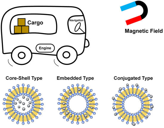
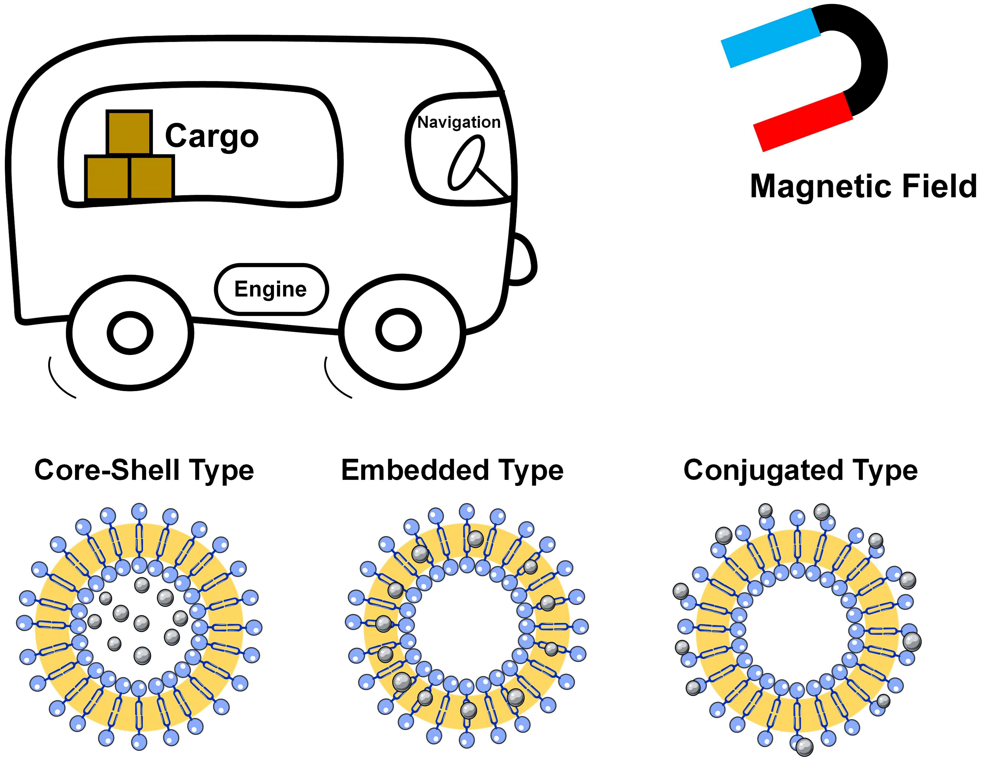
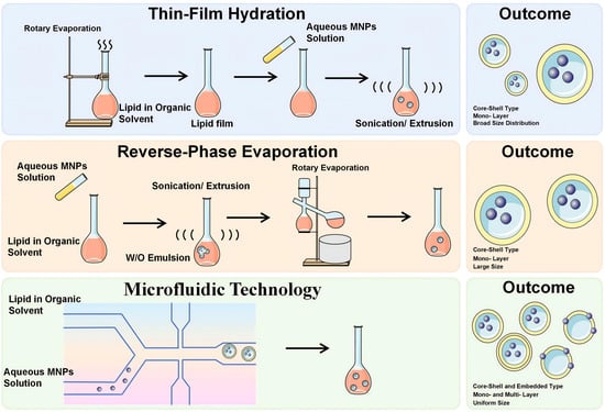
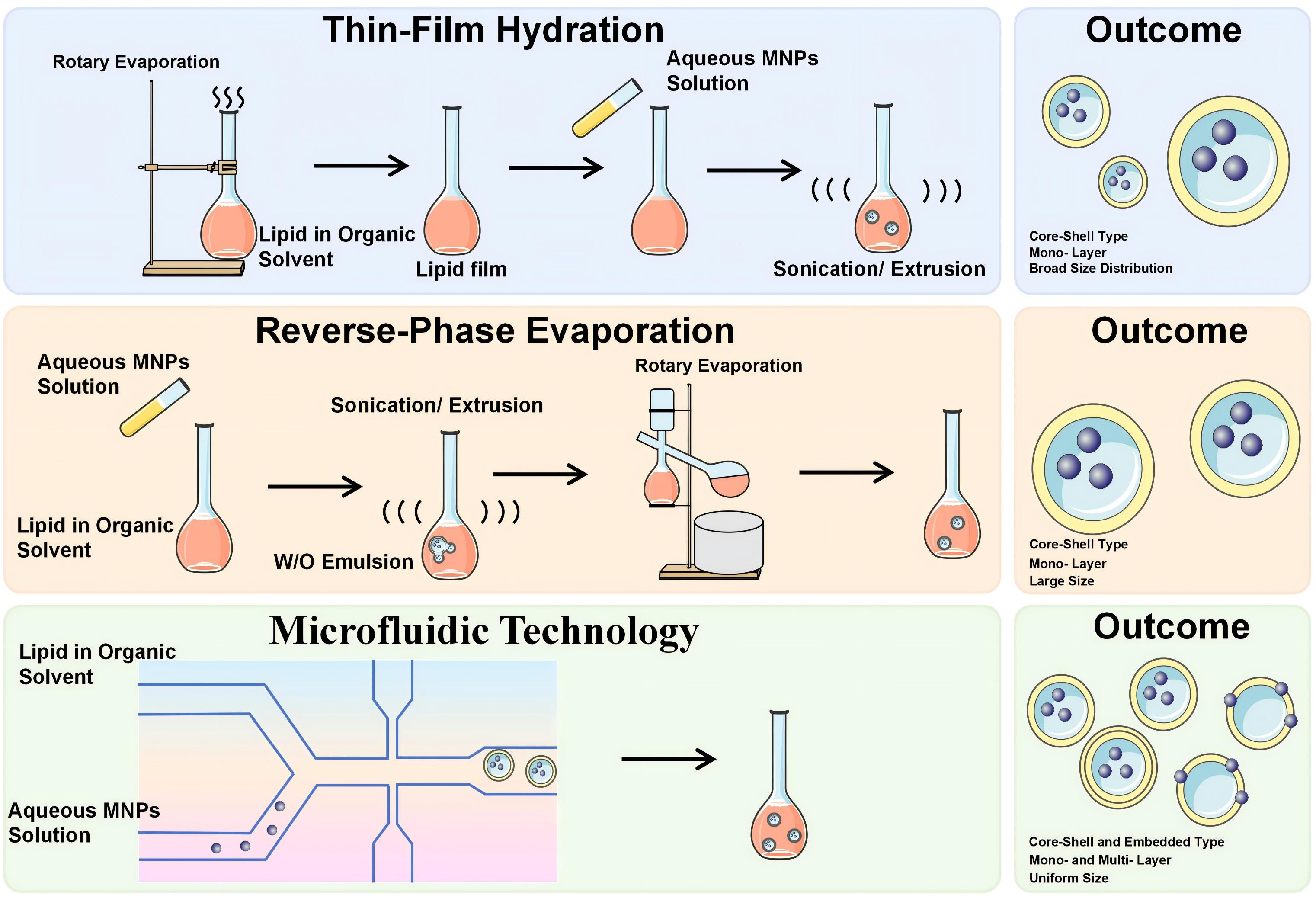
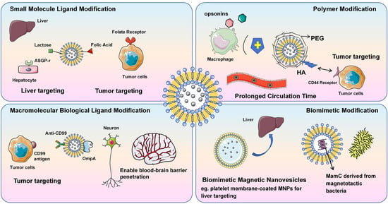
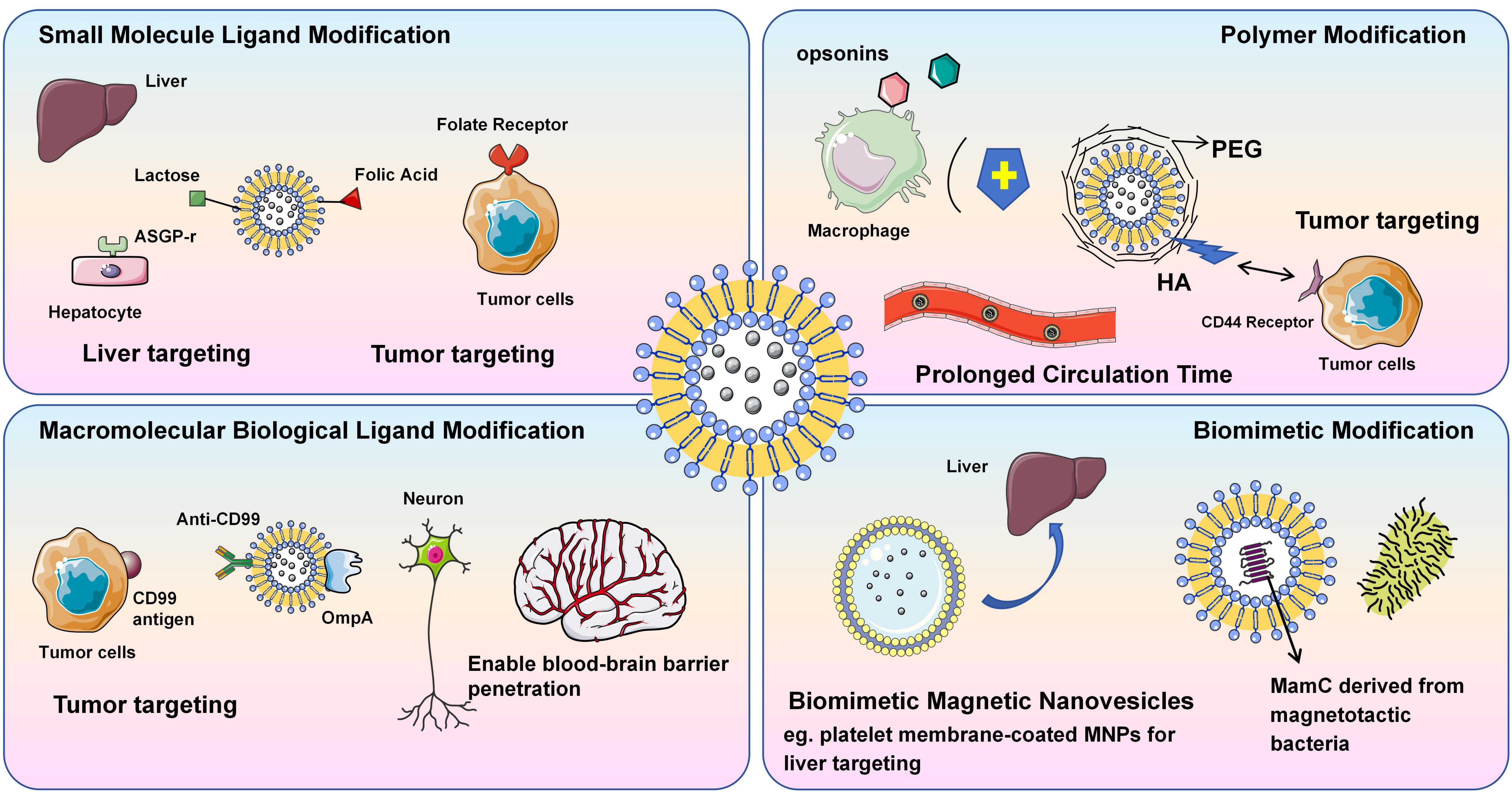
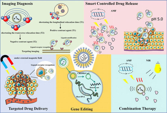
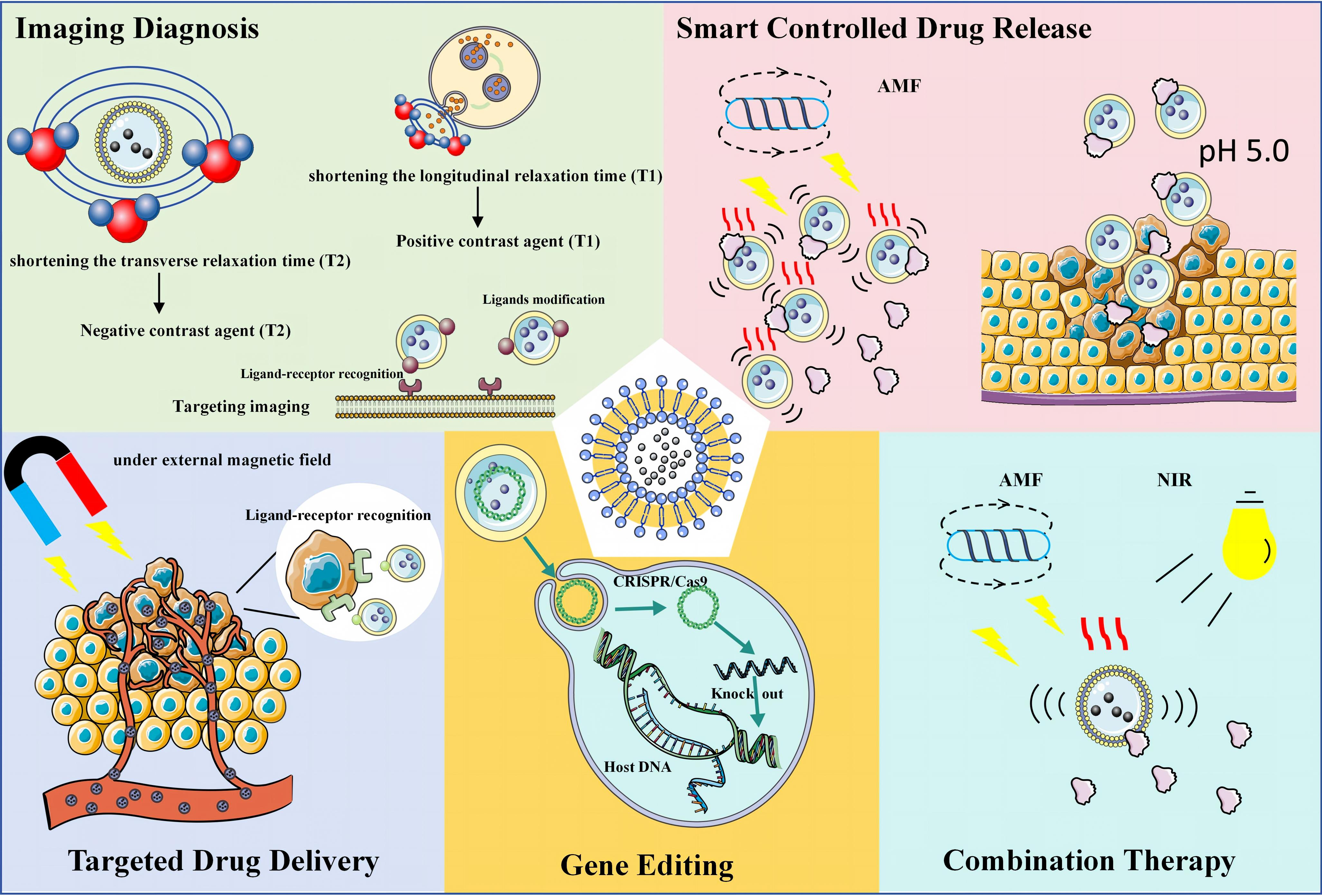

Disclaimer/Publisher’s Note: The statements, opinions and data contained in all publications are solely those of the individual author(s) and contributor(s) and not of MDPI and/or the editor(s). MDPI and/or the editor(s) disclaim responsibility for any injury to people or property resulting from any ideas, methods, instructions or products referred to in the content. |
© 2026 by the authors. Licensee MDPI, Basel, Switzerland. This article is an open access article distributed under the terms and conditions of the Creative Commons Attribution (CC BY) license.