


Abstract
Background/Objectives: Conventional modification of drug release often requires reformulation of the drug-containing dosage form. This study investigated fused deposition modelling (FDM) 3D-printed polymeric coats as external barriers for modifying the dissolution of intact immediate-release tablets without reformulating the drug-containing core. Methods: Commercial tablets were encapsulated within 3D-printed polyvinyl alcohol (PVA) or polylactic acid (PLA) coats incorporating a continuous axial channel with surface apertures of 0, 4.5, or 9 mm diameter. Printing accuracy and dimensional fidelity were evaluated, followed by dissolution testing. Release profiles were characterized using zero-order, first-order, Higuchi, Weibull, and Korsmeyer–Peppas models. Results: Printed coats showed high dimensional fidelity. Fully enclosed PVA coats produced a prolonged lag phase followed by rapid release associated with coat hydration and loss of structural integrity, whereas fully enclosed PLA coats showed no detectable drug release over 6 h. Increasing aperture diameter increased release from both polymers, although the magnitude and pattern of release differed markedly between PVA and PLA. At 2 h, PVA coats with 4.5 and 9 mm apertures released 89.5 ± 10.4% and 97.0 ± 15.6%, respectively, compared with 1.90 ± 0.13% and 58.8 ± 9.5% from the corresponding PLA designs. Korsmeyer–Peppas analysis indicated apparent anomalous release for PVA 4.5 mm (n = 0.647) and PLA 9 mm (n = 0.711), while PVA 9 mm exhibited Super Case-II-like behavior (n = 1.214). Conclusions: FDM-printed external polymeric coats can substantially modify dissolution from intact immediate-release tablets. Release behavior depended on the interaction between aperture geometry and polymer properties, demonstrating the potential of external 3D-printed barriers as a flexible approach for tailoring drug-release profiles without reformulating the tablet core.
1. Introduction
Modulating the temporal release profile of active pharmaceutical ingredients (APIs) is a fundamental objective in oral drug delivery design [1]. While immediate-release solid dosage forms provide rapid drug availability, modified-release systems can extend drug release and, where clinically appropriate, reduce dosing frequency and fluctuations in systemic drug concentrations [2,3]. Conventionally, modified release is achieved through matrix systems or functional film coatings, in which release is governed by interactions among polymer composition, dosage-form architecture, and medium ingress [4]. However, adapting an existing immediate-release product to achieve a different release profile typically requires reformulation and redevelopment of the dosage form, limiting the flexibility with which established formulations can be modified for different release requirements [5].
An alternative strategy is to retain the original tablet core and apply an engineered external barrier around the intact dosage form [3,6]. In reservoir-type architectures, the surrounding polymer can regulate solvent access to the tablet and subsequent drug transport into the dissolution medium [7]. Release from such systems may therefore depend not only on the physicochemical properties of the barrier material, including its thickness, permeability, hydration, and structural integrity, but also on the geometry of fluid-accessible openings within the barrier [3]. Controlling these geometric features independently of tablet formulation could modify dissolution behavior without altering the composition of the drug-containing core.
Additive manufacturing, particularly fused deposition modeling (FDM), provides a suitable platform for fabricating such geometrically defined structures [8]. FDM constructs are produced layer by layer from computer-aided design (CAD) models, allowing dimensions, internal architectures, and apertures to be precisely varied [9]. This design flexibility has enabled geometry to be investigated as a parameter for controlling drug release from 3D-printed dosage forms. When FDM is used to fabricate an external coat around an existing tablet rather than a drug-loaded matrix, the API does not need to be incorporated into the thermoplastic filament, thereby avoiding drug-loaded filament preparation and direct thermal processing of the API during hot-melt extrusion.
However, the effect of an aperture cannot be considered independently of the surrounding material. Hydrophilic polymers such as poly(vinyl alcohol) (PVA) undergo water uptake, swelling, chain relaxation, and ultimately dissolution or erosion, producing a barrier whose properties evolve during exposure to aqueous media [10,11,12,13]. In contrast, hydrophobic polymers such as poly(lactic acid) (PLA) remain comparatively stable under typical dissolution conditions, maintaining a more persistent physical barrier [14]. Consequently, nominally identical channel geometries may produce different dissolution profiles depending on the hydration, solubility, and structural response of the surrounding polymer. Although polymer composition and geometric architecture are both recognized determinants of drug release from 3D-printed dosage forms, their effects are frequently investigated simultaneously within drug-loaded or monolithic systems, making their individual and interactive contributions difficult to distinguish [8].
Encapsulating intact, pre-manufactured tablets within 3D-printed shells has emerged as a potential approach to modify drug release without reformulating the core [6,15,16]. Previous studies have used semi-solid extrusion (SSE) to deposit cellulose acetate or lipid-based shells around immediate-release tablets; however, these approaches typically require multi-component formulations, solvent-based preparations, molten materials, or subsequent drying steps, with release influenced by both shell composition and formulation variables such as pore-former concentration [6,16,17]. FDM has also been applied to compartmentalized and barrier-based dosage forms [15,18,19], although many FDM pharmaceutical systems rely on drug-loaded filaments prepared by hot-melt extrusion [18,19]. Direct FDM encapsulation of an intact commercial tablet using commercially available thermoplastic filaments therefore offers a comparatively simple post-manufacturing strategy in which the drug-containing core remains unchanged and the external polymer and aperture geometry can be varied independently. Avoiding solvent-based coating preparation and drug-loaded filament production may also reduce formulation and material-processing requirements, although the environmental impact of this approach requires dedicated assessment.
The novelty of the present study lies in systematically investigating the interplay between channel geometry and polymer properties in controlling drug release from intact commercial tablets. To achieve this, commercial immediate-release paracetamol tablets were enclosed within FDM-printed PVA and PLA coats, representing hydrophilic, water-responsive and comparatively hydrophobic, structurally persistent polymer systems, respectively. Fully enclosed coats and designs incorporating 4.5 or 9.0 mm through-channels were evaluated under matched geometric conditions, allowing the influence of aperture size to be compared across polymers with contrasting aqueous behavior. Paracetamol was selected as a readily quantifiable model drug to demonstrate the coating concept rather than to develop a specific therapeutic formulation.
The proposed system is intended as a proof-of-concept platform for modifying dissolution profiles through digitally designed external barriers, rather than as a clinically optimized modified-release product. Its potential advantage lies in enabling delayed or retarded release while retaining the original drug-containing core, without requiring tablet reformulation or the preparation of drug-loaded filaments.
2. Materials and Methods
2.1. Materials
Commercial immediate-release paracetamol tablets (500 mg) were used as the model dosage form (Fevadol®; SPIMACO Addwaeih, Al-Qassim, Saudi Arabia). PVA thermoplastic filament (AquaSolve PVA, natural, 1.75 mm; FormFutura, Nijmegen, The Netherlands; recommended printing temperature 190–210 °C) and PLA filament (black, 1.75 mm; Snapmaker, Shenzhen, China; recommended printing temperature 190–210 °C) were utilized as the coating feedstocks. The uncoated tablets had a mean diameter of 12.55 ± 0.00 mm, a thickness of 4.00 ± 0.00 mm, and a weight of 0.55 ± 0.01 g (n = 6).
Phosphate buffer (pH 5.8 ± 0.05) was prepared according to the Pharmacopoeia specifications using analytical-grade potassium dihydrogen phosphate (KH2PO4, anhydrous), sodium dihydrogen phosphate dihydrate (NaH2PO4·2H2O), and disodium hydrogen phosphate (Na2HPO4, anhydrous), which were purchased from Sigma-Aldrich (St. Louis, MO, USA). The pH was adjusted using 0.1 M sodium hydroxide (NaOH, analytical grade; Sigma-Aldrich, St. Louis, MO, USA). Ultrapure water (18.2 MΩ·cm Milli-Q system; Millipore, Bedford, MA, USA) was used for all buffer preparations and dilutions. All other reagents used were of analytical grade.
2.2. Coat Design and 3D Printing
2.2.1. Coat Geometry
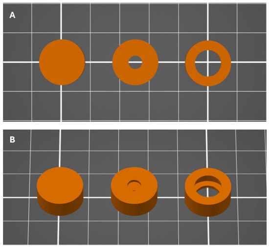
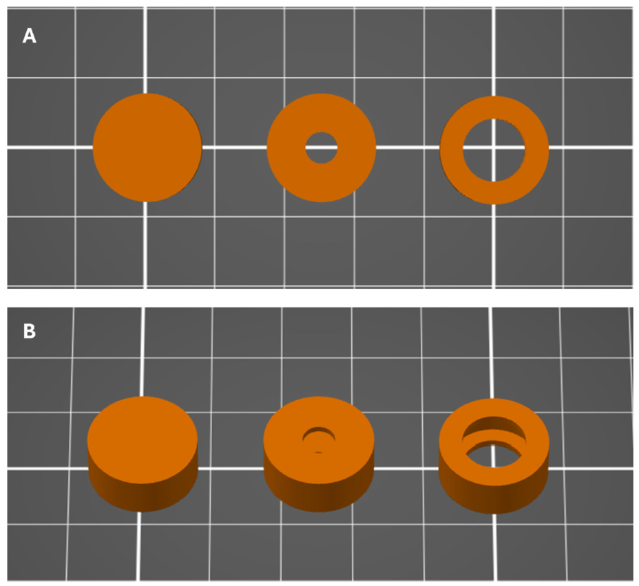
Polymeric barrier coats were designed as hollow cylindrical shells tailored to enclose a standard 500 mg commercial paracetamol tablet. Across all geometries, the coat maintained an outer diameter of 15.5 mm and a height of 6.5 mm, encasing an internal cavity with a diameter of 13.0 mm and a depth of 4.5 mm. The internal cavity was designed based on the dimensions of the commercial tablet to ensure a close fit. The tablet was held securely within the printed coat without observable movement. This geometric configuration established a uniform top and bottom wall thickness of 1.0 mm and a radial lateral wall thickness of 1.25 mm. Three distinct architectural variants were engineered for each polymer matrix, spanning a fully enclosed continuous shell without fluid access (0 mm) and two through-channel designs incorporating coaxial circular apertures of 4.5 mm and 9.0 mm on the opposing planar faces (Figure 1). The relative aperture area was calculated as the combined area of the two circular apertures created by the through-channel, expressed as a percentage of the total external surface area of the fully closed cylindrical coat:
where r is the channel radius, R is the outer radius of the coat, and h is the coat height.
Figure 1.
CAD models of the 3D-printed coat configurations: (A) Top planar view showing through-channel apertures, and (B) perspective isometric view. From left to right: fully closed (0 mm), 4.5 mm channel, and 9.0 mm channel architectures.
The corresponding aperture surface areas and calculated relative aperture areas for each formulation are summarized in Table 1. Three-dimensional CAD models were constructed using Fusion 360 (Autodesk Inc., San Rafael, CA, USA), exported in stereolithography (.STL) format, and sliced using PrusaSlicer (v2.7; Prusa Research a.s., Prague, Czech Republic).
Table 1.
Geometric specifications and theoretical relative aperture area of coat designs.
2.2.2. Printing Parameters
Polymeric coats were fabricated via FDM using a desktop 3D printer (Original Prusa i3 MK3.9, Prusa Research a.s., Prague, Czech Republic) fitted with a 0.4 mm brass nozzle. Tablet encapsulation was achieved using a sequential, interrupted-print protocol. In the initial phase, the lower base and surrounding lateral walls of the cylindrical shell were printed up to approximately 70% of total construct height. The print cycle was then paused via a G-code command, allowing an intact paracetamol tablet to be placed manually into the cavity. Printing was subsequently resumed to deposit the remaining 30% of the upper geometry, fully enclosing the core tablet within the polymeric coat.
Extrusion temperatures were maintained at 200 °C (within the optimized processing range of 190–210 °C) for both PVA and PLA filaments, with the heated build plate set to 60 °C to promote base-layer adhesion. A fixed layer height of 0.20 mm was used across all fabrications, accompanied by three concentric perimeter shells and a 25% grid infill pattern to ensure mechanical barrier integrity. Linear print speeds were configured at 40 mm/s for external perimeters and 60 mm/s for internal infill structures. All constructs were printed individually at the center of the build platform and inspected under magnification prior to use to verify layer cohesion and the absence of interfacial voids or delamination.
2.3. Printing Fidelity Assessment
To evaluate the reproducibility and accuracy of the 3D printing process, printed coat dimensions were systematically compared against their nominal CAD specifications. The outer diameter (D) and total height (h) of each fabricated coat were measured at three equidistant radial positions using a calibrated digital micrometer (SHAHE digital micrometer; Wenzhou Sanhe Measuring Instrument Co., Ltd., Wenzhou, China). Microstructural morphology, inter-layer fusion, surface finish, and aperture boundary definition were examined under a digital optical microscope (Gernie digital biological microscope; Gernie, China). The printed aperture diameter (Ape) for channel-containing designs was quantified from calibrated digital micrographs using ImageJ software (v1.54; National Institutes of Health, Bethesda, MD, USA). Dimensional fidelity was expressed as a percentage deviation from the nominal CAD dimensions:
where Xmeasured is the experimentally determined dimension and XCAD represents the corresponding theoretical CAD target value. Total effective external surface area (Anet) was subsequently calculated from the measured physical dimensions to assess macroscopic geometric shifts.
2.4. Analytical Quantification
Paracetamol concentration in the dissolution aliquots was quantified using a double-beam UV–Vis spectrophotometer (Libra S22, Biochrom Ltd., Cambridge, UK) at a detection wavelength of 243 nm (λmax for paracetamol in phosphate buffer, pH 5.8). A standard calibration curve was generated across a concentration range of 5–100 µg/mL. The calibration curve demonstrated good linearity over the investigated concentration range, with a coefficient of determination (R2) of 0.998. Samples exhibiting absorbance values exceeding the linear dynamic range of the calibration curve were diluted quantitatively with phosphate buffer (pH 5.8) prior to measurement, and the corresponding dilution factors were incorporated into the final concentration calculations. All measurements were performed in quartz cuvettes with a 10 mm optical path length against a buffer blank.
2.5. In Vitro Dissolution Testing
In vitro dissolution studies were performed using a USP Apparatus 1 (basket apparatus) dissolution tester (DT 70, Pharma Test Apparatebau GmbH, Hainburg, Germany). Each vessel contained 500 mL of phosphate buffer (pH 5.8) maintained at 37 ± 0.5 °C, and the baskets were rotated at 75 rpm. Dissolution studies were conducted in triplicate (n = 3) for each 3D-printed coating design and for the uncoated commercial reference tablets. The dissolution medium was selected based on the compendial medium specified for paracetamol tablets [20]. The conditions were modified from the USP monograph for paracetamol tablets. The basket (Apparatus 1) was used instead of the paddle to hold the printed constructs in a fixed position, to prevent the hydrating PVA coats from adhering to the vessel wall or paddle, and to allow the constructs to be retrieved for visual inspection. A rotation speed of 75 rpm, within the 50–100 rpm range commonly applied to Apparatus 1 [20], was selected to avoid stagnant zones around the construct inside the basket while retaining discriminatory power. Aliquots of 5.0 mL were withdrawn at predetermined time intervals (5, 10, 15, and 30 min, and 1, 2, 3, 4, 5, and 6 h). Withdrawn volumes were immediately replaced with an equivalent volume of pre-warmed (37 °C) fresh dissolution medium to preserve sink conditions and maintain a constant total volume throughout the 6 h dissolution period. The collected aliquots were passed through a 0.45 μm syringe filter and immediately analyzed by UV–Vis spectrophotometry at 243 nm. Cumulative drug release was determined after applying mathematical corrections for the cumulative mass removed during sampling.
2.6. Kinetic Modelling of Dissolution Profiles
Dissolution profiles were fitted to zero-order, first-order, Higuchi, Hixson–Crowell, Korsmeyer–Peppas, and Weibull models by nonlinear regression using the Levenberg–Marquardt algorithm in Python 3.12 (SciPy v1.11). The optimal model was identified using the Akaike Information Criterion (AIC), supported by the coefficient of determination (R2) and root-mean-square error (RMSE) [21,22].
For Korsmeyer–Peppas analysis, the release exponent (n) was determined using data corresponding to ≤60% cumulative drug release, where applicable. For formulations that did not reach 60% release during the 6 h dissolution study, the complete observed profile was used. In addition, because substantial structural changes in the open-channel PVA coats became evident after approximately 2 h, a separate early-phase analysis was performed over a fixed interval of 5–120 min for all open-channel designs. This enabled comparison of PVA and PLA during a common early-phase time window before the substantial later-stage structural changes observed for the PVA coats. Results obtained from the ≤60% release analysis and the fixed 5–120 min analysis were treated as separate fitting windows [23,24,25]. The discontinuous lag–burst profile of PVA 0 mm and the PLA 0 mm profile, for which no detectable drug release occurred during the 6 h study, were evaluated descriptively rather than by kinetic modeling.
The Korsmeyer–Peppas release exponent was interpreted according to the conventional framework for cylindrical systems [23,24,25]: n = 0.45 was considered consistent with Fickian (Case I) diffusion, 0.45 < n < 0.89 with anomalous (non-Fickian) transport, n = 0.89 with Case II transport, and n > 0.89 with Super Case II transport. Because the present system consisted of an intact tablet enclosed within an external polymeric barrier rather than a conventional drug-loaded matrix, these classifications were used as comparative descriptors of release behavior rather than as definitive evidence of a single underlying transport mechanism.
2.7. Statistical Analysis
All statistical analyses were performed in Python 3.12 using SciPy v1.11 and NumPy v1.26. Data are presented as mean ± standard deviation (SD). Statistical comparisons of dissolution data were performed at four prespecified time points (30 min, 1 h, 2 h, and 6 h). Within each polymer, the two open-channel designs (4.5 and 9.0 mm; n = 3 independent units per design) were compared using two-sided Welch’s t-tests. The fully enclosed (0 mm) designs were evaluated descriptively and were not included in the inferential comparisons of channel diameter.
Cross-polymer comparisons were performed between matched open-channel designs (PVA 4.5 mm vs. PLA 4.5 mm and PVA 9.0 mm vs. PLA 9.0 mm) using two-sided Welch’s t-tests at the same prespecified time points. A Bonferroni correction was applied separately across the four time-point comparisons within each comparison set, giving a corrected significance threshold of α = 0.0125 (0.05/4). All tests were two-sided, and exact p-values are reported where appropriate.
3. Results and Discussion
3.1. Printing Fidelity of Coat Designs
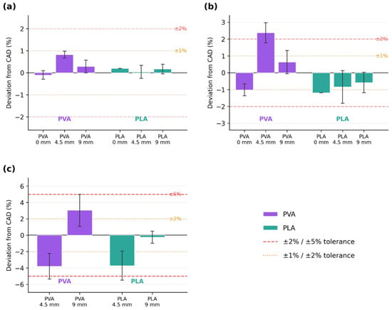
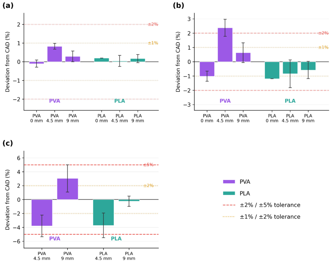
Dimensional characterization demonstrated good agreement between the printed coats and the nominal CAD dimensions (Table 2 and Figure 2). Across both polymers, deviations in outer diameter remained within ±1.0% of the nominal 15.5 mm diameter, while deviations in coat height were within ±2.4%. No consistent polymer-dependent trend toward dimensional over- or under-sizing was observed.
Table 2.
Dimensional characterization of printed coats. D: outer diameter; h: outer coat height; Ape: aperture diameter; Anet: net bilateral coat face area (summed area of both coat faces minus the aperture openings). All values are mean ± SD (n = 5).
Figure 2.
Dimensional fidelity of the printed coats, expressed as percentage deviation from CAD input values. (a) Outer diameter and (b) outer coat height for all six designs; (c) aperture diameter for the four channel-containing designs. Bars represent the mean; error bars represent ± SD (n = 5).
The aperture dimensions showed slightly greater variation than the outer dimensions. For the 4.5 mm designs, the measured aperture diameters were 4.329 ± 0.070 mm for PVA and 4.332 ± 0.080 mm for PLA, corresponding to deviations of −3.80% and −3.73%, respectively. For the 9.0 mm designs, the measured aperture diameters were 9.274 ± 0.177 mm for PVA and 8.978 ± 0.067 mm for PLA, corresponding to deviations of +3.04% and −0.24%, respectively. The calculated net coat face area (Anet) deviated by less than 2.5% of the nominal CAD values across all designs.
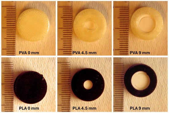
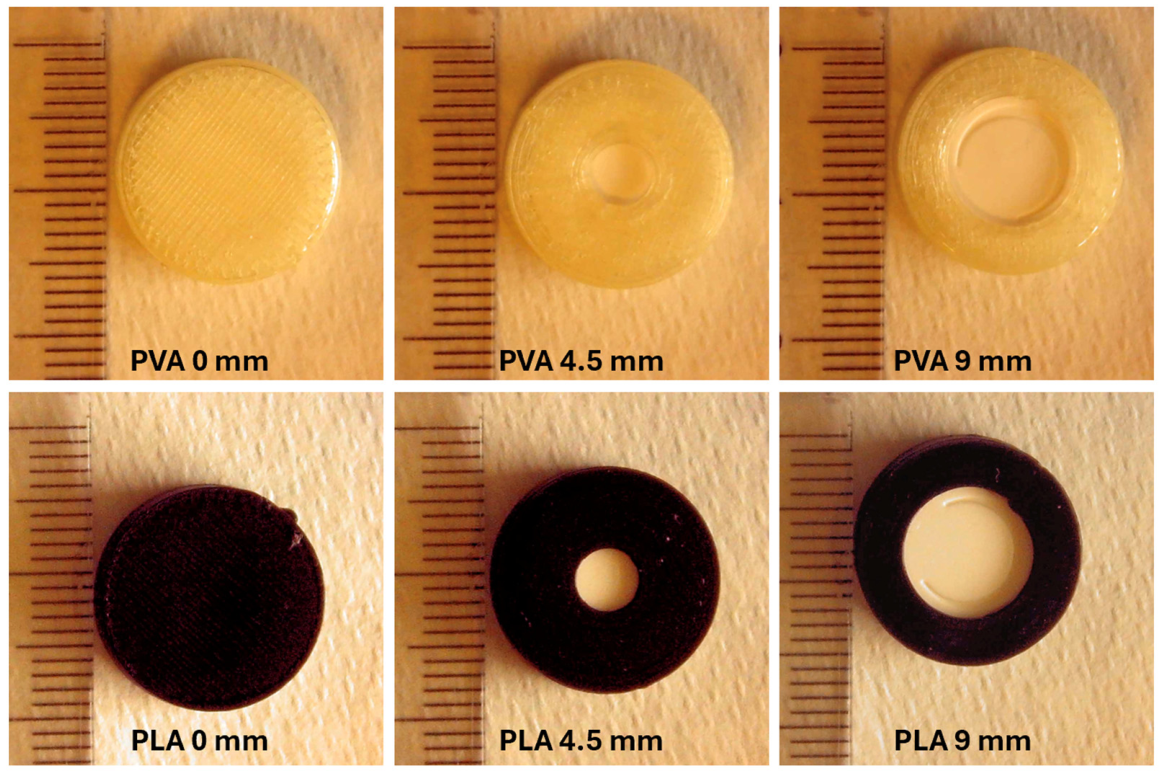
Visual examination of the fabricated coats confirmed continuous layer deposition without visible inter-filament voids, delamination, or structural distortion at the channel boundaries (Figure 3). These dimensional measurements confirmed that structural integrity and geometric accuracy of the printed coats were preserved, supporting subsequent comparison of their dissolution behavior.
Figure 3.
Digital photographs of printed coat designs. A graded ruler (1 mm divisions) is visible in each image for scale.
3.2. In Vitro Drug Dissolution
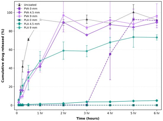
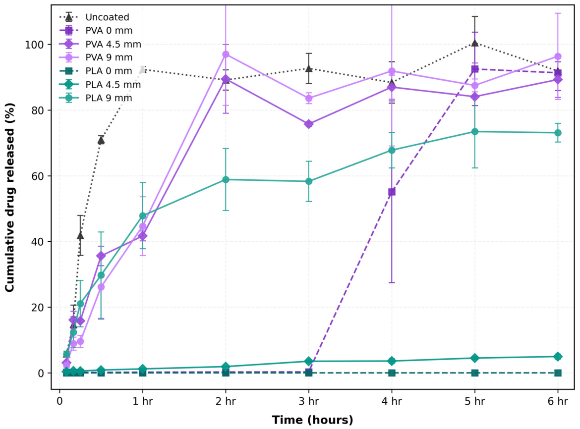
The uncoated commercial paracetamol tablets showed rapid drug release, with cumulative release of 41.8 ± 6.1% at 15 min, 71.0 ± 1.2% at 30 min, and 92.4 ± 0.8% at 60 min (Figure 4). Thereafter, measured cumulative release remained approximately constant, ranging from 88% to 101% over the remainder of the 6 h experiment. The uncoated tablet was used as an internal reference for assessing the effect of the 3D-printed polymeric coats on drug release rather than for evaluation of compendial dissolution compliance.
Figure 4.
Cumulative paracetamol release from uncoated tablets and tablets enclosed within PVA and PLA coats with 0, 4.5, and 9.0 mm channel diameters over 6 h in phosphate buffer (pH 5.8) at 37 ± 0.5 °C. Data are presented as mean ± SD (n = 3).
Marked differences in dissolution behavior were observed among the coated formulations depending on polymer type and channel diameter (Figure 4). Fully enclosed PVA and PLA coats produced distinctly different release profiles, while introduction of 4.5 and 9.0 mm channels increased drug release to different extents depending on the surrounding polymer.
3.2.1. PVA-Coated Tablets
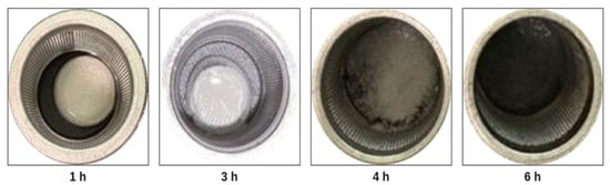
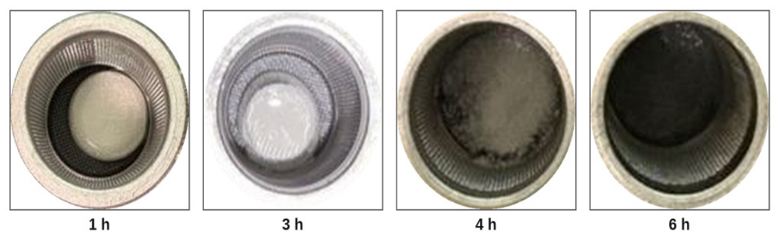
The fully enclosed PVA-coated tablets exhibited a distinct lag–burst release profile (Figure 4). Drug release remained below 0.3% during the first 3 h, indicating that the intact PVA coat effectively restricted drug release during this period. At approximately 4 h, a marked increase in drug release was observed, followed by cumulative release of approximately 87–98% by 6 h. Visual observations during dissolution showed progressive hydration of the PVA coat followed by loss of structural integrity (Figure 5). This behavior is consistent with the water-responsive nature of PVA, in which water penetration, swelling, polymer-chain mobility, and progressive dissolution can alter barrier properties over time [11,13,26,27,28].
Figure 5.
Representative images of the fully enclosed PVA-coated tablets at 1, 3, 4, and 6 h, showing progressive hydration and loss of coat integrity, with dissolution of the coat/tablet evident by 6 h.
The lag period followed by abrupt release may therefore reflect progressive hydration and weakening of the PVA barrier until structural integrity can no longer be maintained. The eventual rupture could also be influenced by internal pressure generated as dissolution medium penetrates the coat and the enclosed tablet begins to disintegrate and dissolve. Pressure-induced rupture of polymeric barriers following water penetration and expansion of an enclosed swelling component has been demonstrated in other pulsatile drug-delivery systems [29,30,31,32,33]. The timing and magnitude of the burst varied among individual dosage units, which may similarly reflect differences in the timing of hydration and structural failure. Because of the discontinuous nature of the lag–burst profile, kinetic models based on continuous release functions were not applied to the fully enclosed PVA design.
Introducing a 4.5 mm channel substantially altered the release profile by providing direct access between the tablet and the dissolution medium while the surrounding PVA coat remained present. Drug release reached 16.1 ± 2.6% at 10 min, 35.6 ± 3.0% at 30 min, 41.7 ± 1.5% at 60 min, and 89.5 ± 10.4% at 120 min (Figure 4). The release profile therefore showed an initial rapid phase followed by slower release before approaching high cumulative release by 2 h. The temporary slowing of release during the early portion of the profile could be associated with progressive hydration and swelling of PVA surrounding the channel, which may increase diffusional resistance as a hydrated polymer layer develops [11,13,26,27,28]. This interpretation is consistent with established behavior of hydrophilic polymer barriers, although changes in gel-layer thickness or diffusional resistance were not directly measured in the present study.
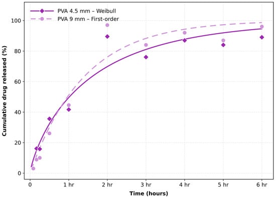
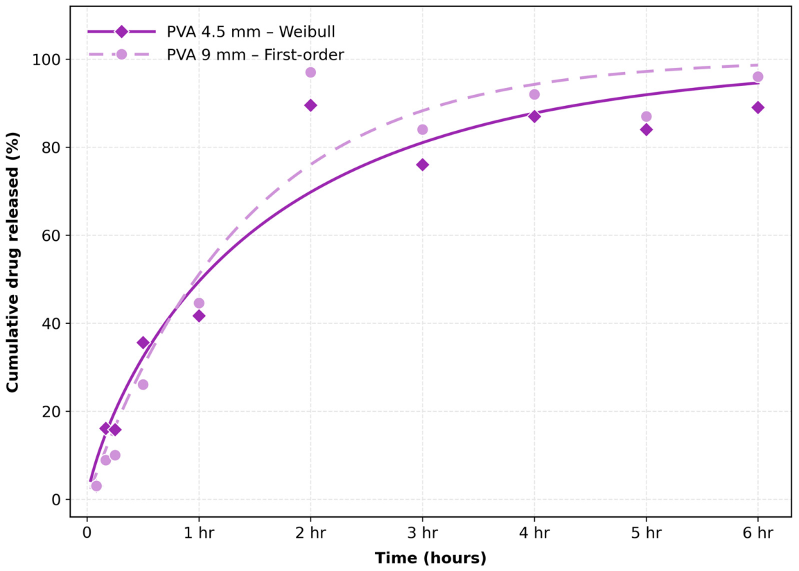
For the full dissolution profile of the PVA 4.5 mm design, the Weibull model provided the lowest AIC (45.41; R2 = 0.943; RMSE = 7.93), with a shape parameter (b) of 0.810. The first-order model showed a similar fit (AIC = 45.46), indicating that the overall profile could not be uniquely distinguished between these empirical models based on goodness-of-fit criteria alone. Korsmeyer–Peppas analysis within the ≤60% release window yielded an apparent release exponent of n = 0.647, with individual-vessel values ranging from 0.582 to 0.886. According to the conventional classification for cylindrical systems, this value is consistent with anomalous transport [23,25]. Such behavior is commonly associated with contributions from both diffusion and polymer relaxation in swellable polymer systems [23,24,25,34,35], and the observed exponent may therefore reflect the combined effects of drug diffusion through the fluid-accessible channel and the evolving hydration state of the surrounding PVA barrier. The fitting results for all evaluated models are presented in Supplementary Table S1.
Increasing the channel diameter to 9.0 mm produced a different release profile. Cumulative release was 8.9 ± 2.0% at 10 min and 26.1 ± 9.9% at 30 min, compared with 16.1 ± 2.6% and 35.6 ± 3.0%, respectively, for the 4.5 mm design. Thus, the larger channel did not produce greater release at the earliest measured time points. Release from the 9.0 mm design subsequently increased to 44.6 ± 8.9% at 60 min and 97.0 ± 15.6% at 120 min, compared with 41.7 ± 1.5% and 89.5 ± 10.4%, respectively, for the 4.5 mm design. Both open-channel PVA designs therefore approached high cumulative release within approximately 2 h, although differences in the shapes of their release profiles were observed over time.
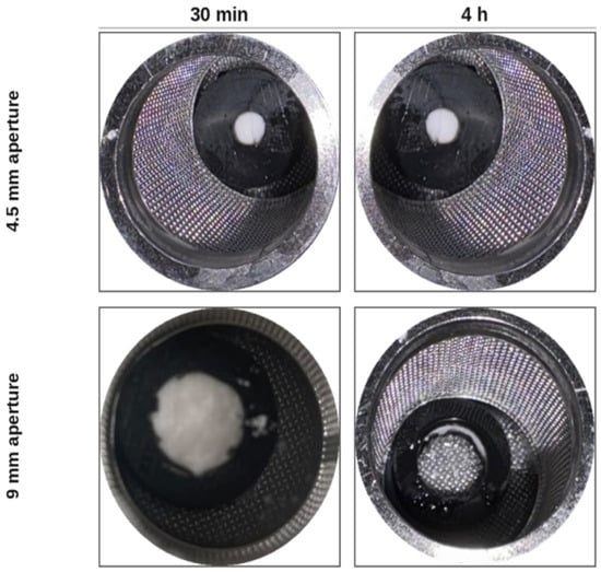
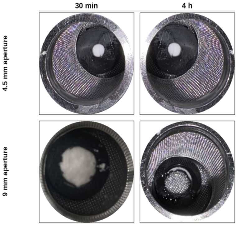
For the full dissolution profile of the PVA 9.0 mm design, the first-order model provided the lowest AIC (44.28; R2 = 0.953; k1 = 0.0119 min−1), while the Hixson–Crowell and Weibull models showed similar fits (ΔAIC < 0.5) (Figure 6). The similarity among these fits indicates that the overall dissolution profile alone does not permit a unique release mechanism to be assigned. Korsmeyer–Peppas analysis within the ≤60% release window yielded an apparent release exponent of n = 1.214, with individual-vessel values ranging from 1.029 to 1.368. According to the conventional classification for cylindrical systems, this corresponds to Super Case-II-like behavior [23,25]. Case II and Super Case-II transport are commonly associated with swelling-controlled systems in which solvent penetration and polymer-chain relaxation make substantial contributions to solute transport as the hydration front advances [34,35,36,37,38]. The high apparent exponent observed for the 9.0 mm design could therefore reflect an increased contribution of PVA hydration, swelling, and polymer relaxation as geometric restriction was reduced. This interpretation is also consistent with the progressive hydration and structural changes observed visually during dissolution (Figure 7), although the relative contributions of diffusion, polymer relaxation, and structural changes cannot be resolved from kinetic fitting alone.
Figure 6.
Best-fit kinetic model curves for PVA-coated designs (4.5 mm: Weibull; 9.0 mm: first-order) overlaid on observed mean release values. Symbols represent experimental data, while lines represent the fitted models.
Figure 7.
Representative images of PVA-coated tablets with 4.5 and 9.0 mm channel designs during dissolution at 30 min and 4 h.
The effect of channel diameter within the PVA-coated designs did not reach statistical significance at any of the evaluated time points (4.5 mm vs. 9.0 mm, all p > 0.18). Considerable inter-vessel variability was observed, particularly at later time points, which may be related to differences in the timing of PVA hydration and structural changes among individual dosage units. Nevertheless, the kinetic analyses showed different apparent release characteristics for the two designs, with anomalous transport for the 4.5 mm design (n = 0.647) and Super Case-II-like behavior for the 9.0 mm design (n = 1.214).
3.2.2. PLA-Coated Tablets
The fully enclosed PLA-coated tablets showed no detectable paracetamol release throughout the 6 h dissolution study (Figure 4), with measured values remaining at approximately 0% at all sampling time points. PLA is comparatively hydrophobic and exhibits substantially lower water uptake and greater structural persistence in aqueous environments than hydrophilic polymers such as PVA [14,39,40,41]. Under the conditions of the present experiment, the PLA coat remained macroscopically intact, and the absence of detectable release from the fully enclosed design indicates that it provided an effective barrier to drug release over the 6 h experimental period. The PLA 0 mm design was therefore evaluated descriptively and was not subjected to kinetic modeling.
Introducing a 4.5 mm channel resulted in slow but progressively increasing drug release. Cumulative release was 0.38 ± 0.19% at 5 min, 1.90 ± 0.13% at 2 h, and 4.96 ± 0.75% at 6 h (Figure 4). No burst release was observed, and cumulative release remained low throughout the experimental period. The persistence of slow release while the PLA coat remained macroscopically intact is consistent with a system in which the external polymeric barrier and restricted fluid-accessible geometry continue to limit drug transport. In contrast to PVA, the comparatively hydrophobic and structurally persistent nature of PLA would be expected to minimize time-dependent changes in the barrier during the experiment [14,39,40,42], although polymer hydration and microscopic structural changes were not directly quantified.
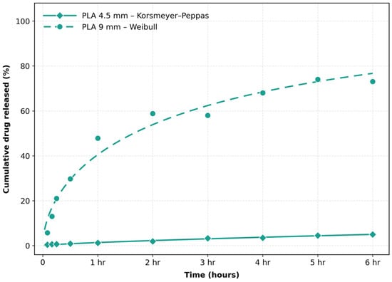
The Korsmeyer–Peppas model provided the best fit to the full PLA 4.5 mm dissolution profile (R2 = 0.982, RMSE = 0.224, AIC = −25.94), with kKP = 0.070 and an apparent release exponent of n = 0.725 (Figure 8). The Weibull model showed a nearly equivalent fit (ΔAIC = 0.04), indicating that model selection based on AIC alone was not unequivocal. Because cumulative release remained below 60% throughout the 6 h experiment, the complete observed profile also constituted the ≤60% release dataset, and the corresponding Korsmeyer–Peppas exponent was therefore n = 0.725. Individual-vessel exponents ranged from 0.543 to 0.730. Under the conventional classification for cylindrical systems, these values are consistent with anomalous transport [23,24,25].
Figure 8.
Best-fit kinetic model curves for the PLA open-channel designs (4.5 mm: Korsmeyer–Peppas; 9.0 mm: Weibull) overlaid on observed mean dissolution data. Symbols represent experimental data, while lines represent the fitted models.
In the separate fixed 5–120 min analysis, the apparent exponent for the PLA 4.5 mm design was n = 0.461, placing the early profile close to the conventional boundary between Fickian and anomalous transport. In a structurally persistent external barrier, deviation from ideal Fickian behavior could arise from geometric restrictions on solvent access and drug transport through the channel rather than from polymer relaxation alone. Geometric architecture is known to influence diffusional path length and dissolution behavior in 3D-printed dosage forms [39,43]. Accordingly, the observed kinetic behavior may reflect the combined effects of diffusion and channel-imposed transport resistance; however, geometric tortuosity was not directly measured in the present study.
The 9.0 mm PLA channel produced substantially greater drug release than the 4.5 mm design, although release remained incomplete at 6 h. Cumulative release reached 5.7 ± 0.8% at 5 min, 47.8 ± 10.0% at 1 h, 58.8 ± 9.5% at 2 h, and 73.1 ± 2.8% at 6 h (Figure 4). Between 2 and 6 h, cumulative release increased by approximately 14 percentage points, indicating a slower increase during the later portion of the dissolution profile. This deceleration could reflect a progressive reduction in the concentration gradient available to drive drug transport, although changes in the tablet core and other transport-related factors cannot be excluded.
Among the models evaluated, the Weibull model provided the best fit to the full PLA 9.0 mm profile (R2 = 0.972, RMSE = 4.06, AIC = 32.00), with a scale parameter (a) of 187.4 min and a shape parameter (b) of 0.576 (Figure 8). A Weibull shape parameter below unity describes a release profile with a decreasing release rate over time and is consistent with the observed progressive deceleration of the PLA 9.0 mm profile [44,45]. Because the Weibull model is empirical, however, these parameters describe the shape of the dissolution profile rather than uniquely identifying the underlying transport mechanism.
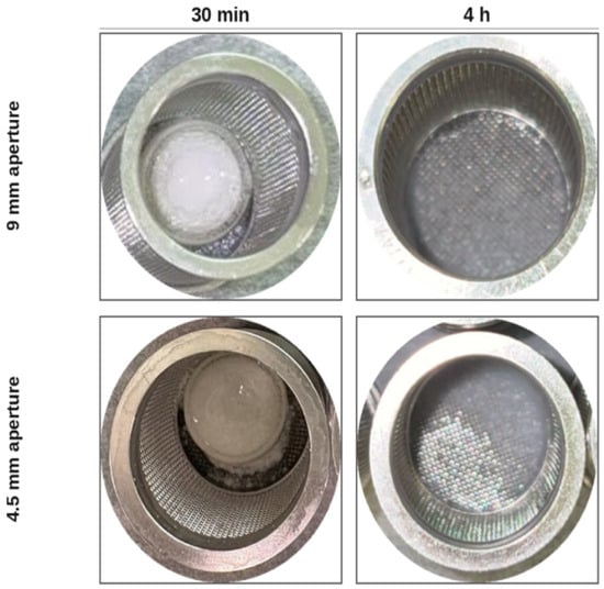
Korsmeyer–Peppas analysis within the ≤60% release window yielded an apparent release exponent of n = 0.711, with individual-vessel values ranging from 0.582 to 0.886. This is consistent with anomalous transport according to the conventional classification for cylindrical systems [23,24,25]. The larger channel provides a greater fluid-accessible area than the 4.5 mm design and could reduce geometric resistance to solvent ingress and drug transport, while the surrounding PLA barrier remains structurally persistent. The greater release observed with the 9.0 mm channel is therefore consistent with an important contribution of channel geometry to fluid penetration, subsequent tablet core disintegration, and drug transport through the PLA-coated system (Figure 9).
Figure 9.
Representative images of PLA-coated tablets with 4.5 and 9.0 mm channel designs during dissolution at 30 min and 4 h.
The influence of channel diameter within the PLA-coated designs became statistically evident at the later time points. The 9.0 mm design showed greater cumulative drug release than the 4.5 mm design at all evaluated time points. Using two-sided Welch’s t-tests with a Bonferroni-corrected significance threshold of α = 0.0125, the difference was not statistically significant at 30 min (p = 0.0625) or 1 h (p = 0.0150), but reached statistical significance at 2 h (p = 0.0090) and 6 h (p < 0.001). These results indicate that the difference in cumulative drug release between the two PLA channel designs became more clearly distinguishable as dissolution progressed.
3.2.3. Cross-Polymer Comparison: Interaction Between Channel Geometry and Polymer Properties
The first 2 h of dissolution provided a common experimental window for direct comparison of the PVA- and PLA-coated systems before the substantial later-stage structural changes observed for PVA. During this interval, drug release from both systems occurred in the presence of the printed polymeric barriers, allowing the combined influence of channel geometry and polymer properties on early release behavior to be evaluated under equivalent dissolution conditions. Cross-polymer release profiles for matched channel designs are shown in Figure 4.
As summarized in Table 3, with the complete model-fitting results provided in Supplementary Table S2, the fixed 5–120 min kinetic analysis provided a common early-phase window for comparing release behavior across the open-channel designs. At the 4.5 mm channel, the fixed 5–120 min Korsmeyer–Peppas analysis yielded apparent release exponents of n = 0.647 for PVA and n = 0.461 for PLA, both within the conventional anomalous transport range, although the PLA value was close to the Fickian boundary [23,24,25]. In the PVA system, the anomalous exponent could reflect contributions from both concentration-gradient-driven diffusion and changes in the hydrated polymer network, as polymer hydration and chain relaxation can introduce time-dependent resistance to solute transport in swellable systems [34,35,36,44]. In contrast, PLA remained macroscopically intact over the experimental period, and the near-Fickian exponent observed during the early phase may therefore be more consistent with diffusion occurring under geometric restriction imposed by the channel and surrounding polymeric barrier [39,46]. These interpretations should be considered in the context of the different physical properties of the two polymers, since the kinetic exponents alone cannot uniquely identify the underlying transport mechanisms.
Despite their broadly similar kinetic classifications, the two 4.5 mm designs differed markedly in the magnitude of drug release. At 2 h, cumulative release was 89.5 ± 10.4% from the PVA-coated tablets compared with only 1.90 ± 0.13% from the PLA-coated tablets. Because the channel dimensions were identical, this large difference indicates an important contribution of polymer properties to the release behavior. The progressive hydration and loss of structural integrity of PVA, compared with the greater aqueous persistence of PLA, could contribute to this divergence [27,39,40].
At the 9.0 mm channel, the difference in apparent kinetic behavior between the polymers was more pronounced. PVA yielded an apparent release exponent of n = 1.214 (individual-vessel range: 1.029–1.368), corresponding to Super Case-II-like behavior according to the conventional classification, whereas PLA yielded n = 0.711 (individual-vessel range: 0.582–0.886), corresponding to anomalous transport [23,24,25]. Case II and Super Case-II behavior in swellable polymer systems has been associated with an increasing contribution of solvent penetration and polymer-chain relaxation to solute transport [34,35,36]. The higher apparent exponent observed for PVA may therefore reflect the increasing influence of hydration and polymer relaxation, whereas release through the macroscopically persistent PLA coat remained constrained by the fluid-accessible channel and surrounding barrier. Thus, identical channel dimensions produced substantially different kinetic behavior depending on the polymer used.
By 2 h, cumulative release reached 97.0 ± 15.6% for PVA 9.0 mm compared with 58.8 ± 9.5% for PLA 9.0 mm, corresponding to an absolute difference of approximately 38 percentage points. Together with the 4.5 mm results, these observations indicate that channel geometry and polymer properties should not be considered independently: the effect of a given channel dimension depends on the physical response of the surrounding polymer to the dissolution environment.
The cross-polymer statistical comparisons further illustrate these differences. For the 4.5 mm designs, cumulative release differed significantly between PVA and PLA at the prespecified comparison times after application of the Bonferroni-corrected significance threshold. In contrast, differences between the PVA and PLA 9.0 mm designs were not statistically significant at 30 min, 1 h, or 2 h under the corrected threshold. The absence of statistical significance for the 9.0 mm comparison should be interpreted cautiously given the small sample size (n = 3 per design) and the associated limited statistical power. Nevertheless, the results suggest that increasing channel diameter reduced the magnitude of the early difference between the two polymer systems.
Beyond 2 h, the physical states of the PVA and PLA barriers increasingly diverged. The PVA systems underwent substantial hydration and progressive loss of structural integrity, whereas the PLA coats remained macroscopically intact throughout the 6 h experiment. Consequently, later-stage differences between the polymers reflect not only channel geometry but also their contrasting responses to the aqueous dissolution environment.
This distinction was particularly evident for the PLA 4.5 mm design, which showed the greatest release retardation among the open-channel formulations. Cumulative release increased from 1.90 ± 0.13% at 2 h to 4.96 ± 0.75% at 6 h and had not reached an observable plateau by the end of the experiment. The continued increase while the PLA coat remained macroscopically intact is consistent with prolonged restriction of drug transport by the external barrier and narrow channel. Similar studies have demonstrated that 3D-printed external structures and geometrical modification can be used to alter drug-release behavior [6,9,15,16,18,19,36]. However, because only approximately 5% of the drug was released within 6 h, longer dissolution studies would be required to determine the subsequent release trajectory and whether complete drug release can ultimately be achieved.
The PLA 9.0 mm design also showed slower release during the later portion of the experiment, increasing from 58.8 ± 9.5% at 2 h to 73.1 ± 2.8% at 6 h. The full profile was best described by the Weibull model (R2 = 0.972, RMSE = 4.06, AIC = 32.00), with a scale parameter of a = 187.4 min and a shape parameter of b = 0.576. A Weibull shape parameter below unity describes a release profile with a decreasing release rate over time [44,45], consistent with the observed deceleration. This later-stage slowing could reflect a reduction in the concentration gradient available to drive drug transport as dissolution progressed, although other changes within the tablet core or transport pathway cannot be excluded. Because release remained incomplete at 6 h, the present data do not establish the eventual plateau or total release achievable from this design. These findings indicate that geometric tunability is polymer-dependent, with channel diameter exerting a pronounced influence on drug release from PLA coats, whereas its effect was less pronounced for PVA coats, potentially due to their hydration and subsequent structural changes.
3.2.4. Design Implications and Release Kinetics
The results demonstrate that channel geometry and polymer properties jointly influence drug release from the 3D-printed coated tablets. Channel diameter determines the area available for direct interaction between the tablet and dissolution medium, while the properties of the surrounding polymer influence how the barrier responds during dissolution. Previous studies have demonstrated that geometry can substantially modify drug release from 3D-printed dosage forms [9,10,43,47], and the present findings extend this principle by showing that identical channel dimensions can produce markedly different dissolution profiles when implemented using polymers with contrasting aqueous behavior.
The lag–burst behavior of the fully enclosed PVA tablets could be associated with progressive hydration and weakening of the polymeric barrier, followed by loss of structural integrity. Similar pressure-induced rupture mechanisms have been described for barrier-controlled drug-delivery systems [32,48,49], although internal pressure and mechanical strength were not measured in the present study and the precise mechanism of coat rupture therefore remains to be established. The full-profile kinetic modelling results are summarized in Table 4. The apparent Super Case-II-like behavior observed for the PVA 9.0 mm design (n = 1.214) is consistent with an increased contribution of solvent penetration and polymer-chain relaxation, processes associated with Case II and Super Case-II transport in glassy and semicrystalline polymer systems [34,35,36,37,38]. In contrast, the anomalous or near-Fickian behavior observed for the PLA designs occurred while the polymeric barrier remained macroscopically intact, suggesting a greater contribution of diffusion under geometric restriction rather than the pronounced hydration-dependent structural changes observed for PVA [23,39,46].
Previous work has shown that changing the geometry and exposed surface area of 3D-printed dosage forms can substantially alter dissolution behavior [9,10]. The present study extends this concept to matched channel geometries in external polymeric coats surrounding intact commercial tablets. At identical channel dimensions, PVA and PLA produced markedly different release profiles and apparent kinetic characteristics, indicating that geometric design cannot be considered independently of the properties of the surrounding polymer. Rather, the effect of a given channel geometry appears to depend on how the polymer interacts with the aqueous dissolution environment.
These findings can also be considered in the context of previous approaches using printed external barriers and geometry-controlled dosage forms. Internal geometry has been used to modify release from FDM-printed dosage forms [9,10], while external printed shells have been applied around immediate-release cores to alter dissolution as a function of shell composition and design [6,15,16]. In the present approach, the coat was fabricated by FDM around an intact commercial tablet using commercially available thermoplastic filaments, thereby avoiding the preparation of drug-loaded filaments and direct thermal compounding of the API. Drug release was modified through the combination of polymer selection and a discrete geometric channel while maintaining the original drug-containing tablet core.
From a formulation-design perspective, polymer properties and channel geometry therefore represent coupled design parameters. With the 9.0 mm channel, PVA produced approximately 97% release within 2 h, whereas PLA produced approximately 59% at 2 h and 73% at 6 h. With the 4.5 mm channel, PVA reached approximately 90% release by 2 h, whereas PLA released only approximately 5% over 6 h. These findings demonstrate that substantial differences in dissolution behavior can be generated from the same tablet core by modifying the material and geometry of the external coat. This approach may therefore provide a flexible basis for further investigation of customized release profiles without reformulating the drug-containing core.
Table 3.
Kinetic model fitting results for PVA- and PLA-coated designs based on dissolution experiments performed in triplicate (n = 3), using 10 sampling time points for the full-profile analysis. The best model was identified by the lowest AIC. KP n values were determined from the Q ≤ 60% subset.
Table 4.
Early-phase kinetic analysis over a fixed 5–120 min interval. The Korsmeyer–Peppas (KP) release exponent was determined within the ≤60% release portion of this interval.
4. Conclusions
This study demonstrates the feasibility of using FDM 3D-printed polymeric coats to modulate the dissolution of intact, commercially available immediate-release paracetamol tablets without reformulating the drug-containing core. The dimensional fidelity of the printed designs enabled systematic evaluation of the combined effects of channel geometry and polymer type on dissolution behavior.
Drug release was strongly influenced by the interaction between channel diameter and polymer properties. PVA coats exhibited time-dependent hydration and loss of structural integrity, with open-channel designs showing release profiles characterized by anomalous to Super Case-II-like kinetics. In contrast, PLA coats remained macroscopically intact throughout the 6 h dissolution study and produced substantially greater release retardation, with a pronounced dependence on channel diameter. These findings demonstrate that identical channel geometries can produce markedly different dissolution profiles depending on the polymer used, highlighting the importance of considering material properties and geometric design together.
Direct encapsulation of finished tablets using commercially available thermoplastic filaments offers a solvent-free, post-manufacturing approach that avoids drug-loaded filament preparation and direct thermal processing of the API during hot-melt extrusion. This strategy may provide a flexible platform for developing customized dissolution profiles while retaining the original tablet formulation.
Further investigations are needed to establish the broader pharmaceutical applicability of this approach, including additional geometries, longer dissolution periods, direct characterization of polymer hydration and structural changes, and post-print stability assessment. Evaluation under physiologically relevant gastrointestinal conditions is also necessary to determine the in vivo relevance of the observed release profiles. In particular, the fate, transit, and elimination of residual PLA shells require investigation before clinical application.
Supplementary Materials
The following supporting information can be downloaded at: https://www.mdpi.com/article/10.3390/pharmaceutics18111400/s1, Table S1: Kinetic model fitting of mean dissolution profiles—full profile (0–6 h). All six models fitted per design; best model (lowest AIC) shaded and marked ✓. Parameters shown for the best-fit model; Table S2: Kinetic model fitting—early phase (5–120 min). Best model (lowest AIC) shaded and markedd.
Author Contributions
R.F.: conceptualization, data curation, formal analysis, methodology, and writing. A.A.: data curation, formal analysis, methodology and writing. N.A.: data curation, formal analysis, and writing. R.F.A. (Riyad F. Alzhrani): formal analysis and writing. F.A.: data curation and formal analysis. R.H.B.: data curation and methodology. N.M.S.: data curation, formal analysis and writing. R.F.S.: data curation, formal analysis and writing. R.F.A. (Rihaf F. Alfaraj): data curation, formal analysis, and writing. All authors have read and agreed to the published version of the manuscript.
Funding
This research project was supported by the Ongoing Research Funding Program. (ORF-2026-1435), King Saud University, Riyadh, Saudi Arabia.
Data Availability Statement
Data is contained within the article.
Conflicts of Interest
The authors declare no conflicts of interest.
References
- Sadosky, A.; Srivastava, K.; Arora, A.; Kataria, A.; Cappelleri; Peterson, A.M. Impact of reducing dosing frequency on adherence to oral therapies: A literature review and meta-analysis. Patient Prefer. Adherence 2013, 7, 419–434. [Google Scholar] [CrossRef] [Scilit] [PubMed]
- Moodley, K.; Pillay, V.; Choonara, Y.E.; Du Toit, L.C.; Ndesendo, V.M.K.; Kumar, P.; Cooppan, S.; Bawa, P. Oral Drug Delivery Systems Comprising Altered Geometric Configurations for Controlled Drug Delivery. Int. J. Mol. Sci. 2011, 13, 18–43. [Google Scholar] [CrossRef] [Scilit] [PubMed]
- Bermejo, M.; Sanchez-Dengra, B.; Gonzalez-Alvarez, M.; Gonzalez-Alvarez, I. Oral controlled release dosage forms: Dissolution versus diffusion. Expert Opin. Drug Deliv. 2020, 17, 791–803. [Google Scholar] [CrossRef] [Scilit] [PubMed]
- Tiwari, S.B.; Rajabi-Siahboomi, A.R. Extended-Release Oral Drug Delivery Technologies: Monolithic Matrix Systems; Springer: Berlin/Heidelberg, Germany, 2008; pp. 217–243. [Google Scholar] [CrossRef] [Scilit] [PubMed]
- Kuentz, M.; Holm, R.; Elder, D.P. Methodology of oral formulation selection in the pharmaceutical industry. Eur. J. Pharm. Sci. 2016, 87, 136–163. [Google Scholar] [CrossRef] [Scilit] [PubMed]
- Algahtani, M.S.; Mohammed, A.A.; Ahmad, J.; Saleh, E. Development of a 3D Printed Coating Shell to Control the Drug Release of Encapsulated Immediate-Release Tablets. Polymers 2020, 12, 1395. [Google Scholar] [CrossRef] [Scilit] [PubMed]
- Coelho, J.F.; Ferreira, P.C.; Alves, P.; Cordeiro, R.; Fonseca, A.C.; Góis, J.R.; Gil, M.H. Drug delivery systems: Advanced technologies potentially applicable in personalized treatments. EPMA J. 2010, 1, 164–209. [Google Scholar] [CrossRef] [Scilit] [PubMed]
- Paccione, N.; Guarnizo-Herrero, V.; Ramalingam, M.; Larrarte, E.; Pedraz, J.L. Application of 3D printing on the design and development of pharmaceutical oral dosage forms. J. Control. Release 2024, 373, 463–480. [Google Scholar] [CrossRef] [Scilit] [PubMed]
- Goyanes, A.; Buanz, A.B.M.; Basit, A.W.; Gaisford, S. Fused-filament 3D printing (3DP) for fabrication of tablets. Int. J. Pharm. 2014, 476, 88–92. [Google Scholar] [CrossRef] [Scilit] [PubMed]
- Xu, X.; Zhao, J.; Wang, M.; Wang, L.; Yang, J. 3D Printed Polyvinyl Alcohol Tablets with Multiple Release Profiles. Sci. Rep. 2019, 9, 12487. [Google Scholar] [CrossRef] [Scilit] [PubMed]
- Narasimhan, B.; Peppas, N.A. Molecular Analysis of Drug Delivery Systems Controlled by Dissolution of the Polymer Carrier. J. Pharm. Sci. 1997, 86, 297–304. [Google Scholar] [CrossRef] [Scilit] [PubMed]
- Ju, R.T.C.; NixoN, P.R.; Patel, M.V.; Tong, D.M. Drug Release From Hydrophilic Matrices. 2. A Mathematical Model Based on the Polymer Disentanglement Concentration and the Diffusion Layer. J. Pharm. Sci. 1995, 84, 1464–1477. [Google Scholar] [CrossRef] [Scilit] [PubMed]
- Snaar, J.E.M.; Bowtell, R.; Melia, C.D.; Morgan, S.; Narasimhan, B.; Peppas, N.A. Self-diffusion and molecular mobility in PVA-based dissolution-controlled systems for drug delivery. Magn. Reson Imaging 1998, 16, 691–694. [Google Scholar] [CrossRef] [Scilit] [PubMed]
- Liao, Y.; Liu, C.; Coppola, B.; Barra, G.; Di Maio, L.; Incarnato, L.; Lafdi, K. Effect of Porosity and Crystallinity on 3D Printed PLA Properties. Polymers 2019, 11, 1487. [Google Scholar] [CrossRef] [Scilit] [PubMed]
- Skalická, B.; Matzick, K.; Komersová, A.; Svoboda, R.; Bartoš, M.; Hromádko, L. 3D-Printed Coating of Extended-Release Matrix Tablets: Effective Tool for Prevention of Alcohol-Induced Dose Dumping Effect. Pharmaceutics 2021, 13, 2123. [Google Scholar] [CrossRef] [Scilit] [PubMed]
- Tsintavi, E.; Rekkas, D.M.; Bettini, R. Partial tablet coating by 3D printing. Int. J. Pharm. 2020, 581, 119298. [Google Scholar] [CrossRef] [Scilit] [PubMed]
- Hu, J.; Fitaihi, R.; Abukhamees, S.; Abdelhakim, H.E. Formulation and Characterisation of Carbamazepine Orodispersible 3D-Printed Mini-Tablets for Paediatric Use. Pharmaceutics 2023, 15, 250. [Google Scholar] [CrossRef] [Scilit] [PubMed]
- Okwuosa, T.C.; Pereira, B.C.; Arafat, B.; Cieszynska, M.; Isreb, A.; Alhnan, M.A. Fabricating a Shell-Core Delayed Release Tablet Using Dual FDM 3D Printing for Patient-Centred Therapy. Pharm. Res. 2017, 34, 427–437. [Google Scholar] [CrossRef] [Scilit] [PubMed]
- Li, R.; Pan, Y.; Chen, D.; Xu, X.; Yan, G.; Fan, T. Design, Preparation and In vitro Evaluation of Core–Shell Fused Deposition Modelling 3D-Printed Verapamil Hydrochloride Pulsatile Tablets. Pharmaceutics 2022, 14, 437. [Google Scholar] [CrossRef] [Scilit] [PubMed]
- USP. ⟨1092⟩ The Dissolution Procedure: Development and Validation; U.S. Pharmacopeial Convention: Rockville, MD, USA, 2026. [Google Scholar] [CrossRef] [Scilit]
- Burnham, K.P.; Anderson, D.R. (Eds.) Model Selection and Multimodel Inference; Springer: New York, NY, USA, 2004. [Google Scholar] [CrossRef] [Scilit]
- Akaike, H. A new look at the statistical model identification. IEEE Trans. Autom. Contr 1974, 19, 716–723. [Google Scholar] [CrossRef] [Scilit]
- Korsmeyer, R.W.; Gurny, R.; Doelker, E.; Buri, P.; Peppas, N.A. Mechanisms of solute release from porous hydrophilic polymers. Int. J. Pharm. 1983, 15, 25–35. [Google Scholar] [CrossRef] [Scilit]
- Ritger, P.L.; Peppas, N.A. A simple equation for description of solute release I. Fickian and non-fickian release from non-swellable devices in the form of slabs, spheres, cylinders or discs. J. Control. Release 1987, 5, 23–36. [Google Scholar] [CrossRef] [Scilit]
- Peppas, N.A. Analysis of Fickian and non-Fickian drug release from polymers. Pharm. Acta Helv. 1985, 60, 110–111. [Google Scholar] [PubMed]
- Peppas, N.A.; Merrill, E.W. Crosslinked poly(vinyl alcohol) hydrogels as swollen elastic networks. J. Appl. Polym. Sci. 1977, 21, 1763–1770. [Google Scholar] [CrossRef] [Scilit]
- Basa, B.; Jakab, G.; Kállai-Szabó, N.; Borbás, B.; Fülöp, V.; Balogh, E.; Antal, I. Evaluation of Biodegradable PVA-Based 3D Printed Carriers during Dissolution. Materials 2021, 14, 1350. [Google Scholar] [CrossRef] [Scilit] [PubMed]
- Tagami, T.; Nagata, N.; Hayashi, N.; Ogawa, E.; Fukushige, K.; Sakai, N.; Ozeki, T. Defined drug release from 3D-printed composite tablets consisting of drug-loaded polyvinylalcohol and a water-soluble or water-insoluble polymer filler. Int. J. Pharm. 2018, 543, 361–367. [Google Scholar] [CrossRef] [Scilit] [PubMed]
- Goyanes, A.; Buanz, A.B.M.; Hatton, G.B.; Gaisford, S.; Basit, A.W. 3D printing of modified-release aminosalicylate (4-ASA and 5-ASA) tablets. Eur. J. Pharm. Biopharm. 2015, 89, 157–162. [Google Scholar] [CrossRef] [Scilit] [PubMed]
- Markl, D.; Zeitler, J.A. A Review of Disintegration Mechanisms and Measurement Techniques. Pharm. Res. 2017, 34, 890–917. [Google Scholar] [CrossRef] [Scilit] [PubMed]
- Siepmann, J. Modeling of drug release from delivery systems based on hydroxypropyl methylcellulose (HPMC). Adv. Drug Deliv. Rev. 2001, 48, 139–157. [Google Scholar] [CrossRef] [Scilit] [PubMed]
- Lin, H.; Lin, S.; Lin, Y.; Ho, H.; Lo, Y.; Sheu, M. Release characteristics and In vitro–in vivo correlation of pulsatile pattern for a pulsatile drug delivery system activated by membrane rupture via osmotic pressure and swelling. Eur. J. Pharm. Biopharm. 2008, 70, 289–301. [Google Scholar] [CrossRef] [Scilit] [PubMed]
- Bussemer, T.; Peppas, N.A.; Bodmeier, R. Evaluation of the swelling, hydration and rupturing properties of the swelling layer of a rupturable pulsatile drug delivery system. Eur. J. Pharm. Biopharm. 2003, 56, 261–270. [Google Scholar] [CrossRef] [Scilit] [PubMed]
- Peppas, N. Hydrogels in pharmaceutical formulations. Eur. J. Pharm. Biopharm. 2000, 50, 27–46. [Google Scholar] [CrossRef] [Scilit] [PubMed]
- Alfrey, T.; Gurnee, E.F.; Lloyd, W.G. Diffusion in glassy polymers. J. Polym. Sci. Part C Polym. Symp. 1966, 12, 249–261. [Google Scholar] [CrossRef] [Scilit]
- Menkudale, A.R.; Poojary, P.V.; Sarkar, S.; Lewis, S.A. From Digital Design to Dosage Performance: Material-Process-Geometry Interactions in 3D-Printed Oral Tablets. AAPS PharmSciTech 2026, 27, 199. [Google Scholar] [CrossRef] [Scilit] [PubMed]
- Thomas, N.L.; Windle, A.H. A theory of case II diffusion. Polymer 1982, 23, 529–542. [Google Scholar] [CrossRef] [Scilit]
- Lyu, P.; Ding, Z.; Doi, M.; Man, X. A Unified Model for Non-Fickian Diffusion and Anomalous Swelling of Glassy Polymer Gels. ACS Macro Lett. 2024, 13, 483–488. [Google Scholar] [CrossRef] [Scilit] [PubMed]
- Sahini, M.G. Polylactic acid (PLA)-based materials: A review on the synthesis and drug delivery applications. Emergent Mater. 2023, 6, 1461–1479. [Google Scholar] [CrossRef] [Scilit]
- Auras, R.; Harte, B.; Selke, S. An Overview of Polylactides as Packaging Materials. Macromol. Biosci. 2004, 4, 835–864. [Google Scholar] [CrossRef] [Scilit] [PubMed]
- Siepmann, J.; Siepmann, F. Mathematical modeling of drug delivery. Int. J. Pharm. 2008, 364, 328–343. [Google Scholar] [CrossRef] [Scilit] [PubMed]
- Siepmann, J.; Siepmann, F. Mathematical modeling of drug dissolution. Int. J. Pharm. 2013, 453, 12–24. [Google Scholar] [CrossRef] [Scilit] [PubMed]
- Lu, H.T.; Wu, Y.H.; Mi, F.L.; Thien, D.V.H.; Ho, M.H. Geometry-driven control of drug release kinetics in 3D printed chitosan scaffolds via an oxygen-self-supplying crosslinking strategy. Carbohydr. Polym. 2026, 390, 125731. [Google Scholar] [CrossRef] [Scilit] [PubMed]
- Colombo, P.; Bettini, R.; Santi, P.; Peppas, N.A. Swellable matrices for controlled drug delivery: Gel-layer behaviour, mechanisms and optimal performance. Pharm. Sci. Technol. Today 2000, 3, 198–204. [Google Scholar] [CrossRef] [Scilit] [PubMed]
- Papadopoulou, V.; Kosmidis, K.; Vlachou, M.; Macheras, P. On the use of the Weibull function for the discernment of drug release mechanisms. Int. J. Pharm. 2006, 309, 44–50. [Google Scholar] [CrossRef] [Scilit] [PubMed]
- Peppas, N.A.; Sahlin, J.J. A simple equation for the description of solute release. III. Coupling of diffusion and relaxation. Int. J. Pharm. 1989, 57, 169–172. [Google Scholar] [CrossRef] [Scilit]
- Pietrzak, K.; Isreb, A.; Alhnan, M.A. A flexible-dose dispenser for immediate and extended release 3D printed tablets. Eur. J. Pharm. Biopharm. 2015, 96, 380–387. [Google Scholar] [CrossRef] [Scilit] [PubMed]
- Peppas, N.A.; Merrill, E.W. Poly(vinyl alcohol) hydrogels: Reinforcement of radiation-crosslinked networks by crystallization. J. Polym. Sci. Polym. Chem. Ed. 1976, 14, 441–457. [Google Scholar] [CrossRef] [Scilit]
- Panraksa, P.; Jantrawut, P.; Teoh, X.Y.; Sengtakdaed, K.; Pornngam, P.; Chaiwarit, T.; Chantadee, T.; Jantanasakulwong, K.; Udomsom, S.; Zhang, B. Formulation and Structural Optimisation of PVA-Fibre Biopolymer Composites for 3D Printing in Drug Delivery Applications. Polymers 2025, 17, 2502. [Google Scholar] [CrossRef] [Scilit] [PubMed]

Disclaimer/Publisher’s Note: The statements, opinions and data contained in all publications are solely those of the individual author(s) and contributor(s) and not of MDPI and/or the editor(s). MDPI and/or the editor(s) disclaim responsibility for any injury to people or property resulting from any ideas, methods, instructions or products referred to in the content. |
© 2026 by the authors. Licensee MDPI, Basel, Switzerland. This article is an open access article distributed under the terms and conditions of the Creative Commons Attribution (CC BY) license.