


Abstract
Poly(lactic-co-glycolic acid) (PLGA), doxorubicin, and bisphosphonates combine a biodegradable drug reservoir, a cytotoxic payload, and a mineral-binding ligand with potential antiresorptive activity. Their integration into a single nanocarrier, however, requires evidence of lesion-specific drug delivery and an improved therapeutic index. This structured critical narrative review evaluates the development and translational potential of bisphosphonate-functionalized PLGA nanocarriers for doxorubicin delivery to primary bone tumors and bone metastases. PubMed/MEDLINE, Scopus, and Web of Science were searched to identify formulation, mechanistic, preclinical, and clinically relevant evidence. The analysis distinguishes hydroxyapatite binding from lesion localization, nanoparticle uptake from intracellular availability of pharmacologically active doxorubicin, and skeletal protection from direct antitumor activity. Preclinical studies support the feasibility of alendronate- and zoledronate-modified carriers, but substantial differences in ligand chemistry, payload, architecture, disease models, and outcome measures prevent generalization of therapeutic superiority. Clinical evidence with bisphosphonates, including the negative OS2006 osteosarcoma trial, further cautions against extrapolating preclinical skeletal activity to clinical antitumor benefit. A staged translational framework is proposed linking product identity, biological stability, mineral interaction, drug-resolved biodistribution, intracellular exposure, comparative efficacy, safety, and reproducible manufacture. Future development should prioritize clearly defined disease settings and demonstration of a measurable exposure advantage over established therapeutic comparators.
1. Introduction
Nanocarriers for malignancies involving bone face two related but distinct problems: delivering an adequate cytotoxic exposure to malignant cells and managing the skeletal microenvironment in which those cells grow. poly(lactic-co-glycolic acid) (PLGA) has attracted sustained interest because its composition and architecture can be adjusted to influence drug loading, degradation, and release. Bisphosphonates add affinity for mineral surfaces, while doxorubicin supplies an established antitumor mechanism. These complementary properties explain the appeal of combining the three components, but they do not ensure that the final product reaches the relevant cells at an effective and tolerable dose [1,2,3,4].
The scope of this review includes both primary malignant bone tumors, particularly osteosarcoma, and bone metastases from extraskeletal cancers, particularly breast cancer. Prostate-cancer skeletal disease is discussed as a contextual example of bisphosphonate-mediated skeletal protection, rather than as direct evidence for doxorubicin nanocarrier efficacy. These disease settings differ in cellular origin, mineral organization, vascular access, and systemic treatment. An antiresorptive intervention may reduce skeletal morbidity without eradicating malignant cells. Likewise, a particle that binds bone mineral may accumulate in healthy remodeling surfaces as well as tumor-associated mineral. The term bone targeting therefore describes an anatomical interaction whose therapeutic meaning must be demonstrated separately [5,6,7].
Doxorubicin is a useful test case because changing its distribution can modify both antitumor activity and toxicity. Yet the development problem is more demanding than increasing total drug content in a tissue homogenate. The drug must leave its carrier, remain chemically active, and reach intracellular targets. At the same time, an apparent improvement in local tumor exposure must be interpreted against cardiac and hematological injury, cumulative treatment, and a clinically relevant comparator [8,9,10].
This review integrates the chemistry and pharmacology of the component materials with a critical assessment of the evidence connecting formulation properties to therapeutic outcomes. Rather than treating PLGA biodegradability, bisphosphonate mineral affinity, or doxorubicin cytotoxicity as sufficient evidence of product performance, it asks which experiments establish each step between administration and benefit. The resulting framework is intended to identify where a proposed formulation has been validated, where evidence remains indirect, and which comparisons would most efficiently resolve the remaining uncertainty.
2. Literature Search and Evidence-Appraisal Framework
2.1. Review Design and Research Question
This study was designed as a structured critical narrative review examining the development and translational potential of bisphosphonate-functionalized PLGA nanocarriers for doxorubicin delivery to bone tumors. The review addressed the evidence linking formulation characteristics, bisphosphonate-mediated mineral affinity, doxorubicin loading and release, bone and tumor-associated biodistribution, intracellular drug availability, antitumor efficacy, safety, and translational readiness.
The narrative synthesis examined the mechanistic and translational relationships between formulation properties and therapeutic outcomes. Literature selection was guided by database searches, predefined eligibility criteria, duplicate removal, and independent screening. Hydroxyapatite binding, lesion localization, intracellular availability of intact doxorubicin, skeletal protection, and direct antitumor activity were evaluated as distinct outcomes within their experimental contexts.
2.2. Information Sources and Search Strategy
PubMed/MEDLINE, Scopus, and Web of Science Core Collection were searched from database inception to 30 September 2026. Search concepts were organized around three complementary areas: (i) PLGA-based systems for doxorubicin formulation and delivery; (ii) bisphosphonate-functionalized or bone-targeted nanocarriers; and (iii) preclinical and translational studies involving bone tumors, osteosarcoma, skeletal metastases, and bone-directed drug delivery.
Search terms included combinations of “PLGA”, “poly(lactic-co-glycolic acid)”, “poly(D,L-lactide-co-glycolide)”, “doxorubicin”, “adriamycin”, “bisphosphonate”, “bisphosphonates”, “alendronate”, “zoledronate”, “zoledronic acid”, “risedronate”, “bone targeting”, “bone-targeted”, “hydroxyapatite”, “bone tumor”, “bone tumour”, “osteosarcoma”, “bone metastasis”, “bone metastases”, “skeletal metastasis”, “skeletal metastases”, “nanoparticle”, “nanocarrier”, “drug delivery”, “biodistribution”, “intracellular uptake”, “cardiotoxicity”, and “therapeutic index”.
Search terms were combined using the Boolean operators AND and OR, with syntax adapted to each database. The searches identified 117 records in PubMed/MEDLINE, 102 records in Scopus, and 116 records in Web of Science, yielding a total of 335 records before duplicate removal.
Reference lists of relevant primary studies and review articles were additionally examined to identify mechanistically or translationally important publications and to verify pivotal studies.
Targeted searches complemented the database selection with five publications from 2022–2025 addressing particle transport, release conditions, formulation characterization, and cardiac safety. These sources were incorporated into the narrative synthesis separately from the 92 records identified through the screening process described in Section 2.3.
2.3. Eligibility Criteria and Study Selection
Experimental, preclinical, translational, and clinical studies were considered eligible when they provided evidence relevant to at least one component of the PLGA–bisphosphonate–doxorubicin development pathway. Review articles, methodological publications, and regulatory documents were also retained when they provided essential mechanistic, comparative, or translational context. Eligible publications included physicochemical and formulation studies, hydroxyapatite or mineral-binding experiments, cellular investigations, pharmacokinetic and biodistribution studies, animal tumor models, toxicological assessments, and clinically relevant studies involving doxorubicin or bisphosphonate therapy. Priority was given to studies directly evaluating bisphosphonate-functionalized PLGA systems containing doxorubicin. Studies involving other antineoplastic cargos, alternative bone-targeted nanocarrier architectures, or established doxorubicin formulations were retained when they provided transferable evidence concerning surface functionalization, mineral affinity, biological transport, biodistribution, efficacy, safety, or clinically relevant comparators. Studies were excluded when they addressed unrelated PLGA applications, lacked relevance to bone targeting or cancer drug delivery, provided insufficient formulation or biological information, consisted of conference abstracts without adequate methodological detail, substantially duplicated previously reported datasets, or were published in languages other than English.
All retrieved records were exported to a reference-management database, and duplicates were removed before screening. Two authors, Renata Maria Văruț (R.M.V.) and Pirscoveanu Denisa Floriana Vasilica (P.D.F.V.), independently performed title and abstract screening and subsequently assessed the full texts of potentially eligible publications. Disagreements were resolved through discussion and consensus; unresolved cases were adjudicated by a third author, Maria Stoica (M.S.).
The database searches identified 335 records, including 117 from PubMed/MEDLINE, 102 from Scopus, and 116 from Web of Science. After removal of 95 duplicate records, 240 unique records remained for title and abstract screening. Following independent screening, 96 records were excluded because they were clearly outside the predefined scope, leaving 144 articles for full-text assessment. Of these, 52 articles were excluded after full-text evaluation because they did not meet the eligibility criteria. The reasons for exclusion were research topics outside the predefined scope (n = 14); insufficient relevance to PLGA-based drug delivery (n = 9); absence of a relevant bisphosphonate or bone-targeting component (n = 8); unrelated cargo or formulation without transferable evidence (n = 6); insufficient formulation, biodistribution, efficacy, or safety information (n = 5); conference abstracts or reports without sufficient accessible full-text information (n = 4); substantially overlapping datasets or publications (n = 3); and non-English publications (n = 3). Consequently, 92 records were retained in the final evidence corpus. The final corpus comprised both primary experimental or clinical studies and selected secondary or contextual sources, including review articles and methodological or regulatory documents, when these were considered necessary to support mechanistic interpretation, comparison with established therapeutic approaches, or translational discussion.
2.4. Data Extraction and Evidence Classification
For primary experimental, preclinical, translational, and clinical studies, the extraction framework is organized into six domains:
- Formulation attributes: polymer composition, lactide–glycolide ratio, molecular mass and end groups; bisphosphonate identity and linkage; surface modification; particle size, distribution and charge; and doxorubicin loading, encapsulation, release and stability.
- Mineral interaction: hydroxyapatite-binding method, medium and exposure conditions, ligand accessibility, and comparison with an otherwise comparable untargeted carrier.
- Biological delivery: experimental model, administration route and schedule, carrier versus drug measurements, plasma and tissue distribution, lesion versus healthy-bone exposure, cellular uptake, and intracellular drug availability.
- Efficacy: drug and carrier doses, comparator, tumor response, dissemination, survival, and skeletal endpoints, with their assessment times and reported uncertainty.
- Safety: cardiac, hematological, renal, hepatic, and local skeletal effects; repeated-dose exposure; follow-up; and controls separating payload toxicity from carrier or ligand effects.
- Translational relevance: cell, rodent, veterinary, or human evidence; indication and route relevance; methodological limitations; reproducibility; and manufacturing considerations.
Evidence is then classified by what the experiment establishes: product identity, mineral affinity, biological access, pharmacologically available drug, comparative efficacy and safety, or clinical benefit. Reviews and methodological or regulatory documents provide context and are not counted as equivalent to primary experiments. A human-derived cell line remains an in vitro model, not human clinical evidence.
2.5. Critical Appraisal
Methodological credibility was evaluated according to the type of evidence. Formulation studies were assessed for adequate characterization of particle composition, size distribution, surface chemistry, drug loading, release behavior, and stability. Bone-targeting studies were examined for appropriate mineral-binding controls and, where available, differentiation between healthy bone and tumor-associated localization.
Cellular studies were evaluated according to model relevance, nanoparticle uptake, intracellular drug release, pharmacodynamic response, and the use of appropriate free-drug, empty-carrier, and untargeted-carrier controls.
Animal studies were considered in relation to disease model, administered drug and carrier doses, route and schedule of administration, comparator selection, biodistribution methodology, lesion-specific drug exposure, antitumor endpoints, skeletal outcomes, and systemic toxicity.
Particular caution was applied when carrier-associated fluorescence, radiolabel retention, or whole-organ accumulation was interpreted as evidence of pharmacologically available doxorubicin at the tumor site. No numerical risk-of-bias or methodological-quality score was assigned because of substantial heterogeneity in formulations, models, and outcome measures.
2.6. Evidence Synthesis
Study-level numerical outcomes were considered alongside the uncertainty estimates and statistical tests reported by the original investigators, where available. Differences in formulations, cargos, disease models, doses, schedules, comparators, and endpoints precluded estimation of a common treatment effect. Tumor response, osteolysis, survival, and toxicity were therefore interpreted separately within each study. Reports describing overlapping experiments were treated as a shared evidence source.
Evidence was synthesized qualitatively across six linked levels: (1) product identity and formulation; (2) behavior and stability in biological media; (3) mineral interaction; (4) tissue and cellular drug exposure; (5) comparative efficacy and safety; and (6) clinical and translational relevance.
Particular emphasis was placed on identifying discontinuities in the evidence chain. Mineral affinity was therefore not regarded as proof of lesion-selective drug delivery, whole-bone accumulation was not considered equivalent to malignant-cell exposure, and skeletal protection was distinguished from direct tumor control.
Positive animal findings were interpreted as preclinical evidence supporting feasibility rather than as predictions of patient benefit. The synthesis was designed to determine whether increasing formulation complexity produces a demonstrable advantage in delivery of pharmacologically available doxorubicin and in the therapeutic index of the final product.
3. PLGA as a Formulation Platform
3.1. Polymer Identity and Degradation
PLGA is a family of copolyesters derived from lactic and glycolic units, rather than a single molecular species with a universal repeat-unit formula or fixed particle size. The lactide–glycolide ratio, stereochemical composition, molecular-mass distribution, end groups, and processing history affect material behavior. Nanoparticles, microspheres, implants, and scaffolds are dosage-form architectures made from PLGA; their dimensions and biological behavior are not intrinsic properties of the polymer name [11,12].
PLGA degradation begins with water uptake and hydrolytic cleavage of backbone ester bonds, producing shorter chains with carboxyl and hydroxyl ends. Chain scission lowers molecular mass before appreciable polymer mass is lost; erosion follows when soluble oligomers and monomers leave the matrix. Water can penetrate the polymer faster than bonds are cleaved, producing bulk degradation rather than a simple receding surface. Accumulated acidic products can catalyze further hydrolysis, although their escape and buffering depend on particle dimensions and the surrounding medium. Drug diffusion and surface desorption can therefore precede substantial erosion [1,11,12].
Degradation rate is governed jointly by composition, stereochemistry, molecular mass, end-group chemistry, porosity, and water access. Increasing the lactide fraction generally reduces water uptake relative to more glycolide-rich amorphous PLGA; acid-terminated chains can favor hydration and autocatalysis. These tendencies are conditional, rather than a universal timetable for every PLGA product. For example, Silva et al. observed a marked molecular-mass decline over 28 days in high-molecular-mass 82:18 PLGA rods; these bulk-specimen kinetics cannot be assigned directly to circulating nanoparticles [11,12].
For doxorubicin carriers, the practical sequence can include an initial surface-associated burst, diffusion through a hydrated matrix, and later pore formation and erosion. Acidic microdomains may also alter drug ionization, polymer–drug association, and chemical stability. Molecular-mass loss, mass loss, medium pH, and intact-drug release should therefore be measured separately; a drug-release curve alone does not demonstrate polymer degradation [1,11,12].
Clinical experience with PLGA-containing products is relevant to material selection but does not establish approval or safety of a new intravenous PLGA nanoparticle. Long-acting leuprolide depots illustrate successful translation of a particular drug–polymer–manufacturing combination. Their route, dimensions, release duration, and biological exposure differ from those of a circulating doxorubicin nanocarrier [13]. Product-specific characterization remains necessary even when individual ingredients have established medical uses [14].
3.2. Loading and Release Must Be Interpreted Together
For doxorubicin formulations, the chemical form of the drug and its partitioning between aqueous and organic phases influence retention during manufacture. Emulsion-based loading, polymer association, and surface adsorption can produce superficially similar encapsulation values while placing the drug in different physical environments. A high fraction of drugs recovered with particles may include readily desorbed surface-associated material. Conversely, strong polymer association can reduce premature leakage but delay the intracellular availability needed for cytotoxic activity [1,2,15].
Encapsulation efficiency and drug loading answer different questions. The former expresses how much of the input drug is retained; the latter relates drug mass to the final carrier or formulation mass. Both denominators must be reported explicitly, together with recovery and the separation method used to quantify unbound drug. A formulation with high encapsulation efficiency but low loading may require an impractical carrier dose. Comparison at the same nominal doxorubicin concentration also becomes misleading if formulations differ substantially in release during the assay.
Release measurements should distinguish total drug recovery from the rate at which chemically intact drug becomes available. Dialysis can introduce membrane-limited transport, while fluorescence can be affected by quenching and the chemical environment. The proposed evaluation framework therefore requires recovery controls, sink-condition assessment, and an analytical method capable of distinguishing intact doxorubicin from interfering matrix components. A fitted release curve is descriptive unless the experiment separately supports the mechanism attributed to it.
Table 1 compares selected release experiments by formulation and assay conditions. Fernandes et al. found faster release from a lipid–PLGA hybrid at pH 5.0 than at pH 7.4 [16], whereas Wang et al. found greater doxorubicin release at pH 7.4 than at pH 5.5 in BSA-stabilized PLGA particles [17]. Thus, an acidic environment is not a universal release trigger for all PLGA–doxorubicin systems. Ionization, drug–matrix interactions, lipid or protein components, and the assay method can modify the observed response.
Comparisons should specify temperature, pH, buffer composition, protein content, agitation, sink conditions, membrane cutoff, sampling replacement, and the intact-drug assay. Neutral buffer, mildly acidic extracellular conditions, and more acidic endolysosomal conditions address different compartments; the selected pH should be justified for the model. Protein-containing medium and mineral-bound particles should be tested separately where relevant. Higuchi or Korsmeyer–Peppas fits may summarize a restricted interval, but a good fit alone cannot distinguish diffusion, desorption, membrane transport, and erosion. The studies in Table 1 do not support a single pooled release constant or a validated in vitro–in vivo correlation.
Table 1.
Selected doxorubicin release experiments and limits of comparison.
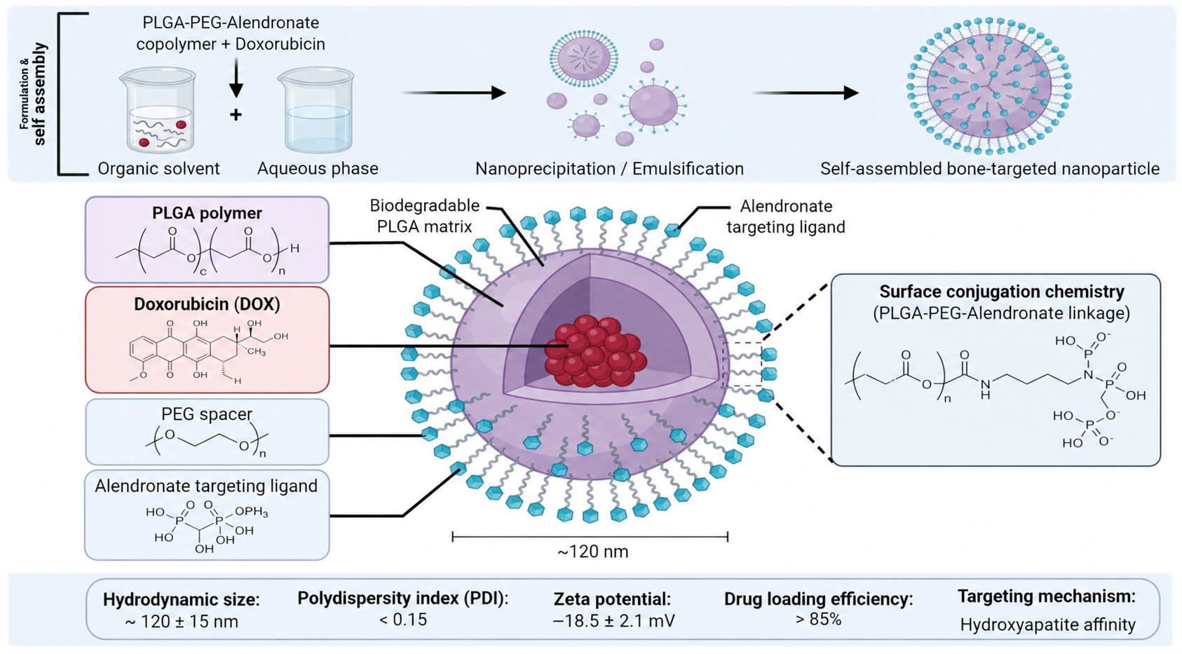
Figure 1 schematically illustrates the formulation and structural organization of a PLGA–PEG–alendronate nanocarrier for doxorubicin delivery, highlighting the integration of a biodegradable drug-loaded matrix, a PEG spacer, and a mineral-binding alendronate ligand, alongside representative physicochemical characteristics reported for related nanoparticle formulations.
Figure 1.
Schematic representation of a PLGA–PEG–alendronate nanocarrier for doxorubicin delivery, showing formulation, structural components, and key physicochemical characteristics. Created with BioRender.com by Varut R.M. (2026); the physicochemical values presented in the figure were obtained from previously published studies on related nanoparticle formulations [19,20,21]; https://app.biorender.com/illustrations/6abcd7f1bf23576f339a88c9?slideId=87dd1e8c-069f-49ba-9091-0b63c6b8232e.
3.3. Particle Size and Delivery Efficiency
Particle size affects both drug release and the barriers encountered before contact with bone. A smaller particle has a greater surface-area-to-volume ratio and shorter diffusion distances, potentially increasing leakage as well as tissue penetration. Larger particles or aggregates may be retained near vessels or captured by phagocytic cells. These effects depend on surface charge, coating, protein adsorption, and tumor architecture; reducing size alone does not ensure greater nuclear doxorubicin exposure [1,22,23].
Reported dimensions illustrate product heterogeneity rather than a size–efficacy rule. Doxorubicin-loaded PLGA–alendronate particles were reported at approximately 245 nm, with PDI 0.250 [21]. A 2025 PLGA–doxorubicin–alendronate composite study reported a number-distribution range of approximately 550–822 nm, with larger particles also contributing to the volume distribution [18]. Different manufacturing methods and size metrics preclude attributing biological differences to diameter alone. In a 2023 breast-tumor spheroid study, stromal composition affected the penetration and efficacy of paclitaxel-loaded polymeric carriers, including PLGA particles; this supports testing tissue barriers but is indirect evidence for bone-targeted doxorubicin [24].
Hydrodynamic size and PDI should be measured after dilution and incubation in relevant biological media, with microscopy or particle-tracking measurements as complementary checks. A valid size comparison should control polymer chemistry, ligand density, loading, dose, and release, then measure lesion-to-healthy-bone exposure, penetration depth, and drug-specific cellular availability. The evidence considered here does not establish a universal optimal diameter for this carrier class.
3.4. What Other PLGA Cargos Can and Cannot Establish
Studies of cisplatin-loaded PLGA systems provide examples of product-specific pharmacodynamic and tolerability assessment in tumor-bearing animals [25,26]. Curcumin formulations demonstrate how formulation can alter the stability and pharmacokinetic behavior of a difficult cargo [27,28]. Docetaxel-loaded PLGA and PLGA–PEG (polyethylene glycol) particles further show the importance of measuring circulation and organ distribution [29]. These studies support the versatility of the platform; they do not establish the exposure or safety of a doxorubicin-loaded, bisphosphonate-functionalized formulation.
The same boundary applies to combination delivery. Irinotecan–stattic PLGA particles, paclitaxel delivery systems, and polymeric cisplatin–doxorubicin co-delivery illustrate distinct strategies for changing exposure or combining mechanisms [30,31,32]. Hu and colleagues investigated hyaluronic acid-functionalized, redox-responsive particles carrying doxorubicin and cyclopamine in breast-cancer models [33]. Their results motivate combination design, but a different polymer architecture, targeting ligand, or disease compartment requires new evidence. Co-encapsulation alone does not demonstrate synergy, which depends on dose ratio, timing, and a prespecified interaction model.
Table 2 summarizes formulation variables whose effects should be assessed jointly. The questions in the final column are proposed development tests, not acceptance limits already validated for this product class.
Table 2.
Formulation variables and their implications for bisphosphonate-functionalized PLGA carriers.
3.5. Local Delivery and Systemic Delivery Are Different Products
PLGA composites used for bone repair demonstrate how mineral phases, porosity, and architecture can be integrated into a local implant. Examples include strontium/magnesium-doped hydroxyapatite structures and hydroxyapatite-microsphere-reinforced scaffolds [34,35]. PLGA–polydopamine particles have also been investigated by intra-articular administration in osteoarthritis [36]. These are informative material studies, but they do not answer the vascular transport questions faced by intravenously administered cancer nanocarriers.
Recent work extends this distinction to post-treatment bone reconstruction. Fan and colleagues studied a PLGA–collagen scaffold containing doxorubicin-loaded mineralized mesoporous silica, with attention to mesenchymal-cell behavior in a post-chemotherapy microenvironment [37]. This supports examining the sequence of tumor treatment and repair. It does not validate a bisphosphonate-functionalized systemic formulation. Local implantation may avoid some circulation barriers while introducing different questions about spatial drug gradients, residual malignant cells, mechanical function, and exposure of regenerating tissue.
4. Doxorubicin Delivery and the Therapeutic Index
4.1. Intracellular Availability Is the Relevant Exposure
Doxorubicin interacts with deoxyribonucleic acid (DNA) and stabilizes topoisomerase-II-associated DNA damage. Its biological activity depends on cellular entry, intracellular distribution, exposure duration, and the susceptibility of the target cell. Carrier uptake and pharmacological drug availability are therefore distinct outcomes. Fluorescent puncta within a cell can show entry of a labeled formulation without establishing delivery of active doxorubicin to its intracellular site of action [8,9,38].
Analytical assessment of doxorubicin delivery encompasses five distinct quantities. Total doxorubicin recovery is the amount of drug recovered after validated extraction of the whole sample. Carrier-associated drug is the fraction remaining with particles after a validated separation, including any surface-associated fraction specified by the method. Released drug is the amount of drug outside the carrier and may still be protein-bound. Intracellular availability concerns chemically intact drugs accessible within cells, particularly the nucleus; pharmacodynamic response concerns the resulting DNA damage, apoptosis, or growth inhibition. Quantitative relationships among these measurements require experimental validation.
Intrinsic doxorubicin fluorescence is useful for localization, but intensity depends on concentration, quenching, pH, binding, and the optical environment. Drug release can increase fluorescence through dequenching without increasing total drug mass. An independent carrier label controls for extracellular or surface-bound signal, and subcellular imaging can complement validated High-Performance Liquid Chromatography (HPLC) or Liquid Chromatography–Tandem Mass Spectrometry (LC–MS/MS) measurements of intact drug and relevant metabolites. A pharmacodynamic assay should then establish biological activity. This is particularly important when fluorescence in endolysosomal puncta is described as successful delivery [16,20,21].
A useful formulation comparison measures the relationship between internalized carrier, released drug, nuclear drug exposure, and a pharmacodynamic response. Drug-resistant and drug-sensitive models should be interpreted separately. Nanoparticle uptake may modify one transport step while leaving efflux, lysosomal sequestration, DNA-damage responses, and cell survival pathways intact. Structural work on verapamil transport by P-glycoprotein informs transporter biology but cannot by itself establish that an efflux-inhibitor combination is clinically effective or that a nanocarrier overcomes resistance [39].
4.2. Cardiac Protection Requires Direct Evidence
Doxorubicin cardiotoxicity cannot be reduced to a single nonspecific oxidative-stress mechanism. Experimental work implicating cardiomyocyte topoisomerase IIβ links DNA damage to mitochondrial and oxidative injury [40]. Other studies examine lipid-peroxidation control and senescence-related pathways: GPX4 activation has been investigated experimentally, while reducing doxorubicin-induced senescence did not necessarily produce cardiac protection in a mouse study [41,42]. These results caution against using improvement in one molecular marker as proof of overall cardioprotection.
For a bone-directed formulation, reduced heart-associated fluorescence does not by itself establish cardioprotection: the signal may reflect a label, intact drug, metabolites, or changes in quenching. Reduced circulating free doxorubicin could lower myocardial exposure, whereas premature leakage or prolonged systemic release could preserve cardiac injury. A convincing comparison should therefore link chemically resolved heart and plasma exposure to serial cardiac function, cardiac troponin, histopathology, cumulative dose, and follow-up after treatment. Free doxorubicin, an untargeted drug-loaded carrier, and an appropriate liposomal comparator help distinguish effects of encapsulation from those of bone targeting. Stable body weight or short-term tumor shrinkage cannot exclude delayed injury.
Formulation-dependent cardiac effects were observed by Drinković et al., who compared conventional, non-PEGylated liposomal, and PLGA-formulated doxorubicin in male and female Wistar Han rats over 28 days using five intravenous administrations at five-day intervals. Cardiac histological injury was less severe with the nanoformulations, but troponin T increased with all three preparations, and the PLGA formulation still showed mitochondrial toxicity [43]. These findings from a non-bone-targeted rat study indicate potential safety benefits of encapsulation alongside residual toxicity. Their relevance to bisphosphonate-functionalized carriers depends on further assessment of long-term cardiac function, tumor exposure, and the contribution of the targeting ligand.
4.3. Liposomal Doxorubicin Sets a Demanding Comparator
Pegylated liposomal doxorubicin provides a clinical benchmark for formulation-dependent cardioprotection. In the randomized study by O’Brien and colleagues, 509 women with metastatic breast cancer received pegylated liposomal doxorubicin (50 mg/m2 every four weeks) or conventional doxorubicin (60 mg/m2 every three weeks). Progression-free survival was comparable (hazard ratio 1.00; 95% confidence interval 0.82–1.22), while protocol-defined cardiotoxicity was more frequent with conventional doxorubicin (hazard ratio 3.16; 95% confidence interval 1.58–6.31; p < 0.001) [44]. These findings concern formulation and population; they do not quantify the cardiac benefit of a PLGA–bisphosphonate carrier.
The long clinical history of liposomal doxorubicin and randomized studies in other disease settings reinforce the need for formulation-specific comparisons [45,46]. Where clinically relevant, free doxorubicin alone is an incomplete benchmark for a more complex experimental carrier. A candidate should also be evaluated against an established delivery formulation or regimen that addresses a similar clinical limitation. The appropriate comparator depends on the intended indication and should not be selected solely because it makes the new formulation appear favorable.
4.4. Additional Functions Create Additional Dependencies
Doxorubicin has been incorporated into gold-based systems, iron-oxide constructs, and pH-responsive carriers, while hybrid compounds provide a further route to changing its properties [38,47,48,49,50]. Each architecture creates different relationships between chemical identity, transport, activation, and elimination. Hyperthermia-responsive delivery likewise requires an exposure that can be generated reproducibly at the intended anatomical site [51].
For deep skeletal lesions, external-energy approaches must account for tissue access and heterogeneous energy deposition. A trigger should be tested under conditions that distinguish the tumor extracellular space from endosomal or lysosomal compartments. Faster release at a strongly acidic laboratory pH is not sufficient evidence that the same release occurs selectively in a patient’s bone lesion. The simplest architecture that demonstrates a reproducible exposure advantage may be a stronger development candidate than a multifunctional system whose individual contributions cannot be separated.
5. Bisphosphonate Chemistry and the Meaning of Bone Targeting
5.1. Mineral Affinity and Pharmacological Potency Are Separate Properties
Bisphosphonates contain a P–C–P backbone that differs from the P–O–P linkage of pyrophosphate. Substituents influence mineral interaction and cellular pharmacology. Nitrogen-containing drugs such as alendronate, risedronate, and zoledronate should not be described as sharing an identical binding strength or potency simply because they belong to the same broad class. Structure–activity analyses and direct hydroxyapatite-binding comparisons demonstrate the importance of the individual compound [5,52,53].
Their affinity for calcium-containing mineral underlies skeletal localization. Distribution studies with labeled alendronate and etidronate provide evidence that localization depends on the skeletal environment [54]. However, the behavior of a free bisphosphonate cannot be transferred quantitatively to a particle displaying multiple ligands. Multivalent presentation, linkage chemistry, steric accessibility, colloidal stability, and competition from biomolecules can all change the effective interaction.
At the mineral interface, phosphonate oxygen atoms coordinate exposed calcium ions; the P–C–P scaffold and, for hydroxybisphosphonates, the central hydroxyl group contribute to this interaction. Side-chain structure, protonation, mineral crystallinity, and solution composition influence affinity. In PLGA–alendronate conjugates, coupling through the terminal amine can leave the phosphonate groups available for mineral association [21,52,53]. On a particle, binding additionally depends on how many groups remain accessible beyond the PEG layer and protein corona. This is mineral-directed binding, not recognition of a tumor-specific receptor; affinity alone cannot discriminate malignant from healthy remodeling bone.
The mineral-binding group is consequently both a chemical motif and a formulation variable. Coupling a bisphosphonate through one functional group may preserve the capacity to interact with mineral while changing its cellular accessibility. A carrier-bound ligand cannot be assumed to reproduce the intracellular pharmacology of the free drug. This distinction is especially important when the same molecule is presented simultaneously as a targeting group and a second anticancer agent.
5.2. Antiresorptive Mechanisms Do Not Establish Direct Tumor Killing
Nitrogen-containing bisphosphonates inhibit farnesyl diphosphate synthase in the mevalonate pathway, disrupting isoprenoid-dependent protein prenylation and osteoclast function. Non-nitrogen-containing compounds can be converted into cytotoxic ATP analogues. These mechanistic classes explain aspects of antiresorptive activity but are not interchangeable descriptions of every bisphosphonate product [55]. The clinical history of the class in osteoporosis provides a foundation for understanding skeletal pharmacology, while safety remains dependent on drug, dose, route, duration, and patient context [56,57].
In a composite product, suppression of osteoclast activity can alter the tumor-associated skeletal niche without proving that more doxorubicin reached malignant cells. Conversely, tumor control may reduce osteolysis secondarily. Measurements of bone preservation, osteoclast number, malignant-cell burden, and drug exposure should therefore be interpreted as complementary outcomes. None can reliably substitute for the others.
5.3. Mineral Binding Must Survive the Biological Interface
Hydroxyapatite assays provide a useful early test of mineral interaction. Their interpretation depends on particle concentration, mineral surface area, incubation time, medium composition, and the method used to distinguish binding from aggregation or sedimentation. The clinically relevant question is whether the interaction remains accessible after exposure to plasma and during passage through the biological barriers that precede contact with mineral.
Evidence from transferrin-functionalized nanoparticles shows that a biomolecular corona can mask a targeting ligand and alter specificity [58]. This is an analogy informing experimental design, not direct proof that every bisphosphonate ligand will be masked. For PLGA systems, surface-dependent differences in cell response and the broader experience with active targeting support testing ligand accessibility in the final biological context [59,60,61].
PEGylation presents a related trade-off. A hydrophilic surface may improve colloidal behavior and circulation while reducing access to a mineral-binding ligand. Choi and Kim explicitly investigated PLGA particles modified with alendronate and PEG as a bone-delivery platform [19]. The appropriate conclusion is that surface architecture is part of the targeting mechanism, not that increasing PEG content or ligand density will improve performance monotonically.
5.4. Reaching Bone Is Not the Same as Reaching the Lesion
Tumor delivery involves circulation, vascular transport, interstitial movement, and cellular access. Classical discussions of the enhanced permeability and retention effect are useful but incomplete descriptions of these processes [22,62,63]. Experimental evidence that active endothelial transport can contribute to nanoparticle entry into tumors further argues against assigning all accumulation to passive passage through vascular gaps [23].
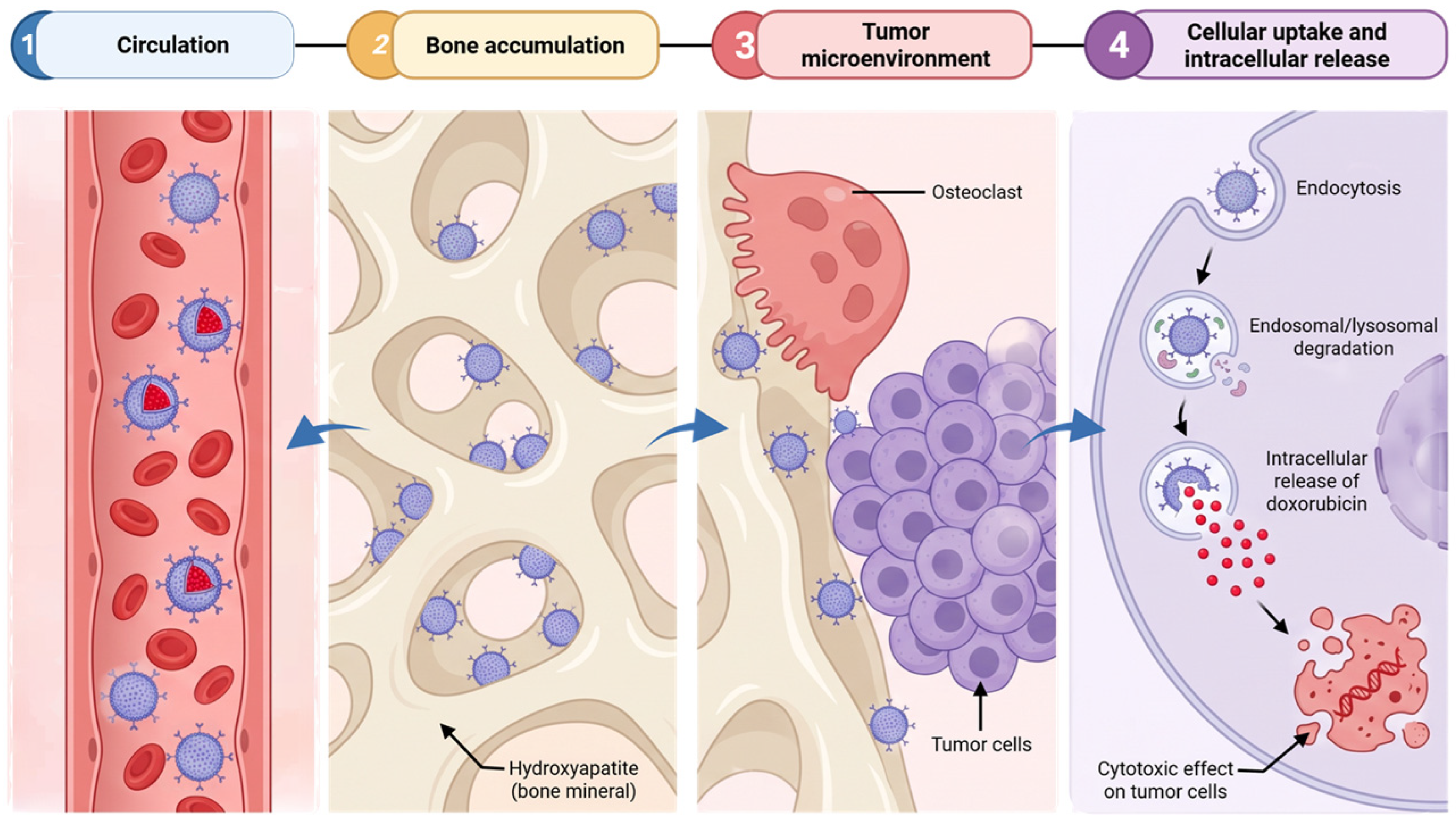
For skeletal disease, the distribution problem includes normal bone, tumor-associated mineral, marrow, stromal cells, osteoclasts, and malignant cells. A whole-bone measurement combines compartments with different therapeutic meanings. Strong mineral binding may be beneficial for maintaining a local reservoir but may also retain a carrier at a surface distant from the viable tumor cells. This is a testable transport trade-off rather than an established universal limitation. Spatially resolved measurements are needed to determine which interpretation applies to a given product (Figure 2).
Figure 2.
Pathway of bisphosphonate-functionalized PLGA nanocarriers from systemic circulation to bone accumulation, tumor-cell uptake, and intracellular doxorubicin release. Created with BioRender.com by Varut R.M. (2026), https://app.biorender.com/illustrations/6abcd7f1bf23576f339a88c9?slideId=d9c54ba9-6efa-4039-82c4-b83424f75925.
5.5. Long-Term Effects on Bone Metabolism
The direct PLGA–alendronate–doxorubicin evidence considered here includes short-term osteolysis and osteoclast measurements but does not establish long-term remodeling safety. In the skeletal-lesion experiments summarized by Salerno et al. and Pignatello et al., assessment extended to approximately six weeks; reduced osteolysis or osteoclast number at that stage cannot establish normal subsequent bone formation, fracture repair, or mechanical strength [20,21]. These reports describe overlapping platform experiments and should not be interpreted as independent long-term confirmations.
Long-term skeletal outcomes may reflect both bisphosphonate-mediated inhibition of resorption and exposure of nonmalignant skeletal cells to released doxorubicin. Bone preservation during tumor treatment can coexist with altered remodeling. Chronic safety findings for free bisphosphonates provide relevant context but cannot quantify risks from a tethered ligand or a mineral-retained drug reservoir [56,57]. Future studies should include post-treatment follow-up, bone-formation and resorption markers, dynamic histomorphometry, micro-CT, and mechanical or healing endpoints in tumor-bearing and healthy bone, with age and skeletal maturity specified. Long-term metabolic benefit or neutrality of targeted doxorubicin delivery remains unproven for this product class.
6. What the Existing Carrier Studies Demonstrate
6.1. Alendronate-Conjugated PLGA and Doxorubicin
Cenni and colleagues investigated the biocompatibility of PLGA–alendronate nanoparticles, providing a foundational assessment of a chemically modified carrier rather than of PLGA alone [64]. This distinction matters because surface conjugation creates a new product with its own interactions. A favorable biocompatibility result in experimental systems should not be restated as proof that the formulation is safe in humans.
Salerno and colleagues subsequently studied doxorubicin-loaded, alendronate-conjugated PLGA nanoparticles in cell models and a mouse model of breast cancer skeletal metastasis. Both free and nanoparticle-associated doxorubicin inhibited cellular growth and reduced metastatic incidence; unloaded particles did not reproduce the antitumor effect, although reductions in osteoclast number were observed with both loaded and unloaded carriers [20]. These findings support delivery feasibility and the need to separate cargo effects from carrier-associated antiresorptive effects. The reported comparison does not justify a blanket statement that the nanoparticles were superior to free doxorubicin.
The polymer-conjugate work described by Pignatello and colleagues adds chemical and biological context to this platform [21]. Thamake and colleagues investigated alendronate-coated PLGA nanoparticles co-loaded with curcumin and bortezomib for metastatic breast cancer targeting [65]. This was a different cargo combination, not a second doxorubicin formulation. Together, these studies establish a preclinical basis for the design concept. They do not constitute a standardized evidence set: differences in linkage, payload handling, surface composition, and disease models preclude treating the formulations as one interchangeable intervention.
A recent formulation study by Postelnicu et al. examined doxorubicin-containing PLGA composites functionalized through polydopamine with alendronate [18]. Its physicochemical characterization and HPLC release measurements update the materials evidence, but the reported antimicrobial assays do not establish antitumor efficacy, lesion-selective delivery, or cardiac protection. The relatively broad particle distribution and the distinction between released-drug concentration and cumulative fractional release are material considerations when relating this formulation to earlier nanocarriers.
6.2. Zoledronate Anchoring and the Cargo Boundary
Chaudhari and colleagues studied zoledronate-anchored PLGA–PEG nanoparticles carrying docetaxel. Their experiments reported changes in cellular uptake and cytotoxicity, together with altered circulation and skeletal distribution in animals [66]. This is directly relevant evidence for bisphosphonate-functionalized PLGA surface design. It is not a doxorubicin study, and the behavior of docetaxel cannot establish doxorubicin release, cardioprotection, or intracellular exposure.
The use of labeled carriers in such studies also illustrates a general interpretive issue. A radiolabel or fluorophore can trace one component while the drug follows a different distribution after release. Demonstrating skeletal retention of the carrier is important, but an exposure claim about the payload requires a payload-specific assay or convincing evidence that the label and drug remain coupled over the relevant interval.
6.3. Alternative Architectures Are Comparators Rather than Confirmations
Morton and colleagues developed alendronate-functionalized layer-by-layer nanoparticles, including doxorubicin-loaded liposomal constructs, and assessed targeting and therapeutic activity in preclinical osteosarcoma systems [67]. The study is a useful architectural comparator because it separates an osteotropic surface from the internal drug reservoir. It should not be described as a PLGA core system, and findings from its xenograft setting should not be generalized to all orthotopic skeletal lesions.
Hydroxyapatite-based surface-modified PLGA nanoparticles carrying risedronate were investigated by Rawat and colleagues with pharmacokinetic and biochemical assessment in an osteoporosis context [68]. This contributes evidence on mineral-oriented delivery but has a different cargo and therapeutic objective. Its success criteria cannot substitute for tumor-cell drug exposure or antitumor efficacy.
A more recent study by Lu and colleagues investigated sequential targeting of PLGA-based biomimetic nanovesicles carrying bortezomib in multiple myeloma [69]. This supports continued interest in separating bone-marrow localization from malignant-cell recognition. Nevertheless, membrane coating, cargo, and disease biology differ from the doxorubicin platforms considered here. The study motivates an experimental comparison; it does not validate a universal dual-targeting solution (Table 3).
Table 3.
Representative published platforms and the boundaries of their evidence.
6.4. Cross-Platform Comparison Requires Common Denominators
Nominal particle size, percentage drug encapsulation, and a reduction in tumor volume are insufficient common denominators for ranking the studies. Meaningful comparison requires the actual administered drug and carrier doses, dosing interval, route, starting disease burden, and comparator. It also requires knowing whether exposure refers to intact nanoparticles, free drug, total drug, or a label. A smaller particle or a higher loading percentage is not intrinsically better if it changes circulation, release, or toxicity in an unfavorable direction.
The evidence also remains uneven across the six levels defined in Section 2. Some studies establish surface chemistry and mineral interaction, others examine animal distribution, and a smaller subset connect these features to drug-specific pharmacology in disease models. The development priority is to close the missing links for one reproducible formulation rather than to combine favorable findings from several different products into a single implied mechanism.
Table 4 summarizes selected quantitative outcomes within their original model, regimen, endpoint, and statistical context. It includes a direct PLGA–alendronate platform, an alternative bone-directed architecture, a PLGA hybrid evaluated outside bone disease, and a clinical formulation benchmark. Small animal groups, differing endpoint definitions, incomplete compatible variance data, and overlapping reports restrict synthesis to descriptive comparisons within the respective experimental settings. Future comparative experiments should report group sizes, allocation and blinding, dose-normalized exposure, effect sizes with confidence intervals, and individual outcome data where feasible.
Table 4.
Selected study-level quantitative outcomes without cross-study pooling.
7. Bisphosphonate Antitumor Evidence and Clinical Reality
7.1. Experimental Osteosarcoma Findings Are Heterogeneous
Animal studies of zoledronate in osteosarcoma have reported effects on pulmonary metastases, tumor growth, and skeletal lesions [70,71,72]. Additional work connected reduced osteolysis to changes in tumor-associated MCP-1 and RANKL, while other experiments examined combined treatment with ifosfamide or broader effects on the tumor microenvironment [73,74,75]. These observations provide biological rationales for intervention at the bone–tumor interface. They should be reported with their model and endpoint rather than compressed into an unqualified anticancer class effect.
The distinction between endpoints is particularly important. A reduction in osteolytic damage can improve a clinically meaningful skeletal outcome even when the direct effect on malignant cells is limited. Conversely, an intervention may influence early dissemination more strongly than established lesions. Alendronate effects on invasion and MMP-2 secretion in osteosarcoma cells illustrate a specific cellular phenotype, not proof of control of established human tumors [76].
7.2. Veterinary and Other Tumor Models Provide Context
Spontaneous canine osteosarcoma offers comparative-oncology evidence from naturally occurring disease but remains veterinary evidence. Studies of zoledronate-associated bone effects, pamidronate for pain palliation, CXCR4-related behavior, and an individual clinical case address different questions [77,78,79,80]. Pain relief measures palliation; suppression of resorption markers measures an antiresorptive response; imaging or histological tumor burden measures local tumor control; pulmonary dissemination measures metastatic behavior; and survival is a separate time-to-event outcome. Improvement in one does not establish improvement in the others, and a case report cannot establish comparative efficacy.
Chondrosarcoma research has examined osteoclast inhibition and tumor-associated bone destruction as well as direct effects of clodronate and zoledronate on cell lines [81,82]. Ewing sarcoma studies have explored adjuvant activity and interference with pulmonary dissemination [83,84]. These data broaden the mechanistic discussion but do not erase the differences between sarcoma subtypes. A future doxorubicin carrier should be selected for a specific biological and clinical indication, not a pooled category of bone cancer.
The evidence hierarchy therefore distinguishes mechanistic cell-line observations, controlled rodent proof-of-concept experiments, spontaneous veterinary disease, and human clinical studies. Rodent xenografts can test delivery under defined conditions but may lack an intact immune system and the vascular or mineral organization of a human skeletal lesion. Veterinary studies add naturally occurring disease and a different body scale, while retaining species-specific pharmacology and treatment constraints. Human evidence for free bisphosphonates or approved liposomes is a clinical comparator; it is not clinical validation of the experimental PLGA carrier.
7.3. Extraosseous Activity and Combination Effects Need Exposure Support
Bisphosphonate activity has also been investigated in colorectal, myeloma, and renal carcinoma models, including apoptosis and mevalonate-pathway effects [85,86,87]. Tumor-associated macrophages have been proposed as additional targets, and nanotechnology has been explored to alter distribution beyond the skeleton [88,89]. These concepts are relevant to carrier design because they indicate that an observed treatment response may involve malignant cells, osteoclasts, or host cells.
Combination studies with farnesyl-transferase inhibition or docetaxel provide examples of context-dependent interactions [90,91]. Their interpretation requires exposure that is feasible for the same cells in vivo. An in vitro combination index obtained with free agents cannot establish synergy when one component is immobilized on a carrier or released at a different rate. The carrier-bound bisphosphonate, any released bisphosphonate, and free drug must be considered separately.
7.4. Clinical Benefit Cannot Be Inferred from Antiresorptive Activity
Clinical evidence for zoledronic acid in metastatic hormone-refractory prostate cancer supports prevention of skeletal complications in the studied population [6]. This should not be rewritten as proof that bisphosphonates prevent all bone metastases or eradicate prostate cancer. Preclinical work on osteoblastic and osteolytic lesions addresses a different question [92], and experience in unresectable benign bone tumors concerns another disease context [93].
The OS2006 randomized phase III trial is a particularly important counterweight to promising animal osteosarcoma data. Adding zoledronate to chemotherapy and surgery did not improve event-free survival, and the trial was stopped for futility. The reported hazard ratio for treatment failure was 1.36, with a 95% confidence interval of 0.95–1.96 [94]. This result does not prove that a bone-targeted nanocarrier will fail, but it does rule out using the preclinical antitumor activity of free zoledronate as sufficient justification for clinical benefit.
A carrier could, in principle, change the exposure responsible for the discrepancy between preclinical and clinical observations. To support that explanation, it must demonstrate the changed exposure and the corresponding pharmacological effect. Merely attaching a bisphosphonate to a particle does not supply the missing evidence (Table 5).
Table 5.
Endpoints that should remain distinct when evaluating skeletal cancer delivery.
8. An Evaluation Framework for Translation
8.1. Define the Clinical Use Before Optimizing the Particle
The target product profile should specify whether the intended use is systemic treatment of established bone metastases, treatment of a primary bone tumor, or local control after surgery. These settings require different routes, drug exposure profiles, comparators, and safety assessments. A system designed to remain on an exposed mineral surface may be appropriate for a local reservoir but insufficient for disseminated soft-tissue disease. Selection of the indication determines which apparent advantages are relevant.
This priority also limits unnecessary complexity. Multifunctional PLGA systems and personalized immunotherapy platforms demonstrate the range of possible modifications [95,96]. However, adding targeting ligands, responsive linkers, membranes, or immunological components creates additional manufacturing and mechanistic dependencies. Each added element should resolve an identified delivery limitation and demonstrate a contribution beyond the simpler parent formulation.
8.2. Establish Product Identity and Accessible Ligand
Chemical characterization should distinguish covalent attachment, adsorption, and physical mixing. A change in a spectroscopic band or surface charge is supportive evidence but is not sufficient alone to quantify conjugation or accessible ligand density. Polymer integrity, residual free bisphosphonate, drug loading, and material recovery should be assessed in a way that allows comparisons between batches.
The functionally relevant ligand measurement is the amount presented to the biological environment after manufacture, storage, dilution, and exposure to proteins. This may differ from the total amount detected after destructive analysis. The proposed decision rule is therefore functional: a candidate should retain reproducible mineral interaction under conditions that also preserve colloidal stability and payload retention. No universal ligand-density threshold is established by the literature considered here.
8.3. Resolve Carrier, Drug, and Lesion Compartments
An informative biodistribution experiment should measure both a carrier-associated signal and the drug, where feasible, and test the stability of their association. Total doxorubicin, released doxorubicin, and relevant metabolites should be distinguished when the analytical method and study design permit. Tissue measurements should include the lesion, contralateral healthy bone, marrow, heart, liver, spleen, kidney, and plasma according to the intended use.
The key comparison is not simply bone versus blood at one time point. Lesion-to-healthy-bone and lesion-to-heart exposure relationships over time are more directly connected to the proposed benefit. Histological or spatial methods should identify whether material is associated with mineral, vasculature, macrophages, osteoclasts, or malignant cells. These recommended measurements address the ambiguity created by whole-organ averages; they are not claimed as completed features of all published studies.
8.4. Match Release to the Site of Pharmacological Action
Mineral-bound retention and intracellular delivery may require different properties. A stable extracellular reservoir must release enough drug to reach nearby malignant cells, whereas an internalized particle must release active payload within a relevant intracellular interval. The preferred architecture depends on which route of action is intended. Studies should therefore state whether therapeutic activity is expected from mineral-associated extracellular release, cellular nanoparticle uptake, or both.
This distinction determines the controls. Release from mineral-bound particles should be measured as well as release from freely dispersed particles. Cellular experiments should compare free drug, drug-loaded untargeted particles, targeted drug-loaded particles, and empty targeted particles. A free-bisphosphonate control can help separate ligand pharmacology from surface-mediated transport. Exposure-matched comparisons are essential when release rates differ.
8.5. Evaluate Efficacy and Safety in the Same Formulation
A persuasive development package connects composition, transport, pharmacodynamics, and safety using the same formulation. It should not combine the biocompatibility of an empty carrier from one study with the efficacy of another product and the pharmacokinetics of a third. Repeated administration should be assessed when repeated treatment is intended, and both local skeletal effects and systemic toxicity should be considered.
Model selection should reflect the proposed indication. Orthotopic disease, established rather than only early lesions, and an appropriate immune context can each resolve a different uncertainty. Study interpretation also depends on allocation procedures, blinding of outcome assessment, prespecified endpoints, and handling of exclusions. The evidence hierarchy proposed here is a framework for asking these questions, not a validated score that ranks heterogeneous platforms on a single scale.
8.6. Manufacturing and Reporting Are Part of the Therapeutic Mechanism
Particle manufacture influences the very properties used to explain efficacy. Scale-dependent mixing, solvent removal, sterilization, storage, and reconstitution can change size distribution, drug leakage, or ligand accessibility. A product that performs well only as a freshly prepared research suspension has not resolved these dependencies. Batch comparisons should therefore include functional release and targeting measurements alongside composition and size.
Food and Drug Administration (FDA)guidance on drug products containing nanomaterials emphasizes product-specific quality, nonclinical, and clinical considerations [14]. Minimum-information reporting proposals in bio–nano research provide a complementary basis for documenting material and biological conditions [97]. Applied here, these principles support a transparent record of how the tested formulation was made and how exposure was measured. They do not provide automatic regulatory acceptance for a material class. Figure 3 outlines a proposed translational evaluation framework for bisphosphonate-functionalized PLGA nanocarriers, linking product characterization, biological stability, and mineral interactions to drug-resolved biodistribution, intracellular exposure, and comparative efficacy and safety, while emphasizing reproducible manufacture and a clearly defined clinical indication as prerequisites for clinical translation.
Figure 3.
Proposed translational evaluation framework for bisphosphonate-functionalized PLGA nanocarriers, from product characterization and biological stability to drug-resolved exposure, comparative efficacy and safety, and clinical translation. Created with BioRender.com by Varut R.M. (2026), https://app.biorender.com/illustrations/6abcd7f1bf23576f339a88c9?slideId=81ffa52e-1663-42cf-b92d-dd5b7a7315bc.
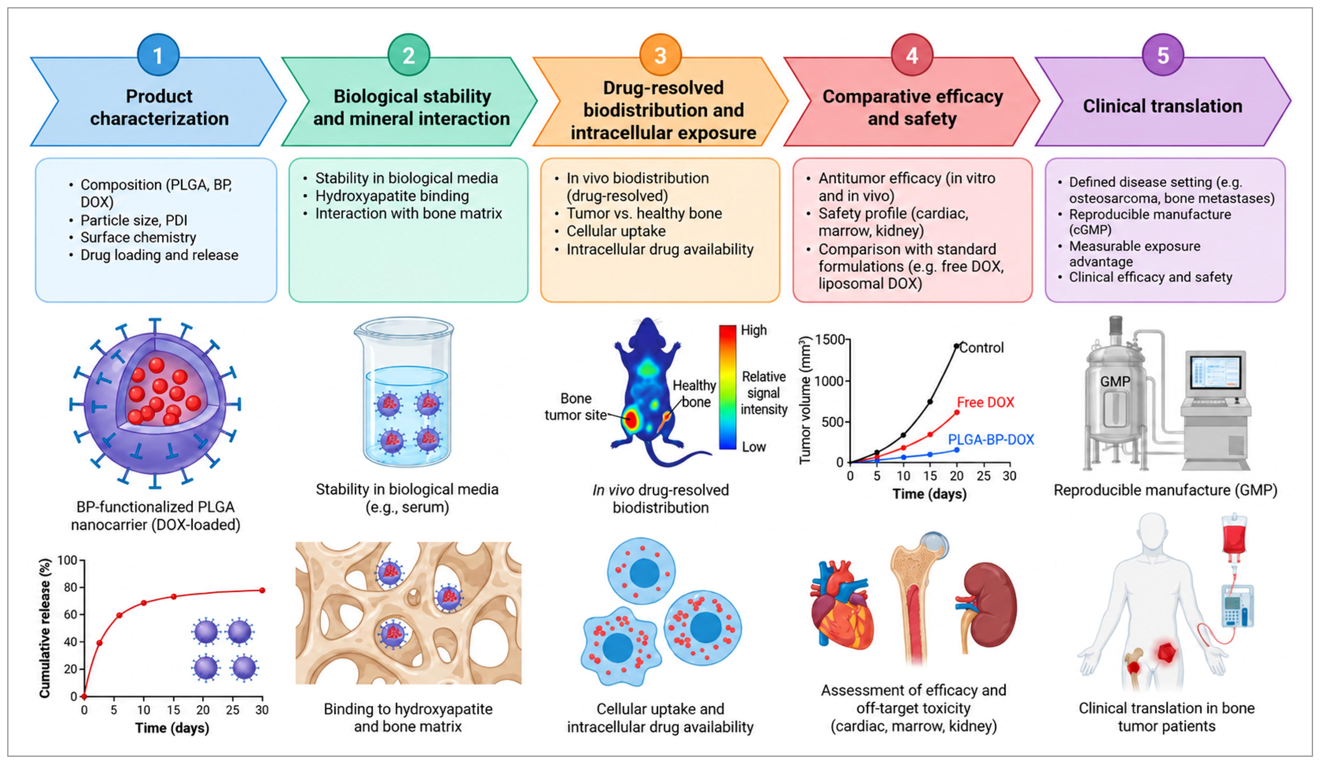
To facilitate the systematic evaluation of bisphosphonate-functionalized PLGA nanocarriers, Table 6 summarizes the key questions that should be addressed at each stage of development, from product characterization and biological stability to drug exposure, therapeutic efficacy, safety, and clinical translation. It also identifies the most informative controls and measurements required to support reliable interpretation and translational relevance.
Table 6.
Proposed decision questions for advancing a candidate formulation.
9. Limitations and Research Priorities
The available evidence is fragmented across products, cargos, disease models, and outcome measures. Substantial heterogeneity in formulation architecture, doses, comparators, and outcome definitions limits quantitative integration. Clinical studies of free bisphosphonates and liposomal doxorubicin provide translational benchmarks, while clinical evaluation of PLGA–doxorubicin–bisphosphonate formulations remains necessary. Research priorities include linking mineral binding to drug-resolved lesion exposure, distinguishing antiresorptive effects from direct tumor control through appropriate comparators, and evaluating efficacy alongside cumulative toxicity and manufacturability in clinically relevant disease settings.
Selection and publication bias may affect the evidence base, and the absence of complete database-specific search strings limits search reproducibility. Differences in release methods and particle-size metrics further constrain numerical comparison, while short skeletal and cardiac follow-up limits assessment of delayed toxicity. The comparative efficacy of bisphosphonate-functionalized PLGA–doxorubicin remains dependent on the formulation, disease model, and comparator evaluated; broader conclusions require directly comparative studies with compatible endpoints and adequate follow-up.
A further priority is the timing of delivery. Early colonization, established osteolytic disease, bulky primary tumors, and postoperative defects present different access routes and exposure requirements. The same formulation may not serve all four settings. Studies that deliberately test these boundaries may be more informative than additional demonstrations of cytotoxicity in a single monolayer culture.
Finally, the balance between binding and availability deserves direct experimental optimization. Maximum mineral affinity is not necessarily the optimum if drug release or transport to malignant cells becomes limiting. The appropriate target is a demonstrable relationship between accessible ligand, lesion exposure, pharmacodynamic response, and safety. That relationship, rather than material novelty alone, would provide a credible basis for clinical development.
10. Conclusions
Bisphosphonate-functionalized PLGA carriers offer a rational way to combine controlled drug delivery with skeletal localization, and published preclinical studies support the feasibility of loading and delivering doxorubicin within this design space. Their promise depends on a chain of distinct events: reproducible formulation, stability during administration, access to mineral-associated lesions, release of active drug, and adequate exposure of malignant cells.
The available evidence does not support treating mineral binding, carrier uptake, skeletal protection, and clinical antitumor benefit as interchangeable findings. Nor does experience with approved component drugs establish the safety or efficacy of a new composite. The strongest next-generation candidates will connect these evidence levels within one formulation and one clearly defined indication, while demonstrating an advantage over a relevant comparator. Progress should be judged by improved delivery of pharmacologically available drugs and a better therapeutic index, not by the number of functionalities assembled on the particle.
Author Contributions
Conceptualization, P.D.F.V., A.M.K., R.M.V. and M.S.; Methodology, P.D.F.V., D.-M.T., A.M.K., R.M.V., P.I.D., D.R., N.B. and M.S.; Validation, D.-M.T., A.M.K., R.M.V., P.I.D., N.B. and M.S.; Formal Analysis, P.D.F.V., D.-M.T., D.R., N.B. and M.S.; Investigation, P.D.F.V., D.-M.T., A.M.K., R.M.V., C.C.A., P.I.D., D.R., N.B. and M.S.; Resources, A.M.K., R.M.V., C.C.A. and P.I.D.; Data Curation, P.D.F.V., D.-M.T., R.M.V., D.R., N.B. and M.S.; Writing—Original Draft Preparation, P.D.F.V., R.M.V. and M.S.; Writing—Review and Editing, P.D.F.V., D.-M.T., A.M.K., R.M.V., C.C.A., P.I.D., D.R., N.B. and M.S.; Visualization, D.-M.T., C.C.A., N.B. and M.S.; Supervision, A.M.K., R.M.V. and M.S.; Project Administration, A.M.K., R.M.V., P.I.D. and M.S. All authors were personally and actively involved in substantial work leading to this paper. All authors have read and agreed to the published version of the manuscript.
Funding
The Article Processing Charges were funded by the University of Medicine and Pharmacy of Craiova, Romania.
Institutional Review Board Statement
Not applicable.
Informed Consent Statement
Not applicable.
Data Availability Statement
No new data were created or analyzed in this study. Data sharing is not applicable to this article.
Conflicts of Interest
The authors declare no conflicts of interest.
Abbreviations
ALN, alendronate; BP, bisphosphonate; DNA, deoxyribonucleic acid; DOX, doxorubicin; EPR, enhanced permeability and retention; HA, hydroxyapatite; HPLC, High-Performance Liquid Chromatography; LbL, layer-by-layer; LC–MS/MS, Liquid Chromatography–Tandem Mass Spectrometry; NP, nanoparticle; PEG, polyethylene glycol; PLGA, poly(lactic-co-glycolic acid); ZOL, zoledronate.
References
- Danhier, F.; Ansorena, E.; Silva, J.M.; Coco, R.; Le Breton, A.; Préat, V. PLGA-based nanoparticles: An overview of biomedical applications. J. Control. Release 2012, 161, 505–522. [Google Scholar] [CrossRef] [Scilit]
- Narmani, A.; Jahedi, R.; Bakhshian-Dehkordi, E.; Ganji, S.; Nemati, M.; Ghahramani-Asl, R.; Moloudi, K.; Hosseini, S.M.; Bagheri, H.; Kesharwani, P.; et al. Biomedical applications of PLGA nanoparticles in nanomedicine: Advances in drug delivery systems and cancer therapy. Expert Opin. Drug Deliv. 2023, 20, 937–954. [Google Scholar] [CrossRef] [Scilit]
- Mbese, Z.; Aderibigbe, B.A. Bisphosphonate-Based Conjugates and Derivatives as Potential Therapeutic Agents in Osteoporosis, Bone Cancer and Metastatic Bone Cancer. Int. J. Mol. Sci. 2021, 22, 6869. [Google Scholar] [CrossRef] [Scilit]
- Cole, L.E.; Vargo-Gogola, T.; Roeder, R.K. Targeted delivery to bone and mineral deposits using bisphosphonate ligands. Adv. Drug Deliv. Rev. 2016, 99, 12–27. [Google Scholar] [CrossRef] [Scilit]
- Russell, R.G.G. Bisphosphonates: The first 40 years. Bone 2011, 49, 2–19. [Google Scholar] [CrossRef] [Scilit]
- Saad, F.; Gleason, D.M.; Murray, R.; Tchekmedyian, S.; Venner, P.; Lacombe, L.; Chin, J.L.; Vinholes, J.J.; Goas, J.A.; Zheng, M. Long-Term Efficacy of Zoledronic Acid for the Prevention of Skeletal Complications in Patients with Metastatic Hormone-Refractory Prostate Cancer. JNCI J. Natl. Cancer Inst. 2004, 96, 879–882. [Google Scholar] [CrossRef] [Scilit]
- Santini, D.; Fratto, M.E.; Vincenzi, B.; Galluzzo, S.; Tonini, G. Zoledronic acid in the management of metastatic bone disease. Expert Opin. Biol. Ther. 2006, 6, 1333–1348. [Google Scholar] [CrossRef] [Scilit]
- Sinha, S.; Kumar, B.; Prasad, C.; Chauhan, S.; Kumar, M. Emerging Research and Future Directions on Doxorubicin: A Snapshot. Asian Pac. J. Cancer Prev. 2025, 26, 5–15. [Google Scholar] [CrossRef] [Scilit]
- Bisht, A.; Avinash, D.; Sahu, K.K.; Patel, P.; Das Gupta, G.; Kurmi, B.D. A comprehensive review on doxorubicin: Mechanisms, toxicity, clinical trials, combination therapies and nanoformulations in breast cancer. Drug Deliv. Transl. Res. 2025, 15, 102–133. [Google Scholar] [CrossRef] [Scilit]
- Lee, J.; Choi, M.-K.; Song, I.-S. Recent Advances in Doxorubicin Formulation to Enhance Pharmacokinetics and Tumor Targeting. Pharmaceuticals 2023, 16, 802. [Google Scholar] [CrossRef] [Scilit]
- Silva, A.T.C.R.; Cardoso, B.C.O.; Silva, M.E.S.R.E.; Freitas, R.F.S.; Sousa, R.G. Synthesis, Characterization, and Study of PLGA Copolymer In Vitro Degradation. J. Biomater. Nanobiotechnol. 2015, 6, 8–19. [Google Scholar] [CrossRef]
- Shive, M.S.; Anderson, J.M. Biodegradation and biocompatibility of PLA and PLGA microspheres. Adv. Drug Deliv. Rev. 1997, 28, 5–24. [Google Scholar] [CrossRef] [Scilit]
- Abulateefeh, S.R. Long-acting injectable PLGA/PLA depots for leuprolide acetate: Successful translation from bench to clinic. Drug Deliv. Transl. Res. 2023, 13, 520–530. [Google Scholar] [CrossRef] [Scilit]
- U.S. Food and Drug Administration. Drug Products, Including Biological Products, That Contain Nanomaterials. Guidance for Industry. 2022. Available online: https://www.fda.gov/regulatory-information/search-fda-guidance-documents/drug-products-including-biological-products-contain-nanomaterials-guidance-industry (accessed on 30 September 2026).
- Alam Khan, S.; Jawaid Akhtar, M. Structural modification and strategies for the enhanced doxorubicin drug delivery. Bioorg. Chem. 2022, 120, 105599. [Google Scholar] [CrossRef] [Scilit]
- Fernandes, R.S.; Arribada, R.G.; Silva, J.O.; Silva-Cunha, A.; Townsend, D.M.; Ferreira, L.A.M.; Barros, A.L.B. In Vitro and In Vivo Effect of pH-Sensitive PLGA-TPGS-Based Hybrid Nanoparticles Loaded with Doxorubicin for Breast Cancer Therapy. Pharmaceutics 2022, 14, 2394. [Google Scholar] [CrossRef] [Scilit]
- Wang, L.; Wang, P.; Liu, Y.; Mustafa Mahayyudin, M.A.; Li, R.; Zhang, W.; Zhan, Y.; Li, Z. The Effect of Different Factors on Poly(lactic-co-glycolic acid) Nanoparticle Properties and Drug Release Behaviors When Co-Loaded with Hydrophilic and Hydrophobic Drugs. Polymers 2024, 16, 865. [Google Scholar] [CrossRef] [Scilit]
- Postelnicu, R.-A.; Ciocîlteu, M.-V.; Manda, C.V.; Bălășoiu, R.-M.; Pîrvu, S.A.; Rău, G.; Nicolicescu, C.; Mocanu, A.G.; Neamțu, J. Formulation and Characterisation of PLGA-Doxorubicin Composites Conjugated with Bisphosphonates in Oncological Therapy. Farmacia 2025, 73, 327–338. [Google Scholar] [CrossRef] [Scilit]
- Choi, S.W.; Kim, J.H. Design of surface-modified poly(d,l-lactide-co-glycolide) nanoparticles for targeted drug delivery to bone. J. Control. Release 2007, 122, 24–30. [Google Scholar] [CrossRef] [Scilit]
- Salerno, M.; Cenni, E.; Fotia, C.; Avnet, S.; Granchi, D.; Castelli, F.; Micieli, D.; Pignatello, R.; Capulli, M.; Rucci, N.; et al. Bone-Targeted Doxorubicin-Loaded Nanoparticles as a Tool for the Treatment of Skeletal Metastases. Curr. Cancer Drug Targets 2010, 10, 649–659. [Google Scholar] [CrossRef] [Scilit]
- Pignatello, R.; Sarpietro, M.G.; Castelli, F. Synthesis and Biological Evaluation of a New Polymeric Conjugate and Nanocarrier with Osteotropic Properties. J. Funct. Biomater. 2012, 3, 79–99. [Google Scholar] [CrossRef] [Scilit]
- Lammers, T.; Kiessling, F.; Hennink, W.E.; Storm, G. Drug targeting to tumors: Principles, pitfalls and (pre-) clinical progress. J. Control. Release 2012, 161, 175–187. [Google Scholar] [CrossRef] [Scilit]
- Sindhwani, S.; Syed, A.M.; Ngai, J.; Kingston, B.R.; Maiorino, L.; Rothschild, J.; MacMillan, P.; Zhang, Y.; Rajesh, N.U.; Hoang, T.; et al. The entry of nanoparticles into solid tumours. Nat. Mater. 2020, 19, 566–575. [Google Scholar] [CrossRef] [Scilit]
- Priwitaningrum, D.L.; Pednekar, K.; Gabriël, A.V.; Varela-Moreira, A.A.; Le Gac, S.; Vellekoop, I.; Storm, G.; Hennink, W.E.; Prakash, J. Evaluation of paclitaxel-loaded polymeric nanoparticles in 3D tumor model: Impact of tumor stroma on penetration and efficacy. Drug Deliv. Transl. Res. 2023, 13, 1470–1483. [Google Scholar] [CrossRef] [Scilit]
- Mattheolabakis, G.; Taoufik, E.; Haralambous, S.; Roberts, M.L.; Avgoustakis, K. In vivo investigation of tolerance and antitumor activity of cisplatin-loaded PLGA-mPEG nanoparticles. Eur. J. Pharm. Biopharm. 2009, 71, 190–195. [Google Scholar] [CrossRef] [Scilit]
- Moreno, D.; Zalba, S.; Navarro, I.; Tros de Ilarduya, C.; Garrido, M.J. Pharmacodynamics of cisplatin-loaded PLGA nanoparticles administered to tumor-bearing mice. Eur. J. Pharm. Biopharm. 2010, 74, 265–274. [Google Scholar] [CrossRef] [Scilit]
- Mohanty, C.; Sahoo, S.K. The in vitro stability and in vivo pharmacokinetics of curcumin prepared as an aqueous nanoparticulate formulation. Biomaterials 2010, 31, 6597–6611. [Google Scholar] [CrossRef] [Scilit]
- Klippstein, R.; Wang, J.T.W.; El-Gogary, R.I.; Bai, J.; Mustafa, F.; Rubio, N.; Bansal, S.; Al-Jamal, W.T.; Al-Jamal, K.T. Passively Targeted Curcumin-Loaded PEGylated PLGA Nanocapsules for Colon Cancer Therapy In Vivo. Small 2015, 11, 4704–4722. [Google Scholar] [CrossRef] [Scilit]
- Rafiei, P.; Haddadi, A. Docetaxel-loaded PLGA and PLGA-PEG nanoparticles for intravenous application: Pharmacokinetics and biodistribution profile. Int. J. Nanomed. 2017, 12, 935–947. [Google Scholar] [CrossRef] [Scilit]
- Arslan, F.B.; Öztürk, K.; Tavukçuoğlu, E.; Öztürk, S.C.; Esendağlı, G.; Çalış, S. A novel combination for the treatment of small cell lung cancer: Active targeted irinotecan and stattic co-loaded PLGA nanoparticles. Int. J. Pharm. 2023, 632, 122573. [Google Scholar] [CrossRef] [Scilit]
- Ma, P.; Huang, J.; Liu, J.; Zhu, Y.; Chen, J.; Chen, J.; Lei, L.; Guan, Z.; Ban, J.; Lu, Z. Nanoformulation of Paclitaxel: Exploring the Cyclodextrin/PLGA Nano Delivery Carrier to Slow Down Paclitaxel Release, Enhance Accumulation In Vivo. J. Cancer 2023, 14, 759–769. [Google Scholar] [CrossRef] [Scilit]
- Zhang, X.; Li, J.; Yan, M. Targeted hepatocellular carcinoma therapy: Transferrin modified, self-assembled polymeric nanomedicine for co-delivery of cisplatin and doxorubicin. Drug Dev. Ind. Pharm. 2016, 42, 1590–1599. [Google Scholar] [CrossRef] [Scilit]
- Hu, K.; Zhou, H.; Liu, Y.; Liu, Z.; Liu, J.; Tang, J.; Li, J.; Zhang, J.; Sheng, W.; Zhao, Y.; et al. Hyaluronic acid functional amphipathic and redox-responsive polymer particles for the co-delivery of doxorubicin and cyclopamine to eradicate breast cancer cells and cancer stem cells. Nanoscale 2015, 7, 8607–8618. [Google Scholar] [CrossRef] [Scilit]
- Yu, S.; Sun, T.; Liu, W.; Yang, L.; Gong, H.; Chen, X.; Li, J.; Weng, J. PLGA Cage-Like Structures Loaded with Sr/Mg-Doped Hydroxyapatite for Repairing Osteoporotic Bone Defects. Macromol. Biosci. 2022, 22, 2200092. [Google Scholar] [CrossRef] [Scilit]
- Wei, J.; Yan, Y.; Gao, J.; Li, Y.; Wang, R.; Wang, J.; Zou, Q.; Zuo, Y.; Zhu, M.; Li, J. 3D-printed hydroxyapatite microspheres reinforced PLGA scaffolds for bone regeneration. Biomater. Adv. 2022, 133, 112618. [Google Scholar] [CrossRef] [Scilit]
- Zong, L.; Wang, Q.; Sun, H.; Wu, Q.; Xu, Y.; Yang, H.; Lv, S.; Zhang, L.; Geng, D. Intra-Articular Injection of PLGA/Polydopamine Core–Shell Nanoparticle Attenuates Osteoarthritis Progression. ACS Appl. Mater. Interfaces 2024, 16, 21450–21462. [Google Scholar] [CrossRef] [Scilit]
- Fan, Z.; Zhang, H.; Wang, L.; Tian, J.; Jiang, H.; Chen, Y.; Guo, B.; Yu, H. Replicating the post-chemotherapy tumor microenvironment via biomimetic scaffolds to regulate MSC differentiation. RSC Adv. 2026, 16, 16841–16853. [Google Scholar] [CrossRef] [Scilit]
- Peter, S.; Alven, S.; Maseko, R.B.; Aderibigbe, B.A. Doxorubicin-Based Hybrid Compounds as Potential Anticancer Agents: A Review. Molecules 2022, 27, 4478. [Google Scholar] [CrossRef] [Scilit]
- Wang, L.; Sun, Y. Efflux mechanism and pathway of verapamil pumping by human P-glycoprotein. Arch. Biochem. Biophys. 2020, 696, 108675. [Google Scholar] [CrossRef] [Scilit]
- Zhang, S.; Liu, X.; Bawa-Khalfe, T.; Lu, L.S.; Lyu, Y.L.; Liu, L.F.; Yeh, E.T.H. Identification of the molecular basis of doxorubicin-induced cardiotoxicity. Nat. Med. 2012, 18, 1639–1642. [Google Scholar] [CrossRef] [Scilit]
- Huang, C.; Guo, Y.; Li, T.; Sun, G.; Yang, J.; Wang, Y.; Xiang, Y.; Wang, L.; Jin, M.; Li, J.; et al. Pharmacological activation of GPX4 ameliorates doxorubicin-induced cardiomyopathy. Redox Biol. 2024, 70, 103024. [Google Scholar] [CrossRef] [Scilit]
- Goshovska, Y.; Lazzarini, E.; Rendon-Angel, A.; Biemmi, V.; Colucci, M.; Alimonti, A.; Barile, L. Reduction of doxorubicine-induced senescence does not provide cardioprotective effect in mice. Vasc. Pharmacol. 2024, 155, 107322. [Google Scholar] [CrossRef] [Scilit]
- Drinković, N.; Beus, M.; Barbir, R.; Debeljak, Ž.; Tariba Lovaković, B.; Kalčec, N.; Ćurlin, M.; Bekavac, A.; Gorup, D.; Mamić, I.; et al. Novel PLGA-based nanoformulation decreases doxorubicin-induced cardiotoxicity. Nanoscale 2024, 16, 9412–9425. [Google Scholar] [CrossRef] [Scilit]
- O’Brien, M.E.R.; Wigler, N.; Inbar, M.; Rosso, R.; Grischke, E.; Santoro, A.; Catane, R.; Kieback, D.G.; Tomczak, P.; Ackland, S.P.; et al. Reduced cardiotoxicity and comparable efficacy in a phase III trial of pegylated liposomal doxorubicin HCl (CAELYX™/Doxil®) versus conventional doxorubicin for first-line treatment of metastatic breast cancer. Ann. Oncol. 2004, 15, 440–449. [Google Scholar] [CrossRef] [Scilit]
- Gabizon, A.A.; Gabizon-Peretz, S.; Modaresahmadi, S.; La-Beck, N.M. Thirty years from FDA approval of pegylated liposomal doxorubicin (Doxil/Caelyx): An updated analysis and future perspective. BMJ Oncol. 2025, 4, e000573. [Google Scholar] [CrossRef] [Scilit]
- Bafaloukos, D.; Linardou, H.; Aravantinos, G.; Papadimitriou, C.; Bamias, A.; Fountzilas, G.; Kalofonos, H.P.; Kosmidis, P.; Timotheadou, E.; Makatsoris, T.; et al. A randomized phase II study of carboplatin plus pegylated liposomal doxorubicin versus carboplatin plus paclitaxel in platinum sensitive ovarian cancer patients: A Hellenic Cooperative Oncology Group study. BMC Med. 2010, 8, 3. [Google Scholar] [CrossRef] [Scilit]
- Choubdar, N.; Avizheh, S.; Karimifard, S.A. Recent Advances in Efficacy of using Doxorubicin Gold Nanoparticles for Chemo-, Radio-, Photothermal, and Photodynamic Therapy. Curr. Drug Deliv. 2022, 19, 745–762. [Google Scholar] [CrossRef] [Scilit]
- Dockery, L.T.; Daniel, M.C. Targeted Doxorubicin-Loaded Dendronized Gold Nanoparticles. Pharmaceutics 2023, 15, 2103. [Google Scholar] [CrossRef] [Scilit]
- Oleksa, V.; Macková, H.; Patsula, V.; Dydowiczová, A.; Janoušková, O.; Horák, D. Doxorubicin-Conjugated Iron Oxide Nanoparticles: Surface Engineering and Biomedical Investigation. ChemPlusChem 2020, 85, 1156–1163. [Google Scholar] [CrossRef] [Scilit]
- Soltani, A.; Faramarzi, M.; Farjadian, F.; Parsa, S.A.M.; Panahi, H.A. pH-responsive glycodendrimer as a new active targeting agent for doxorubicin delivery. Int. J. Biol. Macromol. 2022, 221, 508–522. [Google Scholar] [CrossRef] [Scilit]
- Seynhaeve, A.L.B.; Amin, M.; Haemmerich, D.; van Rhoon, G.C.; ten Hagen, T.L.M. Hyperthermia and smart drug delivery systems for solid tumor therapy. Adv. Drug Deliv. Rev. 2020, 163–164, 125–144. [Google Scholar] [CrossRef] [Scilit]
- Ebetino, F.H.; Sun, S.; Cherian, P.; Roshandel, S.; Neighbors, J.D.; Hu, E.; Dunford, J.E.; Sedghizadeh, P.P.; McKenna, C.E.; Srinivasan, V.; et al. Bisphosphonates: The role of chemistry in understanding their biological actions and structure-activity relationships, and new directions for their therapeutic use. Bone 2022, 156, 116289. [Google Scholar] [CrossRef] [Scilit]
- Lawson, M.A.; Xia, Z.; Barnett, B.L.; Triffitt, J.T.; Phipps, R.J.; Dunford, J.E.; Locklin, R.M.; Ebetino, F.H.; Russell, R.G.G. Differences between bisphosphonates in binding affinities for hydroxyapatite. J. Biomed. Mater. Res. Part B Appl. Biomater. 2010, 92, 149–155. [Google Scholar] [CrossRef] [Scilit]
- Masarachia, P.; Weinreb, M.; Balena, R.; Rodan, G.A. Comparison of the distribution of 3H-alendronate and 3H-Etidronate in rat and mouse bones. Bone 1996, 19, 281–290. [Google Scholar] [CrossRef] [Scilit]
- Coxon, F.; Thompson, K.; Rogers, M. Recent advances in understanding the mechanism of action of bisphosphonates. Curr. Opin. Pharmacol. 2006, 6, 307–312. [Google Scholar] [CrossRef] [Scilit]
- Bellido, T. Bisphosphonates for osteoporosis: From bench to clinic. J. Clin. Investig. 2024, 134, e179942. [Google Scholar] [CrossRef] [Scilit]
- Pazianas, M.; Abrahamsen, B. Safety of bisphosphonates. Bone 2011, 49, 103–110. [Google Scholar] [CrossRef] [Scilit]
- Salvati, A.; Pitek, A.S.; Monopoli, M.P.; Prapainop, K.; Bombelli, F.B.; Hristov, D.R.; Kelly, P.M.; Åberg, C.; Mahon, E.; Dawson, K.A. Transferrin-functionalized nanoparticles lose their targeting capabilities when a biomolecule corona adsorbs on the surface. Nat. Nanotechnol. 2013, 8, 137–143. [Google Scholar] [CrossRef] [Scilit]
- Sims, L.B.; Curry, K.C.; Parupalli, S.; Horner, G.; Frieboes, H.B.; Steinbach-Rankins, J.M. Efficacy of Surface-Modified PLGA Nanoparticles as a Function of Cervical Cancer Type. Pharm. Res. 2019, 36, 66. [Google Scholar] [CrossRef] [Scilit]
- Sudimack, J.; Lee, R.J. Targeted drug delivery via the folate receptor. Adv. Drug Deliv. Rev. 2000, 41, 147–162. [Google Scholar] [CrossRef] [Scilit]
- Zhu, Y.; Feijen, J.; Zhong, Z. Dual-targeted nanomedicines for enhanced tumor treatment. Nano Today 2018, 18, 65–85. [Google Scholar] [CrossRef] [Scilit]
- Nel, A.; Ruoslahti, E.; Meng, H. New Insights into “Permeability” as in the Enhanced Permeability and Retention Effect of Cancer Nanotherapeutics. ACS Nano 2017, 11, 9567–9569. [Google Scholar] [CrossRef] [Scilit]
- Pérez-Herrero, E.; Fernández-Medarde, A. Advanced targeted therapies in cancer: Drug nanocarriers, the future of chemotherapy. Eur. J. Pharm. Biopharm. 2015, 93, 52–79. [Google Scholar] [CrossRef] [Scilit]
- Cenni, E.; Granchi, D.; Avnet, S.; Fotia, C.; Salerno, M.; Micieli, D.; Sarpietro, M.G.; Pignatello, R.; Castelli, F.; Baldini, N. Biocompatibility of poly(d,l-lactide-co-glycolide) nanoparticles conjugated with alendronate. Biomaterials 2008, 29, 1400–1411. [Google Scholar] [CrossRef] [Scilit]
- Thamake, S.I.; Raut, S.L.; Gryczynski, Z.; Ranjan, A.P.; Vishwanatha, J.K. Alendronate coated poly-lactic-co-glycolic acid (PLGA) nanoparticles for active targeting of metastatic breast cancer. Biomaterials 2012, 33, 7164–7173. [Google Scholar] [CrossRef] [Scilit]
- Ramanlal Chaudhari, K.; Kumar, A.; Megraj Khandelwal, V.K.; Ukawala, M.; Manjappa, A.S.; Mishra, A.K.; Monkkonen, J.; Ramachandra Murthy, R.S. Bone metastasis targeting: A novel approach to reach bone using Zoledronate anchored PLGA nanoparticle as carrier system loaded with Docetaxel. J. Control. Release 2012, 158, 470–478. [Google Scholar] [CrossRef] [Scilit]
- Morton, S.W.; Shah, N.J.; Quadir, M.A.; Deng, Z.J.; Poon, Z.; Hammond, P.T. Osteotropic Therapy via Targeted Layer-by-Layer Nanoparticles. Adv. Healthc. Mater. 2014, 3, 867–875. [Google Scholar] [CrossRef] [Scilit]
- Rawat, P.; Ahmad, I.; Thomas, S.C.; Pandey, S.; Vohora, D.; Gupta, S.; Ahmad, F.J.; Talegaonkar, S. Revisiting bone targeting potential of novel hydroxyapatite based surface modified PLGA nanoparticles of risedronate: Pharmacokinetic and biochemical assessment. Int. J. Pharm. 2016, 506, 253–261. [Google Scholar] [CrossRef] [Scilit]
- Lu, C.; Xiao, Z.; Zhang, H.; Zhang, P.; Gao, Y.; Ouyang, L.; He, X.; Han, N.; Zhang, J.; Guan, M.; et al. Sequential dual-targeting biomimetic nanovesicles for bone marrow–specific delivery of bortezomib in multiple myeloma. Front. Bioeng. Biotechnol. 2025, 13, 1714613. [Google Scholar] [CrossRef] [Scilit]
- Ory, B.; Heymann, M.F.; Kamijo, A.; Gouin, F.; Heymann, D.; Redini, F. Zoledronic acid suppresses lung metastases and prolongs overall survival of osteosarcoma-bearing mice. Cancer 2005, 104, 2522–2529. [Google Scholar] [CrossRef] [Scilit]
- Dass, C.R.; Choong, P.F.M. Zoledronic acid inhibits osteosarcoma growth in an orthotopic model. Mol. Cancer Ther. 2007, 6, 3263–3270. [Google Scholar] [CrossRef] [Scilit]
- Labrinidis, A.; Hay, S.; Liapis, V.; Ponomarev, V.; Findlay, D.M.; Evdokiou, A. Zoledronic Acid Inhibits Both the Osteolytic and Osteoblastic Components of Osteosarcoma Lesions in a Mouse Model. Clin. Cancer Res. 2009, 15, 3451–3461. [Google Scholar] [CrossRef] [Scilit]
- Ohba, T.; Cole, H.A.; Cates, J.M.; Slosky, D.A.; Haro, H.; Ando, T.; Schwartz, H.S.; Schoenecker, J.G. Bisphosphonates Inhibit Osteosarcoma-Mediated Osteolysis Via Attenuation of Tumor Expression of MCP-1 and RANKL. J. Bone Miner. Res. 2014, 29, 1431–1445. [Google Scholar] [CrossRef] [Scilit]
- Heymann, D.; Ory, B.; Blanchard, F.; Heymann, M.F.; Coipeau, P.; Charrier, C.; Couillaud, S.; Thiery, J.P.; Gouin, F.; Redini, F. Enhanced tumor regression and tissue repair when zoledronic acid is combined with ifosfamide in rat osteosarcoma. Bone 2005, 37, 74–86. [Google Scholar] [CrossRef] [Scilit]
- Ohba, T.; Cates, J.M.M.; Cole, H.A.; Slosky, D.A.; Haro, H.; Ichikawa, J.; Ando, T.; Schwartz, H.S.; Schoenecker, J.G. Pleiotropic effects of bisphosphonates on osteosarcoma. Bone 2014, 63, 110–120. [Google Scholar] [CrossRef] [Scilit]
- Cheng, Y.Y.; Huang, L.; Lee, K.M.; Li, K.; Kumta, S.M. Alendronate regulates cell invasion and MMP-2 secretion in human osteosarcoma cell lines. Pediatr. Blood Cancer 2004, 42, 410–415. [Google Scholar] [CrossRef] [Scilit]
- Fan, T.M.; De Lorimier, L.P.; Garrett, L.D.; Lacoste, H.I. The Bone Biologic Effects of Zoledronate in Healthy Dogs and Dogs with Malignant Osteolysis. J. Vet. Intern. Med. 2008, 22, 380–387. [Google Scholar] [CrossRef] [Scilit]
- Fan, T.M.; de Lorimier, L.P.; O’Dell-Anderson, K.; Lacoste, H.I.; Charney, S.C. Single-agent Pamidronate for Palliative Therapy of Canine Appendicular Osteosarcoma Bone Pain. J. Vet. Intern. Med. 2007, 21, 431–439. [Google Scholar] [CrossRef] [Scilit]
- Byrum, M.L.; Pondenis, H.C.; Fredrickson, R.L.; Wycislo, K.L.; Fan, T.M. Downregulation of CXCR4 Expression and Functionality After Zoledronate Exposure in Canine Osteosarcoma. J. Vet. Intern. Med. 2016, 30, 1187–1196. [Google Scholar] [CrossRef] [Scilit]
- Spugnini, E.P.; Vincenzi, B.; Caruso, G.; Baldi, A.; Citro, G.; Santini, D.; Tonini, G. Zoledronic acid for the treatment of appendicular osteosarcoma in a dog. J. Small Anim. Pract. 2009, 50, 44–46. [Google Scholar] [CrossRef] [Scilit]
- Otero, J.E.; Stevens, J.W.; Malandra, A.E.; Fredericks, D.C.; Odgren, P.R.; Buckwalter, J.A.; Morcuende, J. Osteoclast inhibition impairs chondrosarcoma growth and bone destruction. J. Orthop. Res. 2014, 32, 1562–1571. [Google Scholar] [CrossRef] [Scilit]
- Streitbuerger, A.; Henrichs, M.; Ahrens, H.; Lanvers-Kaminzky, C.; Gouin, F.; Gosheger, G.; Hardes, J. Cytotoxic effect of clodronate and zoledronate on the chondrosarcoma cell lines HTB-94 and CAL-78. Int. Orthop. 2011, 35, 1369–1373. [Google Scholar] [CrossRef] [Scilit]
- Odri, G.; Kim, P.P.; Lamoureux, F.; Charrier, C.; Battaglia, S.; Amiaud, J.; Heymann, D.; Gouin, F.; Redini, F. Zoledronic acid inhibits pulmonary metastasis dissemination in a preclinical model of Ewing’s sarcoma via inhibition of cell migration. BMC Cancer 2014, 14, 169. [Google Scholar] [CrossRef] [Scilit]
- Odri, G.A.; Dumoucel, S.; Picarda, G.; Battaglia, S.; Lamoureux, F.; Corradini, N.; Rousseau, J.; Tirode, F.; Laud, K.; Delattre, O.; et al. Zoledronic Acid as a New Adjuvant Therapeutic Strategy for Ewing’s Sarcoma Patients. Cancer Res. 2010, 70, 7610–7619. [Google Scholar] [CrossRef] [Scilit]
- Sewing, L.; Steinberg, F.; Schmidt, H.; Göke, R. The bisphosphonate zoledronic acid inhibits the growth of HCT-116 colon carcinoma cells and induces tumor cell apoptosis. Apoptosis 2008, 13, 782–789. [Google Scholar] [CrossRef] [Scilit]
- Guenther, A.; Gordon, S.; Tiemann, M.; Burger, R.; Bakker, F.; Green, J.R.; Baum, W.; Roelofs, A.J.; Rogers, M.J.; Gramatzki, M. The bisphosphonate zoledronic acid has antimyeloma activityin vivoby inhibition of protein prenylation. Int. J. Cancer 2010, 126, 239–246. [Google Scholar] [CrossRef] [Scilit]
- Fujita, M.; Tohi, M.; Sawada, K.; Yamamoto, Y.; Nakamura, T.; Yagami, T.; Yamamori, M.; Okamura, N. Involvement of the mevalonate pathway in the antiproliferative effect of zoledronate on ACHN renal cell carcinoma cells. Oncol. Rep. 2012, 27, 1371–1376. [Google Scholar] [CrossRef] [Scilit]
- Rogers, T.L.; Holen, I. Tumour macrophages as potential targets of bisphosphonates. J. Transl. Med. 2011, 9, 177. [Google Scholar] [CrossRef] [Scilit]
- De Rosa, G.; Misso, G.; Salzano, G.; Caraglia, M. Bisphosphonates and Cancer: What Opportunities from Nanotechnology? J. Drug Deliv. 2013, 2013, 637976. [Google Scholar] [CrossRef] [Scilit]
- Caraglia, M.; D’Alessandro, A.M.; Marra, M.; Giuberti, G.; Vitale, G.; Viscomi, C.; Colao, A.; Prete, S.D.; Tagliaferri, P.; Tassone, P.; et al. The farnesyl transferase inhibitor R115777 (Zarnestra®) synergistically enhances growth inhibition and apoptosis induced on epidermoid cancer cells by Zoledronic acid (Zometa®) and Pamidronate. Oncogene 2004, 23, 6900–6913. [Google Scholar] [CrossRef] [Scilit][Green Version]
- Karabulut, B.; Erten, C.; Gul, M.K.; Cengiz, E.; Karaca, B.; Kucukzeybek, Y.; Gorumlu, G.; Atmaca, H.; Uzunoglu, S.; Sanli, U.A.; et al. Docetaxel/zoledronic acid combination triggers apoptosis synergistically through downregulating antiapoptotic Bcl-2 protein level in hormone-refractory prostate cancer cells. Cell Biol. Int. 2009, 33, 239–246. [Google Scholar] [CrossRef] [Scilit]
- Corey, E.; Brown, L.G.; Quinn, J.E.; Poot, M.; Roudier, M.P.; Higano, C.S.; Vessella, R.L. Zoledronic acid exhibits inhibitory effects on osteoblastic and osteolytic metastases of prostate cancer. Clin. Cancer Res. 2003, 9, 295–306. [Google Scholar]
- Cornelis, F.; Truchetet, M.E.; Amoretti, N.; Verdier, D.; Fournier, C.; Pillet, O.; Gille, O.; Hauger, O. Bisphosphonate therapy for unresectable symptomatic benign bone tumors: A long-term prospective study of tolerance and efficacy. Bone 2014, 58, 11–16. [Google Scholar] [CrossRef] [Scilit]
- Piperno-Neumann, S.; Le Deley, M.C.; Rédini, F.; Pacquement, H.; Marec-Bérard, P.; Petit, P.; Brisse, H.; Lervat, C.; Gentet, J.C.; Entz-Werlé, N.; et al. Zoledronate in combination with chemotherapy and surgery to treat osteosarcoma (OS2006): A randomised, multicentre, open-label, phase 3 trial. Lancet Oncol. 2016, 17, 1070–1080. [Google Scholar] [CrossRef] [Scilit]
- Dinakar, Y.H.; Rajana, N.; Kumari, N.U.; Jain, V.; Mehra, N.K. Recent Advances of Multifunctional PLGA Nanocarriers in the Management of Triple-Negative Breast Cancer. AAPS PharmSciTech 2023, 24, 258. [Google Scholar] [CrossRef] [Scilit]
- Gonzalez-Melero, L.; Santos-Vizcaino, E.; Varela-Calvino, R.; Gomez-Tourino, I.; Asumendi, A.; Boyano, M.D.; Igartua, M.; Hernandez, R.M. PLGA-PEI nanoparticle covered with poly(I:C) for personalised cancer immunotherapy. Drug Deliv. Transl. Res. 2024, 14, 2788–2803. [Google Scholar] [CrossRef] [Scilit]
- Faria, M.; Björnmalm, M.; Thurecht, K.J.; Kent, S.J.; Parton, R.G.; Kavallaris, M.; Johnston, A.P.R.; Gooding, J.J.; Corrie, S.R.; Boyd, B.J.; et al. Minimum information reporting in bio–nano experimental literature. Nat. Nanotechnol. 2018, 13, 777–785. [Google Scholar] [CrossRef] [Scilit]

Disclaimer/Publisher’s Note: The statements, opinions and data contained in all publications are solely those of the individual author(s) and contributor(s) and not of MDPI and/or the editor(s). MDPI and/or the editor(s) disclaim responsibility for any injury to people or property resulting from any ideas, methods, instructions or products referred to in the content. |
© 2026 by the authors. Licensee MDPI, Basel, Switzerland. This article is an open access article distributed under the terms and conditions of the Creative Commons Attribution (CC BY) license.