


Abstract
Background/Objectives: Systemic delivery of oncolytic adenovirus (oAd) remains a major challenge due to rapid immune clearance, hepatic sequestration, and off-target toxicity. To overcome these barriers and enhance tumor-targeting efficacy, we developed Ad-DTX/HER, a novel viral complex engineered by directly co-conjugating the cytotoxic agent docetaxel (DTX) and the site-specifically modified HER2-targeting antibody trastuzumab (Herceptin, HER) onto the oAd surface. Methods: DTX and trastuzumab were site-specifically conjugated onto the naked viral capsid in a polymer-free manner, and the resulting Ad-DTX/HER complex was characterized for size distribution and structural integrity. Its HER2 selectivity and oncolytic activity were evaluated in cancer cells, and its biodistribution and organ toxicity were assessed following systemic administration in tumor-bearing mice. Results: Ad-DTX/HER exhibited a uniform size distribution, sustained structural integrity, and remarkable selectivity toward HER2-overexpressing cancer cells without compromising viral oncolytic activity. Systemic administration in tumor-bearing mice drastically minimized non-specific hepatic uptake while significantly enhancing tumor accumulation, achieving a marked ~2800-fold increase in the tumor-to-liver ratio compared to naked oAd. Histopathological analysis of major organs indicated that systemic administration of Ad-DTX/HER is not associated with overt off-target organ toxicity. Conclusions: This polymer-free, direct capsid-engineering platform provides an effective systemic delivery paradigm, offering potent synergistic chemo-virotherapy for HER2-positive tumor.
1. Introduction
Oncolytic adenovirus (oAd) is one of the virus vectors under development for gene therapy in oncology [1]. oAd selectively replicates in cancer cells to induce tumor cell death, while the progeny virus generated subsequently propagates and infects adjacent tumor cells, thereby potentiating the antitumor effect [2]. Due to these benefits, oAd has been studied frequently in clinical trials as well as animal tumor models [1]. For example, H101 was the first approved oncolytic virus by the State Food and Drug Administration in China for patients with head and neck cancer [3]. More recently, Telomelysin (OBP-301) received regulatory approval in Japan for the treatment of esophageal cancer. In addition, a range of oAd platforms, including ICOVIR and CG0070, are in active clinical development, highlighting oAds’ potential as next-generation cancer therapeutics [4].
Nonetheless, significant challenges remain for systemically administered oAd. Following intravenous delivery, oAd is prone to nonspecific hepatic sequestration that can cause hepatotoxicity [2]. The host immune system recognizes oAd as a foreign antigen, causing elevated proinflammatory cytokines such as IL-6 and TNF-α, which may drive systemic inflammation [5]. In addition, the adaptive immune system induces neutralizing antibodies and clearance via blood cells and proteins, which leads to a short circulating half-life and poor tumor accumulation [6]. Consequently, most ongoing clinical trials of oncolytic viruses rely on intratumoral or locoregional administration, limiting their applicability in treating inaccessible or metastatic tumors [7]. Conventional chemical engineering typically employs bulky hydrophilic polymers, such as polyethylene glycol (PEG), to reduce immune recognition and prolong the half-life in the bloodstream.
To overcome these limitations and enable effective systemic administration, various strategies for oAd delivery have been developed [8]. Among these, chemical shielding has emerged as a highly versatile and direct approach to physically mask the viral surface [9,10,11]. Building upon this concept, several technologies have been reported that further decorate the shielded oAds with antibodies or ligands to achieve targeted delivery [12,13,14]. However, these conventional approaches face significant limitations. First, they heavily rely on bulky polymer carriers to tether the ligands, which enlarges the particle size and complicates the formulation. Second, polymer-tethered antibodies are frequently conjugated in a random orientation, which reduces their antigen-binding affinity compared to site-specific attachment. Finally, these prior systems were designed solely to redirect the virus, lacking an additional secondary therapeutic mechanism.
To bypass the need for bulky polymeric carriers while achieving effective systemic delivery, we have introduced a polymer-free direct surface engineering strategy. By site-specifically conjugating a tumor-targeting antibody and a small-molecule chemotherapeutic directly onto the naked viral capsid, this method establishes an effective chemical shield against nonspecific hepatic clearance without drastically increasing particle size. Because this entirely polymer-free approach attaches the antibody site-specifically and co-conjugates docetaxel directly onto the capsid, a single construct simultaneously provides tumor-selective targeting, reinforced viral replication, and docetaxel-mediated apoptosis, ultimately enhancing the overall antitumor effect.
In this study, we developed a hybrid nanocomplex, oAd-DTX/HER, by site-specifically conjugating a chemotherapeutic agent, docetaxel (DTX), and a HER2-targeting monoclonal antibody, trastuzumab (HER), directly onto the naked viral capsid (Scheme 1). HER2 was rationally selected as the target receptor due to its clinical prevalence, being overexpressed in a significant fraction of breast and other carcinomas [15], and the established safety and specificity of HER as a targeting moiety. Furthermore, because many HER2-positive tumors eventually become refractory to HER monotherapy, coupling HER2-targeted delivery with both oncolytic and chemotherapeutic mechanisms addresses a critical clinical need, highlighting the high translational significance of this approach.
Scheme 1.
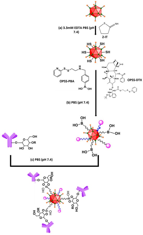
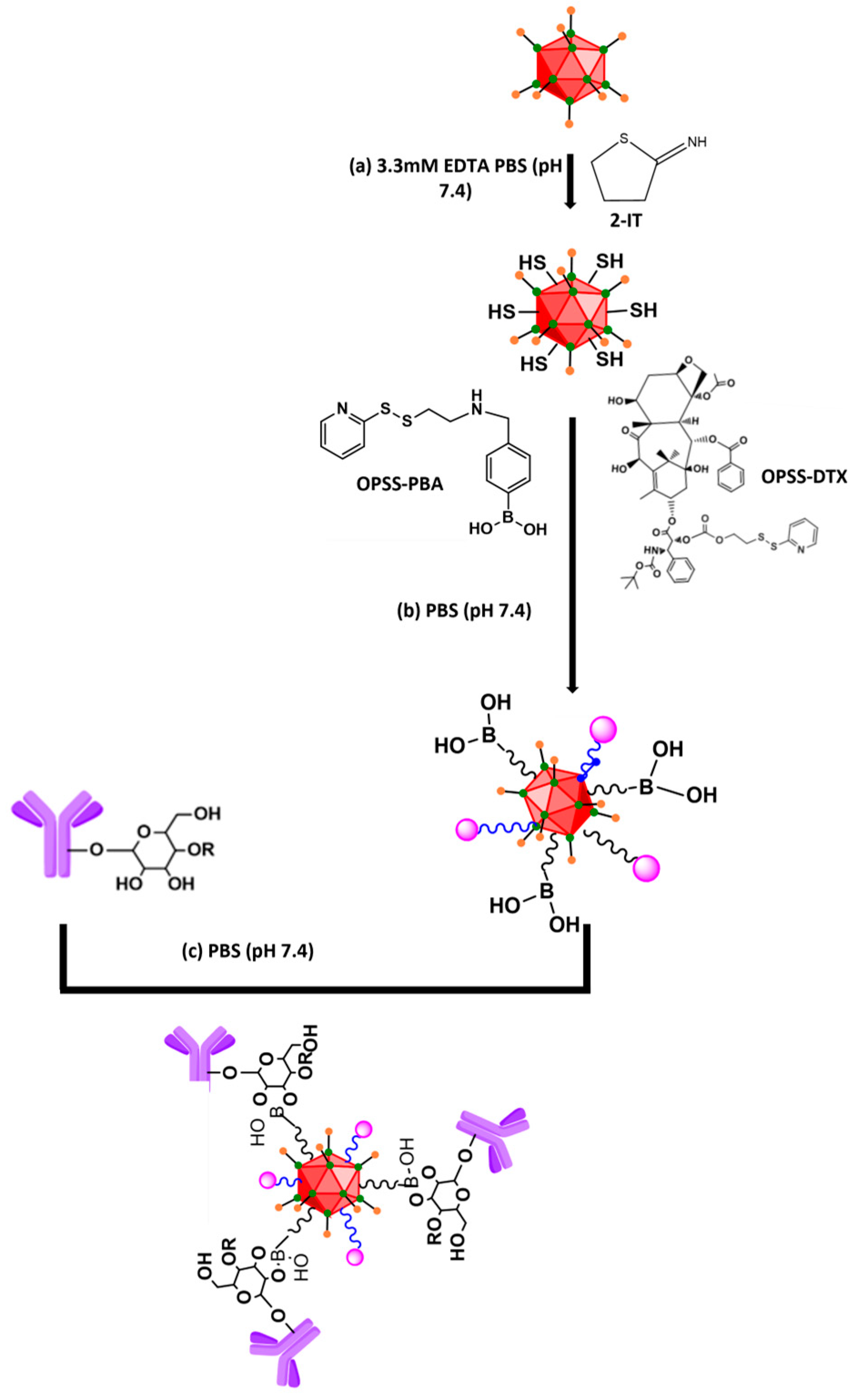
Synthesis scheme used to prepare Ad-DTX/HER. (a) 2-IT was treated to amine groups of Ad in 3.3 mM EDTA PBS for 1 h. (b) Thiol groups on the Ad surface were reacted with OPSS-PBA and OPSS-DTX for 3 h. (c) Ad-DTX/PBA was mixed with Herceptin for 2 h, and Ad-DTX/HER was formed via reaction between the phenylboronic ester of PBA and the diol on the inherent glycosylation site of Herceptin. Color code: red icosahedron, oncolytic adenovirus (oAd) capsid; purple Y-shape, HER2-targeting antibody (trastuzumab, HER); magenta spheres, docetaxel (DTX); blue wavy lines, OPSS disulfide linker; B(OH)2, phenylboronic acid (PBA).
This dual-conjugation creates an effective ‘chemical shield’ that reduces nonspecific hepatic uptake and neutralizes antiviral immune responses without drastically increasing particle size, while simultaneously equipping the virus with targeted chemo-virotherapy capabilities. Within this construct, the antibody serves as a targeting moiety, necessitating only minimal conjugation of HER to confer high-affinity tumor recognition and facilitate receptor-mediated endocytosis into HER2-overexpressing cancer cells. Once internalized into the targeted cancer cell, the complex undergoes intracellular dissociation to release oAd. The released oAd executes tumor-selective oncolytic replication and spread, while DTX enforces chemically induced cell-cycle arrest and apoptosis. This targeted co-delivery potentiates therapeutic efficacy at reduced drug dosages and overcomes the limitations of monotherapies.
In our evaluations, the oAd-DTX/HER complex maintained a uniform size distribution and exhibited robust in vitro cancer cell killing. Importantly, systemic administration in vivo models demonstrated enhanced tumor accumulation and drastically reduced nonspecific hepatic sequestration alongside a safety profile. Collectively, these findings highlight oAd-DTX/HER as a highly translatable, next-generation platform for treating intractable primary and metastatic malignancies.
2. Materials and Methods
2.1. Chemical Reagents
4-(Bromomethyl)phenylboronic acid (PBA), 2-mercaptoethanol, 2-mercaptoethylamine, 2-iminothiolane hydrochloride (2-IT), and triethylamine were purchased from Sigma-Aldrich (St. Louis, MO, USA). 2,2′-Dipyridyl disulfide, paclitaxel (PTX), cabazitaxel (CTX), and docetaxel (DTX) were purchased from TCI (Tokyo, Japan). All reagents were of analytical grade and obtained commercially.
2.2. 1H NMR and MALDI-TOF Analysis
1H NMR spectra was recorded on a Varian NMR spectrometer (NMR, Varian 600 MHz; Agilent Technologies, Santa Clara, CA, USA). The mass was analyzed by matrix assisted laser desorption/ionization–time of flight mass spectrum (MALDI-TOF, AXIMA Confidence, Shimadzu Biotech, Kyoto, Japan).
2.3. Cancer Cell Culture and Preparation of Ad
Human lung cancer cell lines (H460 and A549), human breast cancer cell line (SK-BR-3), human ovarian cancer cell line (SK-OV-3), and human cervical cancer cell line (HeLa) were purchased from the American Type Culture Collection (ATCC, Manassas, VA, USA). Human fibroblast (BJ and HDF) was purchased from the Korea Cell Link Bank (Seoul, Republic of Korea). H460, A549, SK-BR-3, SK-OV-3, HeLa, HDF and BJ were maintained in high glucose Dulbecco’s modified Eagle’s medium (DMEM (PAN-Biotech GmbH, Aidenbach, Germany)). All media were supplemented with 10% fetal bovine serum (FBS; PAN Biotech) and 100 IU/mL penicillin–streptomycin (PAN Biotech). All cell lines were maintained at 37 °C in a humidified atmosphere containing 5% CO2. The replication-incompetent Ad5 expressing green fluorescent protein (GFP) (dE1/GFP; referred to here as dAd) [16] was propagated in 293A cells. The Ad5-based oAd co-expressing decorin and a Wnt-targeting shRNA [17] was produced in A549 cells. The Ad5-based oAd co-expressing decorin and a Wnt-targeting shRNA was generated as previously described [17]. In brief, infected cells were harvested when a complete cytopathic effect was observed, and the virus was released by repeated freeze–thaw cycles. The virus was purified by CsCl gradient ultracentrifugation, and the viral particle (VP) titer was determined by OD260 measurement.
2.4. Synthesis of OPSS-PBA and OPSS-DTX
2.4.1. Synthesis of 2-(Pyridine-2yldisulfanyl) Ethanaminium Chloride (OPSS-NH2·HCl)
2,2′-Dipyridyl disulfide (1.75 g, 8.0 mM) was dissolved in methanol (20.0 mL). Then 2-mercaptoethylamine (0.45 g, 4.0 mM) was dissolved in methanol (10.0 mL containing 0.4 mL of glacial acetic acid) and added dropwise to the above solution. The resulting reaction mixture was stirred at room temperature for 48 h under nitrogen atmosphere in the dark. After that, solvent was concentrated under reduced pressure to 10 mL and the product was precipitated in cold ether. Further, the product was re-dissolved in methanol and precipitated in cold ether, and the resulting solid was filtered and dried to acquire OPSS-NH2, HCl as white powder. Further, the chemical structure was characterized by 1H NMR (600 MHz, DMSO-d6). Characteristic peaks at 3.15 ppm (-S-S-CH2-CH2-NH2), 3.4 ppm (-S-S-CH2-CH2-NH2), and 7.2–8.5 ppm (pyridyl ring) were observed.
2.4.2. Synthesis of OPSS-PBA
OPSS-NH2·HCl (56.0 mg, 0.25 mM) and 4-(bromomethyl)phenylboronic acid (54.0 mg, 0.25 mM) were dissolved in methanol (5.0 mL) and then triethylamine (10 µL) was added. The resulting reaction mixture was stirred at 50 °C under nitrogen atmosphere in the dark, and progression of reaction was monitored by TLC and the reaction was completed in 24 h. Then solvent was concentrated under reduced pressure and the dichloromethane was added. The resulting Et3N·HBr salt was filtered and filtrate was concentrated. Further, the product was re-dispersed in water and lyophilized to yield OPSS-PBA. The chemical structure was characterized by 1H NMR. Characteristic peaks at 3.15 ppm (-S-S-CH2-CH2-NH-), 3.2–3.5 ppm (-S-S-CH2-CH2-NH-CH2-), and 7.2–8.5 ppm (phenyl ring of PBA and pyridyl) were observed.
2.4.3. Synthesis of 2-(Pyridine-2-yl-disulfanyl)ethanol (PDE)
2,2-Dipyridyldisulfide (2.0 g, 9.078 mM) was dissolved in methanol(20 mL) and then glacial acetic acid (0.132 mL) was added; to this, 2-mercaptoethanol (0.320 mL, 4.54 mM in MeOH (5.0 mL) was added slowly under nitrogen atmosphere. The resulting solution was stirred at room temperature for 48 h at room temperature and the reaction was monitored by TLC. The reaction mixture was concentrated and residue washed with water and extracted with ethyl acetate. Further, the organic layer was dried over anhydrous sodium sulfate and the solution was filtered. Subsequently, the filtrate was concentrated under reduced pressure and crude product was purified by silica-gel column chromatography (eluent solvent 20–40% ethyl acetate:hexane) to yield PDE. The chemical structure was confirmed by 1H NMR. Characteristic peaks at 2.95 ppm (-S-S-CH2-CH2-OH), 3.8 ppm (-S-S-CH2-CH2-OH), and 7.2–8.5 ppm (pyridyl ring) were observed.
2.4.4. Synthesis of 4-Nitrophenyl-2-(pyridine-2-disulfanyl)ethyl Carbonate (OPSS-NPC)
2-(Pyridine-2-yl-disulfanyl)ethanol (0.180 g, 0.961 mM) and triethylamine were dissolved in dichloromethane (20 mL) and cooled to 0 °C under nitrogen atmosphere. Subsequently, 4-nitrophenyl chloroformate (0.232 g, 1.153 mM dissolved in DCM (5.0 mL)) was added slowly to the above reaction and stirred at 0 °C for 30 min, followed by 24 h at room temperature, and the reaction was monitored by TLC. After completion of the reaction, the solution was concentrated and purified by silica-gel column chromatography (eluent solvent 10–20% ethyl acetate:hexane) to yield PDE. The chemical structure was confirmed by 1H NMR. Characteristic peaks at 2.95 ppm (-S-S-CH2-CH2-OCOO-), 4.3–4.5 ppm (-S-S-CH2-CH2-OCOO-), and 6.9–8.5 ppm (pyridyl and phenyl ring) were observed.
2.4.5. Synthesis of OPSS-DTX
Docetaxel (40.0 mg, 0.049 mM) and OPSS-NPC (35.2 mg, 0.1 mM) were dissolved in dichloromethane (20.0 mL) and then triethylamine (20.0 µL). The resulting solution was stirred at room temperature under nitrogen atmosphere for 24 h. Then the solution was concentrated under reduced pressure and purified by silica-gel column chromatography with 30–50% ethylacetate:hexane. Further, the product was characterized by 1H NMR and MALDI-TOF.
2.5. Conjugation of Herceptin and Docetaxel on Ad Surface Through Thiol Exchange Reaction
2.5.1. Generation of Ad-DTX/PBA
In the first step, the surface of the adenovirus was thiolated using 2-iminothiolanehydrochloride (2-IT). The reaction was performed at a molar ratio of 1:100 (Ad:2-IT) in 0.1 M sodium phosphate buffer (pH 7.4) containing 3.3 mM EDTA for 1 h at room temperature. Then unreacted 2-IT was removed by purification with a Zeba spin column (MWCO: 7 kDa) (Thermo Fisher Scientific, Waltham, MA, USA). The thiolated Ad was then mixed with OPSS-PBA and OPSS-DTX, and the reaction was allowed to proceed for 3 h at room temperature in a shaking incubator (600 rpm). The reaction mixture was purified using a Zeba spin column (MWCO: 7 kDa).
2.5.2. Generation of Ad-DTX/HER
Before conjugation of Herceptin (HER) with Ad-DTX/PBA, HER was purified and concentrated using an Amicon® Ultra Centrifugal Filter (Merck Millipore, Burlington, MA, USA) (100 kDa, 0.5 mL) to remove formulation stabilizer. After this process, the purified HER was mixed with Ad-DTX/PBA and the reaction was allowed to proceed for 2 h at room temperature in a shaking incubator (600 rpm). The reaction mixture was transferred to a Float-A-Lyzer (MWCO: 1000 kDa) (Spectra/Por, Rancho Dominguez, CA, USA) and dialyzed in PBS containing 0.2% DMSO for 48 h (dialysis buffer was changed eight times).
2.5.3. Cancer Cell Killing Efficacy Test of PTX, CTX, or DTX
H460 and SK-BR-3 were seeded at 5 × 10³ cells/well in a 96-well plate and treated with PBS, free drug (paclitaxel (PTX), cabazitaxel (CTX) or docetaxel (DTX)), and incubated at 37 °C. After 72 h of post-treatment, the supernatants were removed, and then 66.7 μL of 2 mg/mL 3-(4,5-dimethylthiazol-2-yl)-2,5-diphenyl-tetrazolium bromide (MTT; Sigma-Aldrich) was added to each well. The cells were incubated at 37 °C for 4 h and the formazan crystals/precipitate was dissolved in 100 µL of dimethyl sulfoxide after removing the previously treated MTT solution. The absorbance at 540 nm wavelength was recorded in a microplate reader instrument (Tecan Infinite M200; Tecan Deutschland GmbH, Crailsheim, Germany) for the treated wells. Cells treated with PBS alone (contains cell only) showed the highest absorbance value, which was defined as 100 percent viability.
2.5.4. Viral Replication of DTX by Real-Time Quantitative PCR
H460 and SK-BR-3 were seeded at 5 × 10⁴ cells/well in a 24-well plate. After 12 h of incubation, the cells were infected with naked oAd at an MOI of 0 or 0.5. For combination of the taxane drug, cells were co-treated with docetaxel (DTX) at concentrations of 0, 1, 2, 5, or 10 ng/mL under 0.5 MOI conditions. After 4 h of incubation, the medium was replaced with 5% FBS-containing DMEM supplemented with the respective drug concentrations. Supernatants and cell pellets were collected at 72 h post-infection. Supernatants were centrifuged at 400× g for 5 min. Cell pellets were collected after trypsinization and centrifugation at 350× g for 5 min, followed by freezing and thawing to lyse cells. The lysates were centrifuged at 2380× g for 10 min to obtain virus-containing supernatants. The amount of oAd genome in both supernatant and pellet fractions was quantified by qPCR using an Applied Biosystems 7500 Real-Time PCR System (Applied Biosystems, Foster City, CA, USA). The adenoviral genome was detected by a TaqMan assay targeting the gene encoding adenoviral capsid protein IX (pIX), using custom-designed primers and probe (forward primer, 5′-TATTTGACAACGCGCATGCC-3′; reverse primer, 5′-GGTAGTAGAGTTTGCGGGCA-3′; probe, 5′-CCCATGGGCCGGGGTGCGTCAGAATG-3′).
2.5.5. Transduction Efficiency of dAd-DTX/HER
H460 and SK-BR-3 (HER2 and CAR positive) were seeded at a density of 2 × 10⁴ cells/well in a 48-well plate and incubated for 12 h prior to transduction. Next, the cells were transduced with naked dAd, dAd-SH, dAd-DTX/PBA (OPSS-DTX/OPSS-PBA:dAd ratio of 1 × 10⁴, 1 × 10⁵, 1 × 10⁶ each), or dAd-DTX/HER (OPSS-DTX/HER:dAd ratio of 1 × 10⁴, 1 × 10⁵, 1 × 10⁶ each). The multiplicity of infection (MOI) was 30. At 48 h after transduction, GFP expression was imaged and quantified using an IncuCyte® Live-Cell Analysis System (Sartorius, Goettingen, Germany).
2.6. Characterization of oAd-DTX/HER
2.6.1. DLS (Dynamic Light Scattering) and Zeta Potential
The size and surface charge of naked oAd (2 × 10⁹ VP), oAd-SH, oAd-DTX/PBA (DTX:PBA ratio of 4:1 or 1:4), and oAd-DTX/HER (DTX:HER ratio of 4:1 or 1:4) were measured by a Zetasizer 3000HS (Malvern Instrument Inc., Worcestershire, UK) with a He-Ne Laser beam (663 nm, fixed scattering angle of 90°) at 25 °C.
2.6.2. Transmission Electron Microscopy (TEM) Imaging
A 10 μL droplet containing 5 × 10⁹ VPs of naked oAd, oAd-DTX/PBA (DTX:PBA ratio of 4:1 or 1:4), and oAd-DTX/HER (DTX:HER ratio of 4:1 or 1:4) was placed on the copper coated TEM grid. Then, it was dried at room temperature for 5 min. The surplus droplet was swept away using filter paper once the samples had dried. Following that, 10 μL of a 2% (w/v) aqueous uranyl acetate solution was applied to the grid for negative staining and removed using filter paper after drying for 1 min. Finally, the surface morphologies of the control groups and oAd-DTX/HER were observed using TEM: JEOL, JEM-2000EX11, and JEPL (Nikon, Tokyo, Japan) at 200 kV.
2.6.3. Dot Blot Analysis
Naked oAd (2 × 10⁹ VP), oAd-SH, oAd-DTX/PBA (DTX:PBA ratio of 4:1 or 1:4), and oAd-DTX/HER (DTX:HER ratio of 4:1 or 1:4) were spotted onto the PVDF membrane (Merck Millipore, Burlington, MA, USA), which was pre-activated by methanol and dried at room temperature for 90 min. This procedure was repeated four times (dropped VPs of groups were 1 × 10⁹ VPs). Then, the membrane was treated with a primary Ab (hexon-specific Ab) for 12 h at room temperature. After three TBS-T washes, a secondary Ab conjugated with HRP was added and incubated for 50 min at room temperature. The membrane was treated with an enhanced chemoluminescence (ECL) buffer (Sigma Aldrich, St. Louis, MO, USA) to detect the chemoluminescence signal. Finally, ImageQuant LAS 500 (Fujifilm Corporation, Tokyo, Japan) was used to detect the dots on the membrane.
2.6.4. Quantification of Conjugated Antibody by Human IgG ELISA
The number of trastuzumab (Herceptin, a humanized IgG1) molecules conjugated per viral particle was determined by a human IgG ELISA. Unconjugated (free) antibody was first removed from the oAd-DTX/HER complexes by dialysis using a Float-A-Lyzer device, so that the measured IgG reflected only capsid-bound antibody. The oAd-DTX/HER (4:1) and oAd-DTX/HER (1:4) complexes (2.76 × 10⁹ VP per well, n = 3) were diluted 1:100 and analyzed with a Human IgG Total ELISA Kit (Cat# BMS2091, Invitrogen, Thermo Fisher Scientific, Waltham, MA, USA) according to the manufacturer’s instructions. Absorbance was measured at 450 nm with a reference wavelength of 620 nm, and all standards and samples were assayed in duplicate. The IgG concentration of each sample was interpolated from a five-parameter logistic (5-PL) standard curve (0.002–0.1 µg/mL) and multiplied by the dilution factor. The number of antibody molecules per viral particle was calculated by converting the measured IgG mass to moles using a molecular weight of 150 kDa, multiplying by Avogadro’s number (6.022 × 10²³ molecules/mol), and dividing by the number of viral particles analyzed.
2.6.5. Serum Stability Test
To evaluate functional stability of the virus under physiological conditions, naked dAd and the dAd-DTX/HER complex were incubated in PBS supplemented with 10% fetal bovine serum (FBS) at 37 °C. At days 0 and 2, aliquots were collected for analysis. To determine functional stability, the transduction efficiency of the pre-incubated viral samples was evaluated. SK-BR-3 cells were seeded at 2 × 10⁴ cells/well in 48-well plates and treated with the incubated samples at an MOI of 30. After 48 h of infection, the transduction activity was quantified by measuring GFP expression using an IncuCyte® Live-Cell Analysis System (Sartorius, Goettingen, Germany).
2.6.6. Cancer Cell Killing Effect and Cytotoxicity of oAd-DTX/HER
H460, SK-BR-3, SK-OV-3, HeLa, BJ, and HDF were seeded at 5 × 10³ cells/well in a 96-well plate for 12 h prior to MTT assay. The cells were treated with PBS, naked oAd, oAd-SH, oAd-DTX/PBA, or oAd-DTX/HER. H460, SK-BR-3 and HeLa were treated at 4 MOI for each group, SK-OV-3 was treated at 8 MOI. BJ and HDF were treated at 20, 50, or 100 MOI and incubated at 37 °C for 48 h and 72 h. After days of post-treatment, a 66.7 μL of 2 mg/mL 3-(4,5-dimethylthiazol-2-yl)-2,5-diphenyl-tetrazolium bromide (MTT; Sigma-Aldrich) was added to each well. The cells were incubated at 37 °C for 4 h and the formazan crystals/precipitate was dissolved in 100 µL of dimethyl sulfoxide after removing the previously treated MTT solution. Using a microplate reader (Tecan Infinite M200; Tecan Deutschland GmbH, Crailsheim, Germany) set to 540 nm, the absorbance of each well was determined once the precipitate had completely dissolved. It was determined that the absorbance value from the PBS-treated group was 100% viable.
2.6.7. Competition Assay of dAd-DTX/HER with Herceptin
SK-BR-3 and A549 (HER2 and CAR positive) were seeded at 2 × 10⁴ cells/well in a 48-well plate. After 12 h, the medium was replaced with serum-free DMEM, and the cells were pretreated with PBS or free Herceptin (100 μg/mL) at 4 °C for 1 h. Subsequently, the cells were treated with naked dAd and dAd-DTX/HER (at 100 MOI for each group) and incubated at 37 °C for 4 h. The medium was then replaced with DMEM containing 5% FBS after washing the cells three times with PBS. After 48 h, GFP expression was imaged and quantified levels using an IncuCyte® Live-Cell Analysis System (Sartorius, Goettingen, Germany).
2.6.8. Assessment of Bio-Distribution of oAd-DTX/HER
To establish the tumor model with SK-BR-3 (HER2 and CAR positive) cell line, 1 × 10⁷ cells in 100 μL were injected into mice (BALB/c Nude). When the tumor volume reached approximately 200 mm3, the mice were systemically administered three times via the retro-orbital with naked oAd or oAd-DTX/HER (2 × 10¹⁰ VPs). Liver and tumor were harvested at 24 h after the final injection. Subsequently, DNA was extracted from the tissue using a QIAamp DNA Blood Mini Kit (QIAGEN, Hilden, Germany) according to the manufacturer’s instructions. The Ad genomes copy number was quantitated by real-time quantitative PCR (TaqMan PCR detection, Applied Biosystems, Foster City, CA, USA).
2.6.9. In Vivo Cytokine Analysis
To assess the dampening of the innate inflammatory response, C57BL/6 mice were intravenously injected with PBS, naked oAd, or oAd-DTX/HER. Blood samples were collected 6 h after the intravenous injection, and serum was isolated. The levels of the pro-inflammatory cytokine IL-6 in the isolated serum were quantified using a mouse IL-6 ELISA kit (Invitrogen, Thermo Fisher Scientific, Waltham, MA, USA) according to the manufacturer’s instructions.
2.6.10. Histological Analysis for Safety Evaluation
The mice (BALB/c Nude) were injected via the retro-orbital with PBS, naked oAd, or Ad-DTX/HER (2 × 10¹⁰ VPs). All the heart, liver, spleen, lung, kidney, and muscle tissues were collected 2 days after treatments and preserved in 10% formalin for subsequent histological analyses. The tissues sections were embedded in paraffin and sectioned at a 4 µm thickness. Representative sections were stained with hematoxylin and eosin (H&E) to visualize the major organs. Then, sections examined by light microscopy.
2.7. Statistical Analysis
Data were expressed as mean ±standard deviation (SD). Statistical significance was determined by ANOVA (SPSS 13.0 software; SPSS, Chicago, IL, USA). Data with p values less than 0.05 were considered statistically significant.
3. Results
3.1. Synthesis and Characterization of OPSS-PBA and OPSS-DTX
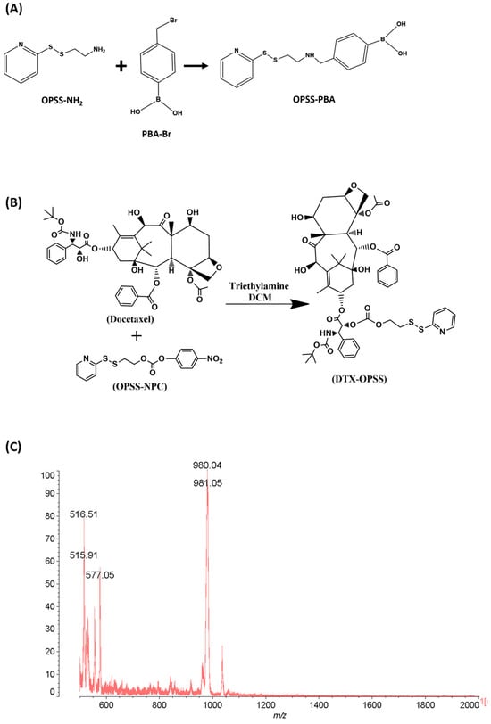
OPSS-NH2 was synthesized by reacting 2,2′-dipyridyl disulfide with 2-mercaptoethylamine (Figure 1A). The successful synthesis was confirmed by 1H NMR spectroscopy, consistent with a previous report [12]. Specifically, the 1H NMR spectra of OPSS-NH2 exhibited resonance peaks at 3.0 to 3.2 ppm and 7.3 to 8.5 ppm, corresponding to the methylene and pyridyl protons, respectively (Figure S1). Subsequently, OPSS-NH2 reacted with 4-(bromomethyl)phenylboronic acid to yield OPSS-PBA. The formation of OPSS-PBA was also validated via 1H NMR. The spectrum revealed new resonance peaks in the aromatic and alkyl regions alongside the conserved OPSS-NH2 protons, confirming the successful conjugation.
Figure 1.
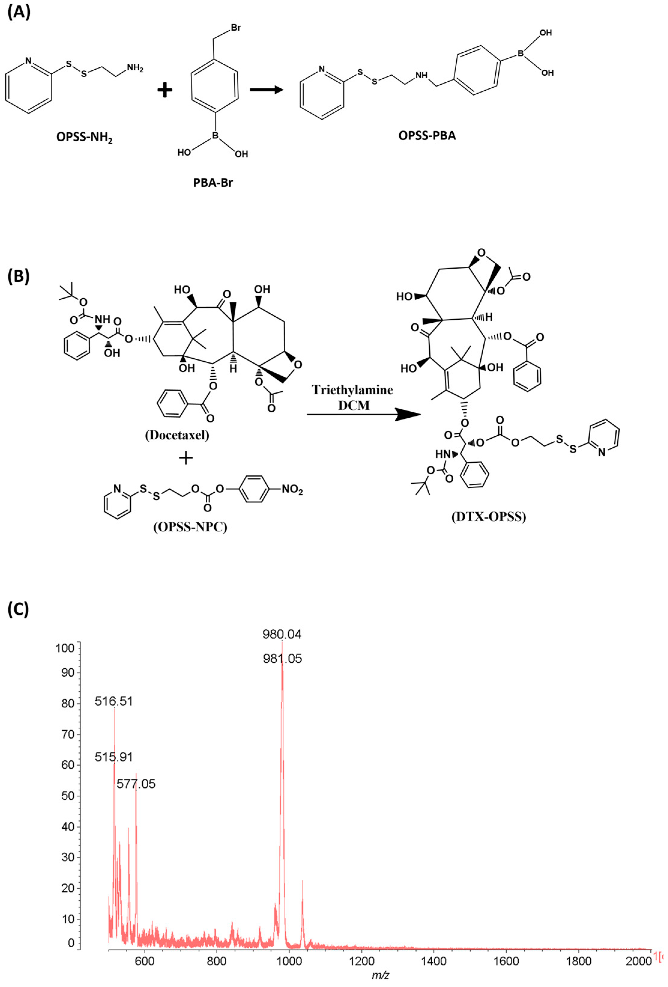
Synthesis and characterization of OPSS-PBA and OPSS-DTX. (A) Molecular structure of OPSS-PBA. (B) Molecular structure of OPSS-DTX. (C) MALDI-TOF mass spectrum of OPSS-DTX.
2,2′-Dipyridyl disulfide was reacted with 2-mercaptoethanol to obtain OPSS-OH. Further, successful synthesis of OPSS-OH was confirmed by 1H NMR. The spectrum shows the resonance peaks at 2.9 and 3.8 ppm related to the methylene protons and 7.15, 7.26, 7.40, 7.58 and 8.5 ppm related to the pyridyl ring protons, which is consistent with a previous report [18]. Next, OPSS-OH was reacted with 4-nitrophenyl chloroformate to obtain OPSS-NPC (Figure 1B). Further, successful synthesis of OPSS-NPC was confirmed by 1H NMR. The 1H NMR spectrum of OPSS-NPC, resonance peaks at 3.0–3.2, 4.5 and 7.0–8.5 ppm, corresponding to the methylene and aromatic ring protons, respectively, indicates successful synthesis of OPSS-NPC. Furthermore, docetaxel was reacted with OPSS-NPC to achieve OPSS-DTX [19]. Further, 1H NMR spectra of OPSS-DTX showed resonance peaks at 3.0–3.2, 4.5, and 7.3–8.5 ppm, corresponding to protons of methylene and pyridyl groups along with characteristic resonance peaks originated from the docetaxel. In addition, OPSS-DTX was characterized by MALDI-TOF, as shown in Figure 1C. Mass spectral analysis showed a mass peak at m/z 1024, along with other characteristic peaks of the fragments, further confirming the formation of OPSS-DTX. Altogether, these results suggest successful synthesis of OPSS-DTX.
3.2. Generation of Ad-DTX/HER Complex
A hetero-bifunctional linker was designed to enable selective conjugation of both the antibody and adenovirus without competitive reactions. The OPSS motif can react with the thiol group on the Ad surface through a thiol exchange reaction. At the opposite terminus, the PBA motif interacts with cis-diol groups on the antibody’s glycosylation site, leading to the formation of a phenylboronic ester complex. The synthesis of the Ad-DTX/HER conjugate is illustrated in Scheme 1. Surface-exposed amines on the adenoviral capsid were thiolated with 2-iminothiolane (2-IT) to generate Ad-SH. Then Ad-SH reacted with OPSS-PBA and OPSS-DTX. A thiol exchange reaction between the OPSS in linker and thiolated Ad resulted in disulfide bond formation, yielding Ad-DTX/PBA. Herceptin, used as the targeting moiety for HER2-overexpressing cancers, was subsequently mixed with Ad-DTX/PBA to produce Ad-DTX/HER conjugates. Purification was performed between each reaction step to remove unreacted reagents.
3.3. Evaluation of Viral Replication and Cell Killing Efficacy of oAd Under Taxane Compounds for Payload Selection
Paclitaxel (PTX), cabazitaxel (CTX), and docetaxel (DTX) are widely used taxane chemotherapeutics with well-established cytotoxic activity in solid tumors [20]. To identify the optimal therapeutic payload for our viral complex, MTT assay was conducted in H460 and SK-BR-3 cells, both of which express HER2 and the coxsackie-adenovirus receptor (CAR). These cells were treated with increasing concentrations of PTX, CTX, or DTX under our experimental conditions. As shown in Figure S2, all three taxanes dose-dependently reduced cell viability over the tested concentration range. Importantly, DTX showed 300% or 150% increased cancer cell killing compared with PTX or CTX at 10 ng/mL in H460 cells, respectively (*** p < 0.001). Similar trends were observed in SK-BR-3, indicating superior anticancer activity of DTX rather than PTX or CTX, with its cytotoxic effects becoming evident at lower concentrations than those of the other taxanes. Based on these findings, DTX was selected as the optimal payload for subsequent evaluations of viral replication assays.
To determine whether the selected taxane DTX interferes with adenoviral replication, replication assays were performed in H460 and SK-BR-3 cells. Across both cell lines, DTX co-treatment yielded replication levels that were maintained or modestly increased relative to drug-free oAd at 72 h, with viral genome copy numbers remaining indistinguishable from drug-free controls. In SK-BR-3 cells, the presence of DTX significantly boosted adenoviral replication across the tested doses (*** p < 0.001). These results indicate that DTX does not compromise adenoviral replication and is therefore compatible as the cytotoxic payload of the oAd-taxane/HER complex in subsequent experiments. The 72 h time point corresponds to the typical adenoviral replication cycle of approximately 48–72 h, which is appropriate for capturing productive viral replication [21,22,23].
3.4. Increased Transduction Efficacy Test of dAd-DTX/HER
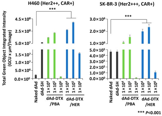
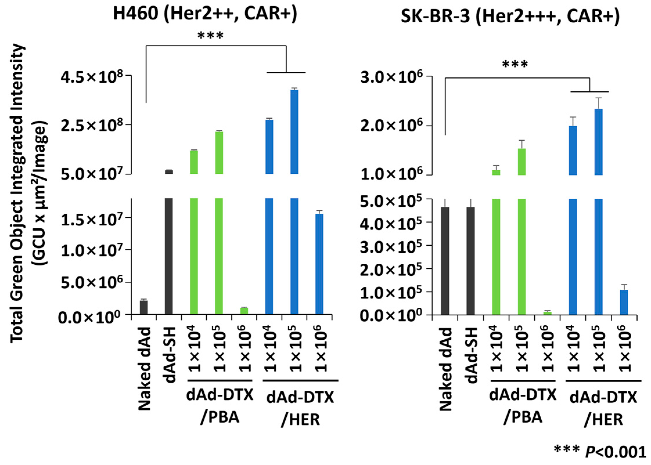
To determine whether the synthesized dAd-DTX/HER undergoes HER2-specific internalization, transduction assays were performed in HER2- and CAR-positive cancer cells (H460 and SK-BR-3) under matched infection conditions (Figure 2). Across formulations, dAd-DTX/HER groups (1 × 10⁴ and 1 × 10⁵) consistently showed higher GFP signals than either naked dAd, dAd-SH or the non-targeted dAd-DTX/PBA control, indicating enhanced HER2-mediated internalization. In SK-BR-3, the dAd-DTX/HER 1 × 10⁵ formulation, containing 1 × 10⁵ equivalents of OPSS-PBA and 1 × 10⁵ equivalents of OPSS-DTX relative to Ad, produced the greatest GFP signal (2.43-fold of naked dAd). The same trend of formulations was observed in H460, indicating that HER2-mediated internalization effectively causes the native CAR-dependent viral entry across multiple HER2-expressing cancer models. By contrast, the highest antibody loading group, the dAd-DTX/HER (1 × 10⁶) group, showed the lowest GFP (0.29-fold of naked dAd) expression in SK-BR-3, with a similar reduction in H460. This pattern is consistent with excess antibody remaining on the viral surface after conjugation and purification, causing steric hindrance and competitive blocking of HER2, thereby limiting complex internalization. Collectively, these results indicate that the dAd-DTX/HER enters cells predominantly via HER2 rather than CAR, and that efficient internalization is achieved only within an optimal linker/drug window exemplified by the 1 × 10⁵ formulation. Accordingly, subsequent experiments were performed sing the dAd-DTX/HER 1 × 10⁵ condition (OPSS-PBA and OPSS-DTX each set at 1 × 10⁵ equivalents relative to Ad). Thereafter, we further refined and systematically varied the Herceptin-to-DTX ratios.
Figure 2.
Transduction efficacy test of dAd-DTX/HER. GFP quantification of all cells transduced with naked dAd, dAd-SH, dAd-DTX/PBA, or dAd-DTX/HER (OPSS-DTX/OPSS-PBA:dAd ratio of 1 × 10⁴, 1 × 10⁵, or 1 × 10⁶). After 48 h, GFP expression was observed and quantified by an IncuCyte® Live-Cell Analysis System. Data are presented as mean ± SD (n = 3 per group). *** p < 0.001.
3.5. Physiochemical Characterization of dAd-DTX/HER
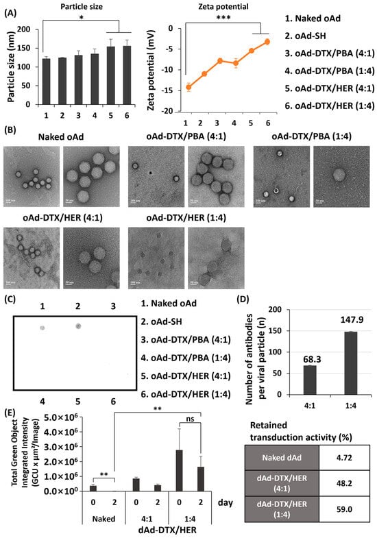
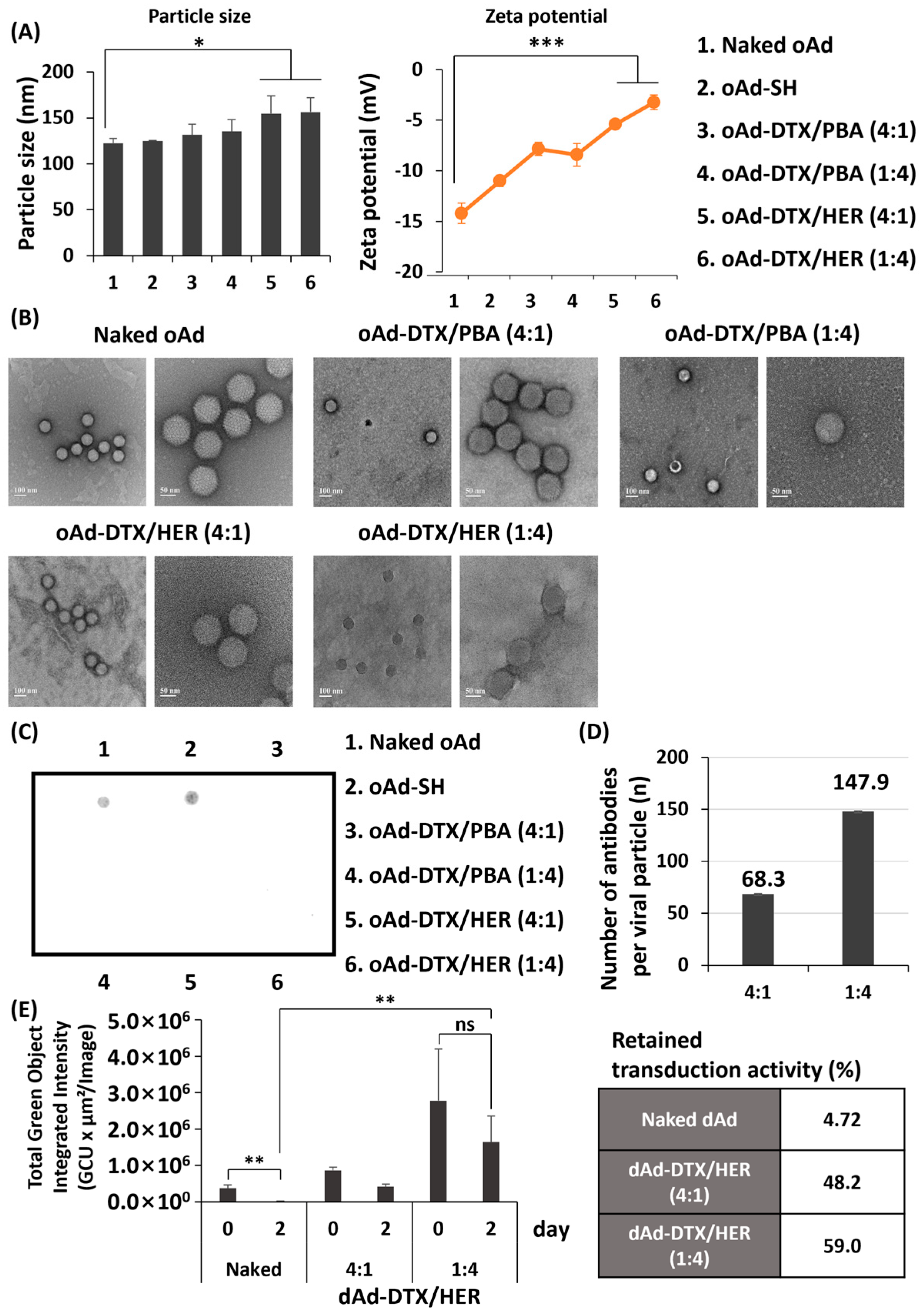
To assess the physiochemical properties of oAd-DTX/HER, we measured the size and surface charge of conjugates by DLS/Zeta instrument (Figure 3A). The average diameter of oAd-SH was not significantly different from that of naked oAd, indicating that thiolation alone does not markedly alter particle size. In contrast, the average size of the oAd-DTX/HER conjugates were significantly increased compared with naked oAd. Conjugation of naked oAd with OPSS-PBA and OPSS-DTX, followed by Herceptin attachment at different molar ratios, resulted in a stepwise increase in the hydrodynamic diameter of the oAd-DTX/HER complexes. Notably, even after this enlargement, the mean diameter of all oAd-DTX/HER formulations remained below 200 nm, a size range generally considered favorable for tumor trafficking and retention via the enhanced permeability and retention (EPR) effect [24].
Figure 3.
Physicochemical characterization of oAd-DTX/HER. (A) Size distribution and ζ-potential of naked oAd, oAd-SH, oAd-DTX/PBA, and oAd-DTX/HER (oAd-DTX/PBA, DTX:PBA ratio of 4:1 or 1:4, oAd-DTX/HER, DTX:HER ratio of 4:1 or 1:4). * p < 0.05, *** p < 0.001. (B) Transmission electron microscopy (TEM) of samples negatively stained with uranyl acetate to analyze the surface structure of naked oAd, oAd-DTX/PBA, and oAd-DTX/HER; the scale bar represents 100 nm (left) and 50 nm (right) in each group. (C) Hexon detection of naked oAd, oAd-SH, oAd-DTX/PBA, and oAd-DTX/HER by dot blot analysis using an anti-adenovirus hexon antibody to confirm surface masking; chemiluminescence was measured by an ImageQuant LAS 500. Data are presented as mean ± SD (n = 3 per group). (D) Number of antibody molecules conjugated per viral particle. The antibody loading of oAd-DTX/HER (4:1 and 1:4) was quantified by a human IgG ELISA after removal of unconjugated antibody and expressed as the number of trastuzumab molecules per viral particle. Data are presented as mean ± SD (n = 3 per group). (E) Transduction efficiency of naked oAd and the complex groups following pre-incubation in 10% FBS-containing PBS at 37 °C for 0 and 2 days. The remaining viral activity of the pre-incubated samples was evaluated in SK-BR-3 cells at an MOI of 30. Data are presented as mean ± SD (n = 3 per group). ** p < 0.01.
For the ζ-potential (surface charge), naked oAd exhibited the lowest value at approximately −15 mV. By contrast, oAd-DTX/HER prepared with increasing amounts of OPSS-PBA/OPSS-DTX and Herceptin showed progressively less negative ζ-potential values compared with naked oAd. For reference, Herceptin alone displayed a ζ-potential of about −10 mV [25]. Increasing the Ad:antibody molar ratio to 1:4 shifted the conjugate ζ-potential to −3.25 mV. This gradual shift toward electrical neutrality indicates that higher levels of antibody and linker incorporation more effectively mask the viral capsid surface. Taken together, these size and ζ-potential profiles confirm that the oAd surface is efficiently covered by the OPSS-PBA, OPSS-DTX, and Herceptin, yielding a compact, partially charge-neutralized nanoparticle that is potentially advantageous for systemic delivery and tumor accumulation. The polydispersity index (PDI) values of naked oAd, oAd-SH, oAd-DTX/PBA(4:1), oAd-DTX/PBA(1:4), oAd-DTX/HER(4:1) and oAd-DTX/HER(1:4) were 0.05, 0.14, 0.21, 0.17, 0.24, and 0.38, respectively. All values remained below 0.4, indicating relatively narrow and homogeneous size distributions, although a modest increase in PDI was observed after dual conjugation, consistent with the surface modification of the viral capsid.
Transmission electron microscopy (TEM) was used to assess the morphology of naked oAd, oAd-DTX/PBA, and oAd-DTX/HER. As shown in Figure 3B, the structural component of naked oAd was clearly observed. The adenoviral capsid, composed of hexon and other structural proteins, displays a characteristic icosahedral morphology [26]. In contrast, the icosahedra were markedly diminished in the oAd-DTX/HER conjugates. In samples where antibody was conjugated to oAd, a dark peripheral edge appeared on the capsid surface. Within these linker-bearing formulations, preparations with a higher antibody:oAd ratio showed more pronounced blurring of capsid features, indicating denser surface coverage. Overall, these TEM observations suggest that increasing the amounts of linker and antibody leads to more complete capsid masking and higher conjugation efficiency in the oAd-DTX/HER complexes. Surface masking was initially inspected by the TEM images of naked oAd, oAd-DTX/PBA, and oAd-DTX/HER. Although the TEM images suggested progressive blurring of the icosahedral capsid in antibody-conjugated samples, this approach did not allow a quantitative assessment of the extent to which capsid proteins were shielded. To directly evaluate Herceptin-mediated masking of hexon, we therefore performed a dot blot assay using an anti-hexon antibody. In addition, the number of antibody molecules conjugated per viral particle was directly quantified by ELISA, providing a quantitative measure of surface modification that complements the qualitative evidence from the TEM and dot blot analyses.
As shown in Figure 3C, naked oAd produced a strong, sharply defined dot at a given viral input, consistent with hexon epitopes being fully accessible on the virion surface. By contrast, conjugate formulations (both oAd-DTX/PBA and oAd-DTX/HER) displayed a complete loss of anti-hexon signals. This result indicates that hexon epitopes are occluded primarily by coating of OPSS-PBA and OPSS-DTX, providing strong shielding of the oAd capsid from the external environment. To quantify the extent of surface conjugation, the dot blot signal was analyzed by densitometry, which indicated that approximately 100% of the accessible hexon epitopes in all conjugate formulations (oAd-DTX/PBA and oAd-DTX/HER at DTX:PBA or DTX:HER ratios of 4:1 and 1:4) were masked relative to naked oAd (Figure 3C and Figure S3). The number of trastuzumab molecules conjugated per viral particle was further determined by ELISA, revealing approximately 68.3 and 147.9 trastuzumab molecules per viral particle for the oAd-DTX/HER 4:1 and 1:4 complexes, respectively (Figure 3D). The amount of DTX conjugated to the complex was below the detection limit of the assay, and an exact DTX loading value could therefore not be determined. Together, these values quantitatively define the conjugation efficiency and the antibody valency of the complex.
To evaluate the stability of the complex under physiological conditions, naked dAd and the final dAd-DTX/HER complex were incubated in 10% FBS-containing PBS at 37 °C, and transduction efficiency was assessed after 0 and 2 days. In terms of viral activity, both complex groups retained transduction activity better than naked dAd over the incubation period, showing approximately 10- to 13-fold higher activity at day 2 (Figure 3E). These results indicate that the dual conjugation improves the functional stability of the virus under physiological conditions, supporting its suitability for systemic administration.
In conclusion, the comprehensive physicochemical characterization demonstrates the successful formulation of oAd-DTX/HER conjugates. The sequential modification with DTX and Herceptin yields highly compact nanoparticles (<200 nm) with near-neutral surface charges and thoroughly masked adenoviral capsids. These structural and surface properties collectively suggest that oAd-DTX/HER is optimally designed to minimize non-specific interactions and evade immune recognition in circulation, thereby maximizing the potential for safe systemic administration and targeted tumor accumulation.
3.6. Cancer-Specific Cell Killing Effect of oAd-DTX/HER
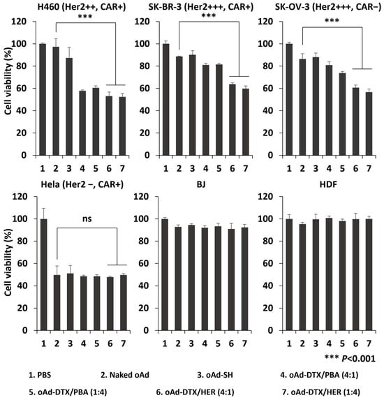
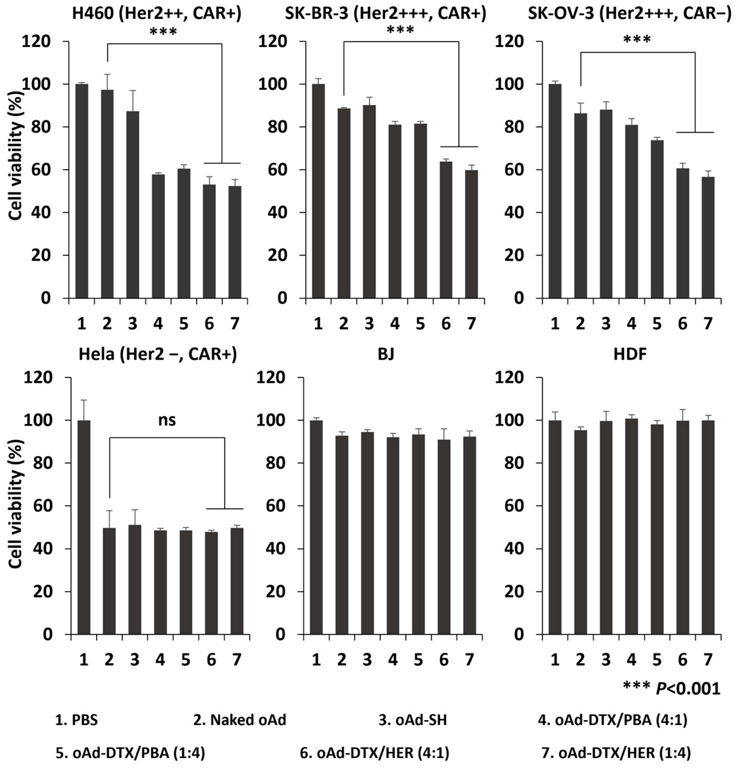
To evaluate cancer-selective cytotoxicity, four preparations, naked oAd, oAd-SH, oAd-DTX/PBA, and oAd-DTX/HER, were compared across cell lines. Overall, oAd-DTX/HER exhibited greater cytotoxicity than the antibody-free formulation and naked virus. In H460, compared with oAd-DTX/PBA, oAd-DTX/HER conjugates revealed 1.11-fold (DTX:HER = 4:1) and 1.20-fold (1:4) greater cell killing than oAd-DTX/PBA, demonstrating 18.15-fold and 18.43-fold higher killing than naked oAd, respectively (Figure 4A). Similarly, in SK-BR-3, oAd-DTX/HER yielded 1.92-fold and 2.18-fold greater cytotoxicity than oAd-DTX/PBA at DTX:HER ratios of 4:1 and 1:4, with 3.18-fold and 5.53-fold increases over naked oAd. The same trend was observed in SK-OV-3 (HER positive and CAR negative), whereas no improvement was detected in HeLa (HER negative and CAR positive). Taken together, these results indicate that oAd-DTX/HER induces HER2-dependent, cancer-specific cell killing. However, in normal fibroblasts (BJ, HDF), none of the formulations including naked oAd, oAd-DTX/PBA, or oAd-DTX/HER induced detectable cytotoxicity. Collectively, these findings demonstrate that oAd-DTX/HER can selectively eradicate HER2-expressing tumor cells while sparing normal cells, thereby achieving tumor-restricted cytotoxicity with a favorable safety profile that is compatible with systemic administration.
Figure 4.
Evaluation of the cancer cell killing effect and cancer selectivity of oAd-DTX/HER. H460, SK-BR-3, SK-OV-3, and HeLa cells were treated with naked oAd, oAd-SH, oAd-DTX/PBA, or oAd-DTX/HER (oAd-DTX/PBA, DTX:PBA ratio of 4:1 or 1:4, oAd-DTX/HER, DTX:HER ratio of 4:1 or 1:4) (MOI 4 for H460, SK-BR-3, and HeLa, MOI 8 for SK-OV-3), and cell viability was measured at 72 h by MTT assay. Normal fibroblasts (BJ and HDF) were treated with the same formulations at 100 MOI, and viability was assessed at 48 h by MTT assay to evaluate off-target cytotoxicity. Data are presented as mean ± SD (n = 3 per group). *** p < 0.001, ns, not significant.
3.7. HER2-Mediated Cellular Entry of dAd-DTX/HER
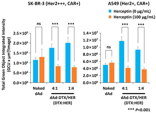
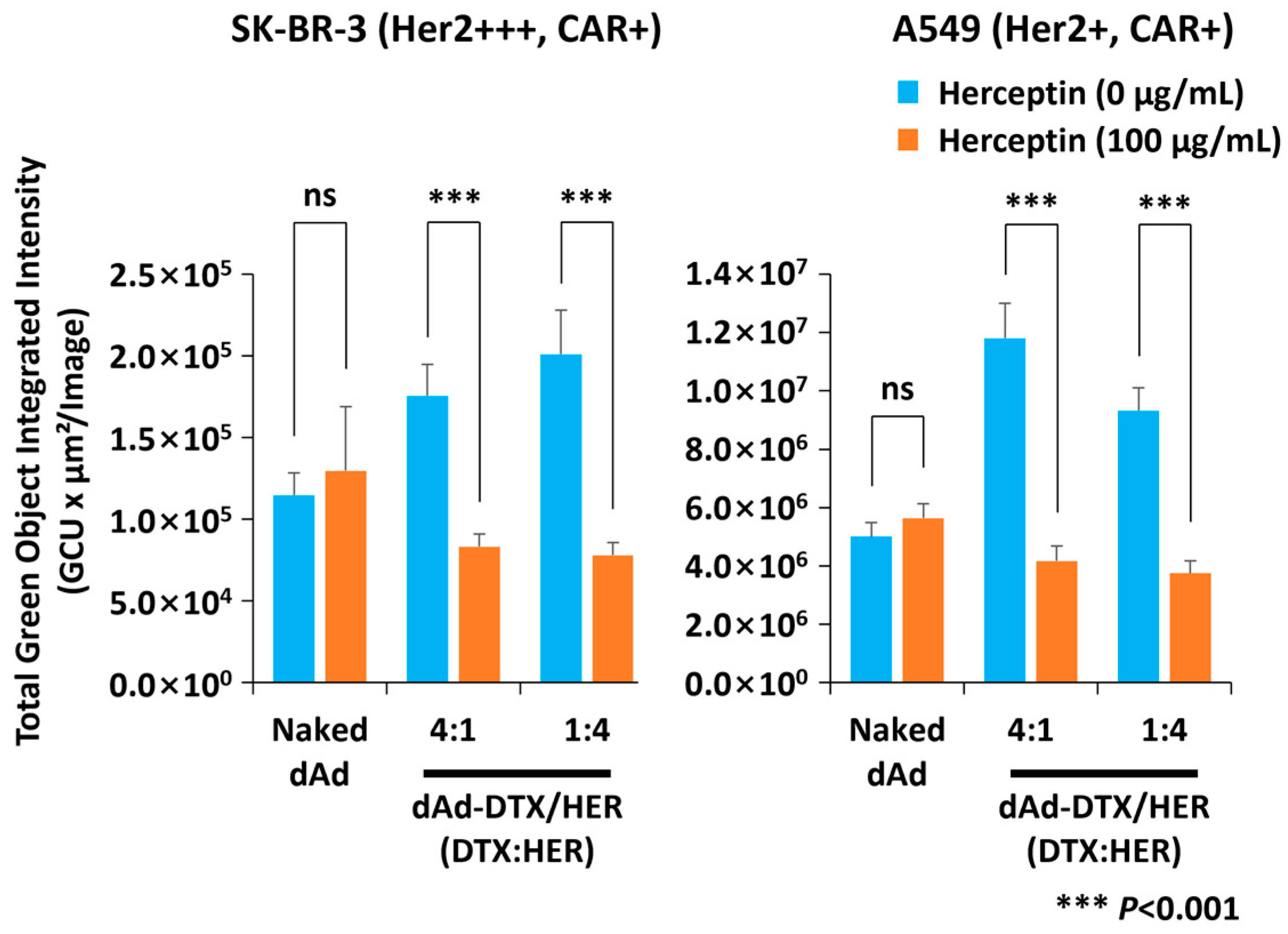
To demonstrate HER2 specific cellular internalization of dAd-DTX/HER, SK-BR-3 and A549 (HER2 and CAR positive) cancer cells were pre-incubated with or without free Herceptin before the transduction experiment. As shown in Figure 5, Herceptin pretreatment did not affect transduction by naked dAd. In contrast, HER2 blockade by Herceptin pretreatment significantly reduced dAd-DTX/HER-mediated transduction. In A549 cells, HER2 blockade reduced dAd-DTX/HER-mediated GFP expression to 0.35-fold and 0.40-fold of the no-block condition for the 4:1 and 1:4 DTX:HER formulations, respectively. These results indicate that Ad-DTX/HER conjugates can be internalized by specific interaction between Herceptin on the surface of the conjugate and HER2 on the surface of the cancer cell. Collectively, these data indicate that cellular entry of dAd-DTX/HER in HER2- and CAR-positive cells is mediated predominantly by HER2-dependent uptake rather than CAR-mediated infection.
Figure 5.
Competition assay of dAd-DTX/HER with free Herceptin. Herceptin (100 µg/mL) was used as a pre-treatment for all cell lines, and both naked dAd and dAd-DTX/HER conjugates (4:1 and 1:4, DTX:HER ratio) were used for transduction at an MOI of 100. Using a fluorescent microscope and an IncuCyte® Live-Cell Analysis System, GFP expression was measured after 48 h. Data are presented as mean ± SD (n = 3 per group). *** p < 0.001, ns, not significant.
3.8. Bio-Distribution Profiling of oAd-DTX/HER
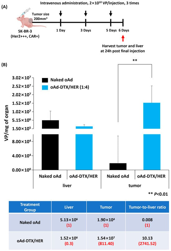
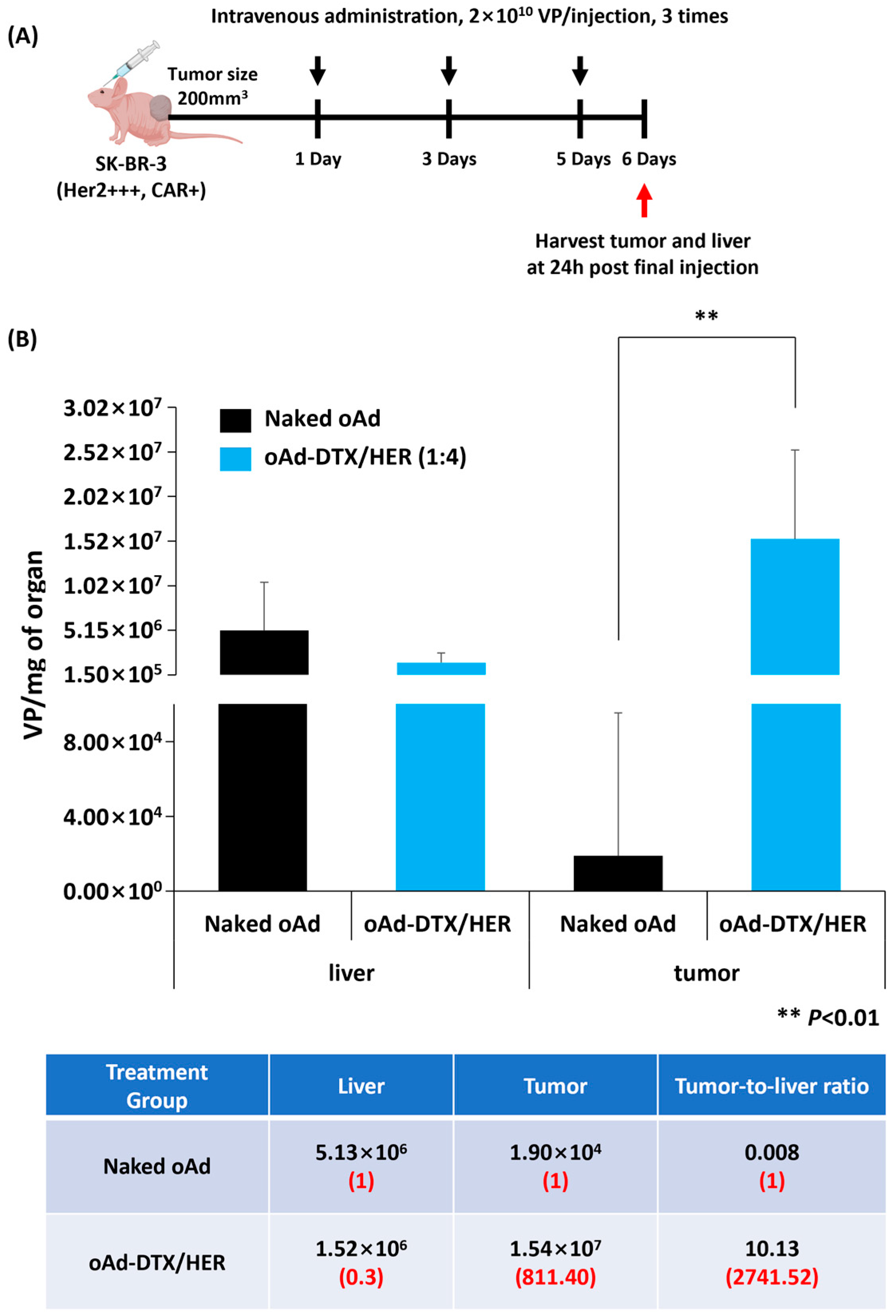
To determine whether antibody and drug decoration could simultaneously reduce nonspecific hepatic uptake and enhance tumor accumulation, bio-distribution of systemically administrated oAd-DTX/HER was profiled in SK-BR-3 xenograft-bearing mice (Figure 6). As expected, naked oAd predominantly accumulated in the liver. It further did not demonstrate tumor accumulation, highlighting the need for additional targeting elements. In contrast, oAd-DTX/HER effectively avoided hepatic trapping, reducing liver accumulation to 0.3-fold compared with naked oAd. Furthermore, tumor accumulation was markedly increased by oAd-DTX/HER. It showed 811.40-fold higher tumor accumulation than naked Ad.
Figure 6.
Assessment of bio-distribution of oAd-DTX/HER. (A) The treatment schedule. (B) Viral genome copy numbers in tumors and livers of mice were quantified using qPCR after the third intravenous administration with 2 × 10¹⁰ VP of naked oAd or oAd-DTX/HER (DTX:HER ratio of 1:4). Data are presented as mean ± SD (n = 3 per group). ** p < 0.01.
As a resultant, the tumor-to-liver ratios of oAd-DTX/HER, the critical consideration factor to measure therapeutic outcome and safety profile simultaneously, significantly increased up to 2741.52 compared to naked Ad. Taken together, these data demonstrate that HER /DTX surface display effectively overcomes residual adenoviral hepatotropism and redirects oAd-DTX/HER to HER2-positive tumors after systemic dosing, thereby improving both safety (reduced liver exposure) and therapeutic index (enhanced intratumoral delivery).
3.9. Immune Evasion of the Ad-DTX/HER
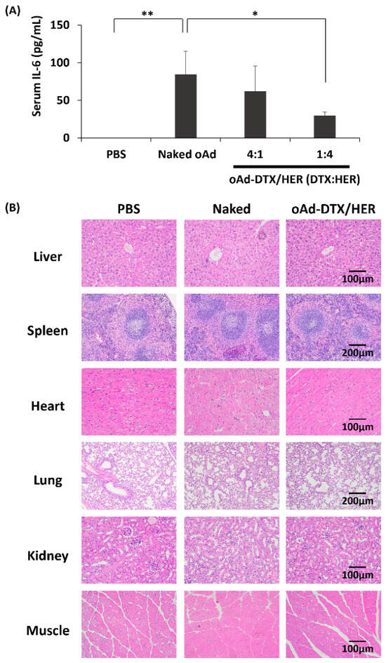
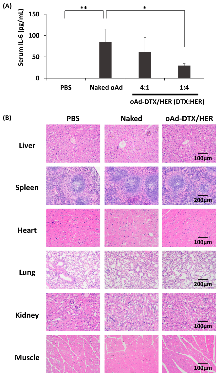
To substantiate the immune-shielding capability of the complex, serum IL-6 levels measured after intravenous injection of oAd-DTX/HER were approximately 1.4- and 2.8-fold lower than those of naked oAd for the 4:1 and 1:4 groups, respectively (Figure 7A), indicating that complex formation attenuates the virus-induced innate immune response.
Figure 7.
In vivo immune-shielding properties and safety profile of oAd-DTX/HER. (A) In vivo cytokine analysis. Serum IL-6 levels were measured by ELISA at 6 h following intravenous injection of PBS, naked oAd, or the complex (4:1 and 1:4) into mice. Data are presented as mean ± SD (n = 3). * p < 0.05, ** p < 0.01. (B) Histopathological analysis of major organs from mice systemically treated with PBS, naked oAd, or oAd-DTX/HER. Major organs were collected 2 days after administration of each formula. Representative sections of liver, spleen, heart, lung, kidney, and muscle (from top to bottom) were stained with H&E for each treatment group. Scale bar, 100 µm.
3.10. Safety Profile of oAd-DTX/HER
To assess off-target toxicity, major organs (liver, spleen, heart, lung, kidney, and skeletal muscle) were collected 2 days after systemic administration of PBS, naked oAd, or oAd-DTX/HER and stained with H&E (Figure 7B). In the naked oAd group, several histological alterations were observed. Hepatic sections showed focal pericentral and perisinusoidal accumulation of inflammatory cells with accompanying sinusoidal congestion, changes compatible with a low-grade inflammatory response. The myocardium contained areas in which fiber orientation was locally disorganized rather than displaying the uniform parallel alignment seen in controls. Lung sections occasionally exhibited thickened alveolar septa with increased interstitial cellularity, indicative of incipient inflammatory remodeling of the alveolar walls.
In contrast, organs from mice treated with oAd-DTX/HER were histologically comparable to those from PBS-treated group. The myocardium showed regularly aligned muscle fibers without structural disruption, the lung displayed intact alveolar walls without septal thickening or inflammatory infiltration, and hepatic lobules exhibited well-preserved central veins without pericentral inflammation. Renal glomeruli and tubules were clearly defined, and the spleen retained a distinct separation between red and white pulp with no signs of necrosis or congestion. Collectively, these findings indicate that systemic administration of oAd-DTX/HER does not confer additional off-target organ toxicity relative to naked oAd. No treatment-related lesions, including necrosis, hemorrhage, or inflammatory infiltrates, were detected in the oAd-DTX/HER group.
4. Discussion
In this study, we successfully engineered a multifunctional oAd platform, oAd-DTX/HER, which integrates tumor targeting, immune evasion, and potent chemo-virotherapy within a single therapeutic platform. A major bottleneck in systemic oAd delivery is rapid immune clearance mediated by major capsid proteins, particularly the hexon [27]. While traditional strategies employ bulky hydrophilic polymers to mask these immunogenic epitopes, such approaches often compromise viral infectivity due to steric hindrance [28]. Our physicochemical and morphological analyses demonstrate that direct site-directed bioconjugation of OPSS-PBA/OPSS-DTX and Herceptin effectively shields the hexon while maintaining a compact hydrodynamic size (Figure 3). This polymer-free masking strategy not only attenuates immune recognition but also preserves the structural integrity required for robust viral entry (Figure 5). Furthermore, by utilizing DTX, which exhibited the highest cytotoxic potency without compromising oAd replication (Figure S2), the complex releases a synergistic dual-assault mechanism such as DTX-mediated microtubule stabilization and subsequent oncolytic replication. Consistent with this, the complex exhibited reduced virus-induced IL-6 production relative to naked oAd (Figure 7A), providing direct functional evidence of immune shielding.
The therapeutic potential of oAd-DTX/HER is heavily dependent on its ability to discriminate between malignant and healthy tissues. Our in vitro and in vivo findings strongly validate this tumor-selective paradigm. The complex exhibited markedly enhanced cytotoxicity in HER2-overexpressing cancer cells, driven by Herceptin-mediated endocytosis, while sparing non-malignant fibroblasts (Figure 4). More importantly, systemic administration of adenoviral vectors is hindered by the liver sink effect, where predominant hepatic sequestration leads to severe hepatotoxicity and low tumor bioavailability (Figure 6). However, oAd-DTX/HER successfully weakened this natural hepatotropism. The dramatically improved tumor-to-liver accumulation ratio following intravenous injection indicates that the chemical shield effectively bypasses nonspecific hepatic uptake while redirecting the viral payload to the tumor microenvironment. Coupled with the absence of treatment-related lesions in major organs, this redistribution profile addresses the most critical safety hurdles in systemic virotherapy, establishing a favorable therapeutic window (Figure 7B).
Ultimately, our present study establishes a novel paradigm for the direct surface engineering of oAd. By site-specifically conjugating a targeted antibody and a chemotherapeutic agent directly onto the naked viral capsid, we successfully bypassed the structural limitations and infectivity loss associated with conventional polymer-based shielding. This streamlined engineering strategy transforms the native virus into a highly sophisticated platform for HER2-targeted chemo-virotherapy. The precise integration of targeted cellular entry, chemical cytotoxicity, and viral oncolysis within a single, polymer-free platform effectively overcomes major systemic delivery barriers, such as hepatic sequestration, while maximizing synergistic tumor eradication. Therefore, this direct surface engineering approach not only validates a potent therapeutic strategy for HER2-positive malignancies but also provides a highly modular and translatable framework for developing next-generation chemo-virotherapeutics.
Supplementary Materials
The following supporting information can be downloaded at: https://www.mdpi.com/article/10.3390/pharmaceutics18111389/s1. Figure S1: 1H NMR (CDCl3, 600 MHz) spectra of OPSS–NH2, PBA–Br, OPSS–PBA, OPSS–NPC, and OPSS–DTX; Figure S2: Evaluation of viral replication and cell-killing efficacy of oAd under taxane compounds for payload selection. (A) At 72 h post-treatment, MTT assay measuring the cancer cell-killing efficacy of free drug (normalized to PBS = 100%); (B) oAd replication in HER2- and CAR-positive cells (H460 and SK-BR-3) with DTX; viral genomes were measured at 72 h by qPCR (drug-free oAd control). Data are presented as mean ± SD (n = 3 per group). *** p < 0.001; Figure S3: Quantification of hexon masking efficiency. Dot-blot signal intensities of oAd-DTX/PBA and oAd-DTX/HER (DTX:PBA or DTX:HER ratio of 4:1 or 1:4) were quantified by densitometry using ImageJ software (version 1.54) and expressed as masking efficiency (%) relative to the hexon signal of naked oAd [100 − (conjugate signal/naked oAd signal) × 100].
Author Contributions
D.K.: Investigation, Writing—original draft. Y.K.: Investigation, Writing—original draft. S.M.C.: Investigation, Methodology. C.-O.Y.: Conceptualization, Project administration, Resources, Supervision. A.-R.Y.: Conceptualization, Project administration, Writing—review & editing, Supervision, Resources. All authors have read and agreed to the published version of the manuscript.
Funding
This research was funded by the Korea Health Technology R&D Project through the Korea Health Industry Development Institute (KHIDI), funded by the Ministry of Health & Welfare, Republic of Korea (grant numbers RS-2024-00439816 to C.-O.Y. and RS-2025-02263117 to A.-R.Y.); the Ministry of Food and Drug Safety, Republic of Korea (grant number RS-2024-00395863 to A.-R.Y.); and the Korea Basic Science Institute (National Research Facilities and Equipment Center), funded by the Ministry of Education (grant number 2023R1A6C101A009).
Institutional Review Board Statement
The animal study protocol was approved by the Institutional Animal Care and Use Committee (IACUC) of Hanyang University (approval no. HYU-2024-0082A). All procedures were conducted in accordance with local legislation and institutional requirements.
Data Availability Statement
The data supporting the findings of this study are available upon reasonable request.
Acknowledgments
The graphical abstract and schematic illustrations were created with BioRender.com. After using this tool, the authors reviewed and edited the figures as needed and take full responsibility for the content of the publication. During the preparation of this work, the authors used ChatGPT, based on GPT-4/5 (OpenAI, San Francisco, CA), to improve the language and readability. After using this tool, the authors reviewed and edited the content as needed and take full responsibility for the content of the publication.
Conflicts of Interest
C-OY is a CEO of GeneMedicine Co., Ltd., which develops oAd for clinical applications. The remaining authors declare that the research was conducted in the absence of any commercial or financial relationships that could be construed as a potential conflict of interest.
References
- Choi, J.-W.; Lee, J.-S.; Kim, S.W.; Yun, C.-O. Evolution of oncolytic adenovirus for cancer treatment. Adv. Drug Deliv. Rev. 2012, 64, 720–729. [Google Scholar] [CrossRef] [Scilit]
- Stepanenko, A.A.; Chekhonin, V.P. Tropism and transduction of oncolytic adenovirus 5 vectors in cancer therapy: Focus on fiber chimerism and mosaicism, hexon and pIX. Virus Res. 2018, 257, 40–51. [Google Scholar] [CrossRef] [Scilit]
- Yamamoto, M.; Curiel, D.T. Current issues and future directions of oncolytic adenoviruses. Mol. Ther. 2010, 18, 243–250. [Google Scholar] [CrossRef] [Scilit]
- Oronsky, B.; Gastman, B.; Conley, A.P.; Reid, C.; Caroen, S.; Reid, T. Oncolytic Adenoviruses: The Cold War against Cancer Finally Turns Hot. Cancers 2022, 14, 4701. [Google Scholar] [CrossRef] [Scilit]
- Wheeler, M.D.; Yamashina, S.; Froh, M.; Rusyn, I.; Thurman, R.G. Adenoviral gene delivery can inactivate Kupffer cells: Role of oxidants in NF-kappaB activation and cytokine production. J. Leukoc. Biol. 2001, 69, 622–630. [Google Scholar] [CrossRef] [Scilit]
- Santos, J.; Heiniö, C.; Quixabeira, D.; Zafar, S.; Clubb, J.; Pakola, S.; Cervera-Carrascon, V.; Havunen, R.; Kanerva, A.; Hemminki, A. Systemic Delivery of Oncolytic Adenovirus to Tumors Using Tumor-Infiltrating Lymphocytes as Carriers. Cells 2021, 10, 978. [Google Scholar] [CrossRef] [Scilit]
- Jirovec, E.; Quixabeira, D.C.A.; Clubb, J.H.A.; Pakola, S.A.; Kudling, T.; Arias, V.; Haybout, L.; Jalkanen, K.; Alanko, T.; Monberg, T.; et al. Single intravenous administration of oncolytic adenovirus TILT-123 results in systemic tumor transduction and immune response in patients with advanced solid tumors. J. Exp. Clin. Cancer Res. 2024, 43, 297. [Google Scholar] [CrossRef] [Scilit]
- Xia, Y.; Li, D.; Yang, K.; Ou, X. Systemic Delivery Strategies for Oncolytic Viruses: Advancing Targeted and Efficient Tumor Therapy. Int. J. Mol. Sci. 2025, 26, 6900. [Google Scholar] [CrossRef] [Scilit]
- Thambi, T.; Hong, J.; Yoon, A.-R.; Yun, C.-O. Challenges and progress toward tumor-targeted therapy by systemic delivery of polymer-complexed oncolytic adenoviruses. Cancer Gene Ther. 2022, 29, 1321–1331. [Google Scholar] [CrossRef] [Scilit]
- Lee, Y.S.; Lim, K.S.; Oh, J.-E.; Yoon, A.-R.; Joo, W.S.; Kim, H.S.; Yun, C.-O.; Kim, S.W. Development of porous PLGA/PEI1.8k biodegradable microspheres for the delivery of mesenchymal stem cells (MSCs). J. Control Release 2015, 205, 128–133. [Google Scholar] [CrossRef] [Scilit]
- Kim, P.-H.; Sohn, J.-H.; Choi, J.-W.; Jung, Y.; Kim, S.W.; Haam, S.; Yun, C.-O. Active targeting and safety profile of PEG-modified adenovirus conjugated with herceptin. Biomaterials 2011, 32, 2314–2326. [Google Scholar] [CrossRef] [Scilit]
- Saito, G.; Velluto, D.; Resmini, M. Synthesis of 1,8-naphthalimide-based probes with fluorescent switch triggered by flufenamic acid. R. Soc. Open Sci. 2018, 5, 172137. [Google Scholar] [CrossRef] [Scilit]
- Yoon, A.-R.; Kasala, D.; Li, Y.; Hong, J.; Lee, W.; Jung, S.-J.; Yun, C.-O. Antitumor effect and safety profile of systemically delivered oncolytic adenovirus complexed with EGFR-targeted PAMAM-based dendrimer in orthotopic lung tumor model. J. Control Release 2016, 231, 2–16. [Google Scholar] [CrossRef] [Scilit]
- Morrison, J.; Briggs, S.S.; Green, N.; Fisher, K.; Subr, V.; Ulbrich, K.; Kehoe, S.; Seymour, L.W. Virotherapy of ovarian cancer with polymer-cloaked adenovirus retargeted to the epidermal growth factor receptor. Mol. Ther. J. Am. Soc. Gene Ther. 2008, 16, 244–251. [Google Scholar] [CrossRef] [Scilit]
- Yan, M.; Schwaederle, M.; Arguello, D.; Millis, S.Z.; Gatalica, Z.; Kurzrock, R. HER2 expression status in diverse cancers: Review of results from 37,992 patients. Cancer Metastasis Rev. 2015, 34, 157–164. [Google Scholar] [CrossRef] [Scilit]
- Choi, J.-W.; Jung, S.-J.; Kasala, D.; Hwang, J.K.; Hu, J.; Bae, Y.H.; Yun, C.-O. pH-sensitive oncolytic adenovirus hybrid targeting acidic tumor microenvironment and angiogenesis. J. Control Release 2015, 205, 134–143. [Google Scholar] [CrossRef] [Scilit]
- Zhao, Y.; Le, T.M.D.; Hong, J.; Jiao, A.; Yoon, A.-R.; Yun, C.-O. Smart Accumulating Dual-Targeting Lipid Envelopes Equipping Oncolytic Adenovirus for Enhancing Cancer Gene Therapeutic Efficacy. ACS Nano 2024, 18, 27869–27890. [Google Scholar] [CrossRef] [Scilit]
- Batisse, C.; Dransart, E.; Sarkouh, R.A.; Brulle, L.; Bai, S.-K.; Godefroy, S.; Johannes, L.; Schmidt, F. A new delivery system for auristatin in STxB-drug conjugate therapy. Eur. J. Med. Chem. 2015, 95, 483–491. [Google Scholar] [CrossRef] [Scilit]
- Kanjilal, P.; Singh, K.; Das, R.; Matte, J.; Thayumanavan, S. Antibody Polymer Conjugates (APCs) for Active Targeted Therapeutic Delivery. Biomacromolecules 2023, 24, 3638–3646. [Google Scholar] [CrossRef] [Scilit]
- Muth, M.; Ojara, F.W.; Kloft, C.; Joerger, M. Role of TDM-based dose adjustments for taxane anticancer drugs. Br. J. Clin. Pharmacol. 2021, 87, 306–316. [Google Scholar] [CrossRef] [Scilit]
- King, C.R.; Dodge, M.J.; MacNeil, K.M.; Tessier, T.M.; Mymryk, J.S.; Mehle, A. Expanding the adenovirus toolbox: Reporter viruses for studying the dynamics of human adenovirus replication. J. Virol. 2024, 98, e0020724. [Google Scholar] [CrossRef] [Scilit]
- Gonzalez, R.; Huang, W.; Finnen, R.; Bragg, C.; Flint, S.J. Adenovirus E1B 55-kilodalton protein is required for both regulation of mRNA export and efficient entry into the late phase of infection in normal human fibroblasts. J. Virol. 2006, 80, 964–974. [Google Scholar] [CrossRef] [Scilit]
- Condezo, G.N.; Martín, C.S. Localization of adenovirus morphogenesis players, together with visualization of assembly intermediates and failed products, favor a model where assembly and packaging occur concurrently at the periphery of the replication center. PLoS Pathog. 2017, 13, e1006320. [Google Scholar] [CrossRef] [Scilit]
- Wu, J. The Enhanced Permeability and Retention (EPR) Effect: The Significance of the Concept and Methods to Enhance Its Application. J. Pers. Med. 2021, 11, 771. [Google Scholar] [CrossRef] [Scilit]
- Schermeyer, M.-T.; Wöll, A.K.; Kokke, B.; Eppink, M.; Hubbuch, J. Characterization of highly concentrated antibody solution-A toolbox for the description of protein long-term solution stability. MAbs 2017, 9, 1169–1185. [Google Scholar] [CrossRef] [Scilit]
- Fuschiotti, P.; Schoehn, G.; Fender, P.; Fabry, C.; Hewat, E.; Chroboczek, J.; Ruigrok, R.; Conway, J. Structure of the Dodecahedral Penton Particle from Human Adenovirus Type 3. J. Mol. Biol. 2006, 356, 510–520. [Google Scholar] [CrossRef] [Scilit]
- Schmid, M.; Ernst, P.; Honegger, A.; Suomalainen, M.; Zimmermann, M.; Braun, L.; Stauffer, S.; Thom, C.; Dreier, B.; Eibauer, M.; et al. Adenoviral vector with shield and adapter increases tumor specificity and escapes liver and immune control. Nat. Commun. 2018, 9, 450. [Google Scholar] [CrossRef] [Scilit]
- Kreppel, F.; Kochanek, S. Modification of adenovirus gene transfer vectors with synthetic polymers: A scientific review and technical guide. Mol. Ther. 2008, 16, 16–29. [Google Scholar] [CrossRef] [Scilit]
Disclaimer/Publisher’s Note: The statements, opinions and data contained in all publications are solely those of the individual author(s) and contributor(s) and not of MDPI and/or the editor(s). MDPI and/or the editor(s) disclaim responsibility for any injury to people or property resulting from any ideas, methods, instructions or products referred to in the content. |
© 2026 by the authors. Licensee MDPI, Basel, Switzerland. This article is an open access article distributed under the terms and conditions of the Creative Commons Attribution (CC BY) license.