


Abstract
Background: This study aimed to evaluate whether a shorter switching interval from intravitreal dexamethasone implant (DEXi) to intravitreal fluocinolone acetonide implant (FAci) improves anatomical and functional outcomes in patients with diabetic macular edema. Methods: This was a retrospective, multicenter, observational extension study including 260 eyes, with a mean follow-up of 141.6 ± 55.4 weeks. Results: Prior to FAci injection, 100% of eyes had received DEXi. The mean interval between the last DEXi and the first FAci was 24.2 ± 41.6 weeks (n = 188), i.e., 0–8 weeks in 54 eyes (28.7%) and >8 weeks in 134 eyes (71.3%). Mean BCVA was comparable between patients receiving their last DEXi ≤8 weeks and those receiving it >8 weeks prior to FAci. Compared to baseline, the ≤8-week group consistently and significantly showed lower central macular thickness (CMT) values at months 1–3, 6, 7, 9–11, 14, 16, 19, and 20 (p < 0.05). At months 12 and 24, 90 (34.6%) and 117 (45.0%) eyes required additional treatments, after a median time of 118 weeks (95% CI: 87.7–191.4). In the ≤8-week group, the normalized cumulative incidence of additional treatments was 0.33 treatments per eye at week 52 and 0.72 at week 104, versus 0.66 and 1.38 in the >8-week group, respectively. Intraocular pressure (IOP) elevation incidence did not significantly differ between the ≤8-week and >8-week groups, regardless of the IOP threshold used (≥25, ≥30, or ≥35 mmHg; all p > 0.05). Conclusions: A shorter interval between DEXi and FAci was associated with greater and sustained macular drying, along with a reduced need for additional treatments.
1. Introduction
According to the International Diabetes Federation Diabetes Atlas (9th edition, 2019), nearly 700 million adults worldwide are projected to live with diabetes by 2045 [1]. The prevalence of ocular complications of diabetes mellitus is estimated at 22.27% for diabetic retinopathy and 4.07% for clinically significant diabetic macular edema (DME) [2], with the latter affecting more than 21 million individuals worldwide [3].
Intravitreal anti-vascular endothelial growth factor (anti-VEGF) injections are considered the first-line treatment for DME. However, despite optimal treatment, 30 to 40% of DME patients respond poorly to anti-VEGF therapy, showing transient or insufficient fluid resolution [4].
Intravitreal corticosteroids provide a therapeutic alternative in patients with a suboptimal response to anti-VEGF or with chronic DME, associated with low-grade retinal inflammation [5,6].
Two slow-release sustained-delivery systems for intraocular corticosteroids are available for DME. They differ in potency, solubility, and pharmacokinetic profiles, particularly regarding burst release and duration of action, leading to dissimilar therapeutic effects in DME patients [7].
The 0.7-mg intravitreal dexamethasone implant (DEXi) (Ozurdex®, Allergan Inc., Irvine, CA, USA) is a bioerodible insert that allows extended drug release over a 6-month period, characterized by an initial burst release of approximately 2 months followed by an exponential decline over the remaining 4 months [8]. The clinical effect of DEXi mirrors its pharmacokinetic profile, with a maximum improvement in DME within the first 2 months followed by a decline, which becomes evident after the 3-month timepoint [9].
The 0.19-mg intravitreal fluocinolone acetonide implant (FAci) (Iluvien®, Alimera Sciences Ltd., Alpharetta, GA, USA) is a non-biodegradable implant providing a mean release rate of 0.2 µg per day for the first 3 months, followed by a stable, continuous, and sustained concentration of 0.5–1.0 ng/mL for up to 36 months [10,11]. Due to its pharmacokinetics, the peak functional and anatomical efficacy appears to occur 6 to 11 months after injection [12,13].
Thus, based on their pharmacokinetic profiles, DEXi is considered more of a bolus treatment with an immediate and substantial drug release after injection, whereas FAci acts more as a basal or background therapy that stabilizes the disease for a much longer duration [14,15].
In clinical practice, the DEXi is used prior to FAci injection, which is considered a third-line treatment, to assess the patient’s response to intravitreal corticosteroids and to identify patients at risk of developing steroid-induced ocular hypertension (OHT) [16,17]. Patients who do not develop steroid-induced OHT after 2–3 DEXi injections are at low risk of increased intraocular pressure (IOP) following FAci injection, with a positive predictive value of 90% [18,19].
The modalities of switching from DEXi to FAci are not well described in the literature [14,15], and data regarding the optimal timing of this switch are scarce. However, given the distinct pharmacokinetics of the two drugs, the time interval between the last DEXi injection and the first FAci injection may be critical.
In a previous study of 113 DME eyes switched from DEXi to FAci, we observed that an interval ≤8 weeks between the last DEXi and the first FAci was more effective in reducing central macular thickness (CMT) fluctuations and the need for additional treatments during the follow-up period (mean follow-up period of 7.6 months) [20]. In two other studies, Rousseau et al. [21] and Lampin et al. [22] injected the first FAci one month after the last DEXi (before DME recurrence) in 41 and 14 DME eyes, respectively. The rate of additional treatments at 12 months was 20.6% and 21%, respectively, which is much lower than that reported in the literature [23]. Bailey et al. [24]. and Cicinelli et al. [25] also observed that rescue therapy was indeed more frequently required when FAci was administered for recurrent DME.
To determine whether it would be more effective to inject FAci into an already dried retina during the peak efficacy period of DEXi, or to wait until DME recurrence at the end of the DEXi efficacy period, we extended the follow-up period of our first study [20] to a mean of 32.6 months and added 147 DME eyes to our initial cohort (total number of 260 eyes). Eyes were retrospectively divided into two cohorts based on the interval between the last DEXi and the first FAci: ≤8 weeks versus >8 weeks. The aim of this new study was to assess efficacy, safety, and the need for additional treatments with an expanded cohort over an extended follow-up period.
2. Materials and Methods
2.1. Study Design
This study was a retrospective, multicenter, observational real-world cohort study conducted in patients with DME who had received at least one FAci injection. At each participating site, patient records were updated and reviewed, and relevant data were extracted.
All sites were members of the CFSR (Club Francophone des Spécialistes de la Rétine), a French retina specialists society, and each received a standardized template to guide data collection. All DME patients who had received at least one intravitreal DEXi injection, followed by at least one FAci injection, with a minimum follow-up of 12 months were considered eligible. Data were gathered between February and September 2024. Patients with macular edema resulting from retinal vein occlusion, uveitis, postsurgical complications, inherited retinal dystrophies, or drug-induced edema were excluded, as well as those with vitreomacular traction or a macular hole.
The study was conducted in accordance with the principles outlined in the declaration of Helsinki and was approved by the ethics committee of the French Ophthalmology Society (IRB00008855).
At baseline, demographics (age, sex), medical history (duration and type of diabetes, insulin therapy, presence of hypertension or high blood pressure treatments, and glycated hemoglobin [HbA1c] level), and ophthalmologic history (stage of diabetic retinopathy, DME duration, previous DME treatments, history of cataract surgery or vitrectomy with or without epiretinal membrane peeling, history of open-angle glaucoma, and use of IOP-lowering medications) were documented. All DME therapies administered before FAci injections were recorded. At baseline (prior to intravitreal FAci injection) and at each follow-up visit (scheduled on a monthly, bi-monthly or quarterly basis depending on the participating site), the best-corrected visual acuity (BCVA; ETDRS letter score), CMT, and IOP were recorded when available. The total number of follow-up visits was also documented.
Overall follow-up was measured from the first FAci to the last documented visit, combining records from the first- and second-implant worksheets. Each distinct post-baseline visit date was counted once per eye. Annualized visit frequency was calculated for each eye as the number of recorded visits divided by its overall follow-up in years (365 days).
All DME treatments administered after FAci injection were recorded, including the timing of each treatment.
After FAci injection, ocular adverse events (cataract surgery, endophthalmitis, elevated IOP, changes in IOP-lowering treatments) and any additional ocular interventions were recorded.
IOP was measured using the investigator’s standard procedure. Elevated IOP was defined as an IOP > 21 mmHg. The specific IOP thresholds of ≥25, ≥30 and ≥35 mmHg were investigated.
2.2. Statistical Analysis
Statistical analysis was performed in Python (3.10.8, 2021, Python Software Foundation) using SciPy (1.15.1, 2025), NumPy (2.2.0, 2024), and Lifelines (0.27.8, 2024). All quantitative variables are reported as mean ± standard deviation, 95% confidence intervals, and median with interquartile range (IQR).
Continuous variables (e.g., CMT, BCVA) were compared using Welch’s t-test to account for unequal variances. Comparisons between independent groups with non-normal distributions and unequal sample sizes were performed using the Mann–Whitney U test. Differences in categorical outcomes between the groups were assessed using the Chi-Squared test.
As the data showed non-normal distributions, Spearman’s rank-order correlation coefficient was used to assess relationships between continuous variables. Comparisons between quantitative measures (age, duration of diabetes mellitus (DM), duration of DME, HbA1c level, BCVA, IOP, CMT, etc.) and categorical factors (whether the interval between the last DEXi injection and the initial FAci injection was ≤8 weeks, associated chronic conditions, etc.) were performed using the non-parametric Kruskal–Wallis test.
Survival analysis was performed using the Kaplan–Meier method with Lifelines. The Kaplan–Meier estimator was fitted to the data, and the median survival and 95% confidence intervals were computed. The resulting survival curve, along with the number of at-risk eyes at predefined timepoints, was visualized to illustrate the probability of survival over time. This analysis accounted for censoring due to loss to follow-up.
BCVA and CMT summaries after the first FAci excluded measurements after the second implantation. Monthly windows were defined by the integer part of elapsed days divided by 30.44. All available measurements were included; an eye could contribute more than once per window.
Statistical significance was considered when p < 0.05.
3. Results
3.1. Study Population
A total of 260 eyes of 194 patients were included across 27 participating sites. Among these eyes, 206 received one FAci injection, and the remaining 54 eyes (20.8%) received two FAci injections.
The mean overall follow-up duration was 141.6 ± 55.4 weeks. All 260 eyes had at least 52 weeks of documented overall follow-up, and 187 (71.9%) had at least 104 weeks.
Across the 260 eyes, 2746 distinct visits were recorded over the overall follow-up, corresponding to 10.6 ± 4.8 visits per eye (median, 10; IQR, 7–14; range, 2–34). The annualized visit frequency was 4.0 ± 1.2 visits per eye-year (median, 4.0; IQR, 3.2–4.8). During the first 52 weeks, the median number of recorded visits per eye was 4 (IQR, 3–5).
Baseline demographics and ocular characteristics are summarized in Table 1.
Table 1.
Demographic and clinical characteristics of the study population (194 patients, 260 eyes).
The study population had a mean age of 71.1 ± 9.0 years, and 49.5% were female. Diabetes status was reported in 190 participants: 6.3% had type 1 and 93.7% had type 2 diabetes, with a mean diabetes duration of 21.2 ± 10.5 years. The mean hemoglobin A1c (HbA1c) level was 7.5 ± 1.0%. Hypertension was present in 75.8% of patients.
Diabetic retinopathy stages were documented in 253 eyes: 5.5% had mild non-proliferative diabetic retinopathy (NPDR), 20.9% had moderate NPDR, 6.3% had severe NPDR, 2.8% had proliferative diabetic retinopathy (PDR), and 64.4% had undergone complete panretinal photocoagulation (PRP). The mean duration of DME was 74.9 ± 46.8 months. Regarding lens status, 5.8% of eyes were phakic, and 94.2% were pseudophakic. Primary open-angle glaucoma (POAG) was documented in 16.2% of eyes, with a mean POAG duration of 5.6 ± 3.4 years. Regarding IOP-lowering treatments, 20 eyes (7.7%) were receiving monotherapy, 25 (9.6%) were on dual therapy, 6 (2.3%) were on triple therapy, and 6 (2.3%) were treated with additional systemic acetazolamide.
Prior vitrectomy was reported in 22.3% of cases. An epiretinal membrane was identified in 30.8% of eyes, including 15.0% that were operated.
Baseline BCVA, IOP, and CMT for the whole cohort and according to the interval between the last DEXi and the first FAci (≤8 weeks or >8 weeks) are presented in Table 2.
Table 2.
Baseline ocular characteristics according to the interval between the last dexamethasone implant and the first fluocinolone acetonide implant.
3.2. Prior DME Treatments
Prior DME treatments are summarized in Table 3.
Table 3.
Prior diabetic macular edema treatments before the first fluocinolone acetonide intravitreal implant (n = 260 eyes).
All patients had received intravitreal DEXi prior to FAci injection. The mean number of prior DEXi injections was 6.1 ± 4.5, and the mean interval between DEXi injections was 18.5 ± 7.6 weeks. The interval between the last DEXi and the first FAci was documented in 188 of 260 eyes (72.3%) and remained undocumented in 72 eyes (27.7%). Among the 188 eyes with a documented interval, the mean interval was 24.2 ± 41.6 weeks; 23 (12.2%) received FAci within 4 weeks, 31 (16.5%) at >4 to ≤8 weeks, and 134 (71.3%) after >8 weeks.
Prior to DEXi and FAci injections, 26.5% of eyes had been treated with macular laser therapy. In total, 49.6% of patients had been treated with intravitreal ranibizumab, 46.9% with aflibercept, and 4.2% with bevacizumab. Intravitreal triamcinolone had been prescribed in 6.9% of eyes, while 3.5% had received periocular triamcinolone.
3.3. Visual Acuity
Prior to the first FAci injection, the mean baseline BCVA was 60.9 ± 17.8 ETDRS letters.
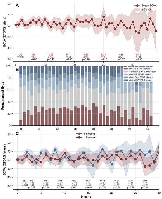
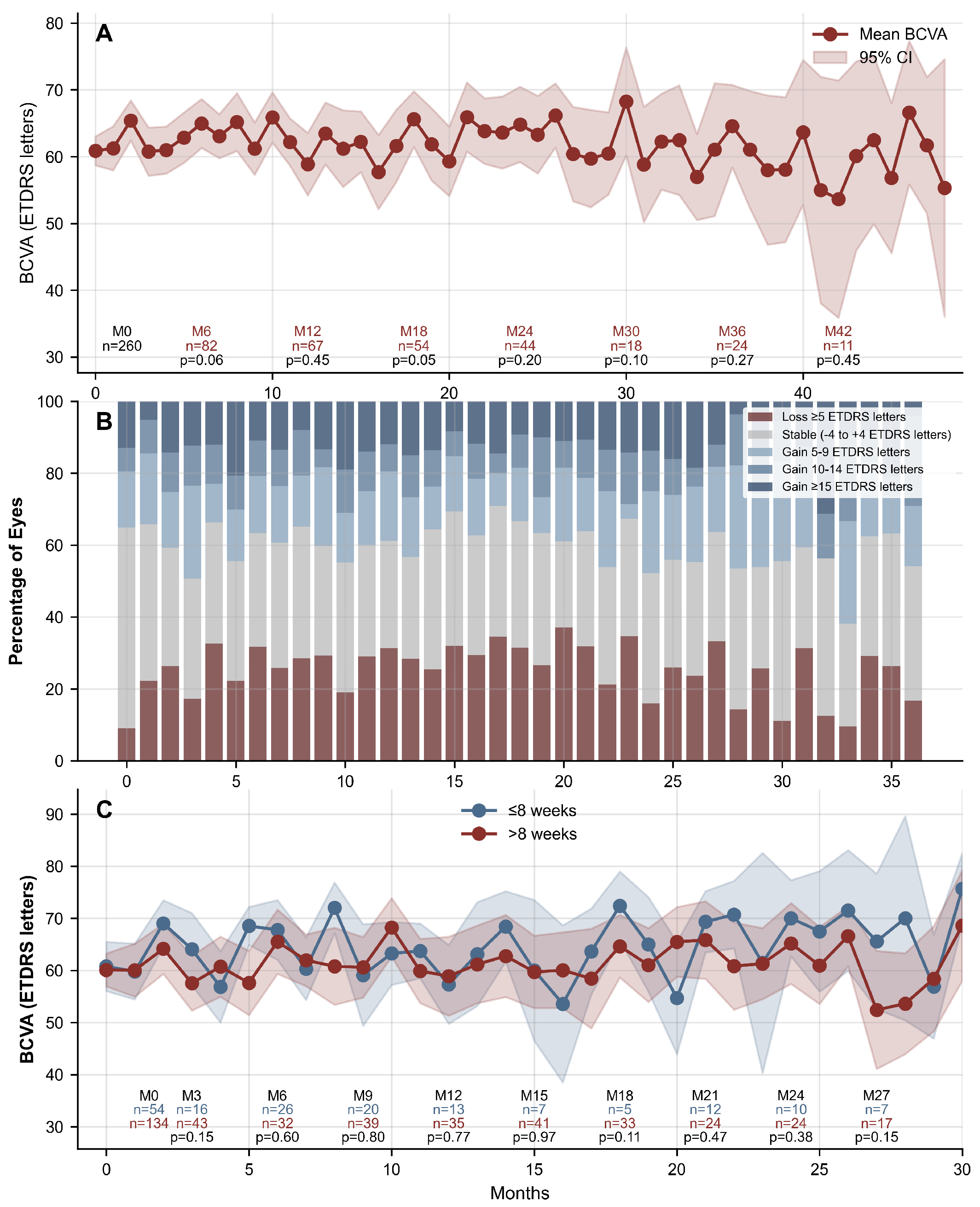
As shown in Figure 1A, the mean BCVA showed an initial slight fluctuation, increasing from 60.9 ± 17.8 ETDRS letters at baseline (n = 260) to 65.0 ± 16.9 ETDRS letters at Month 6 (n = 82), before stabilizing at Months 24 and 36, where the available measurements averaged 64.8 ± 19.1 letters (n = 44) and 64.6 ± 15.4 letters (n = 24), respectively. The Month 48 mean BCVA was 55.3 ± 24.1 letters (n = 6).
Figure 1.
BCVA (ETDRS) over time in the whole cohort (A), visual acuity gain over time in the whole cohort (B), and BCVA over time according to treatment interval between the last DEXi and the first FAci (C). Displayed monthly n values count available measurements. Shaded bands are 95% confidence intervals. BCVA: best-corrected visual acuity; ETDRS: Early Treatment Diabetic Retinopathy Study; DEXi: intravitreal dexamethasone implant; FAci: intravitreal fluocinolone acetonide implant.
At Month 12, 32.3% of eyes had lost ≥5 ETDRS letters, 30.8% remained stable (within −4 to +4 letters), and 36.9% had gained >5 letters (including 20.0% with a 5–9 letter gain, 6.2% with a 10–14 letter gain, and 10.8% with a ≥15 letter gain). At Month 24, these outcomes improved, with only 15.9% losing ≥5 letters, 36.4% remaining stable, and 47.7% gaining >5 letters (22.7% gaining 5–9 letters, 11.4% gaining 10–14 letters, and 13.6% gaining ≥15 letters) (Figure 1B).
The mean BCVA was comparable between eyes that received their last DEXi injection ≤8 weeks prior to the first FAci injection and those that received it >8 weeks before (Figure 1C).
3.4. Retinal Thickness
Prior to the first FAci injection, the mean CMT was 394.4 ± 166.6 µm, corresponding to 321.6 ± 116.6 µm in the ≤8-week group and 413.2 ± 173.7 µm in the >8-week group.
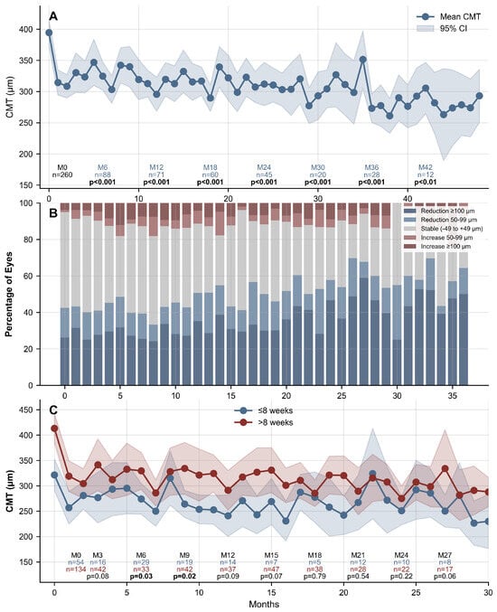
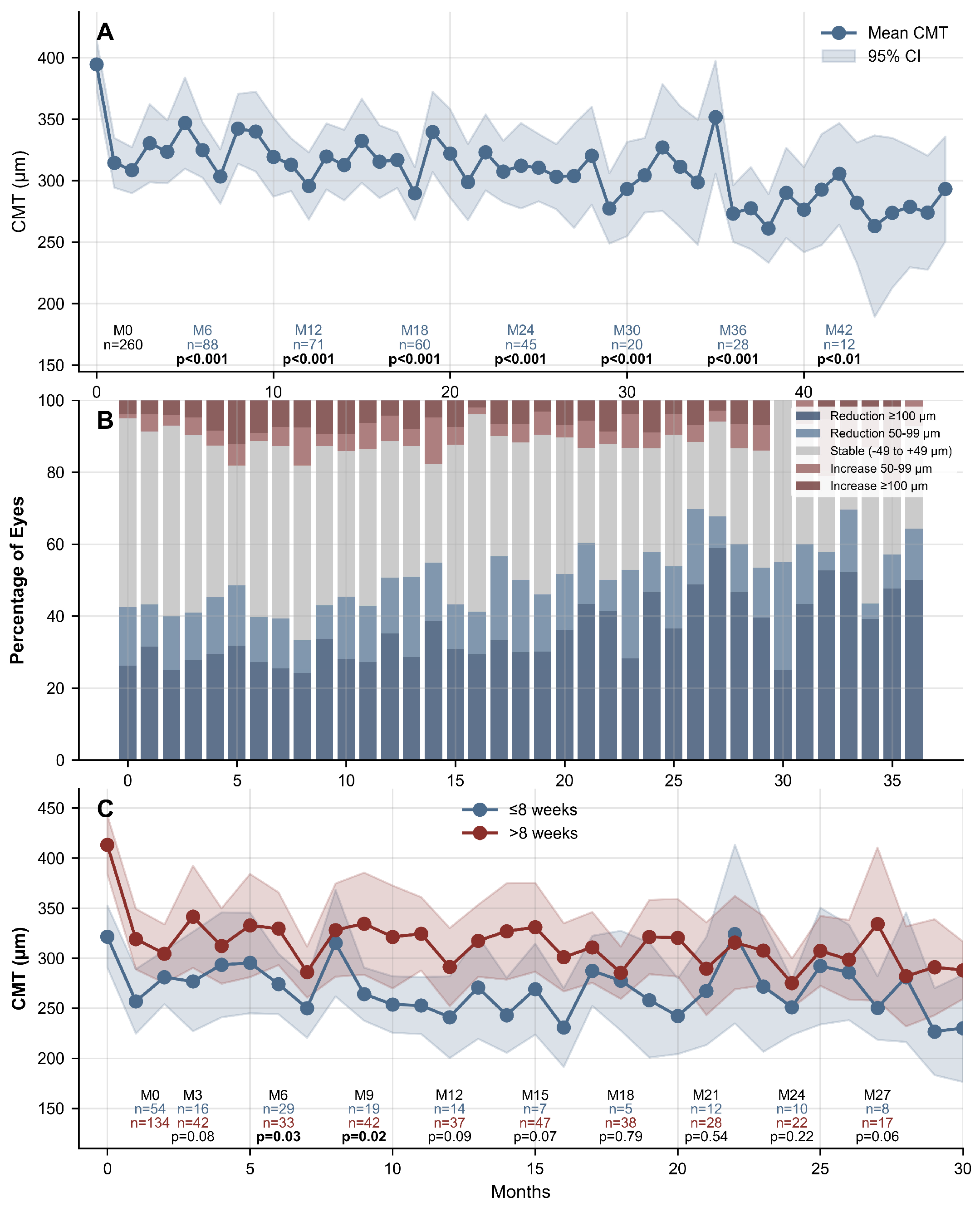
The mean CMT decreased from 394.4 ± 166.6 µm at baseline (n = 260) to 295.7 ± 117.5 µm at Month 12 (n = 71) and 312.1 ± 118.9 µm at Month 24 (n = 45) (Figure 2A).
Figure 2.
Changes in central macular thickness (CMT, µm) over time in the whole population (A), distribution of CMT changes over time in the whole population (B), and CMT according to treatment interval duration between last DEXi and first FAci (C). Eyes with undocumented DEXi–FAci intervals are included in panels (A,B) and excluded from panel (C). Displayed monthly n values count available measurements. Shaded bands are 95% confidence intervals. DEXi: intravitreal dexamethasone implant; FAci: intravitreal fluocinolone acetonide implant.
The mean CMT reductions at Months 12 and 24 were −90.6 ± 156.7 µm and −103.3 ± 176.7 µm, respectively. At Month 12, the CMT was reduced by ≥50 µm in 50.7% of measurements (≥100 µm in 35.2%, 50–99 µm in 15.5%), remained stable in 38.0% (±49 µm), and increased by ≥50 µm in 11.3%. Similar results were observed at Month 24: the CMT was reduced by ≥50 µm in 57.8% of measurements (≥100 µm in 46.7%, 50–99 µm in 11.1%), remained stable in 28.9%, and increased by ≥50 µm in 13.3% (Figure 2B).
A longer interval between the last DEXi and the first FAci correlated with a higher mean CMT (ρ = 0.182, p = 0.012) over the follow-up period. Indeed, significant differences in CMT were observed between eyes that received the last DEXi ≤8 weeks versus >8 weeks prior to FAci injection (Figure 2C). Starting from Month 1, the 8-week group consistently showed lower CMT, with statistically significant differences at several timepoints, including at Months 1–3, 6, 7, 9–11, 14, 16, 19, and 20 (p < 0.05). By contrast, no consistent or statistically significant differences in CMT were observed between subgroups defined by the 4-week cutoff.
3.5. Additional Treatments
Among the 260 eyes included in the study, 132 (50.8%) required additional treatments during the follow-up. At Months 12 and 24, 90 (34.6%) and 117 (45.0%) eyes required additional treatments, respectively.
The most frequently used additional intervention was intravitreal dexamethasone (Ozurdex; 276 injections, 50.7%), followed by aflibercept (201 injections, 36.9%) and macular laser (52 treatments, 9.6%). Other treatments, including ranibizumab, faricimab, and periocular triamcinolone, collectively accounted for less than 3% of all interventions.
The median time to the first additional treatment was 118 weeks (95% CI: 87.7–191.4).
In the ≤8-week group, 20 eyes (37.0%) required additional treatment during the follow-up versus 60 eyes (44.8%) in the >8-week group. In the ≤4-week group, 8 eyes (34.8%) required additional treatment versus 72 eyes (43.6%) in the >4-week group.
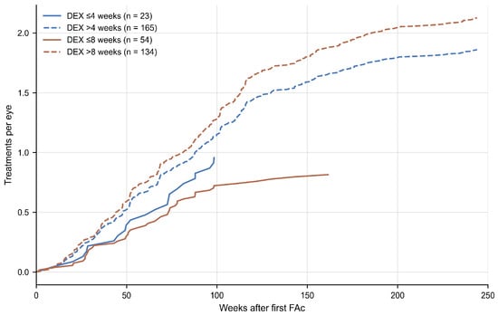
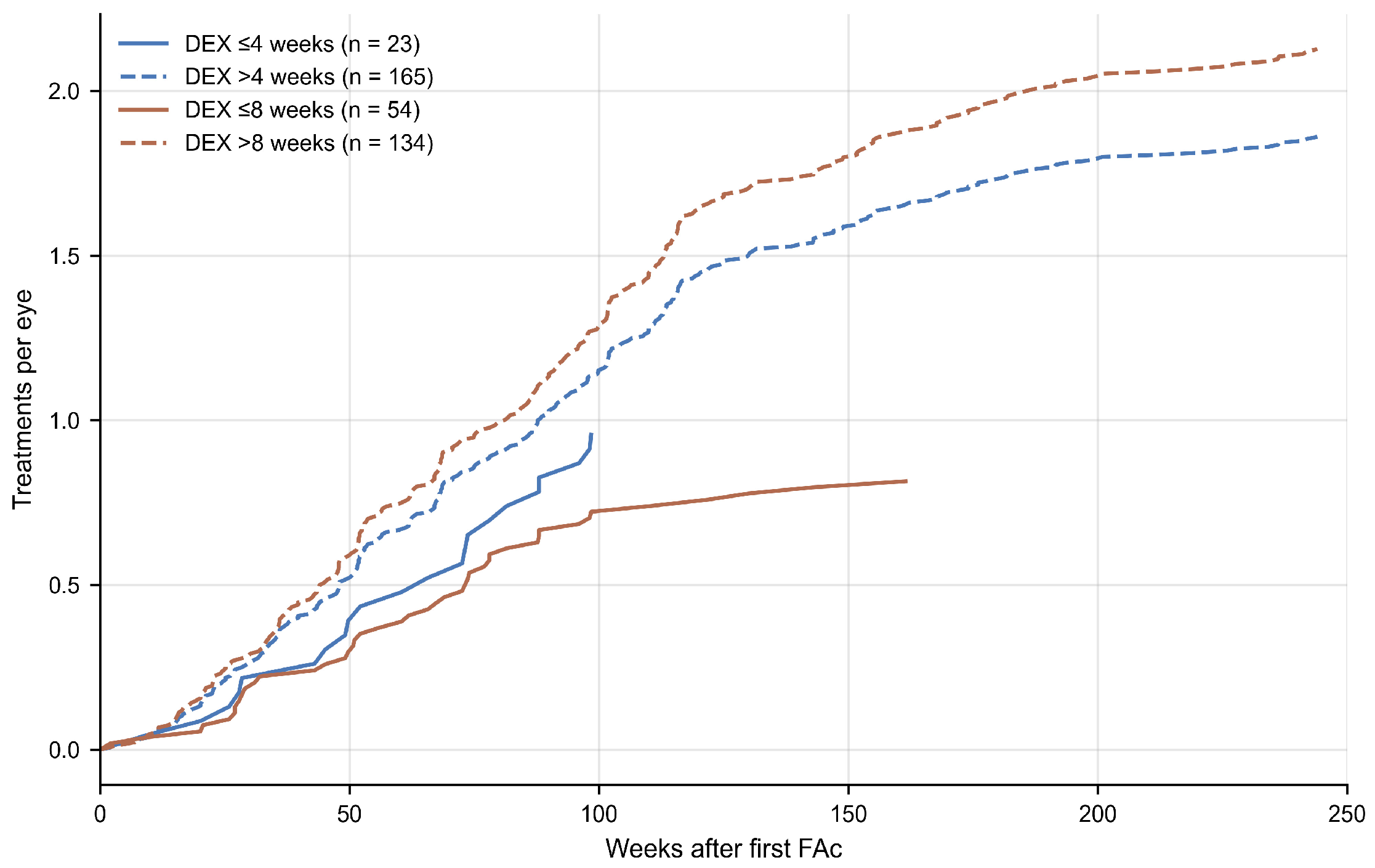
In the ≤8-week group, the normalized cumulative incidence of additional treatments was 0.33 treatments per eye at week 52 and 0.72 at week 104 (n = 54), versus 0.66 and 1.38 in the >8-week group (n = 134) (Figure 3). It was 0.39 at week 52 and 0.96 at week 104 (n = 23) in the ≤4-week group, versus 0.59 and 1.22 in the >4-week group (n = 165).
Figure 3.
Normalized cumulative incidence of additional treatments after first intravitreal fluocinolone acetonide implant (number of additional treatments divided by the number of eyes), according to the interval between the last DEXi and the first FAci: ≤4 weeks (n = 23) versus >4 weeks (n = 165), and ≤8 weeks (n = 54) versus >8 weeks (n = 134). DEXi: intravitreal dexamethasone implant; FAci: intravitreal fluocinolone acetonide implant.
3.6. Safety
Safety events are summarized in Table 4.
Table 4.
Safety events after the first (n = 260 eyes) and the second (n = 54 eyes) fluocinolone acetonide intravitreal implant.
Among phakic eyes, five underwent cataract surgery (33.3%) after a mean time of 41.5 ± 30.6 weeks post-injection. No eye underwent cataract surgery following the second FAci injection.
Endophthalmitis was observed in five eyes (1.6%). Two cases (0.8%) occurred after the first FAci injection (mean time to onset: 91.1 ± 40.3 weeks) and three cases (5.6%) after the second FAci injection (mean time to onset: 44.2 ± 25.5 weeks). A total of 500 and 83 additional injections were performed after the first and second FAci injections, respectively, resulting in an overall endophthalmitis rate of 0.86% for additional injections (0.4% after the first FAci injection and 3.61% after the second). Among these cases, one was caused by Staphylococcus epidermidis, while the remaining four were due to unspecified pathogens.
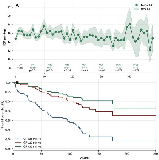
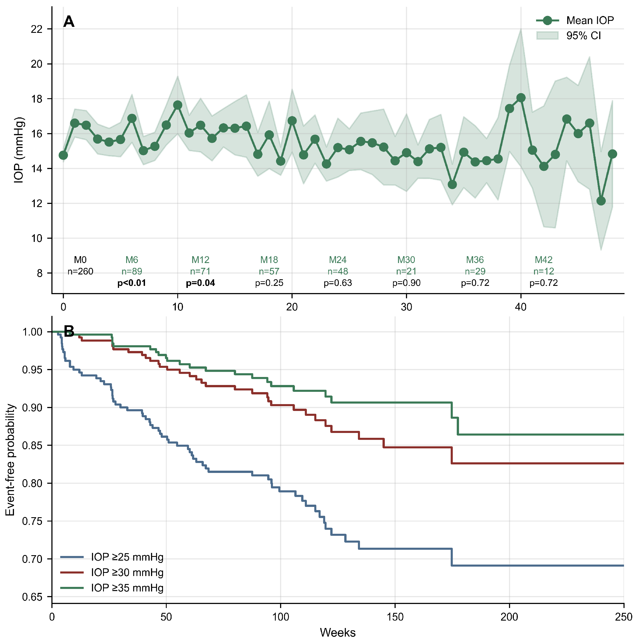
The mean IOP remained ≤18 mmHg throughout the follow-up. Following the first FAci injection, 63 eyes (24.2%) experienced an IOP elevation ≥25 mmHg during the follow-up, whereas 31 eyes (11.9%) reached an IOP ≥30 mmHg, and 22 eyes (8.5%) developed an IOP ≥35 mmHg (Figure 4A). The median time to reach an IOP ≥25 mmHg, ≥30 mmHg, and ≥35 mmHg was 43 weeks, 63 weeks, and 58 weeks, respectively (Figure 4B).
Figure 4.
Intraocular pressure over time in the whole cohort (A); time to intraocular pressure elevation after the first intravitreal fluocinolone acetonide implant (B). Displayed monthly n values count available measurements. Shaded band: 95% confidence interval. Kaplan–Meier estimates of time to IOP ≥25, ≥30, or ≥35 mmHg; follow-up is censored at the second implant when given. IOP: intraocular pressure.
An IOP-lowering treatment was either initiated or intensified in 64 eyes (24.6%) after a mean time of 56.7 ± 41.9 weeks. Among these eyes, 44 (68.8%) had no prior history of IOP-lowering treatment, 8 eyes (12.5%) were on monotherapy, 10 (15.6%) were on dual therapy, 2 (3.1%) were on triple therapy, and no eye had received systemic acetazolamide.
There was no statistically significant difference in mean IOP over time between the first and second FAci injections. No statistically significant difference was observed in the incidence of elevated IOP between eyes receiving the first FAci injection ≤8 weeks versus >8 weeks following the last DEXi injection, regardless of the IOP threshold used (≥25 mmHg, ≥30 mmHg, or ≥35 mmHg; all p-values >0.05).
Following the first FAci injection, selective laser trabeculoplasty (SLT) was performed in 19 eyes (7.3%) after a mean time of 120.6 ± 54.8 weeks. Minimally invasive glaucoma surgery (MIGS) was required in 4 eyes (1.5%) after a mean time of 128.9 ± 54.3 weeks (95% CI: 75.7–182.2), while filtering surgery was performed in 10 eyes (3.8%) after a mean time of 104.1 ± 47.4 weeks (95% CI: 69.1–139.2). Among the 14 eyes that underwent either MIGS or filtering surgery after the first FAci, 1 eye (7.1%) was on triple IOP-lowering therapy at baseline, 4 eyes (28.6%) were on dual therapy, 4 eyes (28.6%) were on monotherapy, and 8 eyes (57.1%) received additional corticosteroids (7 DEXi injections and 1 periocular triamcinolone injection).
Following the second FAci injection, 8 eyes (14.8%) experienced an IOP elevation ≥25 mmHg during the follow-up. Among these eyes, 1 (1.9%) reached an IOP ≥30 mmHg, and the same eye (1.9%) developed an IOP ≥35 mmHg. An IOP-lowering treatment was either initiated or intensified in 7 eyes (13.0%) after a mean time of 40.6 ± 38.5 weeks. Finally, SLT was performed in 5 eyes (9.3%) after a mean time of 49.7 ± 36.0 weeks, and no eyes required MIGS or filtering surgery. Among the 5 eyes treated with SLT, 3 were already on triple therapy before the second FAci injection.
4. Discussion
The pharmacokinetics of the sustained-release intravitreal 0.7 mg dexamethasone implant (DEXi) and the 0.19 mg fluocinolone acetonide implant (FAci) differ markedly. The DEXi immediately delivers a bolus of dexamethasone, reaching its peak efficacy at 8 weeks [26], followed by a rapid decline and loss of clinical efficacy after the third month. The pharmacokinetics of the FAci is slower, with a continuous release of 0.2 μg/day of the drug for up to 3 years, reaching its clinical peak efficacy between 6 and 11 months after injection [11,12]. FAci represents a third-line therapy for DME, functioning primarily as maintenance therapy to stabilize the retina, prevent DME recurrence, and reduce the long-term treatment burden [14,23,27,28]. FAci injection often follows DEX therapy, which is considered a bolus treatment aimed at rapidly and actively drying the retina. A DEXi injection is also recommended prior to FAci implantation as a steroid challenge to monitor both therapeutic efficacy and IOP response in DME eyes [23].
Switching between these two distinct molecules, i.e., from a short-lasting to a long-lasting implant, is crucial to optimize functional and anatomical outcomes in DME patients. However, switching modalities are scarcely described in the literature, and the interval between the last DEXi and the first FAci is usually not reported [14,23,24,29,30,31]. Most of the time, the FAci injection is performed only upon DME recurrence, which could be suboptimal [25].
In our previous study [20] including 113 DME eyes with a mean follow-up of 7.6 months, we found a significantly lower mean baseline CMT in eyes that received the last DEXi injection ≤8 weeks compared to >8 weeks before the first FAci injection. This effectively reduced CMT fluctuations and significantly decreased the need for additional treatments during the follow-up. We aimed to investigate more closely the efficacy of a short interval between the last DEXi and first FAci injections by studying an extended cohort of 260 DME eyes with a longer mean follow-up of 32.6 months.
In this study, the BCVA remained stable with improvements of +3.4 and +3.7 ETDRS letters at Months 24 and 36, respectively, and the mean BCVA was comparable between patients who received their last DEXi injection ≤8 weeks and >8 weeks prior to the FAci injection. Our BCVA gains were lower than those reported in the literature. In a recent systematic review of 9 real-world studies, DME patients treated with FAci achieved mean BCVA improvements of +4.52 and +7.89 ETDRS letters at Months 24 and 36, respectively [32]. However, BCVA improvement depends on many factors. The main baseline characteristic associated with final BCVA and BCVA gain is the initial BCVA, as a poor baseline BCVA is associated with worse final BCVA but greater BCVA gains [33]. The improvement in visual acuity is also related to the reduction in macular edema and in CMT [34]. Other factors, such as DME duration [35] and the clinical response to prior DEXi injections, may also interfere [25]. In the ≤8-week group, as no recurrence of macular edema occurred before FAci injection, we could assume that the highest visual acuity gain had already been reached before the switch.
In our study, a longer interval between the last DEXi and the first FAci correlated with a higher mean CMT (ρ = 0.182, p = 0.012) over the follow-up period. Indeed, we still observed a significantly lower mean baseline CMT in eyes that received the last DEXi injection ≤8 weeks compared to >8 weeks before the first FAci injection. In the ≤8-week group, FAci injections were therefore performed on a drier macula while DEXi drug release was still at its peak. We also observed that the differences in CMT between the two groups remained significant throughout the follow-up. Indeed, starting from Month 1, the ≤8-week group consistently showed lower CMT, with statistically significant differences at several timepoints, including Months 1–3, 6, 7, 9–12, 14–16, 19, 20, and 24 (p < 0.05). Patients injected with a >8-week interval could not achieve similar CMT values over time, even with the help of additional treatments. Cicinelli et al. also reported that patients with higher CMT at the time of FAci injection were more prone to exhibit persistent edema throughout the follow-up [36]. The same authors [37] measured the area under the curve (AUC) of the CMT in 89 eyes with DME injected with FAci, as this allows better assessment of the mean drug effect over time than standard CMT measurements. In their study, eyes with higher baseline CMT (β = 9.61 μm/month; 95% CI: 3.97–15.2, p < 0.001) had a higher AUC for FAci over the study period [37]. In our study, no consistent or statistically significant differences in CMT after baseline were observed between the subgroups defined by the 4-week cutoff. This could be due to the limited sample size in the ≤4-week subgroup (n = 23).
Fusi-Rubiano et al. [38] reported that, in real-world studies, almost 60% of patients treated with FAci injection for DME need additional treatments within 2 years. The Paladin study reported a need for retreatment in 48.1% of patients at one year [16]. In our study, we reported a lower rate of 34.6% and 45.0% of eyes requiring an additional therapeutic intervention at 1 and 2 years, respectively. In our first study, we observed that an extended interval between the last DEXi and the first FAci injections was significantly associated with the need for additional treatment [20]. In this extended study, we confirmed that the need for additional treatments differed with the DEXi-to-FAci interval: the normalized cumulative incidence of additional treatments was lower in the ≤8-week and ≤4-week groups than in the >8-week and >4-week groups. Rousseau et al. [21] also found a much lower additional treatment rate of 20.6% at one year in 41 eyes receiving their first FAci only one month after the last DEXi injection.
These results highlight the need for a short interval when switching from DEXi to FAci, as FAci injection is more effective on an already dried macula during the DEXi efficacy period, allowing sufficient time for FAci to reach its peak efficacy without any macular edema recurrence or worsening [34].
The safety outcomes regarding IOP-related adverse events in our study are consistent with those observed in the literature [39]. The mean IOP remained <21 mmHg throughout the follow-up. The proportion of patients with a recorded IOP >25 or >30 mmHg, regardless of the visit, was 24.2% and 11.9%, respectively, which is less than the rates observed in the FAME study or in other real-life studies [13,19,23,32,40], but higher than those observed in our first study (10.6% and 3.5% with an IOP >25 and >30 mmHg, respectively).
In our two studies, we observed that the change in IOP was similar between patients who received the last DEXi injection ≤8 weeks or >8 weeks before the first FAci injection [20], with no cumulative effect of the two injections on IOP. Lampin et al. [22], who prospectively injected an FAci one month after the last DEXi injection in 14 eyes, also published reassuring results with only 14% of eyes requiring IOP-lowering treatment during the study. In the study by Rousseau et al. [21], where 41 eyes were also switched from DEXi to FAci one month apart, 24% of eyes required IOP-lowering therapy, with no patient experiencing ocular hypertension >30 mmHg. The authors suggested that the OHT rate was low because all the patients who experienced an IOP rise after the previous DEXi injections were excluded from FAci injections [22]. Indeed, as noted by Kodjikian et al. [23], patients with no pressure warning after 2 to 3 DEXi injections have an 85–90% likelihood of maintaining the same tolerance profile after FAci injection [16,19,41], whereas eyes with a history of IOP increase >25 mmHg after DEXi injection are 20 times more likely to experience such IOP elevation after FAci injection [37,42].
In our study, the median time to achieve an IOP ≥25 mmHg, ≥30 mmHg, and ≥35 mmHg was 43 weeks, 63 weeks, and 58 weeks, respectively, which is in line with the literature where most cases of OHT occurred at the end of the first year after FAci injection [34,43]. However, it is advisable to monitor IOP closely even after the end of the first year, as IOP events may occur later [23], with studies showing a mean time to IOP-related events of 14–18 months [24]. Continued monitoring of IOP every ≤3 months, depending on the patient’s IOP status, is thus recommended [15,44].
In our study, most IOP-related adverse events were successfully treated with IOP-lowering medications [44]. However, 14 eyes (5.4%) required MIGS or filtering surgery, which is close to the rate observed in the FAME study (4.8%) [45]. Most of these eyes had identified risk factors for IOP rise, including prior OHT/open-angle glaucoma in 9 eyes (64.3%) [44,46,47], especially glaucoma treated with dual or triple therapy in 5 eyes (35.7%) [48]. These patients should not have been considered eligible for FAci injections. We also observed that 8/14 eyes (57.1%) undergoing filtering surgery received repeated additional DEXi/periocular triamcinolone acetonide injections over the first year following FAci injection. In these cases, the IOP rise occurred 2 to 3 months after additional treatment, when both DEXi and FAci intraocular releases were at their peak [9]. Serrar et al. [49] also observed in DME patients treated with FAci that 43% and 40% of cases of OHT after the first and second FAci injections, respectively, occurred within the 3 months following an additional DEXi injection. Thus, when choosing additional treatments after FAci injection, both the molecule and its injection timing after the last FAci injection should be evaluated.
Our study has several limitations. We were not able to monitor DME OCT biomarkers (such as intraretinal and subretinal fluids, hyperreflective dots) as OCT scans were not collected. The retrospective collection of data limited the quality and exhaustivity of the data analyzed. At some time points, available data were scarce: at baseline, POAG duration was documented in only 24 eyes. During the course of the study, OCT-based anatomical assessments were performed, but BCVA and IOP were not systematically recorded. However, our study included a relatively large number of patients, with an extended follow-up, which allowed us to explore not only the transitional period of the DEXi-to-FAci switch, but also the long-term maintenance phase. This extended timeline enabled us to confirm that the initial benefits of a short switch interval from DEXi to FAci are maintained, without any additional safety events.
5. Conclusions
To conclude, our study showed that the benefits of a short interval from DEXi to FAci were sustained over time. A short interval between the last DEXi and the first FAci injections to avoid DME recurrence reduced CMT variations during the transitional and maintenance phases. It also reduced the incidence of additional treatment throughout the study. The safety profile of the combination of both steroid implants remained reassuring throughout the follow-up.
Author Contributions
Conceptualization, S.B.; methodology, S.B. and P.S.; software, M.M.; validation, S.B., P.S. and L.K.; formal analysis, M.M. and P.S.; investigation, all authors; resources, all authors; data curation, M.M.; writing—original draft preparation, S.B. and M.M.; review, all authors; visualization, M.M.; supervision, S.B.; project administration, S.B. All authors have read and agreed to the published version of the manuscript.
Funding
This research received no external funding.
Institutional Review Board Statement
The study was conducted in accordance with the Declaration of Helsinki and approved by the Ethics Committee of the French Ophthalmology Society (Société Française d’Ophtalmologie, IRB00008855).
Informed Consent Statement
A retrospective study using existing patient data (medical records, data collected during routine care) falls outside the scope of the Jardé law, and no review by the Comité de Protection des Personnes (CPP) is required. Instead, it is governed by the CNIL reference methodology MR-004, which covers retrospective research involving human subjects, as well as studies and evaluations in the health field. Useful links (accessed on 12 October 2026): MR-004 text (CNIL PDF): https://www.cnil.fr/sites/cnil/files/2024-01/mr-004.pdf; CNIL MR-004 page: https://www.cnil.fr/fr/declaration/methodologie-de-reference-04-recherches-nimpliquant-pas-la-personne-humaine-etudes-et-evaluations-dans-le-domaine-de-la-sante; Official reference: Deliberation No. 2018-155 of 3 May 2018 approving MR-004 (AP-HP summary); Practical guide (Health Data Hub): https://health-data-hub.fr/sites/default/files/2020-12/MR004_Mode_emploi.pdf.
Data Availability Statement
The data presented in this study are available on request from the corresponding author due to privacy and ethical restrictions.
Acknowledgments
The authors thank all the members of the Club Francophone des Spécialistes de la Rétine (CFSR) who participated in data collection.
Conflicts of Interest
The following authors are from company: LR: Bayer, Roche; HT: Bayer, Roche; CCG: Bayer, Roche, Horus, Novartis, Abbvie; PYM: Bayer, Roche; MND: Bayer, Roche, Horus, Novartis, Abbvie; CD: Bayer, Roche, Horus, Novartis, Abbvie; VG: Bayer, Roche, Horus, Novartis, Abbvie; PG: Bayer; SN: Bayer; DG: Bayer, Roche; MLLL: Bayer; JBC: Bayer; AC: Abbvie, Bayer, Roche; BN: Bayer, Roche; ALC: Bayer, Roche; FM: Bayer, Roche, Horus, Novartis, Abbvie; LK: Bayer, Roche, Horus, Novartis, Abbvie; SB: Bayer, Roche, Horus, Novartis, Abbvie. The rest of the authors declare no conflicts of interest.
Abbreviations
The following abbreviations are used in this manuscript:
| anti-VEGF | Anti-vascular endothelial growth factor |
| AUC | Area under the curve |
| BCVA | Best-corrected visual acuity |
| CFSR | Club Francophone des Spécialistes de la Rétine |
| CI | Confidence interval |
| CMT | Central macular thickness |
| DEXi | Dexamethasone intravitreal implant |
| DME | Diabetic macular edema |
| ETDRS | Early Treatment Diabetic Retinopathy Study |
| FAci | Fluocinolone acetonide intravitreal implant |
| HbA1c | Glycated hemoglobin |
| IOP | Intraocular pressure |
| IQR | Interquartile range |
| MIGS | Minimally invasive glaucoma surgery |
| NPDR | Non-proliferative diabetic retinopathy |
| OHT | Ocular hypertension |
| PDR | Proliferative diabetic retinopathy |
| POAG | Primary open-angle glaucoma |
| PRP | Panretinal photocoagulation |
| SD | Standard deviation |
| SLT | Selective laser trabeculoplasty |
References
- Saeedi, P.; Petersohn, I.; Salpea, P.; Malanda, B.; Karuranga, S.; Unwin, N.; Colagiuri, S.; Guariguata, L.; Motala, A.A.; Ogurtsova, K.; et al. Global and regional diabetes prevalence estimates for 2019 and projections for 2030 and 2045: Results from the International Diabetes Federation Diabetes Atlas, 9th edition. Diabetes Res. Clin. Pract. 2019, 157, 107843. [Google Scholar] [CrossRef] [Scilit] [PubMed]
- Teo, Z.L.; Tham, Y.C.; Yu, M.; Chee, M.L.; Rim, T.H.; Cheung, N.; Bikbov, M.M.; Wang, Y.X.; Tang, Y.; Lu, Y.; et al. Global Prevalence of Diabetic Retinopathy and Projection of Burden through 2045: Systematic Review and Meta-analysis. Ophthalmology 2021, 128, 1580–1591. [Google Scholar] [CrossRef] [Scilit] [PubMed]
- Yau, J.W.; Rogers, S.L.; Kawasaki, R.; Lamoureux, E.L.; Kowalski, J.W.; Bek, T.; Chen, S.J.; Dekker, J.M.; Fletcher, A.; Grauslund, J.; et al. Global prevalence and major risk factors of diabetic retinopathy. Diabetes Care 2012, 35, 556–564. [Google Scholar] [CrossRef] [Scilit] [PubMed]
- Gonzalez, V.H.; Campbell, J.; Holekamp, N.M.; Kiss, S.; Loewenstein, A.; Augustin, A.J.; Ma, J.; Ho, A.C.; Patel, V.; Whitcup, S.M.; et al. Early and long-term responses to anti-vascular endothelial growth factor therapy in diabetic macular edema: Analysis of Protocol I data. Am. J. Ophthalmol. 2016, 172, 72–79. [Google Scholar] [CrossRef] [Scilit] [PubMed]
- Funatsu, H.; Yamashita, H.; Ikeda, T.; Mimura, T.; Eguchi, S.; Hori, S. Vitreous levels of interleukin-6 and vascular endothelial growth factor are related to diabetic macular edema. Ophthalmology 2003, 110, 1690–1696. [Google Scholar] [CrossRef] [Scilit] [PubMed]
- Augustin, A.J.; Bopp, S.; Fechner, M.; Holz, F.; Sandner, D.; Winkgen, A.M.; Khoramnia, R.; Neuhann, T.; Warscher, M.; Spitzer, M.; et al. Three-year results from the Retro-IDEAL study: Real-world data from diabetic macular edema (DME) patients treated with ILUVIEN® (0.19 mg fluocinolone acetonide implant). Eur. J. Ophthalmol. 2020, 30, 382–391. [Google Scholar] [CrossRef] [Scilit] [PubMed]
- Yang, Y.; Bailey, C.; Loewenstein, A.; Massin, P. Intravitreal corticosteroids in diabetic macular edema: Pharmacokinetic considerations. Retina 2015, 35, 2440–2449. [Google Scholar] [CrossRef] [Scilit] [PubMed]
- Chang-Lin, J.E.; Attar, M.; Acheampong, A.A.; Robinson, M.R.; Whitcup, S.M.; Kuppermann, B.D.; Welty, D. Pharmacokinetics and pharmacodynamics of a sustained-release dexamethasone intravitreal implant. Investig. Ophthalmol. Vis. Sci. 2011, 52, 80–86. [Google Scholar] [CrossRef] [Scilit] [PubMed]
- Boyer, D.S.; Yoon, Y.H.; Belfort, R., Jr.; Bandello, F.; Maturi, R.K.; Augustin, A.J.; Li, X.Y.; Cui, H.; Hashad, Y.; Whitcup, S.M.; et al. Three-year, randomized, sham-controlled trial of dexamethasone intravitreal implant in patients with diabetic macular edema. Ophthalmology 2014, 121, 1904–1914. [Google Scholar] [CrossRef] [Scilit] [PubMed]
- Kane, F.E.; Green, K.E. Ocular pharmacokinetics of fluocinolone acetonide following Iluvien implantation in the vitreous humor of rabbits. J. Ocul. Pharmacol. Ther. 2015, 31, 11–16. [Google Scholar] [CrossRef] [Scilit] [PubMed]
- Campochiaro, P.A.; Nguyen, Q.D.; Hafiz, G.; Bloom, S.; Brown, D.M.; Busquets, M.; Ciulla, T.; Feiner, L.; Sabates, N.; Billman, K.; et al. Aqueous levels of fluocinolone acetonide after administration of fluocinolone acetonide inserts or fluocinolone acetonide implants. Ophthalmology 2013, 120, 583–587. [Google Scholar] [CrossRef] [Scilit] [PubMed]
- Campochiaro, P.A.; Brown, D.M.; Pearson, A.; Chen, S.; Boyer, D.; Ruiz-Moreno, J.; Garretson, B.; Gupta, A.; Hariprasad, S.M.; Bailey, C.; et al. Sustained delivery fluocinolone acetonide vitreous inserts provide benefit for at least 3 years in patients with diabetic macular edema. Ophthalmology 2012, 119, 2125–2132. [Google Scholar] [CrossRef] [Scilit] [PubMed]
- Kodjikian, L.; Baillif, S.; Creuzot-Garcher, C.; Delyfer, M.N.; Matonti, F.; Weber, M.; Mathis, T. Real-world efficacy and safety of fluocinolone acetonide implant for diabetic macular edema: A systematic review. Pharmaceutics 2021, 13, 72. [Google Scholar] [CrossRef] [Scilit] [PubMed]
- Hu, W.; Ni, L.; Ying, H.; Zhang, Z.; Tu, S.; Tao, Y.; Chen, L. Efficacy of fluocinolone acetonide implant in diabetic macular edema patients previously treated with dexamethasone implant: A systematic review and meta-analysis. Pak. J. Med. Sci. 2025, 41, 2388–2398. [Google Scholar] [CrossRef] [Scilit] [PubMed]
- Kodjikian, L.; Bandello, F.; de Smet, M.; Dot, C.; Zarranz-Ventura, J.; Loewenstein, A.; Sudhalkar, A.; Bilgic, A.; Cunha-Vaz, J.; Dirven, W.; et al. Fluocinolone acetonide implant in diabetic macular edema: International experts’ panel consensus guidelines and treatment algorithm. Eur. J. Ophthalmol. 2022, 32, 1890–1899. [Google Scholar] [CrossRef] [Scilit] [PubMed]
- Mansour, S.E.; Kiernan, D.F.; Roth, D.B.; Eichenbaum, D.; Holekamp, N.M.; Kaba, S.; Werts, E. Two-year interim safety results of the 0.2 µg/day fluocinolone acetonide intravitreal implant for the treatment of diabetic macular oedema: The observational PALADIN study. Br. J. Ophthalmol. 2021, 105, 414–419. [Google Scholar] [CrossRef] [Scilit] [PubMed]
- Food and Drug Administration. ILUVIEN 0.19 mg New Drug Application (NDA 201923). Available online: https://www.accessdata.fda.gov/drugsatfda_docs/label/2014/201923s000lbl.pdf (accessed on 27 June 2021).
- Mansour, S.E.; Browning, D.J.; Wong, K.; Flynn, H.W., Jr.; Bhavsar, A.R. The evolving treatment of diabetic retinopathy. Clin. Ophthalmol. 2020, 14, 653–678. [Google Scholar] [CrossRef] [Scilit] [PubMed]
- Eaton, A.; Koh, S.S.; Jimenez, J.; Riemann, C.D. The USER study: A chart review of patients receiving a 0.2 µg/day fluocinolone acetonide implant for diabetic macular edema. Ophthalmol. Ther. 2019, 8, 51–62. [Google Scholar] [CrossRef] [Scilit] [PubMed]
- Baillif, S.; Staccini, P.; Weber, M.; Delyfer, M.N.; Le Mer, Y.; Gualino, V.; Collot, L.; Merite, P.Y.; Creuzot-Garcher, C.; Kodjikian, L.; et al. Management of patients with diabetic macular edema switched from dexamethasone intravitreal implant to fluocinolone acetonide intravitreal implant. Pharmaceutics 2022, 14, 2391. [Google Scholar] [CrossRef] [Scilit] [PubMed]
- Rousseau, N.; Lebreton, O.; Masse, H.; Maucourant, Y.; Pipelart, V.; Clement, M.; Le Lez, M.L.; Nghi, P.; Faure, C.; Abdelhamid, A.; et al. Fluocinolone acetonide implant injected 1 month after dexamethasone implant for diabetic macular oedema: The ILUVI1MOIS study. Ophthalmol. Ther. 2023, 12, 2781–2792. [Google Scholar] [CrossRef] [Scilit] [PubMed]
- Lampin, Q.; Poret, J.; Gherras, M.; Jany, B.; Tran, T.H.C. Fluocinolone acetonide implant (FAci) one month after dexamethasone implant (DEXi) for chronic diabetic macular edema: 1-year results. J. Fr. Ophtalmol. 2025, 48, 104352. [Google Scholar] [CrossRef] [Scilit] [PubMed]
- Kodjikian, L.; Duarte, L.; Singh, P.; Habib, M.; Gonzalez, V. What have we learned from a decade treating patients with diabetic macular oedema with 0.19 mg fluocinolone acetonide intravitreal implant? Eye 2025, 39, 1238–1248. [Google Scholar] [CrossRef] [Scilit] [PubMed]
- Bailey, C.; Chakravarthy, U.; Lotery, A.; Menon, G.; Talks, J.; Medisoft Audit Group. Extended real-world experience with the ILUVIEN® (fluocinolone acetonide) implant in the United Kingdom: 3-year results from the Medisoft® audit study. Eye 2022, 36, 1012–1018. [Google Scholar] [CrossRef] [Scilit] [PubMed]
- Cicinelli, M.V.; Rosenblatt, A.; Grosso, D.; Zollet, P.; Capone, L.; Rabiolo, A.; Lattanzio, R.; Loewenstein, A.; Bandello, F.; International Retina Group. The outcome of fluocinolone acetonide intravitreal implant is predicted by the response to dexamethasone implant in diabetic macular oedema. Eye 2021, 35, 3232–3242. [Google Scholar] [CrossRef] [Scilit] [PubMed]
- Bastakis, G.G.; Dimopoulos, D.; Stavrakakis, A.; Pappas, G. Long-term efficacy and duration of action of dexamethasone implant, in vitrectomised and non-vitrectomised eyes with persistent diabetic macular oedema. Eye 2019, 33, 411–418. [Google Scholar] [CrossRef] [Scilit] [PubMed]
- Veritti, D.; Sarao, V.; Soppelsa, V.; Lanzetta, P. Managing diabetic macular edema in clinical practice: Systematic review and meta-analysis of current strategies and treatment options. Clin. Ophthalmol. 2021, 15, 375–385. [Google Scholar] [CrossRef] [Scilit] [PubMed]
- Sheth, V.S.; Singer, M.; MacCumber, M.; Cutino, A.; Kasper, J.; Coughlin, B.A.; Riemann, C.D. Long-term control of retinal thickness variability and vision following the 0.19 mg fluocinolone acetonide implant. J. Vitreoretin. Dis. 2023, 7, 490–497. [Google Scholar] [CrossRef] [Scilit] [PubMed]
- Rehak, M.; Busch, C.; Unterlauft, J.D.; Jochmann, C.; Wiedemann, P. Outcomes in diabetic macular edema switched directly or after a dexamethasone implant to a fluocinolone acetonide intravitreal implant following anti-VEGF treatment. Acta Diabetol. 2020, 57, 469–478. [Google Scholar] [CrossRef] [Scilit] [PubMed]
- Elbarky, A. Effectiveness and tolerability of the fluocinolone acetonide implant in patients with diabetic macular edema in the UAE: 24 and 36-month outcomes. Eur. J. Ophthalmol. 2023, 33, 1068–1076. [Google Scholar] [CrossRef] [Scilit] [PubMed]
- Ruiz-Moreno, J.M.; Adán, A.; Lafuente, M.; Asencio Durán, M.; Arias Barquet, L.; García Layana, A.; Arévalo, J.F. Effectiveness and safety of fluocinolone acetonide intravitreal implant in diabetic macular edema patients considered insufficiently responsive to available therapies (REACT): A prospective, non-randomized, and multicenter study. Int. Ophthalmol. 2023, 43, 4639–4649. [Google Scholar] [CrossRef] [Scilit] [PubMed]
- Fallico, M.; Maugeri, A.; Lotery, A.; Longo, A.; Bonfiglio, V.; Russo, A.; Avitabile, T.; Pulvirenti, A.; Furino, C.; Cennamo, G.; et al. Fluocinolone acetonide vitreous insert for chronic diabetic macular oedema: A systematic review with meta-analysis of real-world experience. Sci. Rep. 2021, 11, 4800. [Google Scholar] [CrossRef] [Scilit] [PubMed]
- Mathis, T.; Papegaey, M.; Ricard, C.; Rezkallah, A.; Matonti, F.; Sudhalkar, A.; Vartin, C.; Dot, C.; Kodjikian, L. Efficacy and safety of intravitreal fluocinolone acetonide implant for chronic diabetic macular edema previously treated in real-life practice: The REALFAc study. Pharmaceutics 2022, 14, 723. [Google Scholar] [CrossRef] [Scilit] [PubMed]
- Sejournet, L.; Mathis, T.; Vermot-Desroches, V.; Serra, R.; Fenniri, I.; Denis, P.; Kodjikian, L. Efficacy and safety of fluocinolone acetonide implant in diabetic macular edema: Practical guidelines from reference center. Pharmaceutics 2024, 16, 1183. [Google Scholar] [CrossRef] [Scilit] [PubMed]
- Vaz-Pereira, S.; Castro-de-Sousa, J.P.; Martins, D.; Canelas, J.P.; Reis, P.; Sampaio, A.; Urbano, H.; Kaku, P.; Nascimento, J.; Marques-Neves, C. The outcomes of switching from short- to long-term intravitreal corticosteroid implant therapy in patients with diabetic macular edema. Ophthalmic Res. 2020, 63, 114–121. [Google Scholar] [CrossRef] [Scilit] [PubMed]
- Cicinelli, M.V.; Rabiolo, A.; Zollet, P.; Capone, L.; Lattanzio, R.; Bandello, F. Persistent or recurrent diabetic macular edema after fluocinolone acetonide 0.19 mg implant: Risk factors and management. Am. J. Ophthalmol. 2020, 215, 14–24. [Google Scholar] [CrossRef] [Scilit] [PubMed]
- Cicinelli, M.V.; Rabiolo, A.; Capone, L.; Di Biase, C.; Lattanzio, R.; Bandello, F. Factors associated with the response to fluocinolone acetonide 0.19 mg in diabetic macular oedema evaluated as the area-under-the-curve. Eye 2023, 37, 242–248. [Google Scholar] [CrossRef] [Scilit] [PubMed]
- Fusi-Rubiano, W.; Mukherjee, C.; Lane, M.; Tsaloumas, M.D.; Glover, N.; Kidess, A.; Denniston, A.K.; Palmer, H.E.; Manna, A.; Morjaria, R. Treating diabetic macular oedema (DMO): Real world UK clinical outcomes for the 0.19 mg fluocinolone acetonide intravitreal implant (Iluvien™) at 2 years. BMC Ophthalmol. 2018, 18, 62. [Google Scholar] [CrossRef] [Scilit] [PubMed]
- Lebrize, S.; Arnould, L.; Bourredjem, A.; Busch, C.; Rehak, M.; Massin, P.; Barbosa-Breda, J.; Lupidi, M.; Mariotti, C.; Hamza, M.; et al. Intraocular pressure changes after intravitreal fluocinolone acetonide implant: Results from four European countries. Ophthalmol. Ther. 2022, 11, 1217–1229. [Google Scholar] [CrossRef] [Scilit] [PubMed]
- Chakravarthy, U.; Taylor, S.R.; Koch, F.H.J.; Castro de Sousa, J.P.; Bailey, C.; ILUVIEN Registry Safety Study (IRISS) Investigators Group. Changes in intraocular pressure after intravitreal fluocinolone acetonide (ILUVIEN): Real-world experience in three European countries. Br. J. Ophthalmol. 2019, 103, 1072–1077. [Google Scholar] [CrossRef] [Scilit] [PubMed]
- Panos, G.D.; Arruti, N.; Patra, S. The long-term efficacy and safety of fluocinolone acetonide intravitreal implant 190 µg (ILUVIEN®) in diabetic macular oedema in a multi-ethnic inner-city population. Eur. J. Ophthalmol. 2021, 31, 620–629. [Google Scholar] [CrossRef] [Scilit] [PubMed]
- Capone, L.; Airaghi, P.; Aragona, P.; Castellino, N.; Cicinelli, M.V.; Ciucci, F.; Coppola, M.; De Gaetano, C.; Lattanzio, R.; Lorusso, M.; et al. Real-world experience with fluocinolone acetonide intravitreal implant in patients with diabetic macular edema. Eur. J. Ophthalmol. 2024, 34, 1837–1842. [Google Scholar] [CrossRef] [Scilit] [PubMed]
- Merrill, P.T.; Holekamp, N.; Roth, D.; Kasper, J.; Grigorian, R.; PALADIN Study Group. The 0.19-mg fluocinolone acetonide intravitreal implant reduces treatment burden in diabetic macular edema. Am. J. Ophthalmol. 2023, 248, 16–23. [Google Scholar] [CrossRef] [Scilit] [PubMed]
- Goñi, F.J.; Barton, K.; Dias, J.A.; Diestelhorst, M.; Garcia-Feijoo, J.; Hommer, A.; Kodjikian, L.; Nicolò, M. Intravitreal corticosteroid implantation in diabetic macular edema: Updated European consensus guidance on monitoring and managing intraocular pressure. Ophthalmol. Ther. 2022, 11, 15–34. [Google Scholar] [CrossRef] [Scilit] [PubMed]
- Parrish, R.K., II; Campochiaro, P.A.; Pearson, P.A.; Green, K.; Traverso, C.E.; FAME Study Group. Characterization of intraocular pressure increases and management strategies following treatment with fluocinolone acetonide intravitreal implants in the FAME trials. Ophthalmic Surg. Lasers Imaging Retina 2016, 47, 426–435. [Google Scholar] [CrossRef] [Scilit] [PubMed]
- Vié, A.L.; Kodjikian, L.; Malclès, A.; Agard, E.; Voirin, N.; El Chehab, H.; Nguyen, A.M.; Dot, C. Tolerance of intravitreal dexamethasone implants in patients with ocular hypertension or open-angle glaucoma. Retina 2017, 37, 173–178. [Google Scholar] [CrossRef] [Scilit] [PubMed]
- Phulke, S.; Kaushik, S.; Kaur, S.; Pandav, S.S. Steroid-induced glaucoma: An avoidable irreversible blindness. J. Curr. Glaucoma Pract. 2017, 11, 67–72. [Google Scholar] [CrossRef] [Scilit]
- Malclès, A.; Dot, C.; Voirin, N.; Vié, A.L.; Agard, E.; Bellocq, D.; Denis, P.; Kodjikian, L. Safety of intravitreal dexamethasone implant (Ozurdex): The SAFODEX study. Incidence and risk factors of ocular hypertension. Retina 2017, 37, 1352–1359. [Google Scholar] [CrossRef] [Scilit] [PubMed]
- Serrar, Y.; Sejournet, L.; Thomeret, V.; Dot, C.; Bonnin, S.; Soler, V.; Rosier, L.; Matonti, F.; Ghazaryan, M.; Mathis, T.; et al. Efficacy and safety of multiple fluocinolone acetonide implants in diabetic macular oedema: Comparison between first and second intravitreal injections. Eye 2025, 39, 2678–2685. [Google Scholar] [CrossRef] [Scilit] [PubMed]

Disclaimer/Publisher’s Note: The statements, opinions and data contained in all publications are solely those of the individual author(s) and contributor(s) and not of MDPI and/or the editor(s). MDPI and/or the editor(s) disclaim responsibility for any injury to people or property resulting from any ideas, methods, instructions or products referred to in the content. |
© 2026 by the authors. Licensee MDPI, Basel, Switzerland. This article is an open access article distributed under the terms and conditions of the Creative Commons Attribution (CC BY) license.