


Abstract
Background: Anandamide (AEA) is a lipid mediator of the endocannabinoid system that plays a key role in pain perception by regulating multiple molecular targets, including transient receptor potential vanilloid type 1 (TRPV1). This vanilloid receptor is abundantly expressed in nociceptive neurons (nociceptors) and plays a crucial role in mediating pain responses. Methods: We used mouse behavioral tests, cell culture, and Western blot assays to test whether AEA regulates acute pain induced by TRPV1 activation and TRPV1 total protein levels. Results: Our results demonstrate that AEA downregulates the capsaicin-evoked pain response and TRPV1 protein levels in nociceptors. Furthermore, in a transiently heterologous TRPV1 expression system, we found that AEA induces ubiquitination of TRPV1 and its degradation via the proteasome pathway, independent of calcium influx. We also found that AEA induced an upregulation of BiP protein levels, a key endoplasmic reticulum chaperone and stress sensor, without changes in other ER stress sensors. Finally, we explored whether the endocannabinoid 2-arachidonoylglycerol (2-AG) and the phytocannabinoid β-caryophyllene (BCP) modulate capsaicin-evoked pain, finding that 2-AG is less effective than AEA in reducing pain behavior, whereas BCP shows effects similar to AEA in reducing capsaicin-evoked pain. Conclusions: These results highlight AEA and BCP as regulators of TRPV1 protein levels and of the painful response mediated by this molecular pain target.
1. Introduction
An acute painful response is a physiological protective mechanism that warns of potential damage to the organism [1]. This defense mechanism allows us to assess risky situations and respond effectively. In addition to this protective role, pain sometimes goes beyond an alert to a serious health concern, as thousands of people live with ongoing pain every day. Furthermore, the prevalence of living with pain increases with aging, with older people being the most susceptible population group [2]. Accordingly, pain research aims to identify the most effective strategy to produce analgesia with fewer collateral effects. To achieve this, we must understand the mechanisms of action of putative pain modulators and their molecular targets.
Endocannabinoids are among the most promising compounds for analgesia; they are endogenously synthesized cannabinoids with key roles in learning, memory, sleep, appetite, cardiovascular function, immune response, thermoregulation, inflammation, and, of course, pain [3]. These effects are mediated by their interactions with classical G-protein-coupled receptors (GPCRs), the cannabinoid receptors (CB1R and CB2R), orphan GPCRs (GPR55, −110, −119, −18), nuclear hormone receptors (PPAR), and ion channels (Ca2+, K+, Na+ and TRP channels) [4].
These endocannabinoid compounds include N-arachidonoyl-ethanolamine (AEA), also known as anandamide, which was the first endogenously synthesized compound found to compete with exogenous cannabinoids for binding to specific receptors in the brain. 2-arachidonoylglycerol (2-AG) is another cannabinoid receptor ligand, with higher levels in the nervous system than AEA. Several studies have identified numerous endocannabinoid-related compounds, and a new term has been coined: the endocannabinoidome, which refers to endocannabinoids and related compounds, the enzymes that metabolize them, and their molecular targets [5].
In particular, AEA plays a major role in regulating pain perception and analgesia. This endocannabinoid exerts dual effects on pain responses by interacting with key molecular regulators of neuronal excitability: metabotropic receptors CB1R and CB2R and ionotropic receptor TRPV1, producing inhibitory and excitatory effects, respectively [6]. Interestingly, CB1R and TRPV1 are co-expressed in specific cells of the pain pathway, such as nociceptors, specialized sensory neurons that detect noxious stimuli located in the Dorsal Root Ganglia (DRG) [7].
Additionally, CB1R is expressed in specific areas of the central nervous system (CNS), contributing to the inhibitory descending signals that modulate pain [8]. CB1R is a GPCR that primarily signals through Gαi/o proteins, modulating adenylate cyclase activity; thus, its activation is closely linked to cAMP regulation [9]. CB1R expression and activation in nociceptors modulate synaptic transmission of pain signals toward the CNS, since AEA produced by DRG or dorsal horn neurons activates presynaptic CB1R, stimulating Gαi/o signaling and inhibiting adenylyl cyclase [6]. Consequently, calcium conductance decreases and potassium conductance increases, driven by the inhibition of voltage-gated calcium channels and activation of inward-rectifier potassium channels, respectively [10]. These effects impact glutamatergic synaptic transmission between nociceptors and dorsal horn neurons [11].
AEA is also a CB2R agonist, although its analgesic effect through this receptor is mainly produced through 2-AG actions [12]. CB2R also signals through the Gαi/o pathway and is mainly expressed in immune cells [13,14], although some reports show lower expression in the nervous system [15]. Interestingly, CB2R expression is upregulated in medium- and large-sized DRG neurons in rats with spinal nerve ligation (SNL) [16], suggesting that, after nerve injury, CB2R is an important peripheral molecular mediator eliciting analgesia through endocannabinoid actions.
Conversely, AEA actions in nociceptors can also be mediated by TRPV1 activation, thereby enabling the transduction of noxious stimuli [17]. TRPV1 is a tetrameric nonselective cationic channel widely associated with transducing noxious signals in nociceptors and producing a pain response [18]. This ion channel is also known as the capsaicin receptor, since this vanilloid compound, found in chili peppers, is a potent TRPV1 agonist [19]. Likewise, TRPV1 is activated by temperature (≥42 °C), acidic and alkaline pH, and several endogenously synthesized compounds during inflammation and tissue injury (e.g., AEA) [20]. Increased TRPV1 activation and/or expression is strongly associated with the development of acute and chronic pain [21,22]. Activation of the TRPV1 channel produces a conformational change in its ion-conducting pore, allowing Ca2+ influx, which alters the membrane potential and activates voltage-gated ion channels to transmit painful signals to the CNS [23]. Additionally, increased intracellular calcium triggers downstream effects, including the activation of Ca2+-dependent enzymes such as calmodulin and CamKII and the release of excitatory neuropeptides such as calcitonin gene-related peptide (CGRP) and substance P, thereby providing a positive feedback mechanism for neuronal excitability [24].
Interestingly, analgesia can also be produced through TRPV1 activation. For example, prolonged use of capsaicin in topical formulations can provide pain relief [25]. The molecular mechanism is TRPV1 desensitization: persistent TRPV1 activation raises intracellular calcium levels, triggering conformational changes that desensitize the channel and reduce its response to capsaicin, avoiding the Ca2+ influx [26]. Additionally, Ca2+ influx triggers a signaling pathway that dephosphorylates TRPV1, directing it to the lysosome for degradation and reducing its levels in the nociceptor plasma membrane [27], thereby producing pain relief. This pharmaceutical option is an excellent way to produce topical analgesia in patients with localized pain. However, topical capsaicin use causes collateral effects, such as irritation at the application site [28], inviting further exploration into other compounds that produce TRPV1 desensitization without irritant effects.
Here, we explore whether AEA can induce TRPV1 desensitization, as capsaicin does, to regulate protein levels and, consequently, contribute to analgesia. Our results show that AEA decreases capsaicin-evoked pain in mice and downregulates TRPV1 protein levels in DRG neuron primary cultures. We also evaluated this effect in HEK293 cells transiently expressing TRPV1 and observed downregulation of total and surface protein levels. Our results also show that AEA negatively regulates TRPV1 protein levels through the proteasomal degradation pathway, independently of calcium influx through the channel. Finally, our results show that AEA increases BiP protein levels, a marker of the endoplasmic reticulum response, which in turn could affect TRPV1 protein levels. These results strengthen the AEA’s analgesic effects by regulating a crucial pain mediator, such as TRPV1.
2. Materials and Methods
2.1. Animals
Our institution’s animal facility provided 12-week-old male C57BL/6 mice for the pain behavior experiments. The Internal Committee for the Use and Care of Laboratory Animals (CICUAL, protocol SLM127-18) of the Instituto de Fisiología Celular de la Universidad Nacional Autónoma de México approved the experimental procedures. The animals were maintained in a controlled environment under a 12 h/12 h photoperiod, with food and water available ad libitum.
2.2. Capsaicin-Induced Paw Licking Pain Test
Mice were randomly assigned to individual plastic containers and acclimated for 1 h before the test. Stock solutions were prepared in ethanol with 20 μg/μL capsaicin, 3.48 μg/μL anandamide, and 3.78 μg/μL 2-AG (Tocris, Bristol, UK) or 2 μg/μL BCP (all from Sigma-Aldrich, St. Louis, MO, USA). Injection solutions were diluted in saline: capsaicin (0.1 μg/μL), anandamide, 2-AG or BCP (5 ng/μL), and a mix of capsaicin + cannabinoid (0.1 μg/μL + 5 ng/μL, respectively). The control solution was prepared with: 0.64% ethanol (the vehicle of capsaicin and anandamide), 0.63% ethanol (vehicle of capsaicin and 2-AG) and 0.75% ethanol (vehicle of capsaicin and BCP) in saline. A total of 10 μL of injection or control solution was intradermally injected into the plantar surface of the mouse paw using a 33 G × 6 mm gauge needle. Animals were observed for 10 min, and the cumulative paw-licking time (PLT) was recorded and reported in seconds. We scored in a blinded manner, and each animal was used only once; we recorded paw licking only as a nocifensive response.
2.3. Primary Cultures of DRG Neurons
According to previous reports, adult C57BL6 mice were euthanized by decapitation for DRG isolation [29]. Ganglia were homogenized in DMEM (Gibco, Thermo Fisher Scientific, Waltham, MA, USA) containing trypsin (1.25 mg/mL, Sigma-Aldrich, St. Louis, MO, USA) and collagenase II (4 mg/mL, Sigma-Aldrich, St. Louis, MO, USA) and incubated for 30 min at 37 °C. The homogenate was mixed with a micropipette to facilitate complete digestion. Centrifugation at 1000 rpm allowed us to collect the cell pellet, which was then washed three times with DMEM + 10% Fetal Bovine Serum (FBS, HyClone Cytiva, Logan, UT, USA). Finally, we homogenized the cells in DMEM supplemented with 10% FBS, streptomycin (100 µg/mL) andpenicillin (100 U/mL) (Invitrogen, Waltham, MA, USA), and 100 ng NGF (Millipore, Billerica, MA, USA), then seeded them onto 35 mm culture dishes. The cultures were maintained at 37 °C and 5% CO2. After 24 h, neurons were treated with 10 μM AEA or the corresponding vehicle for 20 min. The cells were then harvested, washed with cold PBS, and centrifuged at 1500 rpm, the cell pellet was homogenized in lysis buffer (50 mM Tris-Cl, pH 8, 150 mM NaCl, 1% Triton X-100, 2.5 mM NaF, 1 mM Na3VO4 (all from Sigma-Aldrich, St. Louis, MO, USA) and 1X Complete Protease Inhibitor (Roche, Mannheim, Germany), vortexed, and centrifuged at 13,500 rpm. The supernatant was recovered and used for total protein quantification via bicinchoninic acid assay (Sigma-Aldrich, St. Louis, MO, USA). We used 30 µg of total protein for Western blot analysis (see below).
2.4. Transfection and Treatments of HEK293 Cells
HEK293 cells (CRL-1573, American Type Culture Collection, Manassas, VA, USA) were cultured in high-glucose Dulbecco’s Modified Eagle Medium (DMEM, Gibco Thermo Fisher Scientific, Waltham, MA, USA) supplemented with 10% Fetal Bovine Serum, 100 µg/mL streptomycin, and 100 U/mL penicillin. We maintained cells at 37 °C and 5% CO2. At 24 h before transfection, we seeded cells in 35 mm cell culture dishes to 70% confluence. We performed transient TRPV1 expression using 250 ng of the plasmid pcDNA3.1-rTRPV1 (containing the rat TRPV1 CDS; this plasmid was designed by David Julius’s research group at UCSF and provided by Dra. T. Rosembaum at IFC, UNAM, México City, Mexico) and jetPEI® transfection reagent (Poliplus, Illkirch, France) according to the supplier’s instructions. For cell-surface protein isolation, we seeded HEK293 cells in 100-mm cell culture dishes and transiently transfected them with 1 µg of pcDNA3.1-rTRPV1. At 48 h post-transfection, we treated cells for 20 min with different concentrations of AEA (1, 5, 10, 15 and 25 μM) or 10 μM 2-AG or BCP diluted in DMEM without SFB. Under calcium-free conditions, AEA treatments were administered in a DMEM Ca2+-free medium (Invitrogen). Protein degradation pathways were assessed in cells pretreated with 100 μM chloroquine (Sigma-Aldrich, St. Louis, MO, USA) for 4 h or 25 μM MG132 (Sigma-Aldrich, St. Louis, MO, USA) for 8 h, both treatments in DMEM complemented with 10% SFB. Briefly, the cells were incubated with 10 μM AEA in DMEM under standard culture conditions for 20 min. Co-treatments with 10 μM AEA and 5 μM capsazepine (Sigma-Aldrich) were performed in DMEM for 20 min.
After each treatment, we recovered the cells, centrifuged them (1200× g), and washed them with cold PBS. We homogenized the cell pellet with 150 μL of lysis buffer and centrifuged at 12,000× g for 5 min. Finally, we recovered the supernatants and quantified total protein using the bicinchoninic acid assay (Sigma-Aldrich, St. Louis, MO, USA).
2.5. Cell Viability
We evaluated cell viability using the crystal violet staining protocol. We prepared the cultures by seeding 7500 cells per well in 96-well culture plates, allowing the cells to adhere to the plates for 24 h before staining. After this, cells were treated with 25 µM MG132 for 8 h or with 100 µM chloroquine for 4 h; both treatments were in DMEM with 10% SFB. Then, we discarded the treatment media and washed the cells three times with PBS. We stained the cells with 0.1% crystal violet in 1.75% formaldehyde, 50% ethanol, and 0.25% NaCl for 1 h at room temperature. Afterward, we removed the staining solution, extensively washed the cells with deionized water, and dried them overnight at room temperature. A solution of 2% SDS in water was added to each well to read the absorbances at 570 nm.
2.6. Cell-Surface Protein Isolation
We labeled and isolated cell-surface proteins using the EZ-Link Sulfo-NHS-SS-Biotin kit (Pierce, Thermo Fisher Scientific, Waltham, MA, USA) according to the manufacturer’s instructions. rTRPV1-expressing HEK293 cells were treated with 10 μM AEA or vehicle solution for 20 min, then washed with cold PBS. Briefly, cells were incubated with Sulfo-NHS-SS-Biotin solution. Cells were then lysed, and streptavidin beads were used to precipitate the biotinylated proteins. We assessed the recovered proteins by Western blot.
2.7. Immunoprecipitation Assay
HEK293 cells were cultured in 100-mm wells and transiently transfected with 1 μg of pcDNA3.1-rTRPV1. After 48 h of transfection, cells were treated with control solution or 10 µM AEA for 20 min and immediately lysed to isolate total protein. Recombinant Protein G agarose beads (PGA; Invitrogen, Waltham, MA, USA) were preincubated in 1 mL of immunoprecipitation (IP) buffer (50 mM Tris·Cl (pH 7.5), 150 mM NaCl, 1% Triton X-100) and 2% fatty acid free BSA (Calbiochem; Merck, La Jolla, CA., USA) for 3 h. A total of 2 mg of total protein was mixed with an equal volume of IP buffer and was incubated with 50 μL of prewashed PGA with shaking for 2 h at 4 °C to preclear the cell lysates. Lysates were centrifuged at 5000 × g for 1 min to remove PGA. The preclarified lysates were incubated with 1 μg of rabbit anti-TRPV1 (VR1 ACC-030; Alomone Laboratories, Jerusalem, Israel) or and mouse anti-ubiquitin (sc-8017; Santa Cruz Biotechnology, Inc., Dallas, TX, USA) antibodies and normal IgG and were shaken for 2 h at 4 °C. Finally, 20 μL of prewashed PGA was applied, and samples were incubated overnight at 4 °C. Immunoprecipitants were washed three times with 1 mL of lysis buffer and were shaken for 10 min on ice and twice for 5 min with 1 mL of PBS. Samples were heated for 5 min in 50 μL of SDS sample buffer at 95 °C. Proteins were analyzed by Western blot.
2.8. Western Blot Assay
A total of 30 µg of total and cell-surface proteins were separated by SDS-PAGE on 10% gels and transferred to polyvinylidene fluoride (PVDF) membranes using a semi-dry transfer system (Hoefer, Bridgewater, MA, USA). The membrane was blocked with 6% nonfat dry milk in PBS-T (phosphate-buffered saline plus 0.1% Tween-20) for 1 h, washed with PBS-T and incubated overnight at 4 °C with the primary antibodies diluted in 3% nonfat milk in PBS-T: 1:1000 rabbit anti-TRPV1 (ACC-030 Alomone Laboratories, Jerusalem, Israel), 1:500 rabbit anti-BiP (#3183; Cell Signaling Technology, Danvers, MA, USA), 1:5000 rabbit anti-GAPDH (#14C10; Cell Signaling Technology), 1:500 rabbit anti S1R (#61994; Cell Signaling Technology) and 1:500 mouse anti-Na+/K+-ATPase α (H-300; Santa Cruz Biotechnology, Inc., Dallas, TX, USA). For immunodetection of some ER stress markers we used the anti-rabbit primary antibodies contained in the ER Stress Antibody Sampler Kit #9956 (Cell Signaling Technology, Danvers, MA, USA); these primaries antibodies were used diluted 1:500. After primary antibody incubation, membranes were washed and incubated with HRP-conjugated secondary antibodies for 1 h: anti-rabbit (1:7500 in PBS-T with 3% nonfat dry milk) or anti-mouse (1:5000 in PBS-T). Finally, we exposed the membranes to Western Blotting Luminol Reagent (Santa Cruz Biotechnology, Inc., Dallas, TX, USA) to capture the chemiluminescent signal on photographic film. We analyzed the signals through densitometry using ImageJ (Version 1.54g) and reported them as arbitrary densitometry units (ADU). The n of each assay is indicated in the figure legend.
2.9. Statistical Analysis
We used t-tests to determine significant differences between two groups and one-way ANOVA with Tukey’s post hoc test to analyze three or more groups (GraphPad Prism 7.0). Data are reported as the mean ± SEM, and p ≤ 0.05 indicates statistical significance.
3. Results
3.1. Anandamide Decreases Capsaicin-Evoked Acute Pain and Downregulates TRPV1 Protein Levels in Primary Cultures of DRG Neurons
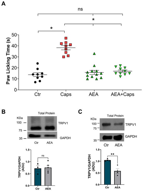
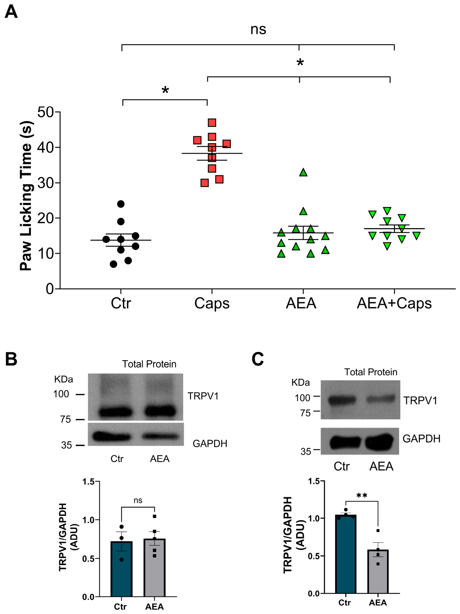
We intradermally injected capsaicin (1 µg), AEA (50 ng), or a mix of capsaicin and AEA (1 µg + 50 ng, respectively) into male mouse paws to evaluate the antinociceptive effects on capsaicin-evoked acute pain. We compared the experimental group’s pain responses with those of the control group. The results show that capsaicin induces pain behavior (38 ± 2 s), whereas AEA does not (16 ± 2 s; Figure 1A). Furthermore, male mice injected with AEA + capsaicin showed a minimal nocifensive response (17 ± 1 s), similar to that observed in mice injected with the control solution (14 ± 2 s; Figure 1A).
Figure 1.
Evaluation of AEA effects on capsaicin-induced acute pain in male mice and on the regulation of TRPV1 protein levels. (A) Paw-licking time (PLT) in response to intradermal injection of vehicle (Ctr, 0.64% ethanol), capsaicin (1 μg), AEA (50 ng), or a combination of AEA and capsaicin in male mice. PLT values show that mice injected with vehicle and AEA do not display nocifensive behavior (Ctr = 14 ± 2 s (n = 9) and AEA = 16 ± 2 s (n = 12)), whereas mice injected with capsaicin show a pain behavior which decrease in the mice co-injected with capsaicin and AEA (Caps = 38 ± 2 s (n = 9) vs. AEA + Caps = 17 ± 1 s (n = 10)) data are mean ± SEM. ANOVA followed by Tukey’s multiple comparisons test was performed. * p < 0.0001; ns: not statistically significant. Each point represents one animal. (B) Immunodetection of TRPV1 and GAPDH proteins from primary cultures of DRG neurons treated with control or 10 µM AEA for 10 min and (C) 10 µM AEA for 20 min, the graph shows that TRPV1 protein levels do not change with AEA treatment for 10 min (Ctr = 0.72 ± 0.12, n = 3 vs. AEA = 0.75 ± 0.9 ADU, n = 5), whereas the AEA treatment for 20 min downregulates TRPV1 protein levels (Ctr = 1.04 ± 0.02, n = 4 vs. AEA = 0.58 ± 0.09 ADU, n = 4); the bars represent the TRPV1 values normalized to GAPDH (mean ± SEM): Student’s t-test was performed ** p < 0.01, ns: not statistically significant; ADU = arbitrary densitometric units.
These data show that AEA reduces TRPV1-induced pain in male mice. Because anandamide is a partial agonist of TRPV1, we asked whether it induces desensitization, as capsaicin does. First, we evaluated whether AEA regulates TRPV1 protein levels in primary DRG neuron cultures treated with 10 µM AEA for 10 min, mimicking the AEA exposure time of nociceptive assay conditions. The results show that TRPV1 protein levels are similar between the anandamide and control treatments (Ctr = 0.72 ± 0.12 vs. AEA = 0.75 ± 0.9 ADU), as shown in Figure 1B.
Next, we evaluated whether long-term AEA exposure could regulate TRPV1 protein levels in primary DRG neuron cultures; we treated cells with 10 µM AEA for 20 min. The results show that anandamide treatment reduces total TRPV1 protein levels compared with control cultures (Ctr = 1.04 ± 0.02 vs. AEA = 0.58 ± 0.09 ADU), as shown in Figure 1C.
Overall, these results show that AEA decreases the capsaicin-evoked pain and downregulates the TRPV1 protein levels in primary DRG neuron cultures after 20 min of treatment.
3.2. Anandamide Downregulates the Total and Surface TRPV1 Protein Levels in a Heterologous Expression System
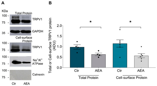
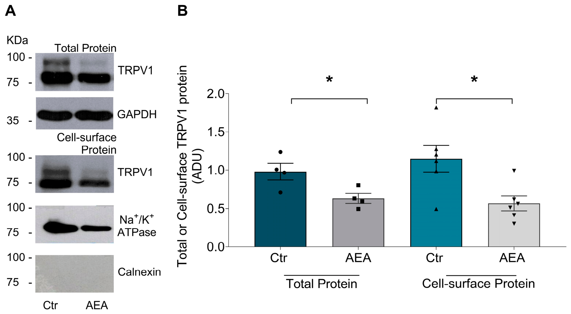
We evaluated AEA effects in rTRPV1-expressing HEK293 cells. We incubated the cells with 10 µM AEA for 20 min and found that total TRPV1 protein levels decreased by 40% relative to control cells (ctr = 1.03 ± 0.08 vs. 0.62 ± 0.04 ADU, Figure 2A,B). We also tested different AEA concentrations on rTRPV1-expressing HEK293 cells and found that lower AEA concentrations (1 and 5 µM) did not affect TRPV1 protein levels, whereas 10 µM decreased total TRPV1 protein levels by approximately 40% (Supplemental Figure S1). Additionally, we exposed TRPV1-expressing HEK293 cells to 10 µM AEA for different incubation times (20, 40, 60, 90, and 120 min), and AEA similarly decreased TRPV1 protein levels at all assessed times (Supplemental Figure S1B). Furthermore, we asked if a short time of AEA treatment, such as 10 min, modifies the TRPV1 total protein levels, finding that 10 µM has not affect the TRPV1 protein levels (Ctr = 0.95 ± 0.07 vs. 10 µM AEA = 0.91 ± 0.015 ADU), whereas higher concentrations (15 and 25 µM) reduced the TRPV1 total protein (Ctr = 1.02 ± 0.1 vs. 15 µM AEA = 0.72 ± 0.03 ADU; Ctr = 0.96 ± 0.03 vs. 25 µM AEA = 0.66 ± 0.02 ADU) (Supplemental Figure S1C).
Figure 2.
Anandamide downregulates total and cell-surface TRPV1 protein in HEK293 cells expressing TRPV1. HEK293 cells were transiently transfected to express TRPV1 protein and exposed to AEA (10 µM, 20 min). (A) Immunodetection of TRPV1 total protein and GAPDH as a loading control (upper panels) and TRPV1 cell surface protein, Na+/K+-ATPase as a loading control, and calnexin as a negative marker to prove the absence of cellular or ER contamination (lower panels). (B) The graphs show that AEA downregulates TRPV1 total and cell surface protein. Bars represent TRPV1 total protein values normalized to GAPDH in cells without treatment (Ctr = 0.97 ± 0.1 and cells treated with AEA 0.63 ± 0.12 ADU). Student’s t-test was performed; * p < 0.01, n = 4. Similarly, TRPV1 cell-surface protein, normalized to Na+/K+-ATPase: Ctr = 1.15 ± 0.17 ADU and AEA = 0.56 ± 0.09 ADU; Student’s t-test was performed; * p < 0.01, n = 6. ADU = arbitrary densitometric units.
We also assessed cell-surface TRPV1 protein levels in TRPV1-expressing HEK293 cells treated with 10 µM anandamide for 20 min, showing a 45% decrease in TRPV1 protein localized at the plasma membrane (Ctr = 0.97 ± 0.1 vs. AEA = 0.63 ± 0.12 ADU; Figure 2A,B).
3.3. Anandamide Induces TRPV1 Protein Degradation
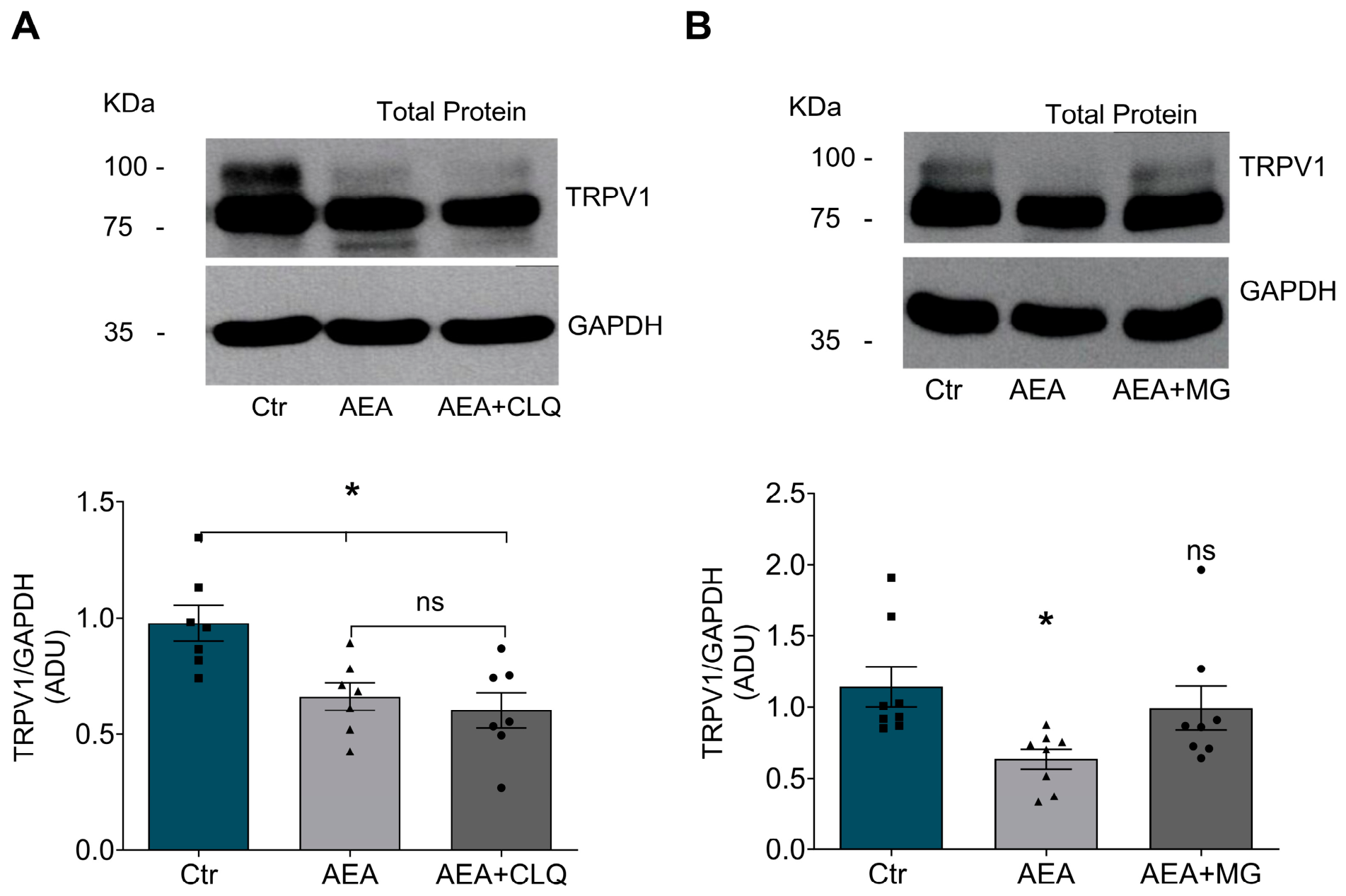
Our results show that 10 µM AEA for 20 min downregulates TRPV1 protein levels, suggesting a protein-degradation-mediated mechanism. To confirm this, we pretreated TRPV1-expressing HEK293 cells with chloroquine, an inhibitor of the lysosomal pathway, or MG132, an inhibitor of the proteasomal pathway. First, we showed that these pre-treatments do not affect cell viability (Supplemental Figure S2A) and do not alter total TRPV1 protein levels (Supplemental Figure S2B). Next, we pretreated TRPV1-expressing HEK293 cells with 25 µM MG132 or 100 µM chloroquine, then we applied AEA.
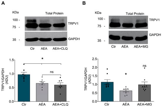
The results show that AEA still decreases TRPV1 protein in cells pretreated with chloroquine (Ctr = 0.97 ± 0.07, AEA = 0.66 ± 0.05, chloroquine pretreatment + AEA = 0.6 ± 0.07, ADU), as shown in Figure 3A. In contrast, pretreatment with MG132 prevents decreasing TRPV1 protein produced under AEA treatment, (Ctr = 1.14 ± 0.14, AEA = 0.63 ± 0.07, MG132 pretreatment + AEA = 0.99 ± 0.15 ADU) as shown in Figure 3B.
Figure 3.
Anandamide induces TRPV1 protein degradation through the proteasomal pathway. (A) Immunodetection of TRPV1 and GAPDH total protein from TRPV1-transiently expressing HEK293 cells in untreated and AEA-treated culture conditions (Ctr and AEA, respectively) and cells pretreated with Chloroquine (CLQ), the inhibitor of the lysosomal pathway, and then with AEA treatment (AEA + CLQ). The lower-left graph shows that CLQ does not rescue the AEA-induced downregulation of total TRPV1 protein. The bars represent the TRPV1 normalized values: Ctr = 0.97 ± 0.07, AEA = 0.66 ± 0.05 and AEA + CLQ = 0.60 ± 0.07 ADU (n = 7). (B) Immunodetection of TRPV1 and GAPDH total protein from TRPV1-transiently expressing HEK293 cells in Ctr and AEA culture conditions, and cells pretreated with MG132 (proteasome inhibitor) and subsequently exposed to AEA. The lower-right graph shows that MG132 prevents AEA-induced downregulation of TRPV1 protein levels. The bars represent TRPV1 normalized values: Ctr = 1.14 ± 0.14, AEA = 0.63 ± 0.07 and AEA + MG132 = 0.99 ± 0.15 ADU (n = 8). We performed ANOVA, followed by Tukey’s multiple comparisons test. * p < 0.05; ns: not statistically significant.
Furthermore, we evaluated whether AEA induces TRPV1 ubiquitination; we immunoprecipitated ubiquitinated proteins from control and AEA-treated HEK293 cells and tested whether TRPV1 co-immunoprecipitated. Supplemental Figure S2C shows that TRPV1 is ubiquitinated in cells treated with AEA.
These data indicate that AEA induces TRPV1 ubiquitination and its proteasomal degradation.
3.4. Anandamide’s Effects on TRPV1 Protein Levels Are Independent of Channel Activation
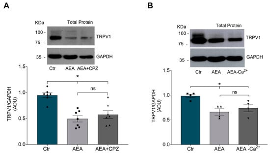
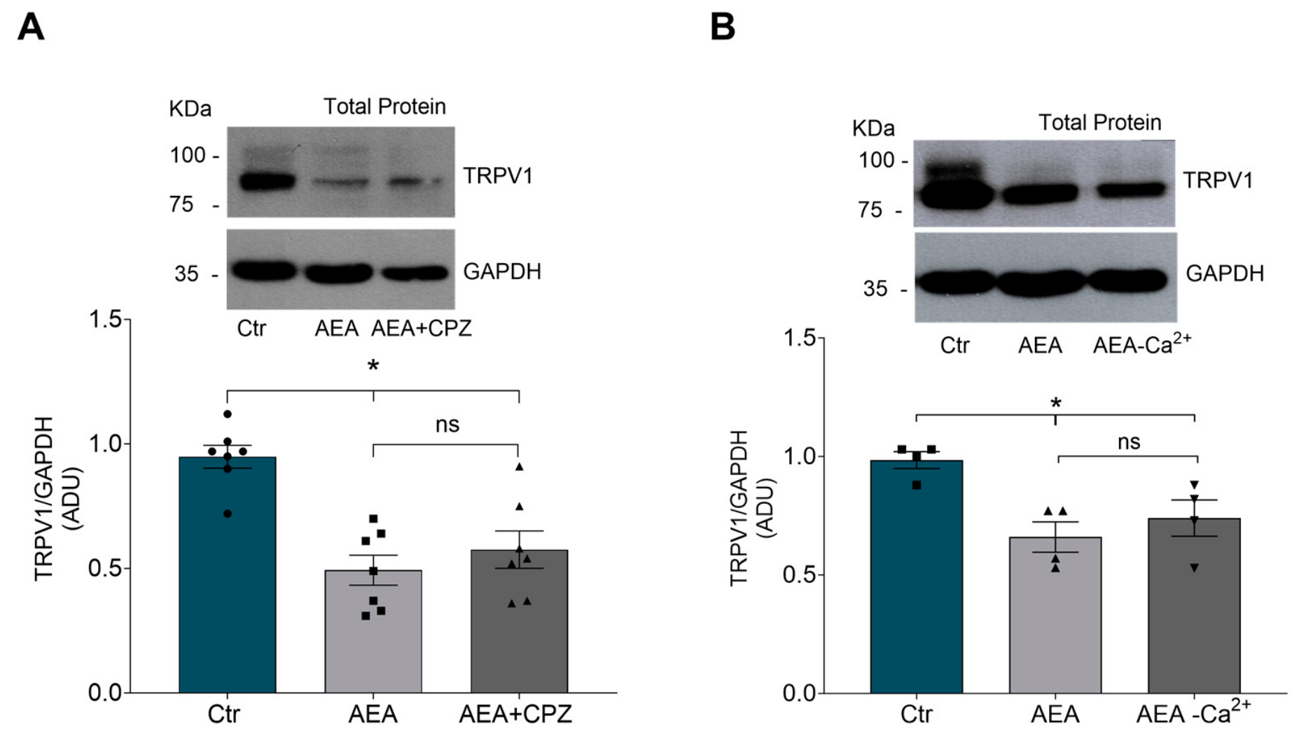
Since AEA is a partial agonist of TRPV1, its negative regulation of TRPV1 protein levels could be mediated via TRPV1 channel activation and Ca2+ influx, leading to TRPV1 desensitization. To evaluate this possibility, we treated TRPV1-expressing HEK293 cells with AEA and capsazepine, a TRPV1 antagonist. Our results show that capsazepine does not prevent the effects of anandamide (Ctr = 0.94 ± 0.04; AEA = 0.49 ± 0.06; AEA + capsazepine = 0.57 ± 0.07, ADU; Figure 4A), suggesting that channel activation is not necessary for the AEA effects. However, this approach does not exclude the possibility that AEA could induce Ca2+ influx through an alternative pathway; therefore, we administered AEA in Ca2+-free medium. We observed that TRPV1-expressing HEK293 cells treated with AEA without calcium also decrease TRPV1 protein levels, as observed with anandamide treatment in the presence of calcium (Ctr = 0.98 ± 0.03; AEA in the presence of calcium = 0.66 ± 0.06, anandamide without calcium: 0.74 ± 0.07 ADU; Figure 4B). These results suggest that AEA-induced decreases in TRPV1 protein levels are independent of Ca2+ influx.
Figure 4.
Anandamide induces TRPV1 protein downregulation independently of TRPV1 activation and Ca2+ influx. (A) Immunodetection of total TRPV1 and GAPDH protein in HEK293 cells transiently expressing TRPV1 in control conditions (Ctr), AEA treatment (AEA 10 µM), and AEA + Capsazepine (AEA 10 µM + CPZ 2.5 µM); all treatments were for 20 min. The graph shows that CPZ does not prevent AEA-induced downregulation of TRPV1. The bars show normalized values of TRPV1: Ctr = 0.94 ± 0.04, AEA = 0.49 ± 0.06, and AEA + CPZ = 0.57 ± 0.07 ADU, n = 7. (B) WB shows the TRPV1 total protein from control culture conditions (Ctr), AEA, and AEA treatment in DMEM Ca2+-free conditions. The graph shows TRPV1 values normalized to GAPDH: Ctr = 0.98 ± 0.03, AEA = 0.66 ± 0.06, AEA-Ca2+ = 0.74 ± 0.07 ADU, n = 4, respectively. ANOVA followed by Tukey’s multiple comparisons test was performed; * p < 0.01; ns: not statistically significant.
3.5. Anandamide Treatment Alters the Protein Levels of BiP, an Endoplasmic Reticulum-Resident Chaperone
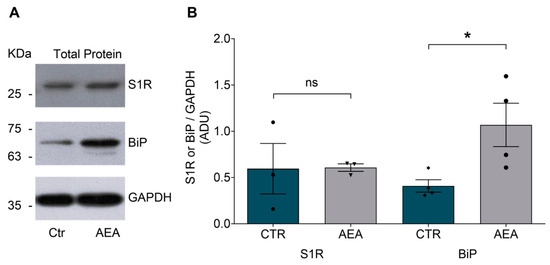
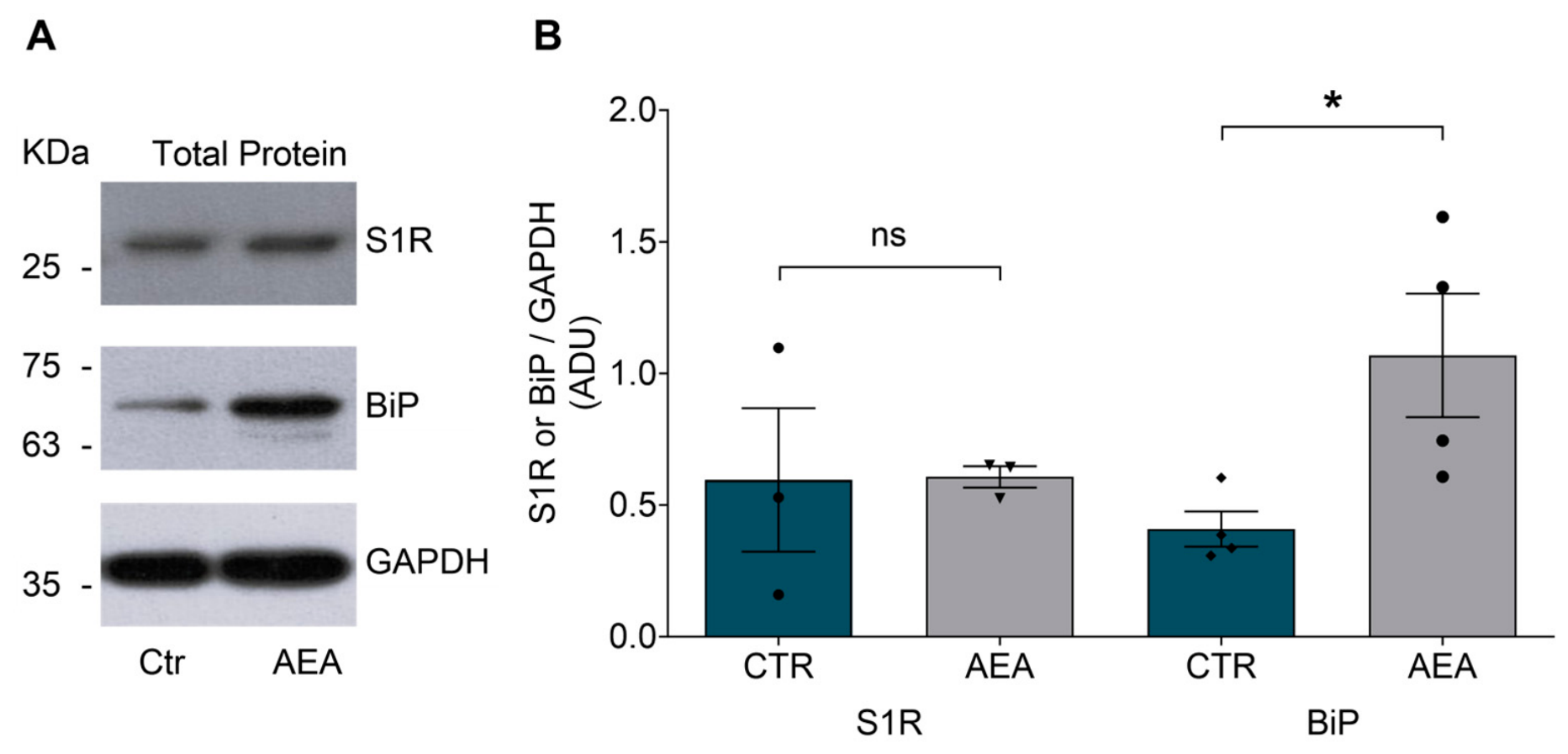
We previously reported that the chaperone Sigma-1 receptor (S1R) regulates TRPV1 by facilitating its folding and trafficking to the plasma membrane [30]. Disruption of this protein complex leads to TRPV1 degradation through the proteasome pathway. Thus, we tested whether AEA downregulates S1R protein levels in AEA-treated cells. Our results show that S1R protein levels in AEA-treated cells are comparable to those in control cells (Ctr = 0.59 ± 0.27 vs. AEA = 0.6 ± 0.04 ADU; Figure 5A,B).
Figure 5.
AEA treatment increases the total BiP protein levels. (A) Immunoblot of Sigma 1 receptor (S1R) and BiP in total protein extracts from HEK293 cells transiently expressing TRPV1 in culture conditions (Ctr) and AEA treatment (AEA 10 µM). (B) The graph shows that AEA does not modify S1R total protein, although this treatment upregulates BiP total protein. The bars represent normalized S1R and BiP protein levels relative to GAPDH. The values for S1R were: Ctr = 0.59 ± 0.27 and AEA = 0.60 ± 0.04 ADU, respectively, n = 3; the BiP normalized values were: Ctr = 0.40 ± 0.06 and AEA = 1.06 ± 0.23 ADU; n = 4, Student’s t test was performed * p < 0.05. ns: not statistically significant.
Next, we evaluated total BiP protein levels, a key chaperone that interacts with S1R and maintains it in a silent state. Our results show that AEA treatment increases BiP protein levels (Ctr = 0.4 ± 0.06 vs. AEA = 1.06 ± 0.23 ADU; Figure 5A,B), suggesting that this increase could maintain S1R in a silent state, thereby allowing TRPV1 protein destabilization in the ER and subsequent targeting to the proteasomal pathway.
Upregulation of BiP expression is linked to endoplasmic reticulum (ER) stress, triggering the unfolded protein response and affecting other ER stress sensors; thus, we evaluated whether AEA treatment induces this response by assessing molecular mediators downstream of BiP regulation, such as PERK, IREα, and PDI. Supplemental Figure S3 shows that none of these proteins change in AEA-treated cells for 20 min.
3.6. Evaluation of 2-AG Effects on Capsaicin-Evoked Pain
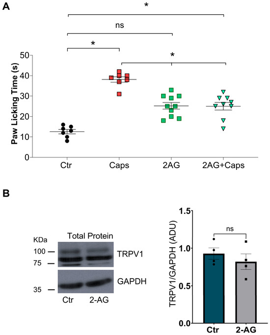
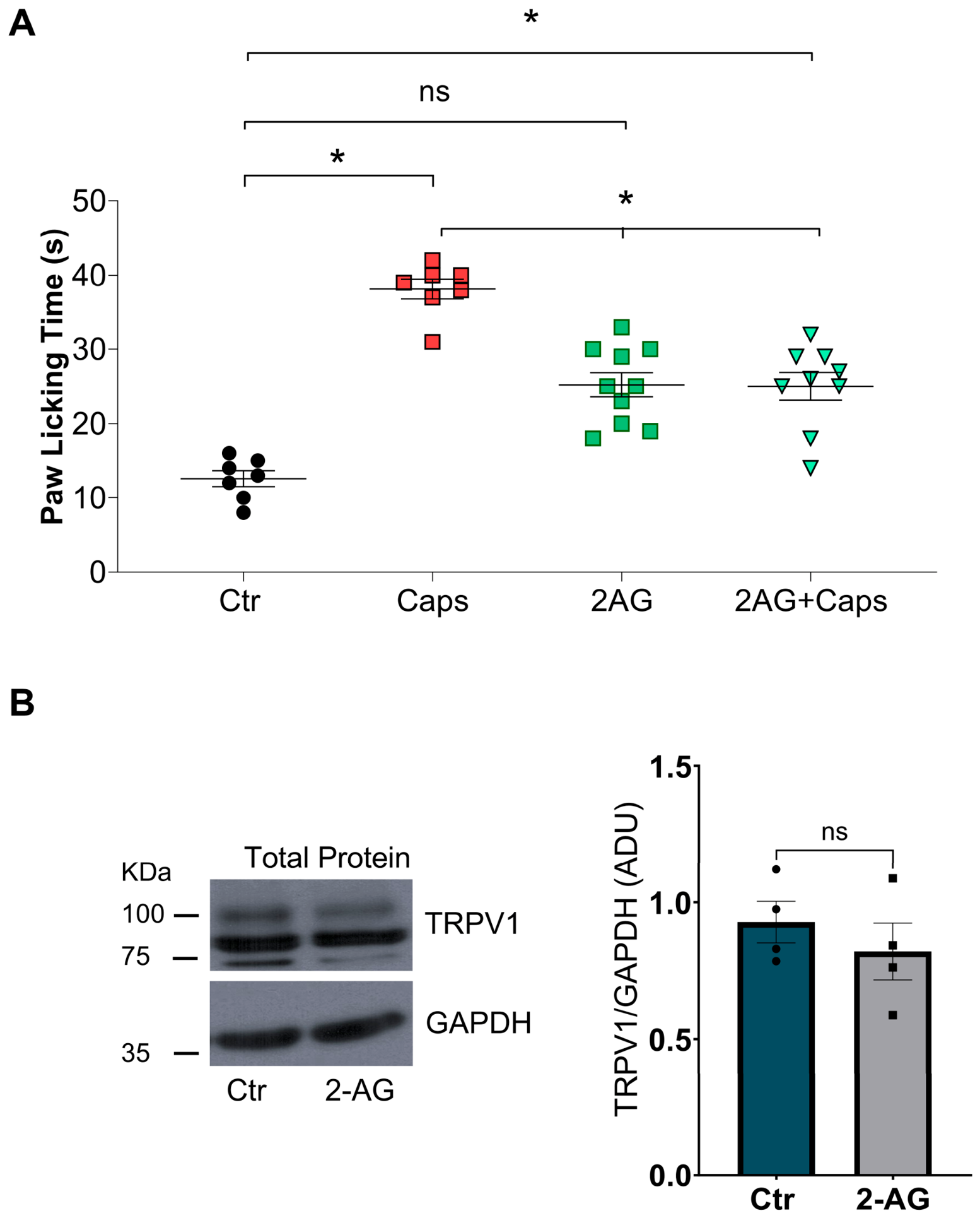
We explored whether 2-AG (a full agonist of CB1R and CB2R) could regulate TRPV1-mediated pain. Injection of 50 ng of 2-AG into the mouse paw elicited similar behavior to injection of control solution (Ctr = 13 ± 1 s vs. 2-AG = 25 ± 1.6 s). Mice injected with capsaicin displayed a typical nocifensive response, which was partially inhibited in animals co-injected with capsaicin plus 2-AG (Caps = 38 ± 1.3 s vs. Caps + 2-AG = 25 ± 1.9 s), Figure 6A. Paw-licking behavior differed significantly between the Ctr and Caps + 2-AG groups, indicating that 2-AG does not completely reduce pain produced through TRPV1 activation.
Figure 6.
Evaluation of capsaicin-induced acute pain in male mice in co-administration with 2-AG and TRPV1 protein levels in HEK293 transfected cultures. (A) Paw-licking time (PLT) in response to intraplantar injection of vehicle (Ctr, 0.63% ethanol), capsaicin (Caps, 1 μg), 2-arachidonoylglycerol (2-AG, 50 ng), or a combination of 2-AG and capsaicin (2-AG + Caps) in male mice. The graph shows that capsaicin evokes a nociceptive response, which 2-AG co-injection partially prevents. PLT values (mean ± SEM) were: Ctr = 13 ± 1 s (n = 7); Caps = 38 ± 1.3 s (n = 7); 2-AG = 25.20 ± 1.64 s (n = 10); 2-AG + Caps = 25.00 ± 1.88 s (n = 9). ANOVA followed by Tukey’s multiple comparisons test was performed; * p < 0.001; ns: not statistically significant. Each point represents one animal. (B) Immunodetection of TRPV1 total protein from HEK293-transiently transfected cells and GAPDH in control culture conditions (Ctr) and 2-AG treatment (10 µM, 20 min). The graph shows that TRPV1 protein levels are similar between the experimental groups. Bars show the values normalized to the loading control (GAPDH), Ctr = 0.87 ± 0.04, 2-AG = 0.75 ± 0.05 ADU; n = 4; Student’s t-test. ns: not statistically significant.
These results led us to evaluate 2-AG’s effects on TRPV1 protein levels. Our results show that TRPV1-expressing HEK293 cells treated with 10 µM 2-AG for 20 min exhibit TRPV1 protein levels comparable to control cultures (Ctr = 0.87 ± 0.04 s vs. 2-AG = 0.75 ± 0.05 ADU; Figure 6B). Overall, our results show that 2-AG is less effective than AEA at regulating TRPV1-mediated pain and modulating TRPV1 protein levels.
3.7. A Phytocannabinoid Also Reduced Capsaicin-Induced Pain by Decreasing TRPV1 Protein Levels
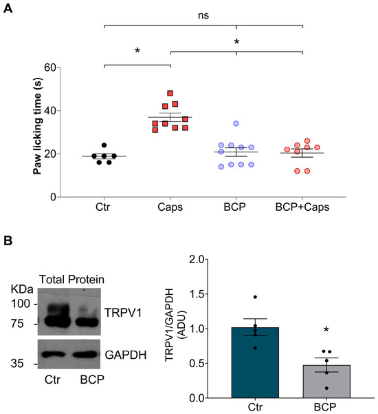
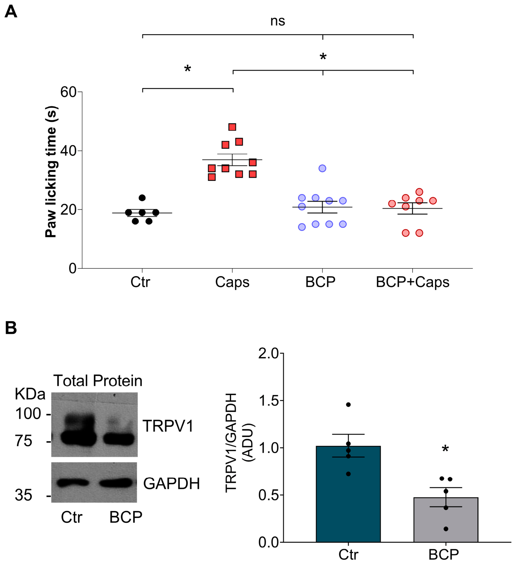
Finally, we evaluated whether the natural product β-caryophyllene (BCP), a selective phytocannabinoid agonist of CB2R [31], could modulate capsaicin-induced pain. Our data show that the intradermal injection of BCP in the mouse paw produces a response similar to that of animals injected with the control solution (Ctr = 18.8 ± 1.2 vs. BCP = 20.8 ± 2 s; Figure 7A), and when we co-injected BCP and capsaicin, we observed a decrease in the nocifensive response compared to that of mice injected with capsaicin alone (Caps = 36.9 ± 2 vs. Caps + BCP = 20.4 ± 1.9 s; Figure 7A).
Figure 7.
β-caryophyllene (BCP) decreases the capsaicin-evoked pain and downregulates TRPV1 protein levels. (A) Paw-licking time (PLT) in response to intradermal injection of vehicle (Ctr, 0.75% ethanol), capsaicin (Caps, 1 μg), BCP (50 ng), or a combination of BCP and capsaicin (BCP + Caps) in male mice. PLT values show that capsaicin-evoked pain is reduced when the mice are co-injected with BCP. The values were (mean ± SEM): Ctr = 18.83 ± 1.19 s (n = 6); Caps = 36.89 ± 1.99 s (n = 9); BCP = 20.80 ± 1.97 s (n = 10); BCP + Caps = 20.38 ± 1.89 s (n = 8). Mean ± SEM; ANOVA followed by Tukey’s multiple comparisons test; * p < 0.001; ns: not statistically significant. Each point represents one animal. (B) Immunodetection of TRPV1 and GAPDH total protein from HEK293-transiently transfected cells in control culture conditions (Ctr) and BCP treatment (10 µM, 20 min). The graph shows that TRPV1 protein levels decrease in AEA-treated cultures. The bars show the values normalized to the loading control (GAPDH), Ctr = 1.02 ± 0.12 and BCP = 0.47 ± 0.1 ADU; n = 5; Student’s t-test was performed. * p < 0.05.
To explore whether BCP modulates capsaicin-evoked pain by downregulating TRPV1 protein levels, we treated TRPV1-expressing HEK293 cells with 10 µM BCP for 20 min. We observed that BCP treatment reduces TRPV1 protein levels by nearly 50% (Ctr = 1.02 ± 0.12 vs. BCP = 0.47 ± 0.10 ADU; Figure 7B).
These data suggest that this natural product has analgesic potential against TRPV1-induced pain and produces downregulation of TRPV1 protein levels.
4. Discussion
Mammals have developed several adaptive mechanisms to survive adverse conditions, highlighting their ability to detect noxious signals that could damage the organism’s integrity. This ability translates into a physiological response: pain, although pain does not always have a protective role and can be a cause of disability and suffering for thousands of people, especially in current times, as all countries face the challenge of an aging population, with pain as a main factor in the debilitation and social isolation of the elderly [2]. Thus, understanding how to regulate molecular mediators of pain is important for developing new analgesic strategies with fewer side effects.
Transient receptor potential vanilloid 1 (TRPV1) has emerged as a key molecular mediator of painful signals in nociceptive, inflammatory, and neuropathic pain [32]; thus, understanding how TRPV1 activity and expression are regulated offers opportunities to develop new analgesic strategies. For example, topical capsaicin formulations have been designed to treat osteoarthritic pain, aiming to alleviate pain while avoiding the collateral effects of systemic analgesic administration [33]. However, these formulations cause severe irritation at the site of application, which is the main reason they are avoided, especially in elderly people [34].
This creates an opportunity to explore new compounds that may induce TRPV1 downregulation without irritant effects, and this work examined endocannabinoids as putative TRPV1 regulators.
Our results show that AEA decreases the capsaicin-evoked pain and that 20 min of AEA treatment downregulates TRPV1 protein levels in primary cultures of DRG neurons (Figure 1). Furthermore, in a heterologous TRPV1 expression system, we observed that AEA treatment also downregulates total TRPV1 protein levels (Figure 2). The molecular mechanism of TRPV1 protein downregulation explored differs from that of capsaicin: AEA induces TRPV1 protein degradation through the proteasomal pathway rather than the lysosomal pathway (Figure 3 [27]).
Notably, our results also show that AEA exerts its effects even with a TRPV1 antagonist (capzasepine) or in a Ca2+-free medium (Figure 4). These results also indicate that AEA treatment does not require TRPV1 activation. Although AEA is a TRPV1 partial agonist [35], our results show that a lower AEA concentration does not produce a nocifensive response, even though it reduces capsaicin-evoked pain, reemphasizing that this endocannabinoid does not produce an irritant sensation. Interestingly, this lower AEA concentration used in the pain behavior experiments, 50 ng in a final volume of 10 µL, gives a 15 µM concentration, which is almost equivalent to the concentration used in all the cell treatment experiments, suggesting that TRPV1 protein downregulation could also be produced locally in the nociceptor, an aspect not evaluated here.
The cues from experiments inhibiting the proteasomal pathway and the co-immunoprecipitation of ubiquitinated TRPV1 protein in cells treated with AEA suggest that AEA triggers TRPV1 degradation through the proteasomal pathway, impacting TRPV1 cell-surface protein levels. Because S1R is a chaperone relevant to TRPV1 stability in the ER [30], we evaluated changes in ER homeostasis under AEA treatment. We found that 20 min of AEA treatment increases BiP protein levels (Figure 5), a crucial ER-resident chaperone that acts as an ER stress sensor [36]; however, we did not find changes in the total protein levels of other markers of ER stress, although we cannot discard this possibility since the initial change in these markers is the phosphorylation state, which we did not evaluate. According to various reports, endocannabinoids (AEA and 2-AG) can induce ER stress [37], activating the Unfolded Protein Response (UPR), in which BiP dissociates from its protein complex with PERK, IRE1α, and ATF6, to associate with unfolded proteins and avoid their accumulation, triggering protein degradation through the proteasome pathway [38].
To expand our knowledge of endocannabinoids’ roles in regulating TRPV1-mediated pain, we tested whether 2-AG could regulate capsaicin-evoked pain as AEA does and found that 2-AG was less effective at reducing the nocifensive response to capsaicin (Figure 6A). Our results are consistent with previous reports showing that 2-AG is less effective at decreasing meningeal nerve-ending nociceptive firing than AEA [39]. Furthermore, we observed that HEK293-TRPV1-expressing cells treated with 2-AG show TRPV1 protein levels similar to control cells (Figure 6B), suggesting that 2-AG is less effective than AEA at downregulating TRPV1 protein levels. Since the HEK293 cell line constitutively expresses monoacylglycerol lipase (MAGL), which degrades 2-AG, the possible effects of this endocannabinoid on TRPV1 protein regulation could be masked in this cell line [40].
Some research has focused on MAGL and FAAH inhibitors, the latter of which is responsible for AEA degradation. Inhibiting these enzymes increases endocannabinoid levels, allowing these molecules to exert their physiological effects for longer periods of time [40]. For example, a dual MAGL/FAAH inhibitor has been shown to reduce migraine nociception by decreasing trigeminal nerve nociceptive firing [39]. These effects are mainly mediated by increased AEA levels and activation of CB1 receptors. This implies that the use of ectopic AEA as analgesic therapy would be limited by the action of FAAH, and we will need a combination of AEA and an FAAH inhibitor to reach adequate pain modulation. However, what about using a compound inert to FAAH activity? Here, a phytocannabinoid such as β-caryophyllene (BCP), a sesquiterpene found in natural products such as clove, cinnamon, oregano, thyme, and others, could be an excellent option. This compound is a selective agonist of the CB2 receptor and is widely associated with several physiological roles, including anti-inflammatory and pain-regulating effects. To date, it is unclear whether this compound regulates capsaicin-evoked pain; here, we show that BCP decreases TRPV1-mediated pain, as does AEA; moreover, we find that BCP also downregulates TRPV1 protein levels in HEK293 cells (Figure 7). BCP’s potential role in analgesia is promising; it lacks psychotropic effects because it is specific for CB2 receptors, and we now have evidence that it negatively regulates the protein levels of a key molecular pain mediator, TRPV1.
Overall, our results provide experimental evidence that the endocannabinoid AEA and phytocannabinoid BCP play pivotal roles in regulating TRPV1-mediated pain, suggesting that topical analgesic formulations could downregulate TRPV1 protein levels and reduce pain induced by this receptor. This could help elicit analgesia, for example, in osteoarthritic cases, so patients avoid the irritant collateral effects common with capsaicin formulations.
A limitation of this study is that we evaluated the nociceptive pain response to capsaicin, which is a TRPV1 selective agonist; we did not evaluate, for example, inflammatory or neuropathic pain, for which TRPV1 expression is exacerbated in the DRG and dorsal horn, breaking new ground in exploration of the effects of AEA and BCP in these contexts. Another limitation is that this study did not examine the specific roles of CB1 and CB2 receptors to determine whether the molecular mechanism is directly related to these receptors; future studies should address this.
Supplementary Materials
The following supporting information can be downloaded at: https://www.mdpi.com/article/10.3390/pharmaceutics18111374/s1, Figure S1: Dose- and time-response curves for AEA treatment. Figure S2: Pretreatment with MG132 and chloroquine does not affect cell viability or TRPV1 protein levels. Figure S3: Evaluation of ER stress markers after AEA treatment.
Author Contributions
Conceptualization, S.L.M.-L.; Methodology, R.J.-C. and S.L.M.-L.; Software, S.L.M.-L.; Validation, S.L.M.-L.; Formal analysis, R.J.-C. and S.L.M.-L.; Investigation, R.J.-C. and S.L.M.-L.; Resources, S.L.M.-L.; Data curation, S.L.M.-L.; Writing—original draft, R.J.-C. and S.L.M.-L.; Writing—review & editing, R.J.-C. and S.L.M.-L.; Visualization, S.L.M.-L.; Supervision, S.L.M.-L.; Project administration, S.L.M.-L.; Funding acquisition, S.L.M.-L. All authors have read and agreed to the published version of the manuscript.
Funding
This research was funded by Dirección General de Asuntos del Personal Académico (DGAPA)-Programa de Apoyo a Proyectos de Investigación e Innovación Tecnológica (PAPIIT) #IN225623, and Secretaría de Ciencia, Humanidades, Tecnología e Innovación (SECIHTI) project number: CBF2023-2024-196. R.J.-C. received scholarship support from SECIHTI #CVU 850914 and from DGAPA-PAPIIT #IN225623.
Institutional Review Board Statement
The Internal Committee for the Use and Care of Laboratory Animals (CICUAL, protocol SLM127-18, 4 May 2022) of the Instituto de Fisiología Celular de la Universidad Nacional Autónoma de México approved the experimental procedures.
Informed Consent Statement
Not applicable.
Data Availability Statement
The original contributions presented in this study are included in the article/supplementary material. Further inquiries can be directed to the corresponding author. Dataset available on request from the authors.
Acknowledgments
We thank Ana Valeria Martínez Silva, Héctor Alfonso Malagón Rivero, and Claudia Verónica Rivera Cerecedo at the Instituto de Fisiología Celular, Universidad Nacional Autónoma de México (IFC-UNAM), for their expert technical support. We also thank Gabriela Xóchitl Ayala Méndez at the IFC-UNAM for her valuable assistance in maintaining and enhancing the well-being of the C57BL6 mouse colony, and David Julius at UCSF for indirectly providing the pcDNA3.1-TRPV1 plasmid. Finally, we sincerely thank Diana Escalante Alcalde for her valuable academic support, and Ricardo González Ramírez for their sincere advice and support with the graphical abstract. This work comprises the Ph.D. thesis of R.J.-C. in the Programa de Posgrado en Ciencias Biológicas de la Universidad Nacional Autónoma de México.
Conflicts of Interest
The authors declare no conflicts of interest.
Abbreviations
| 2-AG | 2-arachidonoylglycerol |
| ADU | Arbitrary Densitometric Units |
| AEA | Anandamide (N-arachidonoylethanolamide) |
| BCP | β-caryophyllene |
| BiP | Binding Immunoglobulin Protein |
| cAMP | cyclic Adenosine 3’,5’-monophosphate |
| CB1 | Cannabinoid Receptor 1 |
| CB2 | Cannabinoid Receptor 2 |
| CGRP | calcitonin gene-related peptide |
| CLQ | Chloroquine |
| CNS | Central Nervous System |
| CPZ | Capsazepine |
| DRG | Dorsal Root Ganglia |
| GPCRs | G-protein-coupled receptors |
| GPR110 | Orphan GPCR 110 |
| GPR119 | Orphan GPCR 119 |
| GPR18 | Orphan GPCR 18 |
| GPR49 | Orphan GPCR 49 |
| GPR55 | Orphan GPCR 55 |
| HEK293 | Human Embryonic Kidney 293 |
| MG | MG 132 |
| PLT | Paw-Licking Time |
| PPAR | Peroxisome proliferator-activated receptor |
| S1R | Sigma-1 receptor |
| SNL | Spinal Nerve Ligation |
| TRPV1 | Transient Receptor Potential Vanilloid 1 |
| UPR | Unfolded Protein Response |
References
- Julius, D.; Basbaum, A.I. Molecular mechanisms of nociception. Nature 2001, 413, 203–210. [Google Scholar] [CrossRef] [Scilit] [PubMed]
- Singh, S.P.; Guindon, J.; Mody, P.H.; Ashworth, G.; Kopel, J.; Chilakapati, S.; Adogwa, O.; Neugebauer, V.; Burton, M.D. Pain and aging: A unique challenge in neuroinflammation and behavior. Mol. Pain 2023, 19, 17448069231203090. [Google Scholar] [CrossRef] [Scilit] [PubMed]
- Battista, N.; Di Tommaso, M.; Bari, M.; Maccarrone, M. The endocannabinoid system: An overview. Front. Behav. Neurosci. 2012, 6, 9. [Google Scholar] [CrossRef] [Scilit] [PubMed]
- Iannotti, F.A.; Di Marzo, V. The endocannabinoidomes: Pharmacological redundancy and promiscuity, and multi-kingdom variety of sources and molecular targets. Pharmacol. Rev. 2025, 77, 100070. [Google Scholar] [CrossRef] [Scilit] [PubMed]
- Maccarrone, M.; Di Marzo, V.; Gertsch, J.; Grether, U.; Howlett, A.C.; Hua, T.; Makriyannis, A.; Piomelli, D.; Ueda, N.; van der Stelt, M. Goods and Bads of the Endocannabinoid System as a Therapeutic Target: Lessons Learned after 30 Years. Pharmacol. Rev. 2023, 75, 885–958. [Google Scholar] [CrossRef] [Scilit] [PubMed]
- Spicarova, D.; Palecek, J. Anandamide-Mediated Modulation of Nociceptive Transmission at the Spinal Cord Level. Physiol. Res. 2024, 73, S435–S448. [Google Scholar] [CrossRef] [Scilit] [PubMed]
- Chen, J.; Varga, A.; Selvarajah, S.; Jenes, A.; Dienes, B.; Sousa-Valente, J.; Kulik, A.; Veress, G.; Brain, S.D.; Baker, D.; et al. Spatial Distribution of the Cannabinoid Type 1 and Capsaicin Receptors May Contribute to the Complexity of Their Crosstalk. Sci. Rep. 2016, 6, 33307. [Google Scholar] [CrossRef] [Scilit] [PubMed]
- Tsou, K.; Brown, S.; Sanudo-Pena, M.C.; Mackie, K.; Walker, J.M. Immunohistochemical distribution of cannabinoid CB1 receptors in the rat central nervous system. Neuroscience 1998, 83, 393–411. [Google Scholar] [CrossRef] [Scilit] [PubMed]
- Ehrlich, B.; Patel, M.; Zheng, X.; Glass, M. Canonical Cannabinoid Receptors. Curr. Top. Behav. Neurosci. 2026, 76, 37–68. [Google Scholar] [PubMed]
- Mackie, K.; Lai, Y.; Westenbroek, R.; Mitchell, R. Cannabinoids activate an inwardly rectifying potassium conductance and inhibit Q-type calcium currents in AtT20 cells transfected with rat brain cannabinoid receptor. J. Neurosci. 1995, 15, 6552–6561. [Google Scholar] [CrossRef] [Scilit] [PubMed]
- Morisset, V.; Urban, L. Cannabinoid-induced presynaptic inhibition of glutamatergic EPSCs in substantia gelatinosa neurons of the rat spinal cord. J. Neurophysiol. 2001, 86, 40–48. [Google Scholar] [CrossRef] [Scilit] [PubMed]
- Sugiura, T.; Kondo, S.; Kishimoto, S.; Miyashita, T.; Nakane, S.; Kodaka, T.; Suhara, Y.; Takayama, H.; Waku, K. Evidence that 2-arachidonoylglycerol but not N-palmitoylethanolamine or anandamide is the physiological ligand for the cannabinoid CB2 receptor. Comparison of the agonistic activities of various cannabinoid receptor ligands in HL-60 cells. J. Biol. Chem. 2000, 275, 605–612. [Google Scholar] [CrossRef] [Scilit] [PubMed]
- Galiegue, S.; Mary, S.; Marchand, J.; Dussossoy, D.; Carriere, D.; Carayon, P.; Bouaboula, M.; Shire, D.; Le Fur, G.; Casellas, P. Expression of central and peripheral cannabinoid receptors in human immune tissues and leukocyte subpopulations. Eur. J. Biochem. 1995, 232, 54–61. [Google Scholar] [CrossRef] [Scilit] [PubMed]
- Munro, S.; Thomas, K.L.; Abu-Shaar, M. Molecular characterization of a peripheral receptor for cannabinoids. Nature 1993, 365, 61–65. [Google Scholar] [CrossRef] [Scilit] [PubMed]
- Gong, J.P.; Onaivi, E.S.; Ishiguro, H.; Liu, Q.R.; Tagliaferro, P.A.; Brusco, A.; Uhl, G.R. Cannabinoid CB2 receptors: Immunohistochemical localization in rat brain. Brain Res. 2006, 1071, 10–23. [Google Scholar] [CrossRef] [Scilit] [PubMed]
- Wotherspoon, G.; Fox, A.; McIntyre, P.; Colley, S.; Bevan, S.; Winter, J. Peripheral nerve injury induces cannabinoid receptor 2 protein expression in rat sensory neurons. Neuroscience 2005, 135, 235–245. [Google Scholar] [CrossRef] [Scilit] [PubMed]
- Fenwick, A.J.; Fowler, D.K.; Wu, S.W.; Shaffer, F.J.; Lindberg, J.E.M.; Kinch, D.C.; Peters, J.H. Direct Anandamide Activation of TRPV1 Produces Divergent Calcium and Current Responses. Front. Mol. Neurosci. 2017, 10, 200. [Google Scholar] [CrossRef] [Scilit] [PubMed]
- Caterina, M.J.; Leffler, A.; Malmberg, A.B.; Martin, W.J.; Trafton, J.; Petersen-Zeitz, K.R.; Koltzenburg, M.; Basbaum, A.I.; Julius, D. Impaired nociception and pain sensation in mice lacking the capsaicin receptor. Science 2000, 288, 306–313. [Google Scholar] [CrossRef] [Scilit] [PubMed]
- Caterina, M.J.; Schumacher, M.A.; Tominaga, M.; Rosen, T.A.; Levine, J.D.; Julius, D. The capsaicin receptor: A heat-activated ion channel in the pain pathway. Nature 1997, 389, 816–824. [Google Scholar] [CrossRef] [Scilit] [PubMed]
- Morales-Lazaro, S.L.; Simon, S.A.; Rosenbaum, T. The role of endogenous molecules in modulating pain through transient receptor potential vanilloid 1 (TRPV1). J. Physiol. 2013, 591, 3109–3121. [Google Scholar] [CrossRef] [Scilit] [PubMed]
- Bolcskei, K.; Helyes, Z.; Szabo, A.; Sandor, K.; Elekes, K.; Nemeth, J.; Almasi, R.; Pinter, E.; Petho, G.; Szolcsanyi, J. Investigation of the role of TRPV1 receptors in acute and chronic nociceptive processes using gene-deficient mice. Pain 2005, 117, 368–376. [Google Scholar] [CrossRef] [Scilit] [PubMed]
- Choi, S.I.; Lim, J.Y.; Yoo, S.; Kim, H.; Hwang, S.W. Emerging Role of Spinal Cord TRPV1 in Pain Exacerbation. Neural Plast. 2016, 2016, 5954890. [Google Scholar] [CrossRef] [Scilit] [PubMed]
- Cao, E.; Liao, M.; Cheng, Y.; Julius, D. TRPV1 structures in distinct conformations reveal activation mechanisms. Nature 2013, 504, 113–118. [Google Scholar] [CrossRef] [Scilit] [PubMed]
- Price, T.J.; Jeske, N.A.; Flores, C.M.; Hargreaves, K.M. Pharmacological interactions between calcium/calmodulin-dependent kinase II alpha and TRPV1 receptors in rat trigeminal sensory neurons. Neurosci. Lett. 2005, 389, 94–98. [Google Scholar] [CrossRef] [Scilit] [PubMed]
- Arora, V.; Campbell, J.N.; Chung, M.K. Fight fire with fire: Neurobiology of capsaicin-induced analgesia for chronic pain. Pharmacol. Ther. 2021, 220, 107743. [Google Scholar] [CrossRef] [Scilit] [PubMed]
- Koplas, P.A.; Rosenberg, R.L.; Oxford, G.S. The role of calcium in the desensitization of capsaicin responses in rat dorsal root ganglion neurons. J. Neurosci. 1997, 17, 3525–3537. [Google Scholar] [CrossRef] [Scilit] [PubMed]
- Sanz-Salvador, L.; Andres-Borderia, A.; Ferrer-Montiel, A.; Planells-Cases, R. Agonist- and Ca2+-dependent desensitization of TRPV1 channel targets the receptor to lysosomes for degradation. J. Biol. Chem. 2012, 287, 19462–19471. [Google Scholar] [CrossRef] [Scilit] [PubMed]
- Ghita, M.A.; Caruntu, C.; Rosca, A.E.; Caruntu, A.; Moraru, L.; Constantin, C.; Neagu, M.; Boda, D. Real-Time Investigation of Skin Blood Flow Changes Induced by Topical Capsaicin. Acta Dermatovenerol. Croat. 2017, 25, 223–227. [Google Scholar] [PubMed]
- Malin, S.A.; Davis, B.M.; Molliver, D.C. Production of dissociated sensory neuron cultures and considerations for their use in studying neuronal function and plasticity. Nat. Protoc. 2007, 2, 152–160. [Google Scholar] [CrossRef] [Scilit] [PubMed]
- Ortiz-Renteria, M.; Juarez-Contreras, R.; Gonzalez-Ramirez, R.; Islas, L.D.; Sierra-Ramirez, F.; Llorente, I.; Simon, S.A.; Hiriart, M.; Rosenbaum, T.; Morales-Lazaro, S.L. TRPV1 channels and the progesterone receptor Sig-1R interact to regulate pain. Proc. Natl. Acad. Sci. USA 2018, 115, E1657–E1666. [Google Scholar] [CrossRef] [Scilit] [PubMed]
- Gertsch, J.; Leonti, M.; Raduner, S.; Racz, I.; Chen, J.Z.; Xie, X.Q.; Altmann, K.H.; Karsak, M.; Zimmer, A. Beta-caryophyllene is a dietary cannabinoid. Proc. Natl. Acad. Sci. USA 2008, 105, 9099–9104. [Google Scholar] [CrossRef] [Scilit] [PubMed]
- Maximiano, T.K.E.; Carneiro, J.A.; Fattori, V.; Verri, W.A. TRPV1: Receptor structure, activation, modulation and role in neuro-immune interactions and pain. Cell Calcium 2024, 119, 102870. [Google Scholar] [CrossRef] [Scilit] [PubMed]
- Gupta, A.; Joshi, R.; Dewangan, L.; Shah, K.; Soni, D.; Patil, U.K.; Chauhan, N.S. Capsaicin: Pharmacological applications and prospects for drug designing. J. Pharm. Pharmacol. 2025, 77, 459–474. [Google Scholar] [CrossRef] [Scilit] [PubMed]
- Pickering, G.; Engelen, S.; Stupar, M.; Ganry, H.; Eerdekens, M. Is the Capsaicin 179 mg (8% w/w) Cutaneous Patch an Appropriate Treatment Option for Older Patients with Peripheral Neuropathic Pain? J. Pain Res. 2024, 17, 1327–1344. [Google Scholar] [CrossRef] [Scilit] [PubMed]
- Li, Y.; Chen, X.; Nie, Y.; Tian, Y.; Xiao, X.; Yang, F. Endocannabinoid activation of the TRPV1 ion channel is distinct from activation by capsaicin. J. Biol. Chem. 2021, 297, 101022. [Google Scholar] [CrossRef] [Scilit] [PubMed]
- Gulow, K.; Bienert, D.; Haas, I.G. BiP is feed-back regulated by control of protein translation efficiency. J. Cell Sci. 2002, 115, 2443–2452. [Google Scholar] [CrossRef] [Scilit] [PubMed]
- Lin, Y.S.; Huang, W.H.; Hsu, K.F.; Tang, M.J.; Chiu, W.T. Reversion of chemoresistance by endocannabinoid-induced ER stress and autophagy activation in ovarian cancer. Am. J. Cancer Res. 2023, 13, 4163–4178. [Google Scholar] [PubMed]
- Kopp, M.C.; Larburu, N.; Durairaj, V.; Adams, C.J.; Ali, M.M.U. UPR proteins IRE1 and PERK switch BiP from chaperone to ER stress sensor. Nat. Struct. Mol. Biol. 2019, 26, 1053–1062. [Google Scholar] [CrossRef] [Scilit] [PubMed]
- Della Pietra, A.; Krivoshein, G.; Ivanov, K.; Giniatullina, R.; Jyrkkanen, H.K.; Leinonen, V.; Lehtonen, M.; van den Maagdenberg, A.; Savinainen, J.; Giniatullin, R. Potent dual MAGL/FAAH inhibitor AKU-005 engages endocannabinoids to diminish meningeal nociception implicated in migraine pain. J. Headache Pain 2023, 24, 38. [Google Scholar] [CrossRef] [Scilit] [PubMed]
- Zygmunt, P.M.; Ermund, A.; Movahed, P.; Andersson, D.A.; Simonsen, C.; Jonsson, B.A.; Blomgren, A.; Birnir, B.; Bevan, S.; Eschalier, A.; et al. Monoacylglycerols activate TRPV1--a link between phospholipase C and TRPV1. PLoS ONE 2013, 8, e81618. [Google Scholar] [CrossRef] [Scilit] [PubMed]
Disclaimer/Publisher’s Note: The statements, opinions and data contained in all publications are solely those of the individual author(s) and contributor(s) and not of MDPI and/or the editor(s). MDPI and/or the editor(s) disclaim responsibility for any injury to people or property resulting from any ideas, methods, instructions or products referred to in the content. |
© 2026 by the authors. Licensee MDPI, Basel, Switzerland. This article is an open access article distributed under the terms and conditions of the Creative Commons Attribution (CC BY) license.