


Abstract
Background/Objectives: Neural infiltration closely correlates with the progression and poor prognosis of gastrointestinal tumors. The vagus nerve regulates tumor cell proliferation and invasion through acetylcholine (ACh). Combining targeted neurotherapy with chemotherapy has recently emerged as a promising anticancer strategy. Methods: In this study, we designed a biodegradable temperature-sensitive hydrogel (PDLLA-PEG-PDLLA, PLEL) that co-delivers 5-fluorouracil (5-FU) and atropine (a vagal nerve blocker) for local gastric cancer chemotherapy. A key feature of this 5-FU + atropine/PLEL hydrogel system is its sol–gel phase transition in response to physiological temperature, enabling sustained drug release in vivo. Results: After intraperitoneal injection, this dual-drug-loaded hydrogel significantly reduced tumor growth in gastric cancer models, outperforming single-drug-loaded hydrogels and free 5-FU plus atropine combinations. This delivery system also reduced side effects. Conclusions: Therefore, this innovative dual-drug-loaded hydrogel shows strong potential for intratumoral gastric cancer chemotherapy and offers a clinically valuable option for postoperative management.
1. Introduction
Gastric cancer (GC) is a common digestive tract malignancy and ranks fifth in both incidence and mortality among all cancers [1]. More than 50% of patients with T3–T4 stage GC experience metastasis after surgical resection. Higher rates of peritoneal metastasis correlate with shorter survival [2]. Chemotherapy and radiotherapy play critical roles in preventing recurrence and metastasis in both operable and inoperable advanced GC. Nevertheless, about 50% of patients with advanced GC still die from recurrence and metastasis [3]. Thus, we urgently need new therapeutic strategies to reduce metastasis and recurrence in GC.
The tumor microenvironment (TME)—including both internal and external factors surrounding tumor cells—plays a key role in tumor development, growth, and metastasis. Nerves, as an essential component of the TME, promote tumor progression by interacting positively with other TME elements [4]. Targeting nerve fibers and signals has therefore become a novel and promising therapeutic approach. Since the early 20th century, researchers have paid increasing attention to tumor-associated nerve fibers, particularly perineural invasion (PNI) [5]. The incidence of PNI in the gastrointestinal tract ranges from 4% to 76% and is strongly associated with poor outcomes [6,7,8]. The location of GC correlates with nerve density, and combining denervation with chemotherapy can reduce tumor recurrence and improve prognosis [9].
The vagus nerve is closely linked to gastrointestinal tumor progression and prognosis, exerting diverse effects. It directly affects tumor cells through neurotransmitters, modulating their growth, proliferation, and metastasis [10]. Additionally, the vagus nerve influences the TME by regulating immune cells [11], affects gastrointestinal microbiota homeostasis via the brain–gut axis [12], and controls angiogenesis to support tumor growth [11].
The vagus nerve regulates tumor cell proliferation, invasion, and metastasis through ACh [13]. Acetylcholine receptors, including muscarinic (mAChRs) and nicotinic (nAChRs) subtypes, mediate these effects. Notably, the muscarinic type 3 receptor (M3R) plays a central role in gastrointestinal tumor progression. ACh comes from both neural and non-neural sources, including tumor and immune cells, and performs tumor-related functions [14]. Increased M3R expression in GC tissues associates with advanced tumor stage and lymph node metastasis, promoting cancer cell proliferation. Conversely, reducing M3R expression halts the cell cycle and triggers apoptosis [15]. Moreover, ACh activates EGFR signaling through M3R, leading to ERK1/2 and AKT phosphorylation. Inhibiting EGFR with AG1478 suppresses ACh-mediated proliferation [16]. This complex interplay among ACh, M3R, and EGFR highlights new opportunities for therapeutic intervention.
Although chemotherapy remains important in cancer treatment, it faces challenges such as severe side effects, drug resistance, and poor targeting [17]. Hydrogels have emerged as promising drug carriers that enable controlled loading, sustained release, and prolonged action. Their crosslinked aqueous structure reduces drug denaturation and aggregation, while their biocompatibility and biodegradability help mitigate side effects. Temperature- and pH-responsive hydrogels allow precise drug delivery, improving efficacy and reducing toxicity [18]. For example, Lee et al. developed a dual-responsive hydrogel that killed 80% of cancer cells within 48 h, showing strong potential to inhibit tumor growth [19]. Similarly, Chen et al. developed spermine-modified polymeric micelles with pH-sensitive drug release for targeted antitumor therapy, demonstrating that stimuli-responsive carriers can enhance drug accumulation at tumor sites while reducing systemic exposure [20]. Temperature-responsive hydrogels offer precise control, ease of use, sustained release, and compatibility with multimodal therapies, making them attractive for oncology [18]. They have therefore gained considerable attention as drug delivery platforms [21]. Injectable forms of these hydrogels gel rapidly at body temperature but remain soluble at room temperature, facilitating administration [22]. They can co-load multiple agents for targeted and sustained release [23,24,25,26,27]. For instance, Lv et al. used a DOX/cytokine-loaded hydrogel for enhanced chemo-immunotherapy [28], and Kim et al. developed a thermosensitive hydrogel for sustained release of nitric oxide donors and antibodies in cancer immunotherapy [29].
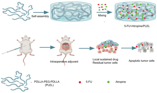
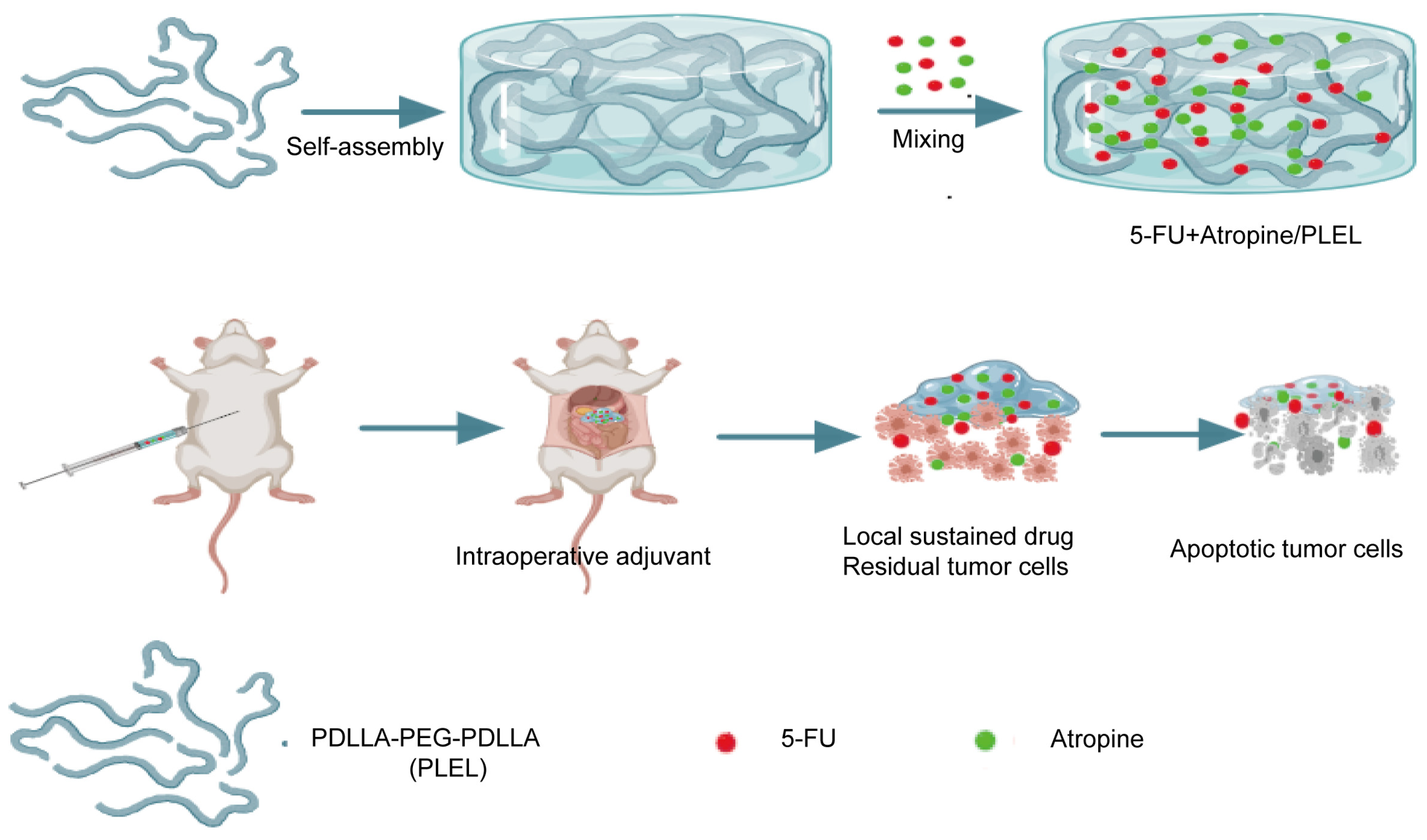
PDLLA-PEG-PDLLA, a biodegradable thermosensitive hydrogel, shows notable antitumor efficacy when co-delivering 5-FU and cisplatin for adjuvant GC chemotherapy [18]. Building on this, we developed a similar hydrogel system co-loaded with 5-FU and atropine for combined intraoperative GC treatment (Scheme 1). We easily prepared the 5-FU + atropine/PLEL hydrogel by physical mixing at room temperature, demonstrating high feasibility and operational flexibility. The hydrogel undergoes sol–gel transition at body temperature, allowing easy application over the tumor site, adjacent tissues, and abdominal cavity to target residual cancer cells. After gelation, the hydrogel provides sustained drug release, prolongs local drug retention, and suppresses postoperative recurrence and metastasis. We further evaluated the antitumor effect and reduced systemic toxicity of this system in a nude mouse model.
Scheme 1.
Schematic illustration of sustained co-delivery of 5-FU and atropine using a biodegradable thermosensitive hydrogel to enhance the antitumor effect of local drug administration in gastric cancer.
2. Materials and Methods
2.1. Samples and Patients
We conducted this study at Ganzhou People’s Hospital and collected gastric tissue samples from 28 patients undergoing radical gastrectomy for GC. The Ethics Committee of Ganzhou People’s Hospital approved the study and granted a full waiver of informed consent for the human tissue study (Approval No. PJB2025-316-01). The study was conducted in accordance with the ethical standards of the Declaration of Helsinki.
2.2. Cell Culture and Reagents
We obtained the GC cell lines MGC803 (RRID:CVCL_5334) and HGC27 (RRID:CVCL_1279) from the American Type Culture Collection (ATCC) in January 2022. After receipt, we expanded and cryopreserved the cell lines to create a master cell bank. We performed all experiments using low-passage cells (passages 5–15) from this bank. We routinely tested cell lines and confirmed they were free of mycoplasma contamination using a MycoAlert™ Mycoplasma Detection Kit (Lonza Rockland, Inc., Rockland, ME, USA, LT07). We cultured GC cells in RPMI-1640 medium (Gibco, Grand Island, NY, USA; Cat. No. C11875500BT) supplemented with 10% fetal bovine serum (FBS; Gibco, Grand Island, NY, USA) in a humidified incubator at 37 °C with 5% CO2. We purchased acetylcholine from Sigma-Aldrich (St. Louis, MO, USA), atropine from MedChemExpress (Monmouth Junction, NJ, USA; Cat. No. HY-B1205), and 5-fluorouracil from MedChemExpress (Monmouth Junction, NJ, USA; Cat. No. HY-90006).
2.3. CCK-8 Assay
We seeded cells into 96-well plates at 1000 cells per well and cultured them in RPMI 1640 medium with 10% FBS for 5 days. We assessed cell proliferation using the Cell Counting Kit-8 (CCK8; Beyotime, Shanghai, China; Cat. No. C0041). Briefly, we added 10 μL of CCK8 solution to each well, incubated the plates at 37 °C for 2 h, and then measured absorbance at 450 nm. We performed all experiments in triplicate.
2.4. Plate Clone Formation Assay
We plated cells in 6-well plates at 100 cells per well and cultured them in RPMI 1640 medium with 10% FBS. We allowed visible colonies to form over 12 days. Then we washed the colonies with phosphate-buffered saline (PBS), fixed them in 4% paraformaldehyde for 15 min, and stained them with 0.1% crystal violet for 15 min. We performed all experiments in triplicate.
2.5. Wound Healing Assay
We seeded cells into 6-well plates at 5 × 10⁵ cells per well and cultured them until they reached 90% confluence. We then replaced the complete medium with serum-free medium and used a sterile 100 μL pipette tip to scratch the cell monolayer. After further incubation for 48 h, we observed the wound gaps under an inverted light microscope at 100× magnification. We calculated the wound healing percentage using ImageJ 1.53 software: (healed wound area)/(initial wound area) × 100%.
2.6. Immunohistochemistry
We cut paraffin-embedded specimens into 4 μm slices. We dewaxed the slices with xylene, rehydrated them with alcohol, and then immersed them in EDTA antigen retrieval buffer, followed by microwave-assisted antigen retrieval. To inhibit endogenous peroxidase activity, we treated the slices with endogenous peroxidase block (Beyotime, Shanghai, China; Cat. No. P0100B) and then blocked nonspecific binding using immunostaining blocking solution (Beyotime, Shanghai, China; Cat. No. P0260). We incubated antibodies against M3R and Ki67 (Abcam, Cambridge, UK; Cat. Nos. ab314013, ab15580; diluted 1:500) with the slices overnight at 4 °C. After incubating with secondary antibodies, we stained the markers with DAB (ZSGBbio, Beijing, China; Cat. No. ZLI-9018) and counterstained the nuclei with hematoxylin. We captured images using an LSM980 (ZEISS) confocal microscope(Carl Zeiss Microscopy GmbH, Jena, Germany).
2.7. Hematoxylin and Eosin (H&E) Staining
We fixed samples in 10% formalin, cut them into 4–5 μm sections, and adjusted the pH. We stained sections sequentially with hematoxylin and eosin (Solarbio, Beijing, China; Cat. No. G1120). After staining, we dehydrated the sections through alcohol, cleared them in xylene, and mounted them using neutral resin. We captured images using the LSM980 confocal microscope (ZEISS).
2.8. Synthesis and Characterization of the Hydrogel
2.8.1. Synthesis of 5-FU + Atropine/PLEL Hydrogel
We purchased the PDLLA-PEG-PDLLA copolymer from Xi’an Ruixi Biotechnology Co., Ltd. (Xi’an, China). Following the manufacturer’s instructions, we fully dissolved the copolymer in sterile water at a ratio of 1:4 at room temperature. After the hydrogel completely dissolved into a solution, we physically mixed 5-FU and atropine with the hydrogel solution at 25 °C to prepare the 5-FU + atropine/PLEL formulation. We kept the 5-FU concentration at 2 mg mL−1 and the atropine concentration at 0.1 mg mL−1.
2.8.2. Characterization of Hydrogels
Polymer nuclear magnetic resonance (1H NMR): We dissolved a specific amount of the polymer in deuterochloroform (CDCl3) and analyzed the solution using a DMX 300 nuclear magnetic resonance spectrometer(Bruker BioSpin GmbH, Rheinstetten, Germany).
Gel permeation chromatography (GPC): We uniformly dissolved the samples in chloroform (CHCl3) for detection.
2.8.3. Sol–Gel Phase Transition of PDLLA-PEG-PDLLA Hydrogel
We observed the thermosensitive sol–gel phase transition behavior of 5-FU + atropine/PLEL hydrogel using the tube inversion method. Specifically, we added 1 mL of the 5-FU + atropine/PLEL hydrogel sample to a 4 mL Eppendorf tube and heated it to 37 °C. We recorded changes in hydrogel state at room temperature and under heated conditions. We also recorded the phase transition behavior of the blank hydrogel at room temperature and 37 °C for comparison.
2.8.4. Rheological Properties of PDLLA-PEG-PDLLA Hydrogels
We prepared PDLLA-PEG-PDLLA hydrogels by dissolving the copolymer in PBS at pH 7.4, with thorough stirring at room temperature to ensure complete dissolution. We evaluated the rheological properties of both blank and drug-loaded hydrogels using a NETZSCH Kinexus dynamic rheometer (NETZSCH-Gerätebau GmbH, Selb, Germany).
2.8.5. Assessment of In Vivo Antitumor Efficacy of 5-FU + Atropine/PLEL Hydrogel
To establish an orthotopic tumor model in BALB/c nude mice, we injected approximately 40 μL of HGC27 GC cells (containing 2 × 10⁶ cells) into the subserosa of the anterior wall of the gastric antrum of 6-week-old nude mice. After successful model establishment, we randomly divided the tumor-bearing mice into six groups (n = 3 per group). We then intraperitoneally injected the mice with 200 μL of various solutions: normal saline (NS), PLEL solution, free 5-FU plus atropine (administered separately), atropine/PLEL hydrogel, 5-FU/PLEL hydrogel, and 5-FU + atropine/PLEL hydrogel. We maintained the doses of 5-FU [18] and atropine [30] at 20 mg/kg and 1 mg/kg, respectively, and administered them once every 6 days for a total of two doses. We recorded the body weight of each mouse every other day throughout the experiment.
On day 14 after treatment, we euthanized the mice and performed laparotomy to remove tumors and major organs. We fixed the resected tissues in 4% phosphate-buffered formalin for subsequent histological and immunohistochemical analyses. We calculated tumor volumes using the modified ellipsoidal formula: (length × width2)/2. All animal experiments followed the NIH Guide for the Care and Use of Laboratory Animals and were approved by the Ethics Committee of Ganzhou People’s Hospital (Approval No. PJD2025-045-01; date of approval: 1 September 2025).
2.9. Statistical Analysis
We present all data as mean ± standard deviation (SD). We performed statistical comparisons between groups using the t-test with GraphPad Prism 11.1.0 (GraphPad Software, San Diego, CA, USA). We considered statistical significance at * p < 0.05, ** p < 0.01, and *** p < 0.001.
3. Results
3.1. M3R Expression Is Upregulated in GC Tissues
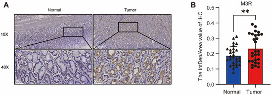
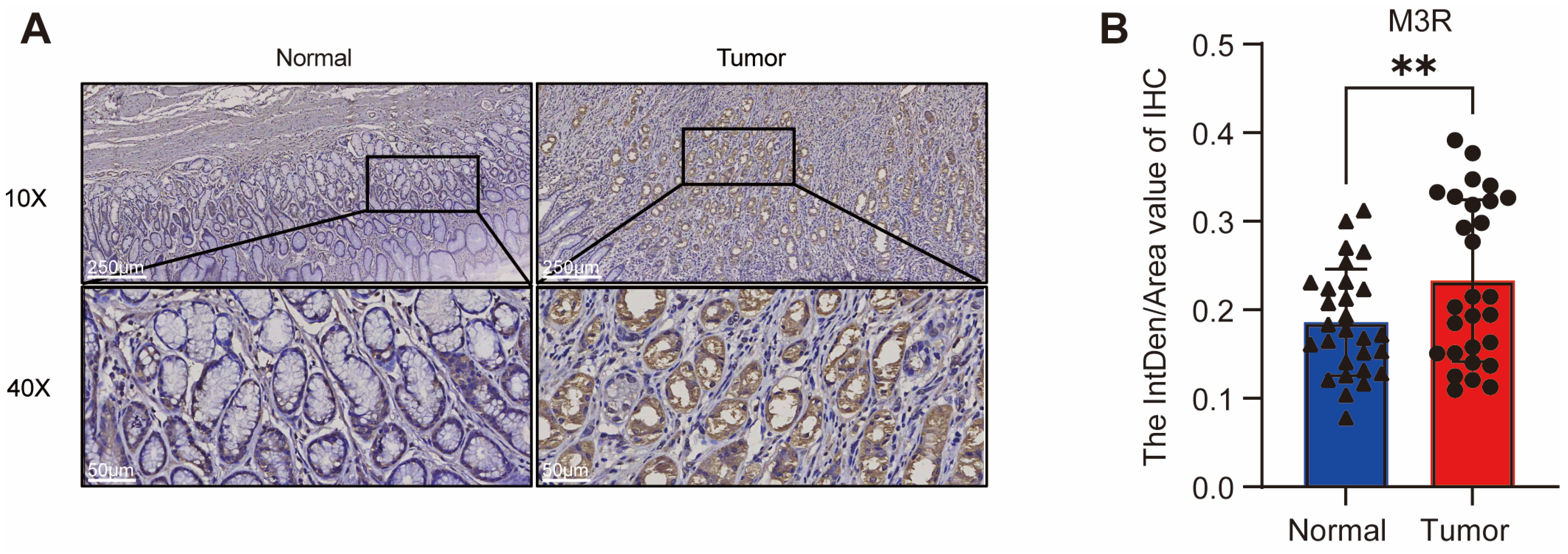
We examined M3R expression levels in 28 GC tissues and their corresponding normal adjacent tissues. The results showed significantly higher M3R expression in GC tissues than in normal tissues (Figure 1A,B). This finding suggests a potential role for M3R in gastric carcinogenesis.
Figure 1.
M3R is upregulated in GC tissues. (A) Representative IHC images of M3R in human GC tissues and adjacent noncancerous tissues. (B) Relative expression of M3R in 28 pairs of GC and adjacent normal tissues (paired t-test). Blue bars, adjacent normal tissues; red bars, GC tissues. ** p < 0.01.
3.2. An Exogenous Muscarinic Receptor Agonist Enhances Proliferation and Migration of HGC27 and MGC803 GC Cells
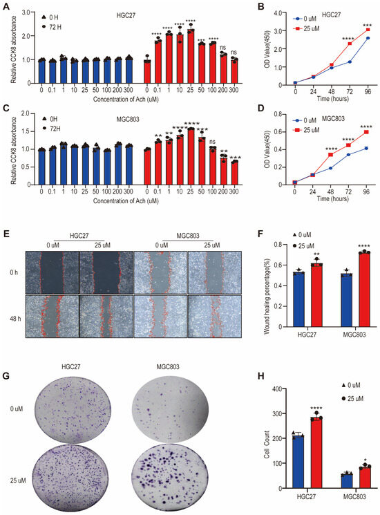
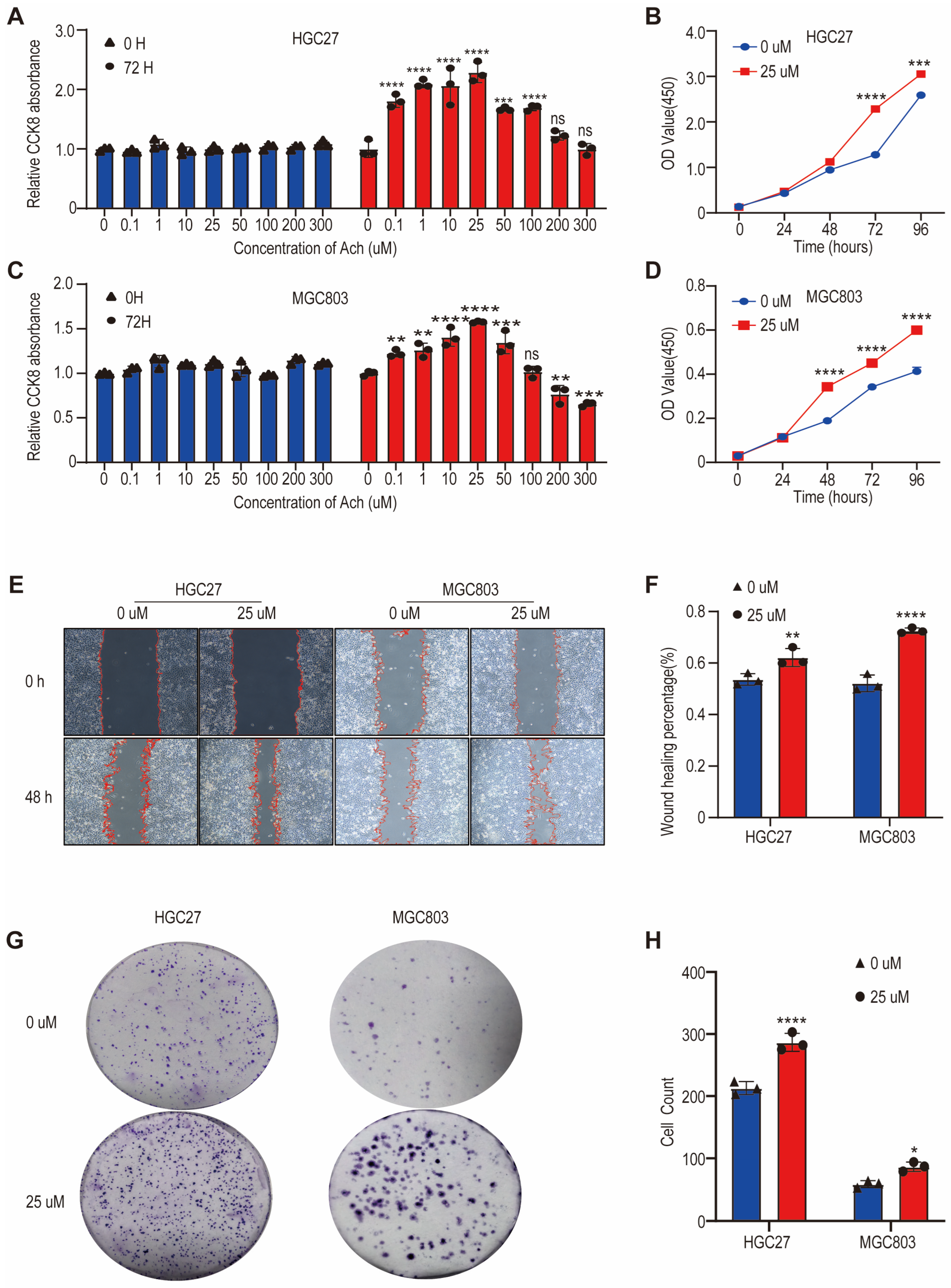
To clarify the functional role of M3R in GC cells, we selected two human GC cell lines, HGC27 and MGC803, both known to express high M3R levels. We treated these cells with different concentrations of exogenous ACh to determine the optimal stimulatory concentration. Our data showed that 25 μM ACh produced the most pronounced effect on cell proliferation (Figure 2A,C). Using this concentration, we observed a time-dependent increase in the growth rate of both HGC27 and MGC803 cells in CCK8 assays (Figure 2B,D). Wound healing assays confirmed that 25 μM ACh stimulation accelerated wound closure, indicating enhanced migration (Figure 2E,F). Plate clone formation assays also showed that 25 μM ACh for 12 days significantly promoted the formation of larger and more numerous colonies in both cell lines (Figure 2G,H). Together, these results demonstrate that exogenous ACh increases proliferation and migration of HGC27 and MGC803 GC cells.
Figure 2.
Effect of ACh on proliferation and migration of MGC803 and HGC27 cells. (A,C) HGC27 and MGC803 cells incubated with different ACh concentrations (0–300 μM) for 3 days; proliferation evaluated by CCK8 assay. (B,D) HGC27 and MGC803 cells incubated with 25 μM ACh for different days; proliferation evaluated by CCK8 assay. (E,F) Wound healing assay of HGC27 and MGC803 cells induced by 25 μM ACh. (G,H) Colony forming capacity of HGC27 and MGC803 cells induced by 25 μM ACh. In (A,C), blue bars, 0 h; red bars, 72 h. In (B,D), blue lines, 0 μM ACh; red lines, 25 μM ACh. In (E), red lines indicate wound edges. In (F,H), blue bars, 0 μM ACh; red bars, 25 μM ACh. ns, not significant. * p < 0.05, ** p < 0.01, *** p < 0.001, **** p < 0.0001.
3.3. Preparation and Characterization of 5-FU + Atropine/PLEL Hydrogel
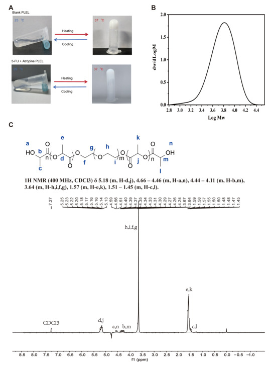
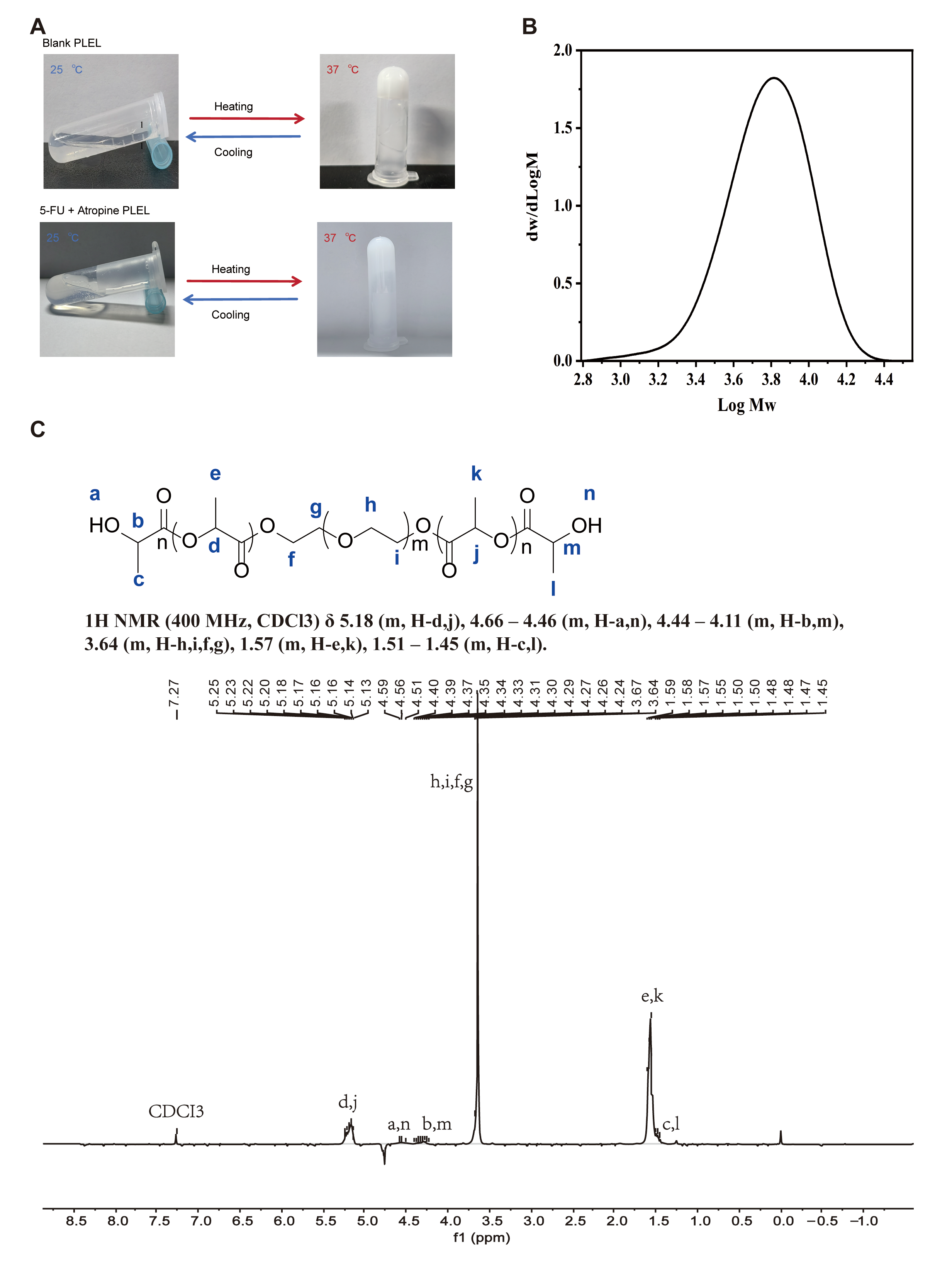
We thoroughly dissolved the PDLLA-PEG-PDLLA polymer in sterile water at room temperature at a polymer-to-water ratio of 1:4. We analyzed the polymer’s structural characteristics and molecular weight by 1H NMR spectroscopy (Figure 3C) and GPC (Figure 3B), respectively. The GPC analysis showed a unimodal molecular weight distribution with Mn = 5207 g/mol, Mw = 6764 g/mol, and PDI = 1.30 (Mw/Mn = 1.299). We then prepared the 5-FU + atropine/PLEL formulation by simple physical mixing of 5-FU and atropine with the PLEL micelle solution at 25 °C, keeping 5-FU at 2 mg mL−1 and atropine at 0.1 mg mL−1.
Figure 3.
Characterization of PDLLA-PEG-PDLLA hydrogels. (A) Reversible sol–gel phase transition of blank PLEL hydrogel and 5-FU + atropine/PLEL hydrogel (polymer concentration: 20 wt%; 5-FU: 2 mg/mL; atropine: 0.1 mg/mL) between 25 and 37 °C. (B) Molecular weight distribution of PDLLA-PEG-PDLLA triblock copolymer. (C) 1H-NMR spectrum of PDLLA-PEG-PDLLA copolymer. The letters a–n in (C) indicate the proton assignments corresponding to the chemical structure of the copolymer; the detailed peak assignments are listed in the text.
A critical property of PLEL hydrogels as drug carriers is their ability to undergo sol–gel transition at physiological temperatures. To evaluate this capability, we used the tube inversion method. The system remained a flowable sol at 25 °C, which facilitates drug loading and injection. As temperature increased, PLEL micelles enlarged, leading to aggregation and bridging, and finally forming a semi-solid hydrogel at 37 °C (Figure 3A). These findings indicate that PLEL hydrogels are injectable and can gel in vivo, making them promising drug carriers. Their sol–gel transition at physiological temperature not only enables easy administration but also ensures stable drug delivery after injection.
3.4. In Vivo Antitumor Efficacy and Toxicity of 5-FU + Atropine/PLEL Hydrogel
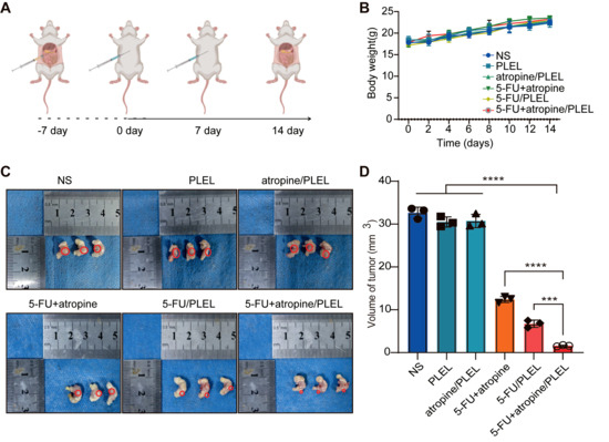
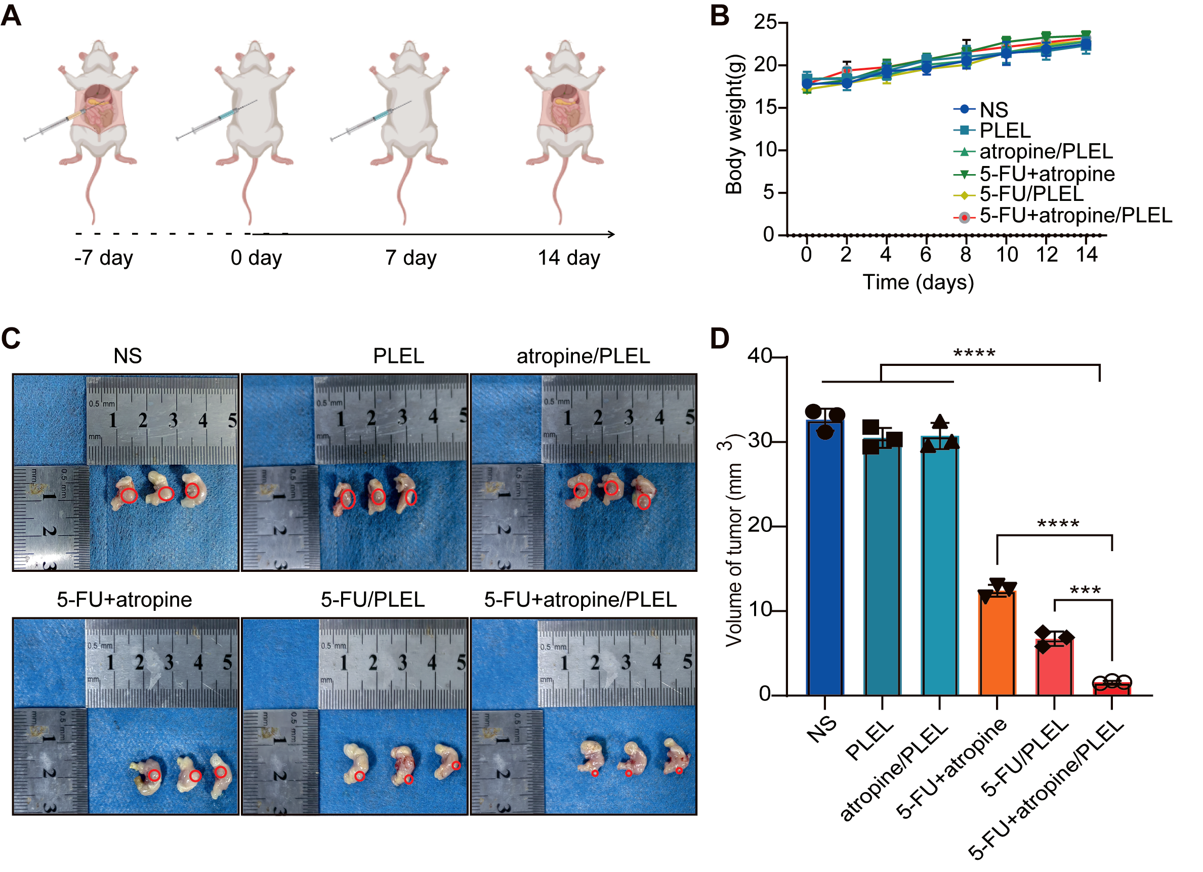
To further investigate the combined antitumor effects of 5-FU + atropine/PLEL hydrogel, we conducted animal experiments using a nude mouse orthotopic GC model. After tumor establishment, we intraperitoneally injected the hydrogels once every 6 days for two doses. We monitored body weights every 2 days until day 14, when we euthanized the mice for data collection (Figure 4A). Mice treated with drug-loaded hydrogels maintained stable body weights, indicating minimal systemic toxicity, in contrast to mice treated with free drug combinations (Figure 4B). Mice treated with NS, PLEL, or atropine/PLEL showed similar tumor volumes, whereas mice treated with 5-FU-containing hydrogels showed various degrees of tumor regression. Among these, the 5-FU + atropine/PLEL group had the strongest antitumor effect, with statistically significant differences compared with other groups (p < 0.05; Figure 4C,D).
Figure 4.
In vivo antitumor effect of different treatments in GC models. (A) Schematic diagram of GC model establishment in nude mice. (B) Body weight changes after drug administration. Data are mean ± SD (n = 3). (C) Tumor images after different treatments. Red circles indicate tumor tissues. (D) Tumor size in different groups. Data are mean ± SD (n = 3). In (B,D), colors correspond to the treatment groups as indicated: NS, PLEL, atropine/PLEL, 5-FU + atropine, 5-FU/PLEL, and 5-FU + atropine/PLEL. *** p < 0.001, **** p < 0.0001.
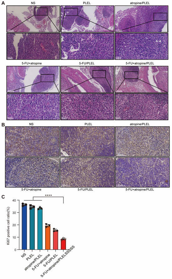
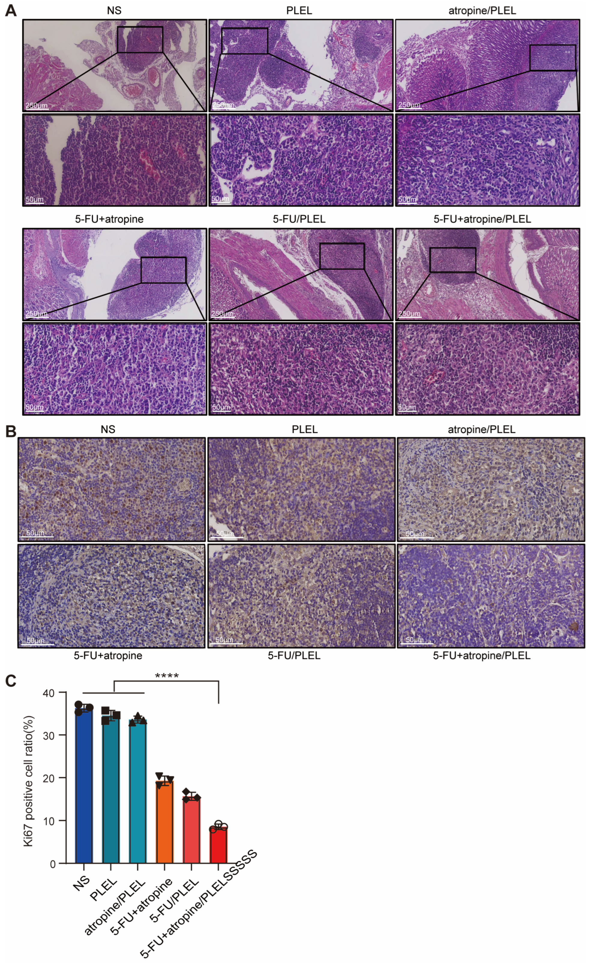
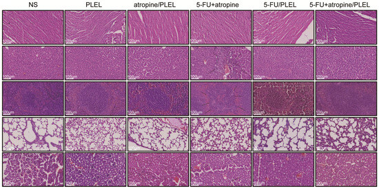
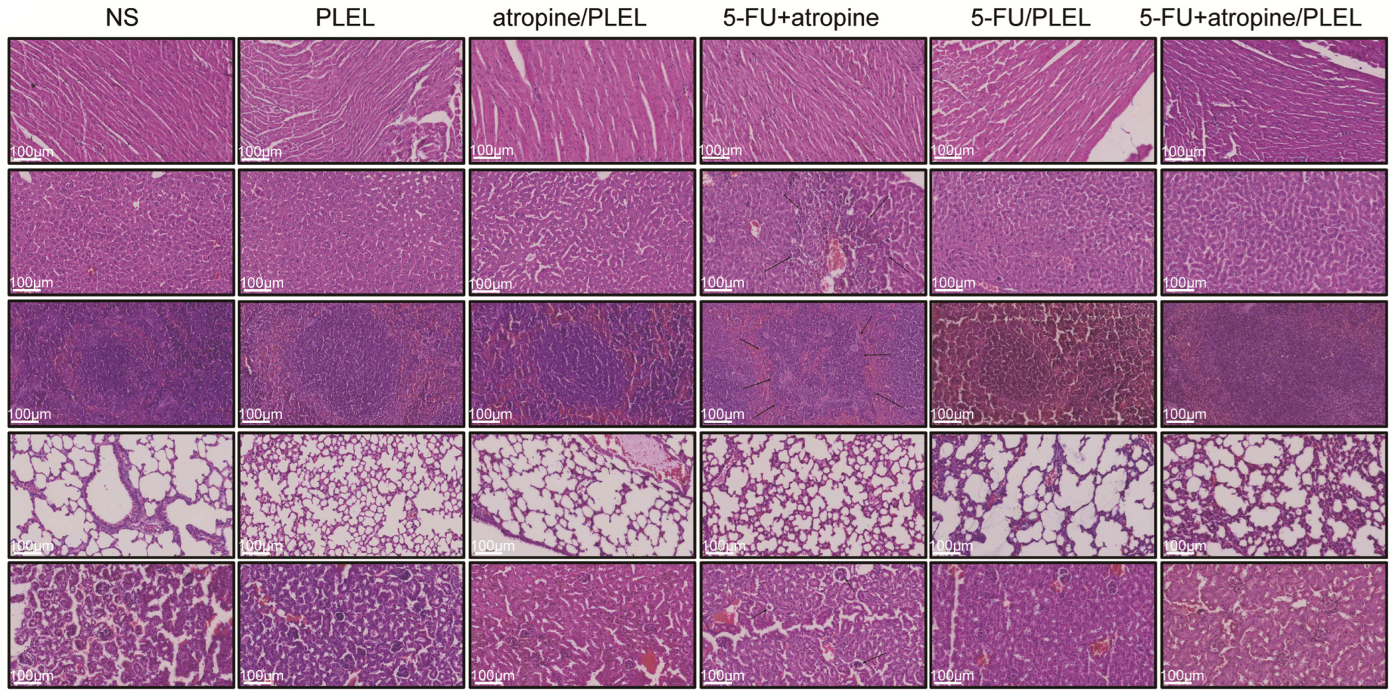
To verify successful establishment of orthotopic gastric tumors, we performed H&E staining of mouse tumor tissue (Figure 5A). Importantly, Ki-67 staining revealed a much lower percentage of positive cells in the 5-FU + atropine/PLEL hydrogel group, indicating a substantial reduction in tumor cell proliferation (Figure 5B,C). Histological analysis confirmed these findings: mice treated with the hydrogel system showed minimal organ damage, whereas mice receiving free 5-FU and atropine displayed significant hepatocellular necrosis, disrupted splenic architecture, and glomerular atrophy (Figure 6). Thus, the 5-FU + atropine/PLEL hydrogel not only enhances the antitumor efficacy of combined GC chemotherapy but also effectively reduces systemic toxicity, making it a promising therapeutic option.
Figure 5.
Tumorigenesis validation and antitumor efficacy of different therapeutic regimens in mice. (A) H&E staining images of mouse tumors (scale bar: 250 μm) and magnification (scale bar: 50 μm). (B) Ki-67 assay images of mouse tumor tissues from different treatments (scale bar: 50 μm). (C) Percentage of Ki67-positive cells after treatment. Data are mean ± SD (n = 3). In (C), colors correspond to the treatment groups as in Figure 4. **** p < 0.0001.
Figure 6.
Safety assessment of the dual-drug loaded hydrogel antitumor regimen. H&E staining of major organs after different treatments. Black arrows indicate tissue damage.
4. Discussion
Nerves are an important part of the tumor microenvironment and interact with multiple TME components to promote tumor development [31,32]. Our preliminary findings showed significantly worse outcomes in GC patients with PNI than in those without, highlighting the clinical relevance of this pathological feature [33]. Other studies agree that denervation can curb tumor progression, and when combined with chemotherapy, it reduces tumor recurrence and improves patient prognosis [9]. Currently, chemotherapy remains a cornerstone of cancer treatment; however, systemic toxicity and side effects from intravenous administration often limit its use [17]. Localized chemotherapy using drug-loaded biomaterials offers a promising clinical strategy to reduce systemic toxicity [34]. In this study, we developed a temperature-sensitive hydrogel delivery system loaded with 5-FU and atropine for local GC chemotherapy and evaluated its antitumor efficacy.
We observed higher M3R expression in GC tissues than in normal tissues. The vagus nerve releases ACh, which binds to M3R on target cells and modulates tumor progression. Previous studies have shown that ACh in tumor tissues promotes cancer cell proliferation and migration [35]. Our in vitro experiments confirmed that the exogenous M3R agonist ACh significantly increased proliferation and migration of HGC27 and MGC803 GC cell lines. These findings align with previous reports on M3R involvement in various cancers, emphasizing the broad relevance of M3R in cancer biology.
The vagus nerve influences tumor cell proliferation, migration, and invasion by releasing neurotransmitters such as ACh. Atropine, as a vagal nerve blocker, interrupts this signaling pathway and inhibits malignant tumor cell behavior [30]. Meanwhile, 5-FU, a classic antitumor drug, inhibits tumor growth by interfering with DNA synthesis and cell division [36]. The combination of these two agents both blocks tumor cell growth signals and directly disrupts proliferative capacity, producing a potent synergistic antitumor effect. In the 5-FU + atropine/PLEL hydrogel treatment group, the marked reduction in Ki-67-positive cells and its downregulation indicate effective suppression of tumor growth. This finding further supports targeting M3R together with chemotherapy for GC treatment.
Hydrogels serve as excellent drug delivery systems and are widely used for antitumor therapy across various tumor types. Their thermosensitive properties and sustained drug release ensure prolonged drug action at tumor sites, thereby improving therapeutic efficacy. Beyond drug delivery, hydrogels have also been explored as intelligent biosensing platforms. For instance, Yao et al. recently developed size-selected DNA hydrogels for efficient intracellular delivery and sensitive mRNA imaging, highlighting the versatility of hydrogel-based systems in biomedical applications [37]. In our study, the 5-FU + atropine/PLEL group showed superior antitumor effects compared with the free 5-FU + atropine group. Moreover, intraperitoneal injection of the drug-loaded hydrogels achieved localized and precise drug release at the tumor site, minimizing systemic toxicity and adverse effects [38]. We observed that nearly all major organs in the hydrogel-treated groups showed histological morphologies similar to those of mice in the NS control group, indicating low systemic toxicity. In contrast, mice treated with free 5-FU showed obvious hepatocyte necrosis, indistinct boundaries between splenic cortex and medulla, and glomerular atrophy. Therefore, the 5-FU + atropine/PLEL hydrogel not only improves the antitumor efficacy of combined GC chemotherapy but also effectively reduces systemic toxicity.
We acknowledge several limitations in this study. Although we have observed that adding a vagal nerve blocker enhances the antitumor efficacy of 5-FU, the precise mechanism underlying the antitumor effect after blocking acetylcholine receptors requires further investigation.
5. Conclusions
Our study demonstrates that M3R is a promising therapeutic target in GC. Inhibiting M3R together with chemotherapy using 5-FU/PLEL hydrogels represents a novel and effective treatment strategy. The local and sustained release of drugs from the hydrogel minimizes systemic toxicity while maximizing antitumor effects. Future studies should focus on optimizing the hydrogel formulation and exploring its clinical potential in larger cohorts of GC patients.
Author Contributions
Conceptualization, T.C., C.F. and X.L. (Xiaoliang Lan); Methodology, C.W., X.L. (Xunjun Li) and Y.L.; Investigation, C.W., X.L. (Xunjun Li), Y.L., W.Z., H.L. and J.Y.; Formal Analysis, C.W., X.L. (Xunjun Li) and S.D.; Data Curation, C.W., X.L. (Xunjun Li), Y.L., W.Z., H.L. and J.Y.; Validation, W.Z., H.L. and J.Y.; Visualization, C.W., X.L. (Xunjun Li) and Y.L.; Writing—Original Draft Preparation, C.W. and X.L. (Xunjun Li); Writing—Review & Editing, T.C., C.F., X.L. (Xiaoliang Lan), S.D., Y.L., W.Z., H.L. and J.Y.; Supervision, T.C., C.F., X.L. (Xiaoliang Lan) and S.D.; Project Administration, T.C. and C.W.; Funding Acquisition, T.C., C.F. and C.W. All authors have read and agreed to the published version of the manuscript.
Funding
This work was supported by the National Natural Science Foundation of China (82472066 and 82272087), the Natural Science Foundation of Jiangxi Province (20242BAB26153), the Jiangxi Provincial Health Commission Science and Technology Project (202410879), Ganzhou Municipal Science and Technology Project (2023LNS17474), and the Ganzhou Municipal Science and Technology + Medical Joint Projects (2025YLCE001 and 2025YLCE073).
Institutional Review Board Statement
The study was conducted in accordance with the Declaration of Helsinki and approved by the Ethics Committee of Ganzhou People’s Hospital (Approval No. PJB2025-316-01; date of approval: 1 September 2025). Written informed consent was obtained from all patients involved in the study. All animal experiments followed the NIH Guide for the Care and Use of Laboratory Animals and were approved by the Ethics Committee of Ganzhou People’s Hospital (Approval No. PJD2025-045-01; date of approval: 1 September 2025).
Informed Consent Statement
Informed consent was waived by the Ethics Committee of Ganzhou People’s Hospital for the human tissue study (Approval No. PJB2025-316-01). No blank informed consent form was used. The Waiver of Informed Consent Application Form and the relevant ethics approval have been provided.
Data Availability Statement
The original contributions presented in the study are included in the article, further inquiries can be directed to the corresponding author.
Conflicts of Interest
The authors declare no conflicts of interest.
References
- Bray, F.; Laversanne, M.; Sung, H.; Ferlay, J.; Siegel, R.L.; Soerjomataram, I.; Jemal, A. Global cancer statistics 2022: GLOBOCAN estimates of incidence and mortality worldwide for 36 cancers in 185 countries. CA Cancer J. Clin. 2024, 74, 229–263. [Google Scholar] [CrossRef] [Scilit] [PubMed]
- Yonemura, Y.; Bandou, E.; Kawamura, T.; Endou, Y.; Sasaki, T. Quantitative prognostic indicators of peritoneal dissemination of gastric cancer. Eur. J. Surg. Oncol. 2006, 32, 602–606. [Google Scholar] [CrossRef] [Scilit] [PubMed]
- Yang, L.; Zheng, R.; Wang, N.; Yuan, Y.; Liu, S.; Li, H.; Zhang, S.; Zeng, H.; Chen, W. Incidence and mortality of stomach cancer in China, 2014. Chin. J. Cancer Res. 2018, 30, 291–298. [Google Scholar] [CrossRef] [Scilit] [PubMed]
- Gysler, S.M.; Drapkin, R. Tumor innervation: Peripheral nerves take control of the tumor microenvironment. J. Clin. Investig. 2021, 131, e147276. [Google Scholar] [CrossRef] [Scilit] [PubMed]
- Chen, S.-H.; Zhang, B.-Y.; Zhou, B.; Zhu, C.-Z.; Sun, L.-Q.; Feng, Y.-J. Perineural invasion of cancer: A complex crosstalk between cells and molecules in the perineural niche. Am. J. Cancer Res. 2019, 9, 1–21. [Google Scholar] [PubMed]
- Liebig, C.; Ayala, G.; Wilks, J.; Verstovsek, G.; Liu, H.; Agarwal, N.; Berger, D.H.; Albo, D. Perineural invasion is an independent predictor of outcome in colorectal cancer. J. Clin. Oncol. 2009, 27, 5131–5137. [Google Scholar] [CrossRef] [Scilit] [PubMed]
- Xu, G.; Feng, F.; Liu, Z.; Liu, S.; Zheng, G.; Xiao, S.; Cai, L.; Yang, X.; Li, G.; Lian, X.; et al. Prognosis and Progression of ESCC Patients with Perineural Invasion. Sci. Rep. 2017, 7, 43828. [Google Scholar] [CrossRef] [Scilit] [PubMed]
- Deng, J.; You, Q.; Gao, Y.; Yu, Q.; Zhao, P.; Zheng, Y.; Fang, W.; Xu, N.; Teng, L. Prognostic value of perineural invasion in gastric cancer: A systematic review and meta-analysis. PLoS ONE 2014, 9, e88907. [Google Scholar] [CrossRef] [Scilit] [PubMed]
- Zhao, C.-M.; Hayakawa, Y.; Kodama, Y.; Muthupalani, S.; Westphalen, C.B.; Andersen, G.T.; Flatberg, A.; Johannessen, H.; Friedman, R.A.; Renz, B.W.; et al. Denervation suppresses gastric tumorigenesis. Sci. Transl. Med. 2014, 6, 250ra115. [Google Scholar] [CrossRef] [Scilit] [PubMed]
- Hutchings, C.; Phillips, J.A.; Djamgoz, M. Nerve input to tumours: Pathophysiological consequences of a dynamic relationship. Biochim Biophys. Acta Rev. Cancer 2020, 1874, 188411. [Google Scholar] [CrossRef] [Scilit] [PubMed]
- Reijmen, E.; Vannucci, L.; De Couck, M.; De Greve, J.; Gidron, Y. Therapeutic potential of the vagus nerve in cancer. Immunol. Lett. 2018, 202, 38–43. [Google Scholar] [CrossRef] [Scilit] [PubMed]
- Ge, Y.; Wang, X.; Guo, Y.; Yan, J.; Abuduwaili, A.; Aximujiang, K.; Yan, J.; Wu, M. Gut microbiota influence tumor development and Alter interactions with the human immune system. J. Exp. Clin. Cancer Res. 2021, 40, 42. [Google Scholar] [CrossRef] [Scilit] [PubMed]
- Vaes, N.; Idris, M.; Boesmans, W.; Alves, M.M.; Melotte, V. Nerves in gastrointestinal cancer: From mechanism to modulations. Nat. Rev. Gastroenterol. Hepatol. 2022, 19, 768–784. [Google Scholar] [CrossRef] [Scilit] [PubMed]
- Schledwitz, A.; Sundel, M.H.; Alizadeh, M.; Hu, S.; Xie, G.; Raufman, J.-P. Differential Actions of Muscarinic Receptor Subtypes in Gastric, Pancreatic, and Colon Cancer. Int. J. Mol. Sci. 2021, 22, 13153. [Google Scholar] [CrossRef] [Scilit] [PubMed]
- Wang, L.; Zhi, X.; Zhang, Q.; Wei, S.; Li, Z.; Zhou, J.; Jiang, J.; Zhu, Y.; Yang, L.; Xu, H.; et al. Muscarinic receptor M3 mediates cell proliferation induced by acetylcholine and contributes to apoptosis in gastric cancer. Tumour Biol. 2016, 37, 2105–2117. [Google Scholar] [CrossRef] [Scilit] [PubMed]
- Yu, H.; Xia, H.; Tang, Q.; Xu, H.; Wei, G.; Chen, Y.; Dai, X.; Gong, Q.; Bi, F. Acetylcholine acts through M3 muscarinic receptor to activate the EGFR signaling and promotes gastric cancer cell proliferation. Sci. Rep. 2017, 7, 40802. [Google Scholar] [CrossRef] [Scilit] [PubMed]
- Smith, P.; Lavery, A.; Turkington, R.C. An overview of acute gastrointestinal side effects of systemic anti-cancer therapy and their management. Best Pract. Res. Clin. Gastroenterol. 2020, 48–49, 101691. [Google Scholar] [CrossRef] [Scilit] [PubMed]
- Chen, W.; Shi, K.; Liu, J.; Yang, P.; Han, R.; Pan, M.; Yuan, L.; Fang, C.; Yu, Y.; Qian, Z. Sustained co-delivery of 5-fluorouracil and cis-platinum via biodegradable thermo-sensitive hydrogel for intraoperative synergistic combination chemotherapy of gastric cancer. Bioact. Mater. 2023, 23, 1–15. [Google Scholar] [CrossRef] [Scilit] [PubMed]
- Lee, J.H.; Tachibana, T.; Yamana, K.; Kawasaki, R.; Yabuki, A. Simple Formation of Cancer Drug-Containing Self-Assembled Hydrogels with Temperature and pH-Responsive Release. Langmuir 2021, 37, 11269–11275. [Google Scholar] [CrossRef] [Scilit] [PubMed]
- Chen, Y.; Yang, C.; Mao, J.; Li, H.; Ding, J.; Zhou, W. Spermine modified polymeric micelles with pH-sensitive drug release for targeted and enhanced antitumor therapy. RSC Adv. 2019, 9, 11026–11037. [Google Scholar] [CrossRef] [Scilit] [PubMed]
- Shatsberg, Z.; Zhang, X.; Ofek, P.; Malhotra, S.; Krivitsky, A.; Scomparin, A.; Tiram, G.; Calderón, M.; Haag, R.; Satchi-Fainaro, R. Functionalized nanogels carrying an anticancer microRNA for glioblastoma therapy. J. Control. Release 2016, 239, 159–168. [Google Scholar] [CrossRef] [Scilit] [PubMed]
- Li, N.; Lin, J.; Liu, C.; Zhang, Q.; Li, R.; Wang, C.; Zhao, C.; Lu, L.; Zhou, C.; Tian, J.; et al. Temperature- and pH-responsive injectable chitosan hydrogels loaded with doxorubicin and curcumin as long-lasting release platforms for the treatment of solid tumors. Front Bioeng. Biotechnol. 2022, 10, 1043939. [Google Scholar] [CrossRef] [Scilit] [PubMed]
- He, P.; Du, X.; Cheng, Y.; Gao, Q.; Liu, C.; Wang, X.; Wei, Y.; Yu, Q.; Guo, W. Thermal-Responsive MXene-DNA Hydrogel for Near-Infrared Light Triggered Localized Photothermal-Chemo Synergistic Cancer Therapy. Small 2022, 18, e2200263. [Google Scholar] [CrossRef] [Scilit] [PubMed]
- Zhao, J.; Peng, Y.-Y.; Wang, J.; Diaz-Dussan, D.; Tian, W.; Duan, W.; Kong, L.; Hao, X.; Narain, R. Temperature-Responsive Aldehyde Hydrogels with Injectable, Self-Healing, and Tunable Mechanical Properties. Biomacromolecules 2022, 23, 2552–2561. [Google Scholar] [CrossRef] [Scilit] [PubMed]
- Khan, S.; Akhtar, N.; Minhas, M.U.; Badshah, S.F. PH/Thermo-Dual Responsive Tunable In Situ Cross-Linkable Depot Injectable Hydrogels Based on Poly(N-Isopropylacrylamide)/Carboxymethyl Chitosan with Potential of Controlled Localized and Systemic Drug Delivery. AAPS PharmSciTech 2019, 20, 119. [Google Scholar] [CrossRef] [Scilit] [PubMed]
- Vu, T.T.; Jo, S.-H.; Kim, S.-H.; Kim, B.K.; Park, S.-H.; Lim, K.T. Injectable and Multifunctional Hydrogels Based on Poly(N-acryloyl glycinamide) and Alginate Derivatives for Antitumor Drug Delivery. ACS Appl. Mater. Interfaces 2024, 16, 15322–15335. [Google Scholar] [CrossRef] [Scilit] [PubMed]
- Mohaghegh, N.; Ahari, A.; Zehtabi, F.; Buttles, C.; Davani, S.; Hoang, H.; Tseng, K.; Zamanian, B.; Khosravi, S.; Daniali, A.; et al. Injectable hydrogels for personalized cancer immunotherapies. Acta Biomater. 2023, 172, 67–91. [Google Scholar] [CrossRef] [Scilit] [PubMed]
- Lv, Q.; He, C.; Quan, F.; Yu, S.; Chen, X. DOX/IL-2/IFN-gamma co-loaded thermo-sensitive polypeptide hydrogel for efficient melanoma treatment. Bioact. Mater. 2018, 3, 118–128. [Google Scholar] [CrossRef] [Scilit] [PubMed]
- Kim, J.; Francis, D.M.; Sestito, L.F.; Archer, P.A.; Manspeaker, M.P.; O’mElia, M.J.; Thomas, S.N. Thermosensitive hydrogel releasing nitric oxide donor and anti-CTLA-4 micelles for anti-tumor immunotherapy. Nat. Commun. 2022, 13, 1479. [Google Scholar] [CrossRef] [Scilit] [PubMed]
- Yang, M.W.; Tao, L.Y.; Jiang, Y.S.; Yang, J.Y.; Huo, Y.M.; Liu, D.J.; Li, J.; Fu, X.L.; He, R.; Lin, C.; et al. Perineural Invasion Reprograms the Immune Microenvironment through Cholinergic Signaling in Pancreatic Ductal Adenocarcinoma. Cancer Res. 2020, 80, 1991–2003. [Google Scholar] [CrossRef] [Scilit] [PubMed]
- Zahalka, A.H.; Frenette, P.S. Nerves in cancer. Nat. Rev. Cancer 2020, 20, 143–157. [Google Scholar] [CrossRef] [Scilit] [PubMed]
- Oya, Y.; Hayakawa, Y.; Koike, K. Tumor microenvironment in gastric cancers. Cancer Sci. 2020, 111, 2696–2707. [Google Scholar] [CrossRef] [Scilit] [PubMed]
- Li, X.; Wang, Y.; Zhai, Z.; Mao, Q.; Chen, D.; Xiao, L.; Xu, S.; Wu, Q.; Chen, K.; Hou, Q.; et al. Predicting response to immunotherapy in gastric cancer via assessing perineural invasion-mediated inflammation in tumor microenvironment. J. Exp. Clin. Cancer Res. 2023, 42, 206. [Google Scholar] [CrossRef] [Scilit] [PubMed]
- Liu, R.; Liang, Q.; Luo, J.; Li, Y.; Zhang, X.; Fan, K.; Du, J. Ferritin-Based Nanocomposite Hydrogel Promotes Tumor Penetration and Enhances Cancer Chemoimmunotherapy. Adv. Sci. 2024, 11, e2305217. [Google Scholar] [CrossRef] [Scilit] [PubMed]
- Kuol, N.; Davidson, M.; Karakkat, J.; Filippone, R.T.; Veale, M.; Luwor, R.; Fraser, S.; Apostolopoulos, V.; Nurgali, K. Blocking Muscarinic Receptor 3 Attenuates Tumor Growth and Decreases Immunosuppressive and Cholinergic Markers in an Orthotopic Mouse Model of Colorectal Cancer. Int. J. Mol. Sci. 2022, 24, 596. [Google Scholar] [CrossRef] [Scilit] [PubMed]
- Vodenkova, S.; Buchler, T.; Cervena, K.; Veskrnova, V.; Vodicka, P.; Vymetalkova, V. 5-fluorouracil and other fluoropyrimidines in colorectal cancer: Past, present and future. Pharmacol. Ther. 2020, 206, 107447. [Google Scholar] [CrossRef] [Scilit] [PubMed]
- Yao, S.; Wang, S.; Meng, Y.; Li, X.; He, Z.; Li, Y.; Chen, F.; Zhao, C. Size-selected DNA hydrogels-based sensor with efficient intracellular delivery for imaging mRNA. Talanta 2026, 298, 128945. [Google Scholar] [CrossRef] [Scilit] [PubMed]
- Ma, H.; He, C.; Chen, X. Injectable Hydrogels as Local Depots at Tumor Sites for Antitumor Immunotherapy and Immune-Based Combination Therapy. Macromol. Biosci. 2021, 21, e2100039. [Google Scholar] [CrossRef] [Scilit] [PubMed]
Disclaimer/Publisher’s Note: The statements, opinions and data contained in all publications are solely those of the individual author(s) and contributor(s) and not of MDPI and/or the editor(s). MDPI and/or the editor(s) disclaim responsibility for any injury to people or property resulting from any ideas, methods, instructions or products referred to in the content. |
© 2026 by the authors. Licensee MDPI, Basel, Switzerland. This article is an open access article distributed under the terms and conditions of the Creative Commons Attribution (CC BY) license.