


Abstract
Canine parvovirus 2 (CPV 2) is a highly contagious viral pathogen responsible for severe gastroenteritis in canine species worldwide. This study provides the first comprehensive molecular characterization of CPV 2 variants circulating in domestic dogs in Botswana. A total of 187 fecal samples from diarrheic dogs presented at veterinary clinics were tested for CPV 2 using an ELISA point-of-care test and molecular assays for confirmation. A CPV 2 prevalence of 62.0% was detected via ELISA, compared to 100% via qPCR. Genetic characterization using minor groove binder probe-based qPCR assays and sequencing of the VP2 gene revealed the predominance of the CPV 2c variant (99.1%), with the CPV 2a variant detected in one sample from Maun village. Amino acid sequence analysis of the VP2 gene identified a key mutation at position 426 (glutamic acid) characteristic of the CPV2c variants. Phylogenetic analysis showed clustering of the Botswana CPV 2c isolates with Nigerian, Ethiopian, Gabon, Egyptian, and Asian CPV2c variants, while the CPV 2a isolate formed a cluster with CPV 2a variants from Nigeria. These findings confirm the circulation of CPV2a and CPV2c variants in Botswana, with CPV2c as the predominant circulating variant. Continuous monitoring of emerging CPV2 variants is essential for effective disease control in Botswana.
1. Introduction
Canine parvovirus 2 emerged in the late 1970s as a highly contagious pathogen causing severe gastroenteritis in domestic dogs and wild Canidae populations worldwide [1]. The virus is classified as canine parvovirus 2 (CPV 2) to differentiate it from the less virulent canine parvovirus 1 (CPV1) [2]. Clinical signs of CPV 2 infection include anorexia, vomiting, hemorrhagic diarrhea and dehydration, and in severe cases, hypovolemic shock and death, particularly in young animals [3,4,5].
CPV 2 belongs to the Parvoviridae family and is 98% genomically similar to the Feline panleukopenia virus (FPLV) [2,6,7]. The virus has an approximately 5.3kb single-stranded DNA genome with open reading frames encoding two non-structural proteins (NSP1 and NSP2) involved in replication and three capsid proteins (VP1, VP2, and VP3) [8,9]. VP2 is the major antigenic determinant, constituting 90% of the viral capsid [10].
CPV 2 undergoes rapid mutation, with substitution rates comparable to RNA viruses [11], and this phenomenon resulted in the emergence of three antigenic variants over the years: CPV 2a, which emerged around 1979 with five key mutations (M87L, I101T, A300G, D305Y, and V555I) in the VP2 protein from the original CPV 2 [10,12,13]; CPV 2b, a new variant that emerged in 1980, with two additional amino acid changes in the VP2 protein (N426D and I555V); and CPV 2c, a variant found in 2000 with a single amino acid change (D426E), which was first reported in Italy [11,13]. Today, CPV 2a, 2b, and 2c are globally distributed, with variable prevalence across regions [5,14,15,16]. In 1990, the Ser297Ala mutation appeared in VP2 of CPV 2a and CPV 2b, and these were designated as new CPV 2a and new CPV 2b, respectively [17,18].
Vaccination remains the most effective strategy for CPV 2 control [19,20], with attenuated vaccines providing strong and long-lasting immunity. Despite widespread availability and use of these vaccines for CPV 2 prevention, CPV 2 cases in vaccinated dogs continue to be recorded globally, raising concerns about vaccine efficacy against emerging variants [21,22,23,24,25].
In Africa, a few studies have described the circulation of CPV 2 variants in countries such as Nigeria, South Africa, Namibia, Ethiopia, Zambia, Mozambique, Morocco, Tunisia, Egypt, and Gambia [8,26,27,28,29]. While serological evidence of CPV 2 has been documented in Ghana [30], Zimbabwe [31], Cape Verde [32] and Botswana [33], few studies have molecularly characterized the virus in these regions.
To date, limited information is available on the prevalence, distribution, and genetic characterization of CPV 2 variants circulating in Botswana. The current study presents the first comprehensive molecular analysis of CPV 2 variants in domestic dogs in Botswana. These findings aim to inform surveillance efforts, guide vaccine policy, and support improved control strategies for CPV 2 in Botswana and the Southern African region.
2. Materials and Methods
2.1. Study Design and Sample Collection
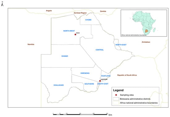
The study was conducted in Botswana and approved by the Bioethics Committee at the Faculty of Animal and Veterinary Science, Botswana University of Agriculture and Natural Resources (BUAN-AEC-2022-07). A total of eight veterinary clinics across Botswana were selected to take part in this study: seven of these clinics were in and around the capital city, Gaborone, situated in the southeastern part of the country, with a population of 250,000 inhabitants [34], while one veterinary clinic was located in Maun village in the northwestern part of the country, with a population of around 80,000 inhabitants [34], as indicated in Figure 1.
Figure 1.
Map of Botswana indicating veterinary clinics and sampling sites.
Dogs presenting with gastrointestinal disorders from participating veterinary clinics were enrolled in the study with consent from their owners. An initial clinical examination of individual cases was conducted at the respective study sites. Demographic dog data, including sex, age, breed, origin, vaccination history, and number of dogs per household, were obtained using a pre-designed form. Dogs presenting clinical symptoms of vomiting, watery or hemorrhagic diarrhea, dehydration, anorexia, and foul smell were exclusively selected by the admitting veterinary clinicians. These dogs (n = 187) were subjected to an initial screening test against CPV 2 using the antigen capture ELISA test (SNAP Parvo, IDEXX Laboratories, Westbrook, ME, USA) following the manufacturer’s instructions, and results were recorded using forms.
Fecal samples were collected from the rectum of each suspected case using sterile swabs and stored at −80 °C until shipped on dry ice to the Pennsylvania Animal Diagnostic Laboratory System, New Bolton Center, School of Veterinary Medicine, University of Pennsylvania, for further testing. Dogs sampled were classified as vaccinated (n = 52), partially vaccinated (n = 34), not vaccinated (n = 98), or of unknown vaccination status (n = 3). The partially vaccinated dogs were those that received a single dose and did not get subsequent booster shots. CPV 2 vaccines commonly used in Botswana are imported from South Africa, including Vanguard Plus 5 Zoetis, Santon, 2196, South Africa, Nobivac® DHPPi Intervet Pty LTD, Spartan, 1619, South Africa and Canigen DHPPi Virbac, SA Reg Number G2839, Santon, South Africa. Nobivac® Puppy DP PLUS, manufactured by Intervet Pty LTD, Spartan, 1619, South Africa, is also routinely used as primary vaccination shots for puppies against parvovirus.
2.2. DNA Extraction and Real-Time PCR
Fecal swabs were suspended in 1 mL of phosphate-buffered saline (PBS, pH 7.4) in 1.5 mL tubes and clarified by centrifugation at 4000× g for 5 min to collect the supernatant. DNA from the supernatant was extracted using QIAamp Fast DNA Stool Mini Kit (Qiagen, Germantown, TN, USA) following the manufacturer’s instructions. For quality control, VetMAX™ Xeno™ Internal Positive Control (IPC) DNA (Thermo Fisher, Waltham, MA, USA) was added to evaluate the extraction process and monitor the presence of any inhibitors. The extracted DNA was stored at −80 °C for further analysis.
All samples were subjected to real-time PCR (qPCR) using previously published CPV 2 primers and a probe set [35]. Real-time PCR was carried out in a 25 µL reaction volume consisting of 12 µL of the VetMAX™ qPCR master mix (Thermo Fisher, Waltham, MA, USA), 1.25 µL of each primer (10 µM), 0.6 µL of the probe (10 µM), 1 µL of VetMAX™ Xeno™ Internal Positive Control DNA (Thermo Fisher, Waltham, MA, USA), 1.4 µL of nuclease-free water, and 7 µL of the DNA template. Amplification was performed on an Applied Biosystems 7500 real-time PCR instrument (Applied Biosystems, Waltham, MA, USA). The cycling conditions were as follows: 95 °C for 10 min as an initial denaturation step, followed by 40 cycles of 95 °C for 15 s and 60 °C for 1 min.
To determine the CPV 2 variants circulating in Botswana, all samples were further characterized using two previously published MGB probe-based multiplex qPCR assays specifically designed for the detection of CPV 2a /2b and CPV 2b /CPV 2c [36] as shown in Table 1.
Table 1.
Real-Time PCR MGB probes and set of primers for characterization of CPV 2 variants, [36].
The total reaction volume for the CPV2a/2b multiplex qPCR was 25 µL, consisting of 12.5 µL of Vetmax qPCR buffer, 1.25 µL each of forward and reverse primers for CPV 2 a/b (10 µM), 0.6 µL each of the CPV 2a and CPV 2b1 probes (10 µM), 1.8 µL of nuclease-free water, and 7 µL of the DNA template. A similar reaction mixture was used for the CPV 2b/c multiplex qPCR, with 1.25 µL each of forward and reverse primers for CPV 2b/c, and 0.6 µL each of CPV 2b2 and CPV 2c probes (10 µM). The reactions were conducted on an Applied Biosystems 7500 real-time PCR machine under the following conditions: 95 °C for 10 min followed by 40 cycles of 95 °C for 15 s and 60 °C for 1 min.
2.3. VP2 Gene Sequencing
A subset of samples (n = 35) was selected for full-length VP2 gene sequencing to enable genetic comparisons and further phylogenetic analyses. The selected samples originated from various veterinary clinics across different regions, including cohorts with different vaccination statuses (vaccinated, partially vaccinated, and non-vaccinated animals), CPV 2 variants, and a diverse range of Ct values. The full-length VP2 gene was amplified by three independent overlapping PCR assays using previously published primers, as shown in Table 2 [37]. Each PCR reaction was conducted in a final volume of 25 µL, consisting of 12.5 µL of Takara Ex-Taq DNA polymerase enzyme master mix (Takara Bio, San Jose, CA, USA), 0.4 µM of each primer, 8 µL of DNase RNase-free water, and 2.5 µL of the DNA template. Amplifications were carried out on SimpliAmp PCR thermocycler instrument (Applied Biosystems, Waltham, MA, USA) using the following cycling conditions: 94 °C for 10 min followed by 40 cycles of 95 °C for 30 s, 50 °C for 1 min, and 72 °C for 1 min with a final extension at 72 °C for 7 min. The PCR products were analyzed using electrophoresis in a 1.4% agarose gel containing ethidium bromide, and their clean-up was performed using ExoSAP-IT (Fisher Scientific, Waltham, MA, USA) according to the manufacturer’s protocol prior to submission for sequencing. The purified products were sent to a commercial company (Eurofins Scientific, Lancaster, PA, USA) for Sanger sequencing.
Table 2.
Primers used for amplification of the full-length VP2 gene of CPV 2.
VP 2 sequence assembly and editing were performed using Geneious version 2023.1 software (http://www.geneious.com, accessed on 17 April 2024), with which the contigs were aligned using the MAFFT algorithm. Low-quality read ends were trimmed, and forward and reverse reads were aligned to generate a single contig, with complete identity required across the overlapping region. The resulting 1755 bp VP2 sequence was manually inspected for ambiguous bases and internal stop codons upon translation.
A total of 21 nucleotide sequences obtained from the study were submitted to GenBank for allocation of accession numbers for future reference. VP2 amino acid predictions were aligned and analyzed in MEGA 11 [38] for CPV 2 variant typing. The obtained sequences were translated into proteins, and comparisons were made with known reference isolates and common commercial CPV 2 vaccine variants available in GenBank; these results were summarized in Tables 5 and 6.
Phylogenetic trees were constructed using Bayesian Markov Chain Monte Carlo (MCMC) in Bayesian Evolutionary Analysis by Sampling Trees (BEAST X v10.5.0) [39] with a chain length of 50,000,000 and sampling at every 5,000,000 generations. The analysis utilized a fixed molecular clock, the Hasegawa, Kishino, and Yano (HKY) model, with gamma-distributed rates of variation among sites and a proportion of invariable sites (G+I). Tracer v1.7.2 [39] was used to visualize results and confirm chain convergence. Every parameter had an effective sample size (ESS) > 500, implying adequate sampling. Tree Annotator v10.5.0 [39] was utilized to choose the maximum clade credibility tree after a 10% burn-in. Posterior probabilities >90% were deemed statistically significant according to BEAST X v10.5.0. [39], and the trees were visualized using Fig Tree version 1.4.4 software (http://tree.bio.ed.ac.uk/software/figtree, accessed on 14 February 2026).
Two phylogenetic trees were constructed. The first was based on partial VP2 sequences (631basepairs) from this study and reference sequences obtained from GenBank (NCBI: http://www.ncbi.nlm.nih.gov, accessed on 14 February 2026), representing CPV 2 variants from Africa and the vaccine strains. The second tree was based on full-length VP2 sequences from this study and reference sequences from Africa, other regions of the world, and vaccine variants obtained from GenBank. Sequences were labeled as follows: GenBank accession number, antigenic variant, country, and year of sample collection.
2.4. Statistical Analysis
The ELISA and qPCR test results were paired proportions/percentages and therefore compared using McNemar’s online test calculator (https://www.medcalc.org/calc/mcnemar.php, version 23.3.5; accessed on 13 August 2025). The Kappa statistic was used to quantify the degree of agreement between the two tests. For the ELISA test results, comparisons among the different categories were performed using the statistical package EpiCal 2000 (available online at https://epicalc-2000.software.informer.com/, version 2; accessed on 13 August 2025). The percentage difference between the categories was assessed (analyzed by the chi-squared test for proportions), and p-values less than 0.05 were considered statistically significant. Since all samples were positive according to the qPCR, no comparisons were made between the different categories for the qPCR test results.
3. Results
The results from the questionnaire, point-of-care (ELISA), and real-time PCR tests are summarized in Table 3.
Table 3.
Summary of ELISA and qPCR results for the samples analyzed.
A total of 187 clinical cases were examined in the study, with most cases observed in young dogs aged 0-3 months (61%) and in male dogs (51.9%). Unvaccinated dogs comprised the majority at 52.4%, 68.4% of which were exotic breeds. Notably, 78.6% of cases from multi-dog dwellings accounted for over 70% of the total cases.
Overall, the positivity rate for the qPCR (100%) was significantly (X2 = 69.01, p < 0.0001) higher than that of the ELISA test (62%), and the results were similar for all dog categories. While qPCR showed consistently higher sensitivity across all tested groups, ELISA detected significantly more positives among dogs from multi-dog dwellings (66%) compared to single-dog dwellings (7.5%, X2 + 4.56, p < 0.033), with no significant (X2 < 3.1, p > 0.05) variation in the positivity rates of the ELISA results for the other categories.
3.1. Real-Time PCR
All samples tested were confirmed positive for CPV 2 by qPCR. A wide range of Ct values were observed (Table 4), with a greater number of unvaccinated and partially vaccinated dogs exhibiting low Ct values (<20), indicating high viral loads. Elevated viral loads are often associated with increased disease severity and progression.
Table 4.
Range of Ct values obtained by qPCR.
3.2. Genotyping Using MGB Probe-Based Multiplex qPCR Assays
MGB probe-based multiplex qPCR assays were carried out to determine the CPV 2 genotypes present in the samples. The CPV 2c strain was detected in all but one sample, which was identified as CPV 2a and originated from Maun village in the northern part of the country (Supplementary Table S1).
3.3. VP2 Gene Sequence Analysis
A total of 35 samples were subjected to Sanger sequencing of the full-length VP2 gene, of which 20 yielded full-length VP2 sequences, and others yielded partial sequences of the gene. A total of 21 sequences were submitted to GenBank and were allocated accession numbers PX920305 to PX920325 (Supplementary Table S2). All sequences were characterized as CPV 2c displaying a glutamic acid (E) residue mutation at position 426 of the VP2 gene amino acid sequence, except one sample, J1 strain accession number PX920325, which was characterized as CPV 2a, displaying asparagine (N) at position 426. A unique mutation among Botswana samples was recorded in the Botswana BC1 strain (accession number PX920306) at amino acid position 218, with leucine substituted for isoleucine. A total of eleven non-synonymous mutations were recorded in the VP2 gene across CPV 2c variants from Botswana samples compared to the reference strain (accession number M38245) (Table 5). The mutations include A5G, I101T, L218I, F267Y, S297A, A300G, D305Y, Y324I, Q370R, N375D and N426E. Mutations at positions 101, 300, 305, and 375 recorded in Botswana samples were also common across selected African reference variants (Table 5). Substitution F267Y was evident in all variants except South African (HQ602977 and HQ602985) and Ethiopian (OM937844) strains, and substitution Y324I was also evident in all variants apart from the South African (HQ602977 and HQ602985) variants. Similarly to Botswana strains, some African variants displayed substitution A5G and Q370R (Ethiopian OM937842; Nigeria MT840294; Gabon OP611195 and OP611196; Egypt OM100698 and OM100699), and CPV 2c (MT840291) from Nigeria also displayed substitution Q370R.
Table 5.
VP2 amino acid substitutions in Botswana CPV 2c variants relative to reference strain M38245 and African reference strains from GenBank.
Comparisons of the deduced amino acid sequences for the VP2 gene from the Botswana samples, commercial vaccine strains, and selected reference strain M38245 from GenBank revealed unique variations in mutation sites and patterns as outlined in Table 6.
Table 6.
VP2 amino acid substitutions in Botswana CPV-2c isolates compared with selected reference and vaccine strains.
The vaccine strains showed variations in both the number and the pattern of mutations compared to Botswana variants and the reference strain from the USA (accession number M38245). Vaccine strains with unique mutation sites not shared with Botswana strains include Vanguard Pfizer (accession number EU914139) with six mutations (positions T44A, I219K, C270V, N375E, Q386R, and Y573F) and only one similarity to Botswana strains at residue N375D; Merial BI/Parvodog (accession number FJ011097) with four unique mutations (T44A, K271R, V316I and K570E); Nobivac Intervet (accession number FJ01011098) with three mutations (K271R, V316I and K570T); and MSD-AH/Nobivac (accession number MJ197846) with one mutation (I219V). These last three vaccines did not share any mutation similarity with the Botswana variants. The Duramen Zoetis (accession number FJ222822) vaccine recorded the highest number of mutation sites at nine (M87L, I101T, S297A, A300G, D305Y, N321K, N375D, N426D, and K570E), seven of which (M87L, I101T, S297A, A300G, D305Y, N375D, and N426D/E) were similar to Botswana strains. Quantum Sharing (accession number GU212739) had the second highest number of mutations at eight (S35F, V38G, T44A, P207S, N375E, N426D, and Y573F), with only two mutations (N375E/D and N426D/E) being similar to the Botswana variant mutant sites.
3.4. Phylogenetic Analysis
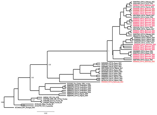
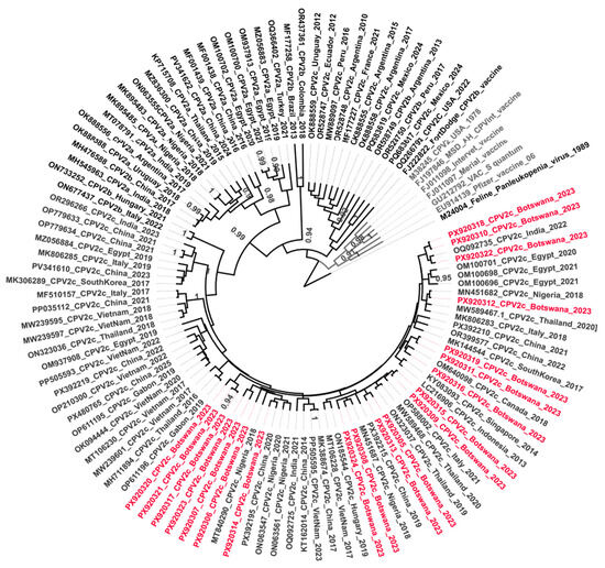
A phylogenetic tree was generated using Bayesian Evolutionary Analysis by Sampling Trees (BEAST), and a comparison was performed between Botswana CPV 2 sequences (21 sequences) and reference sequences obtained from GenBank, including representative sequences from African and vaccine strains for CPV 2 (Figure 2). Almost all Botswana CPV 2c samples (20 sequences) clustered together with other African CPV 2c variants, mainly Nigeria, Egypt, Gabon, and Ethiopia, but only one sample accession number, PX920325 (J1 strain), clustered with CPV 2a from Nigeria. The vaccine variants clustered among themselves, except for the Fort Dodge SHA vaccine, formulated based on the CPV 2b variant, which clustered with South African CPV 2b sequences.
Figure 2.
A phylogenetic tree based on partial VP2 gene sequences of CPV 2 generated using BEAST. The 21 sequences in red indicate variants circulating in Botswana. African and vaccine variant reference sequences were obtained from GenBank (http://www.ncbi.nlm.nih.gov, accessed on 14 February 2026) and contained information such as accession number, variant type, country of origin, and date of sampling.
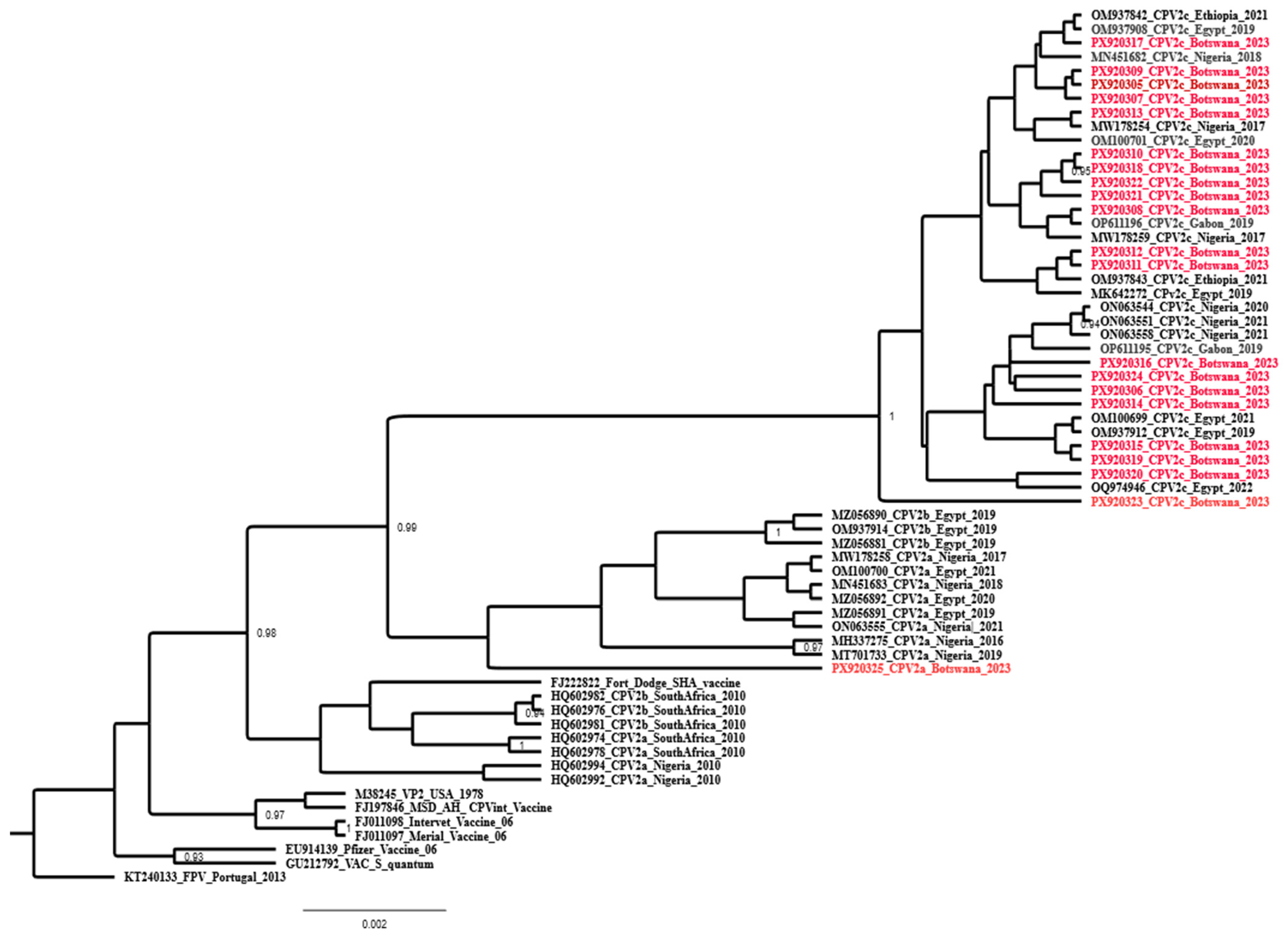
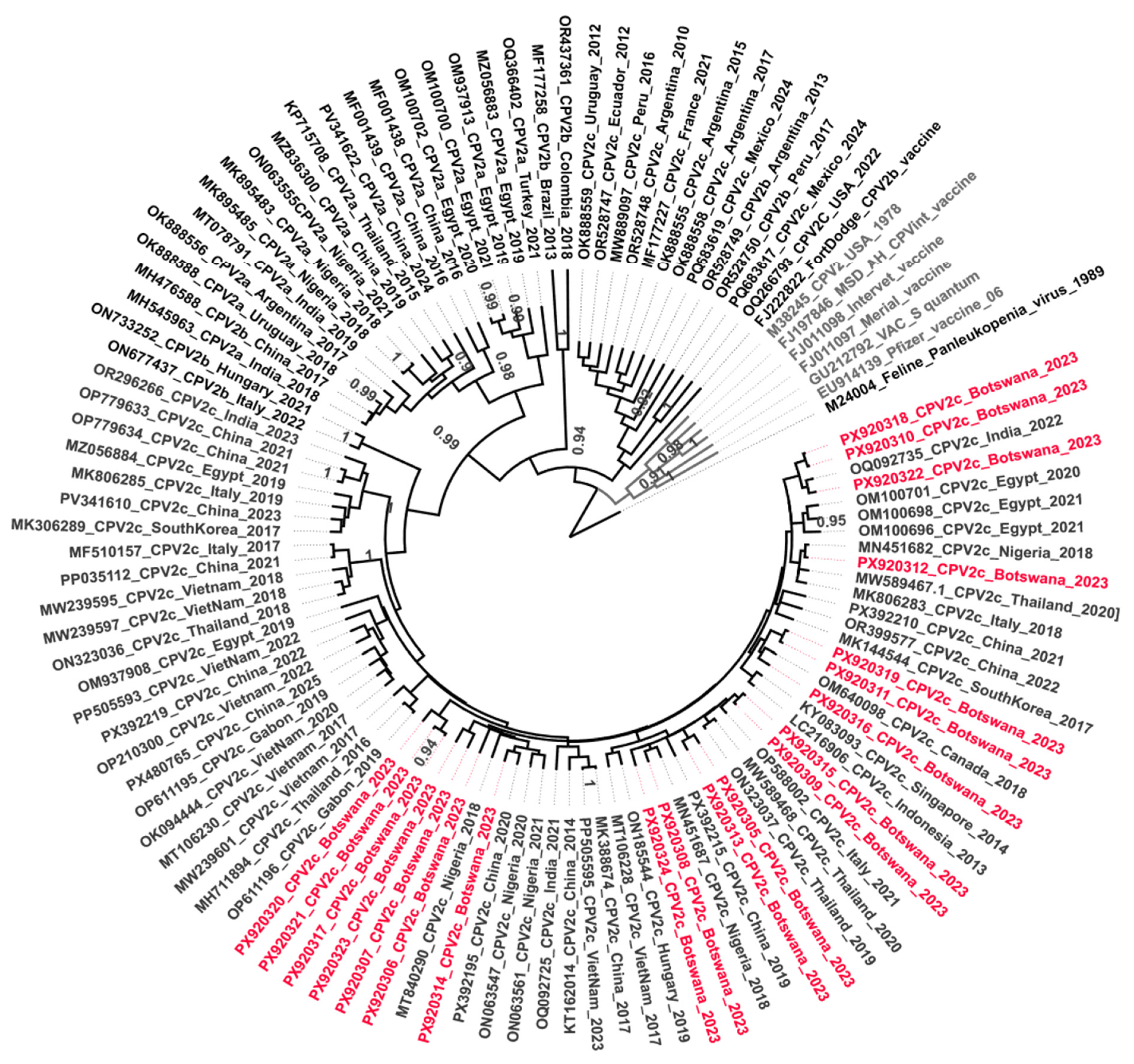
A total of 93 full-length CPV 2 VP2 gene reference sequences obtained from GenBank, representing global isolates, were compared with CPV 2c VP2 sequences from Botswana that were generated in this study (Figure 3). The phylogenetic tree showed tight clustering of Botswana CPV 2c variants with sequences from African CPV 2c and Asian CPV 2c variants.
Figure 3.
A phylogenetic tree based on the full-length VP2 sequence generated using BEAST X v10.5.0. Botswana CPV 2c sequences are marked red. Reference sequences from other global regions and vaccine variants retrieved from GenBank (http://www.ncbi.nlm.nih.gov, accessed on 14 February 2026). Feline panleukopenia (M24004) is an outgroup.
4. Discussion
The present study describes the first detection and genetic characterization of CPV 2 circulating in domestic dog populations in Botswana. In this study, suspected CPV 2 cases from eight veterinary clinics across Botswana were initially screened by ELISA and confirmed by qPCR. The majority of cases (93.9%) occurred in dogs aged 0 to 6 months, consistent with findings from Egypt [41], Morocco [26] and Nigeria [43], where CPV 2 prevalence rates of 84.6%, 85.5%, and 93.3%, respectively, were reported in dogs of similar ages. This observation supports the notion that CPV 2 is predominantly a disease of younger animals [44] likely attributed to waning maternal-derived antibodies between 4 and 16 weeks of age, rendering puppies vulnerable to infection [45].
Multiple-dog households comprised the majority of CPV 2 cases, constituting 78.6% of cases detected in this study, and were identified as a risk factor facilitating the spread of the disease. Overcrowding has been documented as a potential risk factor for CPV 2 infection, especially in boarding kennels [42,46]. Exotic breeds were overrepresented with 68.4% cases, perhaps owing to sentimental care provided by their owners and being most likely to be sent to animal health care compared to local breeds. Overall, age, household density, and breed were the main factors significantly associated with CPV 2 infection, consistent with previous reports [42,47].
A high positivity rate was observed with qPCR (100%), compared to 62% positivity for ELISA, demonstrating the superior sensitivity of qPCR and its ability to detect low viral loads, even in early infection [48,49,50]. In contrast, ELISA is more prone to false-negative results [5,51], partly due to antigen sequestration forming immune complexes and its relatively high detection threshold requirement of approximately 106 viral copies per milligram of feces [50,52]. The high PCR positivity among tested diarrheic dogs is consistent with reports from other regions, including Morocco [26] and Nigeria [43]. Locally, high seroprevalence has also been documented by Thompson et al. [33] in dogs in Maun, suggesting that the virus is endemic in Botswana. However, the high positivity is likely attributed to the cross-sectional design of the study, which focused exclusively on symptomatic dogs. Additionally, socioeconomic factors may delay owners from seeking veterinary care on time, leading to an overrepresentation of severely affected cases presented at veterinary clinics.
In this study, 47.3% of CPV 2 positive cases were from vaccinated or partially vaccinated dogs, while 52.3% were from unvaccinated animals. Similar trends have been reported in China, Morocco, Italy, and Australia, where 48.42%, 40.6%, 32.5% and 3.3% CPV 2-positive cases, respectively, were reported in vaccinated dogs [26,53,54,55]. These findings highlight the widespread occurrence of vaccine failure among vaccinated animals globally. Improper vaccination protocols, poor handling or maintenance of the vaccine cold chain, and use of vaccines with suboptimal cross-protection against circulating variants cannot be ruled out as potential causes of vaccine failure observed in these studies. The low qPCR Ct values (<20) observed in unvaccinated animals indicate higher viral loads, consistent with previous studies showing that unvaccinated or partially immunized dogs tend to exhibit more severe clinical signs and higher mortality rates compared to fully vaccinated animals [20,56].
Molecular genotyping utilizing MGB probe-based multiplex qPCR assays has identified CPV 2c as the predominant variant circulating in Botswana, with only a single CPV 2a variant detected in Maun village. The robustness of probe-based qPCR for CPV 2 differentiation is well established [26,36]. Sequencing of the VP 2 gene corroborated the qPCR findings and confirmed mutations at amino acid position 426, where asparagine was substituted for glutamic acid, thereby classifying the Botswana variants as CPV 2c. This N426E mutation occurs within the VP2 epitope A region comprising amino acid residues 93, 222, 224, and 426 [57,58], which is responsible for the emergence of antigenic variants of CPV-2a, CPV 2b, and CPV2c. Furthermore, mutations at amino acid positions 299 and 300–305, located within epitope B on the threefold axis of the CPV2 capsid structure [58,59], are known to be responsible for virus host range and antigenicity, as these residues are exposed on the viral surface. The Botswana CPV 2c variants exhibit a mutation at position 300 (A300G), which is commonly reported among CPV 2c variants [60]. Additionally, the substitution of tyrosine (Y) with aspartate (D) at position 305 was observed in the Botswana CPV 2c variants, which may contribute to antigenic differences, although its impact is likely modulated by interactions with other residues within the capsid.
Additionally, non-synonymous substitutions were observed in Botswana variants when compared with reference vaccine strains from GenBank (Table 6). This is expected, as currently available vaccines are derived from the earlier CPV2 or CPV2b variants and therefore show genetic divergence from the more recently emerged CPV 2c variants, driven by the high evolutionary rate of CPV 2 [61,62]. Through antigenic drift, three newly acquired mutations in the VP2 gene, F267Y, Y324I, and T440A, have been implicated in vaccine failure [40,63]. The Botswana variants exhibit the F267Y and Y324I mutants, associated with host adaptation and altered host range, respectively [40], but they lack the T440A mutation, which has been more strongly linked to changes in antigenicity [55,64].
Although previous studies have demonstrated cross-protection of CPV 2 and CPV 2b-based vaccines against heterologous variants, including CPV2c [20,62,65,66,67], incorporation of homologous CPV 2c variants into new future vaccine formulations has been proposed to enhance vaccine efficacy. Newer vaccines incorporating emerging genetic variants such as CPV 2c are currently under development [68], and they provide improved vaccine efficacy and overcome the interference of maternal antibodies following primary vaccination.
Amino acid substitutions previously reported in Asian CPV 2c variants, including A5G, F267Y, Y324I, and Q370R, were also detected in the Botswana variants [69,70]. Importantly, while F267Y and Y324I were discussed above in the context of their impact on vaccine failure, their presence in this dataset also supports the broader molecular similarity of Botswana variants to the globally circulating CPV 2c lineage. The A5G mutation is located at the N-terminus of VP2, while the Q370R is located around the protein-binding pocket, and both are believed to be associated with changes in host range and the protein-binding capacity of the virus [71]. The presence of these mutations in Botswana CPV 2c variants suggests that CPV 2c circulating in Southern Africa shares molecular features with globally emerging variants, potentially reflecting viral adaptation. A unique mutation at amino acid position 218 (L218I) was identified in the Botswana BC1 strain (accession number PX920306). Overall, mutations observed in Botswana CPV 2c variants are predominantly located outside the traditionally mapped epitope regions of the CPV2 capsid, which are primary targets for neutralizing antibodies against the virus [72]. Therefore, these substitutions are unlikely to affect the cross-protective efficacy of existing vaccines against the currently circulating CPV2c variant. Some studies have previously demonstrated that CPV 2b and CPV 2c variants are mutually cross-neutralized by antisera raised against either variant, indicating that the 426E substitution does not confer immune escape properties [67,73]. However, given the limited data on in vitro cross-neutralization using Botswana isolates, continued molecular surveillance of circulating CPV 2 variants is recommended. Future studies employing virus neutralization assays to directly assess vaccine-induced antibody cross-reactivity against Botswana CPV 2c field strains are warranted.
A phylogenetic analysis revealed that the Botswana CPV 2c variants formed a monophyletic clade with CPV 2c reference strains from other African countries, including Gabon, Nigeria, Egypt, and Ethiopia (Figure 2), as well as from Asian countries like India, Vietnam, China, Indonesia, and Thailand (Figure 3). This supports the hypothesis regarding the regional and transcontinental spread of this Asian CPV 2c lineage into Africa [8,28,74] likely facilitated by increased trade and pet movement between the two continents.
The single CPV 2a variant from Botswana clustered with CPV 2a variants from Nigeria, which may be attributed to trade or movement of goods between the two countries.
The study had some limitations as CPV 2 cases were not sampled from all districts of the country, which would have provided a more comprehensive epidemiological map of the disease. This was constrained by the limited resources available to undertake such an expanded study. Additionally, the research mainly focused on sick dogs presented to veterinary clinics. This limitation affected the statistical analysis, as all samples tested positive when using qPCR. The absence of negative qPCR results made analytical risk factors difficult to compute and necessitated the use of ELISA results instead. It is therefore advised that the results of the risk factors be interpreted with caution and treated as preliminary findings from this study. Future epidemiological studies should include negative samples to enable more robust estimation of true prevalence and associated risk factors of the disease in the country.
For phylogenetic analysis, most African sequence variants had short partial VP2 sequences available in GenBank, which affected molecular phylogenetic tree reconstruction. A whole-genome sequence of the virus would provide deeper insight into viral evolution as well as phylogenetic and global phylogeographic relationships of the pathogen.
5. Conclusions
This study provides insights into the presence and genetic diversity of CPV 2 variants in Botswana. The findings confirm the active circulation of CPV 2a and CPV 2c variants within the country. Notably, CPV 2c has emerged as the predominant variant associated with most cases of CPV 2. These findings have significant implications for CPV 2 control in Botswana, underscoring the critical importance of routine molecular surveillance to monitor viral evolution and inform future diagnostic and vaccination strategies. This study also highlights the need to improve vaccination coverage, particularly in young dogs and multi-dog households, to mitigate viral spread and disease severity. However, risk analysis findings should be interpreted with caution and considered preliminary, as they were based on ELISA results following the absence of negative qPCR samples.
Future work may cover a comprehensive nationwide epidemiological investigation involving both domestic and wild carnivores, which is essential for fully comprehending the diversity and geospatial distribution of CPV 2 in Botswana. Also, future studies employing virus neutralization assays to directly assess vaccine-induced antibody cross-reactivity against Botswana CPV 2c field strains are warranted.
Supplementary Materials
The following supporting information can be downloaded at https://www.mdpi.com/article/10.3390/v18070772/s1, Supplementary Table S1: Botswana sample distribution showing ELISA, real-time PCR results, and characterization of CPV 2 variants. Supplementary Table S2: Botswana CPV2 samples and their assigned accession numbers from GenBank.
Author Contributions
Conceptualization, K.T., A.A. and E.A.M.A.; methodology, K.T. and E.A.M.A.; formal analysis, K.T., D.P. and E.A.M.A.; investigation, K.T.; resources, E.A.M.A. and S.S.R.; data curation, K.T. and D.P.; writing—original draft preparation, K.T.; writing—review and editing, A.A., S.S.R., D.M.P., D.P. and E.A.M.A.; visualization, K.T., D.M.P., D.P. and E.A.M.A.; supervision, A.A., S.S.R., D.M.P. and E.A.M.A.; project administration, K.T. All authors have read and agreed to the published version of the manuscript.
Funding
This research was partly funded by the Martin and Pamela Winter Infectious Disease Fellowship Program, Institute of Infectious and Zoonotic Diseases (IIZD), University of Pennsylvania, School of Veterinary Medicine, USA, and by the Botswana University of Agriculture and Natural Resources, Botswana.
Institutional Review Board Statement
The animal study protocol was approved by the Bioethics Committee at the Faculty of Animal and Veterinary Science, Botswana University of Agriculture and Natural Resources (BUAN-AEC-2022-07, approval date: 5 October 2022).
Informed Consent Statement
Not applicable.
Data Availability Statement
The original contributions presented in this study are included in the article/Supplementary Materials. Further inquiries can be directed to the corresponding authors.
Acknowledgments
We would like to acknowledge the veterinary practitioners M. Sento, R. Jackson, M. Mazwiduma, B. Radihephi, D. Coyne, Bontsi-Busang, and S. Motsamai from private veterinary clinics, as well as dog owners who participated in the study. Dhruv Desai, Maitumelo Losologolo, and Owen Raboloko are also acknowledged for their assistance in some aspects of the project.
Conflicts of Interest
The authors of this manuscript declare no conflicts of interest. The funders had no role in the design of the study; in the collection, analysis, or interpretation of data; in the writing of the manuscript; or in the decision to publish the results.
References
- Franzo, G.; Mira, F.; Schirò, G.; Canuti, M. Not Asian Anymore: Reconstruction of the History, Evolution, and Dispersal of the “Asian” Lineage of CPV-2c. Viruses 2023, 15, 1962. [Google Scholar] [CrossRef] [Scilit] [PubMed]
- Parrish, C.R. Host range relationships and the evolution of canine parvovirus. Vet. Microbiol. 1999, 69, 29–40. [Google Scholar] [CrossRef] [Scilit] [PubMed]
- Castillo, C.; Neira, V.; Aniñir, P.; Grecco, S.; Pérez, R.; Panzera, Y.; Zegpi, N.A.; Sandoval, A.; Sandoval, D.; Cofre, S.; et al. First Molecular Identification of Canine Parvovirus Type 2 (CPV2) in Chile Reveals High Occurrence of CPV2c Antigenic Variant. Front. Vet. Sci. 2020, 7, 194. [Google Scholar] [CrossRef] [Scilit] [PubMed]
- Decaro, N.; Buonavoglia, C. Canine parvovirus post-vaccination shedding: Interference with diagnostic assays and correlation with host immune status. Vet. J. 2017, 221, 23–24. [Google Scholar] [CrossRef] [Scilit] [PubMed]
- Mazzaferro, E.M. Update on Canine Parvoviral Enteritis. Vet. Clin. N. Am. Small Anim. Pract. 2020, 50, 1307–1325. [Google Scholar] [CrossRef] [Scilit]
- Franzo, G.; Tucciarone, C.M.; Cecchinato, M.; Drigo, M. Canine parvovirus type 2 (CPV-2) and Feline panleukopenia virus (FPV) codon bias analysis reveals a progressive adaptation to the new niche after the host jump. Mol. Phylogenetics Evol. 2017, 114, 82–92. [Google Scholar] [CrossRef] [Scilit]
- Navarro, R.; Nair, R.; Peda, A.; Aung, M.S.; Ashwinie, G.S.; Gallagher, C.A.; Malik, Y.S.; Kobayashi, N.; Ghosh, S. Molecular characterization of canine parvovirus and canine enteric coronavirus in diarrheic dogs on the island of St. Kitts: First report from the Caribbean region. Virus Res. 2017, 240, 154–160. [Google Scholar] [CrossRef] [Scilit] [PubMed]
- Tegegne, D.; Tsegaye, G.; Faustini, G.; Franzo, G. First genetic detection and characterization of canine parvovirus type 2 (Carnivore protoparvovirus 1) in southwestern Ethiopia. Vet. Res. Commun. 2023, 47, 975–980. [Google Scholar] [PubMed]
- Truyen, U. Emergence and recent evolution of canine parvovirus. Vet. Microbiol. 1999, 69, 47–50. [Google Scholar] [CrossRef] [Scilit] [PubMed]
- Umar, S.; Gao, D.; Kim, S.; Cheng, Y.; Fang, Z.; Zhongqi, Q.; Yu, W.; Anderson, B.D. Molecular characterization of canine parvovirus type 2 (CPV2) reveals a high prevalence of the CPV2c genotype among dogs suffering from diarrhea. Anim. Dis. 2024, 4, 1. [Google Scholar] [CrossRef] [Scilit]
- Shackelton, L.A.; Parrish, C.R.; Truyen, U.; Holmes, E.C. High rate of viral evolution associated with the emergence of carnivore parvovirus. Proc. Natl. Acad. Sci. USA 2005, 102, 379–384. [Google Scholar] [PubMed]
- Giraldo-Ramirez, S.; Rendon-Marin, S.; Ruiz-Saenz, J. Phylogenetic, Evolutionary and Structural Analysis of Canine Parvovirus (CPV-2) Antigenic Variants Circulating in Colombia. Viruses 2020, 12, 500. [Google Scholar] [CrossRef] [Scilit] [PubMed]
- Hoelzer, K.; Parrish, C.R. The emergence of parvoviruses of carnivores. Vet. Res. 2010, 41, 39. [Google Scholar] [CrossRef] [Scilit] [PubMed]
- Tuteja, D.; Banu, K.; Mondal, B. Canine parvovirology—A brief updated review on structural biology, occurrence, pathogenesis, clinical diagnosis, treatment and prevention. Comp. Immunol. Microbiol. Infect. Dis. 2022, 82, 101765. [Google Scholar] [CrossRef] [Scilit] [PubMed]
- Miranda, C.; Thompson, G. Canine parvovirus: The worldwide occurrence of antigenic variants. J. Gen. Virol. 2016, 97, 2043–2057. [Google Scholar] [CrossRef] [Scilit] [PubMed]
- Chen, S.; Shang, K.; Chen, J.; Yu, Z.; Wei, Y.; He, L.; Ding, K. Global distribution, cross-species transmission, and receptor binding of canine parvovirus-2: Risks and implications for humans. Sci. Total Environ. 2024, 930, 172307. [Google Scholar] [CrossRef] [Scilit] [PubMed]
- Ohshima, T.; Hisaka, M.; Kawakami, K.; Kishi, M.; Tohya, Y.; Mochizuki, M. Chronological analysis of canine parvovirus type 2 isolates in Japan. J. Vet. Med. Sci. 2008, 70, 769–775. [Google Scholar] [CrossRef] [Scilit] [PubMed]
- Decaro, N.; Desario, C.; Parisi, A.; Martella, V.; Lorusso, A.; Miccolupo, A.; Mari, V.; Colaianni, M.L.; Cavalli, A.; Di Trani, L.; et al. Genetic analysis of canine parvovirus type 2c. Virology 2009, 385, 5–10. [Google Scholar] [CrossRef] [Scilit] [PubMed]
- Atkinson, M. Canine parvovirus vaccination. Vet. Rec. 2006, 159, 895–896. [Google Scholar] [CrossRef] [Scilit] [PubMed]
- Decaro, N.; Buonavoglia, C.; Barrs, V.R. Canine parvovirus vaccination and immunisation failures: Are we far from disease eradication? Vet. Microbiol. 2020, 247, 108760. [Google Scholar] [CrossRef] [Scilit] [PubMed]
- Decaro, N.; Desario, C.; Elia, G.; Campolo, M.; Lorusso, A.; Mari, V.; Martella, V.; Buonavoglia, C. Occurrence of severe gastroenteritis in pups after canine parvovirus vaccine administration: A clinical and laboratory diagnostic dilemma. Vaccine 2007, 25, 1161–1166. [Google Scholar] [CrossRef] [Scilit] [PubMed]
- Miranda, C.; Thompson, G. Canine parvovirus in vaccinated dogs: A field study. Vet. Rec. 2016, 178, 397. [Google Scholar] [CrossRef] [Scilit] [PubMed]
- Paul, B.; Alam, J.; Hossain, M.M.K.; Hoque, S.F.; Bappy, M.N.I.; Akter, H.; Ahmed, N.; Akter, M.; Ali Zinnah, M.; Das, S.; et al. Immunoinformatics for Novel Multi-Epitope Vaccine Development in Canine Parvovirus Infections. Biomedicines 2023, 11, 2180. [Google Scholar] [CrossRef] [Scilit] [PubMed]
- Schirò, G.; Mira, F.; Decaro, N.; Desario, C.; Chiaramonte, G.; Di Bella, S.; Cannella, V.; Purpari, G.; Ventriglia, G.; Randazzo, V.; et al. Persistence of DNA from canine parvovirus modified-live virus in canine tissues. Vet. Res. Commun. 2023, 47, 567–574. [Google Scholar] [PubMed]
- Vila Nova, B.; Cunha, E.; Sepúlveda, N.; Oliveira, M.; São Braz, B.; Tavares, L.; Almeida, V.; Gil, S. Evaluation of the humoral immune response induced by vaccination for canine distemper and parvovirus: A pilot study. BMC Vet. Res. 2018, 14, 348. [Google Scholar] [CrossRef] [Scilit] [PubMed]
- Amrani, N.; Amrani, N.; Desario, C.; Kadiri, A.; Cavalli, A.; Berrada, J.; Zro, K.; Sebbar, G.; Colaianni, M.L.; Parisi, A.; et al. Molecular epidemiology of canine parvovirus in Morocco. Infect. Genet. Evol. 2016, 41, 201–206. [Google Scholar] [CrossRef] [Scilit] [PubMed]
- Dogonyaro, B.B.; Bosman, A.-M.; Sibeko, K.P.; Venter, E.H.; van Vuuren, M. Genetic analysis of the VP2-encoding gene of canine parvovirus strains from Africa. Vet. Microbiol. 2013, 165, 460–465. [Google Scholar] [CrossRef] [Scilit] [PubMed]
- Franzo, G.; De Villiers, L.; De Villiers, M.; Ravandi, A.; Gyani, K.; Van Zyl, L.; Coetzee, L.M.; Khaiseb, S.; Molini, U. Molecular epidemiology of canine parvovirus in Namibia: Introduction pathways and local persistence. Prev. Vet. Med. 2022, 209, 105780. [Google Scholar] [CrossRef] [Scilit] [PubMed]
- Maganga, G.D.; Labouba, I.; Ikapi, S.; Nkili-Meyong, A.; Dikongo, A.; Boundenga, L.; Ngoubangoye, B.; Memvie, C.; Kumulungui, B. Molecular Characterization of Canine Parvovirus Variants CPV-2a and CPV-2c, Associated with Vaccinated Dogs at Libreville, Gabon. Viruses 2023, 15, 1169. [Google Scholar] [CrossRef] [Scilit] [PubMed]
- Folitse, D.R.; Kodie, D.O.; Amemor, E.; Dei, D.; Tasiame, W.; Burimuah, V.; Emikpe, B.O. Detection of canine parvovirus antigen in dogs in kumasi, Ghana. Afr. J. Infect. Dis. 2018, 12, 28–32. [Google Scholar] [PubMed]
- McRee, A.; Wilkes, R.; Dawson, J.; Parry, R.; Foggin, C.; Adams, H.; Odoi, A.; Kennedy, M.; Wilkes, A. Serological detection of infection with canine distemper virus, canine parvovirus and canine adenovirus in communal dogs from Zimbabwe. J. S. Afr. Vet. Assoc. 2014, 85, 2–1110. [Google Scholar] [CrossRef] [Scilit]
- Castanheira, P.; Duarte, A.; Gil, S.; Cartaxeiro, C.; Malta, M.; Vieira, S.; Tavares, L. Molecular and serological surveillance of canine enteric viruses in stray dogs from Vila do Maio, Cape Verde. BMC Vet. Res. 2014, 10, 91. [Google Scholar] [CrossRef] [Scilit] [PubMed]
- Thompson, R.; Adams, H.; Odoi, A.; Kennedy, M. Seroprevalence of viral and vector-borne bacterial pathogens in domestic dogs (Canis familiaris) in northern Botswana. PLoS ONE 2020, 15, e0220593. [Google Scholar] [CrossRef] [Scilit] [PubMed]
- Statistics Botswana. Population and Housing Census 2022: Populations of Cities, Towns, Villages and Associated Localities; Statistics Botswana: Gaborone, Botswana, 2022. [Google Scholar]
- Decaro, N.; Elia, G.; Martella, V.; Desario, C.; Campolo, M.; Trani, L.D.; Tarsitano, E.; Tempesta, M.; Buonavoglia, C. A real-time PCR assay for rapid detection and quantitation of canine parvovirus type 2 in the feces of dogs. Vet. Microbiol. 2005, 105, 19–28. [Google Scholar] [CrossRef] [Scilit] [PubMed]
- Decaro, N.; Elia, G.; Martella, V.; Campolo, M.; Desario, C.; Camero, M.; Cirone, F.; Lorusso, E.; Lucente, M.S.; Narcisi, D.; et al. Characterisation of the canine parvovirus type 2 variants using minor groove binder probe technology. J. Virol. Methods 2006, 133, 92–99. [Google Scholar] [CrossRef] [Scilit] [PubMed]
- Buonavoglia, C.; Martella, V.; Pratelli, A.; Tempesta, M.; Cavalli, A.; Buonavoglia, D.; Bozzo, G.; Elia, G.; Decaro, N.; Carmichael, L. Evidence for evolution of canine parvovirus type 2 in Italy. J. Gen. Virol. 2001, 82, 3021–3025. [Google Scholar] [CrossRef] [Scilit] [PubMed]
- Tamura, K.; Stecher, G.; Kumar, S. MEGA11: Molecular Evolutionary Genetics Analysis Version 11. Mol. Biol. Evol. 2021, 38, 3022–3027. [Google Scholar] [CrossRef] [Scilit] [PubMed]
- Suchard, M.A.; Lemey, P.; Baele, G.; Ayres, D.L.; Drummond, A.J.; Rambaut, A. Bayesian phylogenetic and phylodynamic data integration using BEAST 1.10. Virus Evol. 2018, 4, vey016. [Google Scholar] [CrossRef] [Scilit] [PubMed]
- Zhou, P.; Zeng, W.; Zhang, X.; Li, S. The genetic evolution of canine parvovirus—A new perspective. PLoS ONE 2017, 12, e0175035. [Google Scholar] [CrossRef] [Scilit] [PubMed]
- Adly, M.M.; Elgaml, M.A.; Abdel Khalek, A.F.; Saeed, O.A.; Shalaby, M.A.; Amer, H.M. Molecular Characterization of Full-Length VP2 Gene of Canine Parvovirus Type 2 Strains Circulating in Egypt 2019-2021. Comp. Immunol. Microbiol. Infect. Dis. 2024, 110, 102190. [Google Scholar] [CrossRef] [Scilit] [PubMed]
- Ogbu, K.I.; Chukwudi, I.C.; Mira, F.; Eze, U.U.; Di Bella, S.; Olaolu, O.S.; Tion, M.T.; Purpari, G.; Cannella, V.; Nwosuh, I.C.; et al. Current status and risk factors of canine parvovirus type 2 in North Central Nigeria. Comp. Immunol. Microbiol. Infect. Dis. 2021, 74, 101578. [Google Scholar] [CrossRef] [Scilit] [PubMed]
- Shima, F.K.; Gberindyer, F.A.; Tion, M.T.; Fagbohun, O.A.; Omobowale, T.O.; Nottidge, H.O. Diagnostic Performance of a Rapid Immunochromatographic Test Kit for Detecting Canine Parvovirus Infection. Top. Companion Anim. Med. 2021, 45, 100551. [Google Scholar] [CrossRef] [Scilit] [PubMed]
- Lamm, C.G.; Rezabek, G.B. Parvovirus infection in domestic companion animals. Vet. Clin. N. Am. Small Anim. Pract. 2008, 38, 837–850. [Google Scholar] [CrossRef] [Scilit]
- Mila, H.; Grellet, A.; Desario, C.; Feugier, A.; Decaro, N.; Buonavoglia, C.; Chastant-Maillard, S. Protection against canine parvovirus type 2 infection in puppies by colostrum-derived antibodies. J. Nutr. Sci. 2014, 3, e54. [Google Scholar] [CrossRef] [Scilit] [PubMed]
- Le, H.T.; Nguyen, M.H.D.; Doan, P.H.; Truong, L.P.; Tu, L.T.K.; Nguyen, Q.H.; Nguyen, L.T.B.; Pornchai, P. Risk factors associated with canine parvovirus disease in dogs: A case-control study. J. Agric. Dev. 2020, 19, 32–38. [Google Scholar] [CrossRef] [Scilit]
- Dong, B.; Zhang, G.; Zhang, J.; Bai, J.; Lin, W. A systematic literature review and meta-analysis of characterization of canine parvoviruses 2 prevalent in mainland China. Virol. J. 2020, 17, 195. [Google Scholar] [CrossRef] [Scilit] [PubMed]
- Elbaz, E.; El-Tholoth, M.; Abo Elfadl, E.A.; Mosad, S.M. Molecular investigation on the presence of canine parvovirus in Egypt. Comp. Immunol. Microbiol. Infect. Dis. 2021, 74, 101576. [Google Scholar] [CrossRef] [Scilit] [PubMed]
- Magouz, A.; El-Kon, I.; Raya-Álvarez, E.; Khaled, E.; Alkhalefa, N.; Alhegaili, A.S.; El-khadragy, M.F.; Agil, A.; Elmahallawy, E.K. Molecular typing of canine parvovirus type 2 by VP2 gene sequencing and restriction fragment length polymorphism in affected dogs from Egypt. Front. Microbiol. 2023, 14, 1254060. [Google Scholar] [CrossRef] [Scilit] [PubMed]
- Proksch, A.L.; Unterer, S.; Speck, S.; Truyen, U.; Hartmann, K. Influence of clinical and laboratory variables on faecal antigen ELISA results in dogs with canine parvovirus infection. Vet. J. 2015, 204, 304–308. [Google Scholar] [CrossRef] [Scilit] [PubMed]
- Shima, F.K.; Omobowale, T.O.; Adesina, R.D.; Nottidge, H.O.; Fagbohun, O.A. Molecular characterisation of canine parvoviruses from clinical samples and vaccines in Nigeria. Infect. Genet. Evol. 2020, 85, 104553. [Google Scholar] [CrossRef] [Scilit] [PubMed]
- Decaro, N.; Desario, C.; Billi, M.; Lorusso, E.; Colaianni, M.L.; Colao, V.; Elia, G.; Ventrella, G.; Kusi, I.; Bo, S.; et al. Evaluation of an in-clinic assay for the diagnosis of canine parvovirus. Vet. J. 2013, 198, 504–507. [Google Scholar] [CrossRef] [Scilit] [PubMed]
- Ling, M.; Norris, J.M.; Kelman, M.; Ward, M.P. Risk factors for death from canine parvoviral-related disease in Australia. Vet. Microbiol. 2012, 158, 280–290. [Google Scholar] [CrossRef] [Scilit] [PubMed]
- Battilani, M.; Modugno, F.; Mira, F.; Purpari, G.; Di Bella, S.; Guercio, A.; Balboni, A. Molecular epidemiology of canine parvovirus type 2 in Italy from 1994 to 2017: Recurrence of the CPV-2b variant. BMC Vet. Res. 2019, 15, 393. [Google Scholar] [CrossRef] [Scilit] [PubMed]
- Geng, Y.; Guo, D.; Li, C.; Wang, E.; Wei, S.; Wang, Z.; Yao, S.; Zhao, X.; Su, M.; Wang, X.; et al. Co-Circulation of the Rare CPV-2c with Unique Gln370Arg Substitution, New CPV-2b with Unique Thr440Ala Substitution, and New CPV-2a with High Prevalence and Variation in Heilongjiang Province, Northeast China. PLoS ONE 2015, 10, e0137288. [Google Scholar] [CrossRef] [Scilit] [PubMed]
- Ammar, E.F.; Hegazy, Y.M.; Al-Gaabary, M.; Mosad, S.M.; Salem, M.; Marzok, M.; Housawi, F.; Al-Ali, M.; Alhaider, A.; Tahoun, A. Epidemiological, and molecular investigation of Canine parvovirus-2 infection in Egypt. J. Vet. Sci. 2024, 25, e56. [Google Scholar] [CrossRef] [Scilit] [PubMed]
- Decaro, N.; Buonavoglia, C. Canine parvovirus—A review of epidemiological and diagnostic aspects, with emphasis on type 2c. Vet. Microbiol. 2012, 155, 1–12. [Google Scholar] [CrossRef] [Scilit] [PubMed]
- Tsao, J.; Chapman, M.S.; Agbandje, M.; Keller, W.; Smith, K.; Wu, H.; Luo, M.; Smith, T.J.; Rossmann, M.G.; Compans, R.W.; et al. The three-dimensional structure of canine parvovirus and its functional implications. Science 1991, 251, 1456–1464. [Google Scholar] [CrossRef] [Scilit] [PubMed]
- Parrish, C.R. Mapping specific functions in the capsid structure of canine parvovirus and feline panleukopenia virus using infectious plasmid clones. Virology 1991, 183, 195–205. [Google Scholar] [CrossRef] [Scilit] [PubMed]
- Allison, A.B.; Organtini, L.J.; Zhang, S.; Hafenstein, S.L.; Holmes, E.C.; Parrish, C.R. Single Mutations in the VP2 300 Loop Region of the Three-Fold Spike of the Carnivore Parvovirus Capsid Can Determine Host Range. J. Virol. 2016, 90, 753–767. [Google Scholar] [CrossRef] [Scilit] [PubMed]
- Truyen, U. Evolution of canine parvovirus—A need for new vaccines? Vet. Microbiol. 2006, 117, 9–13. [Google Scholar] [CrossRef] [Scilit] [PubMed]
- Wilson, S.; Illambas, J.; Siedek, E.; Stirling, C.; Thomas, A.; Plevová, E.; Sture, G.; Salt, J. Vaccination of dogs with canine parvovirus type 2b (CPV-2b) induces neutralising antibody responses to CPV-2a and CPV-2c. Vaccine 2014, 32, 5420–5424. [Google Scholar] [CrossRef] [Scilit] [PubMed]
- Mittal, M.; Chakravarti, S.; Mohapatra, J.K.; Chug, P.K.; Dubey, R.; Upmanuyu, V.; Narwal, P.S.; Kumar, A.; Churamani, C.P.; Kanwar, N.S. Molecular typing of canine parvovirus strains circulating from 2008 to 2012 in an organized kennel in India reveals the possibility of vaccination failure. Infect. Genet. Evol. 2014, 23, 1–6. [Google Scholar] [CrossRef] [Scilit] [PubMed]
- Hueffer, K.; Parker, J.S.; Weichert, W.S.; Geisel, R.E.; Sgro, J.Y.; Parrish, C.R. The natural host range shift and subsequent evolution of canine parvovirus resulted from virus-specific binding to the canine transferrin receptor. J. Virol. 2003, 77, 1718–1726. [Google Scholar] [CrossRef] [Scilit] [PubMed]
- Altman, K.; Kelman, M.; Ward, M. Are vaccine strain, type or administration protocol risk factors for canine parvovirus vaccine failure? Vet. Microbiol. 2017, 210, 8–16. [Google Scholar] [CrossRef] [Scilit] [PubMed]
- Siedek, E.M.; Schmidt, H.; Sture, G.H.; Raue, R. Vaccination with canine parvovirus type 2 (CPV-2) protects against challenge with virulent CPV-2b and CPV-2c. Berl. Und Munch. Tierarztl. Wochenschr. 2011, 124, 58–64. [Google Scholar] [CrossRef] [Scilit]
- Spibey, N.; Greenwood, N.M.; Sutton, D.; Chalmers, W.S.K.; Tarpey, I. Canine parvovirus type 2 vaccine protects against virulent challenge with type 2c virus. Vet. Microbiol. 2008, 128, 48–55. [Google Scholar] [CrossRef] [Scilit] [PubMed]
- Pearce, J.; Spibey, N.; Sutton, D.; Tarpey, I. Development of a Novel Canine Parvovirus Vaccine Capable of Stimulating Protective Immunity in Four-Week-Old Puppies in the Face of High Levels of Maternal Antibodies. Vaccines 2023, 11, 1499. [Google Scholar] [CrossRef] [Scilit] [PubMed]
- Liu, C.; Gao, J.; Li, H.; Sun, F.; Liang, H.; Liu, H.; Yi, J. Phylogenetic Characteristics of Canine Parvovirus Type 2c Variant Endemic in Shanghai, China. Viruses 2021, 13, 2257. [Google Scholar] [CrossRef] [Scilit] [PubMed]
- Hoang, M.; Lin, W.H.; Le, V.P.; Nga, B.T.T.; Chiou, M.T.; Lin, C.N. Molecular epidemiology of canine parvovirus type 2 in Vietnam from November 2016 to February 2018. Virol. J. 2019, 16, 52. [Google Scholar] [CrossRef] [Scilit] [PubMed]
- Hao, X.; Li, Y.; Xiao, X.; Chen, B.; Zhou, P.; Li, S. The Changes in Canine Parvovirus Variants over the Years. Int. J. Mol. Sci. 2022, 23, 11540. [Google Scholar] [CrossRef] [Scilit] [PubMed]
- Strassheim, M.L.; Gruenberg, A.; Veijalainen, P.; Sgro, J.-Y.; Parrish, C.R. Two Dominant Neutralizing Antigenic Determinants of Canine Parvovirus Are Found on the Threefold Spike of the Virus Capsid. Virology 1994, 198, 175–184. [Google Scholar] [CrossRef] [Scilit] [PubMed]
- Wilson, S.; Stirling, C.; Borowski, S.; Thomas, A.; King, V.; Salt, J. Vaccination of dogs with Duramune DAPPi+LC protects against pathogenic canine parvovirus type 2c challenge. Vet. Rec. 2013, 172, 662. [Google Scholar] [CrossRef] [Scilit] [PubMed]
- Ogbu, K.I.; Mira, F.; Purpari, G.; Nwosuh, C.; Loria, G.R.; Schirò, G.; Chiaramonte, G.; Tion, M.T.; Di Bella, S.; Ventriglia, G.; et al. Nearly full-length genome characterization of canine parvovirus strains circulating in Nigeria. Transbound. Emerg. Dis. 2020, 67, 635–647. [Google Scholar] [PubMed]

Disclaimer/Publisher’s Note: The statements, opinions and data contained in all publications are solely those of the individual author(s) and contributor(s) and not of MDPI and/or the editor(s). MDPI and/or the editor(s) disclaim responsibility for any injury to people or property resulting from any ideas, methods, instructions or products referred to in the content. |
© 2026 by the authors. Licensee MDPI, Basel, Switzerland. This article is an open access article distributed under the terms and conditions of the Creative Commons Attribution (CC BY) license.