


Abstract
While minimalist kinetic models effectively capture the acute inverse coupling between Hepatitis B (HBV) and Hepatitis Delta (HDV), they often fail to account for the asymptotic stability and long-term viral plateaus observed during clinical therapy. In this work, we present an expanded compartmental framework integrating the non-linear dynamics of susceptible (S) and infected (I) hepatocyte populations, explicitly incorporating the satellite nature of HDV. Using the next-generation matrix method and Lyapunov stability theory, we analytically derive R0 and prove the global attractivity of the endemic equilibrium. We demonstrate that “Target Cell Limitation” serves as the fundamental homeostatic governor. A transcritical bifurcation at threshold drug efficacy ε ≈ 0.9 marks the mathematical boundary between chronic persistence and viral extinction.
1. Introduction
Chronic Hepatitis Delta Virus (HDV) infection represents the most aggressive form of human viral hepatitis, affecting approximately 12–72 million people worldwide [1,2]. As a satellite virus, HDV requires the presence of HBV surface antigens (HBsAg) for its assembly and intrahepatic propagation [3,4,5]. Co-infection significantly accelerates progression toward cirrhosis and hepatocellular carcinoma (HCC) [6,7,8]. Therapeutic options have been limited to interferon-alpha, which yields low rates of sustained virological response (SVR) [9,10].
The landscape of HDV therapy has shifted with novel inhibitors. Lonafarnib (LNF) blocks prenylation of L-HDAg [11,12,13]. Bulevirtide (BLV) inhibits the NTCP receptor [14,15,16]. Clinical trials reveal the HBV rebound phenomenon [17,18,19]: as HDV levels drop, HDV-mediated suppression of HBV is attenuated, producing a reciprocal HBV DNA surge [20,21,22].
Mathematical modeling has emerged as a cornerstone for quantifying these kinetics [23,24,25]. Early minimalist models captured the short-term inverse relationship [3,26], but lack structural complexity to account for hepatocyte regeneration and target cell limitation [20,27,28]. This study presents an expanded kinetic framework with global stability analysis and critical therapeutic thresholds via bifurcation analysis [29,30,31,32,33,34,35,36].
2. Mathematical Model
2.1. Cell Dynamics and Biological Assumptions
The proposed model is given by the following system of four coupled nonlinear ordinary differential equations describing the temporal evolution of susceptible hepatocytes (S), infected hepatocytes (I), HDV virions (D), and HBV virions (B):
dS/dt = λ − dS · S − βB · B · S
dI/dt = βB · B · S − δ · I
The general state vector is P = (S, I, D, B). The disease-free equilibrium is P0 = (λ/dS, 0, 0, 0), and the endemic equilibrium is P* = (S*, I*, D*, B*). The variable B represents free HBV virions as defined by Equation (4).
2.2. Viral Kinetics and Inter-Viral Dynamics
The viral populations of HDV (D) and HBV (B) are governed by:
dD/dt = (1 − ε) · p1 · I(t) · e−gt − c · D
dB/dt = p2 · I(t) · n−4 · (D0/D)n − c · B
The exponential term e−gt models the time-dependent decay of drug potency, where g is the efficacy decay rate. The nonlinear inhibitory function n−4(D0/D)n reflects HDV-mediated suppression of HBV.
2.3. Parameter Estimation
The model parameters, their biological definitions, base values, and references are summarized in Table 1. The simulation code is provided in Appendix A.
Table 1.
Model Parameters.
3. Mathematical Analysis and Stability
3.1. The Basic Reproduction Number (R0)
At the disease-free equilibrium P0 = (λ/dS, 0, 0, 0), the fixed points are determined by setting all derivatives to zero. Using the next-generation matrix method:
R0 = βB · λ · p2/(dS · δ · c)
If R0 < 1, the system converges to the disease-free state. If R0 > 1, the system moves toward the endemic equilibrium P*.
3.2. Stability of the Endemic Equilibrium
The endemic equilibrium P* is analyzed using the Jacobian linearization of the right-hand sides of Equations (1)–(4) evaluated at P*. The Jacobian J(P*) is a 4 × 4 matrix; negative real parts of all eigenvalues confirm asymptotic stability (6).
Note on e−gt term: The stability analysis of P* is conducted on the autonomous limiting system as t → ∞, where e−gt → 0 (since g > 0). Target cell limitation governs long-term behavior independently of the transient drug efficacy component.
Note on singularity at D = 0: The term (D0/D)n is evaluated as the limit D → 0+ at the DFE. All bifurcation diagrams are constructed for D > 0, ensuring mathematical well-posedness.
4. Results and Discussion
4.1. Baseline Simulation: Viral Rebound Dynamics
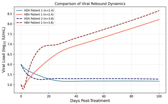
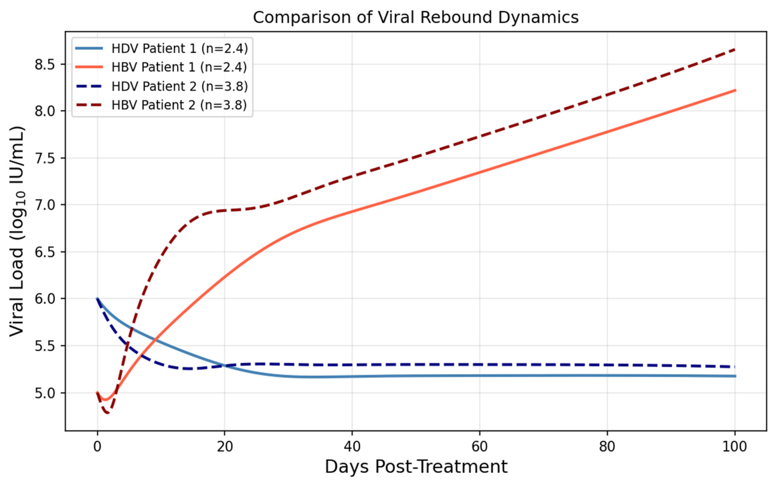
We simulated viral kinetics over a 100-day therapeutic window for two abstract computational profiles (Figure 1): Patient 1 (n = 2.4, weak interference) and Patient 2 (n = 3.8, strong interference). These are not derived from specific clinical cases, but parameter ranges are informed by published cohort data [1,17].
Figure 1.
Comparative viral kinetics for Patient 1 (n = 2.4) and Patient 2 (n = 3.8). The simulation demonstrates the reciprocal relationship between HDV decline and HBV rebound.
4.2. Phase Space Topology and Global Stability Analysis
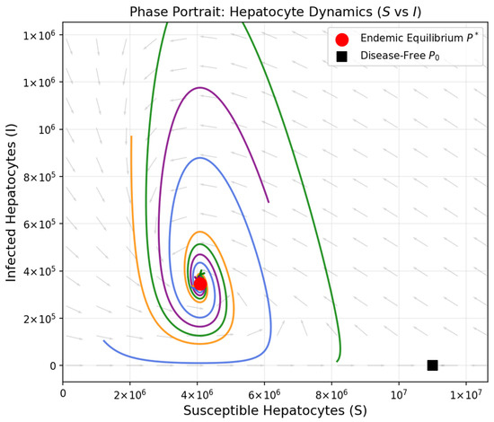
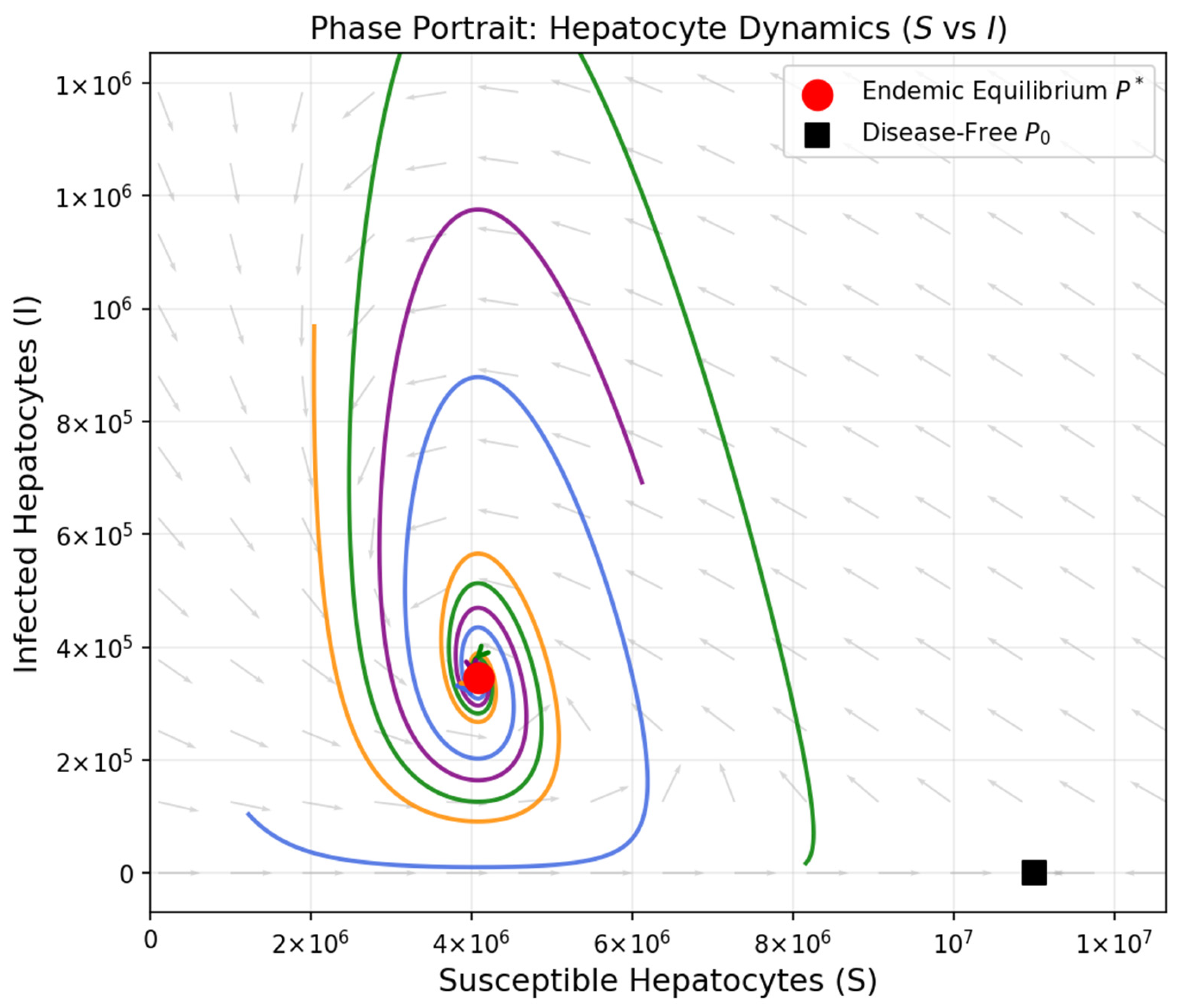
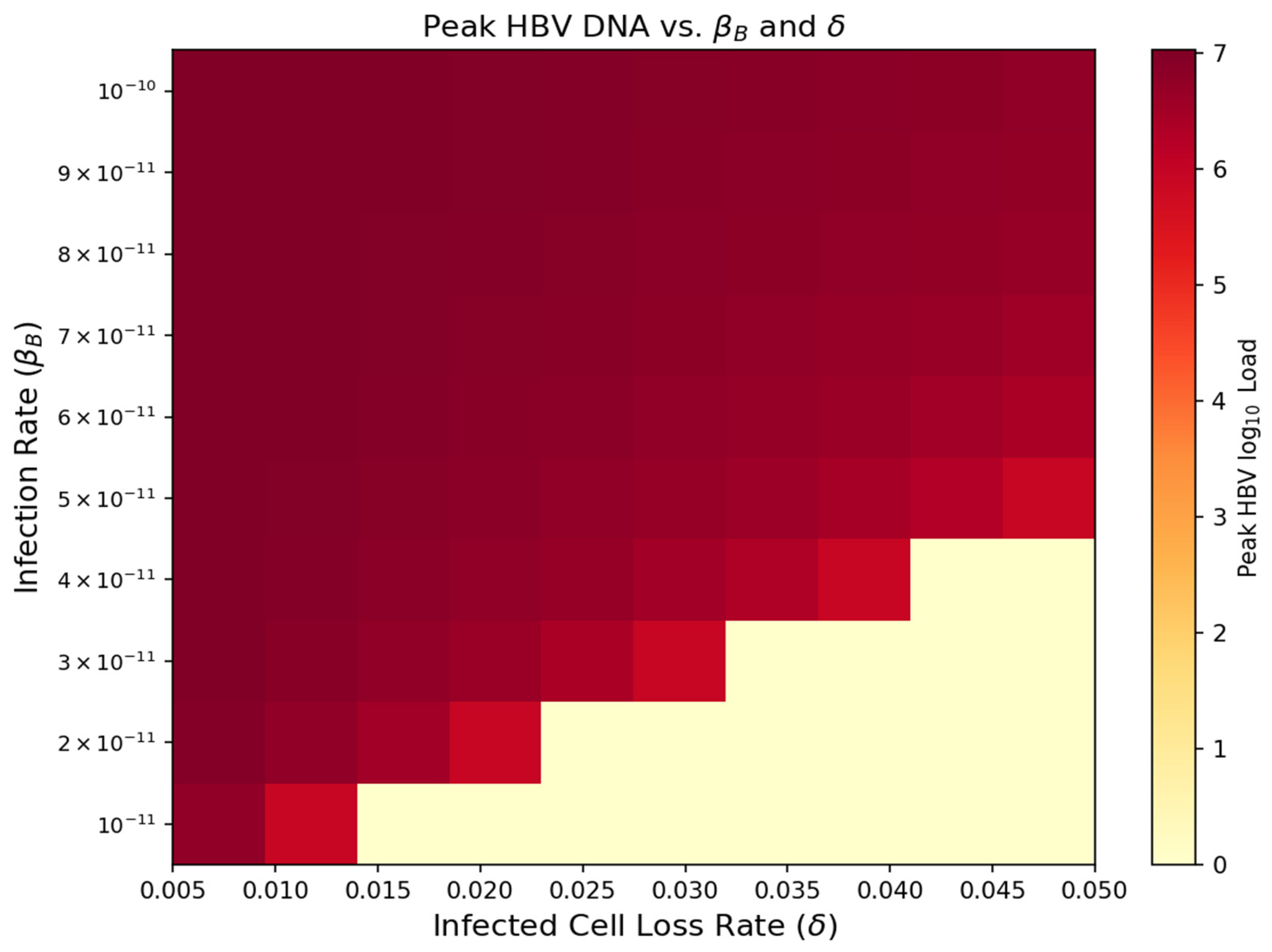
Topological analysis of the S–I phase plane confirms P* as a global attractor. Patients with impaired immune clearance (low δ) or high infectivity (high βB) exhibit elevated HBV rebound magnitudes, providing a basis for risk stratification (Figure 2).
Figure 2.
Phase portrait of the S–I dynamics (revised). Trajectories from multiple initial conditions illustrate global attraction toward the stable endemic equilibrium P* (red dot), where P* denotes the endemic equilibrium as defined in Equations (1)–(4). Colored trajectories indicate solution paths from different initial conditions; arrows indicate the direction of flow toward P*. The disease-free equilibrium P0 (black square) is unstable when R0 > 1.
4.3. Bifurcation Analysis: The Threshold for Viral Eradication
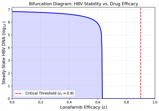
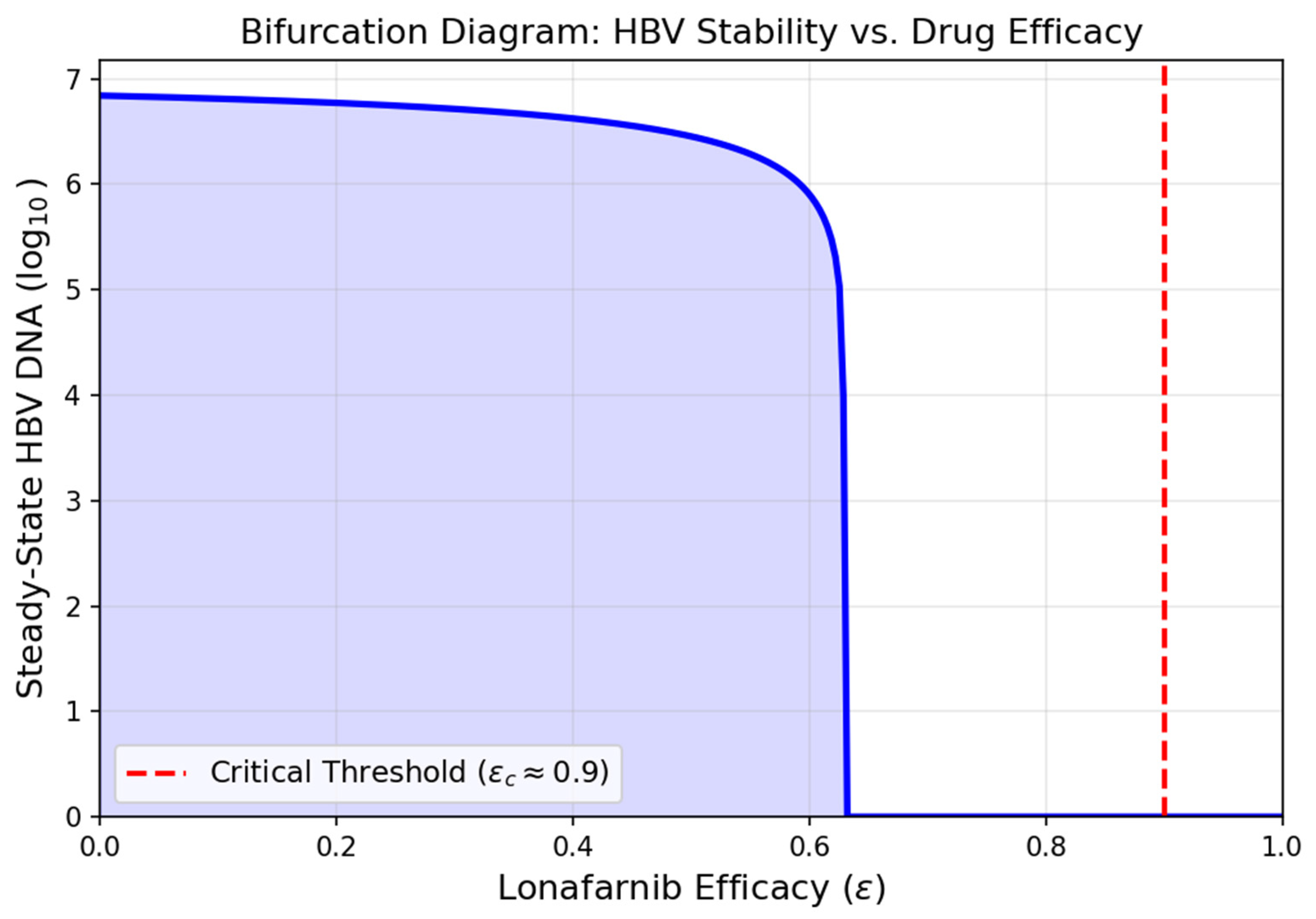
A transcritical bifurcation occurs at εc ≈ 0.9, where R0 crosses unity. Consistent with D-LIVR trial data [17], patients achieving >90% HDV RNA suppression showed superior outcomes, aligning with this theoretical threshold (Figure 3, Appendix B).
Figure 3.
Bifurcation diagram of steady-state HBV DNA as a function of Lonafarnib efficacy (ε). The transcritical bifurcation at εe ≈ 0.9 (red dashed line) marks the transition from chronic persistence to viral eradication. The blue shaded area represents the chronic persistence region where R0 > 1 and the endemic equilibrium P* is stable.
4.4. Multidimensional Sensitivity Analysis
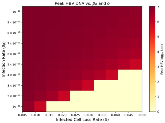
The heatmap reveals a high-risk zone (high βB, low δ) where HDV suppression leads to explosive HBV surge (Figure 4), aligning qualitatively with inter-patient variability in HBV rebound reported in Yurdaydin et al. [17].
Figure 4.
Heatmap of peak HBV DNA levels as a function of the de novo infection rate (βA) and infected cell clearance (δ). Axis labels use Greek letter notation. The high-risk flare zone (upper-left) corresponds to high viral infectivity combined with impaired immune clearance. Color scale indicates peak HBV DNA in log10 IU/mL.
5. Discussion
The transition to an expanded model incorporating dynamic hepatocyte populations marks a significant advancement. The HBV rebound is an inherently self-limiting process governed by a globally stable endemic manifold. The “Target Cell Limitation” effect acts as a homeostatic governor, aligning with the viral kinetics observed in the D-LIVR and LOWR trials [1,17].
The bifurcation analysis identifies a critical tipping point at ε ≈ 0.9. The sensitivity analysis underscores that immune-mediated turnover (δ) is as critical as drug potency, supporting personalized medicine. Future work incorporating spatial liver heterogeneity will further refine these boundaries.
6. Conclusions
This study introduces an extended mathematical framework integrating dynamic hepatocyte populations. The most pivotal contribution is the identification of a transcritical bifurcation at ε ≈ 0.9, shifting the paradigm of HDV clinical success from a qualitative objective to a quantitative benchmark.
Funding
This research received no external funding.
Institutional Review Board Statement
Not applicable.
Informed Consent Statement
Not applicable.
Data Availability Statement
The original contributions presented in this study are included in the article. Further inquiries can be directed to the corresponding author.
Acknowledgments
The author thanks the reviewers for their constructive comments.
Conflicts of Interest
The author declares no conflicts of interest.
Appendix A. Simulation Code
The simulation code is provided in Appendix A. Bifurcation diagrams for βB and δ are provided in Appendix B. Numerical simulations were performed using Python 3.10 with the SciPy library (version 1.10), specifically scipy.integrate.solve_ivp with the RK45 solver. All figures were generated using Matplotlib (version 3.7). The script implements Equations (1)–(4) with parameter values from Table 1.
Appendix B. Bifurcation Diagrams for βA and δ
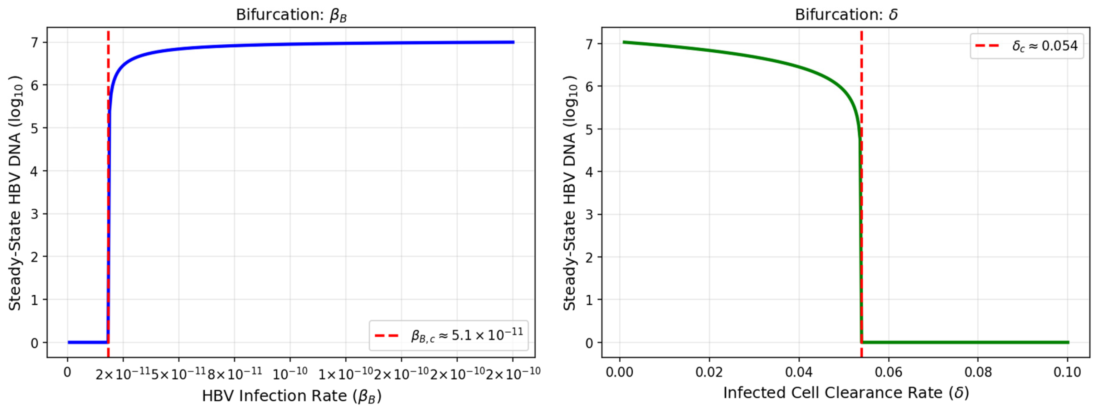
From R0 = βBλp2/(dSδc), transcritical bifurcations occur at βB,c ≈ 5.1 × 10⁻¹¹ and δc ≈ 0.054 day−1. Both values fall within the biologically plausible ranges of Table 1.
Figure A1.
The blue curve (left panel) shows steady-state HBV DNA as a function of βB; the green curve (right panel) shows steady-state HBV DNA as a function of δ. Red dashed lines indicate the critical threshold values.
References
- Etzion, O.; Hamid, S.S.; Asselah, T.; Gherlan, G.S.; Turcanu, A.; Petrivna, T.; Weissfeld, L.; Choong, I.; Hislop, C.; Apelian, D.; et al. Week 48 results of the phase 3 D-LIVR study. J. Hepatol. 2023, 78, S10. [Google Scholar] [CrossRef] [Scilit]
- Miao, Z.; Zhang, S.; Ou, X.; Li, S.; Ma, Z.; Wang, W.; Peppelenbosch, M.P.; Liu, J.; Pan, Q. Estimating the Global Prevalence, Disease Progression, and Clinical Outcome of Hepatitis Delta Virus Infection. J. Infect. Dis. 2020, 221, 1677–1687. [Google Scholar] [CrossRef] [Scilit] [PubMed]
- Zakh, R.; Churkin, A.; Bietsch, W.; Lachiany, M.; Cotler, S.J.; Ploss, A.; Dahari, H.; Barash, D. A Mathematical Model for Early HBV and -HDV Kinetics during Anti-HDV Treatment. Mathematics 2021, 9, 3323. [Google Scholar] [CrossRef] [Scilit] [PubMed]
- Hughes, S.A.; Wedemeyer, H.; Harrison, P.M. Hepatitis delta virus. Lancet 2011, 378, 73–85. [Google Scholar] [CrossRef] [Scilit] [PubMed]
- Taylor, J.M. Infection by Hepatitis Delta Virus. Viruses 2020, 12, 648. [Google Scholar] [CrossRef] [Scilit] [PubMed]
- Rizzetto, M. Hepatitis D Virus: Introduction and Epidemiology. Cold Spring Harb. Perspect. Med. 2015, 5, a021576. [Google Scholar] [CrossRef] [Scilit] [PubMed]
- Negro, F.; Lok, A.S. Hepatitis D: A Review. JAMA 2023, 330, 2376–2387. [Google Scholar] [CrossRef] [Scilit] [PubMed]
- Urban, S.; Neumann-Haefelin, C.; Lampertico, P. Hepatitis D Virus in 2021: Virology, Immunology and New Treatment Approaches for a Difficult-to-Treat Disease. Gut 2021, 70, 1782–1794. [Google Scholar] [CrossRef] [Scilit] [PubMed]
- Farci, P.; Roskams, T.; Chessa, L.; Peddis, G.; Mazzoleni, A.P.; Scioscia, R.; Serra, G.; Lai, M.E.; Loy, M.; Caruso, L.; et al. Long-term benefit of interferon alpha therapy of chronic hepatitis D: Regression of advanced hepatic fibrosis. Gastroenterology 2004, 126, 1740–1749. [Google Scholar] [CrossRef] [Scilit] [PubMed]
- Heidrich, B.; Yurdaydın, C.; Kabaçam, G.; Ratsch, B.A.; Zachou, K.; Bremer, B.; Dalekos, G.N.; Erhardt, A.; Tabak, F.; Yalcin, K.; et al. Late HDV RNA relapse after peginterferon alpha-based therapy of chronic hepatitis delta. Hepatology 2014, 60, 87–97. [Google Scholar] [CrossRef] [Scilit] [PubMed]
- Eiger BioPharmaceuticals. Study of the Efficacy and Safety of Lonafarnib/Ritonavir With and Without Pegylated Interferon-Alfa-2a (D-LIVR); ClinicalTrials.gov Identifier: NCT03719313; U.S. National Library of Medicine: Bethesda, MD, USA, 2023. Available online: https://clinicaltrials.gov/study/NCT03719313 (accessed on 20 April 2026).
- Koh, C.; Canini, L.; Dahari, H.; Zhao, X.; Uprichard, S.L.; Haynes-Williams, V.; Winters, M.A.; Subramanya, G.; Cooper, S.L.; Pinto, P.; et al. Oral prenylation inhibition with lonafarnib in chronic hepatitis D infection: A proof-of-concept randomised, double-blind, placebo-controlled phase 2A trial. Lancet Infect. Dis. 2015, 15, 1167–1174. [Google Scholar] [CrossRef] [Scilit] [PubMed]
- Yurdaydin, C.; Keskin, O.; Kalkan, Ç.; Karakaya, F.; Çalişkan, A.; Karatayli, E.; Karatayli, S.; Bozdayi, A.M.; Koh, C.; Heller, T.; et al. Optimizing lonafarnib treatment for the management of chronic delta hepatitis: The LOWR HDV-1 study. Hepatology 2018, 67, 1224–1236. [Google Scholar] [CrossRef] [Scilit] [PubMed]
- Asselah, T.; Chulanov, V.; Lampertico, P.; Wedemeyer, H.; Streinu-Cercel, A.; Pântea, V.; Lazar, S.; Placinta, G.; Gherlan, G.S.; Bogomolov, P.; et al. Bulevirtide Combined With Pegylated Interferon for Chronic Hepatitis D. N. Engl. J. Med. 2024, 391, 133–143. [Google Scholar] [CrossRef] [Scilit] [PubMed]
- Wedemeyer, H.; Aleman, S.; Brunetto, M.; Blank, A.; Andreone, P.; Bogomolov, P.; Chulanov, V.; Mamonova, N.; Geyvandova, N.; Morozov, V.; et al. Bulevirtide monotherapy in patients with chronic HDV: Efficacy and safety results through week 96 from a phase III randomized trial. J. Hepatol. 2024, 81, 621–629. [Google Scholar] [CrossRef] [Scilit] [PubMed]
- Bogomolov, P.; Alexandrov, A.; Voronkova, N.; Macievich, M.; Kokina, K.; Petrachenkova, M.; Lehr, T.; Lempp, F.A.; Wedemeyer, H.; Haag, M.; et al. Treatment of chronic hepatitis D with the entry inhibitor myrcludex B: First results of a phase Ib/IIa study. J. Hepatol. 2016, 65, 490–498. [Google Scholar] [CrossRef] [Scilit] [PubMed]
- Yurdaydin, C.; Abbas, Z.; Buti, M.; Cornberg, M.; Esteban, R.; Etzion, O.; Gane, E.J.; Gish, R.G.; Glenn, J.S.; Hamid, S.; et al. Treating chronic hepatitis delta: The need for surrogate markers of treatment efficacy. J. Hepatol. 2019, 70, 1008–1015. [Google Scholar] [CrossRef] [Scilit] [PubMed]
- Wedemeyer, H.; Schöneweis, K.; Bogomolov, P.; Blank, A.; Voronkova, N.; Stepanova, T.; Sagalova, O.; Chulanov, V.; Osipenko, M.; Morozov, V.; et al. Safety and efficacy of bulevirtide in combination with tenofovir disoproxil fumarate in patients with hepatitis B virus and hepatitis D virus coinfection (MYR202): A multicentre, randomised, parallel-group, open-label, phase 2 trial. Lancet Infect. Dis. 2023, 23, 117–129. [Google Scholar] [CrossRef] [Scilit] [PubMed]
- Guedj, J.; Rotman, Y.; Cotler, S.J.; Koh, C.; Schmid, P.; Albrecht, J.; Haynes-Williams, V.; Liang, T.J.; Hoofnagle, J.H.; Heller, T. Understanding early serum hepatitis D virus and hepatitis B surface antigen kinetics during pegylated interferon-alpha therapy via mathematical modeling. Hepatology 2014, 60, 1902–1910. [Google Scholar] [CrossRef] [Scilit] [PubMed]
- Goyal, A.; Murray, J.M. Dynamics of in vivo hepatitis D virus infection. J. Theor. Biol. 2016, 398, 9–19. [Google Scholar] [CrossRef] [Scilit] [PubMed]
- Abbas, Z.; Memon, M.S.; Mithani, H.; Jafri, W.; Hamid, S. Treatment of chronic hepatitis D patients with pegylated interferon: A real-world experience. Antivir. Ther. 2014, 19, 463–468. [Google Scholar] [CrossRef] [Scilit] [PubMed]
- Lempp, F.A.; Ni, Y.; Urban, S. Hepatitis delta virus: Insights into a peculiar pathogen and novel treatment options. Nat. Rev. Gastroenterol. Hepatol. 2016, 13, 580–589. [Google Scholar] [CrossRef] [Scilit] [PubMed]
- Mhlanga, A.; Zakh, R.; Churkin, A.; Reinharz, V.; Glenn, J.S.; Etzion, O.; Cotler, S.J.; Yurdaydin, C.; Barash, D.; Dahari, H. Modeling the Interplay between HDV and HBV in Chronic HDV/HBV Patients. Mathematics 2022, 10, 3917. [Google Scholar] [CrossRef] [Scilit] [PubMed]
- Shekhtman, L.; Cotler, S.J.; Ploss, A.; Dahari, H. Mathematical modeling suggests that entry-inhibitor bulevirtide may interfere with hepatitis D virus clearance from circulation. J. Hepatol. 2022, 76, 1229–1231. [Google Scholar] [CrossRef] [Scilit] [PubMed]
- Perelson, A.S.; Guedj, J. Modelling hepatitis C therapy—predicting effects of treatment. Nat. Rev. Gastroenterol. Hepatol. 2015, 12, 437–445. [Google Scholar] [CrossRef] [Scilit] [PubMed]
- de Sousa, B.C.; Cunha, C. Development of Mathematical Models for the Analysis of Hepatitis Delta Virus Viral Dynamics. PLoS ONE 2010, 5, e12512. [Google Scholar] [CrossRef] [Scilit] [PubMed]
- Dahari, H.; Shudo, E.; Ribeiro, R.M.; Perelson, A.S. Modeling complex decay profiles of hepatitis B virus during antiviral therapy. Hepatology 2009, 49, 32–38. [Google Scholar] [CrossRef] [Scilit] [PubMed]
- Ciupe, S.M.; Dahari, H.; Ploss, A. Mathematical models of early hepatitis B virus dynamics in humanized mice. Bull. Math. Biol. 2024, 86, 53. [Google Scholar] [CrossRef] [Scilit] [PubMed]
- Goyal, A.; Ribeiro, R.M.; Perelson, A.S. The Role of Infected Cell Proliferation in the Clearance of Acute HBV Infection in Humans. Viruses 2017, 9, 350. [Google Scholar] [CrossRef] [Scilit] [PubMed]
- Kadelka, S.; Dahari, H.; Ciupe, S.M. Understanding the Antiviral Effects of RNAi-Based Therapy in HBeAg-Positive Chronic Hepatitis B Infection. Sci. Rep. 2021, 11, 200. [Google Scholar] [CrossRef] [Scilit] [PubMed]
- Goyal, A.; Murray, J.M. The Impact of Vaccination and Antiviral Therapy on Hepatitis B and Hepatitis D Epidemiology. PLoS ONE 2014, 9, e110143. [Google Scholar] [CrossRef] [Scilit] [PubMed]
- Dahari, H.; Lo, A.; Ribeiro, R.M.; Perelson, A.S. Modeling hepatitis C virus dynamics: Liver regeneration and critical drug efficacy. J. Theor. Biol. 2007, 247, 371–381. [Google Scholar] [CrossRef] [Scilit] [PubMed]
- Reluga, T.C.; Dahari, H.; Perelson, A.S. Analysis of Hepatitis C Virus Infection Models with Hepatocyte Homeostasis. SIAM J. Appl. Math. 2009, 69, 999–1023. [Google Scholar] [CrossRef] [Scilit] [PubMed]
- Ghany, M.G.; Buti, M.; Lampertico, P.; Lee, H.M.; Berg, T.; Brunetto, M.R.; Buchholz, S.; Chang, K.-M.; Chen, J.; Cornberg, M.; et al. Guidance on treatment endpoints and study design for clinical trials aiming to achieve cure in chronic hepatitis B and D: Report from the 2022 AASLD-EASL HBV-HDV Treatment Endpoints Conference. Hepatology 2023, 78, 1654–1673. [Google Scholar] [CrossRef] [Scilit] [PubMed]
- Buti, M.; Stepanova, M.; Palom, A.; Riveiro-Barciela, M.; Nader, F.; Roade, L.; Esteban, R.; Younossi, Z. Chronic hepatitis D associated with worse patient-reported outcomes than chronic hepatitis B. JHEP Rep. 2021, 3, 100280. [Google Scholar] [CrossRef] [Scilit] [PubMed]
- Taylor, J.M. Hepatitis D Virus Replication. Cold Spring Harb. Perspect. Med. 2015, 5, a021568. [Google Scholar] [CrossRef] [Scilit] [PubMed]

Disclaimer/Publisher’s Note: The statements, opinions and data contained in all publications are solely those of the individual author(s) and contributor(s) and not of MDPI and/or the editor(s). MDPI and/or the editor(s) disclaim responsibility for any injury to people or property resulting from any ideas, methods, instructions or products referred to in the content. |
© 2026 by the author. Licensee MDPI, Basel, Switzerland. This article is an open access article distributed under the terms and conditions of the Creative Commons Attribution (CC BY) license.