


Abstract
(1) Background: SARS-CoV-2 variants clinical presentation and severity may differ in children. This systematic review evaluated changes that occurred in pediatric COVID-19 across successive viral variants. (2) Methods: PubMed/MEDLINE, Embase, and Web of Science were searched through 26 September 2026 for comparative studies of SARS-CoV-2 variants in patients aged 18 years or younger. We assessed their methodological quality with the Newcastle–Ottawa Scale (NOS) and their clinical manifestations and severity outcomes were synthesized narratively. (3) Results: Twenty-nine studies were included. Delta was generally associated with a greater symptomatic burden than Alpha, even though the differences in severe outcomes were inconsistent. Omicron was associated with a distinct clinical phenotype, more frequent croup and seizures, some of which were reported as febrile seizure, while gastrointestinal findings varied across studies. Population-based studies generally indicated lower risks of hospitalization or severe disease with Omicron than Delta, but findings among hospitalized children were less consistent. Studies of Omicron subvariants identified differences in individual symptoms without a consistent trend in disease severity. (4) Conclusions: The clinical phenotype of pediatric COVID-19 evolved across SARS-CoV-2 variants. Omicron was particularly associated with increased upper-airway, particularly croup, while several studies also reported increased seizure frequency, including febrile seizures. Population-based studies generally suggesting less severe disease than Delta infections. Differences in age, study setting, variant ascertainment, and outcome definitions should be considered when interpreting these findings.
1. Introduction
Coronavirus disease 2019 (COVID-19) is caused by the severe acute respiratory syndrome coronavirus 2 (SARS-CoV-2) and emerged in late 2019 spreading into a global pandemic [1,2]. The spectrum of SARS-CoV-2 infection ranged from acute symptoms to persistent symptoms in some infected children, psychological and social effects of the pandemic, and even concerns about neurodevelopment following maternal infection during pregnancy [3,4,5,6,7]. Acute COVID-19 has shown a large clinical spectrum, from asymptomatic and mild infection to severe respiratory disease [8].
During early pandemic, fever and cough were among the most frequently reported symptoms, but gastrointestinal and other systemic manifestations were also described [9,10]. As SARS-CoV-2 evolved, successive variants of the virus had spread and created new questions about whether the clinical presentation of pediatric infection also changed over time [11,12].
Following the circulation of the original SARS-CoV-2 variant, several other variants have emerged. Alpha spread widely from late 2020, while Beta and Gamma circulated prominently only in particular regions. Delta became dominant in many parts of the world during 2021, before being replaced by Omicron, which emerged toward the end of that year. Omicron later diversified into multiple subvariants, including BA.1, BA.2, BA.4/BA.5 and subsequent descendants [13,14,15,16]. Comparative pediatric studies have reported differences in symptoms across variant periods. Infections during the Omicron era have been associated in some cohorts with more frequent upper respiratory tract presentations, including croup, while other studies have described changes in the frequency of seizures and gastrointestinal symptoms [17,18,19]. These observations suggest that the clinical profile of pediatric COVID-19 may have evolved much more than a simple change in the overall number of cases. A previous systematic review by Wiedenmann et al. examined the clinical impact of SARS-CoV-2 variants in children using evidence available through May 2022 [20]. Since then, additional studies have provided further comparisons between Delta and Omicron, as well as data on more recent Omicron subvariants. The present review extends this evidence by examining changes in clinical manifestations and disease severity across successive variants, including differences between population-based and hospitalized pediatric cohorts.
This systematic review aims to describe the evolution of the acute clinical presentation of COVID-19 in children across the major SARS-CoV-2 variant eras and also to examine how differences in study population, variant identification and outcome definitions affect the interpretation of reported changes in pediatric disease severity.
2. Materials and Methods
2.1. Review Design
This systematic review was designed to examine differences in the acute clinical presentation of SARS-CoV-2 infection in children across viral variants. The review was conducted and reported in accordance with the Preferred Reporting Items for Systematic Reviews and Meta-Analyses (PRISMA) 2020 statement. The completed PRISMA 2020 checklist is provided in Supplementary Table S1. The review was not registered, and no formal review protocol was prepared.
2.2. Search Strategy
We searched PubMed/MEDLINE, Embase and Web of Science from database inception to 26 September 2026, using terms related to SARS-CoV-2, COVID-19, children, adolescents, variants, and periods of variant predominance. Variant names, including Alpha, Beta, Gamma, Delta and Omicron, and relevant Omicron subvariants were included in the search strategy.
Two reviewers independently conducted literature searches in PubMed/MEDLINE, Embase, and Web of Science using predefined search terms related to SARS-CoV-2 infection, pediatric populations, and viral variants. The searches covered publications from 1 January 2020 to 26 September 2026.
For PubMed/MEDLINE, the search strategy was: (“COVID-19”[Title/Abstract] OR “SARS-CoV-2”[Title/Abstract] OR “coronavirus disease 2019”[Title/Abstract]) AND (child*[Title/Abstract] OR pediatric*[Title/Abstract] OR paediatric*[Title/Abstract] OR adolescen*[Title/Abstract] OR infant*[Title/Abstract]) AND (variant*[Title/Abstract] OR Alpha[Title/Abstract] OR Beta[Title/Abstract] OR Gamma[Title/Abstract] OR Delta[Title/Abstract] OR Omicron[Title/Abstract] OR subvariant*[Title/Abstract] OR lineage*[Title/Abstract] OR BA.1[Title/Abstract] OR BA.2[Title/Abstract] OR BA.4[Title/Abstract] OR BA.5[Title/Abstract] OR XBB[Title/Abstract] OR JN.1[Title/Abstract]).
For Embase (Elsevier), the search strategy was: (‘COVID-19’,ab OR ‘SARS-CoV-2’,ab OR ‘coronavirus disease 2019’,ab) AND (child*,ab OR pediatric*,ab OR paediatric*,ab OR adolescen*,ab OR infant*,ab) AND (variant*,ab OR alpha,ab OR beta,ab OR gamma,ab OR delta,ab OR omicron,ab OR subvariant*,ab OR lineage*,ab OR ‘ba.1’,ab OR ‘ba.2’,ab OR ‘ba.4’,ab OR ‘ba.5’,ab OR xbb,ab OR ‘jn.1’,ab).
For Web of Science, the search strategy was: TS = (“COVID-19” OR “SARS-CoV-2” OR “coronavirus disease 2019”) AND TS = (child* OR pediatric* OR paediatric* OR adolescen* OR infant*) AND TS = (variant* OR Alpha OR Beta OR Gamma OR Delta OR Omicron OR subvariant* OR lineage* OR “BA.1” OR “BA.2” OR “BA.4” OR “BA.5” OR XBB OR “JN.1”).
The search terms were adapted to the syntax of each database. No language restrictions were applied during the database searches. However, eligibility was restricted to articles published in English, and non-English publications were excluded during title and abstract screening. No geographical restrictions were applied.
2.3. Eligibility Criteria
Original comparative studies evaluating acute SARS-CoV-2 infection in patients aged 18 years or younger were eligible for inclusion (The eligibility threshold was defined as age 18 years old or younger. Studies reporting broader age ranges were included only when data for participants 18 years or younger could be extracted separately.) Eligible studies were required to compare at least two SARS-CoV-2 variants or clearly defined periods characterized by the predominance of different variants and to report pediatric results separately. Studies were eligible if SARS-CoV-2 variants were identified through genomic sequencing, variant-specific molecular testing, or clearly defined calendar periods corresponding to the predominance of particular variants. For calendar-period studies, variant assignment was based on the predominant circulating variant during the study period, as reported by the original authors. Studies without a sufficiently defined variant comparison were excluded.
Eligible studies reported at least one outcome related to the acute clinical presentation or severity of COVID-19. Clinical outcomes included respiratory, neurologic, gastrointestinal, and other acute manifestations. Severity outcomes included emergency-department attendance, hospitalization, oxygen requirement or respiratory support, intensive care unit (ICU) admission, mechanical ventilation, and mortality.
Studies were excluded if they:
- Combined pediatric and adult or young-adult populations without separately extractable data for the pediatric population.
- Evaluated a single SARS-CoV-2 variant without an appropriate comparator.
- Did not report acute clinical manifestations or severity outcomes.
- Were case reports, non-comparative case series, reviews, editorials, commentaries, or conference abstracts without sufficient extractable data.
- Focused exclusively on multisystem inflammatory syndrome in children (MIS-C), long COVID, or other post-acute sequelae.
- Lacked sufficient full-text information to confirm eligibility and reliably extract the prespecified data.
- Were a language other than the English language (n = 13).
2.4. Study Selection and Data Extraction
Records identified through the database searches were screened after we removed the duplicates. Studies were included only when the full text provided sufficient information to confirm eligibility and extract the required comparative data.
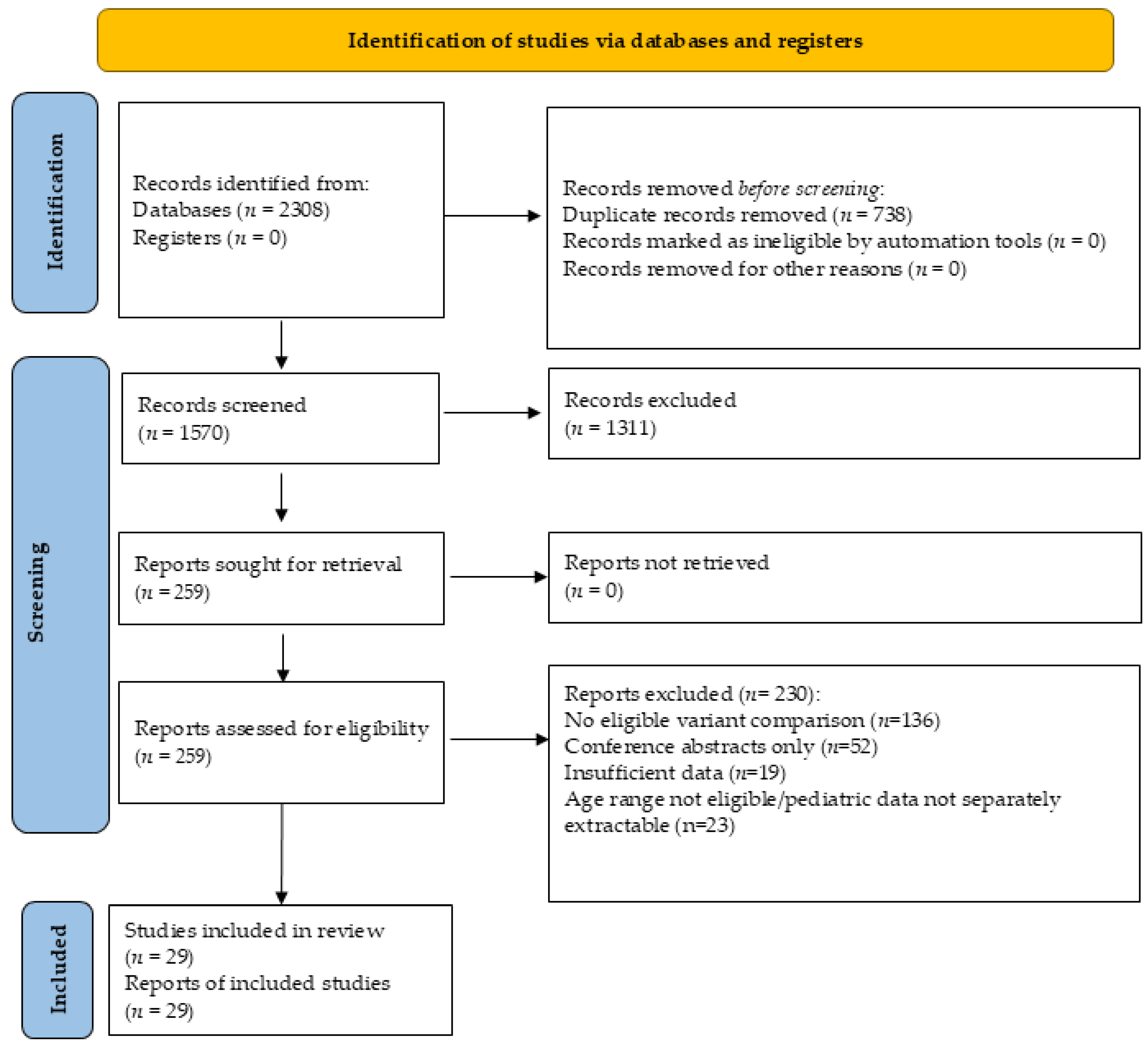
After duplicate removal, titles and abstracts were screened for eligibility, followed by full-text assessment of potentially relevant articles. Studies were included only when sufficient information was available to confirm eligibility and extract the required comparative data. Reasons for full-text exclusion are presented in the PRISMA flow diagram (Figure 1). Two reviewers independently screened titles and abstracts, assessed full-text eligibility, and extracted data from the included studies. Their results were subsequently compared, and any discrepancies were jointly reassessed and resolved by consensus. Potential overlap between study populations was assessed by comparing study settings, recruitment periods, data sources, and participating institutions. When publications appeared to originate from the same underlying cohort, their findings were examined for possible duplication and interpreted as related evidence rather than independent observations. Acute COVID-19 outcomes were considered separately from MIS-C. Where reported, we also distinguished hospitalizations attributable to COVID-19 from admissions with incidental SARS-CoV-2 detection.
The extracted information included study setting, recruitment period, patient age, sample size, method of variant identification, vaccination and prior infection status where reported, clinical manifestations, severity outcomes, and outcome definitions. We distinguished studies in which variants were identified through sequencing or variant-specific testing from those in which variants were assigned according to the predominant variant during a calendar period.
2.5. Risk of Bias and Synthesis
Two reviewers independently assessed the methodological quality of the included studies using the Newcastle–Ottawa Scale (NOS) for cohort studies. The NOS evaluates 3 categories: selection of study groups, comparability of groups and ascertainment of outcomes. It has a maximum score of 9 points. Studies scoring 7–9 points were considered high quality, 4–6 points moderate quality and 0–3 points low quality. Disagreements between the 2 reviewers were resolved by consensus. The NOS assessment considered the selection of study populations, the comparability of variant groups, and the reliability of outcome ascertainment. Particular attention was given to adjustment for potential confounders, including age, comorbidities, and prior immunity, where relevant and reported.
Findings were synthesized narratively by variant comparison, clinical manifestation, and healthcare setting. Outcomes measured among all children with confirmed infection, such as hospitalization, were presented separately from outcomes measured only among hospitalized children, such as oxygen treatment or intensive care admission. When there were conflicting results we considered the differences in study design, population and variant identification. Findings from studies with limited adjustment for confounding factors, indirect variant assignment based on calendar periods, or heterogeneous outcome definitions were interpreted with greater caution. NOS scores were not used as formal statistical weights in the narrative synthesis.
Figure 1.
PRISMA 2020 flow diagram of study identification, screening, eligibility assessment, and inclusion.
No formal assessment of reporting bias or certainty of evidence was performed.
3. Results
3.1. Study Characteristics
We included 29 studies. The included studies compared SARS-CoV-2 variants from the Alpha period through Gamma, Delta, Omicron, and subsequent Omicron variants. Several studies compared Delta directly with Omicron, while more recent investigations examined differences among BA.1/BA.2, BA.5/BQ.1, XBB and later Omicron subvariants. The study populations ranged from neonates and young children to adolescents. Methodological quality was generally moderate to high, with Newcastle–Ottawa Scale scores ranging from 5 to 8 points. Thirteen studies were classified as high quality and 16 as moderate quality; no study was classified as low quality (Supplementary Table S2). The studies included in our manuscript are summarized in Table 1.
Table 1.
Characteristics and main findings of comparative studies evaluating pediatric COVID-19 across SARS-CoV-2 variants.
Two publications by Shoji et al. were included. The first (Shoji et al. [26], 2022a) compared pediatric hospitalizations during the pre-Delta and Delta periods and the second study (Shoji et al., 2022 [28]) compared the Delta and Omicron periods. As both analyses used the Japanese national inpatient registry, some overlap in the Delta-period population is possible. Their findings were therefore considered as related evidence rather than entirely independent cohorts.
3.2. Clinical Presentation Before Omicron
Studies comparing pre-Omicron variants suggested that Delta variant was associated with higher frequency of symptomatic illness than Alpha variant, but findings were not uniform across settings. In an Israeli ambulatory cohort published by Ben-Tov et al. in 2022, symptomatic infection increased from 37.5% during Alpha to 49.9% during Delta, while fever prevalence increased from 13.6% to 33.0% [23]. In the UK prospective COVID Symptom Study published by Molteni et al. (2022), symptoms were recorded longitudinally through the ZOE COVID Study mobile application. The analysis included 694 symptomatic SARS-CoV-2-positive children during the Alpha-predominant period and 706 during the Delta-predominant period. Median illness duration was 5 days in both periods, but symptom burden was slightly greater during Delta. In children aged 5–11 years, the median number of symptoms increased from 3 (IQR 2–5) with Alpha to 4 (IQR 2–7) with Delta, while among those aged 12–17 years it increased from 5 (IQR 3–8) to 6 (IQR 3–9). Several individual symptoms were more frequent during Delta, including headache, rhinorrhea, sore throat, dysosmia, fever, dizziness, chills or shivering, eye soreness, and hoarse voice. For example, among children aged 5–11 years, headache increased from 39.9% during Alpha to 60.8% during Delta, while fatigue increased from 38.8% to 48.9% [22]. An Italian sequencing study published by Alteri et al. (2022) in the same period of time found no consistent association between viral variant (Alpha, Gamma, and Delta) and specific clinical manifestations, but Alpha (B.1.1.7) was negatively associated with hospitalization [24]. Yılmaz Çelebi et al. (2022) evaluated 680 symptomatic RT-PCR-positive children in Turkey who underwent individual SARS-CoV-2 variant analysis. Hospitalization occurred in 19.4% of children with Delta, compared with 9.4% with Alpha and 10.1% among children in whom variant analysis was negative. The hospitalization rate was also relatively high in the combined Beta/Gamma group (18%), although this group included only 17 children [31].
Differences in severe outcomes were also heterogeneous. Among sequenced infections in England, hospitalization after exclusion of injury-related admissions increased from 1.9% during Alpha to 2.5% during Delta, although the difference was small when restricted to symptomatic children (1.5% vs. 1.6%) [27]. In the Japanese national inpatient registry, ICU admission increased from 0.1% in the pre-Delta period to 1.4% during Delta, with no deaths, mechanical ventilation, or extracorporeal membrane oxygenation (ECMO) reported in either period [26]. A US sequencing-based cohort provided additional evidence regarding differences among earlier variants. Edward et al. (2022) evaluated 714 children aged ≤18 years whose SARS-CoV-2 samples underwent individual Illumina sequencing. After adjustment for age, race, ethnicity, high-risk medical conditions, and community COVID-19 incidence, Alpha, Delta, and Omicron were not independently associated with increased disease severity compared with non-VOC infections. In contrast, Gamma was associated with higher odds of hospitalization (OR 6.7, 95% CI 2.0–22.1), pharmacologic treatment (OR 5.7, 95% CI 1.2–26.8), respiratory support (OR 11.9, 95% CI 2.7–62.4), and severe disease according to the WHO Clinical Progression Scale (OR 11.7, 95% CI 2.1–90.5). However, one of the limitations is that only 38 Gamma infections were identified, resulting in wide confidence intervals [25]. In the study published by Yılmaz Çelebi et al. Delta was associated with a significantly higher hospitalization rate than Alpha and variant-negative infections [31].
3.3. Changes in Clinical Manifestations During the Omicron Period
Across studies comparing Delta and Omicron, several differences were observed in the clinical presentation of pediatric COVID-19, particularly in respiratory and neurologic manifestations.
3.3.1. Respiratory Manifestations
One of the most consistent clinical differences after the emergence of Omicron involved upper-airway manifestations, particularly croup. In a Korean hospital cohort, croup increased from 0.8% during Delta to 13.4% during Omicron, whereas pneumonia decreased from 12.9% to 4.2% [36]. Iijima et al. reported similar finding with an increase croup from 1.5% to 11.1% among children aged 0–4 years during the Omicron period [29]. Among 558 matched pairs from a Malaysian cohort, Omicron was associated with more croup (3.8% vs. 0.4%; OR 10.87, 95% CI 2.54–46.59) and lower respiratory tract infection (11.6% vs. 5.4%; OR 2.32, 95% CI 1.48–3.64) than Delta, however this comparison was restricted to hospitalized children [19].
In a Canadian multicenter emergency-department cohort, both Delta and Omicron were associated with increased odds of fever and cough compared with original-type SARS-CoV-2, but hospital and ICU admission did not differ significantly across variants.
Akaishi et al. (2022) found similar overall clinical presentation among Japanese children aged 0–3 years between the Delta and Omicron periods. The rates of fever, rhinorrhea, sore throat, vomiting, diarrhea, and headache did not differ significantly between the two periods, whereas cough increased from 29.7% during Delta to 43.8% during Omicron (pairwise p = 0.045) [30].
Kenney et al. (2023) also observed a change in the spectrum of clinical presentations among hospitalized US children during the Omicron BA.1.1 wave. Respiratory-related admissions were more prominent during Delta and presentations during Omicron were more heterogeneous and included an increased representation of croup and seizures [40].
3.3.2. Neurologic Manifestations
Neurologic manifestations, particularly seizures, were repeatedly reported more frequently during Omicron. In the Japanese national inpatient registry, seizures at presentation increased from 2.0% during Delta to 6.2% during Omicron [28]. In a Korean cohort, febrile seizures increased from 0.8% to 8.8% [36]. This pattern was particularly more pronounced in the Malaysian cohort published by Ng et al. (2024), in which seizures occurred in 21.9% of Omicron admissions compared with 3.2% of Delta admissions (OR 8.39, 95% CI 5.04–13.99) [19]. A similar shift toward neurological manifestations was observed by Sahin et al. (2023) among 283 hospitalized children in Turkey. Neurological symptoms were more frequent during the Omicron than the Delta period (31.1% vs. 14.0%, p = 0.03). Seizures were particularly prominent, occurring in 23/103 (22.3%) children during Omicron compared with 18/180 (10.0%) during the combined pre-Omicron periods (p = 0.005). Altered mental status was reported only during Omicron (3.9% vs. 0%, p = 0.017) [38].
3.3.3. Gastrointestinal Manifestations
Gastrointestinal findings were less consistent. Vomiting increased from 8.5% during Delta to 16.7% during Omicron in the Japanese inpatient registry and from 21.7% to 47.2% among children aged 5–11 years in the Iijima cohort [28,29]. In a Brazilian national registry that included 11,153 children younger than 2 years-old with SARS-CoV-2-associated severe acute respiratory syndrome, diarrhea was less frequently associated with Omicron than with Delta after adjustment for potential confounders (aOR 0.71 (95% CI 0.61–0.97)) [46]. Sahin et al. (2023) reported a decrease in gastrointestinal symptoms among 283 hospitalized children in Turkey, from 41.9% during Delta to 22.3% during Omicron (p = 0.018) [38]. Gastrointestinal manifestations also differed in the study published by Taytard et al. (2022), with diarrhea occurring in 33% of Omicron admissions compared with none during Delta (p = 0.01), and refusal to eat in 46% versus 7%, respectively (p = 0.01) [34].
3.4. Hospitalization and Severe Outcomes During Delta and Omicron
Population-based studies generally suggested a lower risk of severe outcomes with Omicron than Delta, although this was not consistently observed among cohorts restricted to hospitalized children.
In Qatar, Butt et al. (2022) compared 985 matched pairs of unvaccinated children without documented previous infection and found substantially lower odds of moderate/severe disease with Omicron than Delta (aOR 0.12, 95% CI 0.07–0.18) [33]. Similar reductions in acute clinical outcomes were reported by Wang et al. (2022) in a large US electronic health-record cohort of children younger than five years. After matching children with Omicron and Delta infections for age, sex, race and ethnicity, and relevant comorbidities, Omicron was associated with significantly lower 14-day risks of emergency-department attendance, hospitalization, ICU admission, and mechanical ventilation compared with Delta [32].
A study from Norway by Whittaker et al. (2022) reported that among 135,807 unvaccinated children without previous documented infection, acute COVID-19 hospitalization occurred in 21/10,538 Alpha, 28/42,362 Delta, and 48/82,907 Omicron BA.1 infections. Compared with Alpha, the adjusted risk was lower during both Delta (aRR 0.53) and Omicron (aRR 0.40), while no clear difference in acute-COVID hospitalization was observed between Omicron and Delta. The risk of multisystem inflammatory syndrome in children (MIS-C), however, was lower during Omicron than Delta, with MIS-C being diagnosed in 14 of 42,362 children during the Delta wave (0.03%) compared with 7 of 82,907 during the Omicron BA.1 wave (0.008%). After adjustment for demographic characteristics and underlying comorbidities, the risk of MIS-C was approximately 74% lower during Omicron than during Delta (aRR 0.26, 95% CI 0.10–0.63) [35].
Bahl et al. (2023) examined 4517 pediatric emergency-department encounters across Alpha-, Delta-, and Omicron-predominant periods in a large US healthcare system, of which 566 resulted in hospitalization. Hospital admissions increased numerically across the three periods, from 138 during Alpha and 179 during Delta to 249 during Omicron. Despite this increase, the adjusted odds of the composite severe-disease outcome were significantly lower during Omicron than during Alpha (aOR 0.35, 95% CI 0.21–0.60) [39].
In the Malaysian matched cohort, the moderate/severe disease was more frequent during Omicron than Delta (64.9% vs. 23.1%; OR 6.14, 95% CI 4.72–7.99), largely reflecting greater treatment requirements. However, severe disease alone did not differ significantly (1.3% vs. 2.2%), nor did PICU admission or mechanical ventilation [19]. Similar results were described by Choi et al. (2023) in a multicenter South Korean cohort of 612 hospitalized children, including 211 during the Delta-predominant period and 401 during the Omicron-predominant period. The proportion classified as having serious illness, defined as moderate, severe, or critical disease, was higher during Omicron than Delta (21.2% vs. 11.8%, p = 0.034). The increase was particularly evident for moderate illness among children aged 0–4 years (14.2% vs. 3.4%) and 5–11 years (18.6% vs. 4.2%) [41].
In the Canadian multicenter emergency-department cohort, Sumner et al. (2023) found no significant differences across SARS-CoV-2 variants in either hospitalization or ICU admission [37]. Similarly, in a US study restricted to hospitalized children younger than five years, Andersen et al. (2025) found comparable rates of ICU admission across the pre-Delta, Delta, and Omicron periods (20%, 21%, and 21%, respectively; p = 0.77). Invasive mechanical ventilation was also similar across the three periods (7%, 8%, and 7%, respectively; p = 0.35) [45].
3.5. Age-Specific Findings and Omicron Subvariants
Age substantially influenced the interpretation of variant-associated severity. Neonates represented a distinct high-risk population. In the UK national surveillance, 27 of 57 neonates hospitalized during the Delta period required invasive ventilation, and all four deaths attributed to neonatal SARS-CoV-2 infection occurred during Delta [43]. However, prematurity, maternal COVID-19, and the very small Omicron sample complicated direct variant comparisons.
Among 657 sequenced infections in Italian children aged ≤12 years, moderate/severe presentations occurred in 14.9% with BA.1, 6.4% with BA.2, 1.9% with BA.5, and 7.1% with BQ.1, although hospitalization did not differ significantly across variants [44]. A subsequent Italian genomic cohort encompassing BA.2-like, BA.5-like, XBB, and JN.1 infections found no statistically significant adjusted association between individual variants and hospitalization [47].
Pokorska-Śpiewak et al. (2024) compared 1098 children hospitalized during the predominance of early and later Omicron subvariants in Poland. The early-Omicron group included 575 children hospitalized between January and June 2022, when BA.1 and BA.2 predominated, whereas 523 children were hospitalized between July 2022 and April 2023, during the predominance of BA.5, BA.2.75, BQ.1, and XBB.1.5. Fever was significantly more frequent during the later Omicron period (87.0% vs. 79.0%), as was croup (3.0% vs. 1.2%, p = 0.03). In contrast, febrile seizures remained uncommon and did not differ significantly between the two periods (1.5% vs. 0.87%). Radiological pulmonary lesions were also infrequent, occurring in 6.5% of children during the later Omicron period compared with 4.0% during early Omicron. Despite these differences in clinical presentation, outcomes were favorable in both groups: no deaths, acute respiratory distress syndrome, or ICU admissions were reported. The authors also observed a longer mean duration of hospitalization and greater use of antiviral treatment during the later Omicron period [42].
More recent Japanese data also suggest that Omicron variants may differ in symptom profile without clear evidence of major differences in severity. In the Kobe emergency-department cohort of 4085 children, cough occurred in 37.9% during BA.1/BA.2, 39.1% during BA.5/BQ.1, and 46.0% during XBB, while nasal discharge was also more frequent during XBB. Vomiting was more frequent during BA.5/BQ.1 than BA.1/BA.2 [48].
4. Discussion
Our systematic review provides an updated synthesis of the clinical manifestations and severity of pediatric SARS-CoV-2 infection across successive viral variants, from early and pre-Alpha strains through Alpha, Gamma, Delta, Omicron, and more recent Omicron subvariants.
4.1. Changes in Clinical Presentation Across SARS-CoV-2 Variants
Our findings suggest that changes in SARS-CoV-2 variants were accompanied by measurable changes in the clinical phenotype of infection in children.
Comparisons between Alpha and Delta generally showed a greater symptomatic burden during Delta. Ben-Tov et al. reported an increase in symptomatic infection and fever during Delta, while Molteni et al. found a modest increase in the number of reported symptoms, including headache and fever, although the median illness duration remained 5 days during both periods [22,23]. This pattern was also supported by Yılmaz Çelebi et al., who used individual variant testing and found that hospitalization was approximately twice as frequent among children with Delta as among those with Alpha (19.4% vs. 9.4%) [31]. These findings suggest that the transition from Alpha to Delta affected the clinical expression of pediatric infection in some setting even when differences in severe outcomes were relatively modest.
Evidence regarding Gamma was more limited. The sequencing-based study by Edward et al. identified Gamma as being associated with substantially higher adjusted odds of hospitalization, respiratory support, and severe disease compared with non-VOC infections, but the wide confidence intervals surrounding these estimates, resulting partly from the relatively small number of Gamma infections, warrant caution [25]. These findings are consistent with the previous pediatric systematic review by Wiedenmann et al., in which more severe disease was particularly reported with Delta and Gamma variants [20]. We should also mention that their review included evidence available only through May 2022, whereas ours incorporates subsequent Delta–Omicron comparisons and more recent Omicron subvariants.
The transition to Omicron produced some changes in clinical presentation. There were several geographically independent studies that identified an increased in the frequency of croup or croup-like illness. This finding was observed across Asian, European, and North American populations, although the absolute frequencies differed substantially [29,36,40,42]. A possible biological explanation may be differences in respiratory tract replication. Experimental work by Hui et al. demonstrated that Omicron replicated more efficiently in ex vivo human bronchial tissue but less efficiently in the lung parenchyma than earlier SARS-CoV-2 variants [49]. In contrast with our other findings in the Malaysian cohort reported by Ng et al., lower respiratory tract infection was more frequent among hospitalized children with Omicron than among those with Delta [19]. The respiratory phenotype therefore appears to depend substantially on age, study setting, and the population from which patients were selected.
4.2. Neurologic and Gastrointestinal Manifestations During Omicron
A second relatively consistent finding was the increased frequency of seizures during the Omicron period. Increased seizures or febrile seizures were reported in several independent cohorts from Japan, South Korea, Malaysia, Turkey, and the United States [19,28,36,38,40]. This association was more pronounced in the Malaysian matched cohort reported by Ng et al., in which seizures were substantially more frequent with Omicron than Delta (21.9% vs. 3.2%) and in the Turkish study by Sahin et al. that found seizures in 22.3% of hospitalized children during Omicron compared with 10.0% during the combined pre-Omicron periods [19,38]. The interpretation requires caution because Omicron cohorts frequently contained a larger proportion of infants and young children, who inherently have a greater susceptibility to febrile seizures. Sahin et al., for example, reported a markedly younger population during the Omicron period [38]. Age distribution may therefore explain part of the observed increase. Furthermore, studies did not use identical definitions: some reported all seizures at presentation, whereas others specifically identified febrile seizures. Febrile seizures should be distinguished from other neurological manifestations, and their increased frequency does not necessarily indicate direct central nervous system involvement.
Gastrointestinal manifestations showed substantially less consistency. Vomiting increased during Omicron in the Japanese cohorts reported by Shoji et al. and Iijima et al., particularly among school-aged children in the latter study, whereas Sahin et al. reported fewer gastrointestinal symptoms during Omicron than Delta [28,29,38]. Gonçalves et al. also found lower adjusted odds of diarrhea with Omicron than Delta among Brazilian children younger than two years with severe acute respiratory syndrome [46]. Taytard et al. similarly reported more gastrointestinal involvement among hospitalized French children during Omicron than Delta, including diarrhea in 33% versus 0% and refusal to eat in 46% versus 7%, but the small sample size warrants caution [34]. Differences in age, disease severity, healthcare setting, and definitions of gastrointestinal involvement probably contributed to this heterogeneity. Therefore, unlike croup and seizures, the available evidence does not support a single consistent Omicron-associated gastrointestinal phenotype.
4.3. Disease Severity Across Delta and Omicron Variants
The relationship between viral variant and disease severity was more complex than the differences in symptom profile. At the population level, the most consistent finding was a reduction in severe outcomes during Omicron compared with Delta. In Qatar, Butt et al. found substantially lower adjusted odds of moderate or severe disease with Omicron than Delta among matched unvaccinated children without documented previous infection [33]. Similar reductions in acute healthcare outcomes were observed by Wang et al. in a large US cohort of children younger than five years, while Whittaker et al. reported very low absolute risks of acute COVID-19 hospitalization during both Delta and Omicron in Norway and a substantially lower risk of MIS-C during Omicron [32,35].
Findings from hospitalized pediatric populations were notably less consistent. Ng et al. reported substantially higher odds of the combined moderate/severe category during Omicron, while Choi et al. also found a greater proportion of moderate, severe, or critical disease among hospitalized South Korean children during the Omicron period [19,41].
Interpretation of these findings depends substantially on the population in which outcomes were assessed. Population-based cohorts address the probability that an infected child will subsequently require hospitalization or develop severe disease. In contrast, hospital-based studies begin after the decision to admit has already occurred and therefore describe the clinical spectrum conditional on hospitalization. Changes in admission thresholds, age distribution, healthcare-seeking behavior, testing practices, and incidental SARS-CoV-2 detection can substantially alter the composition of hospitalized cohorts. Consequently, a variant associated with a lower population-level risk of hospitalization may nevertheless account for a hospitalized population with a different or apparently more severe clinical profile. The definition of severity also differed considerably between studies. In the Malaysian cohort reported by Ng et al., the increased frequency of the combined moderate/severe category during Omicron was accompanied by greater treatment requirements, whereas severe disease considered separately, PICU admission, and mechanical ventilation did not show a significant increase [19]. Thus, treatment-defined or composite severity categories should not automatically be interpreted as evidence of greater intrinsic virulence. Another important issue is incidental infection. During periods of high community transmission, routine testing of hospitalized children can identify SARS-CoV-2 infection unrelated to the reason for admission. Kenney et al. found that incidental detection increased from 19% during Delta to 34% during Omicron. Failure to distinguish hospitalization with SARS-CoV-2 from hospitalization because of COVID-19 may therefore distort comparisons of severity across waves. This distinction is particularly important for Omicron because of its exceptionally high community incidence [40].
4.4. Age as an Important Factor of Variant-Associated Disease
Age emerged as an important determinant of both clinical presentation and apparent severity. Infants and younger children were disproportionately represented in several Omicron cohorts that may potentially reflect both biological susceptibility and changes in the clinical phenotype [36,38,41]. The increased frequency of croup during Omicron is particularly relevant to preschool-aged children, while differences in age distribution may also contribute to the observed increase in febrile seizures. Butt et al. additionally found that children aged 6–11 years and 12 to <18 years had lower adjusted odds of moderate or severe/critical disease than children younger than six years. These findings reinforce the need to consider age when comparing variant periods rather than treating the pediatric population as a homogeneous group [33].
Neonates represent an especially distinct population. UK national surveillance demonstrated substantial respiratory support requirements among hospitalized neonates during the Delta period, including invasive ventilation and deaths attributed to neonatal SARS-CoV-2 infection [43]. However, the interpretation of variant effects in neonates is particularly difficult because prematurity, maternal infection, perinatal transmission, and the small number of infections during some variant periods may strongly influence outcomes. Variant-specific conclusions in this age group therefore remain less certain than those for older children.
4.5. Evolution Within the Omicron Period
The available evidence suggests that differences exist among Omicron subvariants, but these differences are more apparent for individual symptoms than for major severity outcomes. In the Polish multicenter cohort reported by Pokorska-Śpiewak et al., later Omicron subvariants were associated with higher frequencies of fever and croup than BA.1/BA.2, while febrile seizures did not differ significantly and no deaths, ARDS, or ICU admissions occurred in either period [42]. Italian sequencing data reported by Scutari et al. (2024) similarly demonstrated differences in the proportion of moderate/severe presentations among BA.1, BA.2, BA.5, and BQ.1 infections, but hospitalization did not differ significantly among subvariants [44]. Another Italian genomic study published also by Scutari et al. at one year distance (2025) that consist of a cohort encompassing BA.2-like, BA.5-like, XBB, and JN.1 infections likewise found no statistically significant adjusted association between individual subvariant and hospitalization, whereas comorbidity remained strongly associated with hospitalization [47]. Japanese data from Yamaguchi et al. also suggest a continuous evolution of the symptom profile, with differences in cough, nasal discharge, and vomiting across BA.1/BA.2, BA.5/BQ.1, and XBB periods [48]. All together these findings suggest that Omicron should not be treated as a single clinically homogeneous period. Nevertheless, current pediatric evidence does not demonstrate a consistent progression toward increasing or decreasing severity across successive Omicron subvariants.
Future advances in SARS-CoV-2 variant identification may also benefit from emerging biosensing technologies based on two-dimensional materials [50]. These materials have promising electronic and optical properties, as demonstrated by studies of ultrathin tellurium nanosheets. More recently, a graphene-based photoelectrochemical biosensing platform combined with CRISPR/Cas12a demonstrated highly specific detection of genetic sequences associated with the Omicron BA.5 variant [51].
Differences in the classification of COVID-19-related and incidental hospitalizations may have influenced comparisons across variant periods. Nonsignificant findings should not be interpreted as evidence of equivalence between variants, particularly when event numbers were small or estimates were imprecise.
4.6. Strengths and Limitations
The strengths of this review are the following, it focuses specifically on direct comparisons between SARS-CoV-2 variants or clearly defined variant-predominant periods rather than describing isolated waves independently. It incorporates evidence from different healthcare settings and geographical regions and extends previous pediatric reviews to include more recent Omicron subvariants. Studies using individual sequencing or variant-specific testing were distinguished from those relying on calendar-period assignment, and population-level severity outcomes were considered separately from outcomes among already hospitalized children. This distinction was important for interpreting apparently conflicting findings.
Several limitations should also be acknowledged. Most included studies were observational, and comparisons were often based on sequential variant-predominant periods. Changes in population immunity, vaccination coverage, previous infection, testing practices, and hospitalization thresholds may therefore have influenced the observed differences. Variant ascertainment was also heterogeneous, ranging from individual sequencing or molecular testing to assignment based on calendar periods, with a potential for misclassification during periods of variant cocirculation. The potential for variant misclassification depended on the timing of the study periods. For example, Thelwall et al. reported that Delta accounted for less than 5% of sequenced SARS-CoV-2 infections in England in early April 2021, but more than 95% by June 2021 [27]. This rapid change suggests that studies using calendar periods close to the transition between variants may have included infections caused by different variants. Such misclassification would generally reduce observed differences in symptoms or severity if it occurred similarly across groups. However, the magnitude of this bias could not be reliably estimated because variant proportions within the pediatric study populations were not consistently available.
Considerable heterogeneity was also present in study populations and outcome definitions. Studies ranged from population-based cohorts to emergency-department and hospitalized populations, while definitions of disease severity and individual manifestations were not uniform. Age distributions also differed between variant periods, particularly during Omicron, when younger children were frequently overrepresented. This may have influenced the reported differences in clinical presentation, as manifestations such as croup are more common in younger children, regardless of the SARS-CoV-2 variant [35,41]. Age differences may also have affected comparisons of disease severity, since the risk of hospitalization and respiratory support varies across pediatric age groups [35]. Therefore, some of the observed changes between variants may reflect differences in age composition rather than variant-specific effects. However, the extent of age-related confounding could not be reliably quantified across studies because age-stratified or age-adjusted comparisons were not consistently available.
The NOS assessment highlighted differences in methodological quality, particularly regarding adjustment for potential confounders. Findings from studies with limited adjustment or indirect variant identification were therefore interpreted more cautiously, especially when results regarding hospitalization or disease severity were inconsistent. These differences limited direct comparisons across studies and precluded meaningful quantitative pooling, supporting the use of a narrative synthesis.
Another limitation concerns the distinction between hospitalization due to COVID-19 and hospitalization with an incidental SARS-CoV-2 infection. This may have affected comparisons of disease severity, particularly when admission screening practices differed between variant periods. Kenney et al. reported a higher proportion of incidental infections during Omicron than Delta (34% vs. 19%) and separately analyzed hospitalization rates after excluding incidental cases [40]. Future studies should distinguish these groups and to perform where possible sensitivity analyses excluding incidental infections or admissions unrelated to COVID-19. Comparing results before and after these exclusions would help determine whether incidental infections influenced the observed differences in hospitalization and severity across variants.
5. Conclusions
The clinical presentation of pediatric COVID-19 changed across successive SARS-CoV-2 variants. Delta was generally associated with a greater symptomatic burden than Alpha. Omicron was characterized by a shift in clinical phenotype with an increased frequency in upper-airway manifestations such as croup. Several studies also reported increased seizure frequency, including febrile seizures, although these findings should not be interpreted as evidence of direct central nervous system involvement. Gastrointestinal manifestations were less consistent across studies.
At the population level, Omicron was generally associated with a lower risk of hospitalization and severe outcomes than the Delta variant, although findings were less consistent among children already hospitalized. Differences in study setting, age distribution, prior immunity, variant ascertainment, and definitions of severity are important when interpreting these apparently conflicting results. Available evidence on later Omicron subvariants suggests variation in individual symptoms but no consistent trend in disease severity. Continued pediatric surveillance combining genomic characterization with standardized clinical outcomes is needed to define the clinical impact of emerging SARS-CoV-2 variants.
Supplementary Materials
The following supporting information can be downloaded at https://www.mdpi.com/article/10.3390/v18111227/s1, Table S1: PRISMA 2020 Checklist for the Systematic Review [52]; Table S2: Newcastle–Ottawa Scale assessment of the studies included in the review.
Author Contributions
Conceptualization, I.M.O.L., I.F.Ţ., A.D., A.C.D. and D.A.P.; methodology, I.M.O.L., A.D. and D.A.P.; software, I.M.O.L. and A.D.; validation, I.F.Ţ. A.C.D. and D.A.P.; writing—original draft preparation, I.M.O.L. and A.D.; writing—review and editing, I.F.Ţ., A.C.D. and D.A.P.; supervision, I.F.Ţ., A.C.D. and D.A.P.; All authors have read and agreed to the published version of the manuscript.
Funding
This research received no external funding.
Institutional Review Board Statement
Not applicable.
Informed Consent Statement
Not applicable.
Data Availability Statement
All data used in this systematic review were obtained from previously published studies. The main extracted data are presented in Table 1, and the Newcastle–Ottawa Scale quality assessments are provided in Supplementary Table S2. Additional extracted data and quality-assessment materials are available from the corresponding author upon request.
Conflicts of Interest
The authors declare no conflicts of interest.
Abbreviations
The following abbreviations are used in this manuscript:
| COVID-19 | Coronavirus disease 2019 |
| SARS-CoV-2 | Severe acute respiratory syndrome coronavirus 2 |
| NOS | Newcastle–Ottawa Scale |
| PICU | Pediatric intensive care unit |
References
- Chatterjee, P.; Nagi, N.; Agarwal, A.; Das, B.; Banerjee, S.; Sarkar, S.; Gupta, N.; Gangakhedkar, R.R. The 2019 Novel Coronavirus Disease (COVID-19) Pandemic. Indian J. Med. Res. 2020, 151, 147–159. [Google Scholar] [CrossRef] [Scilit] [PubMed]
- El Zawily, A.; Eckert, S.; Adajar, R.; Wagih, N.; Elmaidomy, A.H.; Helmy, A.M.; Mustafa, M.; Elshorbagi, M.; Ghali, E.; Fadl, R.G.; et al. Comprehensive Review on COVID-19: Etiology, Pathogenicity, and Treatment. Front. Med. 2025, 12, 1569013. [Google Scholar] [CrossRef] [Scilit] [PubMed]
- Păcurar, D.; Dinulescu, A.; Prejmereanu, A.; Palcău, A.C.; Dijmărescu, I.; Pavelescu, M.-L. Neurodevelopmental Outcomes in Children Born to Mothers Infected with SARS-CoV-2 During Pregnancy: A Narrative Review. J. Clin. Med. 2025, 14, 6202. [Google Scholar] [CrossRef] [Scilit] [PubMed]
- Kim, J.; Ewing, C.R.; Larson, A.; Illapperuma-Wood, C.; Fischer, A.; Duru, E.E.; Kim, Y.; Wilson, F. Incidence of Depression and Anxiety in Children and Adolescents Following COVID-19 Infection. PLoS ONE 2025, 20, e0331984. [Google Scholar] [CrossRef] [Scilit] [PubMed]
- Kamel, D.; Vu, M.H.; Bender, J.; Warburton, D.; Wood, J.C.; Mohandas, S. Exploring Long COVID in Pediatric Patients: Clinical Insights from a Long COVID Clinic. Front. Pediatr. 2025, 13, 1640747. [Google Scholar] [CrossRef] [Scilit] [PubMed]
- Păcurar, D.; Dinulescu, A.; Totu, A.-V.; Pavelescu, M.-L.; Dijmărescu, I. How Were Functional Somatic Symptoms in Children Influenced by the COVID-19 Pandemic? A Retrospective Study of Children Admitted to a Tertiary Pediatric Emergency Hospital in Bucharest, Romania. Life 2026, 16, 713. [Google Scholar] [CrossRef] [Scilit] [PubMed]
- Rothensteiner, M.; Leeb, F.; Götzinger, F.; Tebruegge, M.; Zacharasiewicz, A. Long COVID in Children and Adolescents: A Critical Review. Children 2024, 11, 972. [Google Scholar] [CrossRef] [Scilit] [PubMed]
- Zumla, A.; Hui, D.S.; Peiris, M.; Perlman, S. Respiratory Infections Due to Human Common Cold Coronaviruses, SARS-CoV, MERS-CoV, and SARS-CoV-2: Epidemiology, Pathogenesis, Clinical Features, Diagnostics, Therapeutics, and Vaccine Landscapes. Lancet Respir. Med. 2026, 14, 545–566. [Google Scholar] [CrossRef] [Scilit] [PubMed]
- de Souza, T.H.; Nadal, J.A.; Nogueira, R.J.N.; Pereira, R.M.; Brandão, M.B. Clinical Manifestations of Children with COVID-19: A Systematic Review. Pediatr. Pulmonol. 2020, 55, 1892–1899. [Google Scholar] [CrossRef] [Scilit] [PubMed]
- Galanopoulos, M.; Gkeros, F.; Doukatas, A.; Karianakis, G.; Pontas, C.; Tsoukalas, N.; Viazis, N.; Liatsos, C.; Mantzaris, G.J. COVID-19 Pandemic: Pathophysiology and Manifestations from the Gastrointestinal Tract. World J. Gastroenterol. 2020, 26, 4579–4588. [Google Scholar] [CrossRef] [Scilit] [PubMed]
- Trempelis, K.P.; Kosmeri, C.; Kalavas, P.; Ladomenou, F.; Siomou, E.; Makis, A. SARS-CoV-2 Variants and Their Impact on Pediatric COVID-19: Clinical Manifestations and Hematological Profiles. Diseases 2025, 13, 48. [Google Scholar] [CrossRef] [Scilit] [PubMed]
- Eun, B.W. The Changing Landscape of Pediatric Infectious Diseases before, during, and after the COVID-19 Pandemic. Child Health Nurs. Res. 2025, 31, 79–84. [Google Scholar] [CrossRef] [Scilit] [PubMed]
- World Health Organization (WHO). Tracking SARS-CoV-2 Variants. Available online: https://www.who.int/activities/tracking-SARS-CoV-2-variants (accessed on 26 September 2026).
- Carabelli, A.M.; Peacock, T.P.; Thorne, L.G.; Harvey, W.T.; Hughes, J.; de Silva, T.I.; Peacock, S.J.; Barclay, W.S.; de Silva, T.I.; Towers, G.J.; et al. SARS-CoV-2 Variant Biology: Immune Escape, Transmission and Fitness. Nat. Rev. Microbiol. 2023, 21, 162–177. [Google Scholar] [CrossRef] [Scilit] [PubMed]
- Liu, W.; Huang, Z.; Xiao, J.; Wu, Y.; Xia, N.; Yuan, Q. Evolution of the SARS-CoV-2 Omicron Variants: Genetic Impact on Viral Fitness. Viruses 2024, 16, 184. [Google Scholar] [CrossRef] [Scilit] [PubMed]
- Alhamlan, F.S.; Al-Qahtani, A.A. SARS-CoV-2 Variants: Genetic Insights, Epidemiological Tracking, and Implications for Vaccine Strategies. Int. J. Mol. Sci. 2025, 26, 1263. [Google Scholar] [CrossRef] [Scilit] [PubMed]
- Quintero, A.M.; Eisner, M.; Sayegh, R.; Wright, T.; Ramilo, O.; Leber, A.L.; Wang, H.; Mejias, A. Differences in SARS-CoV-2 Clinical Manifestations and Disease Severity in Children and Adolescents by Infecting Variant. Emerg. Infect. Dis. 2022, 28, 2278–2288. [Google Scholar] [CrossRef] [Scilit] [PubMed]
- Di Chiara, C.; Boracchini, R.; Sturniolo, G.; Barbieri, A.; Costenaro, P.; Cozzani, S.; De Pieri, M.; Liberati, C.; Zin, A.; Padoan, A.; et al. Clinical Features of COVID-19 in Italian Outpatient Children and Adolescents during Parental, Delta, and Omicron Waves: A Prospective, Observational, Cohort Study. Front. Pediatr. 2023, 11, 1193857. [Google Scholar] [CrossRef] [Scilit] [PubMed]
- Ng, D.C.; Liew, C.; Tan, K.K.; Chin, L.; Awang, E.H.B.; Chandirasekharan, D.A.; Surdi Roslan, M.J.B.; Jamil, M.B.; Zainol Abidin, N.Z.; Cheah, Y.K.; et al. Clinical Severity of Omicron and Delta SARS-CoV-2 Infections in Children. Pediatr. Int. 2024, 66, e15777. [Google Scholar] [CrossRef] [Scilit] [PubMed]
- Wiedenmann, M.; Ipekci, A.M.; Araujo-Chaveron, L.; Prajapati, N.; Lam, Y.T.; Alam, M.I.; L’Huillier, A.G.; Zhelyazkov, I.; Heron, L.; Low, N.; et al. SARS-CoV-2 Variants of Concern in Children and Adolescents with COVID-19: A Systematic Review. BMJ Open 2023, 13, e072280. [Google Scholar] [CrossRef] [Scilit] [PubMed]
- Somekh, I.; Stein, M.; Karakis, I.; Simões, E.A.F.; Somekh, E. Characteristics of SARS-CoV-2 Infections in Israeli Children During the Circulation of Different SARS-CoV-2 Variants. JAMA Netw. Open 2021, 4, e2124343. [Google Scholar] [CrossRef] [Scilit] [PubMed]
- Molteni, E.; Sudre, C.H.; Canas, L.D.S.; Bhopal, S.S.; Hughes, R.C.; Chen, L.; Deng, J.; Murray, B.; Kerfoot, E.; Antonelli, M.; et al. Illness Characteristics of COVID-19 in Children Infected with the SARS-CoV-2 Delta Variant. Children 2022, 9, 652. [Google Scholar] [CrossRef] [Scilit] [PubMed]
- Ben-Tov, A.; Lotan, R.; Gazit, S.; Chodick, G.; Perez, G.; Mizrahi-Reuveni, M.; Patalon, T. Dynamics in COVID-19 Symptoms during Different Waves of the Pandemic among Children Infected with SARS-CoV-2 in the Ambulatory Setting. Eur. J. Pediatr. 2022, 181, 3309–3318. [Google Scholar] [CrossRef] [Scilit] [PubMed]
- Alteri, C.; Scutari, R.; Costabile, V.; Colagrossi, L.; Yu La Rosa, K.; Agolini, E.; Lanari, V.; Chiurchiù, S.; Romani, L.; Markowich, A.H.; et al. Epidemiological Characterization of SARS-CoV-2 Variants in Children over the Four COVID-19 Waves and Correlation with Clinical Presentation. Sci. Rep. 2022, 12, 10194. [Google Scholar] [CrossRef] [Scilit] [PubMed]
- Edward, P.R.; Lorenzo-Redondo, R.; Reyna, M.E.; Simons, L.M.; Hultquist, J.F.; Patel, A.B.; Ozer, E.A.; Muller, W.J.; Heald-Sargent, T.; McHugh, M.; et al. Severity of Illness Caused by Severe Acute Respiratory Syndrome Coronavirus 2 Variants of Concern in Children: A Single-Center Retrospective Cohort Study. J. Pediatr. Infect. Dis. Soc. 2022, 11, 440–447. [Google Scholar] [CrossRef] [Scilit] [PubMed]
- Shoji, K.; Akiyama, T.; Tsuzuki, S.; Matsunaga, N.; Asai, Y.; Suzuki, S.; Iwamoto, N.; Funaki, T.; Ohmagari, N. Comparison of the Clinical Characteristics and Outcomes of COVID-19 in Children before and after the Emergence of Delta Variant of Concern in Japan. J. Infect. Chemother. 2022, 28, 591–594. [Google Scholar] [CrossRef] [Scilit] [PubMed]
- Thelwall, S.; Aiano, F.; Harman, K.; Dabrera, G.; Ladhani, S.N. Risk of Hospitalisation and Death in Children with SARS-CoV-2 Delta (B.1.612.2) Infection. Lancet Child Adolesc. Health 2022, 6, e16–e17. [Google Scholar] [CrossRef] [Scilit] [PubMed]
- Shoji, K.; Akiyama, T.; Tsuzuki, S.; Matsunaga, N.; Asai, Y.; Suzuki, S.; Iwamoto, N.; Funaki, T.; Ohmagari, N. Clinical Characteristics of COVID-19 in Hospitalized Children during the Omicron Variant Predominant Period. J. Infect. Chemother. 2022, 28, 1531–1535. [Google Scholar] [CrossRef] [Scilit] [PubMed]
- Iijima, H.; Kubota, M.; Ogimi, C. Clinical Characteristics of Pediatric Patients with COVID-19 between Omicron Era vs. Pre-Omicron Era. J. Infect. Chemother. 2022, 28, 1501–1505. [Google Scholar] [CrossRef] [Scilit] [PubMed]
- Akaishi, T.; Ishii, T. Coronavirus Disease 2019 Transmission and Symptoms in Young Children during the Severe Acute Respiratory Syndrome Coronavirus 2 Delta Variant and Omicron Variant Outbreaks. J. Int. Med. Res. 2022, 50. [Google Scholar] [CrossRef] [Scilit] [PubMed]
- Yılmaz Çelebi, M.; Kıymet, E.; Böncüoğlu, E.; Şahinkaya, Ş.; Cem, E.; Düzgöl, M.; Akaslan Kara, A.; Ayhan, F.Y.; Bayram, S.N.; Devrim, İ. Evaluation of Childhood Hospitalization Rates and Degree of Severity of SARS-CoV-2 Variants, Including B.1.1.7 (Alpha), B.1.315/P.1 (Beta/Gamma), and B.1.617.2 (Delta). J. Med. Virol. 2022, 94, 2050–2054. [Google Scholar] [CrossRef] [Scilit] [PubMed]
- Wang, L.; Berger, N.A.; Kaelber, D.C.; Davis, P.B.; Volkow, N.D.; Xu, R. Incidence Rates and Clinical Outcomes of SARS-CoV-2 Infection With the Omicron and Delta Variants in Children Younger Than 5 Years in the US. JAMA Pediatr. 2022, 176, 811. [Google Scholar] [CrossRef] [Scilit] [PubMed]
- Butt, A.A.; Dargham, S.R.; Loka, S.; Shaik, R.M.; Chemaitelly, H.; Tang, P.; Hasan, M.R.; Coyle, P.V.; Yassine, H.M.; Al-Khatib, H.A.; et al. Coronavirus Disease 2019 Disease Severity in Children Infected With the Omicron Variant. Clin. Infect. Dis. 2022, 75, e361–e367. [Google Scholar] [CrossRef] [Scilit] [PubMed]
- Taytard, J.; Prevost, B.; Schnuriger, A.; Aubertin, G.; Berdah, L.; Bitton, L.; Dupond-Athenor, A.; Thouvenin, G.; Nathan, N.; Corvol, H. SARS-CoV-2 B.1.1.529 (Omicron) Variant Causes an Unprecedented Surge in Children Hospitalizations and Distinct Clinical Presentation Compared to the SARS-CoV-2 B.1.617.2 (Delta) Variant. Front. Pediatr. 2022, 10, 932170. [Google Scholar] [CrossRef] [Scilit] [PubMed]
- Whittaker, R.; Greve-Isdahl, M.; Bøås, H.; Suren, P.; Buanes, E.A.; Veneti, L. COVID-19 Hospitalization Among Children <18 Years by Variant Wave in Norway. Pediatrics 2022, 150, e2022057564. [Google Scholar] [CrossRef] [Scilit] [PubMed]
- Han, M.S.; Kim, K.M.; Oh, K.J.; Chang, J.Y.; Lee, S.Y.; Choi, J.E.; Shin, S.-M.; Sun, J. Distinct Clinical and Laboratory Features of COVID-19 in Children During the Pre-Delta, Delta and Omicron Wave. Pediatr. Infect. Dis. J. 2023, 42, 423–428. [Google Scholar] [CrossRef] [Scilit] [PubMed]
- Sumner, M.W.; Xie, J.; Zemek, R.; Winston, K.; Freire, G.; Burstein, B.; Kam, A.; Emsley, J.; Gravel, J.; Porter, R.; et al. Comparison of Symptoms Associated With SARS-CoV-2 Variants Among Children in Canada. JAMA Netw. Open 2023, 6, e232328. [Google Scholar] [CrossRef] [Scilit] [PubMed]
- Sahin, A.; Karadag-Oncel, E.; Buyuksen, O.; Ekemen-Keles, Y.; Ustundag, G.; Elvan-Tuz, A.; Tasar, S.; Didinmez-Taskirdi, E.; Baykan, M.; Kara-Aksay, A.; et al. The Diversity in the Clinical Features of Children Hospitalized with COVID-19 during the Nonvariant, Alpha (B.1.1.7), Delta (B.1.617.2), and Omicron (B.1.1.529) Variant Periods of SARS CoV-2: Caution for Neurological Symptoms in Omicron Variant. J. Med. Virol. 2023, 95, e28628. [Google Scholar] [CrossRef] [Scilit] [PubMed]
- Bahl, A.; Mielke, N.; Johnson, S.; Desai, A.; Qu, L. Severe COVID-19 Outcomes in Pediatrics: An Observational Cohort Analysis Comparing Alpha, Delta, and Omicron Variants. Lancet Reg. Health Am. 2023, 18, 100405. [Google Scholar] [CrossRef] [Scilit] [PubMed]
- Kenney, P.O.; Chang, A.J.; Krabill, L.; Hicar, M.D. Decreased Clinical Severity of Pediatric Acute COVID-19 and MIS-C and Increase of Incidental Cases during the Omicron Wave in Comparison to the Delta Wave. Viruses 2023, 15, 180. [Google Scholar] [CrossRef] [Scilit] [PubMed]
- Choi, S.-H.; Choi, J.H.; Lee, J.K.; Eun, B.-W.; Song, S.H.; Ahn, B.; Kim, Y.K.; Yun, K.W. Clinical Characteristics and Outcomes of Children with SARS-CoV-2 Infection During the Delta and Omicron Variant-Dominant Periods in Korea. J. Korean Med. Sci. 2023, 38, e65. [Google Scholar] [CrossRef] [Scilit] [PubMed]
- Pokorska-Śpiewak, M.; Pawłowska, M.; Ciechanowski, P.; Peregrym, M.; Dobrzeniecka, A.; Sobolewska-Pilarczyk, M.; Franczak, J.; Majda-Stanisławska, E.; Szczepańska, B.; Zaleska, I.; et al. Differences in Clinical Presentation of COVID-19 in Children Hospitalized During Domination of Early (BA.1, BA.2) and Late (BA.5, BA.2.75, BQ.1 and XBB.1.5) SARS-CoV-2 Omicron Subvariants. Pediatr. Infect. Dis. J. 2024, 43, 149–154. [Google Scholar] [CrossRef] [Scilit] [PubMed]
- Gale, C.; Sharkey, D.; Fitzpatrick, K.E.; Mactier, H.; Morelli, A.; Nakahara, M.; Hurd, M.; Placzek, A.; Knight, M.; Ladhani, S.N.; et al. Characteristics and Outcomes of Neonates Hospitalised with SARS-CoV-2 Infection in the UK by Variant: A Prospective National Cohort Study. Arch. Dis. Child. Fetal Neonatal Ed. 2024, 109, 279–286. [Google Scholar] [CrossRef] [Scilit] [PubMed]
- Scutari, R.; Fox, V.; Fini, V.; Granaglia, A.; Vittucci, A.C.; Smarrazzo, A.; Lancella, L.; Calo’ Carducci, F.; Romani, L.; Cursi, L.; et al. Molecular Characterization of SARS-CoV-2 Omicron Clade and Clinical Presentation in Children. Sci. Rep. 2024, 14, 5325. [Google Scholar] [CrossRef] [Scilit] [PubMed]
- Andersen, K.M.; McColgan, M.D.; Cane, A.; Moran, M.M.; Reimbaeva, M.; Lopez, S.M.C. Comparison of COVID−19-Associated Inpatient Burden by Variant Period in Hospitalized Children Age <5 Years in the United States. Front. Pediatr. 2025, 13, 1573933. [Google Scholar] [CrossRef] [Scilit] [PubMed]
- Gonçalves, B.M.M.; Francisco, R.P.V.; Rodrigues, Á.S.; Junior, J.C.S. Coronavirus Disease 2019 Infection Severity among Different Variants in Children under 2-Years Old in Brazil. Clinics 2025, 80, 100592. [Google Scholar] [CrossRef] [Scilit] [PubMed]
- Scutari, R.; Fox, V.; Nguyen, J.L.; Colagrossi, L.; Smarrazzo, A.; Vittucci, A.C.; Cursi, L.; Mastropaolo, M.; Forquè Rodriguez, L.; D’amore, C.; et al. Clinical Manifestations and Severity of COVID-19 Caused by Omicron among Paediatric Patients Aged 0–17 Years in Italy. Sci. Rep. 2025, 15, 36638. [Google Scholar] [CrossRef] [Scilit] [PubMed]
- Yamaguchi, H.; Sameshima, T.; Fujioka, K.; Horinouchi, T.; Asagai, Y.; Ohashi, H.; Fujikawa, T.; Nishimura, A.; Tamura, A.; Ishimori, S.; et al. Pediatric Coronavirus Disease 2019 Clinical Characteristics in a Primary Emergency Medical Center: Comparative Analysis of BA.1/BA.2, BA.5/BQ.1, and XBB Periods. J. Infect. Chemother. 2026, 32, 102948. [Google Scholar] [CrossRef] [Scilit] [PubMed]
- Hui, K.P.Y.; Ho, J.C.W.; Cheung, M.; Ng, K.; Ching, R.H.H.; Lai, K.; Kam, T.T.; Gu, H.; Sit, K.-Y.; Hsin, M.K.Y.; et al. SARS-CoV-2 Omicron Variant Replication in Human Bronchus and Lung Ex Vivo. Nature 2022, 603, 715–720. [Google Scholar] [CrossRef] [Scilit] [PubMed]
- Xie, Z.; Xing, C.; Huang, W.; Fan, T.; Li, Z.; Zhao, J.; Xiang, Y.; Guo, Z.; Li, J.; Yang, Z.; et al. Ultrathin 2D Nonlayered Tellurium Nanosheets: Facile Liquid-Phase Exfoliation, Characterization, and Photoresponse with High Performance and Enhanced Stability. Adv. Funct. Mater. 2018, 28, 1705833. [Google Scholar] [CrossRef] [Scilit]
- Zhang, Y.; Chen, Z.; Wei, S.; Zhang, Y.; Fu, H.; Zhang, H.; Li, D.; Xie, Z. Detection of Biological Loads in Sewage Using the Automated Robot-driven Photoelectrochemical Biosensing Platform. Exploration 2024, 4, 20230128. [Google Scholar] [CrossRef] [Scilit] [PubMed]
- Page, M.J.; McKenzie, J.E.; Bossuyt, P.M.; Boutron, I.; Hoffmann, T.C.; Mulrow, C.D.; Shamseer, L.; Tetzlaff, J.M.; Akl, E.A.; Brennan, S.E.; et al. The PRISMA 2020 statement: An updated guideline for reporting systematic reviews. BMJ 2021, 372, n71. [Google Scholar] [CrossRef] [Scilit]

Disclaimer/Publisher’s Note: The statements, opinions and data contained in all publications are solely those of the individual author(s) and contributor(s) and not of MDPI and/or the editor(s). MDPI and/or the editor(s) disclaim responsibility for any injury to people or property resulting from any ideas, methods, instructions or products referred to in the content. |
© 2026 by the authors. Licensee MDPI, Basel, Switzerland. This article is an open access article distributed under the terms and conditions of the Creative Commons Attribution (CC BY) license.