


Abstract
The influence of some retroelement-derived genes is conflicting: their adaptive role is essential to promote specific biological processes, while their dysregulation can have profound consequences in disease pathology. A prominent example of this is Paternally Expressed Gene 10 (PEG10), a member of the Mart family of retrotransposons that has both gag- and pol-like domains. PEG10 preserves many features that are shared with retrotransposons, including the ability to bind and package RNA into virus-like particles and to undergo self-processing through its aspartic protease domain. Here, we explore the role of PEG10 in both adaptive and pathological biology. Throughout evolution, PEG10 has been co-opted to support placental development, playing a vital role in fetal vasculature formation within the mouse placenta and in trophoblast cell differentiation. However, the accumulation of PEG10 has been identified in two neurological diseases, Angelman’s syndrome and Amyotrophic Lateral Sclerosis, and in numerous malignancies, where its activity may influence disease pathology. Although considerable progress has been made, the adaptive and putative pathological mechanisms of PEG10 activity remain elusive, and future work will aim to isolate the exact processes that govern PEG10’s ability to influence both placental development and disease pathology.
1. Introduction
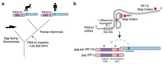
Nearly half of the human genome comprises transposable element-derived sequences that were a result of ancient viral and transposon DNA insertions [1,2]. For many years, these elements were referred to as “junk” DNA; however, it is now clear that throughout mammalian evolution, many transposable elements have been domesticated, co-opted, or exapted to assist in essential biological processes [3,4,5]. Broadly, two types of retroelements are found within the human genome: long terminal repeat (LTR)-containing elements, and non-LTR retrotransposons, which include the abundant LINE and SINE elements [6,7]. LTR retrotransposons, which include endogenous retroviruses, encode multiple reading frames, including a ‘gag’ structural protein, ‘pol’ enzymatic polyprotein that canonically includes protease, polymerase, and integrase, and envelope proteins that are necessary for replication within the genome [8]. In humans, there is limited evidence of LTR retrotransposon duplication; instead, most of these elements are degenerated and have lost LTRs as well as parts of their open reading frame [9,10]. Throughout evolution, however, some of these LTR retrotransposons have undergone a process of molecular domestication, where mutation of the genetic element has resulted in development of an adaptive biological function [5,11,12,13,14,15,16]. Paternally Expressed Gene 10 (PEG10) is one of these domesticated retrotransposons that is essential for placental development [17,18,19,20,21,22]. PEG10 belongs to the Mart (MAmmalian RetroTransposon) family of 11 neofunctionalized retrotransposon-derived genes and shares the greatest homology with the sushi-ichi retrotransposons of the Ty3/Gypsy family, Metaviridae [11,21,23,24,25,26,27]. Each member of the Mart family has varying degenerated forms of gag-like and pol- or protease-like protein domains, which roughly correlate with their nomenclature: Mar1/Retrotransposon Gag-like protein 1 (RTL1) or Hur1 is the longest gene in the Mart family, containing a gag and a pol-like domain, while Mar8/RTL8 encodes only 113 amino acids of a truncated gag domain (RTL9 is unique and encodes a pol-like fusion protein). PEG10, also known as Mar2, RTL2, or Edr, is the second-longest and, like Mar1, also contains gag and pol regions [24,25,26,27]. Not all Mart family members were inserted into the mammalian genome simultaneously: genomic analysis has shown that PEG10 first appeared in the genome of a therian ancestor during the emergence of placentation in mammals (Figure 1a) [15]. PEG10 is not found in monotremes, but does exist in therians, including marsupials, where it contains gag and pol regions, frameshifts, and only differs from placental mammals in a small portion of the very C-terminal region of the protein [15,28,29]. The conservation of PEG10 in placental mammals and marsupials, but not in non-placental mammals, suggests its adaptation assists in placental development [11,14]. Tissue expression profiling of adult humans and mice for PEG10 RNA levels shows low levels of expression in brain and endocrine tissues, with abundant PEG10 protein expression in the placenta, further supporting that this retrotransposon-derived element was adapted to promote placental reproduction [21,30,31]. As its name alludes, PEG10 is subject to monoallelic genomic imprinting in humans, mice, and wallaby, in which the paternal allele is expressed while the maternal allele is silenced through DNA methylation, meaning that placental tissues, derived from the fetus in early development, predominantly express the paternal allele of PEG10 [21,22,28]. A major challenge in studying PEG10 biology is that its expression is regulated on multiple levels. Some tissues with low levels of PEG10 mRNA show abundant protein expression; in contrast, some tissues demonstrate clear RNA expression of PEG10 with very little protein production.
Figure 1.
Schematic of PEG10 evolution and schematic of PEG10 mRNA and protein. (a) Schematized phylogenetic tree depicting the insertion of PEG10 into the genome of a mammalian ancestor around 125–200 million years ago (MYA), following the divergence from egg-laying monotremes. Marsupials express a truncated form of PEG10, as depicted by the dotted line at the C-terminus of gag-pol. (b) Simplified schematic of the gag and gag-pol proteins that are translated from PEG10 mRNA, which has a unique pseudoknot structure that causes −1 ribosomal frameshifting. The slippery sequence immediately 5′ of the pseudoknot is highlighted in blue. Stop codons are depicted with red octagons indicating the frame in which they are read. The gag region retains a capsid domain (CA) depicted in purple and a nucleocapsid domain (NC) depicted in dark pink. The pol domain contains the catalytically active aspartic protease domain depicted in dark blue, which promotes self-proteolysis at two locations, depicted by dotted lines and scissor symbols.
Although PEG10 has been adapted throughout mammalian evolution, there are key examples of conserved features that are reminiscent of its retroviral ancestry, including −1 ribosomal frameshifting, virus-like particle formation, and self-proteolysis [15,24,30,32]. PEG10 is one of very few domesticated genetic elements that consistently utilizes −1 ribosomal frameshifting to produce 2 protein products, sometimes referred to as reading frame 1 (RF1) and reading frame 1/2 (RF1/2), and alternatively as gag and gag-pol, from a single mRNA transcript (Figure 1b) [24,30,33,34]. Frameshifting is governed by the formation of an RNA pseudoknot surrounding the stop codon at the end of RF1/gag, which causes ribosomes to pause on a ‘slippery’ RNA sequence (XXX YYY Z, which in PEG10 is GGG AAA C), during unwinding of the pseudoknot [35,36]. About 30% of the time, stalling on this sequence causes ribosomes to slip backwards by one nucleotide (often referred to as a −1 frameshift), which results in a change in reading frame and translation of the RF1/2 or gag-pol fusion protein [30,33,37]. PEG10 gag includes a capsid (CA)-forming domain comprising the prototypical N-terminal domain (NTD) and C-terminal domain (CTD) lobes found in retrovirus and retrotransposon capsids, as well as an RNA-binding CCHC-type zinc finger, sometimes referred to as nucleocapsid (NC) [31,38,39,40]. RF1/2 or gag-pol harbors the catalytically active aspartic protease domain, which strongly resembles retroviral proteases like HIV-1 [41]. Like retroviruses, the PEG10 aspartic protease is capable of cleaving itself in trans in both gag and pol regions [29,38,41]. Protease activity is necessary for the formation of fetal vasculature during mid-to-late gestation in mice [18], and has been implicated in neurological disease [29]. However, the precise mechanism and cellular consequences of PEG10 protease activity remain largely unexplored.
PEG10 is also capable of forming virus-like particles (VLPs) that can incorporate mRNA [29,38,39,42,43]. Within domesticated genes derived from Metaviridae, there are examples of VLP assembly from members of the paraneoplastic Ma antigen (PNMA) family of domesticated gag-like capsid genes (PNMA1, PNMA2, and PNMA4) [44,45,46]. Of the Mart family, it appears that only RTL1 and PEG10 are sufficient to form VLPs [26,38], perhaps because many of the Mart genes have truncated CA domains that may be missing key sites for self-assembly [24,38]. However, some Mart family members have been shown to incorporate into PEG10 VLPs [39,43]. The CA domain of PEG10 consists of NTD and CTD lobes that have close homology to the corresponding domain in HIV-1 and Ty3, and typically mediate gag multimerization in retroviruses and retrotransposons [42,47,48,49,50]. Indeed, expression of either gag or gag-pol are both sufficient to promote the release of PEG10-containing particles from cells in culture [39], though no high-resolution structure of a PEG10 VLP has been solved. It is thought that PEG10 VLPs package mRNA via interactions between RNA and the NC domain [31,38,40,51] and PEG10-derived VLPs can deliver mRNA to neighboring cells, showing promising applications as drug delivery platforms [38,52,53]. Beyond their synthetic applications, PEG10 VLPs may play a physiological role in facilitating intercellular communication, similar to the domesticated retrotransposon, Arc, that has been co-opted to support neuronal function through VLP-mediated mRNA transfer across synapses [54,55].
2. PEG10 Is Essential for Placental Development
PEG10 emerged as a domesticated gene in a common mammalian ancestor at least 125 million years ago, and appears to be universal amongst placental mammals [15,26,56]. Notably, PEG10 is not the only retrovirus-derived domesticated element essential for placental development: ERVW-1 and ERVW-2, also known as Syncytin-A and -B, are retroviral envelope genes that have been adapted to promote placental cell fusion [57,58]. RTL1, another imprinted Mart family member, is also involved in placental development by maintaining fetal capillaries within the mouse placenta [59]. The mammalian genome has similarly adapted the gag-pol-derived PEG10 gene to support placental development, where it provides a vital role in fetal vasculature formation and trophoblast cell differentiation and function [17,18,19,20,31]. Consequently, its dysregulation has been implicated in pregnancy complications [60,61,62].
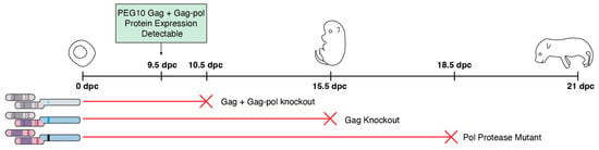
Peg10 plays a stage-dependent role to support placental development in mice. In mice, both Peg10 gag and gag-pol proteins are present starting at 9.5 days post coitum (dpc) and peak shortly thereafter, remaining highly expressed until birth [30]. The clearest evidence that Peg10 has developed an adaptive role in mammalian biology comes from a series of elegant genetic mouse models that demonstrate the essentiality of Peg10 to placental development [17,18,19,20] (Figure 2). Complete knockout of the Peg10 gene results in early embryonic lethality by 10.5 dpc, and examination of placental morphology reveals an underdeveloped labyrinth layer and a lack of spongiotrophoblast cells [17]. Mice harboring a mutant Peg10 protease (Peg10-ASG) have normal embryo and placental morphology at 12.5 dpc, but exhibit growth retardation beginning at 15.5 dpc and considerable fetal death at 18.5 dpc. Mutant Peg10-ASG mice have severe inflammation and abnormal organization of fetal capillaries within the labyrinth layers, highlighting the essential role of Peg10 protease activity from mid- to late-gestation [18]. Loss of Peg10 gag, while maintaining Peg10 gag-pol expression, also results in fetal growth retardation beginning at 12.5 dpc and lethality beginning at 15.5 dpc, owing to structural abnormalities in the fetal vasculature and inhibited cell differentiation [19].
Figure 2.
Peg10 is essential for placental development in mice. A series of studies in mice, as reviewed in Shiura et al. 2026 [20], demonstrated embryonic lethality and placental defects at various time points (dpc: days post coitum) following manipulation of Peg10, including deletion of both gag and gag-pol, deletion of solely gag, and inactivation of the Peg10 aspartic protease domain.
PEG10 is vital for trophoblast cell differentiation and function. Successful placentation requires careful orchestration of the differentiation and migration of multiple trophoblast stem cell (TSC) populations during fetal development [63,64,65,66]. In mice, Peg10 deletion in TSCs isolated from early blastocysts prevented their proper differentiation into functional trophoblast subtypes [31]. In humans, PEG10 silencing in JEG-3 cells and primary chorionic villi reduces cellular proliferation and invasion through modulating the expression of tissue inhibitor of metalloproteinases-1 (TIMP-1) [67]. PEG10-derived VLPs may play a role in these pathways; VLPs containing PEG10 are abundantly secreted by trophoblasts [31,39,62], and encapsulate numerous mRNAs, including its own. Recent work even suggests that PEG10 VLPs are capable of facilitating transfer of paternal alleles of mRNA transcripts from fetal tissue to maternal cells; however, other sources of transferred RNA, such as exosomes, have not been ruled out as a contributor [62].
PEG10 abundance may influence pregnancy complications. There is contradictory evidence for the influence of PEG10 in pregnancy complications. Tissue expression profiles show abundant PEG10 protein in both the human and mouse placental tissues [17,30,31,67]. In humans, PEG10 shows increased mRNA abundance at 11–12 weeks of gestation, which remains elevated at 38 weeks [68]. Some groups have observed elevated mRNA transcript levels in patients with preeclampsia [61,62], a serious hypertensive disorder which is one of the leading causes of maternal and neonatal morbidity and mortality worldwide [69,70,71]. However, early-onset preeclamptic placentas have also been reported to show decreased PEG10 expression [60]. Given that Peg10-deficient mouse models show underdeveloped placentas, the low PEG10 expression in early-onset preeclampsia could point to insufficient PEG10 as a cause of the disease; however, elevated PEG10 levels could also reflect failed compensatory mechanisms. Further investigation is required to generate a comprehensive understanding of the role that PEG10 may play in preeclampsia and whether its dysregulation is a driver or consequence of disease.
3. PEG10 in Neurological Diseases
Neurodegenerative diseases, from Alzheimer’s, to Parkinson’s, Huntington’s, and ALS, are often characterized by disruptions to protein homeostasis, resulting in the loss or accumulation of proteins that may contribute to disease pathology [72]. Mounting evidence has also linked dysregulation of transposable elements and their protein products in neurodegenerative disease [73,74,75], and more broadly in neurological and neuropsychiatric disease [74,76]. In particular, PEG10 protein levels are elevated and may contribute to disease pathology in the neurodevelopmental disorder Angelman syndrome (AS) [43] and neurodegenerative disease amyotrophic lateral sclerosis (ALS-FTD) [29, 77].
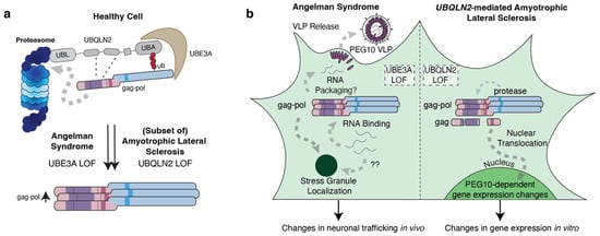
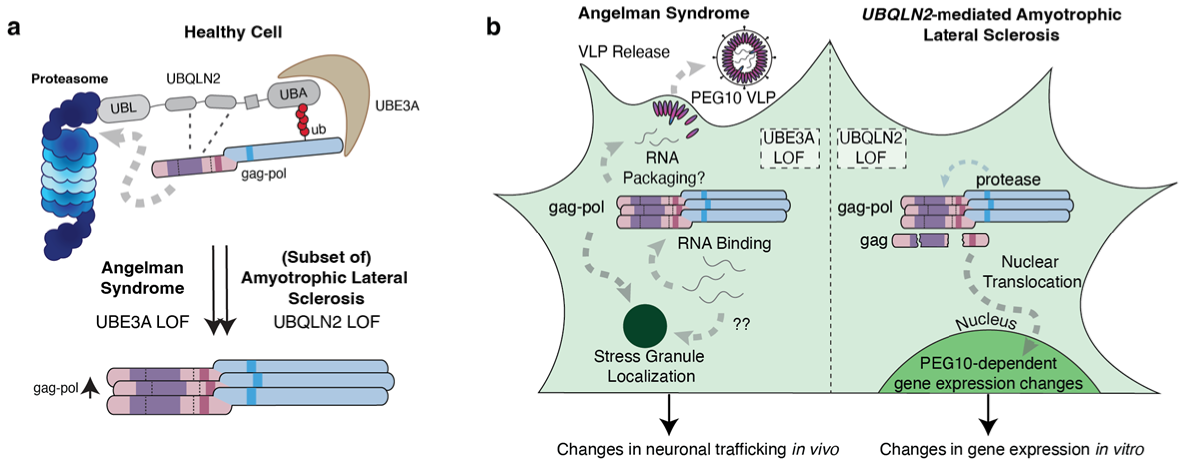
PEG10 gag-pol is degraded through the Ubiquitin-Proteasome System (UPS), which is disrupted in some neurological diseases. In two different neurological diseases, Angelman syndrome (AS) and ALS-FTD, PEG10 degradation via the ubiquitin-proteasome system (UPS) fails, which leads to the specific accumulation of gag-pol protein [29,43]. The neurodevelopmental disorder, Angelman syndrome, results from a genetic mutation of the maternal allele of UBE3A, an E3 ubiquitin ligase [78,79,80]. A subset of ALS-FTD cases results from mutations in the gene UBQLN2, a proteasomal shuttle factor that facilitates the degradation of client proteins [81,82,83,84]. Notably, UBE3A and UBQLN2 coordinate to regulate the proteasomal degradation of the gag-pol form of PEG10 (Figure 3a), but not gag. In both conditions, PEG10 accumulation appears to be limited to the protein level, with no evident changes to PEG10 mRNA in AS or ALS [43,85].
Figure 3.
PEG10 gag-pol is elevated in two neurological disorders: Angelman syndrome (AS) and Amyotrophic Lateral Sclerosis (ALS). (a) UBE3A, an E3 ligase, and UBQLN2, a proteasomal shuttle factor, coordinate to facilitate the degradation of PEG10 gag-pol via the proteasome in healthy cells. Angelman’s Syndrome is caused by UBE3A loss of function (LOF), and a subset of ALS, familial ALS with fronto-temporal dementia (ALS-FTD), is caused by mutations in UBQLN2 that leave it non-functional. As a result of these mutations, PEG10 gag-pol specifically accumulates in both diseases. (b) Elevated PEG10 gag-pol expression in AS neurons showed stress granule localization and enrichment in virus-like particles. In ALS, PEG10 gag-pol accumulation results in neuronal gene expression changes due to the small protein fragment containing a nucleic acid-binding domain that is liberated upon self-proteolysis.
While a molecular understanding is still lacking, the current model of PEG10 degradation seems to involve a two-step process: a first step, where either gag or gag-pol can engage with UBQLN2 and UBE3A, and a second step, where UBE3A-dependent ubiquitination of lysine residues present in the pol region dictates its degradation by the proteasome [43,85,86]. Consequently, disruptions to these two regulators both lead to the observed accumulation of PEG10 gag-pol, but not gag, in these diseases [43,85,87].
PEG10 gag-pol is elevated and may influence neuronal health in AS. AS is a neurodevelopmental disorder that is characterized by developmental delays, seizures, ataxia, microcephaly, and hyperactivity, and is caused by loss of function of the imprinted gene UBE3A [80,88,89]. Using AS patient-derived iPSCs and post-mortem AS patient neurons, Pandya et al. identified PEG10 gag-pol as being one of the most significantly elevated proteins in disease, owing to the disruptions in UBE3A-mediated regulation [43]. In AS neurons, PEG10 gag-pol, but not gag, was recruited to stress granules, consistent with previous reports that PEG10, as well as UBQLN2, localize to these compartments [43,90,91,92,93]. In addition, PEG10 gag-pol protein, including what appeared to be proteolytic products of self-processing, was enriched in extracellular vesicles from AS patients (Figure 3b) [43]. RNA-seq highlighted that elevated PEG10 expression results in the dysregulation of genes involved in neuronal migration and development, and in vivo overexpression of PEG10 gag-pol in embryonic AS mouse models restricted the ability of neurons to migrate effectively [43]. Although this study provides evidence of PEG10 as a potential contributor to neuronal dysfunction, it remains unclear whether PEG10 is a driver, modifier, or bystander of AS. UBE3A regulates the degradation of many different proteins, of which PEG10 is one, and it is likely that AS develops as a result of the dysregulation of many UBE3A substrates that regulate neuronal function [94,95,96]. Future work will be required to identify the precise role of PEG10 in neural function and its contribution to AS pathology.
PEG10 gag-pol protein also accumulates in Amyotrophic Lateral Sclerosis (ALS). Classical ALS is a neurodegenerative disease that typically manifests in the 5th to 7th decade of life and is hallmarked by the progressive loss of both upper and lower motor neuron function, resulting in progressive paralysis and death within 3–5 years post-diagnosis [97,98]. Some cases of ALS are complicated by the co-development of fronto-temporal dementia (FTD), which involves degeneration of neurons of the frontal cortex and frequently results in mood, behavior, and personality changes, and executive dysfunction [99,100]. Approximately 90% of ALS cases are sporadic, with no known genetic component; however, about 10% of ALS cases are familial and can often be traced to the mutation of over three dozen genes, including the proteasome shuttle factor UBQLN2 [81,84,101,102]. In animal models of Ubqln2-mediated ALS, and in a small cohort of human ALS patient tissues taken from sporadic disease, PEG10 gag-pol protein levels are significantly higher than controls and are amongst the most dramatically changed proteins in spinal cord tissue [29,77]. In cells overexpressing PEG10, activity of the PEG10 protease domain on gag and gag-pol proteins leads to the formation of a small RNA-binding protein fragment which moves to the nucleus (Figure 3b) [29]. RNA-seq following overexpression of just the small RNA-binding fragment in both HEK293 and SH-SY5Y cells showed cell-type-specific changes to axon and dendrite remodeling and RNA splicing pathways, respectively [29,40]. Notably, neuregulin 3 (NRG3), a gene involved in the neuregulin-ErbB4 pathway, which is involved in neuronal processes, exhibited altered mRNA splicing upon NC overexpression in SH-SY5Y cells and may represent one potential way that PEG10 dysregulation contributes to neuronal dysfunction [40]. Together, there is a growing body of evidence that elevated levels of PEG10 can influence neuronal phenotypes; however, it should be emphasized that a majority of work so far has been limited to overexpression studies in cell culture systems. Future work is necessary to tease apart the molecular mechanism behind PEG10’s ability to influence gene expression, and specifically, modulation of PEG10 in animal models of AS and ALS will be required to provide a causal link between PEG10 expression and disease phenotypes.
4. Similarities and Differences Amongst Neurological Diseases with PEG10 Accumulation
The independent investigations of PEG10 in AS and ALS-FTD highlight some notable convergent themes and suggest molecular mechanisms of PEG10 activity that may contribute to neuronal dysfunction. First, both studies specifically identify the long, gag-pol form of PEG10 as being accumulated in disease, while the gag form of PEG10 is unperturbed [29,43]. This specificity to gag-pol may suggest that the protease domain specifically contributes to the pathological role of PEG10, perhaps through formation of the nuclear, RNA-binding fragment that changes gene expression [29,40]. At the same time, PEG10 localization to stress granules (SGs) is also suspect, as these compartments influence cellular responses to stress and are altered in neurodegenerative disease [103], though it is unclear whether they play a role in Angelman’s syndrome. It is possible that increased gag-pol localization within SGs alters the pool of SG-sequestered mRNAs, resulting in changes to gene expression that alter neuron behavior. Another possibility is that elevated levels of PEG10 VLPs disrupt neuronal function by altering transcript abundance or availability; however, further work is required to isolate the mechanism by which PEG10 VLPs are involved. Collectively, PEG10 gag-pol presents itself as a promising biomarker and potential therapeutic target relevant to both AS and ALS-FTD.
Although PEG10 gag-pol accumulates in both AS and ALS-FTD, the disease presentation and onset have notable differences, which requires an explanation. To account for these differences, it could be speculated that both UBE3A and UBQLN2 have unique sets of client proteins that ultimately dictate the differences seen between both diseases. UBE3A has a diverse, but well-established, substrate pool of neuronal proteins that accumulate upon UBE3A mutation or loss [94,95,96]. UBQLN2 client proteins remain comparatively poorly defined, but also include proteins such as Huntingtin in addition to PEG10 [104,105]. The unique targets of UBE3A and UBQLN2 could contribute to the different phenotypes observed in these diseases. However, future work is required to determine precise PEG10-dependent effects that are shared between both disorders, and ultimately whether PEG10 can be considered a causative driver of disease.
Finally, PEG10 deletion has also been implicated in human disease, though the details of this rare disease, and the role of PEG10, remain poorly understood. A recent study found deletion of both PEG10 and its neighboring gene, epsilon-sarcoglycan gene (SGCE), in two individuals with Silver–Russell syndrome, a rare genetic disorder characterized by growth retardation, facial abnormalities, body asymmetry, and difficulty feeding [106]. Using patient samples, it was found that the co-deletion of PEG10 and SGCE led to an increase in CDKN1C in both patients, which was speculated to contribute to the growth defects observed. Although this study provided evidence of PEG10 in regulating the cell cycle in vitro, which will be discussed in further detail below, its co-deletion with SGCE means that the causal contribution of PEG10 and SGCE cannot be fully teased apart, and further work is required to gain a molecular understanding of the involvement of PEG10 in Silver–Russell Syndrome.
5. PEG10 in Cancer
Tissue invasion, proliferation, and migration are hallmarks of both trophoblasts and cancer cells [107,108]. PEG10 mRNA and protein levels are elevated in hepatocellular carcinoma (HCC), breast cancer, pancreatic cancer, and neuroendocrine prostate cancer (NEPC) [109,110,111,112,113,114], and its expression has been correlated with cancer cell proliferation, invasion, and migration in experimental models [111,112,115]. However, the mechanisms governing PEG10’s potential contribution to cancer remain poorly understood.
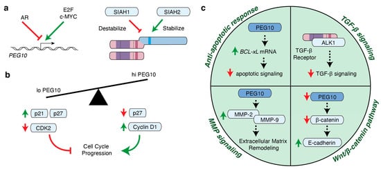
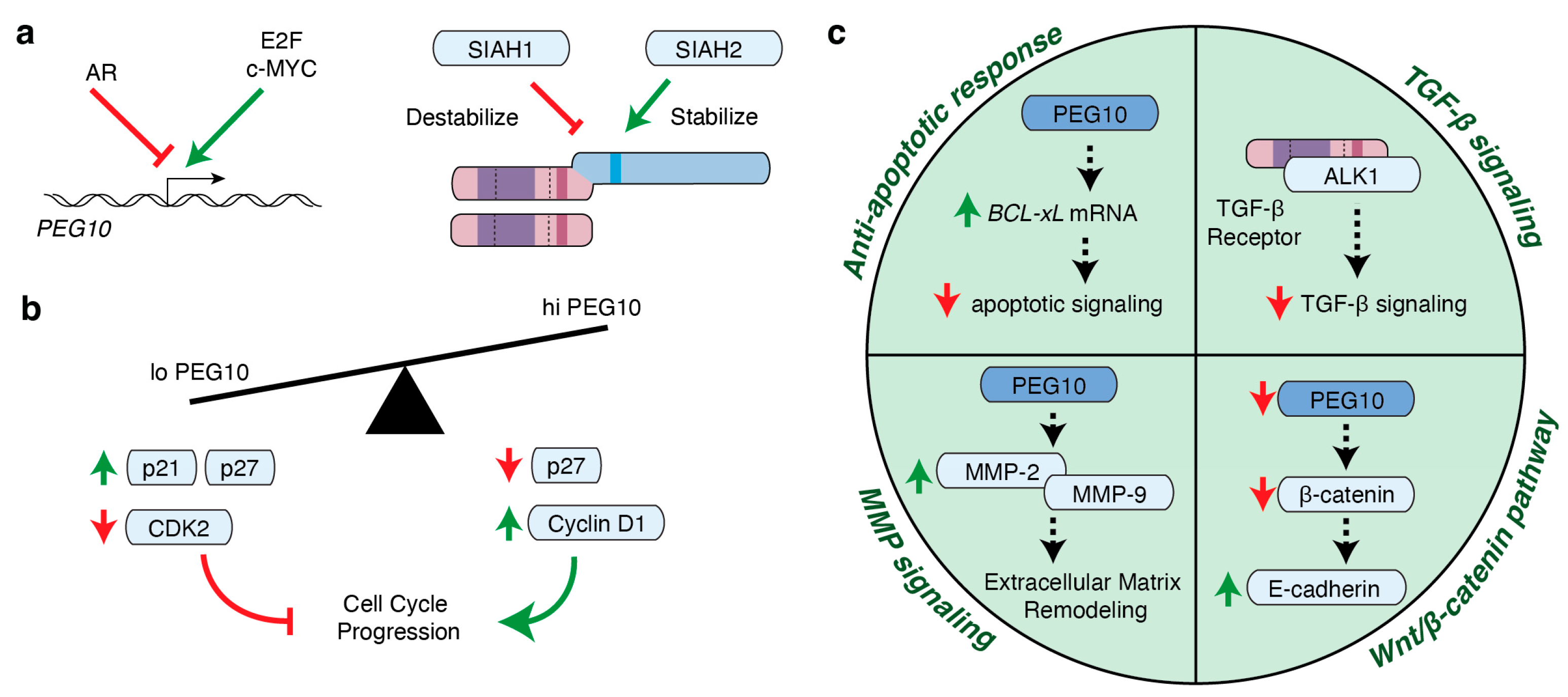
PEG10 is dynamically regulated by transcription factors in multiple cancer types. Consistent with many cancer-associated genes, PEG10 is regulated by multiple cancer-associated transcription factors including the androgen receptor (AR), members of the E2F family, and c-MYC. In healthy cells, the AR directly binds to the promoter element of PEG10 and is responsible for suppressing its expression [112,113] (Figure 4a). NEPC is a rare and aggressive AR-negative form of prostate cancer that typically emerges as a resistance mechanism to AR inhibition [116]. This loss of AR signaling coincides with increased PEG10 expression, which was shown to promote the proliferative and invasive nature of NEPC cells [112].
Figure 4.
PEG10 in cancer. (a) Simplified schematic of PEG10 regulation. Androgen receptor (AR) suppresses PEG10 expression (indicated by a red line), while E2F and c-MYC promote its expression (indicated by a green arrow). SIAH1 and SIAH2 regulate PEG10 protein expression by destabilizing (red line) and stabilizing PEG10 (green arrow), respectively. (b) Schematic of the effect of low or high PEG10 expression on cell cycle progression. Green upward arrows indicate an accumulation of the corresponding protein while red downward arrows indicate a loss of corresponding protein. The large green arrow or red line indicates the promotion or inhibition of the cell cycle, respectively. (c) Simplified schematic of the influence of PEG10 in signaling pathways, including anti-apoptotic response, TGF- β signaling, MMP signaling, and Wnt/β -catenin signaling. Green upward arrows indicate an accumulation of the corresponding transcript/protein while red downward arrows indicate a loss of corresponding transcript/protein.
E2Fs are a family of transcription factors that play essential roles in gene expression and cell cycle regulation; however, their aberrant activity has a profound impact on cancer cell proliferation [117]. In hepatocellular carcinoma (HCC) models, E2F-1 and E2F-4 directly bind to the PEG10 promoter and activate its expression [110]. E2F-1 also positively regulates PEG10 expression in models of pancreatic cancer, lung epithelial cells, and NEPC [112,115,118,119] (Figure 4a).
Finally, PEG10 was identified as a c-MYC target gene in Panc1 pancreatic cancer cells and HepG2 hepatocellular carcinoma cells [120]. c-MYC is an oncogenic transcription factor that has been extensively linked to tumorigenesis [121]. It was found that c-MYC binds to an enhancer region of PEG10, thereby promoting its expression. (Figure 4a) In murine c-MYC-driven mammary carcinomas, Peg10 mRNA was significantly elevated in comparison to normal mammary tissue. Additionally, PEG10 expression was found to positively correlate with c-MYC expression in both human breast cancer and prostate cancer [120]. However, other model systems suggest that PEG10 is not universally regulated by c-MYC signaling: semi-quantitative RT-PCR of c-MYC overexpressing HEK293 and HepG2 cells did not demonstrate significant changes to PEG10 expression compared to control cells [118]. Regardless, it is evident that the transcriptional regulation of PEG10 is coordinated by multiple transcription factors, and that its expression is elevated in many malignancies.
PEG10 promotes proliferation in cell culture systems through anti-apoptotic response and cell cycle regulation. Several studies have shown that PEG10 promotes proliferation in cell culture [113,120,122] through both the suppression of apoptosis and regulation of cell-cycle progression [109,110,111,112,115]. PEG10 interacts with SIAH1 and SIAH2, mediators of cellular apoptosis that promote and suppress apoptosis, respectively [109,123]. SIAH1 and SIAH2 are E3 ubiquitin ligases that mediate K48/63-linked ubiquitination of PEG10 but regulate PEG10 in opposing ways; SIAH1 destabilizes PEG10 while SIAH2 stabilizes PEG10 [123] (Figure 4a). Following SIAH1 overexpression, PEG10 cotransfection ameliorates apoptosis phenotypes, suggesting that SIAH1-mediated apoptosis is at least in part mediated by PEG10 [109,124]. Consistent with this finding, overexpression of PEG10 in hepatic cell lines reduced apoptosis and was accompanied by an accumulation of BCL-xL, an important anti-apoptotic protein [110] (Figure 4c). Although these studies suggest that PEG10 may inhibit apoptosis, they relied on the overexpression of PEG10 and, therefore, may not reflect its effects at physiological expression levels. Others found no statistical difference in apoptosis upon PEG10 depletion using different model systems, indicating that there may be cancer-specific or cell-type-specific effects of PEG10 expression [115].
In addition to apoptosis, PEG10 may influence cell cycle progression [111,112,115]. In breast cancer cells and HEK293 cells, overexpression of PEG10 promoted the progression of cells into S and G2/M phases [111,114]. In contrast, PEG10 knockdown in prostate and pancreatic cancer cells resulted in retention of cells in G0/G1 and cell cycle delay [112,115]. Further investigation identified a role for gag-pol in regulating the expression of p21 and p27, which inhibit critical cell cycle regulators, including Cyclin D1 and CDK2 [112,115]. Gag-pol overexpression results in the downregulation of p27 and results in upregulation of Cyclin D1, which is consistent with the observed rapid cell cycle progression in PEG10 overexpressing cancer cells [112]. In contrast, PEG10 knockdown results in upregulation of p21 and p27 and downregulation of CDK2 [112,115] (Figure 4b). Together, these provide evidence that PEG10 may promote cell proliferation through both the suppression of apoptotic factors and the regulation of the cell cycle, although further work is needed to determine the contribution of PEG10 to cancer proliferation in vivo.
PEG10 promotes tumor invasion and migration. Degradation of the extracellular matrix (ECM) allows malignant cells to remodel the tumor microenvironment while releasing growth factors that promote cancer cell migration and eventual metastasis [125,126]. Two key proteins involved in this are MMP-2 and MMP-9, which not only function in placental cell invasion but are also responsible for ECM degradation by cleaving type IV collagens [127,128]. Consistent with its role in the placenta, PEG10 knockdown in cancer cells results in a reduction in both the mRNA and protein abundance of MMPs, while overexpression in the same cell types promotes their accumulation [111,115,122,129] (Figure 4c).
PEG10 also influences ECM remodeling as well as cancer cell proliferation through its involvement in signaling pathways. TGF-β signaling promotes ECM stiffness by increasing collagen production in fibroblasts, and can have complex effects on proliferation depending on the cell type involved [126,130]. The gag region of PEG10 directly interacts with activin receptor-like kinase 1 (ALK1), a TGF-β type 1 receptor, which results in decreased TGF-β signaling in luciferase reporter assays when PEG10 is overexpressed [131] (Figure 4c). Additional evidence points to TGF-β and PEG10 regulating mutually exclusive signaling outcomes: PEG10 depletion in chondrosarcoma cells increased the abundance of phosphorylated SMADs, which are regulated by TGF-β [132]. However, knockdown of PEG10 rendered NEPC cell lines nonresponsive to TGF-β treatment in invasion and scratch assays, and even resulted in decreased SMAD phosphorylation, suggesting that PEG10 is in some cases necessary for TGF-β signaling [112]. PEG10 has also been connected to Wnt/β-catenin signaling, which is involved in mediating cell adhesion in many cancers, where silencing of PEG10 reduced the expression of β-catenin and, in turn, increased the expression of E-cadherin [122] (Figure 4c).
6. Possible Mechanisms Involve Signaling Hubs and Transcriptional Reprogramming
A major question that remains surrounds the specific mechanism by which PEG10 regulates cell behavior in placentation and disease. Much of PEG10’s presumed function stems from its RNA-binding domain, which has been shown to bind to its own 3′ UTR [38], as well as diverse cellular mRNAs [31,38,43,133]. Considerable focus has also been given to the biological implications of the formation and contents of PEG10-derived VLPs [31,43], thought to be generated through self-assembly of the capsid domains of the gag region. However, it remains unclear how these two features govern PEG10-dependent cellular behavior. In some cases, PEG10 binding to particular RNA transcripts correlates with their increased abundance by RNA-Seq: this was hypothesized to contribute to trophoblast differentiation in Peg10-expressing mouse TSCs [31]. In TSCs, these transcripts were enriched for those involved in TSC differentiation [31]; in neurons and HEK293 cells, PEG10 overexpression leads to upregulation of transcripts involved in cytoskeletal remodeling and migration [29,40,43]. Consequently, there appear to be considerable cell-type-specific effects of PEG10 expression that warrant further investigation. However, it remains unclear whether the dominant effect of PEG10 expression is through direct RNA binding and enhancement of RNA half-life.
Another putative mechanism is through regulation of mRNA splicing. Recent work in SH-SY5Y cells showed that PEG10 overexpression causes additional changes to the expression of splicing regulators such as HNRNP family members and SFPQ [40], which in some cases also bind to PEG10 via protein:protein interactions [43]. PEG10 overexpression alters RNA splicing patterns, which can have profound effects on gene expression by generating alternative isoforms, or perhaps by generating dysfunctional mRNA products. Future work is necessary to determine whether this splicing effect is direct or indirect, and how PEG10 influences other disease-relevant splicing factors, such as TDP-43. PEG10 localization may also play a large role in determining whether binding to RNA influences pre-mRNA splicing or mRNA half-life, and given the complex self-regulation and localization patterns of PEG10 and its proteolytic fragments [29,43,90,131], additional work is necessary to tease apart these possibilities.
In addition to RNA interactions, PEG10 influences cell behavior through the regulation of signal transduction cascades, like the TGF- β and Wnt/β-catenin signaling pathways, which have so far only been investigated in the context of cancers, highlighted above [122,131,132]. Perhaps most importantly, it is unknown whether these interactions are dependent on PEG10’s RNA-binding ability, or are somehow indirectly linked.
Finally, there remains significant interest in determining how VLPs influence the ability of PEG10 to alter cellular behavior. Spontaneous release of PEG10-derived VLPs has been observed from placenta [31], neurons [39,43], and multiple cell lines [39]. In some cases, these VLPs have been found to contain RNA, which has ranged from a diversity of cellular mRNAs [43] to predominantly its own mRNA [38]. While it is still unclear how VLPs contribute to PEG10-dependent changes in cell behavior, the domesticated gag-like gene Arc provides key clues [134]. Like PEG10, Arc forms VLPs released from neurons that contain RNA [54,55], and changes to the expression of Arc have significant effects on neuronal function. Dysregulation of Arc results in memory deficits [135,136], and it appears that formation of the VLP is likely central to its ability to influence neuronal function through the intercellular delivery of RNA [54,55]. Future work is underway to determine to what extent PEG10 dysregulation does something similar. While PEG10 VLPs have been experimentally verified to transfer engineered RNAs from one cell to another [38,53,137], it is unclear whether this also occurs in the context of placentation, neurological disease, or cancer. However, it remains a strong possibility that PEG10 can influence cell behavior in trans by delivering RNAs via VLP from one cell to another.
7. Conclusions
Scattered throughout the human genome are remnants of retroelement-derived genes, some of which have been domesticated and contribute to adaptive biological processes [3,4,5]. The physiological influence of domesticated retroelements is sometimes, however, two-fold, where they not only contribute to biology, but their aberrant activity has been implicated in disease. PEG10, a retroviral gag-pol element, was inserted into the genome of a mammalian ancestor nearly 200 million years ago and is essential for placental development [15,17,18]. However, PEG10 has also been implicated in neurological disease and cancer [29,43,109,111,112,115]. Continued investigation into the activities of domesticated elements, including PEG10, will provide vital insight into how these rare genes have shaped human physiology.
Author Contributions
Conceptualization, A.M.W. and M.E.R.; writing—original draft preparation, M.E.R.; writing—review and editing, A.M.W. and M.E.R.; visualization, A.M.W. and M.E.R.; funding acquisition, A.M.W. and M.E.R. All authors have read and agreed to the published version of the manuscript.
Funding
This research was funded by NIH R01 NS131660, T32 GM145437, and DoD CDMRP HT94252510110.
Data Availability Statement
No new data were created or analyzed in this study. Data sharing is not applicable to this article.
Acknowledgments
We sincerely apologize for those whose contributions could not be cited due to space restrictions.
Conflicts of Interest
A.M. Whiteley is a co-founder of Endios Bio and is an author on patents surrounding the inhibition of PEG10 as therapeutics.
References
- Lander, E.S.; Linton, L.M.; Birren, B.; Nusbaum, C.; Zody, M.C.; Baldwin, J.; Devon, K.; Dewar, K.; Doyle, M.; FitzHugh, W.; et al. Initial sequencing and analysis of the human genome. Nature 2001, 409, 860–921. [Google Scholar] [CrossRef] [Scilit]
- Cordaux, R.; Batzer, M.A. The impact of retrotransposons on human genome evolution. Nat. Rev. Genet. 2009, 10, 691–703. [Google Scholar] [CrossRef] [Scilit]
- Modzelewski, A.J.; Gan Chong, J.; Wang, T.; He, L. Mammalian genome innovation through transposon domestication. Nat. Cell Biol. 2022, 24, 1332–1340. [Google Scholar] [CrossRef] [Scilit]
- Almeida, M.V.; Vernaz, G.; Putman, A.L.K.; Miska, E.A. Taming transposable elements in vertebrates: From epigenetic silencing to domestication. Trends Genet. 2022, 38, 529–553. [Google Scholar] [CrossRef] [Scilit]
- Volff, J.-N. Turning junk into gold: Domestication of transposable elements and the creation of new genes in eukaryotes. BioEssays 2006, 28, 913–922. [Google Scholar] [CrossRef] [Scilit]
- Wicker, T.; Sabot, F.; Hua-Van, A.; Bennetzen, J.L.; Capy, P.; Chalhoub, B.; Flavell, A.; Leroy, P.; Morgante, M.; Panaud, O.; et al. A unified classification system for eukaryotic transposable elements. Nat. Rev. Genet. 2007, 8, 973–982. [Google Scholar] [CrossRef] [Scilit]
- Bourque, G.; Burns, K.H.; Gehring, M.; Gorbunova, V.; Seluanov, A.; Hammell, M.; Imbeault, M.; Izsvák, Z.; Levin, H.L.; Macfarlan, T.S.; et al. Ten things you should know about transposable elements. Genome Biol. 2018, 19, 199. [Google Scholar] [CrossRef] [Scilit]
- Thompson, P.J.; Macfarlan, T.S.; Lorincz, M.C. Long Terminal Repeats: From Parasitic Elements to Building Blocks of the Transcriptional Regulatory Repertoire. Mol. Cell 2016, 62, 766–776. [Google Scholar] [CrossRef] [Scilit]
- Vargiu, L.; Rodriguez-Tomé, P.; Sperber, G.O.; Cadeddu, M.; Grandi, N.; Blikstad, V.; Tramontano, E.; Blomberg, J. Classification and characterization of human endogenous retroviruses; mosaic forms are common. Retrovirology 2016, 13, 7. [Google Scholar] [CrossRef] [Scilit]
- Griffiths, D.J. Endogenous retroviruses in the human genome sequence. Genome Biol. 2001, 2, reviews1017.1–reviews1017.5. [Google Scholar] [CrossRef] [Scilit]
- Kokošar, J.; Kordiš, D. Genesis and Regulatory Wiring of Retroelement-Derived Domesticated Genes: A Phylogenomic Perspective. Mol. Biol. Evol. 2013, 30, 1015–1031. [Google Scholar] [CrossRef] [Scilit]
- Etchegaray, E.; Naville, M.; Volff, J.-N.; Haftek-Terreau, Z. Transposable element-derived sequences in vertebrate development. Mob. DNA 2021, 12, 1. [Google Scholar] [CrossRef] [Scilit]
- Wang, J.; Han, G.-Z. Frequent Retroviral Gene Co-option during the Evolution of Vertebrates. Mol. Biol. Evol. 2020, 37, 3232–3242. [Google Scholar] [CrossRef] [Scilit]
- Kaneko-Ishino, T. The role of genes domesticated from LTR retrotransposons and retroviruses in mammals. Front. Microbiol. 2012, 3, 262. [Google Scholar] [CrossRef] [Scilit]
- Henriques, W.S.; Young, J.M.; Nemudryi, A.; Nemudraia, A.; Wiedenheft, B.; Malik, H.S. The Diverse Evolutionary Histories of Domesticated Metaviral Capsid Genes in Mammals. Mol. Biol. Evol. 2024, 41, msae061. [Google Scholar] [CrossRef] [Scilit]
- Jangam, D.; Feschotte, C.; Betrán, E. Transposable Element Domestication As an Adaptation to Evolutionary Conflicts. Trends Genet. 2017, 33, 817–831. [Google Scholar] [CrossRef] [Scilit]
- Ono, R.; Nakamura, K.; Inoue, K.; Naruse, M.; Usami, T.; Wakisaka-Saito, N.; Hino, T.; Suzuki-Migishima, R.; Ogonuki, N.; Miki, H.; et al. Deletion of Peg10, an imprinted gene acquired from a retrotransposon, causes early embryonic lethality. Nat. Genet. 2006, 38, 101–106. [Google Scholar] [CrossRef] [Scilit]
- Shiura, H.; Ono, R.; Tachibana, S.; Kohda, T.; Kaneko-Ishino, T.; Ishino, F. PEG10 viral aspartic protease domain is essential for the maintenance of fetal capillary structure in the mouse placenta. Development 2021, 148, dev199564. [Google Scholar] [CrossRef] [Scilit]
- Shiura, H.; Fujii, M.; Ooga, M.; Wakayama, S.; Ito, D.; Kohda, T.; Kaneko-Ishino, T.; Ishino, F. PEG10-ORF1 programs trophoblast progenitor development for placental labyrinth formation. bioRxiv 2025. bioRxiv:2025.10.08.681076. [Google Scholar] [CrossRef] [Scilit]
- Shiura, H.; Kitazawa, M.; Kaneko-Ishino, T.; Ishino, F. Placenta-Driven Evolution: Viral Gene Acquisition and PEG10’s Essential Roles in Eutherian Placenta. Biomolecules 2026, 16, 161. [Google Scholar] [CrossRef] [Scilit]
- Ono, R.; Kobayashi, S.; Wagatsuma, H.; Aisaka, K.; Kohda, T.; Kaneko-Ishino, T.; Ishino, F. A Retrotransposon-Derived Gene, PEG10, Is a Novel Imprinted Gene Located on Human Chromosome 7q21. Genomics 2001, 73, 232–237. [Google Scholar] [CrossRef] [Scilit]
- Koppes, E.; Himes, K.P.; Chaillet, J.R. Partial Loss of Genomic Imprinting Reveals Important Roles for Kcnq1 and Peg10 Imprinted Domains in Placental Development. PLoS ONE 2015, 10, e0135202. [Google Scholar] [CrossRef] [Scilit]
- Campillos, M.; Doerks, T.; Shah, P.K.; Bork, P. Computational characterization of multiple Gag-like human proteins. Trends Genet. 2006, 22, 585–589. [Google Scholar] [CrossRef] [Scilit]
- Brandt, J.; Veith, A.M.; Volff, J.N. A family of neofunctionalized Ty3/gypsy retrotransposon genes in mammalian genomes. Cytogenet. Genome Res. 2005, 110, 307–317. [Google Scholar] [CrossRef] [Scilit]
- Volff, J.-N.; Körting, C.; Schartl, M. Ty3/Gypsy Retrotransposon Fossils in Mammalian Genomes: Did They Evolve into New Cellular Functions? Mol. Biol. Evol. 2001, 18, 266–270. [Google Scholar] [CrossRef] [Scilit][Green Version]
- Brandt, J.; Schrauth, S.; Veith, A.-M.; Froschauer, A.; Haneke, T.; Schultheis, C.; Gessler, M.; Leimeister, C.; Volff, J.-N. Transposable elements as a source of genetic innovation: Expression and evolution of a family of retrotransposon-derived neogenes in mammals. Gene 2005, 345, 101–111. [Google Scholar] [CrossRef] [Scilit]
- Naville, M.; Warren, I.A.; Haftek-Terreau, Z.; Chalopin, D.; Brunet, F.; Levin, P.; Galiana, D.; Volff, J.-N. Not so bad after all: Retroviruses and long terminal repeat retrotransposons as a source of new genes in vertebrates. Clin. Microbiol. Infect. 2016, 22, 312–323. [Google Scholar] [CrossRef] [Scilit]
- Suzuki, S.; Ono, R.; Narita, T.; Pask, A.J.; Shaw, G.; Wang, C.; Kohda, T.; Alsop, A.E.; Graves, J.A.M.; Kohara, Y.; et al. Retrotransposon Silencing by DNA Methylation Can Drive Mammalian Genomic Imprinting. PLoS Genet. 2007, 3, e55. [Google Scholar] [CrossRef] [Scilit]
- Black, H.H.; Hanson, J.L.; Roberts, J.E.; Leslie, S.N.; Campodonico, W.; Ebmeier, C.C.; Holling, G.A.; Tay, J.W.; Matthews, A.M.; Ung, E.; et al. UBQLN2 restrains the domesticated retrotransposon PEG10 to maintain neuronal health in ALS. eLife 2023, 12, e79452. [Google Scholar] [CrossRef] [Scilit]
- Clark, M.B.; Jänicke, M.; Gottesbühren, U.; Kleffmann, T.; Legge, M.; Poole, E.S.; Tate, W.P. Mammalian Gene PEG10 Expresses Two Reading Frames by High Efficiency –1 Frameshifting in Embryonic-associated Tissues. J. Biol. Chem. 2007, 282, 37359–37369. [Google Scholar] [CrossRef] [Scilit]
- Abed, M.; Verschueren, E.; Budayeva, H.; Liu, P.; Kirkpatrick, D.S.; Reja, R.; Kummerfeld, S.K.; Webster, J.D.; Gierke, S.; Reichelt, M.; et al. The Gag protein PEG10 binds to RNA and regulates trophoblast stem cell lineage specification. PLoS ONE 2019, 14, e0214110. [Google Scholar] [CrossRef] [Scilit]
- Cagliani, R.; Forni, D.; Mozzi, A.; Fuchs, R.; Tussia-Cohen, D.; Arrigoni, F.; Pozzoli, U.; De Gioia, L.; Hagai, T.; Sironi, M. Evolution of Virus-like Features and Intrinsically Disordered Regions in Retrotransposon-derived Mammalian Genes. Mol. Biol. Evol. 2024, 41, msae154. [Google Scholar] [CrossRef] [Scilit]
- Manktelow, E.; Shigemoto, K.; Brierley, I. Characterization of the frameshift signal of Edr, a mammalian example of programmed −1 ribosomal frameshifting. Nucleic Acids Res. 2005, 33, 1553–1563. [Google Scholar] [CrossRef] [Scilit]
- Shigemoto, K.; Brennan, J.; Walls, E.; Watson, C.J.; Stott, D.; Rigby, P.W.J.; Reith, A.D. Identification and characterisation of a developmentally regulated mammalian gene that utilises –1 programmed ribosomal frameshifting. Nucleic Acids Res. 2001, 29, 4079–4088. [Google Scholar] [CrossRef] [Scilit]
- Brierley, I.; Jenner, A.J.; Inglis, S.C. Mutational analysis of the “slippery-sequence” component of a coronavirus ribosomal frameshifting signal. J. Mol. Biol. 1992, 227, 463–479. [Google Scholar] [CrossRef] [Scilit]
- Kontos, H.; Napthine, S.; Brierley, I. Ribosomal pausing at a frameshifter RNA pseudoknot is sensitive to reading phase but shows little correlation with frameshift efficiency. Mol. Cell Biol. 2001, 21, 8657–8670. [Google Scholar] [CrossRef] [Scilit]
- Cardno, T.S.; Shimaki, Y.; Sleebs, B.E.; Lackovic, K.; Parisot, J.P.; Moss, R.M.; Crowe-McAuliffe, C.; Mathew, S.F.; Edgar, C.D.; Kleffmann, T.; et al. HIV-1 and Human PEG10 Frameshift Elements Are Functionally Distinct and Distinguished by Novel Small Molecule Modulators. PLoS ONE 2015, 10, e0139036. [Google Scholar] [CrossRef] [Scilit]
- Segel, M.; Lash, B.; Song, J.; Ladha, A.; Liu, C.C.; Jin, X.; Mekhedov, S.L.; Macrae, R.K.; Koonin, E.V.; Zhang, F. Mammalian retrovirus-like protein PEG10 packages its own mRNA and can be pseudotyped for mRNA delivery. Science 2021, 373, 882–889. [Google Scholar] [CrossRef] [Scilit]
- Campodonico, W.; Mohan, H.M.; Huynh, P.T.; Black, H.H.; Lau, C.I.; Paulson, H.L.; Sharkey, L.M.; Whiteley, A.M. The gag-like gene RTL8 antagonizes PEG10-mediated virus like particles. PLoS ONE 2024, 19, e0310946. [Google Scholar] [CrossRef] [Scilit]
- Matthews, A.M.; Whiteley, A.M. The retroelement-derived human protein PEG10 is a regulator of mRNA splicing in neurons. bioRxiv 2026. bioRxiv:2026.05.21.727000. [Google Scholar] [CrossRef] [Scilit]
- Golda, M.; Mótyán, J.A.; Mahdi, M.; Tőzsér, J. Functional Study of the Retrotransposon-Derived Human PEG10 Protease. Int. J. Mol. Sci. 2020, 21, 2424. [Google Scholar] [CrossRef] [Scilit]
- Zurowska, K.; Alam, A.; Ganser-Pornillos, B.K.; Pornillos, O. Structural evidence that MOAP1 and PEG10 are derived from retrovirus/retrotransposon Gag proteins. Proteins 2022, 90, 309–313. [Google Scholar] [CrossRef] [Scilit]
- Pandya, N.J.; Wang, C.; Costa, V.; Lopatta, P.; Meier, S.; Zampeta, F.I.; Punt, A.M.; Mientjes, E.; Grossen, P.; Distler, T.; et al. Secreted retrovirus-like GAG-domain-containing protein PEG10 is regulated by UBE3A and is involved in Angelman syndrome pathophysiology. Cell Rep. Med. 2021, 2, 100360. [Google Scholar] [CrossRef] [Scilit]
- Wood, T.W.P.; Henriques, W.S.; Cullen, H.B.; Romero, M.; Blengini, C.S.; Sarathy, S.; Sorkin, J.; Bekele, H.; Jin, C.; Kim, S.; et al. The retrotransposon-derived capsid genes PNMA1 and PNMA4 maintain reproductive capacity. Nat. Aging 2025, 5, 765–779. [Google Scholar] [CrossRef] [Scilit]
- Xu, J.; Erlendsson, S.; Singh, M.; Holling, G.A.; Regier, M.; Ibiricu, I.; Einstein, J.; Hantak, M.P.; Day, G.S.; Piquet, A.L.; et al. PNMA2 forms immunogenic non-enveloped virus-like capsids associated with paraneoplastic neurological syndrome. Cell 2024, 187, 831–845.e19. [Google Scholar] [CrossRef] [Scilit]
- Madigan, V.; Zhang, Y.; Raghavan, R.; Wilkinson, M.E.; Faure, G.; Puccio, E.; Segel, M.; Lash, B.; Macrae, R.K.; Zhang, F. Human paraneoplastic antigen Ma2 (PNMA2) forms icosahedral capsids that can be engineered for mRNA delivery. Proc. Natl. Acad. Sci. USA 2024, 121, e2307812120. [Google Scholar] [CrossRef] [Scilit]
- Gamble, T.R.; Yoo, S.; Vajdos, F.F.; von Schwedler, U.K.; Worthylake, D.K.; Wang, H.; McCutcheon, J.P.; Sundquist, W.I.; Hill, C.P. Structure of the Carboxyl-Terminal Dimerization Domain of the HIV-1 Capsid Protein. Science 1997, 278, 849–853. [Google Scholar] [CrossRef] [Scilit]
- Freed, E.O. HIV-1 assembly, release and maturation. Nat. Rev. Microbiol. 2015, 13, 484–496. [Google Scholar] [CrossRef] [Scilit]
- Dodonova, S.O.; Prinz, S.; Bilanchone, V.; Sandmeyer, S.; Briggs, J.A.G. Structure of the Ty3/Gypsy retrotransposon capsid and the evolution of retroviruses. Proc. Natl. Acad. Sci. USA 2019, 116, 10048–10057. [Google Scholar] [CrossRef] [Scilit]
- Zhang, M.; Larsen, L.S.; Irwin, B.; Bilanchone, V.; Sandmeyer, S. Two-hybrid analysis of Ty3 capsid subdomain interactions. Mob. DNA 2010, 1, 14. [Google Scholar] [CrossRef] [Scilit]
- Steplewski, A.; Krynska, B.; Tretiakova, A.; Haas, S.; Khalili, K.; Amini, S. MyEF-3, a Developmentally Controlled Brain-Derived Nuclear Protein Which Specifically Interacts with Myelin Basic Protein Proximal Regulatory Sequences. Biochem. Biophys. Res. Commun. 1998, 243, 295–301. [Google Scholar] [CrossRef] [Scilit]
- Li, M.; Liu, Z.; Wang, D.; Ye, J.; Shi, Z.; Pan, C.; Zhang, Q.; Ju, R.; Zheng, Y.; Liu, Y. Intraocular mRNA delivery with endogenous MmPEG10-based virus-like particles. Exp. Eye Res. 2024, 243, 109899. [Google Scholar] [CrossRef] [Scilit]
- Tang, R.; Guo, L.; Wei, T.; Chen, T.; Yang, H.; Ye, H.; Lin, F.; Zeng, Y.; Yu, H.; Cai, Z.; et al. Engineering PEG10-assembled endogenous virus-like particles with genetically encoded neoantigen peptides for cancer vaccination. eLife 2024, 13, RP98579. [Google Scholar] [CrossRef]
- Pastuzyn, E.D.; Day, C.E.; Kearns, R.B.; Kyrke-Smith, M.; Taibi, A.V.; McCormick, J.; Yoder, N.; Belnap, D.M.; Erlendsson, S.; Morado, D.R.; et al. The Neuronal Gene Arc Encodes a Repurposed Retrotransposon Gag Protein that Mediates Intercellular RNA Transfer. Cell 2018, 172, 275–288.e18. [Google Scholar] [CrossRef] [Scilit]
- Ashley, J.; Cordy, B.; Lucia, D.; Fradkin, L.G.; Budnik, V.; Thomson, T. Retrovirus-like Gag Protein Arc1 Binds RNA and Traffics across Synaptic Boutons. Cell 2018, 172, 262–274.e11. [Google Scholar] [CrossRef] [Scilit]
- Liu, Y.; Yu, W.; Tang, J.; Zhang, Y.; Yang, J.; Wu, Q.; Li, H.; Zhu, Z.; Li, A.; Han, J.; et al. Evolutionary dynamics of PEG10 and its interacting proteins during early and late-stage placental development in ruminants. Int. J. Biol. Macromol. 2025, 309, 142761. [Google Scholar] [CrossRef] [Scilit]
- Mi, S.; Lee, X.; Li, X.; Veldman, G.M.; Finnerty, H.; Racie, L.; LaVallie, E.; Tang, X.-Y.; Edouard, P.; Howes, S.; et al. Syncytin is a captive retroviral envelope protein involved in human placental morphogenesis. Nature 2000, 403, 785–789. [Google Scholar] [CrossRef] [Scilit]
- Roberts, R.M.; Ezashi, T.; Schulz, L.C.; Sugimoto, J.; Schust, D.J.; Khan, T.; Zhou, J. Syncytins expressed in human placental trophoblast. Placenta 2021, 113, 8–14. [Google Scholar] [CrossRef] [Scilit]
- Sekita, Y.; Wagatsuma, H.; Nakamura, K.; Ono, R.; Kagami, M.; Wakisaka, N.; Hino, T.; Suzuki-Migishima, R.; Kohda, T.; Ogura, A.; et al. Role of retrotransposon-derived imprinted gene, Rtl1, in the feto-maternal interface of mouse placenta. Nat. Genet. 2008, 40, 243–248. [Google Scholar] [CrossRef] [Scilit]
- Baird, L.; Cannon, P.; Kandel, M.; Nguyen, T.-V.; Nguyen, A.; Wong, G.; Murphy, C.; Brownfoot, F.C.; Kadife, E.; Hannan, N.J.; et al. Paternal Expressed Gene 10 (PEG10) is decreased in early-onset preeclampsia. Reprod. Biol. Endocrinol. 2023, 21, 65. [Google Scholar] [CrossRef] [Scilit]
- Chen, H.; Sun, M.; Zhao, G.; Liu, J.; Gao, W.; Si, S.; Meng, T. Elevated expression of PEG10 in human placentas from preeclamptic pregnancies. Acta Histochem. 2012, 114, 589–593. [Google Scholar] [CrossRef] [Scilit]
- Gong, X.; He, W.; Jin, W.; Ma, H.; Wang, G.; Li, J.; Xiao, Y.; Zhao, Y.; Chen, Q.; Guo, H.; et al. Disruption of maternal vascular remodeling by a fetal endoretrovirus-derived gene in preeclampsia. Genome Biol. 2024, 25, 117. [Google Scholar] [CrossRef] [Scilit]
- Lawless, L.; Qin, Y.; Xie, L.; Zhang, K. Trophoblast Differentiation: Mechanisms and Implications for Pregnancy Complications. Nutrients 2023, 15, 3564. [Google Scholar] [CrossRef] [Scilit]
- Li, X.; Li, Z.-H.; Wang, Y.-X.; Liu, T.-H. A comprehensive review of human trophoblast fusion models: Recent developments and challenges. Cell Death Discov. 2023, 9, 372. [Google Scholar] [CrossRef] [Scilit]
- Soncin, F.; Natale, D.; Parast, M.M. Signaling pathways in mouse and human trophoblast differentiation: A comparative review. Cell Mol. Life Sci. 2014, 72, 1291–1302. [Google Scholar] [CrossRef] [Scilit]
- Red-Horse, K.; Zhou, Y.; Genbacev, O.; Prakobphol, A.; Foulk, R.; McMaster, M.; Fisher, S.J. Trophoblast differentiation during embryo implantation and formation of the maternal-fetal interface. J. Clin. Investig. 2004, 114, 744–754. [Google Scholar] [CrossRef]
- Chen, H.; Sun, M.; Liu, J.; Tong, C.; Meng, T. Silencing of Paternally Expressed Gene 10 Inhibits Trophoblast Proliferation and Invasion. PLoS ONE 2015, 10, e0144845. [Google Scholar] [CrossRef] [Scilit]
- Smallwood, A.; Papageorghiou, A.; Nicolaides, K.; Alley, M.K.R.; Jim, A.; Nargund, G.; Ojha, K.; Campbell, S.; Banerjee, S. Temporal Regulation of the Expression of Syncytin (HERV-W), Maternally Imprinted PEG10, and SGCE in Human Placenta1. Biol. Reprod. 2003, 69, 286–293. [Google Scholar] [CrossRef] [Scilit]
- Rana, S.; Lemoine, E.; Granger, J.P.; Karumanchi, S.A. Preeclampsia. Circ. Res. 2019, 124, 1094–1112. [Google Scholar] [CrossRef] [Scilit]
- Al-Jameil, N.; Aziz Khan, F.; Fareed Khan, M.; Tabassum, H. A Brief Overview of Preeclampsia. J. Clin. Med. Res. 2014, 6, 1–7. [Google Scholar] [CrossRef] [Scilit]
- Young, B.C.; Levine, R.J.; Karumanchi, S.A. Pathogenesis of Preeclampsia. Annu. Rev. Pathol. Mech. Dis. 2010, 5, 173–192. [Google Scholar] [CrossRef] [Scilit]
- Ciechanover, A.; Kwon, Y.T. Degradation of misfolded proteins in neurodegenerative diseases: Therapeutic targets and strategies. Exp. Mol. Med. 2015, 47, e147. [Google Scholar] [CrossRef] [Scilit]
- Frost, B.; Dubnau, J. The Role of Retrotransposons and Endogenous Retroviruses in Age-Dependent Neurodegenerative Disorders. Annu. Rev. Neurosci. 2024, 47, 123–143. [Google Scholar] [CrossRef] [Scilit]
- Jönsson, M.E.; Garza, R.; Johansson, P.A.; Jakobsson, J. Transposable Elements: A Common Feature of Neurodevelopmental and Neurodegenerative Disorders. Trends Genet. 2020, 36, 610–623. [Google Scholar] [CrossRef] [Scilit]
- Whiteley, A.M.; Shepherd, J.D. Retrotransposons unplugged: Rewiring the nervous system and wreaking havoc. Neuron 2026, 114, 33–45. [Google Scholar] [CrossRef] [Scilit]
- Misiak, B.; Ricceri, L.; Sąsiadek, M.M. Transposable Elements and Their Epigenetic Regulation in Mental Disorders: Current Evidence in the Field. Front. Genet. 2019, 10, 580. [Google Scholar] [CrossRef] [Scilit]
- Whiteley, A.M.; Prado, M.A.; de Poot, S.A.H.; Paulo, J.A.; Ashton, M.; Dominguez, S.; Weber, M.; Ngu, H.; Szpyt, J.; Jedrychowski, M.P.; et al. Global proteomics of Ubqln2-based murine models of ALS. J. Biol. Chem. 2021, 296, 100153. [Google Scholar] [CrossRef] [Scilit]
- Fang, P.; Lev-Lehman, E.; Tsai, T.-F.; Matsuura, T.; Benton, C.S.; Sutcliffe, J.S.; Christian, S.L.; Kubota, T.; Halley, D.J.; Meijers-Heijboer, H.; et al. The Spectrum of Mutations in UBE3A Causing Angelman Syndrome. Hum. Mol. Genet. 1999, 8, 129–135. [Google Scholar] [CrossRef] [Scilit]
- Matsuura, T.; Sutcliffe, J.S.; Fang, P.; Galjaard, R.-J.; Jiang, Y.; Benton, C.S.; Rommens, J.M.; Beaudet, A.L. De novo truncating mutations in E6-AP ubiquitin-protein ligase gene (UBE3A) in Angelman syndrome. Nat. Genet. 1997, 15, 74–77. [Google Scholar] [CrossRef] [Scilit]
- Buiting, K.; Williams, C.; Horsthemke, B. Angelman syndrome—Insights into a rare neurogenetic disorder. Nat. Rev. Neurol. 2016, 12, 584–593. [Google Scholar] [CrossRef] [Scilit]
- Deng, H.-X.; Chen, W.; Hong, S.-T.; Boycott, K.M.; Gorrie, G.H.; Siddique, N.; Yang, Y.; Fecto, F.; Shi, Y.; Zhai, H.; et al. Mutations in UBQLN2 cause dominant X-linked juvenile and adult-onset ALS and ALS/dementia. Nature 2011, 477, 211–215. [Google Scholar] [CrossRef] [Scilit]
- Matthews, A.M.; Whiteley, A.M. UBQLN2 in neurodegenerative disease: Mechanistic insights and emerging therapeutic potential. Biochem. Soc. Trans. 2025, 53, 823–833. [Google Scholar] [CrossRef] [Scilit]
- Zheng, T.; Yang, Y.; Castañeda, C.A. Structure, dynamics and functions of UBQLNs: At the crossroads of protein quality control machinery. Biochem. J. 2020, 477, 3471–3497. [Google Scholar] [CrossRef] [Scilit]
- Gorrie, G.H.; Fecto, F.; Radzicki, D.; Weiss, C.; Shi, Y.; Dong, H.; Zhai, H.; Fu, R.; Liu, E.; Li, S.; et al. Dendritic spinopathy in transgenic mice expressing ALS/dementia-linked mutant UBQLN2. Proc. Natl. Acad. Sci. USA 2014, 111, 14524–14529. [Google Scholar] [CrossRef] [Scilit]
- Roberts, J.E.; Huynh, P.T.; Carale, L.O.; Whiteley, A.M. UBQLN2 is necessary for UBE3A-mediated proteasomal degradation of the domesticated retroelement PEG10. J. Cell Sci. 2025, 138, jcs264105. [Google Scholar] [CrossRef] [Scilit]
- Buel, G.R.; Chen, X.; Myint, W.; Kayode, O.; Folimonova, V.; Cruz, A.; Skorupka, K.A.; Matsuo, H.; Walters, K.J. E6AP AZUL interaction with UBQLN1/2 in cells, condensates, and an AlphaFold-NMR integrated structure. Structure 2023, 31, 395–410.e6. [Google Scholar] [CrossRef] [Scilit]
- Kim, S.H.; Boos, C.E.; Scalf, M.; Wilkemeyer, A.K.; Smith, L.M.; Tibbetts, R.S. Interactome screening implicates BAG6 as a suppressor of UBQLN2 misfolding in ALS/FTD. Front. Mol. Neurosci. 2026, 18, 1720347. [Google Scholar] [CrossRef] [Scilit]
- Margolis, S.S.; Sell, G.L.; Zbinden, M.A.; Bird, L.M. Angelman Syndrome. Neurotherapeutics 2015, 12, 641–650. [Google Scholar] [CrossRef] [Scilit]
- Clayton-Smith, J.; Laan, L. Angelman syndrome: A review of the clinical and genetic aspects. J. Med. Genet. 2003, 40, 87–95. [Google Scholar] [CrossRef] [Scilit]
- Mohan, H.M.; Fernandez, M.G.; Huang, C.; Lin, R.; Ryou, J.H.; Seyfried, D.; Grotewold, N.; Barget, A.J.; Whiteley, A.M.; Basrur, V.; et al. Endogenous retrovirus-like proteins recruit UBQLN2 to stress granules and shape their functional biology. Sci. Adv. 2025, 11, eadu6354. [Google Scholar] [CrossRef] [Scilit]
- Peng, G.; Gu, A.; Niu, H.; Chen, L.; Chen, Y.; Zhou, M.; Zhang, Y.; Liu, J.; Cai, L.; Liang, D.; et al. Amyotrophic lateral sclerosis (ALS) linked mutation in Ubiquilin 2 affects stress granule assembly via TIA-1. CNS Neurosci. Ther. 2022, 27, 105–115. [Google Scholar] [CrossRef] [Scilit]
- Dao, T.P.; Kolaitis, R.-M.; Kim, H.J.; O’Donovan, K.; Martyniak, B.; Colicino, E.; Hehnly, H.; Taylor, J.P.; Castañeda, C.A. Ubiquitin Modulates Liquid-Liquid Phase Separation of UBQLN2 via Disruption of Multivalent Interactions. Mol. Cell 2018, 69, 965–978.e6. [Google Scholar] [CrossRef] [Scilit]
- Alexander, E.J.; Ghanbari Niaki, A.; Zhang, T.; Sarkar, J.; Liu, Y.; Nirujogi, R.S.; Pandey, A.; Myong, S.; Wang, J. Ubiquilin 2 modulates ALS/FTD-linked FUS–RNA complex dynamics and stress granule formation. Proc. Natl. Acad. Sci. USA 2018, 115, E11485–E11494. [Google Scholar] [CrossRef] [Scilit]
- Khatri, N.; Man, H.-Y. The Autism and Angelman Syndrome Protein Ube3A/E6AP: The Gene, E3 Ligase Ubiquitination Targets and Neurobiological Functions. Front. Mol. Neurosci. 2019, 12, 109. [Google Scholar] [CrossRef] [Scilit]
- Martínez-Noël, G.; Luck, K.; Kühnle, S.; Desbuleux, A.; Szajner, P.; Galligan, J.T.; Rodriguez, D.; Zheng, L.; Boyland, K.; Leclere, F.; et al. Network analysis of UBE3A/E6AP-associated proteins provides connections to several distinct cellular processes. J. Mol. Biol. 2018, 430, 1024–1050. [Google Scholar] [CrossRef] [Scilit]
- Pandya, N.J.; Meier, S.; Tyanova, S.; Terrigno, M.; Wang, C.; Punt, A.M.; Mientjes, E.J.; Vautheny, A.; Distel, B.; Kremer, T.; et al. A cross-species spatiotemporal proteomic analysis identifies UBE3A-dependent signaling pathways and targets. Mol. Psychiatry 2022, 27, 2590–2601. [Google Scholar] [CrossRef] [Scilit]
- Brown, R.H.; Al-Chalabi, A. Amyotrophic Lateral Sclerosis. N. Engl. J. Med. 2017, 377, 162–172. [Google Scholar] [CrossRef] [Scilit]
- Feldman, E.L.; Goutman, S.A.; Petri, S.; Mazzini, L.; Savelieff, M.G.; Shaw, P.J.; Sobue, G. Amyotrophic lateral sclerosis. Lancet 2022, 400, 1363–1380. [Google Scholar] [CrossRef] [Scilit]
- Couratier, P.; Corcia, P.; Lautrette, G.; Nicol, M.; Marin, B. ALS and frontotemporal dementia belong to a common disease spectrum. Rev. Neurol. 2017, 173, 273–279. [Google Scholar] [CrossRef] [Scilit]
- Bang, J.; Spina, S.; Miller, B.L. Frontotemporal dementia. Lancet 2015, 386, 1672–1682. [Google Scholar] [CrossRef] [Scilit]
- Williams, K.L.; Warraich, S.T.; Yang, S.; Solski, J.A.; Fernando, R.; Rouleau, G.A.; Nicholson, G.A.; Blair, I.P. UBQLN2/ubiquilin 2 mutation and pathology in familial amyotrophic lateral sclerosis. Neurobiol. Aging 2012, 33, 2527.e3–2527.e10. [Google Scholar] [CrossRef] [Scilit]
- Ghasemi, M.; Brown, R.H. Genetics of Amyotrophic Lateral Sclerosis. Cold Spring Harb. Perspect. Med. 2018, 8, a024125. [Google Scholar] [CrossRef] [Scilit]
- Wolozin, B.; Ivanov, P. Stress granules and neurodegeneration. Nat. Rev. Neurosci. 2019, 20, 649–666. [Google Scholar] [CrossRef] [Scilit]
- Gerson, J.E.; Safren, N.; Fischer, S.; Patel, R.; Crowley, E.V.; Welday, J.P.; Windle, A.K.; Barmada, S.; Paulson, H.L.; Sharkey, L.M. Ubiquilin-2 differentially regulates polyglutamine disease proteins. Hum. Mol. Genet. 2020, 29, 2596–2610. [Google Scholar] [CrossRef] [Scilit]
- Hjerpe, R.; Bett, J.S.; Keuss, M.J.; Solovyova, A.; McWilliams, T.G.; Johnson, C.; Sahu, I.; Varghese, J.; Wood, N.; Wightman, M.; et al. UBQLN2 Mediates Autophagy-Independent Protein Aggregate Clearance by the Proteasome. Cell 2016, 166, 935–949. [Google Scholar] [CrossRef] [Scilit]
- Vimercati, A.; Patti, G.; Tannorella, P.; Guzzetti, S.; Calzari, L.; Branca, L.; Bellini, M.; Larizza, L.; Napoli, F.; Allegri, A.E.M.; et al. PEG10 loss of function causes Silver-Russell syndrome: A familial case with paternal deletion. Sci. Rep. 2025, 15, 45070. [Google Scholar] [CrossRef] [Scilit]
- Costanzo, V.; Bardelli, A.; Siena, S.; Abrignani, S. Exploring the links between cancer and placenta development. Open Biol. 2018, 8, 180081. [Google Scholar] [CrossRef] [Scilit]
- Ferretti, C.; Bruni, L.; Dangles-Marie, V.; Pecking, A.P.; Bellet, D. Molecular circuits shared by placental and cancer cells, and their implications in the proliferative, invasive and migratory capacities of trophoblasts. Hum. Reprod. Update 2007, 13, 121–141. [Google Scholar] [CrossRef] [Scilit]
- Okabe, H.; Satoh, S.; Furukawa, Y.; Kato, T.; Hasegawa, S.; Nakajima, Y.; Yamaoka, Y.; Nakamura, Y. Involvement of PEG10 in human hepatocellular carcinogenesis through interaction with SIAH1. Cancer Res. 2003, 63, 3043–3048. [Google Scholar]
- Wang, C.; Xiao, Y.; Hu, Z.; Chen, Y.; Liu, N.; Hu, G. PEG10 directly regulated by E2Fs might have a role in the development of hepatocellular carcinoma. FEBS Lett. 2008, 582, 2793–2798. [Google Scholar] [CrossRef] [Scilit]
- Li, X.; Xiao, R.; Tembo, K.; Hao, L.; Xiong, M.; Pan, S.; Yang, X.; Yuan, W.; Xiong, J.; Zhang, Q. PEG10 promotes human breast cancer cell proliferation, migration and invasion. Int. J. Oncol. 2016, 48, 1933–1942. [Google Scholar] [CrossRef] [Scilit]
- Akamatsu, S.; Wyatt, A.W.; Lin, D.; Lysakowski, S.; Zhang, F.; Kim, S.; Tse, C.; Wang, K.; Mo, F.; Haegert, A.; et al. The Placental Gene PEG10 Promotes Progression of Neuroendocrine Prostate Cancer. Cell Rep. 2015, 12, 922–936. [Google Scholar] [CrossRef] [Scilit]
- Kim, S.; Thaper, D.; Bidnur, S.; Toren, P.; Akamatsu, S.; Bishop, J.L.; Colins, C.; Vahid, S.; Zoubeidi, A. PEG10 is associated with treatment-induced neuroendocrine prostate cancer. J. Mol. Endocrinol. 2019, 63, 39–49. [Google Scholar] [CrossRef] [Scilit]
- Tsou, A.-P.; Chuang, Y.-C.; Su, J.-Y.; Yang, C.-W.; Liao, Y.-L.; Liu, W.-K.; Chiu, J.-H.; Chou, C.-K. Overexpression of a Novel Imprinted Gene, PEG10, in Human Hepatocellular Carcinoma and in Regenerating Mouse Livers. J. Biomed. Sci. 2003, 10, 625–635. [Google Scholar] [CrossRef] [Scilit]
- Peng, Y.-P.; Zhu, Y.; Yin, L.-D.; Zhang, J.-J.; Wei, J.-S.; Liu, X.; Liu, X.-C.; Gao, W.-T.; Jiang, K.-R.; Miao, Y. PEG10 overexpression induced by E2F-1 promotes cell proliferation, migration, and invasion in pancreatic cancer. J. Exp. Clin. Cancer Res. 2017, 36, 30. [Google Scholar] [CrossRef] [Scilit]
- Yamada, Y.; Beltran, H. Clinical and Biological Features of Neuroendocrine Prostate Cancer. Curr. Oncol. Rep. 2021, 23, 15. [Google Scholar] [CrossRef] [Scilit]
- Chen, H.-Z.; Tsai, S.-Y.; Leone, G. Emerging roles of E2Fs in cancer: An exit from cell cycle control. Nat. Rev. Cancer 2009, 9, 785–797. [Google Scholar] [CrossRef] [Scilit]
- Lux, H.; Flammann, H.; Hafner, M.; Lux, A. Genetic and Molecular Analyses of PEG10 Reveal New Aspects of Genomic Organization, Transcription and Translation. PLoS ONE 2010, 5, e8686. [Google Scholar] [CrossRef] [Scilit]
- Wang, D.; Zhao, J.; Li, S.; Wei, J.; Nan, L.; Mallampalli, R.K.; Weathington, N.M.; Ma, H.; Zhao, Y. Phosphorylated E2F1 is stabilized by nuclear USP11 to drive Peg10 gene expression and activate lung epithelial cells. J. Mol. Cell Biol. 2018, 10, 60–73. [Google Scholar] [CrossRef] [Scilit]
- Li, C.-M.; Margolin, A.A.; Salas, M.; Memeo, L.; Mansukhani, M.; Hibshoosh, H.; Szabolcs, M.; Klinakis, A.; Tycko, B. PEG10 Is a c-MYC Target Gene in Cancer Cells. Cancer Res. 2006, 66, 665–672. [Google Scholar] [CrossRef] [Scilit]
- Dhanasekaran, R.; Deutzmann, A.; Mahauad-Fernandez, W.D.; Hansen, A.S.; Gouw, A.M.; Felsher, D.W. The MYC oncogene—The grand orchestrator of cancer growth and immune evasion. Nat. Rev. Clin. Oncol. 2022, 19, 23–36. [Google Scholar] [CrossRef] [Scilit]
- Deng, X.; Hu, Y.; Ding, Q.; Han, R.; Guo, Q.; Qin, J.; Li, J.; Xiao, R.; Tian, S.; Hu, W.; et al. PEG10 plays a crucial role in human lung cancer proliferation, progression, prognosis and metastasis. Oncol. Rep. 2014, 32, 2159–2167. [Google Scholar] [CrossRef] [Scilit]
- Liu, Z.; Luo, Q.; Hu, Q.; Wu, K.; Sun, J.; Xia, N.; Xu, J.; Cao, K.; Shi, H.; Zhang, B. Polyubiquitination and accumulation of PEG10 regulated by SIAH1/2 affiliates the progression of hepatocellular carcinoma. Cell Commun. Signal 2026, 24, 402. [Google Scholar] [CrossRef] [Scilit]
- Yoshibayashi, H.; Okabe, H.; Satoh, S.; Hida, K.; Kawashima, K.; Hamasu, S.; Nomura, A.; Hasegawa, S.; Ikai, I.; Sakai, Y. SIAH1 causes growth arrest and apoptosis in hepatoma cells through β-catenin degradation-dependent and -independent mechanisms. Oncol. Rep. 2007, 17, 549–556. [Google Scholar] [CrossRef] [Scilit]
- Winkler, J.; Abisoye-Ogunniyan, A.; Metcalf, K.J.; Werb, Z. Concepts of extracellular matrix remodelling in tumour progression and metastasis. Nat. Commun. 2020, 11, 5120. [Google Scholar] [CrossRef] [Scilit]
- Huang, J.; Zhang, L.; Wan, D.; Zhou, L.; Zheng, S.; Lin, S.; Qiao, Y. Extracellular matrix and its therapeutic potential for cancer treatment. Signal Transduct. Target. Ther. 2021, 6, 153. [Google Scholar] [CrossRef] [Scilit]
- Novikov, N.M.; Zolotaryova, S.Y.; Gautreau, A.M.; Denisov, E.V. Mutational drivers of cancer cell migration and invasion. Br. J. Cancer 2021, 124, 102–114. [Google Scholar] [CrossRef] [Scilit]
- Niland, S.; Riscanevo, A.X.; Eble, J.A. Matrix Metalloproteinases Shape the Tumor Microenvironment in Cancer Progression. Int. J. Mol. Sci. 2022, 23, 146. [Google Scholar] [CrossRef] [Scilit]
- Xiong, J.; Qin, J.; Zheng, Y.; Peng, X.; Luo, Y.; Meng, X. PEG10 promotes the migration of human Burkitt’s lymphoma cells by up-regulating the expression of matrix metalloproteinase-2 and -9. Clin. Investig. Med. 2012, 35, E117–E125. [Google Scholar] [CrossRef] [Scilit]
- Deng, Z.; Fan, T.; Xiao, C.; Tian, H.; Zheng, Y.; Li, C.; He, J. TGF-β signaling in health, disease and therapeutics. Signal Transduct. Target. Ther. 2024, 9, 61. [Google Scholar] [CrossRef] [Scilit]
- Lux, A.; Beil, C.; Majety, M.; Barron, S.; Gallione, C.J.; Kuhn, H.-M.; Berg, J.N.; Kioschis, P.; Marchuk, D.A.; Hafner, M. Human Retroviral gag- and gag-pol-like Proteins Interact with the Transforming Growth Factor-β Receptor Activin Receptor-like Kinase 1. J. Biol. Chem. 2005, 280, 8482–8493. [Google Scholar] [CrossRef] [Scilit]
- Shinohara, N.; Maeda, S.; Yahiro, Y.; Sakuma, D.; Matsuyama, K.; Imamura, K.; Kawamura, I.; Setoguchi, T.; Ishidou, Y.; Nagano, S.; et al. TGF-β signalling and PEG10 are mutually exclusive and inhibitory in chondrosarcoma cells. Sci. Rep. 2017, 7, 13494. [Google Scholar] [CrossRef] [Scilit]
- Ray, D.; Laverty, K.U.; Jolma, A.; Nie, K.; Samson, R.; Pour, S.E.; Tam, C.L.; Von Krosigk, N.; Nabeel-Shah, S.; Albu, M.; et al. RNA-binding proteins that lack canonical RNA-binding domains are rarely sequence-specific. Sci. Rep. 2023, 13, 5238. [Google Scholar] [CrossRef] [Scilit]
- Tyagi, M.; de Hoog, E.; Grega, M.; Sullivan, K.R.; Walker, A.C.; Chadha, R.; Northrop, A.; Fábián, B.; Hummer, G.; Fuxreiter, M.; et al. Arc mediates intercellular tau transmission via extracellular vesicles. Cell 2026, 189, 4706–4723.e13. [Google Scholar] [CrossRef] [Scilit]
- Shepherd, J.D.; Rumbaugh, G.; Wu, J.; Chowdhury, S.; Plath, N.; Kuhl, D.; Huganir, R.L.; Worley, P.F. Arc/Arg3.1 mediates homeostatic synaptic scaling of AMPA receptors. Neuron 2006, 52, 475–484. [Google Scholar] [CrossRef] [Scilit]
- Plath, N.; Ohana, O.; Dammermann, B.; Errington, M.L.; Schmitz, D.; Gross, C.; Mao, X.; Engelsberg, A.; Mahlke, C.; Welzl, H.; et al. Arc/Arg3.1 is essential for the consolidation of synaptic plasticity and memories. Neuron 2006, 52, 437–444. [Google Scholar] [CrossRef] [Scilit]
- Chen, P.-Y.; Tien, T.-L.; Truong, V.A.; Dang, Q.T.; Nguyen, N.T.K.; Chen, P.-H.; Hu, Y.-C. Engineering human PEG10-based nanoparticles for RNA self-packaging, delivery and cancer therapy. Nat. Commun. 2026, 17, 7479. [Google Scholar] [CrossRef] [Scilit]
Disclaimer/Publisher’s Note: The statements, opinions and data contained in all publications are solely those of the individual author(s) and contributor(s) and not of MDPI and/or the editor(s). MDPI and/or the editor(s) disclaim responsibility for any injury to people or property resulting from any ideas, methods, instructions or products referred to in the content. |
© 2026 by the authors. Licensee MDPI, Basel, Switzerland. This article is an open access article distributed under the terms and conditions of the Creative Commons Attribution (CC BY) license.