


Abstract
Canker and dieback diseases caused by fungal pathogens represent an increasing threat to woody plants in both urban and forest environments, where sustainable management options are urgently needed. In this study, the biocontrol potential of Bacillus strain N1 was investigated against Neofusicoccum parvum and Gnomoniopsis smithogilvyi, causal agents of canker diseases on Eucalyptus globulus and Castanea sativa, respectively. The whole-genome sequence confirmed the taxonomic identification of strain N1 as B. velezensis, showing high average nucleotide identity and digital DNA–DNA hybridization values with reference strains. AntiSMASH analysis revealed the presence of multiple biosynthetic gene clusters associated with the production of antimicrobial secondary metabolites, including polyketides, non-ribosomal peptides, and lipopeptides, reflecting strain N1’s genomic potential to produce compounds that may contribute to its antifungal activity. Moreover, B. velezensis strain N1 significantly inhibited the growth of N. parvum and G. smithogilvyi and showed a biocontrol efficacy on detached eucalyptus and chestnut shoots. In both preventive and curative treatments and pathosystems, the application of B. velezensis N1 resulted in a significant reduction in the length of necrotic lesions, compared to pathogen-only controls, while no phytotoxic effects were observed on treated shoots. Overall, this study supported B. velezensis N1 as a promising candidate for the sustainable control of canker-associated pathogens in woody plants.
1. Introduction
Urban and peri-urban forests are increasingly recognized as key components of climate adaptation strategies, providing ecosystem services such as cooling, air purification, carbon storage, and improvements in human well-being.
In this context, Eucalyptus L’Hér. and Castanea Mill., besides their economic importance, are two tree genera particularly appreciated in forestry and increasingly used in urban green areas [1,2]. The genus Eucalyptus, with more than 900 species [1], is one of the most widely planted forest tree genera worldwide, covering over 20 million ha [3]. In particular, E. globulus, commonly known as Tasmanian blue gum, is appreciated for its importance in the phytoremediation, pulp and paper industries, and traditional medicine [4]. Similarly, the genus Castanea holds a substantial ecological, cultural, and economic importance, especially in Europe and Asia. For instance, in Europe alone, C. sativa Mill., common name sweet chestnut, occupies more than 1.8 million hectares [2], and it is cultivated for both fruit and timber production. In recent years, both genera have become increasingly valued in urban environments. Eucalyptus species are appreciated for their fast growth, colorful barks, evergreen leaves and high resilience [5]. Similarly, C. sativa is particularly appreciated for its ecological adaptability, aesthetic and functional traits. In green areas, both genera are commonly cultivated in nurseries and planted in parks and streets [6,7,8,9]. However, sometimes they are found outside their native niches [5]. The area of trees cultivation, together with the impacts of climate change, may increase their exposure to abiotic and biotic stresses, affecting plant health and, consequently, hampering the provision of ecosystem services [10,11,12]. Both eucalyptus and chestnut trees are highly susceptible to a variety of diseases which can impact growth, vitality, and survival [13,14,15,16,17,18,19,20,21,22,23,24]. Among the various pathogens known to affect eucalyptus, particular attention in recent years has focused on Neofusicoccum parvum (Pennycook & Samuels) Crous, Slippers & A.J.L. Phillips, teleomorph Botryosphaeria parva (Pennycook & Samuels) Crous, Slippers & A.J.L. Phillips, especially in urban environments. In a recent study, Bertetti et al. [25] described cankers and other diseases associated with N. parvum on 50-year-old E. globulus trees located in two public parks in Hyères (Toulon Province, France), highlighting the pathogen’s impact even on mature specimens. Previously, N. parvum had already been reported on E. globulus in China [26], Australia [27], and Portugal [28], indicating its broad geographic distribution. Comparable concerns about fungal pathogens impacting woody hosts are also evident in chestnut, where Gnomoniopsis smithogilvyi L.A. Shuttlew., E.C.Y. Liew & D.I. Guest (syn. G. castaneae Tamietti) has emerged as a key disease agent of chestnut brown rot, which can seriously compromise nut quality and can cause important economic losses. The pathogen has also been associated with branch and stem/bark cankers, as reported by Lewis et al. [29] in the United Kingdom, Trapiello et al. [30] in Spain, O’Loinsigh et al. [31] in Ireland, and Dar & Rai [32] in India.
The development of management strategies of canker- and dieback-associated pathogens of woody hosts is of relevance because they weaken branches and increase the risk of breakage, with serious implications for public safety in urban areas. At the same time, cankered tissues can serve as persistent inoculum reservoirs, elevating disease pressure and facilitating pathogen spread within urban areas, orchards and stands. In this context, the adoption of sustainable control options becomes increasingly necessary, and in particular biological control with antagonists fits well within the EU’s current shift toward IPM-based, low-impact plant protection strategies [33,34]. Over the past decade, the biocontrol potential of Bacillus spp. against fungal plant pathogens has attracted increasing attention [35,36]. Thus, this study aimed to evaluate the efficacy of B. velezensis Ruiz-García et al. strain N1 in inhibiting the growth of N. parvum and G. smithogilvyi in vitro tests, and in reducing disease development caused by these pathogens on eucalyptus and chestnut shoots, respectively.
2. Materials and Methods
2.1. Antagonistic Bacteria and Fungal Plant Pathogens
Bacillus velezensis strain N1 was obtained from a straw digestate and described by Antonelli et al. [37]. Its complete genome has been deposited in the NCBI database under the accession number GCF_053470005.1. Two fungal isolates, namely G. smithogilvyi strain G1 and N. parvum strain MR1, were obtained from the fungal collection of Prof. Vettraino (University of Tuscia, Viterbo, Italy). For long-term storage, the bacterial isolate was preserved at −80 °C in tryptic soy broth (TSB; Oxoid, Milan, Italy) supplemented with glycerol (20%), while fungal isolates were stored on Potato Dextrose Agar medium (PDA: 39 g/L, VWR International, Milan, Italy) at 4 ± 1 °C.
2.2. Genome Sequencing and Data Analysis
While 16S rRNA gene sequence analysis provides a valuable first indication of taxonomic affiliation, in this study genome-based analyses were performed to strengthen the taxonomic framework and improve species-level resolution. B. velezensis strain N1 genomic DNA was extracted using the NucleoSpin® Plant II kit (Macherey-Nagel, Düren, Germany). The total DNA was quantified fluorometrically using a Qubit fluorometer (Thermo Fisher Scientific, Waltham, MA, USA) and sequenced on an Illumina NovaSeq platform (Illumina, San Diego, CA, USA) in paired-end mode (2 × 150 bp) by Eurofins (Luxemburg). Raw sequencing reads were processed and assembled into contigs using the PATRIC assembly pipeline with default parameters [38] and the assembly quality was evaluated using checkM2 v. 1.1.0 [39]. The resulting contigs were submitted to the NCBI RefSeq Genome database and annotated through the NCBI Prokaryotic Genome Annotation Pipeline (PGAP) v. 5.3 [40]. A whole-genome phylogenetic tree based on the Genome Blast Distance Phylogeny (GBDP) approach was constructed using the TYGS software v. 10 [41]. The phylogenetic tree comprised the genome of 8 isolates belonging to the B. subtilis species complex (Table 1). Digital DDH values and confidence intervals were calculated using the recommended settings of the GGDC v. 4.0 [42,43]. Additionally, the taxonomic assignment was verified using the average nucleotide identity analysis tool FastANI v.1.3 [44].
Table 1.
Bacillus strains used in construction of the whole-genome phylogenetic tree.
Branch support was inferred from 100 pseudo-bootstrap replicates each. Biosynthetic gene clusters (BGCs) of potential antimicrobial compounds in the B. velezensis strain N1 genome were identified by antiSMASH 8.0 [55].
2.3. In Vitro Antagonism of Bacillus velezensis Toward Gnomoniopsis smithogilvyi and Neofusicoccum parvum
The in vitro antifungal activity of B. velezensis N1 against G. smithogilvyi G1 and N. parvum MR1 was evaluated using a dual culture plate assay. To prepare a bacterial suspension, N1 was cultured overnight on Luria–Bertani media (LB; Oxoid, Milan, Italy). Single colonies were then transferred to a 50 mL falcon tube containing 25 mL of LB broth and incubated at 28 ± 1 °C for 24 h, under shaking conditions (180 rpm). Ten microliters of the bacterial suspension adjusted to OD600 = 0.1 (6 × 10⁷ CFU/mL) was streaked in the center of PDA plates and incubated at 28 ± 1 °C for 24 h. Phytopathogens were grown on PDA at 25 ± 1 °C for 5 days. Two fungal mycelial plugs (5 mm in diameter), taken from peripheral areas of actively growing plates, were put 4 cm away from the bacterial inoculum on either side of the Petri dishes. PDA plates inoculated only with the pathogens served as controls. The colony growth of each pathogen was measured after 6 days of incubation at 25 ± 1 °C. Each treatment was performed twice in 5 replicates.
2.4. Biocontrol Efficacy of Bacillus velezensis N1 Against Gnomoniopsis smithogilvyi and Neofusicoccum parvum on Chestnut and Eucalyptus Shoots
In spring 2025, 1-year-old shoots (≅30 cm in length and 1 cm in diameter) were collected from chestnut (50 scions) and eucalyptus trees (50 scions). Twigs were surface sterilized with 70% ethanol, rinsed three times with sterile water, and inoculated according to Ciaffi et al. [56], with minor modification. Briefly, two 5 mm2 wounds, reaching the stem cambium and with a distance of almost 10 cm in between, were made in two different points of each shoot using a cork borer. Each treatment included the following thesis: (a) wounded but untreated shoots (control 1); (b) shoots receiving sterile (uncolonized) PDA plugs (control 2); (c) wounded twigs inoculated with the bacteria (B. velezensis-BV: 10 µL of a bacterial cell suspension 109 cells/mL); (d) wounded shoots inoculated with the pathogens (G. smithogilvyi/N. parvum-GC/NP); (e) wounded scions inoculated with the bacteria plus pathogens (G. smithogilvyi + B. velezensis and N. parvum + B. velezensis, GC + BV and NP + BV, respectively).
For preventive treatment the bacteria were applied to the wounds prior to pathogens inoculation. After 1 day, a mycelial plug (5 mm diameter), collected from the margin of a 7-day-old fungal culture of each pathogen, was inserted into each scar (preventive: GC + BV/NP + BV).
For curative treatment, for each pathosystem, a mycelial plug was lodged into the wounds. After 24 h of incubation, the fungal plug was removed, and the wound was treated with the bacterial suspension prepared as described for the preventive treatment (curative: GC + BV and NP + BV). For all treatments, the inoculation sites were sealed with Parafilm, and twigs were placed in a plastic container (40 cm × 20 cm) and maintained at 25 ± 1 °C, 100% relative humidity with a 12 h light/12 h dark photoperiod.
After 6 days of incubation, the length of bark necrosis was measured on each shoot with a ruler. The experiment was repeated once using a completely randomized design, with 5 shoots per treatment. Koch’s postulates were fully filled by pathogen re-isolation.
2.5. Data Analysis
Data were subjected to the Shapiro–Wilk test to assess normality. Differences in dual growth values for G. smithogilvyi (treated vs. untreated with B. velezensis) and for N. parvum (treated vs. untreated) were assessed through a two-sample t-test to determine the statistical significance of treatment effects. Homogeneity of variances was assessed using the Brown–Forsythe test. Welch’s ANOVA, followed by the Games-Howell’s multiple comparison test, was used. Effect size was reported as partial eta squared (ηp2), calculated as described by Lakens [57].
All analyses were performed using the Graphpad Instat® software v. 8.0.1 (San Diego, CA, USA). The threshold for statistical significance was set at p < 0.05. All experimental data were expressed as average ± standard deviation (SD).
3. Results
3.1. Genome Sequencing of Bacillus velezensis Strain N1
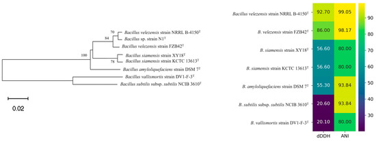
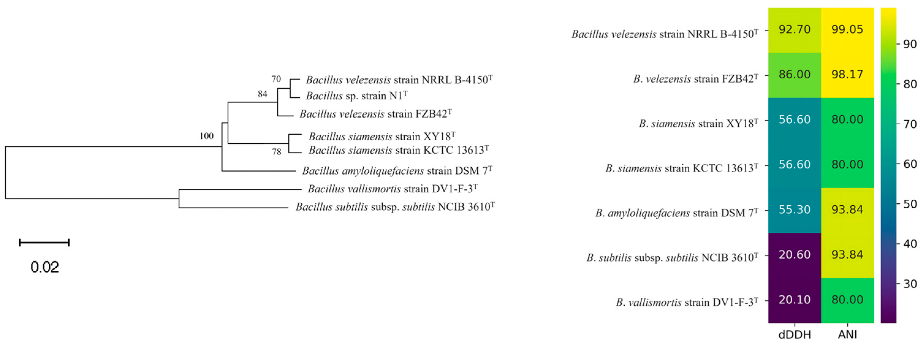
The B. velezensis N1 genome (4.3 Mb) was assembled into 46 contigs, with an N50 of approximately 437 kb and a final sequencing coverage of 405×. The whole-genome phylogenetic reconstruction clearly showed that the analyzed isolates grouped into well-defined, species-associated clades. In particular, B. velezensis strain N1 clustered tightly with B. velezensis NRRL B-41580 and B. velezensis FZB42, forming a distinct evolutionary branch that reflects their close genetic relatedness. This phylogenomic placement was fully supported by pairwise genome comparison: the ANI values between B. velezensis N1 and the two reference B. velezensis strains were 99.05% and 98.17%, while the corresponding dDDH values were 92.7% and 86.0%, respectively. Both parameters exceed the accepted species thresholds (ANI > 95%–96%; dDDH > 70%). In contrast, strains belonging to other Bacillus taxa (B. siamensis, B. amyloliquefaciens, B. subtilis, B. vallismortis) displayed much lower similarity values (ANI ≤ 94% and dDDH ≤ 57%), confirming that they are genetically more distant from N1 and fall outside the B. velezensis genomic boundary. Overall, these findings indicate that N1 is a B. velezensis strain, and it is therefore referred to as B. velezensis N1 (Figure 1).
Figure 1.
Phylogenetic tree (on the left), dDDH, and ANI values (on the right) for B. velezensis strain N1 and the other 7 species belonging to the B. subtilis species complex. Distances are computed by the Genome Blast Distance Phylogeny (GBDP) approach that was constructed using the TYGS software. GBDP pseudo-bootstrap (100 replications) support values above 60% are shown.
3.2. Secondary Metabolite Biosynthetic Gene Clusters in Bacillus velezensis Strain N1
Bacillus velezensis N1 genome harbors different types of BGCs, including non-ribosomal peptide synthase (NRPS), transAT polyketide synthase (PKS), type-III PKS (T3PKS), PKS-like, hybrid PKS–NRPS, terpene, phosphonate and bacilysin-type BGCs (Table 2). Several clusters showed 100% conservation with respect to well-known BGCs, suggesting the genomic potential to produce secondary metabolites with antimicrobial activity. These include the BGCs responsible for difficidin (Region 1.1), bacilysin (Region 2.1), bacillibactin (Regions 2.2 and 3.1), macrolactin H (Region 5.1), and bacillaene biosynthesis (Region 6.1).
Table 2.
Biosynthetic gene clusters identified in the genome of B. velezensis N1. For each region, the table reports the BGC type, the closest known homologous metabolite, and the percentage of sequence similarity to characterized reference clusters.
Some of the multiple NRPS clusters detected corresponded to well-known lipopeptides. The surfactin cluster (Region 7.1) showed an 82% identity score, while the fengycin/plipastatin clusters (Regions 9.1, 10.2, 20.1, and 22.1) displayed variable degrees of identity ranging from 13% to 80% (based on the reference cluster in the antiSMASH database). This variation is likely due to the cluster being split across multiple contigs in the draft assembly, rather than the absence of core biosynthetic genes. Two terpene clusters (Regions 4.1 and 9.1) were identified, although both lacked clear homology to known pathways, suggesting the presence of uncharacterized terpenes biosynthetic clusters. Similarly, several NRPS clusters (Regions 23.1 and 24.1) showed no detectable homology, indicating the possibility of novel peptide products.
3.3. Inhibition of Fungal Pathogen Growth by Bacillus velezensis Strain N1
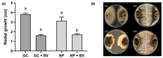
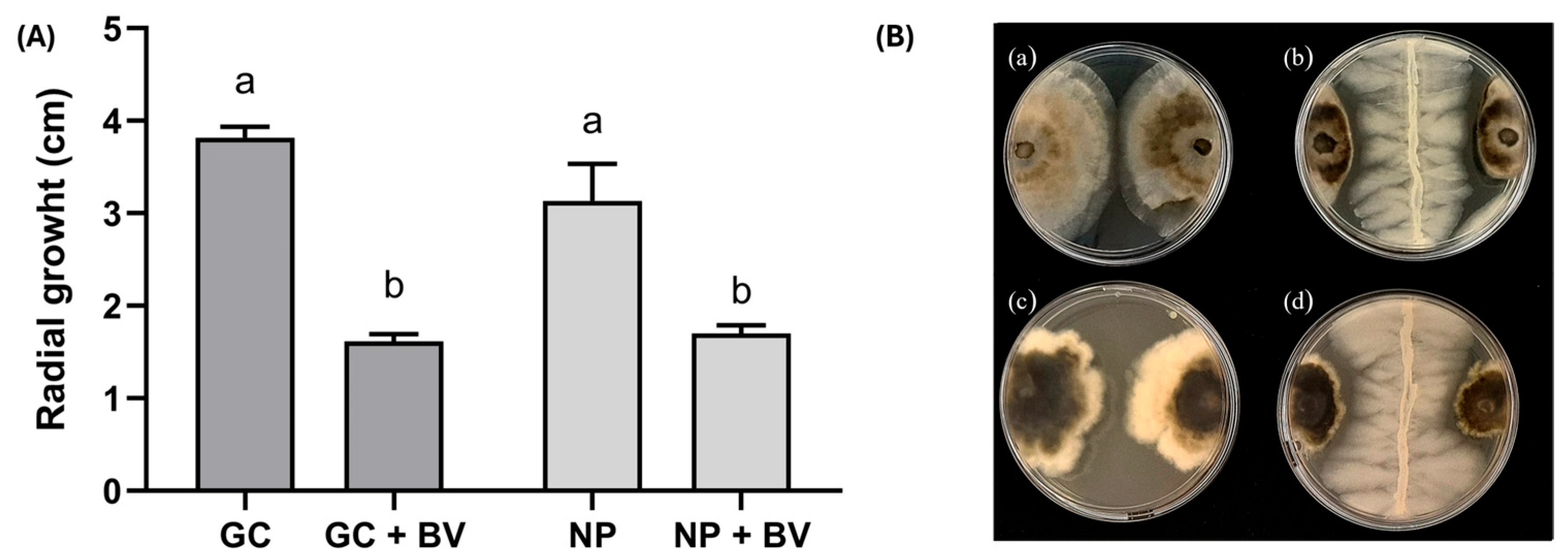
Dual-culture confrontation assays revealed a strong antagonistic effect of B. velezensis N1 against both wood-infecting pathogens. The bacterium significantly inhibited G. smithogilvyi G1 and N. parvum MR1 growth by 57.6% ± 1.2% and 45.0% ± 7.9% (t-test; p < 0.0001) as reflected by the reduced colony expansion shown in Figure 2.
Figure 2.
Radial growth of G. smithogilvyi G1 (GC) and N. parvum MR1 (NP) alone or in presence of B. velezensis N1 (GC + BV and NP + BV) (A); dual culture tests (B): (a) GC (b) GC + BV; (c) NP (d) NP + BV. Different lowercase letters indicate statistical differences according (p ≤ 0.05).
3.4. Biocontrol Effect of Bacillus velezensis Strain N1 on Eucalyptus and Chestnut Shoots
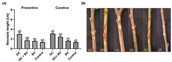
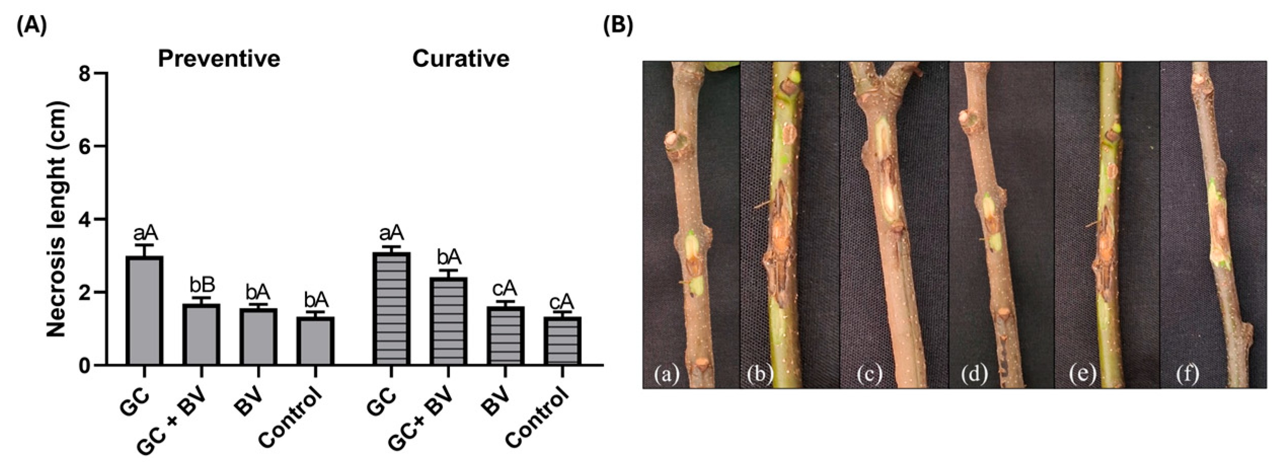
No significant differences in necrotic lesion size were observed between the untreated controls (control 1 and control 2); therefore, the data were pooled for statistical analysis. No phytotoxic effects were observed on shoots inoculated only with the bacterium (BV). Both G. smithogilvyi G1 and N. parvum MR1 caused necrotic lesions on chestnut and eucalyptus shoots, respectively (Figure 3 and Figure 4). For the chestnut dataset, both the Brown–Forsythe ANOVA (F (DFn, DFd) = 26.11 (7, 41.33), p = 2.97 × 10⁻¹³) and Welch’s ANOVA (W (DFn, DFd) = 23.25 (7, 30.64), p = 1.33 × 10⁻¹⁰) showed significant differences among the groups, with a large effect (ηp2 ≈ 0.816 and 0.842, respectively). On chestnut shoots, preventive and curative treatments with B. velezensis N1 significantly limited necrosis development caused by G. smithogilvyi (GC + BV). In particular, in the preventive assay, the application of the bacteria prior to pathogen inoculation provided effective protection; the lesion did not differ statistically from the control. Conversely, although the curative treatment significantly reduced disease severity, lesions were larger than in the control group. Twigs treated after inoculation with the pathogen presented lesions of 2.4 ± 0.6 cm, significantly smaller than those caused by G. smithogilvyi alone (3.1 ± 0.4 cm) but bigger than scars developed on control shoots (Figure 3).
Figure 3.
Efficacy of B. velezensis N1 (BV) in reducing necrotic lesion size caused by G. smithogilvyi G1 (GC) on chestnut shoots. (A) lesion size in preventive (solid columns) and curative (striped columns) assays; (B) representative symptoms on untreated and treated shoots in preventive/curative assays ((a,d) = control; (b,e) = pathogen; (c,f) = pathogen treated with B. velezensis N1). Lowercase letters indicate statistical significance within each individual test, whereas uppercase letters indicate significance between treatments (Welch’s ANOVA, p < 0.05).
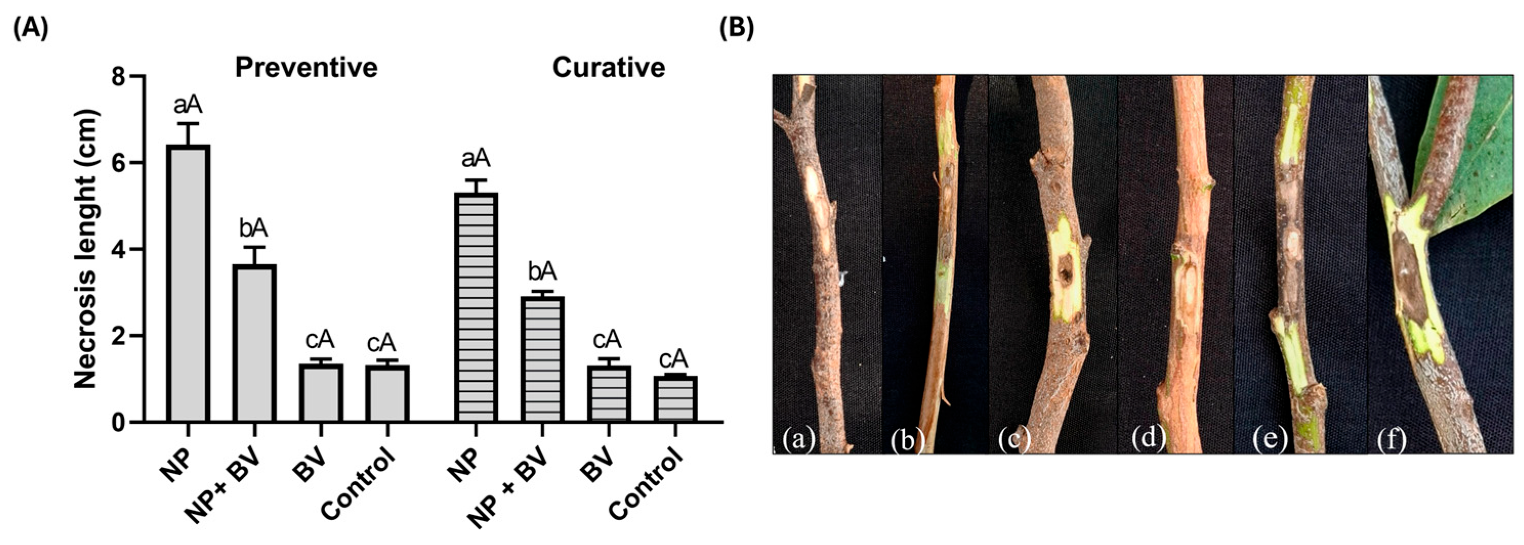
Figure 4.
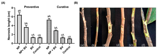
Efficacy of B. velezensis N1 (BV) in reducing necrotic lesion size caused by N. parvum MR1 (NP) on eucalyptus shoots. (A) lesion size in preventive (solid columns) and curative (striped columns) assays; (B) representative symptoms on untreated and treated shoots in preventive/curative assays ((a,d) = control; (b,e) = pathogen; (c,f) = pathogen treated with B. velezensis N1). Lowercase letters indicate statistical significance within each individual test, whereas uppercase letters indicate significance between treatments (Welch’s ANOVA, p < 0.05).
In the eucalyptus dataset, Welch’s one-way ANOVA showed significant differences among the groups (W (DFn, DFd) = 80.87 (7, 28.92), p = 2.89 × 10⁻¹⁷), with a very large effect (ηp2 ≈ 0.951). Bacillus velezensis treatments significantly reduced the necrotic lesion size (NP + BV) compared with scars observed on pathogen-inoculated shoots (NP). Preventive and curative applications of B. velezensis showed comparable efficacy in reducing necrotic scars (Figure 4).
Fungal strains re-isolated from inoculated twigs exhibited morphological traits identical to those of the original inocula. Thus, Koch’s postulates were accomplished.
4. Discussion
The implementation of effective and sustainable management strategies for canker- and dieback diseases in woody hosts remains a significant challenge. In this framework, the present study provided preliminary evidence that B. velezensis strain N1 inhibited the growth of N. parvum and G. smithogilvyi in in vitro assays and limited the disease severity on eucalyptus and chestnut shoots. Bacillus velezensis is a Gram-positive, spore-forming bacterium. Phylogenomic and genomic analyses have shown that B. velezensis was originally considered a subspecies of B. amyloliquefaciens (Fukumoto) Priest et al. but is now recognized as a distinct species [45,58]. In recent years, B. velezensis has attracted considerable interest for its applications in agriculture and biotechnology, particularly as a biological control agent against plant pathogenic fungi [36,59,60,61]. For instance, Cao et al. [62] reported that B. velezensis strains Y6 and F7 exhibited potent antagonistic activity against Fusarium oxysporum f. sp. cubense (E.F.Smith) Snyder & Hansen, the causal agent of banana Fusarium wilt, as well as against Ralstonia solanacearum (Smith) Yabuuchi et al. emend. Safni et al., a pathogen affecting several crops, including potato and tomato. Similarly, Li et al. [63] demonstrated that B. velezensis FX-6 fostered tomato plant growth and strongly inhibited Botrytis cinerea Persoon, achieving an in vitro mycelial inhibition rate of 78.6%. The biocontrol mechanisms of some B. velezensis strains are largely attributed to the production of diverse antimicrobial secondary metabolites, including lipopeptides (surfactin, fengycin, plipastatin), polyketides (difficidin, bacillaene, macrolactin), and other bioactive compounds, such as the non-ribosomal dipeptide bacilysin, and the siderophore bacillibactin [64,65,66]. While these metabolites can directly suppress pathogen growth or interfere with pathogen development, some Bacillus strains also promote plant defense responses through induced systemic resistance [67,68,69]. In fact, in addition to antifungal and antibacterial activity, many B. velezensis strains possess plant growth-promoting traits, like the production of siderophores, indole-3-acetic acid, as well as hydrolytic enzymes (e.g., chitinases, lipases, amylases, and cellulases) which further enhance host health and resilience.
Whole-genome phylogenetic analysis confirmed that N1 belongs to the B. velezensis species, and is therefore phylogenetically related to reference strains NRRL B-41580 and FZB42 (Figure 1). Bacillus velezensis N1 harbors multiple biosynthetic gene clusters, encompassing NRPS, PKS, hybrid PKS–NRPS, terpene, phosphonate, and bacilysin-like clusters. Several of these BGCs are completely conserved relative to known antimicrobial compounds, such as difficidin, bacilysin, bacillibactin, macrolactin H, and bacillaene. These observations align with previous reports on FZB42 and other B. velezensis/B. amyloliquefaciens strains, which showed that lipopeptides, such as surfactin, fengycin, and bacillomycin D together with polyketides, are key determinants of antagonistic activity against pathogens including Rhizoctonia solani and F. oxysporum Schlechtendal [70,71,72].
Furthermore, several studies have highlighted the dual role of secondary metabolites produced by FZB42, which can both directly inhibit pathogens and trigger plant immune defenses. For instance, surfactin has been shown to activate plant defense pathways (jasmonic acid/ethylene) and related defense genes (PDF1.2, PR1), thereby enhancing plant resistance to infections [73,74,75].
While these findings highlight the promising potential of B. velezensis N1 to produce compounds that support its role as a biocontrol agent, several methodological and experimental limitations should be considered when interpreting the results. Given the number of replicates, the statistical resolution of our assays does not allow us to confidently detect very small differences between treatments and the control. Moreover, because the experiments were conducted on detached shoots, the system does not fully reproduce whole-plant physiology, including vascular transport, systemic immune responses, and natural humidity conditions. These methodological constraints limit the direct extrapolation of our findings to field conditions, and validation on intact plants will be necessary. Further, the observed effects cannot be interpreted as evidence of induced resistance, as the current experimental design does not allow us to distinguish between direct antimicrobial activity and potential plant-mediated responses. These interpretations should therefore be regarded as hypotheses rather than demonstrated mechanisms. Finally, although genome mining revealed several secondary metabolite gene clusters, metabolite production remains predicted rather than experimentally validated.
However, the findings of the present study indicate that B. velezensis N1 is a promising biocontrol agent against G. smithogilvyi and N. parvum, expanding its documented antagonistic spectrum beyond the previously reported suppression of Phytophthora infestans [37]. Notably, available information on the biological control of N. parvum is still scarce and mainly limited to horticultural pathosystems, including mango postharvest rot [76] and almond canker [77]. Likewise, for G. smithogilvyi, biocontrol evidence remains limited; earlier studies reported the effectiveness of Trichoderma spp. endotherapy in reducing nut rot in chestnut groves [78]. In addition, Pasche et al. [79] highlighted the potential of B. amyloliquefaciens and T. atroviride as biocontrol agents against G. smithogilvyi on chestnut scions. Together, these studies underscore the need for additional biological solutions, and our results support B. velezensis N1 as an effective candidate for managing both pathogens in systems where the current literature remains limited.
In urban environments, where chemical treatments are often restricted due to public safety concerns, environmental regulations, and the proximity of people and pets, the use of microbial biocontrol agents represents a particularly valuable strategy. The ability of B. velezensis N1 to suppress N. parvum and G. smithogilvyi therefore highlights the relevance of biological solutions compatible with the ecological and regulatory constraints of urban forestry. By combining antagonistic activity with plant-beneficial traits [37], B. velezensis-based interventions may offer a sustainable, low-impact tool for managing canker and dieback diseases in trees growing in parks, avenues, and other public green spaces.
5. Conclusions
This study provided novel evidence for the efficacy of Bacillus velezensis N1 to reduce disease severity caused by N. parvum and G. smithogilvyi on eucalyptus and chestnut twigs, respectively. These results highlight the potential of strain N1 as a biocontrol agent for woody hosts and support its possible use in sustainable disease management strategies. Further validation on whole plants, combined with a larger experimental scale and metabolomic analyses for the detection of secondary metabolites with antifungal activity, will be essential to confirm the robustness of these effects and to clarify the underlying mechanisms involved. These findings broaden the potential use of B. velezensis as a species in sustainable disease management strategies, both as preventive or curative treatments on woody hosts.
Author Contributions
Conceptualization, A.M.V. and A.F.; methodology, A.M.V. and A.F.; formal analysis, A.M.V.; investigation, A.M.V. and M.N.; resources, A.M.V.; data curation, C.A.; writing—original draft preparation, A.M.V., A.F. and B.T.L.; writing—review and editing, A.M.V. and A.F. All authors have read and agreed to the published version of the manuscript.
Funding
This research received no external funding.
Data Availability Statement
Dataset available on request from the authors.
Acknowledgments
The authors have reviewed and edited the output and take full responsibility for the content of this publication.
Conflicts of Interest
The authors declare no conflicts of interest.
References
- Brooker, M.I.H.; Kleinig, D.A. Field Guide to Eucalypts. 1: South-Eastern Australia; Inkata Press: Melbourne, Australia, 1983. [Google Scholar]
- Marques, T.; Ferreira-Pinto, A.; Fevereiro, P.; Pinto, T.; Gomes-Laranjo, J. Current biological insights of Castanea sativa Mill. to improve crop sustainability to climate change. Plants 2025, 14, 335. [Google Scholar] [CrossRef] [Scilit] [PubMed]
- Booth, T.H. Eucalypt plantations and climate change. For. Ecol. Manag. 2013, 301, 28–34. [Google Scholar] [CrossRef] [Scilit]
- Shala, A.Y.; Gururani, M.A. Phytochemical properties and diverse beneficial roles of Eucalyptus globulus Labill.: A review. Horticulturae 2021, 7, 450. [Google Scholar] [CrossRef] [Scilit]
- Esperon-Rodriguez, M.; Tjoelker, M.G.; Lenoir, J.; Laugier, B.; Gallagher, R.V. Wide climatic niche breadth and traits associated with climatic tolerance facilitate eucalypt occurrence in cities worldwide. Glob. Ecol. Biogeogr. 2024, 33, e13833. [Google Scholar] [CrossRef] [Scilit]
- Antonelli, C.; Soulioti, N.; Linaldeddu, B.T.; Tsopelas, P.; Biscontri, M.; Tsoukas, C.; Paplomatas, E.; Kuzminsky, E.; Vettraino, A.M. Phytophthora nicotianae and Ph. mediterranea: A biosecurity threat to Platanus orientalis and P. x acerifolia in urban green areas in Greece. Urban For. Urban Green. 2024, 95, 128281. [Google Scholar] [CrossRef] [Scilit]
- Davis, A.; Major, R.E.; Taylor, C.E. Do trees flower longer in the city? A comparison of flowering eucalyptus trees in streets, remnants and continuous forest and their association with nectarivorous birds. Urban. Ecosyst. 2016, 19, 735–747. [Google Scholar] [CrossRef] [Scilit]
- González-Jaramillo, S.I.; Lozano-Amaro, I.; Ceccon, E.; Pérez-Muñoz, T.; Cano-Santana, Z. Risk of Eucalyptus tree failure and social acceptance of its removal in urban green areas of Mexico City. Urban For. Urban Green. 2026, 115, 129191. [Google Scholar] [CrossRef] [Scilit]
- Haltofová, P.; Jankovský, L. Distribution of sweet chestnut Castanea sativa Mill. in the Czech Republic. J. For. Sci. 2003, 49, 259–272. [Google Scholar] [CrossRef] [Scilit]
- Esperon-Rodriguez, M.; Tjoelker, M.G.; Lenoir, J.; Baumgartner, J.B.; Beaumont, L.J.; Nipperess, D.A.; Power, S.A.; Richard, B.; Rymer, P.D.; Gallagher, R.V. Climate change increases global risk to urban forests. Nat. Clim. Change 2022, 12, 950–955. [Google Scholar] [CrossRef] [Scilit]
- Deidda, A.; Buffa, F.; Linaldeddu, B.; Pinna, C.; Scanu, B.; Deiana, V.; Satta, A.; Franceschini, A.; Floris, I. Emerging pests and diseases threaten Eucalyptus camaldulensis plantations in Sardinia, Italy. iForest 2016, 9, 883–891. [Google Scholar] [CrossRef] [Scilit]
- Egerer, M.; Schmack, J.M.; Vega, K.; Barona, C.O.; Raum, S. The challenges of urban street trees and how to overcome them. Front. Sustain. Cities 2024, 6, 1394056. [Google Scholar] [CrossRef] [Scilit]
- Balmelli, G.; Simeto, S.; Altier, N.; Marroni, V.; Diez, J.J. Long term losses caused by foliar diseases on growth and survival of Eucalyptus globulus in Uruguay. New For. 2013, 44, 249–263. [Google Scholar] [CrossRef] [Scilit]
- Barber, P.A.; Carnegie, A.J.; Burgess, T.I.; Keane, P.J. Leaf diseases caused by Mycosphaerella species in Eucalyptus globulus plantations and nearby native forest in the Green Triangle Region of Southern Australia. Austral. Plant Pathol. 2008, 37, 472. [Google Scholar] [CrossRef] [Scilit]
- Diogo, E.; Machado, H.; Reis, A.; Valente, C.; Phillips, A.J.L.; Bragança, H. Phytophthora alticola and Phytophthora cinnamomi on Eucalyptus globulus in Portugal. Eur. J. Plant Pathol. 2023, 165, 255–269. [Google Scholar] [CrossRef] [Scilit]
- EFSA Panel on Plant Health (PLH); Jeger, M.; Bragard, C.; Chatzivassiliou, E.; Dehnen-Schmutz, K.; Gilioli, G.; Jaques Miret, J.A.; MacLeod, A.; Navajas Navarro, M.; Niere, B.; et al. Risk assessment and reduction options for Cryphonectria parasitica in the EU. EFSA J. 2016, 14, e04641. [Google Scholar] [CrossRef] [Scilit]
- Lione, G.; Danti, R.; Fernandez-Conradi, P.; Ferreira-Cardoso, J.V.; Lefort, F.; Marques, G.; Meyer, J.B.; Prospero, S.; Radócz, L.; Robin, C.; et al. The emerging pathogen of chestnut Gnomoniopsis castaneae: The challenge posed by a versatile fungus. Eur. J. Plant Pathol. 2019, 153, 671–685. [Google Scholar] [CrossRef] [Scilit]
- Miller, A.C.; Lewis Ivey, M.L. The disease triangle of chestnut: A review of host, pathogen, and environmental interactions of chestnuts cultivated in the Eastern United States. Plant Dis. 2025, 109, 245–256. [Google Scholar] [CrossRef] [Scilit]
- Topalidou, E.; Lagiotis, G.; Bosmali, I.; Stefanidou, E.; Tsirogiannis, D.; Vettraino, A.M.; Madesis, P. Incidence of brown rot disease caused by Gnomoniopsis smithogilvyi on buds, flowers and chestnuts and rapid HRM-based detection of the disease. Fungal Biol. 2024, 128, 1968–1981. [Google Scholar] [CrossRef] [Scilit]
- Turchetti, T.; Maresi, G. Biological control and management of chestnut diseases. In Integrated Management of Diseases Caused by Fungi, Phytoplasma and Bacteria; Ciancio, A., Mukerji, K.G., Eds.; Springer: Dordrecht, The Netherlands, 2008; Volume 3, pp. 85–118. [Google Scholar]
- Vettraino, A.M.; Morel, O.; Perlerou, C.; Robin, C.; Diamandis, S.; Vannini, A. Occurrence and distribution of Phytophthora species in european chestnut stands, and their association with ink disease and crown decline. Eur. J. Plant Pathol. 2005, 111, 169–180. [Google Scholar] [CrossRef] [Scilit]
- Vettraino, A.M.; Bonants, P.; Tomassini, A.; Bruni, N.; Vannini, A. Pyrosequencing as a tool for the detection of Phytophthora species: Error rate and risk of false Molecular Operational Taxonomic Units. Lett. Appl. Microbiol. 2012, 55, 390–396. [Google Scholar] [CrossRef] [Scilit]
- Vettraino, A.M.; Luchi, N.; Rizzo, D.; Pepori, A.L.; Pecori, F.; Santini, A. Rapid diagnostics for Gnomoniopsis smithogilvyi (syn. Gnomoniopsis castaneae) in chestnut nuts: New challenges by using LAMP and Real-Time PCR methods. AMB Expr. 2021, 11, 105. [Google Scholar] [CrossRef] [Scilit]
- Wingfield, M.; Slippers, B.; Hurley, B.; Coutinho, T.; Wingfield, B.; Roux, J. Eucalypt pests and diseases: Growing threats to plantation productivity. South. For. A J. For. Sci. 2008, 70, 139–144. [Google Scholar] [CrossRef] [Scilit]
- Bertetti, D.; Rettori, A.; Martinis, R.; Guarnaccia, V.; Tabone, G.; Garibaldi, A.; Gullino, M.L. First report of Neofusicoccum parvum causing stem cankers and woody rot on Eucalyptus globulus in France. Plant Dis. 2023, 107, 2548. [Google Scholar] [CrossRef] [Scilit]
- Li, G.; Slippers, B.; Wingfield, M.J.; Chen, S. Variation in Botryosphaeriaceae from Eucalyptus plantations in YunNan province in Southwestern China across a climatic gradient. IMA Fungus 2020, 11, 22. [Google Scholar] [CrossRef] [Scilit] [PubMed]
- Burgess, T.I.; Wingfield, M.J. Pathogens on the move: A 100-year global experiment with planted eucalypts. BioScience 2017, 67, 14–25. [Google Scholar] [CrossRef] [Scilit]
- Barradas, C.; Pinto, G.; Correia, B.; Jesus, C.; Alves, A. Impact of Botryosphaeria, Diplodia and Neofusicoccum species on two Eucalyptus species and a hybrid: From pathogenicity to physiological performance. For. Pathol. 2019, 49, e12493. [Google Scholar] [CrossRef] [Scilit]
- Lewis, A.; Gorton, C.; Rees, H.; Webber, J.; Pérez-Sierra, A. First report of Gnomoniopsis smithogilvyi causing lesions and cankers of sweet chestnut in the United Kingdom. New Dis. Rep. 2017, 35, 20. [Google Scholar] [CrossRef] [Scilit]
- Trapiello, E.; Feito, I.; González, A.J. First report of Gnomoniopsis castaneae causing canker on hybrid plants of Castanea sativa × C. crenata in Spain. Plant Dis. 2018, 102, 1040. [Google Scholar] [CrossRef] [Scilit]
- O’Loinsigh, B.; McAuley, D.; Bréchon, A.L.; Lopez Vernaza, M.; Ryan, C.; Destefanis, M.L.; O’Hanlon, R. First report of the fungus Gnomoniopsis smithogilvyi causing cankers on sweet chestnut (Castanea sativa) in Ireland. New Dis. Rep. 2022, 45, e12072. [Google Scholar] [CrossRef] [Scilit]
- Dar, M.A.; Rai, M. Gnomoniopsis smithogilvyi, a canker causing pathogen on Castanea sativa: First report. Mycosphere 2015, 6, 327–336. [Google Scholar] [CrossRef] [Scilit]
- Regulation (EC). No 1107/2009 of the European Parliament and of the Council of 21 October 2009 concerning the placing of plant protection products on the market and repealing Council Directives 79/117/EEC and 91/414/EEC. EEC 2009, 91, 1–50. [Google Scholar]
- Vettraino, A.M.; Soulioti, N.; Matosevic, D.; Tuğba Doğmuş Lehtijarvi, H.; Woodward, S.; Santini, A.; Luchi, N. Management of fungal diseases of Platanus under changing climate conditions: Case studies in urban areas. Urban For. Urban Green. 2025, 107, 128750. [Google Scholar] [CrossRef] [Scilit]
- Etesami, H.; Jeong, B.R.; Glick, B.R. Biocontrol of plant diseases by Bacillus spp. Physiol. Mol. Plant Pathol. 2023, 126, 102048. [Google Scholar] [CrossRef] [Scilit]
- Zhang, N.; Wang, Z.; Shao, J.; Xu, Z.; Liu, Y.; Xun, W.; Miao, Y.; Shen, Q.; Zhang, R. Biocontrol mechanisms of Bacillus: Improving the efficiency of green agriculture. Microb. Biotechnol. 2023, 16, 2250–2263. [Google Scholar] [CrossRef] [Scilit] [PubMed]
- Antonelli, C.; Narduzzi, M.; Ruzzi, M.; Testa, A.; Vettraino, A.M. Mining microbial niches: Sources of bacteria for enhancing plant growth and resilience to pH, salinity, drought and Phytophthora infestans. Environ. Microbiol. Rep. 2025, 17, e70217. [Google Scholar] [CrossRef] [Scilit]
- Olson, R.D.; Assaf, R.; Brettin, T.; Conrad, N.; Cucinell, C.; Davis, J.J.; Dempsey, D.M.; Dickerman, A.; Dietrich, E.M.; Kenyon, R.W.; et al. Introducing the Bacterial and Viral Bioinformatics Resource Center (BV-BRC): A resource combining PATRIC, IRD and ViPR. Nucleic Acids Res. 2023, 51, D678–D689. [Google Scholar] [CrossRef] [Scilit]
- Chklovski, A.; Parks, D.H.; Woodcroft, B.J.; Tyson, G.W. CheckM2: A rapid, scalable and accurate tool for assessing microbial genome quality using machine learning. Nat. Methods 2023, 20, 1203–1212. [Google Scholar] [CrossRef] [Scilit]
- Li, W.; O’Neill, K.R.; Haft, D.H.; Di Cuccio, M.; Chetvernin, V.; Badretdin, A.; Coulouris, G.; Chitsaz, F.; Derbyshire, M.K.; Durkin, A.S.; et al. RefSeq: Expanding the Prokaryotic Genome Annotation Pipeline reach with protein family model curation. Nucleic Acids Res. 2021, 49, D1020–D1028. [Google Scholar] [CrossRef] [Scilit]
- Meier-Kolthoff, J.P.; Göker, M. TYGS is an automated high-throughput platform for state-of-the-art genome-based taxonomy. Nat. Commun. 2019, 10, 2182. [Google Scholar] [CrossRef] [Scilit]
- Meier-Kolthoff, J.P.; Auch, A.F.; Klenk, H.-P.; Göker, M. Genome sequence-based species delimitation with confidence intervals and improved distance functions. BMC Bioinform. 2013, 14, 60. [Google Scholar] [CrossRef] [Scilit]
- Meier-Kolthoff, J.P.; Carbasse, J.S.; Peinado-Olarte, R.L.; Göker, M. TYGS and LPSN: A database tandem for fast and reliable genome-based classification and nomenclature of prokaryotes. Nucleic Acids Res. 2022, 50, D801–D807. [Google Scholar] [CrossRef] [Scilit] [PubMed]
- Jain, C.; Rodriguez-R, L.M.; Phillippy, A.M.; Konstantinidis, K.T.; Aluru, S. High throughput ANI analysis of 90K prokaryotic genomes reveals clear species boundaries. Nat. Commun. 2018, 9, 5114. [Google Scholar] [CrossRef] [Scilit] [PubMed]
- Dunlap, C.A.; Kim, S.J.; Kwon, S.W.; Rooney, A.P. Bacillus velezensis is not a later heterotypic synonym of Bacillus amyloliquefaciens; Bacillus methylotrophicus, Bacillus amyloliquefaciens subsp. plantarum and ‘Bacillus oryzicola’ are later heterotypic synonyms of Bacillus velezensis based on phylogenomics. Int. J. Syst. Evol. Microbiol. 2016, 66, 1212–1217. [Google Scholar] [CrossRef] [Scilit] [PubMed]
- Chen, X.H.; Koumoutsi, A.; Scholz, R.; Eisenreich, A.; Schneider, K.; Heinemeyer, I.; Morgenstern, B.; Voss, B.; Hess, W.R.; Reva, O.; et al. Comparative analysis of the complete genome sequence of the plant growth–promoting bacterium Bacillus amyloliquefaciens FZB42. Nat. Biotechnol. 2007, 25, 1007–1014. [Google Scholar] [CrossRef] [Scilit]
- Dunlap, C.A. Phylogenomic analysis shows that ‘Bacillus vanillea’ is a later heterotypic synonym of Bacillus siamensis. Int. J. Syst. Evol. Microbiol. 2015, 65, 3507–3510. [Google Scholar] [CrossRef] [Scilit]
- Jeong, H.; Jeong, D.-E.; Kim, S.H.; Song, G.C.; Park, S.-Y.; Ryu, C.-M.; Park, S.-H.; Choi, S.-K. Draft genome sequence of the plant growth-promoting bacterium Bacillus siamensis KCTC 13613T. J. Bacteriol. 2012, 194, 4148–4149. [Google Scholar] [CrossRef] [Scilit]
- Priest, F.G.; Goodfellow, M.; Todd, C. A numerical classification of the genus Bacillus. J. Gen. Microbiol. 1988, 134, 1847–1882. [Google Scholar] [CrossRef] [Scilit]
- Wang, L.T.; Lee, F.L.; Tai, C.J.; Kasai, H. Comparison of gyrB gene sequences, 16S rRNA gene sequences and DNA-DNA hybridization in the Bacillus subtilis group. Int. J. Syst. Evol. Microbiol. 2007, 57, 1846–1850. [Google Scholar] [CrossRef] [Scilit]
- Cohn, F. Untersuchungen über Bacterien. In Beitrage zur Biologie der Pflanzen Heft 2; J.U. Kern: Breslau, Poland, 1872; Volume 1, pp. 127–224. [Google Scholar]
- Conn, H.J. The identity of Bacillus subtilis. J. Infect. Dis. 1930, 46, 341–350. [Google Scholar] [CrossRef] [Scilit]
- Ehrenberg, C.G. Physikalische Abhandlungen der Königlichen Akademie der Wissenschaften zu Berlin; Realschul-Buchhandlung: Berlin, Germany, 1835. [Google Scholar]
- Earl, A.M.; Eppinger, M.; Fricke, W.F.; Rosovitz, M.J.; Rasko, D.A.; Daugherty, S.; Losick, R.; Kolter, R.; Ravel, J. Whole-genome sequences of Bacillus subtilis and close relatives. J. Bacteriol. 2012, 194, 2378–2379. [Google Scholar] [CrossRef] [Scilit]
- Blin, K.; Shaw, S.; Vader, L.; Szenei, J.; Reitz, Z.L.; Augustijn, H.E.; Cediel-Becerra, J.D.D.; de Crécy-Lagard, V.; Koetsier, R.A.; Williams, S.E.; et al. antiSMASH 8.0: Extended gene cluster detection capabilities and analyses of chemistry, enzymology, and regulation. Nucleic Acids Res. 2025, 53, W32–W38. [Google Scholar] [CrossRef] [Scilit] [PubMed]
- Ciaffi, M.; Vettraino, A.M.; Alicandri, E.; Tomao, A.; Adducci, F.; Kuzminsky, E.; Agrimi, M. Dimensional and genetic characterization of the last oriental plane trees (Platanus orientalis L.) of historical sites in Lazio (central Italy). Urban For. Urban Green. 2022, 69, 127506. [Google Scholar] [CrossRef] [Scilit]
- Lakens, D. Calculating and reporting effect sizes to facilitate cumulative science: A practical primer for t-tests and ANOVAs. Front. Psychol. 2013, 4, 863. [Google Scholar] [CrossRef] [Scilit]
- Fan, B.; Blom, J.; Klenk, H.-P.; Borriss, R. Bacillus amyloliquefaciens, Bacillus velezensis, and Bacillus siamensis form an “operational group B. amyloliquefaciens” within the B. subtilis species complex. Front. Microbiol. 2017, 8, 22. [Google Scholar] [CrossRef] [Scilit]
- Kim, Y.S.; Lee, Y.; Cheon, W.; Park, J.; Kwon, H.-T.; Balaraju, K.; Kim, J.; Yoon, Y.J.; Jeon, Y. Characterization of Bacillus velezensis AK-0 as a biocontrol agent against apple bitter rot caused by Colletotrichum gloeosporioides. Sci. Rep. 2021, 11, 626. [Google Scholar] [CrossRef] [Scilit] [PubMed]
- Jiang, C.-H.; Liao, M.-J.; Wang, H.-K.; Zheng, M.-Z.; Xu, J.-J.; Guo, J.-H. Bacillus velezensis, a potential and efficient biocontrol agent in control of pepper gray mold caused by Botrytis cinerea. Biol. Control 2018, 126, 147–157. [Google Scholar] [CrossRef] [Scilit]
- Rabbee, M.F.; Hwang, B.-S.; Baek, K.-H. Bacillus velezensis: A beneficial biocontrol agent or facultative phytopathogen for sustainable agriculture. Agronomy 2023, 13, 840. [Google Scholar] [CrossRef] [Scilit]
- Cao, Y.; Pi, H.; Chandrangsu, P.; Li, Y.; Wang, Y.; Zhou, H.; Xiong, H.; Helmann, J.D.; Cai, Y. Antagonism of two Plant-Growth Promoting Bacillus velezensis isolates against Ralstonia solanacearum and Fusarium oxysporum. Sci. Rep. 2018, 8, 4360. [Google Scholar] [CrossRef] [Scilit]
- Li, Z.; Li, J.; Yu, M.; Quandahor, P.; Tian, T.; Shen, T. Bacillus velezensis FX-6 suppresses the infection of Botrytis cinerea and increases the biomass of tomato plants. PLoS ONE 2023, 18, e0286971. [Google Scholar] [CrossRef] [Scilit]
- Jin, P.; Chu, L.; Xuan, Z.; Lin, Z.; Fang, Y.; Pan, X.; Wang, J.; Liu, W.; Miao, W. Bacillus velezensis, a new valuable source of bioactive molecules within plant microbiomes and natural weapons for the biocontrol of plant pathogens. Trop. Plants 2025, 4, e001. [Google Scholar] [CrossRef] [Scilit]
- Rabbee, M.F.; Ali, M.S.; Choi, J.; Hwang, B.S.; Jeong, S.C.; Baek, K.-H. Bacillus velezensis: A valuable member of bioactive molecules within plant microbiomes. Molecules 2019, 24, 1046. [Google Scholar] [CrossRef] [Scilit]
- Karpiński, T.M. Marine macrolides with antibacterial and/or antifungal activity. Mar. Drugs 2019, 17, 241. [Google Scholar] [CrossRef] [Scilit]
- Li, P.; Zhao, L.; Jiao, J.; Bai, L.; Ding, J.; Tang, G.; Fu, X.; Sun, Q. Enhanced disease resistance and growth promotion in prickly ash by the rhizobacterium Bacillus velezensis P87. Ind. Crops Prod. 2025, 236, 121972. [Google Scholar] [CrossRef] [Scilit]
- Yan, Y.; Xu, W.; Hu, Y.; Tian, R.; Wang, Z. Bacillus velezensis YYC promotes tomato growth and induces resistance against bacterial wilt. Biol. Control 2022, 172, 104977. [Google Scholar] [CrossRef] [Scilit]
- Zhong, X.; Jin, Y.; Ren, H.; Hong, T.; Zheng, J.; Fan, W.; Hong, J.; Chen, Z.; Wang, A.; Lu, H.; et al. Research progress of Bacillus velezensis in plant disease resistance and growth promotion. Front. Ind. Microbiol. 2024, 2, 1442980. [Google Scholar] [CrossRef] [Scilit]
- Cawoy, H.; Debois, D.; Franzil, L.; De Pauw, E.; Thonart, P.; Ongena, M. Lipopeptides as main ingredients for inhibition of fungal phytopathogens by Bacillus subtilis/amyloliquefaciens. Microb. Biotechnol. 2015, 8, 281–295. [Google Scholar] [CrossRef] [Scilit] [PubMed]
- Chen, X.-H.; Vater, J.; Piel, J.; Franke, P.; Scholz, R.; Schneider, K.; Koumoutsi, A.; Hitzeroth, G.; Grammel, N.; Strittmatter, A.W.; et al. Structural and functional characterization of three polyketide synthase gene clusters in Bacillus amyloliquefaciens FZB 42. J. Bacteriol. 2006, 188, 4024–4036. [Google Scholar] [CrossRef] [Scilit] [PubMed]
- Chowdhury, S.P.; Uhl, J.; Grosch, R.; Alquéres, S.; Pittroff, S.; Dietel, K.; Schmitt-Kopplin, P.; Borriss, R.; Hartmann, A. Cyclic lipopeptides of Bacillus amyloliquefaciens subsp. plantarum colonizing the lettuce rhizosphere enhance plant defense responses toward the bottom rot pathogen Rhizoctonia solani. Mol. Plant-Microbe Interact. 2015, 28, 984–995. [Google Scholar] [CrossRef] [Scilit] [PubMed]
- Cawoy, H.; Mariutto, M.; Henry, G.; Fisher, C.; Vasilyeva, N.; Thonart, P.; Dommes, J.; Ongena, M. Plant defense stimulation by natural isolates of Bacillus depends on efficient surfactin production. Mol. Plant-Microbe Interact. 2014, 27, 87–100. [Google Scholar] [CrossRef] [Scilit]
- Han, Q.Q.; Lü, X.P.; Bai, J.P.; Qiao, Y.; Paré, P.W.; Wang, S.M.; Zhang, J.L.; Wu, Y.N.; Pang, X.P.; Xu, W.B.; et al. Beneficial soil bacterium Bacillus subtilis (GB03) augments salt tolerance of white clover. Front. Plant Sci. 2014, 5, 525. [Google Scholar] [CrossRef] [Scilit]
- Ongena, M.; Jourdan, E.; Adam, A.; Paquot, M.; Brans, A.; Joris, B.; Arpigny, J.L.; Thonart, P. Surfactin and fengycin lipopeptides of Bacillus subtilis as elicitors of induced systemic resistance in plants. Environ. Microbiol. 2007, 9, 1084–1090. [Google Scholar] [CrossRef] [Scilit]
- Guirado-Manzano, L.; Tienda, S.; Gutiérrez-Barranquero, J.A.; De Vicente, A.; Cazorla, F.M.; Arrebola, E. Biological control and cross infections of the Neofusicoccum spp. causing mango postharvest rots in Spain. Horticulturae 2024, 10, 166. [Google Scholar] [CrossRef] [Scilit]
- Romero-Cuadrado, L.; Picos, M.C.; Camacho, M.; Ollero, F.J.; Capote, N. Biocontrol of almond canker diseases caused by Botryosphaeriaceae fungi. Pest. Manag. Sci. 2024, 80, 1839–1848. [Google Scholar] [CrossRef] [Scilit]
- Benigno, A.; Aglietti, C.; Cacciola, S.O.; Moricca, S. Trunk Injection delivery of biocontrol strains of Trichoderma spp. effectively suppresses nut rot by Gnomoniopsis castaneae in chestnut (Castanea sativa Mill.). Biology 2024, 13, 143. [Google Scholar] [CrossRef] [Scilit]
- Pasche, S.; Calmin, G.; Auderset, G.; Crovadore, J.; Pelleteret, P.; Mauch-Mani, B.; Barja, F.; Paul, B.; Jermini, M.; Lefort, F. Gnomoniopsis smithogilvyi causes chestnut canker symptoms in Castanea sativa shoots in Switzerland. Fungal Genet. Biol. 2016, 87, 9–21. [Google Scholar] [CrossRef] [Scilit]

Disclaimer/Publisher’s Note: The statements, opinions and data contained in all publications are solely those of the individual author(s) and contributor(s) and not of MDPI and/or the editor(s). MDPI and/or the editor(s) disclaim responsibility for any injury to people or property resulting from any ideas, methods, instructions or products referred to in the content. |
© 2026 by the authors. Licensee MDPI, Basel, Switzerland. This article is an open access article distributed under the terms and conditions of the Creative Commons Attribution (CC BY) license.