


Abstract
Heterobasidion and Armillaria may infect tree roots and cause internal decay without visible changes in the crown. Decay is therefore often discovered only after windthrow or harvesting. Conventional remote-sensing observations do not identify the causal fungus directly. They record changes in trees or stand conditions that may be associated with root rot. We reviewed these indicators and the technologies used in studies of Heterobasidion and Armillaria, with particular attention to their possible use in forest management. Three semantic searches in OpenAlex and Semantic Scholar identified 1500 records on disease indicators, sensing technologies and research gaps. Screening retained 106 records, representing 84 unique publications. Information on disease stage, sensing method, reference data and validation was compiled. Site, stand and management data were mainly used to estimate the probability of root rot occurrence. LiDAR and aerial images were effective when damage was already visible as crown decline, mortality or canopy gaps. RGB images were used to identify decay exposed after harvesting. Detecting internal decay in standing trees was more difficult. Hyperspectral studies reported moderate and mainly site-specific results. Small affected classes, inconsistent reference standards and limited independent validation made it difficult to determine whether the results would apply in other forests. Remote sensing could not reliably distinguish root rot from other causes of tree stress from the evaluation data and is currently better suited to locating areas for field inspection than to confirming the causal pathogen.
1. Introduction
Root and butt rot caused by Heterobasidion and Armillaria is an important problem in conifer stands across boreal, hemiboreal and temperate regions. The fungi can remain in infected roots, stumps and other woody material after a tree has died or been harvested. This allows the diseases to persist across forest rotations and spread to neighbouring trees through root contact or, in the case of Armillaria, through rhizomorphs [1,2]. The diseases result in reduced tree growth and volume, butt and stem decay, loss of mechanical stability and increased susceptibility to windthrow. Advanced disease may lead to crown decline and tree mortality [3,4].
Economic losses from root rot are not limited to tree mortality. Decay inside the stem lowers timber quality and may affect how the stem is cut into different assortments. Trees may remain asymptomatic for years while economic losses continue to occur [5,6]. Information on the spatial distribution of root rot may also be useful when planning regeneration. Holmström et al. (2025) used spatial information on decay to optimise tree-species selection for the regeneration of affected spruce stands [7].
Tree mortality and windthrow associated with root rot can create canopy gaps and increase the amount of coarse woody debris, with further effects on forest microclimate and fungal communities [8,9]. Root disease may also affect forest carbon storage [10]. Carbon-related effects received little attention in the remote-sensing studies included in this review.
Root rot is often unevenly distributed within a stand. Apparently healthy trees may occur alongside trees showing crown decline or mortality. Routine forest inventories therefore provide limited information on the fine-scale distribution of the disease. Crown assessments can identify declining trees, but they cannot confirm the cause of decline. Internal decay is usually confirmed through destructive assessment, after windthrow or when trees are felled. Harvester records and stump observations can provide detailed spatial information on decay after harvesting [11,12]. By that stage, however, timber losses have already occurred.
Satellites, aircraft, UAVs and ground-based sensors provide different types of information about forest condition. They may show changes in foliage, canopy temperature, crown or stem form, or patterns of tree mortality. They do not usually detect Heterobasidion or Armillaria themselves. Instead, they record changes in the host tree or identify conditions under which root rot is more likely to occur.
Infection may remain confined to the roots, root collar, butt or heartwood without producing visible changes in the crown. Morrison et al. (2000) found that the actual incidence of Armillaria root disease in young conifer stands was considerably higher than indicated by visible symptoms [13]. Similarly, infected radiata pine with green crowns could not be distinguished in aerial photographs, although dying and dead trees were detected [14]. Early airborne research also found that root disease produced weak changes in short-wave reflectance and that infected trees could be classified as healthy [15].
Also, detectable changes do not necessarily indicate root rot. Drought, nutrient limitation, insect damage and mechanical injury can produce similar changes in chlorophyll absorption, canopy water content and temperature. Remote sensing may therefore distinguish stressed trees from apparently healthy trees without identifying the cause of stress [16,17].
The reference data introduce further uncertainty. The absence of visible decay in a stem cross-section does not show that the roots are free from infection. Visible decay also does not reveal which fungus caused it. Errors in matching roots, stems, crowns and harvested trees, as well as time differences between image acquisition and diagnosis, can further weaken the relationship between the remotely sensed signal and the recorded disease status. The main limitation is therefore not sensor resolution alone, but the weak, delayed and non-specific relationship between internal infection and externally observable tree properties.
Forest pathology studies have addressed disease biology, symptoms, inoculum, diagnostic methods and control measures, often separately from remote-sensing applications [1,2,18]. Broader remote-sensing studies and reviews of forest health have focused mainly on drought, fire, insects and defoliation, while root and butt rot have received limited attention [19,20]. Despite this limited coverage in broader reviews, individual studies have used aerial photography, satellite time series, airborne hyperspectral imagery, UAV-based sensors, LiDAR, terrestrial laser scanning and post-harvest computer vision. Their results are difficult to compare because they concern different biological targets. Mapping dead trees in a known disease centre is not equivalent to detecting internal infection in a tree with an apparently healthy crown. Similarly, identifying decay on an exposed stump differs from attempting to detect internal decay in a standing tree.
Reported accuracy alone may therefore be misleading. High values may reflect advanced disease, strongly contrasting classes or the absence of other causes of tree stress. However, biological evidence alone does not show whether a method is suitable for operational use. This review considers the biological target, sensing method, reference standard, validation and possible management application. Four tasks are distinguished: detecting pathogen infection, identifying internal decay, mapping visible decline or mortality, and predicting disease risk.
This review examines how remote sensing and spatial data have been used to assess Heterobasidion and Armillaria root and butt rot in boreal, hemiboreal and adjacent temperate forests. It also considers the limitations of current methods and the research needed before they can be used operationally. Three questions are addressed:
RQ1: Which spectral, structural, thermal, textural, phenological, spatial and environmental indicators have been associated with Heterobasidion or Armillaria root rot? Do these indicators reflect root rot specifically or tree stress more generally?
RQ2: What approaches have been used with satellite, airborne, UAV and ground-based data? How were the results assessed? Can these approaches be applied elsewhere, and are they feasible for forest management in terms of cost and data requirements?
RQ3: What limits reliable pre-symptomatic and pathogen-specific mapping of root rot? Which study designs, reference standards and validation or monitoring approaches could address these limitations?
2. Materials and Methods
2.1. Review Design
This study was conducted as a structured critical review of remote-sensing and spatial methods used to detect, map or predict root and butt rot associated with Heterobasidion and Armillaria. The review covered three related areas: indicators associated with infection or disease development; satellite, airborne, UAV and ground-based approaches; and the scientific, methodological and operational limitations affecting their use in forest management.
The review design followed the critical-review typology described by Grant and Booth [21], combining a structured literature search with critical evaluation and conceptual synthesis. The search and selection process was documented using selected elements adapted from ROSES [22]. The review was not intended to provide an exhaustive systematic search of the literature. Semantic searches were relevance-ranked, and a predefined number of records was retrieved for each evidence stream. Because the studies differed in their biological targets, sensing methods, reference standards and reported outcomes, the evidence was synthesised narratively.
2.2. Information Sources and Search Strategy
The review used three complementary semantic evidence streams. Searches, screening and structured data extraction were completed on 1 July 2026. Records indexed in Semantic Scholar and OpenAlex were identified using relevance-ranked semantic searches. The 500 records ranked as most relevant within each evidence stream were exported, giving 1500 records in total.
The first search addressed spectral, structural, thermal, textural, phenological, spatial and environmental indicators associated with Heterobasidion or Armillaria infection and disease development. It also considered other factors that may produce similar indicators, including drought, nutrient limitation, competition, bark beetle damage and stand age.
The second search addressed satellite, crewed-airborne, UAV and ground-based approaches. It covered sensor type, spectral range, spatial and temporal resolution, LiDAR and terrain variables, analytical methods, reference data, validation and possible use in forest management.
The third search addressed factors limiting early or pathogen-specific mapping. It covered reference-standard quality, multi-sensor integration, time-series analysis, external validation, uncertainty, reproducibility and links with forest inventory, harvesting and silvicultural planning.
No restriction was placed on publication year. The retained publications were published between 1970 and 2025. The complete search questions, eligibility criteria and screening rules are provided in Section S1.
2.3. Eligibility and Study Selection
The review focused on boreal, hemiboreal and temperate moist forests. Where the climate zone was not reported, relevance was assessed from the study location, forest type and dominant tree species. Studies from other regions were included only when they presented methods or findings considered applicable to the forest conditions covered by the review. These studies were treated as supporting or methodological evidence rather than direct evidence for the target forest regions.
Studies addressing the detection, mapping or prediction of Heterobasidion or Armillaria infection and disease were eligible. Studies of root, butt, heartwood or stem decay without confirmation of the causal pathogen were also included when they addressed a relevant sensing or mapping problem. These studies were treated as evidence of decay detection rather than pathogen diagnosis. Studies of other forest disturbances were included only as methodological analogues or when they provided information on sensor performance, competing stressors or transferable analytical methods.
Eligible technologies included satellite imagery, aerial photography, airborne hyperspectral imaging, airborne LiDAR, UAV-based RGB, multispectral, hyperspectral and thermal imaging, photogrammetry, terrestrial laser scanning and spatially referenced proximal imaging. Molecular analysis, fungal culture, microscopy, resistance drilling, acoustic measurements, root excavation and destructive inspection were considered reference or supporting methods rather than remote-sensing technologies.
Studies were retained when they provided information on at least one of the following: remotely observable indicators, detection or mapping of infection or decay, visible decline or mortality, susceptibility or risk mapping, reference methods, validation, uncertainty, transferability or application in forest management.
Titles and abstracts were screened separately within the three evidence streams. Individual eligibility criteria were classified as yes, maybe or no and combined into an overall screening judgement; no fixed numerical score was used as the sole threshold for inclusion. Screening and extraction were computer-assisted, while eligibility decisions and extracted information were synthesised manually. The process was not independently repeated by a second reviewer, and inter-reviewer agreement was therefore not calculated.
Screening retained 80 records from the indicator stream, 16 from the technology-comparison stream and 10 from the research-gap stream. These 106 entries were standardised by title and DOI and deduplicated, resulting in 84 unique publications. Of these, 63 had a recorded DOI, and 21 did not. The source available for each publication is reported in Table S1.
2.4. Data Extraction and Evidence Classification
The extracted information included publication year, country or region, forest type, host species, pathogen, study objective, disease stage, platform, sensor, spatial unit, spectral range, acquisition design, analytical method, sample size, class prevalence, reference method, validation design, reported performance and potential management application. Table S1 provides the publication-level bibliography, evidence-stream membership, evidence tier, bibliographic-verification status and source used for assessment.
For evaluating remote-sensing capabilities, four principal analytical tasks were distinguished:
- detection of pathogen infection in living trees;
- detection of internal decay in roots, butt or stem;
- mapping of visible decline, mortality, windthrow or canopy gaps;
- prediction of disease occurrence, prevalence or susceptibility.
Each included publication was assigned to one primary analytical-target category based on its main focus.
In this review, detection refers to locating a signal, object or anomaly, whereas classification refers to assigning an observation to a predefined class. Identification is used for determining the causal organism, and confirmation for verifying a result using an independent field, destructive or laboratory reference method. These terms are distinguished throughout the review.
The evidence was also classified into three tiers according to its directness in relation to the review questions. Tier 1 comprised remote-sensing or imaging studies addressing verified infection, internal decay or disease-related decline and mortality. Tier 2 comprised supporting biological, epidemiological and operational studies, including studies of physiological responses, growth, structural changes, disease-centre development and decay recorded during harvesting. Tier 3 comprised methodological analogues involving other forest-health disturbances. The tiers described the relevance and directness of the evidence, not its methodological quality.
2.5. Assessment of Evidence Limitations and Applicability
Evidence limitations were assessed by domain rather than combined into a single quality score. The domains were informed by principles from QUADAS-2 and PROBAST but were adapted to the remote-sensing and forest-pathology context [23,24]. Publications lacking sufficient information were marked as not assessable rather than assigned a formal risk category.
The assessment considered sample and study-area selection, definition of the biological target, reference-standard quality, spatial and temporal correspondence between remotely sensed and field observations, sensor acquisition and processing, class balance, feature selection, internal validation, external validation, competing stressors, performance reporting, reproducibility and operational relevance.
Molecular analysis, fungal culture or microscopy at the individual-tree level provided the strongest reference for pathogen-specific conclusions. Destructive observation could confirm the presence of decay but did not necessarily identify the causal fungus. Crown symptoms and mortality were treated as evidence of tree condition rather than pathogen identity. Stand, terrain and management variables were treated as indicators of disease risk or susceptibility.
Validation based on a random division of neighbouring trees from the same stand was considered weaker than validation using independent stands, acquisition years, sensors or geographical regions. Particular limitations included small affected classes, selection of strongly affected and apparently healthy trees, uncertain matching between crowns, stems and harvested trees, time differences between image acquisition and field diagnosis, and failure to include other causes of tree stress.
2.6. Evidence Synthesis
The studies differed in pathogen, host species, disease stage, spatial unit, sensor, reference standard, class definition and validation design. They were therefore first grouped according to the four principal remote-sensing tasks and then compared by disease stage, platform, evidence directness, reference-standard quality and validation design.
Where available, class-specific measures such as sensitivity, specificity, producer’s accuracy, user’s accuracy, precision, F1 score and balanced accuracy were considered alongside overall accuracy and Cohen’s kappa. Class prevalence was also examined because overall accuracy can be misleading when affected trees form a small proportion of the sample. Results based on independent validation were given greater interpretive weight than results based only on training data or internal data splitting.
A statistical meta-analysis was not performed because the studies addressed different biological targets and used heterogeneous class definitions, reference standards, spatial units and performance measures. Some publications also involved overlapping study areas, while confusion matrices and class-specific results were not consistently reported. The synthesis was therefore based on comparisons among individual studies, ranges of reported performance and interpretation of what the remotely sensed signals represented biologically.
3. Results
3.1. Characteristics and Quality of the Evidence Base
3.1.1. Study Selection and Overall Composition
The three evidence streams yielded 1500 records, with 500 retrieved for each stream. Screening retained 106 entries: 80 concerning remotely observable indicators and their biological interpretation, 16 comparing sensing technologies and 10 addressing research and operational gaps. After deduplication by normalised title and DOI, these entries represented 84 unique publications. The identification, screening and deduplication process is summarised in Figure 1.
Figure 1.
Identification, screening and deduplication of records included in the structured critical review. The searches were relevance-ranked and limited to the first 500 records in each evidence stream; the diagram therefore documents a structured semantic search rather than an exhaustive systematic search.
Publication output increased over time, with 45 of the 84 publications appearing between 2010 and 2025. However, only 18 publications provided direct diagnostic or remote-mapping evidence, compared with 61 supporting biological, epidemiological or operational publications and five methodological analogues. Thus, most of the reviewed literature informed the biological or operational interpretation of remote-sensing signals rather than directly evaluating remote detection or diagnosis.
3.1.2. Geographic, Pathogen and Host Representation
Geographic, pathogen and host representation was uneven. Earlier Armillaria research was concentrated in North America and commonly examined visible symptoms, disease-centre development, mortality and site susceptibility. More recent imaging studies were concentrated in northern Europe and mainly concerned Norway spruce, Scots pine, Heterobasidion-associated decay and decay recorded after harvesting. Norway spruce was the most frequently studied host, and the available evidence therefore provides the strongest basis for conclusions about this species and Heterobasidion-associated decay. Evidence concerning other hosts, mixed stands and broadleaved forests was more limited. Aggregate distributions are summarised in Table 1, while publication-level information on pathogen, host, country, climate zone, study type and analytical target is provided in Table S2.
Table 1.
Characteristics, distribution and principal interpretive implications of the 84 publications included in the structured evidence synthesis.
3.1.3. Platforms, Targets and Disease Stages
The direct platform-comparison subset contained 15 publications. Eight used UAV systems and five used crewed airborne sensors or aerial photography, while satellite imagery and terrestrial laser scanning were each represented by one publication. Supporting evidence also drew on harvester observations, forest inventories, terrain models and field-based structural measurements.
The literature covered different stages and consequences of disease. Thirty-one publications concerned general disease biology, incidence, spatial epidemiology, symptom association or the operational identification of infectious stumps. Susceptibility, prevalence or risk prediction was addressed in 14 publications. Physiological, growth-related or structural responses were addressed in 11 publications, visible crown decline, mortality or canopy gaps in 10, and cryptic infection, internal decay or structural responses in 9. Post-harvest decay confirmation was addressed in 4 publications, management response or regeneration in 3, and methodological analogues or general forest health in 2, as summarised in Table 1.
This distribution is important when interpreting reported performance. Dead trees, severe crown decline and exposed stump decay provide stronger and more visible signals than internal infection associated with subtle or absent crown responses. Consequently, high accuracy in detecting visible damage does not demonstrate that the same platform can detect early infection in trees with apparently healthy crowns.
3.1.4. Reference Standards and Risk of Bias
Laboratory-based pathogen assessment was reported in 10 publications: three used culture-based identification, two used molecular identification, three combined culture and molecular methods, and two used microscopic identification. These approaches did not provide uniform taxonomic resolution. Microscopic detection of Heterobasidion conidiophores supported genus-level confirmation, whereas culture-based and molecular approaches supported genus- or species-level identification depending on the protocol and reporting. Where species-level identification was available, the reported taxa included several Armillaria and Heterobasidion species with differing ecological roles and pathogenicity. Pooling these taxa at genus or species-complex level may therefore obscure biologically and operationally relevant differences. Destructive or post-harvest assessment of decay was used in 13 publications, and visual, symptom or operational assessments were used in 16. Six publications relied on stand or site risk proxies or epidemiological observations, while instrumental or proximal confirmation was used in one publication. In 38 publications, the reference condition was unclear or incompletely reported. Each publication was assigned to one primary reference-standard category. Reference methods, taxonomic resolution, sample information, validation procedures and principal limitations are presented in Table S4. These differences affect the conclusions that can be drawn from the studies. Laboratory assessment can support pathogen-specific identification, but the achievable taxonomic resolution depends on the method, and its cost may restrict the number of trees examined. Destructive assessment of stems or stumps confirms decay but may not identify the causal fungus. It may also miss infections confined to the roots. Visual symptoms and mortality can be useful for operational mapping, but they are not specific to root rot or pathogen identity. Several studies included only small numbers of infected trees, particularly when confirmation required destructive assessment. Other studies compared clearly healthy trees with trees showing advanced disease. Such sampling may produce greater separation between classes than would occur in operational forest conditions, where intermediate stages are also present. Harvester-based studies sometimes treated stems without visible decay as unaffected, although the absence of decay on the observed stem surface does not exclude infection elsewhere in the tree.
Spatial and temporal correspondence between remotely sensed and reference observations introduced further uncertainty. Individual-tree studies required roots, stems, crowns and harvested trees to be matched correctly. Crown-delineation errors, omitted suppressed trees, and incorrect stem-to-crown matching could affect both training data and estimated performance. Time intervals between image acquisition and harvesting introduced additional uncertainty because disease condition may have changed between observations.
Independent validation was limited. Most of the primary platform-comparison studies were conducted within a single stand, study site or regional landscape, and models were generally evaluated using internal or random data partitions. Validation using independent forests, acquisition years, sensors or geographic regions was uncommon. No convincing demonstration of transfer to substantially different regions was identified in the reviewed evidence.
Some sites and datasets were used in more than one publication. Four identifiable publication families, comprising 13 publications, shared experimental areas or related observations. These publications may provide distinct analyses, but they should not be interpreted as fully independent evidence of model transferability. The identified publication families and their likely areas of overlap are presented in Section S2.
The heterogeneity of biological targets, reference standards, class definitions, spatial units and reported metrics prevented meaningful comparison of accuracy among reference-standard categories or calculation of a common effect size. High reported accuracy often referred to classification of visibly or severely affected trees rather than pathogen identification. Confusion matrices and uncertainty estimates were also inconsistently available, and overlapping datasets would have violated independence assumptions. Pooling the reported accuracy values would therefore have combined fundamentally different tasks and risked double-counting related evidence; a statistical meta-analysis was not undertaken.
3.1.5. Overall Quality and Applicability
The strongest evidence concerned the mapping of advanced crown decline, mortality, canopy gaps, stem deformation and decay exposed after harvesting. Environmental, stand and management variables were also used to estimate disease occurrence, prevalence or susceptibility.
Evidence for early or pathogen-specific detection was much more limited. Direct imaging studies commonly involved small affected classes, restricted study areas, uncertain reference labels and little independent external validation. High reported accuracy therefore did not necessarily indicate reliable detection of cryptic infection in apparently healthy trees.
The principal limitations were not related to sensor capability alone. Reference-standard quality, sample representativeness and independent validation also affected the strength and applicability of the results. Reported performance should therefore be interpreted in relation to the biological target, reference method, validation design and intended management use. Table 1 summarises the composition and principal limitations of the evidence base. The publication-level appraisal is provided in Table S6.
3.2. Indicators and Comparative Platform Performance
Comparisons between remote-sensing approaches depend on the biological indicator being measured, the stage of disease and the reference condition used. A spectral comparison of healthy and affected crowns, a LiDAR-based map of tree mortality and a model based on soil or stand variables address different parts of the root-rot problem. Their reported performance therefore cannot be compared without considering the target of the analysis and its possible management use.
Observable signals generally became stronger as disease progressed from internal infection to physiological or structural impairment, crown decline and mortality, but no approach reliably distinguished between Heterobasidion and Armillaria across host species and geographic locations. Platform performance was therefore considered together with disease stage, biological target and reference standard. Sensor and acquisition characteristics are summarised in Table S3.
3.2.1. Spectral and Physiological Indicators
Visible-spectrum imagery records crown colour, discolouration, needle loss, shadow patterns and decay exposed on cut surfaces. RGB cameras are comparatively inexpensive, provide high spatial resolution and can be mounted on UAVs, aircraft or harvesting equipment. Their usefulness for standing trees is nevertheless largely restricted to changes that appear after infection has affected the crown. Discolouration and defoliation may indicate advanced impairment, but neither symptom is specific to root rot.
This limitation was already apparent in studies using aerial photography. Firth and Brownlie (2002) reported that 24 of 25 visibly infected trees taller than 2 m were correctly classified in aerial photographs acquired at a scale of 1:3000 [14]. However, 66% of all confirmed infected trees retained green crowns and could not be distinguished reliably from healthy trees. The photographs therefore performed well for trees with visible damage but detected only part of the infected population.
Closer-range RGB imagery may reveal smaller differences in crown condition. Youssef (2023) related UAV images of Scots pine crowns to microscopic observations of the roots [25]. A threshold based on the blue chromatic coordinate correctly classified 73% of infected trees and 71% of reference trees. This result indicates that infection may sometimes be associated with visible differences before pronounced crown decline. However, the threshold was not tested in independent stands or with different sensors and acquisition conditions.
RGB imagery is more reliable when decay is directly visible. Holmström et al. (2024) used convolutional neural networks to classify root rot and resin on exposed Scots pine stem surfaces [26]. RGB information produced an accuracy of 63%, which increased to approximately 70% after texture transformations were included. Puliti et al. (2018) used UAV images to classify decay on exposed stumps [27]. Among successfully detected stumps, overall classification accuracy was 78.3%, with a kappa value of 0.60 and a class-specific accuracy of 82.1% for root and butt rot. Across the complete workflow, however, only about 56% of all rot-affected stumps were identified because some stumps were missed or segmented incompletely. These results show that classification performance for detected objects should be distinguished from the performance of the complete detection workflow.
Visible imagery is best supported for exposed decay and visible damage, while evidence for internal decay in green-crowned trees remains limited. Lighting, shadows, background conditions, crown age and unrelated damage may introduce further variation.
Red-edge and near-infrared wavelengths have also been examined as indicators of foliage condition. The red-edge region covers the transition between strong absorption in visible red wavelengths and high near-infrared reflectance. It is influenced by chlorophyll content, while near-infrared reflectance is affected by foliage and canopy structure.
Root dysfunction may alter these properties through changes in water uptake, nutrient transport, carbon allocation or foliage retention. Differences between affected and reference trees have been reported in individual red-edge and near-infrared bands, but the direction and magnitude of these differences have varied among studies. No index has consistently identified root rot across different hosts and study sites.
Boman et al. (2025), for example, reported a difference in near-infrared response between rot-affected and reference Norway spruce trees in a study combining UAV multispectral data and LiDAR [28]. The reported p-value was 0.0244, while LiDAR intensity differed at p = 0.0312. These results demonstrate statistical separation within the study population, but an externally validated classifier was not reported. Statistical differences between groups should therefore not be treated as evidence of operational detection.
Drought, nutrient limitation, insect damage and differences in crown structure may produce similar red-edge and near-infrared responses. Vegetation indices can reduce illumination effects, but the resulting variables should be interpreted as measures of foliage condition rather than fungal identity. Information on competing biotic and abiotic stressors, co-infection and their effects on classification performance was generally insufficient for a study-level comparison.
Short-wave infrared wavelengths provide information related to canopy water content and biochemical constituents such as cellulose and lignin. Root dysfunction may influence canopy hydration and chemical composition before extensive needle loss occurs, making this region relevant to the study of internal decay. Nevertheless, any resulting response may still be weak and non-specific.
Evidence for the contribution of short-wave infrared wavelengths comes mainly from hyperspectral studies of Norway spruce. Allen, Dalponte, Ørka, et al. (2022) compared UAV hyperspectral systems covering different wavelength ranges [29]. Classification using a full-range sensor covering approximately 400–2500 nm produced an overall accuracy of 76% and a kappa value of approximately 0.24. A system covering approximately 450–895 nm produced an accuracy of 60% and a kappa value of approximately 0.13. The broader system included water- and biochemistry-sensitive short-wave infrared wavelengths, but the two datasets also differed in sensor, study site, reference data and acquisition procedure. The difference in performance cannot therefore be attributed to spectral range alone.
Allen, Dalponte, Hietala, et al. (2022) used airborne full-range hyperspectral data to classify 7288 Norway spruce trees, of which 1652 were recorded as affected by rot during harvesting [30]. The reported overall accuracy was 64.8%, with a kappa value of 0.27. Producer’s accuracy for the rot class was 70.0%, and user’s accuracy was 61.8%. The results indicate that canopy spectra contained information associated with internal decay at the population level, although agreement beyond chance remained limited.
These studies are particularly relevant because they attempted to identify internal decay in living trees. Their reference condition, however, was decay observed during harvesting rather than species-specific confirmation of Heterobasidion infection at the time of image acquisition. Trees recorded as unaffected may have contained infection confined to the roots, while visible decay could have been caused by more than one fungal species. Hyperspectral sensing can measure subtle variation in canopy physiology and biochemistry, but its diagnostic value remains constrained by weak host responses, uncertain class labels and environmental variation.
Thermal and moisture-related measurements provide another possible means of identifying physiological impairment. Root dysfunction may reduce water uptake and transpiration, potentially increasing canopy temperature or changing leaf water content. Such responses might make infected trees more susceptible to water stress, but evidence for early detection remains limited.
No independently validated thermal classifier for Heterobasidion or Armillaria infection was identified in the reviewed evidence. Canopy temperature is affected by solar angle, wind speed, vapour-pressure deficit, soil moisture, crown illumination, tree height and the timing of image acquisition. Thermal studies would therefore require carefully standardised measurements, accompanying environmental observations and repeated acquisitions.
Water-sensitive hyperspectral bands may be less affected by short-term temperature variation than absolute thermal measurements, but they remain non-specific. Drought, restricted rooting depth, competition and physical root damage can produce responses similar to those associated with root disease. Thermal and moisture-related indicators are therefore more appropriately interpreted as evidence of physiological stress and are likely to be most useful as part of repeated, multi-source assessments rather than as independent indicators of pathogen infection.
3.2.2. Structural, Textural and Spatial Indicators
Structural measurements may provide information that is not available from canopy reflectance alone. Root and butt rot can affect stem taper, butt geometry and mechanical stability, while disease-related crown decline may alter crown density and structure. Pitkänen et al. (2021) examined a mature Scots pine stand affected by Heterobasidion using terrestrial laser scanning [31]. Infected trees generally had more pronounced swelling at the stem base and less dense crowns. Combining butt geometry with vertical crown-point distribution correctly classified 17 of 20 trees. However, this leave-one-out result came from selected trees in one stand; uncertain cases were excluded, and the method was not validated independently.
LiDAR data can describe crown dimensions, foliage density, vertical canopy structure and canopy gaps. Although these measurements are less dependent on illumination than optical reflectance, LiDAR intensity varies with distance, incidence angle, footprint, target moisture and sensor settings. Boman et al. (2025) reported differences in LiDAR intensity between rot-affected and reference Norway spruce trees, but did not demonstrate independently validated pre-symptomatic classification or an improvement over spectral data. More convincing results were obtained after visible structural changes had developed [28]. Oblinger et al. (2022) combined airborne LiDAR and orthoimagery in an Armillaria-affected landscape in Oregon [32]. Overall accuracy was approximately 83%, and disease-related mortality was identified in all 27 validation stands. The method located mortality and affected areas, but mainly detected crown deterioration, dead trees and spatially associated damage rather than cryptic infection. In Norwegian hyperspectral and satellite studies, airborne laser scanning was also used to delineate crowns and match image data with harvested stems rather than as the main source of information on decay [28,29].
Spatial patterns can add context to observations of individual trees. Pathogens that persist in roots and stumps may spread to neighbouring trees, producing clusters of decline, mortality and canopy gaps [1,2]. Bendel et al. (2006) associated gap formation in unmanaged mountain pine forests with Heterobasidion and Armillaria, while Andrés et al. (2024) reported pathogen-associated gaps of 582–1072 m2 in silver fir forests [8,9]. Similar patterns may also result from windthrow, drought, insects or snow damage. A declining crown within a cluster of affected host trees and near a confirmed infected stump is therefore more informative than an isolated spectral anomaly, but the spatial pattern alone cannot identify the causal pathogen.
Image texture describes local variation in colour or brightness and can provide information complementary to spectral values. Evidence for root-rot-specific crown texture was limited, and texture was generally included among several image features. Texture may also vary with illumination, viewing geometry and background conditions. It was more useful when decay was exposed: Holmström et al. [26] reported that classification accuracy for decay on Scots pine stem surfaces increased from 63% with RGB information to approximately 70% after texture transformations were added.
Photogrammetric data can provide information on crown and canopy structure. Repeated measurements might document structural decline, but none of the reviewed studies demonstrated a validated temporal trajectory for detecting root rot in otherwise asymptomatic trees.
3.2.3. Temporal, Phenological and Environmental Indicators
Repeated observations can help distinguish persistent changes from short-term variation associated with weather, phenology or illumination. Dalponte et al. [33] combined approximately three years of 3 m Dove satellite imagery with LiDAR-derived crown boundaries and harvester observations for 16,163 Norway spruce trees. A weighted support vector machine produced a balanced accuracy of 56.3%. For the rot class, producer’s accuracy was 48.4% and user’s accuracy was 32.7%. The large dataset allowed satellite observations to be linked with decay recorded at harvest, although the 3 m pixels contained signals from more than one tree or background feature. Honkavaara et al. [34] used repeated UAV hyperspectral and multispectral observations to classify healthy Norway spruce, root rot and bark beetle damage. Reported accuracies were approximately 40%–55% for hyperspectral imagery and 40%–50% for multispectral imagery. Only 16 trees belonged to the root-rot class, and crown responses associated with root rot and bark beetle damage overlapped.
Neither study established a disease-specific temporal or phenological pattern. Persistent departures from expected seasonal behaviour may be more informative than reflectance measured on one date, although they may also reflect environmental variation or previous disturbance. Reductions in radial or volume growth have been reported in trees or stands affected by Armillaria and Heterobasidion [5,35]. However, growth responses are not disease-specific. The reviewed evidence did not include a validated multi-year growth trajectory for diagnostically confirmed infection.
Phenological changes such as delayed spring development, premature leaf fall or unusual seasonal reflectance have received little attention in root-rot studies. Their interpretation may be difficult because environmental variation among years can obscure disease-related changes. The reviewed evidence therefore does not support a phenological indicator specific to Heterobasidion or Armillaria.
Environmental and stand variables were more widely used to estimate disease occurrence at stand or landscape level. These included host composition, stand age, tree diameter, site fertility, soil moisture, drainage, elevation, temperature, previous land use, thinning history and proximity to infected stumps. Wiensczyk et al. (1997), Thor et al. (2005) and Suvanto et al. (2026) related disease occurrence or recorded decay to combinations of environmental, tree, stand and management variables [12,36,37]. Many of these variables can be mapped using forest inventories, terrain models and soil or climate data. They indicate where disease may be more likely but do not confirm infection. Their main use is therefore risk or susceptibility mapping, which can guide later remote observations and field inspection. Table 2 summarises the biological interpretation, principal confounding factors and most defensible use of each indicator group.
Table 2.
Remotely observable indicator groups associated with forest root disease, their biological interpretation, principal confounders and current evidential status.
3.2.4. Comparative Performance of Sensing Platforms
Satellite imagery offers frequent acquisition and broad spatial coverage, making it suitable for regional surveillance, monitoring changes in mortality and stratifying areas by disease risk [38]. Its main limitations are mixed pixels, the spectral resolution of operational multispectral sensors and the weak or absent external response of trees with internal infection. Higher spatial resolution cannot reveal infection if it has not produced a detectable change in the crown or surrounding stand. Satellite data are therefore most defensible for identifying stand-level patterns, monitoring known disease centres and directing higher-resolution surveys. Dalponte et al. (2022) compared several years of Dove imagery with decay recorded during harvesting [33]. The classification results for individual trees were moderate. Aircraft can carry high-resolution cameras, hyperspectral sensors and LiDAR over larger areas than UAVs [30,33]. Airborne hyperspectral studies of internal decay in living Norway spruce reported moderate classification performance, whereas airborne LiDAR and orthoimagery performed better when mortality or pronounced crown deterioration was the target [32]. Airborne campaigns are costly and usually less frequent than satellite or targeted UAV acquisitions. Their most defensible use is mapping visible damage over large forest areas, particularly when the same dataset can support several forest-inventory or monitoring tasks.
UAVs dominated the recent experimental literature because they can acquire centimetre-scale RGB, multispectral, hyperspectral, thermal or LiDAR data at times selected by the operator [39]. They are suitable for repeated observations of individual crowns and selected stands. UAV hyperspectral studies attempting to identify internal decay in living trees reported accuracies of approximately 60%–76%, but kappa values were low, and validation was limited to individual study sites [29]. Constraints include limited area coverage, large data volumes, crown-segmentation errors, variation in radiometric conditions between flights, weather and aviation regulations. UAVs are likely to be most useful for examining stands selected through regional risk mapping or wider surveillance. Terrestrial laser scanning and close-range cameras can then be used to inspect individual trees, including the lower stem, root collar, exposed wood and parts of the crown. These methods may reveal structural details that are not visible in airborne data. However, their limited spatial coverage and demanding acquisition or processing requirements restrict their use for stand- or regional-scale surveys. They are better considered targeted assessment tools than independent landscape-monitoring systems.
Post-harvest approaches include UAV stump imagery, stem-end imaging and decay observations recorded during harvesting. Because decay is exposed, these methods provide more direct evidence than crown-based observations and can generate detailed spatial datasets. These observations are obtained only after felling, when timber losses have already occurred. However, they can be used to estimate the amount of affected timber, compare observed decay with pre-harvest predictions, revise risk maps and support regeneration planning. Decay recorded on a stump or stem does not necessarily identify the fungus that caused it.
3.2.5. Comparative Performance and Interpretation
Reported performance depended on the biological target, disease stage, class prevalence, reference standard and validation design. Table 3 summarises representative studies covering satellite, airborne, UAV, terrestrial and post-harvest approaches. The values should not be interpreted as a direct ranking of sensing platforms because the studies differed in their targets, samples and evaluation procedures.
Table 3.
Representative performance of satellite, airborne, UAV, terrestrial and post-harvest imaging approaches for detecting internal decay, visible impacts or exposed rot.
The highest reported accuracies generally concerned visible crown damage, structural changes or exposed decay. Studies attempting to classify internal decay or infection in living trees produced more moderate results, often with low kappa values and no independent geographical validation. High accuracy for detecting advanced symptoms cannot therefore be interpreted as evidence of early or pathogen-specific detection. Table S5 provides the study-level performance metrics together with their analytical targets, reference standards and validation designs.
3.2.6. Analytical Methods and Overall Synthesis
The reviewed studies used Random Forest, support vector machines, discriminant methods and convolutional neural networks. No algorithm performed consistently better across platforms or disease stages. Results depended strongly on the target definition, reference data, class balance, object segmentation and validation design.
Class imbalance was common because affected trees often formed a small part of the dataset. Overall accuracy could therefore appear high even when sensitivity or precision for the affected class was low. Balanced accuracy, producer’s and user’s accuracies, sensitivity, specificity, precision, F1 score and kappa provided additional information, although the choice of metric depended on the study design and intended use [40].
Object detection and segmentation also affected end-to-end performance. Accuracy calculated only for successfully detected crowns or stumps did not necessarily describe the complete workflow. Puliti et al. (2018), for example, reported an overall classification accuracy of 78.3% among detected stumps and a class-specific accuracy of 82.1% for root and butt rot, while only approximately 56% of all rot-affected stumps were identified by the complete workflow [27]. Conditional classification results should therefore be distinguished from operational performance.
4. Discussion
The reviewed evidence does not support a simple conclusion that forest root rot is either detectable or undetectable by remote sensing. What can be detected depends on the purpose of the assessment, disease stage, spatial scale and reference data. A distinction must also be made between mapping conditions or damage associated with root disease and identifying the fungus responsible for that damage.
Remote sensing currently provides the strongest evidence at two ends of the disease process. Environmental, stand and management data can indicate where root rot is more likely to occur [36,37]. Visible crown decline, mortality, canopy gaps and decay exposed after harvesting can also be mapped [14,27,31,32]. The main difficulty lies between these endpoints: identifying internal decay or pathogen infection in a living tree whose crown still appears healthy [25,29,30,33].
4.1. Current Remote-Sensing Capabilities and Limitations
Forest inventories combined with soil, terrain, climate and management data can be used to estimate susceptibility at regional or stand level [36,37]. These models can help prioritise stands for surveillance and field assessment, but they estimate risk rather than demonstrate the presence of infection.
Visible crown decline and mortality can be mapped using UAV imagery, LiDAR, orthoimagery and aerial photography. Oblinger et al. (2022) reported an overall accuracy of approximately 83% when mapping crown-condition and mortality classes in an Armillaria-affected landscape [32]. Firth and Brownlie (2002) detected approximately 96% of dying or dead Armillaria-infected radiata pines in aerial photographs [14]. These results show that visible damage can be mapped successfully. However, mortality and canopy gaps may also result from drought, insects, windthrow and other diseases. Their detection does not by itself confirm Armillaria or Heterobasidion.
Structural measurements of individual trees have also shown potential. Pitkänen et al. (2021) found differences in butt form and crown structure between healthy and Heterobasidion-affected Scots pines [31]. A combination of structural variables correctly classified 17 of 20 selected trees. The result shows that disease-associated changes can be measured within a stand, but the method was not tested using independent trees from another stand.
Post-harvest decay mapping is another comparatively well-developed application. Harvester observations, stem-end cameras, close-range imaging and UAV surveys of exposed stumps can provide spatial information on decay [11,12,26,27]. These data can be used to estimate timber losses, assess pre-harvest predictions, revise risk maps and support regeneration planning. They are nevertheless retrospective, and visible decay does not necessarily identify the fungus responsible.
Hyperspectral studies indicate that crowns of trees containing internal decay may differ from reference trees. Reported overall accuracies have ranged from approximately 60% to 76%, but kappa values were low, affected classes were often small, and validation was generally restricted to the original study sites. Reference data were also commonly based on decay recorded during harvesting rather than pathogen confirmation at the time of image acquisition. These methods may help rank trees for further examination, but they cannot yet provide an independent diagnosis or justify irreversible management decisions.
Youssef (2023) reported differences in UAV-derived crown colour between Scots pine trees classified as infected or unaffected using microscopic root assessment [25]. This suggests that infection may sometimes be associated with subtle crown differences. However, the result came from a restricted study population and was not validated independently. It should therefore be treated as a local proof of concept rather than a generally applicable diagnostic method.
Repeated observations may help distinguish persistent decline from short-term variation, but no disease-specific temporal pattern was demonstrated before visible symptoms appeared. Weather, drought, phenology and competition may produce similar temporal responses [16,17,18,34,38].
The reviewed evidence did not demonstrate reliable identification of Heterobasidion or Armillaria from canopy imagery alone. No spectral, thermal or structural signature was shown to remain consistent across hosts, locations and acquisition conditions. Transferable pre-symptomatic diagnosis was also not demonstrated.
Remote-sensing classifications should therefore not be presented as maps of pathogen-confirmed infection without suitable diagnostic reference data. Sanitation felling or other irreversible actions should not be based solely on internally validated classifications, and published thresholds should be tested locally before operational use. Figure 2 summarises the distinction between currently supported applications, applications requiring further validation and those that cannot presently be justified using remote sensing alone.
Figure 2.
Current capability boundary for remote sensing of forest root rot. Applications on the left are sufficiently supported for operational surveillance, targeting and post-harvest planning.
Table 4 synthesises these limitations into an evidence-to-decision framework, linking the remotely mapped target to the maturity of the available evidence, management uses that can currently be justified, and the level of independent confirmation required.
Table 4.
Evidence-to-decision matrix for current remote-sensing applications in Heterobasidion and Armillaria root-disease management.
4.2. Why Reported Accuracy Values Appear Contradictory
Reported performance ranged from results close to chance agreement to detection rates above 90%. These values are not directly comparable because the studies differed in disease stage, target definition, reference data, class prevalence and validation design.
Disease stage accounts for much of this variation. Dead branches, crown loss, mortality and exposed stump decay provide clearer signals than internal infection in a tree with a green crown. Firth and Brownlie (2002), for example, detected approximately 96% of dying or dead infected trees in aerial photographs, whereas most infected trees that retained green crowns could not be identified [14]. The high detection rate therefore applied to visibly affected trees, not to the entire infected population.
The definition of the target also matters. Separating severely damaged trees from healthy trees is easier than distinguishing root rot from drought, insect damage or other sources of stress. Some studies combined several vitality classes into a broader healthy-versus-damaged classification [6,16,17,34,38]. This may simplify the classification task, but it does not demonstrate that the causal pathogen can be identified. Infection, internal decay, physiological stress, mortality and susceptibility represent different biological conditions and should not be compared as if they were equivalent targets.
Interpretation also depends on the reference standard. Laboratory methods based on culture, microscopy or molecular analysis can confirm the presence of a pathogen, whereas visual inspection of a stump or stem confirms decay but may not identify its cause [1,2,3,4,6,11,12,13]. Crown assessment records symptoms, and environmental models estimate susceptibility or risk. The specificity of the model cannot exceed that of its reference labels. A stem without visible decay may still contain infection confined to the roots, while visible decay may have been caused by a fungus other than the pathogen under investigation. Consequently, both the positive and negative reference classes may contain uncertain cases.
Class prevalence and the selected performance metric can further change the apparent result. When affected trees form a small proportion of the dataset, overall accuracy may remain high even if few affected trees are identified correctly. Sensitivity and precision for the affected class, F1 score, balanced accuracy and Cohen’s kappa should therefore accompany overall accuracy. Dalponte et al. (2022), for example, reported a balanced accuracy of approximately 56%, while the user’s accuracy for the rot class was approximately 33% [33]. In practical terms, only about one-third of the crowns classified as rot-affected were confirmed as such during harvesting. This level of performance may support preliminary screening, but not decisions to fell individual trees. Similarly, the low kappa values reported in some hyperspectral studies indicate that apparently moderate overall accuracy corresponded to limited agreement beyond chance.
Operational performance also depends on the stages preceding classification. Trees, crowns, stems or stumps must first be detected, delineated and matched to the correct reference observations. Puliti et al. (2018) obtained an overall classification accuracy of 78.3% for successfully detected stumps, with a class-specific accuracy of 82.1% for root and butt rot, but the complete workflow identified only approximately 56% of all rot-affected stumps [27]. Similar differences may occur in studies of standing trees when suppressed or overlapping crowns and uncertain crown-to-stem matches are removed before classification. The reported value may then describe performance for a selected group of readily delineated trees rather than for the whole stand.
Validation design provides another explanation for optimistic results. Randomly dividing neighbouring trees from the same site between training and test datasets does not provide a geographically independent evaluation [23,24]. Trees within a site share soil conditions, management history, illumination, image characteristics and patterns of disease exposure. A model may therefore learn local relationships that do not apply elsewhere. Encouraging results from terrestrial laser scanning, hyperspectral or multispectral studies should not be treated as evidence of operational transferability until the models have been tested in independent stands, years, regions or sensor campaigns [25,27,29,30,31,32,33].
The choice of algorithm appears to be less important than these biological and methodological constraints. Random Forests, support-vector machines and convolutional neural networks have all performed well in particular studies, but no algorithm consistently performed best. More complex models may represent nonlinear relationships more effectively, but they cannot recover a pathogen-specific signal from ambiguous reference labels or separate disease from competing stressors when those factors produce similar observations.
Reported performance should therefore be interpreted by asking:
- Was the target infection, internal decay, physiological stress, mortality or disease risk?
- What stage of disease was represented?
- How was the reference condition established?
- What was the unit of classification?
- What proportion of the sample belonged to the affected class?
- Were class-specific performance measures reported?
- Was the model evaluated in independent stands, years or regions?
4.3. Principal Research and Implementation Gaps
A major gap is the absence of well-characterised longitudinal studies following trees from confirmed infection through physiological and structural change to visible decline. Most studies examined trees on a single occasion or established the presence of decay only after harvesting [28,29,30,31,32,33]. Future studies should begin with trees of known pathogen status and monitor them repeatedly using spectral, thermal, structural and physiological measurements. This would help determine when observable changes first appear, whether they persist and how they develop as disease progresses.
Pathogen specificity remains another fundamental problem. Most remotely sensed responses indicate tree stress rather than the identity of the causal organism [6,16,34,38]. Comparisons between infected and visually healthy trees cannot establish whether the same response would occur under drought, nutrient limitation, bark beetle attack, wind damage or infection by other decay fungi. Studies making pathogen-specific claims therefore require appropriate competing classes and confirmation by molecular, culture-based or microscopic methods. Decay observed on a stump or stem should not automatically be attributed to Heterobasidion or Armillaria.
No single reference method provides all the necessary information. Laboratory diagnosis offers relatively strong evidence of pathogen presence but may miss spatially localised infection. Harvester observations can provide large datasets but record only decay visible on cut surfaces. Crown symptoms are inexpensive to assess but are non-specific [6,11,12,13]. Where feasible, studies should combine pathogen diagnosis with information on the location and severity of decay, resistance drilling or acoustic measurements, crown condition and post-harvest observations. Uncertainty in the reference classification should be recorded rather than forcing every tree into an unquestioned binary category.
Sampling also requires greater attention. Affected classes have often been small or selected from clearly developed disease centres [25,28,29,30,31]. Models trained using severely affected and clearly healthy trees may not perform similarly in operational stands containing many trees with intermediate or uncertain conditions. Sampling should reflect the expected prevalence and full range of disease severity. Trees should be selected independently of their appearance in the imagery, and spatial clustering should be considered in both model development and validation. If suppressed, overlapping or poorly delineated trees are excluded, the population to which the results apply should be stated.
Repeated observations may help separate persistent disease-related change from temporary effects of illumination, weather and phenology. Multi-sensor studies should, however, be guided by the biological process rather than by adding as many variables as possible. Environmental and management data can describe prior susceptibility; spectral and thermal measurements can characterise physiological condition; LiDAR and photogrammetry can describe structural change; and spatial information can show proximity to confirmed infections or established disease centres.
External validation remains one of the most important methodological gaps [24,28,29,30,31,32,33]. Multi-site studies should reserve entire stands, regions or acquisition years for final testing rather than distribute neighbouring trees randomly between training and test sets. Where feasible, a common protocol should be applied across sensors, host species, age classes and contrasting site conditions, followed by leave-one-site-out evaluation and testing in a geographically independent dataset. Internal and external results should be reported separately, together with any decline in performance and the effect of local recalibration. A model that requires recalibration may still have practical value, but it should not be described as geographically transferable.
Uncertainty, reproducibility and operational cost have received comparatively little attention. Future studies should report confidence intervals, probability calibration and the stability of results across resampling procedures or acquisition dates. Code, model settings and fixed test datasets should be made available where possible. Operational assessments should also include the cost of reference sampling, data acquisition, calibration, processing, quality control and confirmatory fieldwork. Sensor price or flight time alone does not represent the full cost of producing a defensible disease map.
4.4. Recommended Research and Monitoring Architecture
Development towards operational use can be divided into three stages:
- Mechanistic studies. Small, intensively sampled cohorts should be used to determine whether confirmed infection is followed by reproducible physiological or structural changes, and at what stage those changes become detectable.
- Multi-site diagnostic validation. Candidate indicators should be tested in stands containing infected and uninfected trees as well as trees affected by other stressors. Reference methods and acquisition procedures should be standardised across sites. Entire sites should be reserved for independent testing, or a leave-one-site-out analysis should quantify between-site transferability before any final evaluation in a geographically independent dataset.
- Operational demonstration. Methods that perform adequately in independent validation should be incorporated into a tiered monitoring system. Regional susceptibility assessment would be followed by repeated broad-area observations, targeted UAV or airborne surveys and diagnostic confirmation where required.
Figure 3 presents this progression from mechanistic investigation to operational deployment together with the proposed nested monitoring system. The strength of evidence required should increase with the specificity and consequences of the intended management decision. Operational demonstrations should therefore include uncertainty estimates, explicit decision thresholds and the full cost of applying the method.
Figure 3.
Proposed staged research pathway and nested monitoring architecture for remote sensing of Heterobasidion and Armillaria root and butt rot.
The monitoring system would narrow the area of interest and reduce uncertainty at each stage. Forest inventory, terrain, soil, climate and management data would first be used to identify stands with an elevated likelihood of root disease. Satellite or airborne time series could then be used to locate persistent canopy anomalies or expanding centres of mortality. Selected areas could be examined at patch or individual-tree level using UAV-based spectral and structural measurements. Where the cause must be established, remote observations would be followed by field assessment, proximal measurements or laboratory diagnosis. Harvester and stump observations could subsequently be used to evaluate earlier predictions and update the underlying risk models.
4.5. Minimum Standards for Future Studies
Reported performance depended not only on the sensor and analytical method, but also on the definition of disease, the reference evidence, sampling design and validation procedure. Future studies should therefore report a consistent minimum set of biological, technical, analytical and operational information. The principal requirements are summarised in Table 5.
Table 5.
Minimum study-design, validation, operational-interpretation and reporting standards for future remote-sensing studies of forest root and butt rot.
The biological target should be defined before data acquisition and model development. “Root-rot detection” is not a sufficiently precise target because pathogen infection, internal decay, physiological stress, visible decline, mortality and susceptibility represent different conditions. The target definition must correspond to the evidence provided by the reference method.
Separation of training, tuning and test data should take place before any data-dependent preprocessing, feature selection or model optimisation. Preprocessing parameters should be estimated from the training data and then applied unchanged to the test data. Random tree-level cross-validation within one stand is insufficient to demonstrate transferability [23,24]. Spatially separated stands, acquisition years or study regions should be included in the evaluation wherever possible. Performance should also be reported for the complete workflow, including objects that were missed during crown, stem or stump detection and segmentation.
Reported metrics should reflect class prevalence and the intended application. A confusion matrix and appropriate class-specific measures should accompany overall accuracy [36,37]. For imbalanced datasets, these should include sensitivity and precision for the affected class, F1 score, balanced accuracy and Cohen’s kappa. For probabilistic outputs, calibration and uncertainty should also be examined. When several sensors, acquisition dates, feature sets or algorithms are compared, the reporting should describe the full comparison rather than only the best-performing combination.
Applying these standards would make studies easier to compare and help distinguish methodological progress from high performance resulting from easily visible disease stages, simplified class definitions, uncertain reference labels or non-independent validation. The immediate objective should not be a universal system for diagnosing root infection under all conditions. A more realistic goal is a transparent, tiered framework combining susceptibility assessment, repeated observation, targeted diagnosis and post-harvest feedback. The spatial resolution, confidence and biological specificity of its outputs should correspond to the management decision they are intended to support.
4.6. Implications for Forest Management
Remote sensing can support the management of root and butt rot, although it cannot currently replace diagnosis in the field. Its immediate value lies in reducing the area that needs to be inspected and indicating where more detailed investigation may be worthwhile. The use of a resulting map depends on what it represents. Susceptibility, visible crown damage, internal decay and confirmed pathogen infection are different targets and provide different levels of evidence. The consequences of an incorrect classification should also be considered, particularly when the proposed action is costly or irreversible.
One of the clearest applications is risk assessment at regional and stand levels. Forest inventory data can be combined with stand age, host composition, soil and terrain conditions, climate, management history and previous records of decay to identify places where Heterobasidion or Armillaria root disease is more likely to occur. The resulting products can guide field inspections and the allocation of UAV or airborne surveys. They may also draw attention to stands before thinning or harvesting. However, they represent susceptibility or expected prevalence rather than confirmed infection. Repeated satellite or aerial observations can add information about changes in forest condition. Persistent canopy anomalies, localised mortality, windthrow and expanding canopy gaps may indicate where damage is developing. A susceptible stand in which decline is also progressing provides a stronger reason for investigation than either source of information alone. Even then, the imagery does not establish whether root disease is responsible.
At a finer scale, spectral or structural models with moderate performance may still be useful for selecting trees or patches for field examination. UAV-based hyperspectral, multispectral or LiDAR data could reduce the number of trees requiring resistance drilling, acoustic assessment, root-collar inspection, root sampling or laboratory analysis. Remote sensing would then serve as a screening step rather than as the final diagnosis. A false-positive result would lead mainly to additional inspection rather than the removal of a healthy tree, making this use easier to justify than direct management based on the classification alone. Both predicted affected and predicted unaffected trees should be sampled when such models are evaluated. Restricting confirmation to trees classified as affected would not reveal how many diseased trees had been missed. Sampling both groups is also needed to estimate operational performance and obtain data for later recalibration.
Aerial imagery, LiDAR, orthomosaics and UAV data can be used to delineate areas of crown decline, mortality, windthrow and canopy opening [14,32]. These maps can focus field inspections, identify potentially unstable trees, estimate the area or volume affected and support the planning of access, salvage or sanitation activities. Their boundaries should not be interpreted as the limits of infection below ground. Roots and inoculum may extend beyond visible canopy damage, while some trees inside a mapped centre may have declined because of drought, insects, wind or another pathogen.
Greater caution is required when remote-sensing results are used to support sanitation or felling. Current evidence does not justify sanitation felling of apparently healthy trees solely because a model predicts cryptic infection. False-positive classifications could result in unnecessary timber loss, and felling can produce fresh stump surfaces that require appropriate preventive treatment where Heterobasidion infection is a concern. Visibly dead, declining or unstable trees may be removed for operational or safety reasons, but this does not confirm the cause of their condition. Decisions concerning green-crowned trees require stronger evidence, potentially including resistance drilling, acoustic assessment, root inspection or laboratory diagnosis. The decision threshold should therefore depend on the proposed action. A relatively inclusive threshold may be acceptable when the result is used only to select trees for inspection, whereas a substantially stronger threshold is needed when trees would be felled or another irreversible intervention undertaken.
Pre-harvest information on the likely distribution of decay may assist with estimates of timber quality, affected volume and expected economic loss. During harvesting, georeferenced operator observations, stem-surface images and bucking data can document the location and severity of visible decay [11,12,26]. These records may be used to estimate affected timber volume, assess losses, support bucking decisions, compare observed decay with pre-harvest predictions and update stand-level maps. In this setting, performance should be assessed for the complete workflow, including tree positioning, object detection, matching and data transfer, rather than for image classification alone.
Risk information may also contribute to decisions about infection prevention. Host composition, previous disease, harvesting period, temperature and proximity to known disease centres can help identify stands where preventive stump treatment may be particularly relevant. Remote sensing does not detect individual spore deposition, but it can contribute spatial information to a wider decision-support system. The absence of visible crown decline should not by itself be interpreted as evidence that treatment is unnecessary, because inoculum or cryptic infection may occur in stands that appear healthy.
Post-harvest decay maps can subsequently inform regeneration. Spatial records may indicate where decay was concentrated and where persistent inoculum is more likely. Depending on site conditions and the pathogen involved, managers may consider replacing a susceptible host, introducing a species mixture or varying regeneration among different parts of the harvested area. Holmström et al. (2025) showed that spatial information on Heterobasidion-associated decay could improve the selection of regeneration strategies and their estimated economic return in affected spruce stands [7]. This is particularly relevant where repeated planting of the same susceptible host could maintain the disease across rotations. Management does not always need to be uniform across an entire stand. When decay is concentrated in particular patches, regeneration or monitoring may differ among microsites or sub-stands. Harvester and stump observations can then be returned to the monitoring system to evaluate earlier predictions and update risk models. This feedback is included in the nested architecture presented in Figure 3. The practical value of remote sensing should therefore be judged not only from classification accuracy, but also from whether it reduces unnecessary fieldwork, identifies affected areas sooner, improves harvest or regeneration planning and limits avoidable losses.
4.7. Limitations of the Review
This structured critical review and evidence map has several limitations. The searches were conducted in Semantic Scholar and OpenAlex using relevance-ranked semantic queries. Only the first 500 records returned for each evidence stream were exported. The search was therefore broad but not exhaustive. Relevant publications may have ranked below this threshold, used terminology not adequately represented by the queries, lacked sufficient metadata or not been indexed in either source. Changes to the databases or their ranking systems could also affect the records returned by the same queries at a later date.
Screening and data extraction were model-assisted and were not repeated independently by two reviewers. Final decisions and the synthesis of extracted information were checked manually, but inter-reviewer agreement could not be calculated. Uncertainty may therefore remain in some eligibility decisions and extracted fields. In addition, the exported workflow used broad exclusion categories, which limited the analysis of specific reasons for exclusion.
The completeness of methodological reporting varied among publications. Details of sampling, disease severity, class prevalence, reference procedures, preprocessing, model development and validation were not consistently available. Numerical results and technical details were therefore interpreted only where they could be verified from the reported information, and uncertainty arising from incomplete reporting was considered in the evidence appraisal.
Terminology was not standardised across the literature. Terms such as root rot, butt rot, heart rot, stem decay, infection, disease, decline and mortality were sometimes used for different biological states or without a clear distinction between them. Some publications referred to Heterobasidion or Armillaria while reporting only visible decay or crown symptoms. This review attempted to separate pathogen infection, internal decay, physiological response, visible decline and mortality, but some classifications remained dependent on the information reported by the authors.
The evidence base was also highly heterogeneous. Studies differed in pathogen, host, disease stage, sensing platform, spatial unit, reference standard, class definition, validation design and performance metric. A statistical meta-analysis was therefore not considered appropriate, and reported performance was compared narratively. The accuracy values presented in this review should not be interpreted as pooled estimates of platform performance. The apparent size of the evidence base also exceeds the number of fully independent datasets: at least 6 identifiable study families, comprising 13 publications, reused study areas or related observations.
Geographic and taxonomic imbalances further limit the scope of the conclusions. Much of the earlier Armillaria literature came from North America, whereas recent imaging studies were concentrated in northern European systems dominated by Norway spruce or Scots pine. Evidence for broadleaved hosts, mixed stands and contrasting climatic regions was limited. Transferability beyond the hosts and environments represented in the reviewed studies therefore remains uncertain.
Publication bias is also possible. Studies reporting detectable spectral or structural differences and successful classifications may be more likely to appear in the published literature than studies reporting weak or absent relationships. The reported performance may consequently present an overly favourable view of current operational capability. Despite these limitations, the overall distinction was consistent across the evidence base: advanced damage and exposed decay were easier to map than cryptic infection in apparently healthy trees. The conclusions should nevertheless be read in light of the restricted search, incomplete full-text coverage, variable terminology, heterogeneous methods and limited independent validation.
5. Conclusions
The reviewed literature shows that current remote-sensing methods can map several conditions associated with Heterobasidion and Armillaria root and butt rot. Environmental and stand data can be used to estimate susceptibility, while aerial imagery, LiDAR and UAV data can locate crown decline, mortality, windthrow, canopy gaps and some structural changes. Visible decay on harvested stumps or stems can also be recorded using close-range imaging. These applications can support regional surveillance, field-inspection planning, assessment of damaged timber and regeneration decisions.
Evidence for detecting infection in apparently healthy standing trees was much weaker. Hyperspectral studies found associations between canopy reflectance and internal decay, and individual studies reported differences in crown colour or tree structure. However, performance was generally moderate or based on small, unbalanced or geographically restricted samples. The reviewed literature did not demonstrate that these models could be transferred reliably among regions, host species, sensors or acquisition years.
The main obstacle is not sensor resolution alone. Infection may remain confined to the roots, butt or heartwood without producing an observable change in the crown. When physiological or structural changes do appear, they may resemble responses to drought, insects, nutrient limitation or other disturbances. More complex algorithms cannot compensate for a weak or non-specific relationship between the remotely sensed signal and the biological condition being predicted. Progress will require longitudinal observations of trees with confirmed pathogen status, representative sampling across disease stages, inclusion of competing stressors and independent evaluation in new stands or regions. Reference methods, uncertainty and the cost of the complete workflow also need to be reported more consistently.
For operational use, the most realistic approach is a nested monitoring system. Inventory, terrain, soil, climate and management data can first identify stands requiring attention. Repeated satellite or aerial observations can then locate persistent changes, followed by targeted UAV or field assessment and diagnostic confirmation where the planned decision requires it. Harvester and stump observations can subsequently be used to evaluate predictions and update risk maps. Remote sensing cannot presently replace forest-pathology diagnosis, but it can reduce the area requiring inspection and direct diagnostic and management resources to locations where they are most useful.
Supplementary Materials
The following supporting information can be downloaded at: https://www.mdpi.com/article/10.3390/f17101251/s1, Section S1: Complete search questions and eligibility criteria; Section S2: Duplicate and related publication families; Table S1: Included Publications and Bibliographic Information [5,6,7,8,9,11,12,13,14,15,16,17,25,26,27,28,29,30,31,32,33,34,35,36,37,39,41,42,43,44,45,46,47,48,49,50,51,52,53,54,55,56,57,58,59,60,61,62,63,64,65,66,67,68,69,70,71,72,73,74,75,76,77,78,79,80,81,82,83,84,85,86,87,88,89,90,91,92,93,94,95,96,97,98]; Table S2: Pathogen, Host, Geography, Climate Zone and Study Type; Table S3: Sensor, Platform and Acquisition Characteristics; Table S4: Reference Standards, Sample Sizes and Validation Procedures; Table S5: Reported Performance Metrics; Table S6: Domain-Level Risk-of-Bias and Applicability Appraisal.
Author Contributions
Conceptualisation, A.L. and A.Z.; methodology, A.L.; software, A.L.; validation, G.P. and S.K.; formal analysis, G.P.; investigation, S.K.; resources, A.L.; data curation, A.L. and G.P.; writing—original draft preparation, A.L.; writing—review and editing, G.P.; visualisation, A.L.; supervision, A.Z.; project administration, A.Z.; funding acquisition, A.Z. All authors have read and agreed to the published version of the manuscript.
Funding
This research was funded by lzp-2023/1-0550 Elaboration of harvester production data-based forest management decision support and reporting tools (HprForMan).
Data Availability Statement
The data supporting the findings of this review are included in the article and its Supplementary Materials.
Acknowledgments
During the preparation of this manuscript, the authors used OpenAI’s ChatGPT (GPT-5) solely for English-language editing and improvement of clarity and readability. All scientific content, interpretations and references were reviewed and verified by the authors, who take full responsibility for the final manuscript.
Conflicts of Interest
The authors declare no conflict of interest. The funders had no role in the design of the study; in the collection, analysis, or interpretation of data; in the writing of the manuscript; or in the decision to publish the results.
References
- Garbelotto, M.; Gonthier, P. Biology, epidemiology, and control of Heterobasidion species worldwide. Annu. Rev. Phytopathol. 2013, 51, 39–59. [Google Scholar] [CrossRef] [Scilit] [PubMed]
- Heinzelmann, R.; Dutech, C.; Tsykun, T.; Labbé, F.; Soularue, J.-P.; Prospero, S. Latest advances and future perspectives in Armillaria research. Can. J. Plant Pathol. 2019, 41, 1–23. [Google Scholar] [CrossRef] [Scilit]
- Asiegbu, F.O.; Adomas, A.; Stenlid, J. Conifer root and butt rot caused by Heterobasidion annosum (Fr.) Bref. s.l. Mol. Plant Pathol. 2005, 6, 395–409. [Google Scholar] [CrossRef] [Scilit] [PubMed]
- Kim, M.-S.; Heinzelmann, R.; Labbé, F.; Ota, Y.; Elías-Román, R.D.; Pildain, M.B.; Stewart, J.E.; Woodward, S.; Klopfenstein, N.B. Armillaria root diseases of diverse trees in widespread global regions. In Forest Microbiology: Forest Tree Health; Asiegbu, F.O., Kovalchuk, A., Eds.; Elsevier Academic Press: Amsterdam, The Netherlands, 2022; Volume 2, pp. 361–378. [Google Scholar] [CrossRef] [Scilit]
- Cherubini, P.; Fontana, G.; Rigling, D.; Dobbertin, M.; Brang, P.; Innes, J. Tree-life history prior to death: Two fungal root pathogens affect tree-ring growth differently. J. Ecol. 2002, 90, 839–850. [Google Scholar] [CrossRef] [Scilit]
- Kurkela, T. Crown condition as an indicator of the incidence of root rot caused by Heterobasidion annosum in Scots pine stands. Silva Fenn. 2002, 36, 451–457. [Google Scholar] [CrossRef] [Scilit]
- Holmström, E.; Honkaniemi, J.; Ahtikoski, A.; Rajala, T.; Hantula, J.; Piri, T.; Heikkinen, J.; Suvanto, S.; Räsänen, T.; Sorsa, J.-A.; et al. Optimizing the regeneration of spruce-dominated stands suffering from Heterobasidion root rot in Finland. Comput. Electron. Agric. 2025, 232, 110134. [Google Scholar] [CrossRef] [Scilit]
- González de Andrés, E.; Gazol, A.; Camarero, J.J.; Bonet, J.A.; Caballol, M.; Ceausu, A.; Oliva, J. Forest and soil fungal community dynamics are fuelled by root rot pathogen-induced gaps. J. Ecol. 2024, 112, 1952–1966. [Google Scholar] [CrossRef] [Scilit]
- Bendel, M.; Kienast, F.; Bugmann, H.; Rigling, D. Incidence and distribution of Heterobasidion and Armillaria and their influence on canopy gap formation in unmanaged mountain pine forests in the Swiss Alps. Eur. J. Plant Pathol. 2006, 116, 85–93. [Google Scholar] [CrossRef] [Scilit]
- Healey, S.P.; Raymond, C.L.; Lockman, I.B.; Hernandez, A.J.; Garrard, C.; Huang, C. Root disease can rival fire and harvest in reducing forest carbon storage. Ecosphere 2016, 7, e01569. [Google Scholar] [CrossRef] [Scilit]
- Lara, W.; Astrup, R.; Hietala, A.; Maleki, K.; Belbo, H.; Antón-Fernández, C. Mapping pathogenic fungi in decayed Norway spruce stands: Insights from harvester positional data. J. Appl. Ecol. 2024, 61, 3114–3125. [Google Scholar] [CrossRef] [Scilit]
- Suvanto, S.; Heikkinen, J.; Holmström, E.; Honkaniemi, J.; Piri, T.; Hantula, J.; Räsänen, T.; Riekki, K.; Sorsa, J.-A.; Hytönen, H.; et al. Rot or not? Uncovering the spatial patterns and drivers of Norway spruce root rot with harvester data. Forestry 2026, 99, cpaf061. [Google Scholar] [CrossRef] [Scilit]
- Morrison, D.J.; Pellow, K.W.; Norris, D.J.; Nemec, A.F.L. Visible versus actual incidence of Armillaria root disease in juvenile coniferous stands in the southern interior of British Columbia. Can. J. For. Res. 2000, 30, 405–414. [Google Scholar] [CrossRef]
- Firth, J.; Brownlie, R. Armillaria root disease of Pinus radiata in New Zealand. 5: Stand assessment using aerial photography. N. Z. J. For. Sci. 2002, 32, 141–147. [Google Scholar]
- Weber, F.P.; Wear, J.F. The Development of Spectro-Signature Indicators of Root Disease Impacts on Forest Stands: Annual Progress Report, 30 September 1970; NASA-CR-127450; Forestry Remote Sensing Laboratory, School of Forestry and Conservation, University of California: Berkeley, CA, USA, 1970; 46p. Available online: https://ntrs.nasa.gov/citations/19720020671 (accessed on 17 August 2026).
- Holuša, J.; Lubojacký, J.; Čurn, V.; Tonka, T.; Lukášová, K.; Horák, J. Combined effects of drought stress and Armillaria infection on tree mortality in Norway spruce plantations. For. Ecol. Manag. 2018, 427, 434–445. [Google Scholar] [CrossRef] [Scilit]
- Linares, J.C.; Camarero, J.J.; Bowker, M.A.; Ochoa, V.; Carreira, J.A. Stand-structural effects on Heterobasidion abietinum-related mortality following drought events in Abies pinsapo. Oecologia 2010, 164, 1107–1119. [Google Scholar] [CrossRef] [Scilit] [PubMed]
- Devkota, P.; Hammerschmidt, R. Responses of woody hosts to Armillaria species infections: Current insights and knowledge gaps. Physiol. Mol. Plant Pathol. 2025, 138, 102686. [Google Scholar] [CrossRef] [Scilit]
- Torres, P.; Rodes-Blanco, M.; Viana-Soto, A.; Nieto, H.; García, M. The role of remote sensing for the assessment and monitoring of forest health: A systematic evidence synthesis. Forests 2021, 12, 1134. [Google Scholar] [CrossRef] [Scilit]
- Soubry, I.; Doan, T.; Chu, T.; Guo, X. A systematic review on the integration of remote sensing and GIS to forest and grassland ecosystem health attributes, indicators, and measures. Remote Sens. 2021, 13, 3262. [Google Scholar] [CrossRef] [Scilit]
- Grant, M.J.; Booth, A. A typology of reviews: An analysis of 14 review types and associated methodologies. Health Inf. Libr. J. 2009, 26, 91–108. [Google Scholar] [CrossRef] [Scilit] [PubMed]
- Haddaway, N.R.; Macura, B.; Whaley, P.; Pullin, A.S. ROSES RepOrting standards for Systematic Evidence Syntheses: Pro forma, flow-diagram and descriptive summary of the plan and conduct of environmental systematic reviews and systematic maps. Environ. Evid. 2018, 7, 7. [Google Scholar] [CrossRef] [Scilit]
- Whiting, P.F.; Rutjes, A.W.S.; Westwood, M.E.; Mallett, S.; Deeks, J.J.; Reitsma, J.B.; Leeflang, M.M.G.; Sterne, J.A.C.; Bossuyt, P.M.M. QUADAS-2: A revised tool for the quality assessment of diagnostic accuracy studies. Ann. Intern. Med. 2011, 155, 529–536. [Google Scholar] [CrossRef] [Scilit] [PubMed]
- Wolff, R.F.; Moons, K.G.M.; Riley, R.D.; Whiting, P.F.; Westwood, M.; Collins, G.S.; Reitsma, J.B.; Kleijnen, J.; Mallett, S.; PROBAST Group. PROBAST: A tool to assess the risk of bias and applicability of prediction model studies. Ann. Intern. Med. 2019, 170, 51–58. [Google Scholar] [CrossRef] [Scilit] [PubMed]
- Youssef, K. Heterobasidion Root Rot Infections on Scots Pine: A Cryptic Threat to Sustainable Forest Management in Sweden. Ph.D. Thesis, Swedish University of Agricultural Sciences, Uppsala, Sweden, 2023. [Google Scholar] [CrossRef] [Scilit]
- Holmström, E.; Kainulainen, H.; Raatevaara, A.; Pohjankukka, J.; Piri, T.; Honkaniemi, J.; Uusitalo, J.; Peltoniemi, M.; Lehtonen, A. Automatic detection of root rot and resin in felled Scots pine stems using convolutional neural networks. Int. J. For. Eng. 2024, 35, 153–165. [Google Scholar] [CrossRef] [Scilit]
- Puliti, S.; Talbot, B.; Astrup, R. Tree-stump detection, segmentation, classification, and measurement using unmanned aerial vehicle (UAV) imagery. Forests 2018, 9, 102. [Google Scholar] [CrossRef] [Scilit]
- Boman, G.; Dahy, B.; Sengun, E.; Witzell, J.; Fransson, J.E.S. Detecting root rot infected Norway spruce trees using multispectral and LiDAR UAV data. Int. Arch. Photogramm. Remote Sens. Spat. Inf. Sci. 2025, XLVIII-2/W11-2025, 33–38. [Google Scholar] [CrossRef] [Scilit]
- Allen, B.; Dalponte, M.; Ørka, H.O.; Næsset, E.; Puliti, S.; Astrup, R.; Gobakken, T. UAV-based hyperspectral imagery for detection of root, butt, and stem rot in Norway spruce. Remote Sens. 2022, 14, 3830. [Google Scholar] [CrossRef] [Scilit]
- Allen, B.; Dalponte, M.; Hietala, A.M.; Ørka, H.O.; Næsset, E.; Gobakken, T. Detection of root, butt, and stem rot presence in Norway spruce with hyperspectral imagery. Silva Fenn. 2022, 56, 10606. [Google Scholar] [CrossRef] [Scilit]
- Pitkänen, T.P.; Piri, T.; Lehtonen, A.; Peltoniemi, M. Detecting structural changes induced by Heterobasidion root rot on Scots pines using terrestrial laser scanning. For. Ecol. Manag. 2021, 492, 119239. [Google Scholar] [CrossRef] [Scilit]
- Oblinger, B.W.; Bright, B.C.; Hanavan, R.P.; Simpson, M.; Hudak, A.T.; Cook, B.D.; Corp, L.A. Identifying conifer mortality induced by Armillaria root disease using airborne lidar and orthoimagery in south central Oregon. For. Ecol. Manag. 2022, 511, 120126. [Google Scholar] [CrossRef] [Scilit]
- Dalponte, M.; Correa, Y.T.S.; Ørka, H.O.; Gobakken, T.; Næsset, E. Detection of heartwood rot in Norway spruce trees with lidar and multi-temporal satellite data. Int. J. Appl. Earth Obs. Geoinf. 2022, 109, 102790. [Google Scholar] [CrossRef] [Scilit]
- Honkavaara, E.; Näsi, R.; Oliveira, R.; Viljanen, N.; Suomalainen, J.; Khoramshahi, E.; Hakala, T.; Nevalainen, O.; Markelin, L.; Vuorinen, M.; et al. Using multitemporal hyper- and multispectral UAV imaging for detecting bark beetle infestation on Norway spruce. Int. Arch. Photogramm. Remote Sens. Spat. Inf. Sci. 2020, XLIII-B3-2020, 429–434. [Google Scholar] [CrossRef] [Scilit]
- Bloomberg, W.J.; Morrison, D.J. Relationship of growth reduction in Douglas-fir to infection by Armillaria root disease in southeastern British Columbia. Phytopathology 1989, 79, 482–487. [Google Scholar] [CrossRef] [Scilit]
- Wiensczyk, A.M.; Dumas, M.T.; Irwin, R.N. Predicting Armillaria ostoyae infection levels in black spruce plantations as a function of environmental factors. Can. J. For. Res. 1997, 27, 1630–1634. [Google Scholar] [CrossRef] [Scilit]
- Thor, M.; Ståhl, G.; Stenlid, J. Modelling root rot incidence in Sweden using tree, site and stand variables. Scand. J. For. Res. 2005, 20, 165–176. [Google Scholar] [CrossRef] [Scilit]
- Lausch, A.; Erasmi, S.; King, D.J.; Magdon, P.; Heurich, M. Understanding forest health with remote sensing—Part I—A review of spectral traits, processes and remote-sensing characteristics. Remote Sens. 2016, 8, 1029. [Google Scholar] [CrossRef] [Scilit]
- Manase, A.; Manyevere, A.; Abd Elbasit, M.A.M.; Mashamaite, C.V. The use of UAV-based systems in monitoring forest health: Potentials and challenges. Sci. Afr. 2025, 28, e02724. [Google Scholar] [CrossRef] [Scilit]
- Tharwat, A. Classification assessment methods. Appl. Comput. Inform. 2021, 17, 168–192. [Google Scholar] [CrossRef] [Scilit]
- Hudak, J.; Singh, P. Incidence of Armillaria root rot in balsam fir infested by balsam woolly aphid. Can. Plant Dis. Surv. 1970, 50, 99–101. [Google Scholar]
- Olson, C.E.; Rhode, W.G.; Ward, J.M. Remote Sensing of Changes in Morphology and Physiology of Trees Under Stress: Annual Progress Report, 30 September 1970; Forestry Remote Sensing Laboratory, School of Forestry and Conservation, University of California: Berkeley, CA, USA, 1970. [Google Scholar] [CrossRef] [Scilit][Green Version]
- Williams, R.E. Color Infrared Aerial Photography for Root Disease Detection in the Northern Region; Report No. 73-22; U.S. Department of Agriculture, Forest Service, Division of State and Private Forestry, Northern Region: Missoula, MT, USA, 1973.
- Roth, L.F.; Rolph, L.; Cooley, S. Identifying infected ponderosa pine stumps to reduce costs of controlling Armillaria root rot. J. For. 1980, 78, 145–151. [Google Scholar] [CrossRef] [Scilit]
- Kile, G.A. Armillaria root rot in eucalypt forests: Aggravated endemic disease. Pac. Sci. 1983, 37, 459–464. [Google Scholar]
- Myers, B.J.; Kile, G.A.; Runnalls, R. Evaluation of large-scale aerial photography for identifying areas of Armillaria root rot in mixed-species eucalypt forest. Aust. For. 1983, 46, 39–42. [Google Scholar] [CrossRef] [Scilit]
- Tkacz, B.M.; Schmitz, R.F. Association of an Endemic Mountain Pine Beetle Population with Lodgepole Pine Infected by Armillaria Root Disease in Utah; Research Note INT-353; U.S. Department of Agriculture, Forest Service, Intermountain Research Station: Ogden, UT, USA, 1986.
- Filip, G.M. Symptom Expression of Root-Diseased Trees in Mixed Conifer Stands in Central Washington. West. J. Appl. For. 1986, 1, 46–48. [Google Scholar] [CrossRef] [Scilit]
- McDonald, G.I.; Martin, N.E.; Harvey, A.E. Armillaria in the Northern Rockies: Pathogenicity and Host Susceptibility on Pristine and Disturbed Sites; U.S. Department of Agriculture, Forest Service, Intermountain Research Station: Ogden, UT, USA, 1987. [Google Scholar] [CrossRef] [Scilit]
- McDonald, G.I.; Martin, N.E.; Harvey, A.E. Occurrence of Armillaria spp. in Forests of the Northern Rocky Mountains; U.S. Department of Agriculture, Forest Service, Intermountain Forest and Range Experiment Station: Ogden, UT, USA, 1987. [Google Scholar] [CrossRef] [Scilit]
- Whitney, R.D. Armillaria root rot damage in softwood plantations in Ontario. For. Chron. 1988, 64, 345–351. [Google Scholar] [CrossRef] [Scilit][Green Version]
- Schmitt, C. Diagnosis of Annosus Root Disease in Mixed Conifer Forests in the Northwestern United States. In Proceedings of the Symposium on Research and Management of Annosus Root Disease (Heterobasidion annosum) in Western North America, Monterey, CA, USA, 18–21 April 1989. [Google Scholar]
- Mallett, K.I.; Volney, W.J.A. Relationships among jack pine budworm damage, selected tree characteristics, and Armillaria root rot in jack pine. Can. J. For. Res. 1990, 20, 1791–1795. [Google Scholar] [CrossRef] [Scilit]
- Byler, J.W.; Marsden, M.A.; Hagle, S.K. The probability of root disease on the Lolo National Forest, Montana. Can. J. For. Res. 1990, 20, 987–994. [Google Scholar] [CrossRef] [Scilit]
- Mallett, K.I. Armillaria Root Rot in the Canadian Prairie Provinces; Information Report NOR-X-329; Forestry Canada, Northwest Region, Northern Forestry Centre: Edmonton, AB, Canada, 1992; 22p. [Google Scholar]
- Van Der Kamp, B.J. The spatial distribution of Armillaria root disease in an uneven-aged, spatially clumped Douglas-fir stand. Can. J. For. Res. 1995, 25, 1008–1016. [Google Scholar] [CrossRef] [Scilit]
- Wiensczyk, A.M.; Dumas, M.T.; Irwin, R.N. Effect of Northwestern Ontario Forest Ecosystem Classification treatment units on the infection levels of Armillaria in black spruce plantations. Can. J. For. Res. 1996, 26, 1248–1255. [Google Scholar] [CrossRef] [Scilit]
- Bruhn, J.N.; Mihail, J.D.; Meyer, T.R. Using spatial and temporal patterns of Armillaria root disease to formulate management recommendations for Ontario’s black spruce (Picea mariana) seed orchards. Can. J. For. Res. 1996, 26, 298–305. [Google Scholar] [CrossRef] [Scilit]
- Cruickshank, M.G.; Morrison, D.J.; Punja, Z.K. Incidence of Armillaria species in precommercial thinning stumps and spread of Armillaria ostoyae to adjacent Douglas-fir trees. Can. J. For. Res. 1997, 27, 481–490. [Google Scholar] [CrossRef]
- Morrison, D.J.; Pellow, K.W.; Nemec, A.F.; Norris, D.J.; Semenoff, P. Effects of selective cutting on the epidemiology of Armillaria root disease in the southern interior of British Columbia. Can. J. For. Res. 2001, 31, 59–70. [Google Scholar] [CrossRef]
- Hood, I.A.; Kimberley, M.O.; Gardner, J.F.; Sandberg, C.J. Armillaria root disease of Pinus radiata in New Zealand. 3: Influence of thinning and pruning. N. Z. J. For. Sci. 2002, 32, 116–132. [Google Scholar]
- Hood, I.A.; Kimberley, M.O. Armillaria root disease of Pinus radiata in New Zealand: 4. Assessment of stand infection. N. Z. J. For. Sci. 2002, 32, 133–140. [Google Scholar]
- Navarro Cerrillo, R.M.; Calzado Martínez, C.; Sánchez Hernández, M.E.; López Quintanilla, J.; Trapero Casas, A. Censo de focos de Heterobasidion annosum (Fr.) Bref. en ecosistemas de pinsapo. Bol. San. Veg. Plagas 2003, 29, 581–592. [Google Scholar]
- Lung-Escarmant, B.; Maugard, F. Conséquences phytosanitaires de la tempête du 27 décembre 1999: Évaluation du risque fomès (Heterobasidion annosum) dans le massif landais. Les Cah. Du DSF 2004, 1, 79–81. [Google Scholar]
- Omdal, D.W.; Shaw, C.G., III; Jacobi, W.R. Symptom expression in conifers infected with Armillaria ostoyae and Heterobasidion annosum. Can. J. For. Res. 2004, 34, 1210–1219. [Google Scholar] [CrossRef] [Scilit]
- Szewczyk, W. Monitoring Armillaria root rot in young (up to 20 yrs) Scots pine plantations in Zielonka Forest District. Acta Sci. Pol. Silvarum Colendarum Ratio Ind. Lignaria 2005, 4, 91–100. [Google Scholar]
- Punja, Z.K. Research news highlights. Can. J. Plant Pathol. 2006, 28, 499–500. [Google Scholar] [CrossRef] [Scilit]
- Bendel, M. The Spread of Root Rot Fungi in Mountain Pine Stands in the Swiss National Park. Ph.D. Thesis, Eidgenössische Technische Hochschule Zürich, Zürich, Switzerland, 2006. [Google Scholar] [CrossRef] [Scilit]
- Bendel, M.; Rigling, D. Signs and symptoms associated with Heterobasidion annosum and Armillaria ostoyae infection in dead and dying mountain pine (Pinus mugo ssp. uncinata). For. Pathol. 2008, 38, 61–72. [Google Scholar] [CrossRef] [Scilit]
- Hanna, J.W.; Smith, A.L.; Maffei, H.M.; Kim, M.-S.; Klopfenstein, N.B. Survey of Armillaria spp. in the Oregon East Cascades: Baseline data for predicting climatic influences on Armillaria root disease. In Proceedings of the 56th Annual Western International Forest Disease Work Conference, Missoula, MT, USA, 27–31 October 2008; Baker, F., Jamieson, C., Palacios, P., Eds.; USU Department of Wildland Resources: Logan, UT, USA, 2008; pp. 53–59. [Google Scholar]
- Mykhayliv, O.; Sierota, Z. Threat caused to forests by the root rot Heterobasidion annosum (Fr.) Bref. in relation to soil temperature and precipitation. For. Res. Pap. 2010, 71, 51–59. [Google Scholar] [CrossRef] [Scilit]
- Morrison, D.J. Epidemiology of Armillaria root disease in Douglas-fir plantations in the cedar-hemlock zone of the southern interior of British Columbia. For. Pathol. 2011, 41, 31–40. [Google Scholar] [CrossRef] [Scilit]
- Zhang, J. A Study of Root Distribution and the Effect of Heterobasidion spp. Root Infection on the Growth of Live Scots Pines (Pinus sylvestris) in Southern Sweden. Master’s Thesis, Swedish University of Agricultural Sciences, Alnarp, Sweden, 2012. [Google Scholar]
- Hanna, J.; Ashiglar, S.; Case, A.E.; Fairweather, M.; Hoffman, C.; Kim, M.-S.; Maffei, H.; Mathiasen, R.; McDonald, G.I.; Nelson, E.; et al. Armillaria root disease in the western USA. J. For. 2012, 110, 489. [Google Scholar]
- Gruduls, K.; Gaitnieks, T.; Donis, J.; Liepa, I.; Treija, S.; Skuja, I. Heterobasidion spp. in Picea abies understory: Incidence and impact on radial growth of trees. In Research for Rural Development 2012: Annual 18th International Scientific Conference Proceedings; Latvia University of Agriculture: Jelgava, Latvia, 2012. [Google Scholar]
- Klutsch, J.G.; Kallas-Richlefs, M.A.; Reich, R.M.; Harris, J.L.; Jacobi, W.R. Relationship of site and stand characteristics to Armillaria root disease incidence on ponderosa pine in the Black Hills, South Dakota. For. Pathol. 2012, 42, 160–170. [Google Scholar] [CrossRef] [Scilit]
- Gruduls, K.; Donis, J.; Gaitnieks, T.; Treija, S.; Skujeniece, S. Comparison of different electronic devices for detecting Heterobasidion root rot. In Research for Rural Development 2013: Annual 19th International Scientific Conference Proceedings; Latvia University of Agriculture: Jelgava, Latvia, 2013. [Google Scholar]
- Marçais, B.; Husson, C.; Godart, L.; Caël, O. Influence of site and stand factors on Hymenoscyphus fraxineus—induced basal lesions. Plant Pathol. 2016, 65, 1452–1461. [Google Scholar] [CrossRef] [Scilit]
- Lee, C.A. The Ecology of Root Disease Fungi in Missouri Forests. Ph.D. Thesis, University of Missouri Libraries, Columbia, MO, USA, 2016. [Google Scholar] [CrossRef] [Scilit][Green Version]
- Mesanza, N.; Patten, C.L.; Iturritxa, E. Distribution and Characterization of Armillaria Complex in Atlantic Forest Ecosystems of Spain. Forests 2017, 8, 235. [Google Scholar] [CrossRef] [Scilit]
- Mesanza Iturricha, N. Heterobasidion Bref. and Armillaria (Fr.) Staude Pathosystems in the Basque Country: Identification, Ecology and Control. Ph.D. Thesis, University of the Basque Country (UPV/EHU), Leioa, Spain, 2017. [Google Scholar]
- Gudmundson, M.; Lutz, J. Honey Fungus: A Silent Killer—Distribution of Armillaria and Its Effect on an Old-Growth Douglas-Fir/Western Hemlock Forest; Research Poster; Department of Biology, Utah State University: Logan, UT, USA, 2017. [Google Scholar]
- Jankovský, L.; Cudlín, P.; Moravec, I. Root decays as a potential predisposition factor of a bark beetle disaster in the Šumava Mts. J. For. Sci. 2003, 49, 125–132. [Google Scholar] [CrossRef] [Scilit]
- Brovkina, O.; Cienciala, E.; Surový, P.; Janata, P. Unmanned aerial vehicles (UAV) for assessment of qualitative classification of Norway spruce in temperate forest stands. Geo-Spat. Inf. Sci. 2018, 21, 12–20. [Google Scholar] [CrossRef] [Scilit]
- Mukrimin, M.; Conrad, A.O.; Kovalchuk, A.; Julkunen-Tiitto, R.; Bonello, P.; Asiegbu, F.O. Fourier-transform infrared (FT-IR) spectroscopy analysis discriminates asymptomatic and symptomatic Norway spruce trees. Plant Sci. 2019, 289, 110247. [Google Scholar] [CrossRef] [Scilit] [PubMed]
- Gaitnieks, T.; Zaļuma, A.; Kenigsvalde, K.; Brūna, L.; Kļaviņa, D.; Burņeviča, N.; Stenlid, J.; Jankovský, L.; Vasaitis, R. Natural infection and colonization of pre-commercially cut stumps of Picea abies and Pinus sylvestris by Heterobasidion rot and its biocontrol fungus Phlebiopsis gigantea. Biol. Control 2020, 143, 104208. [Google Scholar] [CrossRef] [Scilit]
- Dálya, L.B.; Sedlák, P. Prevalence of major wood-decay Agaricomycetes in artificial, managed near-natural and undisturbed forests of South Moravia, Czechia. For. Pathol. 2020, 50, e12636. [Google Scholar] [CrossRef] [Scilit]
- Otto, E.; Held, B.; Redford, S.; Blanchette, R.A. Detecting Heterobasidion irregulare in Minnesota and Assessment of Indigenous Fungi on Pines. Forests 2021, 12, 57. [Google Scholar] [CrossRef] [Scilit]
- Lee, C.A.; Holdo, R.M.; Muzika, R.M. Feedbacks between forest structure and an opportunistic fungal pathogen. J. Ecol. 2021, 109, 4092–4102. [Google Scholar] [CrossRef] [Scilit]
- Honkaniemi, J.; Heikkinen, J.; Henttonen, H.; Peltoniemi, M. Management legacies and forest structure shape the root rot risk of coniferous forests in Finland. In EGU General Assembly 2021; United Nations General Assembly: New York, NY, USA, 2021. [Google Scholar] [CrossRef] [Scilit]
- Bāders, E.; Romāns, E.; Desaine, I.; Krišāns, O.; Seipulis, A.; Donis, J.; Jansons, Ā. An Integration of Linear Model and ‘Random Forest’ Techniques for Prediction of Norway Spruce Vitality: A Case Study of the Hemiboreal Forest, Latvia. Remote Sens. 2022, 14, 2122. [Google Scholar] [CrossRef] [Scilit]
- Szczepkowski, A.; Kowalczuk, W.; Sikora, K.; Damszel, M.; Sierota, Z. Fungi Occurring in Norway Spruce Wood Decayed by Heterobasidion parviporum in Puszcza Borecka Stands (Northeastern Poland). Forests 2022, 13, 229. [Google Scholar] [CrossRef] [Scilit]
- Dálya, L.B.; Dvořák, M.; Sedlák, P. Volumetric Spore Traps Are a Viable Alternative Tool for Estimating Heterobasidion Infection Risk. Forests 2022, 13, 2146. [Google Scholar] [CrossRef] [Scilit]
- Dalponte, M.; Kallio, A.J.I.; Ørka, H.O.; Næsset, E.; Gobakken, T. Wood Decay Detection in Norway Spruce Forests Based on Airborne Hyperspectral and ALS Data. Remote Sens. 2022, 14, 1892. [Google Scholar] [CrossRef] [Scilit]
- Sazonov, A.A.; Zvyagintsev, V.B.; Zaitseva, E.M. Belarusian oak forests afflicted with Armillaria root rot during their mass desiccation. Lesovedenie 2023, 5, 451–461. [Google Scholar] [CrossRef] [Scilit]
- Youssef, K.; Dambrauskaite, M.; Witzell, J.; Rönnberg, J. Survey Study Reveals High Prevalence of Heterobasidion Root Rot Infection in Scots Pine (Pinus sylvestris) Stands Established on Seemingly Low-Risk Sites. Forests 2023, 14, 1018. [Google Scholar] [CrossRef] [Scilit]
- Piri, T.; Vainio, E.J. Significance of Heterobasidion species among wood decay fungi in northern peatland forests. For. Ecol. Manag. 2024, 568, 122148. [Google Scholar] [CrossRef] [Scilit]
- Mudrak, O.V.; Morozova, T.V. Dieback of Picea abies (L.) H. Karst. as an indicator of ecosystem instability. Agroecol. J. 2025, 2, 14–23. [Google Scholar] [CrossRef] [Scilit]
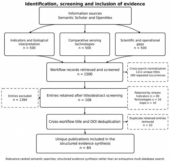
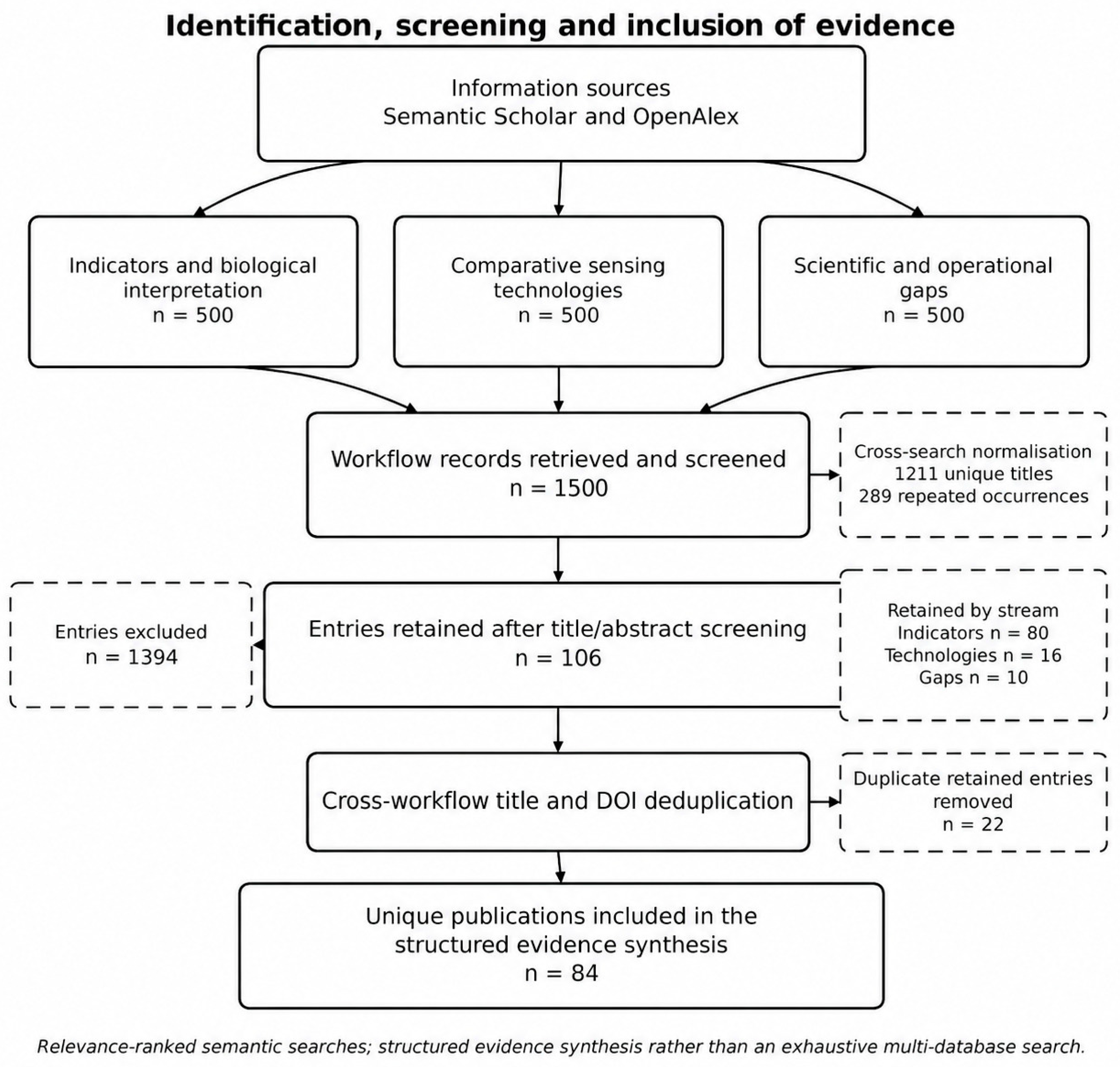
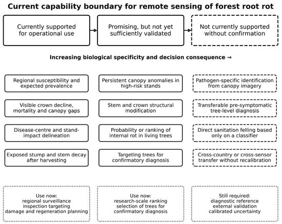
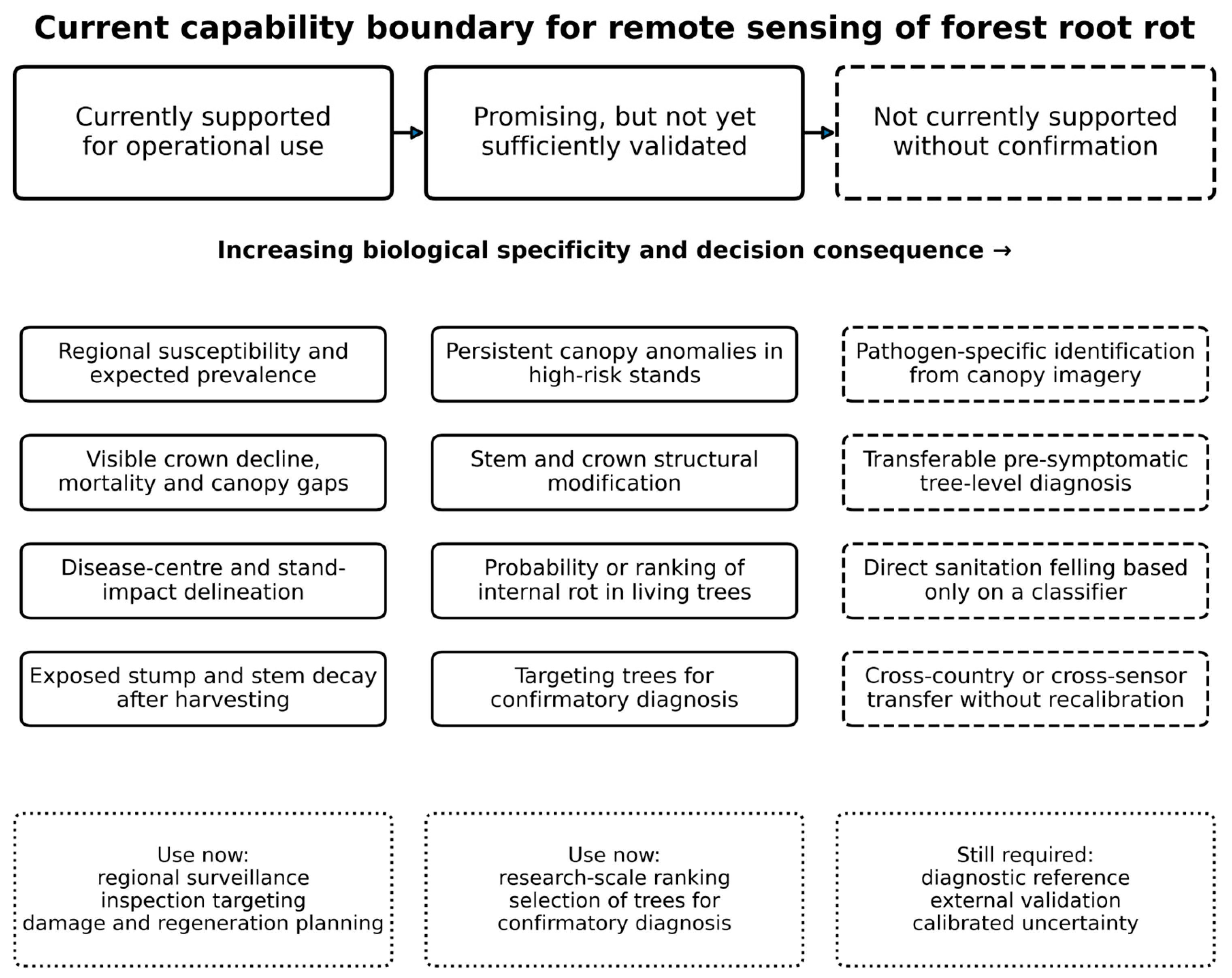
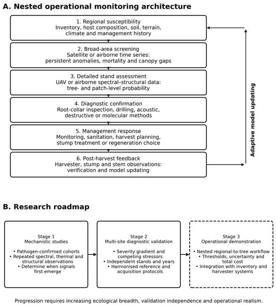

Disclaimer/Publisher’s Note: The statements, opinions and data contained in all publications are solely those of the individual author(s) and contributor(s) and not of MDPI and/or the editor(s). MDPI and/or the editor(s) disclaim responsibility for any injury to people or property resulting from any ideas, methods, instructions or products referred to in the content. |
© 2026 by the authors. Licensee MDPI, Basel, Switzerland. This article is an open access article distributed under the terms and conditions of the Creative Commons Attribution (CC BY) license.